Structure of the Taz2 domain of p300: insights into ligand ...
9
electronic reprint Acta Crystallographica Section D Biological Crystallography ISSN 0907-4449 Editors: E. N. Baker and Z. Dauter Structure of the Taz2 domain of p300: insights into ligand binding Maria Miller, Zbigniew Dauter, Scott Cherry, Joseph E. Tropea and Alexander Wlodawer Acta Cryst. (2009). D65, 1301–1308 Copyright c International Union of Crystallography Author(s) of this paper may load this reprint on their own web site or institutional repository provided that this cover page is retained. Republication of this article or its storage in electronic databases other than as specified above is not permitted without prior permission in writing from the IUCr. For further information see http://journals.iucr.org/services/authorrights.html Acta Crystallographica Section D: Biological Crystallography welcomes the submission of papers covering any aspect of structural biology, with a particular emphasis on the struc- tures of biological macromolecules and the methods used to determine them. Reports on new protein structures are particularly encouraged, as are structure–function papers that could include crystallographic binding studies, or structural analysis of mutants or other modified forms of a known protein structure. The key criterion is that such papers should present new insights into biology, chemistry or structure. Papers on crystallo- graphic methods should be oriented towards biological crystallography, and may include new approaches to any aspect of structure determination or analysis. Crystallography Journals Online is available from journals.iucr.org Acta Cryst. (2009). D65, 1301–1308 Miller et al. · Taz2 domain of p300
Transcript of Structure of the Taz2 domain of p300: insights into ligand ...
electronic reprintActa Crystallographica Section D
BiologicalCrystallography
ISSN 0907-4449
Editors: E. N. Baker and Z. Dauter
Structure of the Taz2 domain of p300: insights into ligandbinding
Maria Miller, Zbigniew Dauter, Scott Cherry, Joseph E. Tropea andAlexander Wlodawer
Acta Cryst. (2009). D65, 1301–1308
Copyright c© International Union of Crystallography
Author(s) of this paper may load this reprint on their own web site or institutional repository provided thatthis cover page is retained. Republication of this article or its storage in electronic databases other than asspecified above is not permitted without prior permission in writing from the IUCr.
For further information see http://journals.iucr.org/services/authorrights.html
Acta Crystallographica Section D: Biological Crystallography welcomes the submission ofpapers covering any aspect of structural biology, with a particular emphasis on the struc-tures of biological macromolecules and the methods used to determine them. Reportson new protein structures are particularly encouraged, as are structure–function papersthat could include crystallographic binding studies, or structural analysis of mutants orother modified forms of a known protein structure. The key criterion is that such papersshould present new insights into biology, chemistry or structure. Papers on crystallo-graphic methods should be oriented towards biological crystallography, and may includenew approaches to any aspect of structure determination or analysis.
Crystallography Journals Online is available from journals.iucr.org
Acta Cryst. (2009). D65, 1301–1308 Miller et al. · Taz2 domain of p300
research papers
Acta Cryst. (2009). D65, 1301–1308 doi:10.1107/S0907444909040153 1301
Acta Crystallographica Section D
BiologicalCrystallography
ISSN 0907-4449
Structure of the Taz2 domain of p300: insights intoligand binding
Maria Miller,a* Zbigniew
Dauter,b Scott Cherry,c
Joseph E. Tropeac and
Alexander Wlodawera
aProtein Structure Section, Macromolecular
Crystallography Laboratory, NCI-Frederick,
Frederick, Maryland 21702-1201, USA,bSynchrotron Radiation Research Section,
Macromolecular Crystallography Laboratory,
National Cancer Institute, Argonne, IL 60439,
USA, and cProtein Purification Core,
Macromolecular Crystallography Laboratory,
NCI-Frederick, Frederick, Maryland
21702-1201, USA
Correspondence e-mail: [email protected]
# 2009 International Union of Crystallography
Printed in Singapore – all rights reserved
CBP and its paralog p300 are histone acetyl transferases
that regulate gene expression by interacting with multiple
transcription factors via specialized domains. The structure
of a segment of human p300 protein (residues 1723–1836)
corresponding to the extended zinc-binding Taz2 domain has
been investigated. The crystal structure was solved by the
SAD approach utilizing the anomalous diffraction signal of
the bound Zn ions. The structure comprises an atypical helical
bundle stabilized by three Zn ions and closely resembles the
solution structures determined previously for shorter peptides.
Residues 1813–1834 from the current construct form a helical
extension of the C-terminal helix and make extensive crystal-
contact interactions with the peptide-binding site of Taz2,
providing additional insights into the mechanism of the
recognition of diverse transactivation domains (TADs) by
Taz2. On the basis of these results and molecular modeling,
a hypothetical model of the binding of phosphorylated p53
TAD1 to Taz2 has been proposed.
Received 8 September 2009
Accepted 1 October 2009
PDB Reference: Taz2 domain
of p300, 3io2, r3io2sf.
1. Introduction
p300/CBP coactivators link DNA-bound transcription factors
(TFs) to the basal transcriptional machinery. Both are large
proteins composed of several folded globular domains that are
connected by flexible linkers (Dyson & Wright, 2005). Apart
from the domains necessary for the acetyltransferase activity,
p300 and CBP share highly conserved domains that serve as
structural scaffolds for protein ligand binding, including two
copies of a zinc-finger domain (Taz1 and Taz2), Kix, Ibid and
IHD (Goodman & Smolik, 2000; McManus & Hendzel, 2001).
Detailed structural knowledge of these discrete domains and
their interactions with transcriptional activators is necessary in
order to understand the mechanism of assembly of functional
promoter-specific pre-initiation complexes.
The Taz2 domain binds specifically to the acidic transacti-
vation domains (TADs) of several TFs, including the p53
tumor suppressor, ETF2 and members of the STAT and
C/EBP families, which exhibit very little sequence similarity.
The mechanism of this specific recognition and stimulus-
dependent selectivity is poorly understood. Three-dimensional
structures have been determined by NMR for unliganded
CBP-Taz2 (De Guzman et al., 2000) and its complexes with
peptides derived from the TAD of STAT1 (Wojciak et al.,
2009) and from the conserved region 1 (CR1) domain of the
adenoviral oncoprotein E1A (Ferreon, Martinez-Yamout et
al., 2009), as well as for the p300-Taz2–p53-TAD1 complex
(Feng et al., 2009). Both STAT1-TAD and p53-TAD1 form
short helices upon binding and interact with the Taz2 domain
electronic reprint
through an extended interface. However, their binding sites
overlap only partially and the two polypeptide chains are
positioned in opposite directions, with their amphipathic
helices occupying distinct sites on the Taz2 surface. In the
crystal structure of free Taz2 reported here, the C-terminal
helix from a symmetry-generated molecule interacts with the
same surface, indicating yet another possible mode of peptide
binding to Taz2.
The interaction of CBP/p300 with the TAD of the tumor
suppressor protein p53 is of particular interest and has been
the focus of intensive studies. It has been shown that p53
interacts with the Taz1, Kix, Ibid and Taz2 domains and
probably with the IHD domain (Teufel et al., 2007). The
strongest binding of unphosphorylated p53 was observed to
the Taz2 domain. The cellular levels and functions of p53
depend on interactions with multiple partners and are regu-
lated by an array of post-transcriptional modifications (for
reviews, see Appella & Anderson, 2001; Kruse & Gu, 2009).
CBP/p300 competes for binding to the same region of p53 with
two negative regulators, MDM2 and MDMX, which are
responsible for p53 degradation. Intrinsically disordered p53-
TAD is composed of two independent transcriptional sub-
domains, TAD1 and TAD2, each containing an amphipathic
helical motif, ’xx’’ (where ’ denotes a hydrophobic residue),
which mediates distinct protein interactions. p53 interacts with
MDM2 and MDMX primarily via TAD1 (Bottger et al., 1999;
Kussie et al., 1996), whereas TAD2 is a major binding site for
RPA (Bochkareva et al., 2005) and for the p62 subunit of
TFIIH (Di Lello et al., 2006).
The contributions of the two subdomains to Taz2 binding
depend on phosphorylation, since p53 is stabilized and acti-
vated by phosphorylation of the TAD region in response to
DNA damage and other stresses. It has been demonstrated
that phosphorylation of Thr18 within p53-TAD1 abrogates its
binding to MDM2 and significantly enhances binding to Taz2.
The effect of p53 phosphorylation on its binding to Taz2 and
MDM2 has previously been investigated by isothermal titra-
tion calorimetry, fluorescence anisotropy and NMR spectro-
scopy (Miller Jenkins et al., 2009; Teufel et al., 2009). However,
no satisfactory model for the regulation of p53 activity has
emerged from these studies. In particular, structural infor-
mation on the interaction of Taz2 with the whole TAD of p53
and the structural basis of the increased binding affinity upon
phosphorylation of TAD1 at various sites is still lacking. Here,
we propose a hypothetical model of p53-TAD1 phosphoryl-
ated at Ser15 and Thr18 bound to Taz2 that could reconcile
some of the seemingly contradictory published results.
2. Materials and methods
2.1. Protein production and crystallization
Several versions of the Taz2 peptide of varying lengths
derived from the human p300 protein were prepared for
crystallographic studies: Taz2(1723–1812) and Taz2(1723–1816)
correspond to an evolutionarily conserved domain (Yuan &
Giordano, 2002), whereas the longer construct Taz2(1723–
1836) was designed based on secondary-structure predictions.
To prevent misfolding and/or aggregation arising from
oxidation, the four Cys residues which are not involved in
coordination of the zinc ions (Cys1738, Cys1746, Cys1789 and
Cys1790) were mutated to alanines using the QuikChange
mutagenesis kit (Stratagene, La Jolla, California, USA). The
proteins were expressed in Escherichia coli strain BL21-
CodonPlus(DE3)-RIL cells (Stratagene) and purified as
described previously (Miller Jenkins et al., 2009). The fractions
eluted from a Superdex 75 column corresponding to the
monomer were collected and concentrated to 2–3 mM. The
protein concentration was determined spectrophotometrically
and/or using a Bio-Rad assay. The molecular weights of the
recombinant proteins were confirmed by electrospray ioniza-
tion mass spectroscopy and their proper folding was assessed
by far-UV CD.
We attempted to grow crystals of the three constructs and
their complexes with peptides derived from the p53 TAD. No
positive results were obtained on screening for crystallization
conditions using vapor diffusion, which was performed with
Hydra Plus One and Phoenix robots utilizing a variety of
commercial screens or manually in Linbro plates using
specially designed conditions with DTT, TCEP or MME being
employed as reducing agents. Finally, in order to better pre-
vent oxidation, microbatch crystallization screens were set up
manually under paraffin oil in 72-well plasma-treated hydro-
philic microbatch plates (Hampton Research). The initial ‘hit’
was found for the longest peptide, Taz2(1723–1836), using
the Precipitant Synergy (Emerald BioSystems) Primary 64
formulations kit. A shower of small crystals appeared when
2.5 M potassium/sodium phosphate buffer pH 7.5 and 20%
glycerol (formulation No. 10) were used as the precipitant.
research papers
1302 Miller et al. � Taz2 domain of p300 Acta Cryst. (2009). D65, 1301–1308
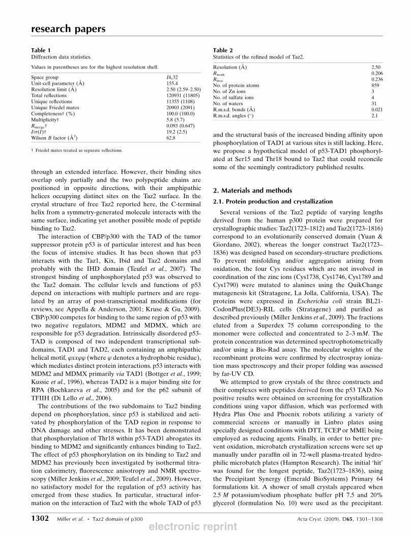
Table 1Diffraction data statistics.
Values in parentheses are for the highest resolution shell.
Space group I4132Unit-cell parameter (A) 155.4Resolution limit (A) 2.50 (2.59–2.50)Total reflections 120931 (11805)Unique reflections 11355 (1108)Unique Friedel mates 20903 (2091)Completeness† (%) 100.0 (100.0)Multiplicity† 5.8 (5.7)Rmerge† 0.093 (0.647)I/�(I)† 19.2 (2.5)Wilson B factor (A2) 62.8
† Friedel mates treated as separate reflections.
Table 2Statistics of the refined model of Taz2.
Resolution (A) 2.50Rwork 0.206Rfree 0.236No. of protein atoms 859No. of Zn ions 3No. of sulfate ions 4No. of waters 31R.m.s.d. bonds (A) 0.021R.m.s.d. angles (�) 2.1
electronic reprint

Subsequently, crystallization trials were set up under oil using
mixtures of various salts and alcohols. Crystals grew in the pH
range 6–7.4 from 1.5–1.7 M ammonium sulfate (AMS) solu-
tion containing 10–20% glycerol, ethylene glycol or ethanol.
No crystals grew when NaCl was used as the precipitant. The
best results were obtained when protein at a concentration of
30 mg ml�1 in 25 mM MES buffer pH 6.3, 100 mM NaCl, 6%
glycerol, 10 mM TCEP was mixed under oil with an equal
amount (1 ml) of precipitant containing 3.2 M AMS in MES
buffer pH 6.0 and 10% ethylene glycol. Crystals grew at 277 K
within 2–4 d. The cryoprotectant solution was composed of
1.5 M AMS and 25% glycerol. However, the shorter Taz2
peptides failed to crystallize under these or any other condi-
tions.
2.2. Data collection, structure solution and refinement
Diffraction data were collected on SER-CAT beamline
22-ID (APS, Argonne National Laboratory) using a MAR 300
CCD detector. The crystal was cryocooled to 100 K in a stream
of cold nitrogen gas. 45 images of 1� oscillation were collected
using a wavelength of 1.2827 A, corresponding to the high-
energy remote region of the Zn absorption edge. The data
were processed with HKL-2000 (Otwinowski & Minor, 1997)
and the resulting data statistics are summarized in Table 1.
The structure was solved with HKL-3000 (Minor et al.,
2006) using the SAD technique based on the anomalous signal
of Zn ions present in Taz2. Four fragments of the main chain
encompassing 98 residues were built automatically in the
initial SAD map. The model of the Taz2 molecule was itera-
tively completed using Coot (Emsley & Cowtan, 2004) and
refined with REFMAC (Murshudov et al., 1997), with the final
model encompassing residues 1726–1834 of the Taz2 sequence.
Each long helix of the Taz2 molecule was treated as a separate
TLS fragment in the final refinement cycles. The results are
presented in Table 2.
Structure analysis and modeling were performed using
Coot and INSIGHT II (Accelrys). Comparisons of three-
dimensional models were performed using the SSM (Secon-
dary Structure Matching; Krissinel & Henrick, 2004) method
as implemented in Coot.
3. Results and discussion
3.1. Description of the crystal structure
The Taz2 protein (residues 1723–1836) crystallized in space
group I4132 with a = 155.4 A (Table 1). The crystals contained
one molecule in the asymmetric unit and the solvent content
was quite high at 81%. The packing diagram depicted in Fig. 1
research papers
Acta Cryst. (2009). D65, 1301–1308 Miller et al. � Taz2 domain of p300 1303
Figure 1Crystal structure of the Taz2 domain. (a) Sequence features of human p300 Taz2 and the secondary structure. Arg and Lys residues are shown in navyblue and Zn2+ ligands are highlighted in magenta and orange for His and Cys, respectively. �-Helices are marked by rectangles above the sequence. Thezinc-finger subdomains are indicated below the sequence. (b) Packing diagram of the 48 molecules contained in the unit cell. (c) Taz2 crystallographicmodel. Helices are represented by brown cylinders, loops are shown in navy blue and Zn ions are depicted as cyan spheres. The molecule of Taz2 formsintimate contacts with three symmetry-generated molecules, which are shown as yellow, gray and green ribbons.
electronic reprint

shows clusters of symmetry-generated molecules and vast
solvent channels running parallel to the unit-cell axes. The
structure was refined at 2.5 A resolution to an R factor of
20.6% (Rfree = 23.6%) with the statistics shown in Table 2. In
addition to the protein, the final model
included three zinc ions, four sulfate ions
and 31 water molecules. The polypeptide
chain assumes a compact structure that
consists of four �-helices organized by three
zinc fingers (Znfs) with HCCC-type coor-
dination (Fig. 1).
It has been established (De Guzman et al.,
2000) that the folding of Taz2 into an
ordered structure requires the presence of
three molar equivalents of Zn ion. Three
Zn-binding subdomains are composed of
loops linking pairs of helices (�1–�2, �2–�3and �3–�4) and include the C- and N-
termini of consecutive helices (Fig. 1). Each
Zn ion is coordinated by a His residue
located at the C-terminus of the first helix,
two Cys residues from the interhelical loop
and a Cys residue located at the N-terminus
of the second helix. The requirement for
tetrahedral coordination of the Zn ion dictates the crossing
angles between helices. The configuration of each zinc finger is
stabilized by a set of conserved hydrophobic and electrostatic
interactions as described by De Guzman et al. (2000). The Zn-
coordinating His residues at the C-termini of �1 (His1744), �2(His1767) and �3 (His1792) contact the side chains of residues
from the loops linking the helices (Leu1755, Ile1780 and
Val1803), whereas the Cys ligands residing in the loops are
stabilized by hydrogen bonds between their S atoms and the
backbone NH groups. In the crystal structure, Znf3 is further
stabilized by a set of three electrostatic interactions between
the positively charged amine group on the side chain of
Lys1810 from �4 and (i) the Cys1796 S atom, (ii) the main-
chain carbonyl of Cys1796 and (iii) the main-chain carbonyl of
Glu1798 (Fig. 2).
The hydrophobic core of the molecule is formed by residues
from the three N-terminal helices (�1, �2 and �3) and from
the N-terminal part of the fourth helix (�4). 21 residues from
the end of �4 protrude outside the globular structure. The
helical conformation of this segment is stabilized by a network
of electrostatic interactions between polar/charged side
chains. Intrahelical links are formed by Gln1816, Lys1812 and
Gln1815, by Gln1817 and Arg1821, by Gln1823, His1820 and
Gln1824, and by Gln1826 and Arg1830, as well as by the
interaction of the Met1827 S atom with the guanidinium group
of Arg1831. A continuous hydrophobic patch on the helix
surface is generated by the exposed Leu1818, Leu1822,
Ala1825, Leu1828 and Met1832, together with the aliphatic
portions of the Arg1814 and Arg1821 side chains. This region
provides the majority of crystal-contact interactions. The last
14 residues of the molecule generated by the twofold axis
(hereafter referred to as �4-end0) make extensive interactions
with the residues that maintain the packing of helices �1, �2and �3 (Fig. 3). The segment Arg18210–Arg18290 (where the
prime denotes a symmetry mate) interacts with residues from
the �2–�3 interface. The interactions are predominantly
hydrophobic and include aliphatic portions of the Arg18210,
research papers
1304 Miller et al. � Taz2 domain of p300 Acta Cryst. (2009). D65, 1301–1308
Figure 2Stereoview of the Znf3 region with the 2Fo � Fc electron-density map contoured at 1.8� (blue)and 8.0� (magenta).
Figure 3Important crystal contacts: interactions between side chains of Taz2(chocolate) and the C-terminal part of �4 from a symmetry-relatedmolecule (green). For clarity, side chains that were not involved inintermolecular contacts were omitted.
electronic reprint
Arg18290 and Gln18240 side chains. In addition, the guanidi-
nium group of Arg18290 makes polar contacts with the side-
chain carbonyl O atom of Gln1784. Leu18280 is entirely buriedin a deep hydrophobic cavity at the crossing of three helices
which is lined by the side chains of Met1761, Val1764, Ile1781,
Gln1784, Leu1785 and Leu1788. Met18320 packs against
Ile1735 from �1 and Ala1787, Leu1788 and Tyr1791 from �3.Arg18310 and Ser18340 at the C-terminus are bound exclu-
sively by residues from the �1 helix. Ala1738 C� contacts
Arg18310 C�, while Ser1741 and Ser1734 provide a hydrophilic
environment for its guanidinium group. The C-terminal
carboxylate and main-chain carbonyl O atoms of Ser18340
interact electrostatically with the guanidinium group of
Arg1731. The loop connecting helices �3 and �4 is also
involved in crystal-contact inter-
actions, in which Gln1797 and
Glu1798 form a network of
hydrogen bonds with Gln17470,Arg17490 and Glu17980 that alsoinvolve several water molecules
(data not shown). These intimate
contacts between molecules in
the crystal lattice are facilitated
by bound sulfate (SO4) ions.
(SO4)204 is tightly bound by the
amine groups of Lys1772 and
Arg1773, as well as by their
backbone NH groups. (SO4)205
interacts with Arg1737 and
its symmetry mate, Gln1740,
Ser17410 and Ser17570. (SO4)206
is located in the vicinity of
Arg1829, whereas (SO4)207
bridges Arg1773 and Arg18140.These contacts explain the
requirement for sulfate or phos-
phate anions for crystallization of
the basic Taz2 domain.
3.2. Comparison with the NMR
structure of unliganded Taz2
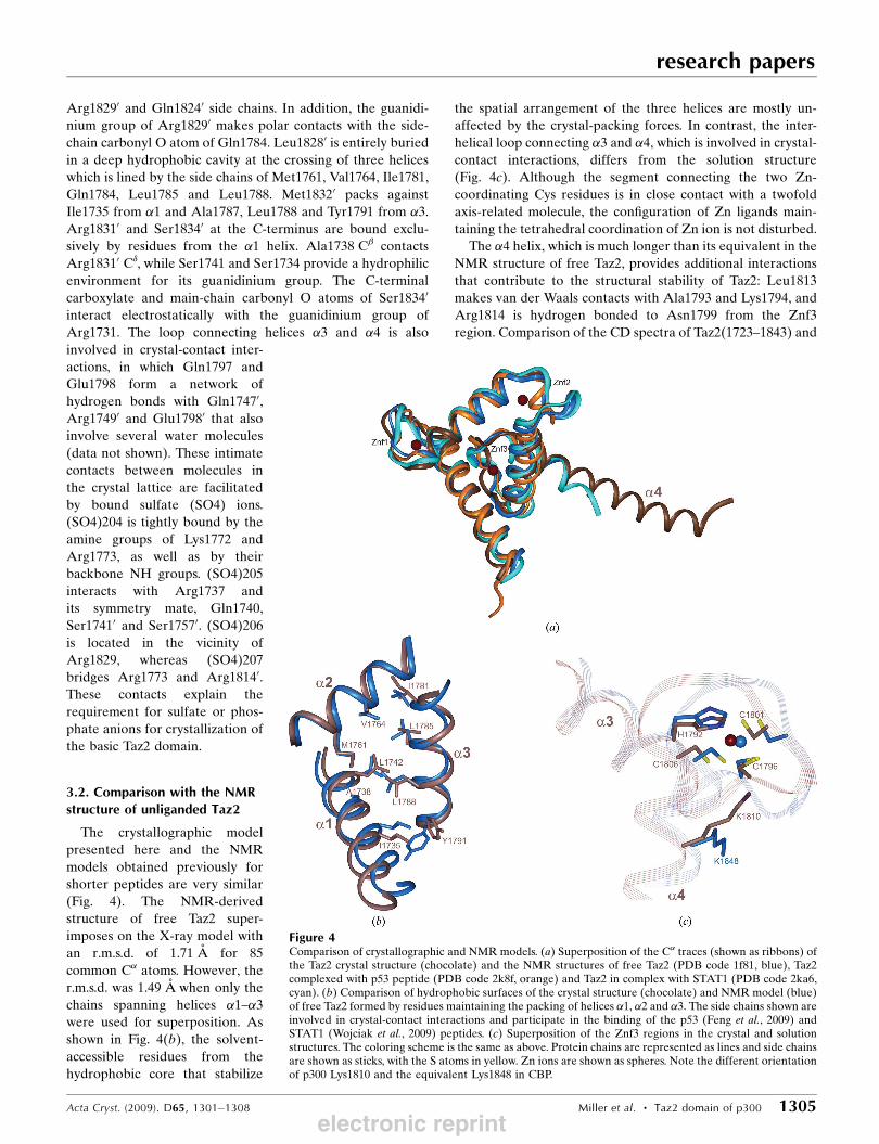
The crystallographic model
presented here and the NMR
models obtained previously for
shorter peptides are very similar
(Fig. 4). The NMR-derived
structure of free Taz2 super-
imposes on the X-ray model with
an r.m.s.d. of 1.71 A for 85
common C� atoms. However, the
r.m.s.d. was 1.49 A when only the
chains spanning helices �1–�3were used for superposition. As
shown in Fig. 4(b), the solvent-
accessible residues from the
hydrophobic core that stabilize
the spatial arrangement of the three helices are mostly un-
affected by the crystal-packing forces. In contrast, the inter-
helical loop connecting �3 and �4, which is involved in crystal-contact interactions, differs from the solution structure
(Fig. 4c). Although the segment connecting the two Zn-
coordinating Cys residues is in close contact with a twofold
axis-related molecule, the configuration of Zn ligands main-
taining the tetrahedral coordination of Zn ion is not disturbed.
The �4 helix, which is much longer than its equivalent in the
NMR structure of free Taz2, provides additional interactions
that contribute to the structural stability of Taz2: Leu1813
makes van der Waals contacts with Ala1793 and Lys1794, and
Arg1814 is hydrogen bonded to Asn1799 from the Znf3
region. Comparison of the CD spectra of Taz2(1723–1843) and
research papers
Acta Cryst. (2009). D65, 1301–1308 Miller et al. � Taz2 domain of p300 1305
Figure 4Comparison of crystallographic and NMR models. (a) Superposition of the C� traces (shown as ribbons) ofthe Taz2 crystal structure (chocolate) and the NMR structures of free Taz2 (PDB code 1f81, blue), Taz2complexed with p53 peptide (PDB code 2k8f, orange) and Taz2 in complex with STAT1 (PDB code 2ka6,cyan). (b) Comparison of hydrophobic surfaces of the crystal structure (chocolate) and NMR model (blue)of free Taz2 formed by residues maintaining the packing of helices �1, �2 and �3. The side chains shown areinvolved in crystal-contact interactions and participate in the binding of the p53 (Feng et al., 2009) andSTAT1 (Wojciak et al., 2009) peptides. (c) Superposition of the Znf3 regions in the crystal and solutionstructures. The coloring scheme is the same as above. Protein chains are represented as lines and side chainsare shown as sticks, with the S atoms in yellow. Zn ions are shown as spheres. Note the different orientationof p300 Lys1810 and the equivalent Lys1848 in CBP.
electronic reprint

the shorter Taz2 protein indicates that this segment retains a
helical conformation in solution (see Miller Jenkins et al.,
2009).
3.3. Implications forTaz2 ligand binding
The interaction surface for binding of protein ligands
revealed by the structures of the Taz2 domain complexed with
p53-TAD1 (Feng et al., 2009) and with STAT1-TAD (Wojciak
et al., 2009) is shown in Fig. 5. The central hydrophobic area
formed by residues maintaining the packing of helices �1, �2and �3 is flanked by clusters of positively charged residues:
Arg1731, Arg1732 and Arg1737 from �1, Lys1760 and
Arg1763 from �2 and Lys1772, Arg1773 and Lys1774 from the
Znf1 region. The binding site also includes Gln1784, which is
an important determinant of binding specificity.
In the crystal structure, the C-terminal end of the �4 helix
from a symmetry-related molecule (�4-end0) coincides with
the position of the p53-TAD1 helix bound to Taz2. Although
the two helices are positioned in opposite directions, Leu22 of
p53 and Leu18280 of Taz2 are buried in the same hydrophobic
pocket (Fig. 5b). Previous studies (Miller Jenkins et al., 2009)
revealed that the peptides representing p53 transcriptional
subdomains, TAD1 and TAD2, bind in an anticooperative
manner to two overlapping sites on the Taz2 domain. The
relatively weak binding of p53-TAD1 to Taz2 is greatly
enhanced by the phosphorylation of six possible phospho-
acceptors (Ser9, Ser15, Thr18, Ser20, Ser33 and Ser37), with
phosphorylation of Ser15 or Thr18 producing the strongest
effect. NMR titration showed a sevenfold and 11-fold increase
in affinity for Taz2 upon monophosphorylation at Ser15 and
Thr18, respectively. Interestingly, only a sevenfold increase
was generated upon diphosphorylation at these sites (Feng et
al., 2009). Several findings pointed towards the possibility of
two distinct modes of interaction with Taz2 for phosphoryl-
ated and unphosphorylated forms of p53-TAD1. In the
p53-TAD1–Taz2 complex Ser15 is entirely solvent-exposed
and mutation of the closest positively charged amino acid,
Arg1737, to Ala increased fourfold rather than decreased the
binding affinity of p53-TAD1 phosphorylated at this position
[p53-(Ser15p)TAD1]. Analogous mutations of Arg1731 or
Arg1732, which are proximal to Thr18, decreased the binding
affinity of p53-(Thr18p)TAD1 by only twofold. Furthermore,
calorimetric as well as chemical shift mapping results for p53-
(Thr18p)TAD1 binding indicated changes in the hydrophobic
interface of p53-TAD1 with Taz2. Isothermal titration calori-
metry experiments showed a significant increase in the con-
tribution of hydrophobic interactions to p53-(Thr18p)TAD1
binding and NMR spectroscopy revealed pronounced differ-
ences in the amide chemical shifts of several residues on
titration with p53-TAD1 and p53-(Thr18p)TAD (e.g. Leu1733,
Ile1735 and Ala1738).
Therefore, we investigated the possibility that a phos-
phorylated p53 peptide could bind in a manner indicated by
the intermolecular interactions in the crystal lattice. As
described above, the crystal contacts are mediated by bound
sulfate anions and the aliphatic portions of the Arg18210,Arg18290 and Gln18240 side chains make apolar contacts with
residues forming the hydrophobic groove on the Taz2 surface.
Thus, under crystallization conditions the �4-end0 can mimic
an amphipathic helix bound to Taz2. The side chains from the
�4-end0 segment were replaced with those from the p53-TAD1
helix (residues 15–26) based on the sequence alignment shown
in Fig. 6. The modeling of this interface required only a few
adjustments to the Taz2 side chains: Lys1760 was positioned to
make a salt bridge with Asp21, Arg1763 was positioned to
interact with Thr18 O� and the aliphatic portions of Lys1783
and Arg1737 were used to complete a hydrophobic environ-
ment for Phe19 and Leu25, respectively. Upon in silico
phosphorylation of Ser15 and Thr18, the side-chain confor-
mations of Lys1760 and Arg1763 were altered to generate the
network of electrostatic interactions depicted in Fig. 6. As in
the NMR structure of the complex, Phe19, Leu22 and Leu25
provide the most important contributions to the hydrophobic
interface, whereas Trp23 is pushed away by the polar amide
group of Gln1784, which is partially buried upon complex
formation. However, unlike in the unphosphorylated form
research papers
1306 Miller et al. � Taz2 domain of p300 Acta Cryst. (2009). D65, 1301–1308
Figure 5(a) Superposition of peptides in the bound conformation (based onstructural superposition of Taz2 domains from relevant complexes). Thebackbones of STAT1, p53-TAD1 and the C-terminus of the symmetry-related molecule in the crystal structure (�4-end0) are rendered asmagenta, orange and green ribbons, respectively. The molecular surfaceof the Taz2 crystal structure is shown in beige, with the Arg side chainsin navy blue. Note the reversed directions of the STAT1 and p53polypeptide chains. (b) An enlarged area showing the surface of ahydrophobic cavity (light green) comprised of the side chains of Met1761,Val1764, Ile1781, Leu1785, Leu1788 and the aliphatic portion of Glu1784(yellow). The side chains of p53 Leu22 from the p53-TAD1–Taz2 complexand Leu18280 from the �4-end0 are rendered as sticks and shown inorange and green, respectively.
electronic reprint

Leu26 is also important for binding. This residue is positioned
in a hydrophobic pocket created by the side chains of Ile1735,
Ala1738, Tyr1791 and Leu1788. These contacts may provide
an explanation for the increase in the extent of hydrophobic
interactions for p53-(Thr18p)TAD1 compared with the un-
phosphorylated form, as well as for the observed changes in
amide chemical shifts. Also, in this direction of p53 poly-
peptide binding the phosphorylated Ser9 could potentially
interact with Lys1772 and/or Arg1773. Furthermore, p53
Glu28 would be in the vicinity of Arg1731 (Fig. 5a), explaining
the moderately decreased binding affinity of the Taz2 R1731A
mutant protein. On the other hand, mutation of Arg1737 to
Ala would remove the unfavorable hydrophilic environment
for Leu25 of p53 and should increase the binding affinity,
which is in agreement with previous results (Feng et al., 2009).
The C-terminus of the �4-end0 overlaps with the N-terminus
of the amphipathic helix from the bound STAT1 peptide
(Fig. 5), which could indicate the position of binding for the
helical binding motif from p53-TAD2. As is the case with
STAT1-TAD, this second amphipathic sequence within p53-
TAD is preceded by negatively charged amino acids and its
affinity of interaction with Taz2 is not affected by phosphor-
ylation (Miller Jenkins et al., 2009). Evidence for overlapping
binding sites for p53 TAD1 and TAD2 peptides was provided
by 1H–15NHSQC titration experiments on 15N-labeled Taz2
(Feng et al., 2009). Two binding motifs from p53 TAD are
separated by a 20-residue flexible linker. According to recent
reports (Ferreon, Lee et al., 2009; Teufel et al., 2009), the
binding of full-length p53-TAD to Taz2 is dominated by the
second motif, with only a minor contribution from TAD1. If
this assessment is correct, then only a short helical segment of
p53-TAD1 (residues 15–22) would interact with Taz2, whereas
Ile1735, Ala1787, Leu1788 and Tyr1791, together with
Arg1731 and Arg1732 from �1, would mediate the inter-
actions with TAD2. It is likely that the role of two overlapping
sites for p53 binding on the Taz2 surface is to facilitate the
formation of multiprotein complexes of varying compositions
in response to different stress stimuli and environmental
changes. Ternary complexes of MDM2–p53-TAD–CBP/p300
have been observed in vitro (Ferreon, Lee et al., 2009) and in
vivo (Kobet et al., 2000). Ferreon and coworkers demonstrated
that unphosphorylated p53-TAD can bind simultaneously to
MDM2 through TAD1 and to Taz2 (and also to other CBP
domains) through TAD2. The two subdomains behave inde-
pendently in forming the ternary complex. Phosphorylation of
Thr18 abrogates p53 binding to MDM2 and increases its
affinity towards CBP/p300. It is therefore conceivable to
envision that p53-(Ser15p/Thr18p)TAD1 could be bound to
Taz2, whereas TAD2 could interact with another protein
partner, with RPA (Bochkareva et al., 2005) and the p62
subunit of TFIIH (Di Lello et al., 2006) being the best
candidates of those that have been studied. Further experi-
mental results will be needed to further our understanding of
the interactions of p53 with the Taz2 domain of CBP/p300.
Whereas determination of the interactions involving the
whole p53-TAD may not be feasible, finding the site for p53-
TAD2 subdomain binding should be straightforward with the
application of the NMR technique.
4. Concluding remarks
We determined the crystal structure of the Taz2 domain of
human p300. Comparison with NMR structures confirms that
Taz2 is a rigid scaffold for interactions with protein ligands. We
observed that the C-terminal helix from a symmetry-related
molecule in the crystal lattice interacts with the peptide-
binding surface of Taz2, suggesting a putative novel mode of
Taz2–peptide ligand interaction. Based on careful analysis of
crystal contacts, we examined whether such a mode of inter-
research papers
Acta Cryst. (2009). D65, 1301–1308 Miller et al. � Taz2 domain of p300 1307
Figure 6A hypothetical model of a diphosphorylated p53-TAD1–Taz2 complex.(a) Sequence alignment of the C-terminal end of Taz2 with the p53-TAD1peptide used for modeling. Amino acids are colored according to theirside chains: hydrophobic, polar uncharged, acidic and basic residues areshown in green, magenta, red and blue text, respectively. Identicalresidues are boxed. (b) Molecular interface of doubly phosphorylatedp53-TAD1 (green) with Taz2 (chocolate). For clarity, only the side chainsinvolved in intermolecular contacts are shown. Taz2 side chains arerepresented as sticks and p53 is shown in ball-and-stick representation.The hydrophobic environment for p53 Phe19, Leu22, Leu25 and Leu26 isprovided by Pro1780, Ile1781, Leu1785, the aliphatic portions of Lys1783and Glu1784, Ile1786, Ala1787, Leu1788 and Tyr1791 from Taz2 �3, andIle1735, Ala1738 and the aliphatic portion of Arg1737 from �1. Themethyl group of Thr18 packs against Ile1781. The complex is stabilized byelectrostatic interactions of phosphate groups from pSer15 and pThr18with Lys1760, Arg1763 and His1767 from Taz2 �2. In addition, there is apossibility of hydrogen-bond formation between p53 Asp21 and Lys1760.
electronic reprint
action is possible in the case of p53-TAD1 binding. Modeling
indicated that in the absence of TAD2 the isolated unphos-
phorylated p53-TAD1 peptide may bind to the same site on
Taz2 in two alternative orientations. However, upon phos-
phorylation at Ser15 and/or Thr18 the preferred orientation
appears to be determined by electrostatic interactions of
the phosphate groups with Lys1760, Arg1763 and possibly
Arg1773 of Taz2. The proposed model of phosphorylated
p53-TAD1 binding accounts for several experimental results
obtained with isothermal titration calorimetry, as well as with
NMR spectroscopy, and provides a clue to the mode of
interaction of the whole p53-TAD with Taz2.
The polypeptide representing the Taz2 domain used in
this study was designed on the basis of secondary-structure
predictions and corresponds more to a structural rather than
to a functional domain. The resulting structure described here
provokes an intriguing question: what is the biological role of
the helical C-terminal segment that extends beyond the stable
globular structure of Taz2?
We would like to thank Dr Stewart Durell for critical
reading of the manuscript. We acknowledge the use of
beamline 22-ID of the Southeast Regional Collaborative
Access Team (SER-CAT), located at the Advanced Photon
Source, Argonne National Laboratory. Use of the APS was
supported by the U.S. Department of Energy, Office of
Science, Office of Basic Energy Sciences, under Contract
No. W-31-109-Eng-38. This project was supported by the
Intramural Research Program of the NIH, National Cancer
Institute, Center for Cancer Research.
References
Appella, E. & Anderson, C. W. (2001). Eur. J. Biochem. 268, 2764–2772.
Bochkareva, E., Kaustov, L., Ayed, A., Yi, G. S., Lu, Y., Pineda-Lucena, A., Liao, J. C., Okorokov, A. L., Milner, J., Arrowsmith,C. H. & Bochkarev, A. (2005). Proc. Natl Acad. Sci. USA, 102,15412–15417.
Bottger, V., Bottger, A., Garcia-Echeverria, C., Ramos, Y. F., van derEb, A. J., Jochemsen, A. G. & Lane, D. P. (1999). Oncogene, 18,189–199.
De Guzman, R. N., Liu, H. Y., Martinez-Yamout, M., Dyson, H. J. &Wright, P. E. (2000). J. Mol. Biol. 303, 243–253.
Di Lello, P., Jenkins, L. M., Jones, T. N., Nguyen, B. D., Hara, T.,Yamaguchi, H., Dikeakos, J. D., Appella, E., Legault, P. &Omichinski, J. G. (2006). Mol. Cell, 22, 731–740.
Dyson, H. J. & Wright, P. E. (2005). Nature Rev. Mol. Cell Biol. 6,197–208.
Emsley, P. & Cowtan, K. (2004). Acta Cryst. D60, 2126–2132.Feng, H., Jenkins, L. M., Durell, S. R., Hayashi, R., Mazur, S. J.,Cherry, S., Tropea, J. E., Miller, M., Wlodawer, A., Appella, E. &Bai, Y. (2009). Structure, 17, 202–210.
Ferreon, J. C., Lee, C. W., Arai, M., Martinez-Yamout, M. A., Dyson,H. J. & Wright, P. E. (2009). Proc. Natl Acad. Sci. USA, 106, 6591–6596.
Ferreon, J. C., Martinez-Yamout, M. A., Dyson, H. J. & Wright, P. E.(2009). Proc. Natl Acad. Sci. USA, 106, 13260–13265.
Goodman, R. H. & Smolik, S. (2000). Genes Dev. 14, 1553–1577.Kobet, E., Zeng, X., Zhu, Y., Keller, D. & Lu, H. (2000). Proc. NatlAcad. Sci. USA, 97, 12547–12552.
Krissinel, E. & Henrick, K. (2004). Acta Cryst. D60, 2256–2268.Kruse, J. P. & Gu, W. (2009). Cell, 137, 609–622.Kussie, P. H., Gorina, S., Marechal, V., Elenbaas, B., Moreau, J.,Levine, A. J. & Pavletich, N. P. (1996). Science, 274, 948–953.
McManus, K. J. & Hendzel, M. J. (2001). Biochem. Cell Biol. 79,253–266.
Miller Jenkins, L. M., Yamaguchi, H., Hayashi, R., Cherry, S., Tropea,J. E., Miller, M., Wlodawer, A., Appella, E. & Mazur, S. J. (2009).Biochemistry, 48, 1244–1255.
Minor, W., Cymborowski, M., Otwinowski, Z. & Chruszcz, M. (2006).Acta Cryst. D62, 859–866.
Murshudov, G. N., Vagin, A. A. & Dodson, E. J. (1997). Acta Cryst.D53, 240–255.
Otwinowski, Z. & Minor, W. (1997). Methods Enzymol. 276, 307–326.
Teufel, D. P., Bycroft, M. & Fersht, A. R. (2009). Oncogene, 28, 2112–2118.
Teufel, D. P., Freund, S. M., Bycroft, M. & Fersht, A. R. (2007). Proc.Natl Acad. Sci. USA, 104, 7009–7014.
Wojciak, J. M., Martinez-Yamout, M. A., Dyson, H. J. & Wright, P. E.(2009). EMBO J. 28, 948–958.
Yuan, L. W. & Giordano, A. (2002). Oncogene, 21, 2253–2260.
research papers
1308 Miller et al. � Taz2 domain of p300 Acta Cryst. (2009). D65, 1301–1308
electronic reprint