Passive carriage of rabies virus by dendritic cells - SpringerPlus
Structure of a rabies virus polymerase complex fromelectron cryo-microscopyJoshua A. Horwitza,b, Simon Jennia, Stephen C. Harrisona,c,1
, and Sean P. J. Whelanb,d,1
aDepartment of Biological Chemistry and Molecular Pharmacology, Harvard Medical School, Boston, MA 02115; bDepartment of Microbiology, HarvardMedical School, Boston, MA 02115; cHoward Hughes Medical Institute, Harvard Medical School, Boston, MA 02115; and dDepartment of MolecularMicrobiology, Washington University School of Medicine, St. Louis, MO 63110
Contributed by Stephen C. Harrison, December 9, 2019 (sent for review October 28, 2019; reviewed by Stephen Cusack and Rachel Fearns)
Nonsegmented negative-stranded (NNS) RNA viruses, among themthe virus that causes rabies (RABV), include many deadly humanpathogens. The large polymerase (L) proteins of NNS RNA virusescarry all of the enzymatic functions required for viral messenger RNA(mRNA) transcription and replication: RNA polymerization, mRNA cap-ping, and cap methylation. We describe here a complete structure ofRABV L bound with its phosphoprotein cofactor (P), determined byelectron cryo-microscopy at 3.3 Å resolution. The complex closelyresembles the vesicular stomatitis virus (VSV) L-P, the one otherknown full-length NNS-RNA L-protein structure, with key localdifferences (e.g., in L-P interactions). Like the VSV L-P structure,the RABV complex analyzed here represents a preinitiation con-formation. Comparison with the likely elongation state, seen intwo structures of pneumovirus L-P complexes, suggests differencesbetween priming/initiation and elongation complexes. Analysis ofinternal cavities within RABV L suggests distinct template and prod-uct entry and exit pathways during transcription and replication.
rabies lyssavirus | NNS RNA viruses | vesicular stomatitis virus |transcription | replication
Rabies virus (RABV) and other viruses with nonsegmented,negative-strand (NNS) RNA genomes have, as the catalytic
core of their replication machinery, a large, multifunctional RNApolymerase (L). Many of these viruses are serious human patho-gens, including Ebola virus, respiratory syncytial virus, and mea-sles. Vaccines are available for some, including descendants of thestoried work on rabies by Louis Pasteur and Pierre Paul ÉmileRoux, but specific, small-molecule therapeutics are still underdevelopment. By analogy with inhibitors of viral polymerases ofmany other types, L would be a suitable candidate for inhibitordevelopment, for which structural and mechanistic studies areessential precursors.For all mononegaviruses, including the rhabdoviruses RABV
and vesicular stomatitis virus (VSV), L associates with a cofactorknown as P (for phosphoprotein). P bridges L with the viralnucleocapsid, an antisense RNA genome fully encapsidated bythe viral nucleoprotein (N). The complete structure of L fromVSV (1), the sole example published so far of an NNS viralpolymerase in which all five domains are clearly resolved, showsthe global features that sequence comparisons suggest it has incommon with most other NNS RNA viral L proteins. Its multi-domain organization includes three enzymatic modules: an RNA-dependent RNA polymerase (RdRp), a capping domain (CAP),and a dual-specificity methyltransferase (MT) domain. CAP is aGDP:polyribonucleotidyltransferase (PRNTase) that transfers a 5′monophosphate of the nascent RNA transcript onto a GDPacceptor (2); the single MT domain methylates the ribose 2′-Oposition on the first nucleotide of the transcript and then theN-7 position of the capping guanylate (3). Transcription initi-ation and cap addition require several residues within orproximal to a priming loop from CAP, which extends into theRdRp core (4). Residues at related positions in the RABV Lamino acid sequence are similarly essential for its activities. Aconnector domain (CD) and a C-terminal domain (CTD), both
with nonenzymatic functions, flank the MT domain and, inconjunction with P, likely facilitate the large structural rearrange-ments that appear to coordinate the three enzymatic activitiesduring transcription and replication.We report here an atomic model of L from RABV SAD-B19
in complex with a 91-residue, N-terminal fragment of P (P1–91)from electron cryo-microscopy (cryo-EM) at an average reso-lution of 3.3 Å. The atomic structure resembles that of VSVL-P in many respects, consistent with the 34.1% amino acidsequence conservation between the two L proteins. As in theVSV L-P complex, binding of P1–91 locks the CD, MT domain,and CTD into a fixed, “closed” arrangement with respect to thelarge RdRp-CAP module. This closed conformation appears torepresent the L protein poised for initiation at the 3′ end of thegenome or antigenome. Comparison with structures of tworecently published pneumovirus L-P complexes (5, 6) as wellas with that of a VSV L-P reconstruction determined at 3.0 Åresolution (7) suggests that replication and transcription havealternative priming configurations and alternative product exitsites.
Significance
Rabies virus (RABV) and other viruses with single-segment,negative-sense, RNA genomes have a multi-functional polymer-ase protein (L) that carries out the various reactions required fortranscription and replication. Many of these viruses are serioushuman pathogens, and L is a potential target for antiviral ther-apeutics. Drugs that inhibit polymerases of HCV and HIV-1 pro-vide successful precedents. The structure described here of theRABV L protein in complex with its P-protein cofactor shows aconformation poised for initiation of transcription or replication.Channels in the molecule and the relative positions of catalyticsites suggest that L couples a distinctive capping reaction withpriming and initiation of transcription, and that replication andtranscription have different priming configurations and differentproduct exit sites.
Author contributions: J.A.H., S.J., S.C.H., and S.P.J.W. designed research; J.A.H. performedresearch; S.C.H. analyzed data; J.A.H. and S.C.H. wrote the paper; and S.J. and S.P.J.W.edited the paper.
Reviewers: S.C., European Molecular Biology Laboratory; and R.F., Boston UniversitySchool of Medicine.
The authors declare no competing interest.
This open access article is distributed under Creative Commons Attribution-NonCommercial-NoDerivatives License 4.0 (CC BY-NC-ND).
Data deposition: Data for the cryo-EM 3D reconstruction reported in this paper have beendeposited in the Electron Microscopy Data Bank, https://www.ebi.ac.uk/pdbe/emdb/ (ac-cession no. EMD-20753), and data for coordinates fit to 3D reconstruction have beendeposited in the Protein Data Bank, https://www.rcsb.org/ (PDB ID 6UEB).
See online for related content such as Commentaries.1To whom correspondence may be addressed. Email: [email protected] [email protected].
This article contains supporting information online at https://www.pnas.org/lookup/suppl/doi:10.1073/pnas.1918809117/-/DCSupplemental.
First published January 17, 2020.
www.pnas.org/cgi/doi/10.1073/pnas.1918809117 PNAS | January 28, 2020 | vol. 117 | no. 4 | 2099–2107
MICRO
BIOLO
GY
Dow
nloa
ded
by g
uest
on
Mar
ch 1
6, 2
020
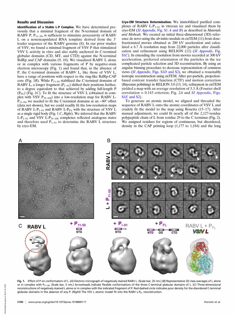
Results and DiscussionIdentification of a Stable L-P Complex. We have determined pre-viously that a minimal fragment of the N-terminal domain ofRABV P, P11–50, is sufficient to stimulate processivity of RABVL on a nonencapsidated RNA template derived from the 3′leader sequence of the RABV genome (8). In our prior studiesof VSV, we found a minimal fragment of VSV P that stimulatedVSV L activity in vitro and also stably anchored its C-terminalglobular domains (CD, MT, and CTD) against the N-terminalRdRp and CAP domains (9, 10). We visualized RABV L aloneor in complex with various fragments of P by negative-stainelectron microscopy (Fig. 1) and found that, in the absence ofP, the C-terminal domains of RABV L, like those of VSV L,have a range of positions with respect to the ring-like RdRp-CAPcore (Fig. 1B). While P11–50 stabilized the C-terminal domains ofRABV L, a longer fragment (P1–91) shifted their positions further,to a degree equivalent to that achieved by adding full-length P(PFL) (Fig. 1C). To fit the structure of VSV L (obtained in com-plex with VSV P35–106) into a low-resolution map for RABV L-P11–50, we needed to fit the C-terminal domains at an ∼80° offset(data not shown), but we could readily fit the low-resolution mapsof RABV L-P1–91 and RABV L-PFL with the structure of VSV Las a single rigid body (Fig. 1 C, Right). We inferred that the RABVL-P1–91 and VSV L-P35–106 complexes reflected analogous statesand therefore used P1–91 to determine the RABV L structureby cryo-EM.
Cryo-EM Structure Determination. We immobilized purified com-plexes of RABV L-P1–91 in vitreous ice and visualized them bycryo-EM (SI Appendix, Fig. S1 A and B) as described in Materialsand Methods. We created an initial three-dimensional (3D) refer-ence de novo using the ab-initio module in cisTEM (11) from dose-fractionated movies obtained at 200 kV acceleration and calcu-lated a 6.7 Å resolution map from 22,000 particles after classifi-cation and refinement using RELION (12) (SI Appendix, Fig.S1C). In extending the resolution from movies recorded at 300 kVacceleration, preferred orientation of the particles in the icecomplicated particle selection and 3D reconstruction. By using anangular binning procedure to decrease representation of commonviews (SI Appendix, Figs. S1D and S2), we obtained a reasonablyisotropic reconstruction using cisTEM. After per-particle, projection-based contrast transfer function (CTF) and motion correction(Bayesian polishing) in RELION 3.0 (13, 14), refinement in cisTEMyielded a map with an average resolution of 3.3 Å (Fourier shellcorrelation = 0.143 criterion; Fig. 2A and SI Appendix, Figs.S1E and S2).To generate an atomic model, we aligned and threaded the
sequence of RABV L onto the atomic coordinates of VSV L andcrudely fit the model to the map using Rosetta (15–17). Aftermanual adjustment, we could fit nearly all of the 2,127-residuepolypeptide chain of L from residue 29 to the C terminus (Fig. 2).We assigned residues for regions of continuous, but disordered,density in the CAP priming loop (1,177 to 1,184) and the long
Fig. 1. Effect of P on conformation of L. (A) Electron micrograph of negatively stained RABV L. (Scale bar, 25 nm.) (B) Representative 2D class averages of L aloneor in complex with P11–50. (Scale bar, 5 nm.) Arrowheads indicate flexible conformations of the three C-terminal globular domains of L. (C) Three-dimensionalreconstructions of negatively stained L alone or in complex with the indicated fragment of P. Red dashed circle indicates poor density for the disordered C-terminalglobular domains in the absence of any P. (Right) The VSV L atomic model fit into the RABV L-PFL reconstruction.
2100 | www.pnas.org/cgi/doi/10.1073/pnas.1918809117 Horwitz et al.
Dow
nloa
ded
by g
uest
on
Mar
ch 1
6, 2
020
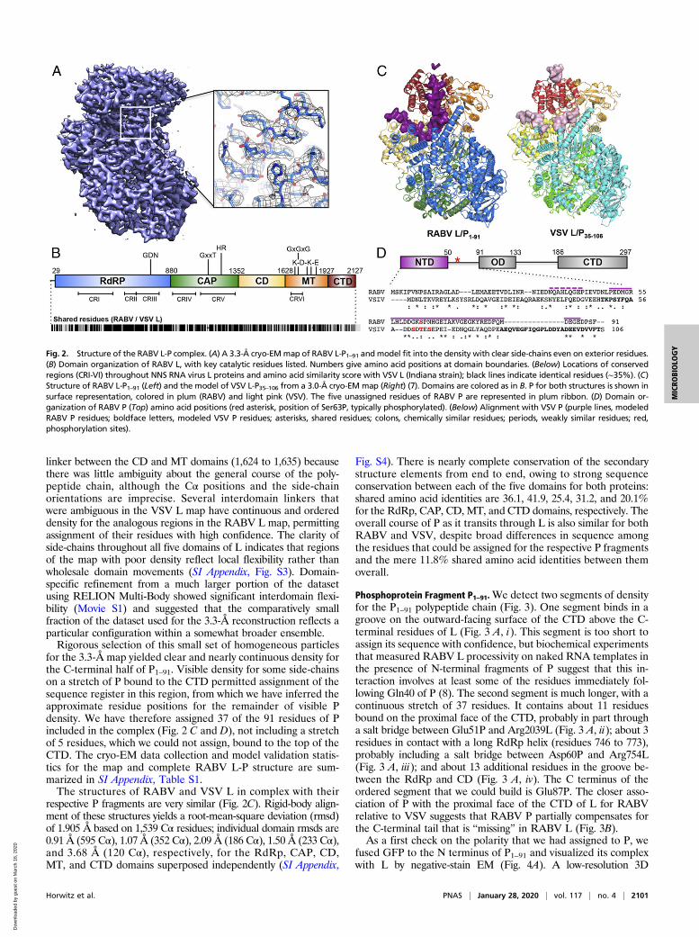
linker between the CD and MT domains (1,624 to 1,635) becausethere was little ambiguity about the general course of the poly-peptide chain, although the Cα positions and the side-chainorientations are imprecise. Several interdomain linkers thatwere ambiguous in the VSV L map have continuous and ordereddensity for the analogous regions in the RABV L map, permittingassignment of their residues with high confidence. The clarity ofside-chains throughout all five domains of L indicates that regionsof the map with poor density reflect local flexibility rather thanwholesale domain movements (SI Appendix, Fig. S3). Domain-specific refinement from a much larger portion of the datasetusing RELION Multi-Body showed significant interdomain flexi-bility (Movie S1) and suggested that the comparatively smallfraction of the dataset used for the 3.3-Å reconstruction reflects aparticular configuration within a somewhat broader ensemble.Rigorous selection of this small set of homogeneous particles
for the 3.3-Å map yielded clear and nearly continuous density forthe C-terminal half of P1–91. Visible density for some side-chainson a stretch of P bound to the CTD permitted assignment of thesequence register in this region, from which we have inferred theapproximate residue positions for the remainder of visible Pdensity. We have therefore assigned 37 of the 91 residues of Pincluded in the complex (Fig. 2 C and D), not including a stretchof 5 residues, which we could not assign, bound to the top of theCTD. The cryo-EM data collection and model validation statis-tics for the map and complete RABV L-P structure are sum-marized in SI Appendix, Table S1.The structures of RABV and VSV L in complex with their
respective P fragments are very similar (Fig. 2C). Rigid-body align-ment of these structures yields a root-mean-square deviation (rmsd)of 1.905 Å based on 1,539 Cα residues; individual domain rmsds are0.91 Å (595 Cα), 1.07 Å (352 Cα), 2.09 Å (186 Cα), 1.50 Å (233 Cα),and 3.68 Å (120 Cα), respectively, for the RdRp, CAP, CD,MT, and CTD domains superposed independently (SI Appendix,
Fig. S4). There is nearly complete conservation of the secondarystructure elements from end to end, owing to strong sequenceconservation between each of the five domains for both proteins:shared amino acid identities are 36.1, 41.9, 25.4, 31.2, and 20.1%for the RdRp, CAP, CD, MT, and CTD domains, respectively. Theoverall course of P as it transits through L is also similar for bothRABV and VSV, despite broad differences in sequence amongthe residues that could be assigned for the respective P fragmentsand the mere 11.8% shared amino acid identities between themoverall.
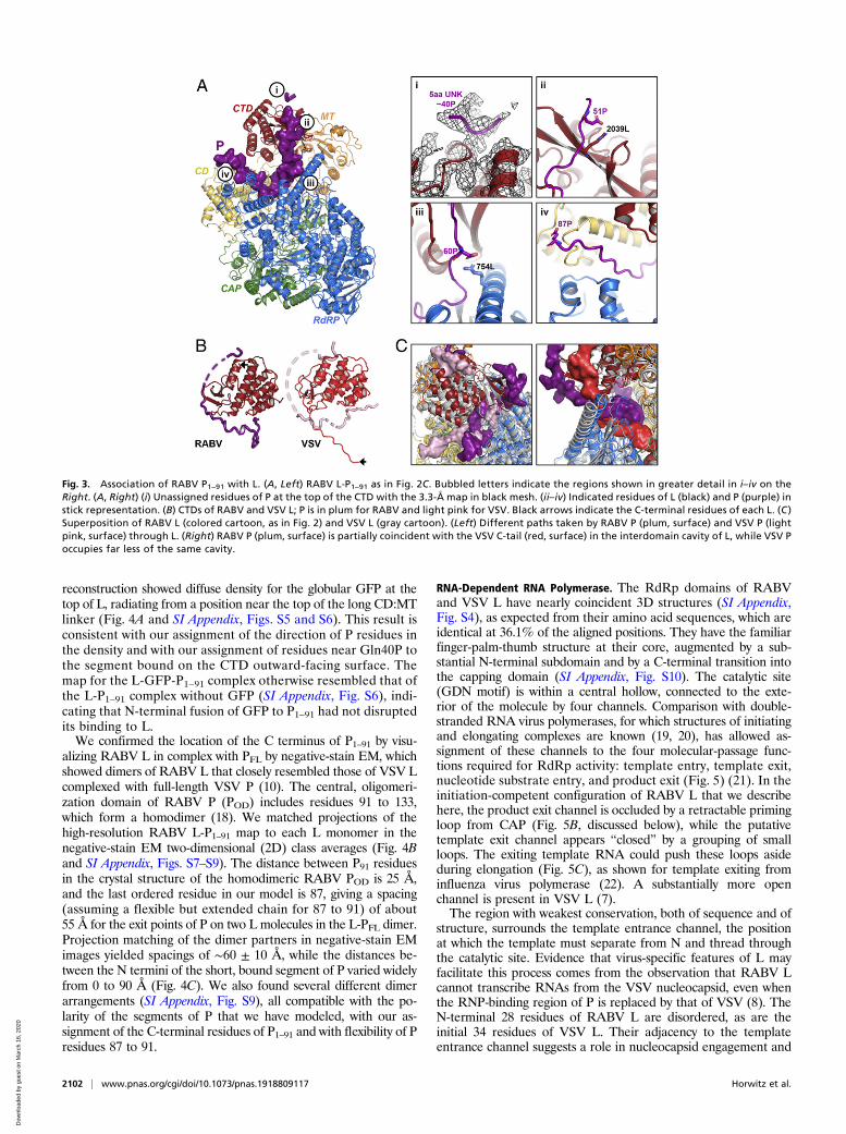
Phosphoprotein Fragment P1–91.We detect two segments of densityfor the P1–91 polypeptide chain (Fig. 3). One segment binds in agroove on the outward-facing surface of the CTD above the C-terminal residues of L (Fig. 3 A, i). This segment is too short toassign its sequence with confidence, but biochemical experimentsthat measured RABV L processivity on naked RNA templates inthe presence of N-terminal fragments of P suggest that this in-teraction involves at least some of the residues immediately fol-lowing Gln40 of P (8). The second segment is much longer, with acontinuous stretch of 37 residues. It contains about 11 residuesbound on the proximal face of the CTD, probably in part througha salt bridge between Glu51P and Arg2039L (Fig. 3 A, ii); about 3residues in contact with a long RdRp helix (residues 746 to 773),probably including a salt bridge between Asp60P and Arg754L(Fig. 3 A, iii); and about 13 additional residues in the groove be-tween the RdRp and CD (Fig. 3 A, iv). The C terminus of theordered segment that we could build is Glu87P. The closer asso-ciation of P with the proximal face of the CTD of L for RABVrelative to VSV suggests that RABV P partially compensates forthe C-terminal tail that is “missing” in RABV L (Fig. 3B).As a first check on the polarity that we had assigned to P, we
fused GFP to the N terminus of P1–91 and visualized its complexwith L by negative-stain EM (Fig. 4A). A low-resolution 3D
Fig. 2. Structure of the RABV L-P complex. (A) A 3.3-Å cryo-EMmap of RABV L-P1–91 andmodel fit into the density with clear side-chains even on exterior residues.(B) Domain organization of RABV L, with key catalytic residues listed. Numbers give amino acid positions at domain boundaries. (Below) Locations of conservedregions (CRI-VI) throughout NNS RNA virus L proteins and amino acid similarity score with VSV L (Indiana strain); black lines indicate identical residues (∼35%). (C)Structure of RABV L-P1–91 (Left) and the model of VSV L-P35–106 from a 3.0-Å cryo-EM map (Right) (7). Domains are colored as in B. P for both structures is shown insurface representation, colored in plum (RABV) and light pink (VSV). The five unassigned residues of RABV P are represented in plum ribbon. (D) Domain or-ganization of RABV P (Top) amino acid positions (red asterisk, position of Ser63P, typically phosphorylated). (Below) Alignment with VSV P (purple lines, modeledRABV P residues; boldface letters, modeled VSV P residues; asterisks, shared residues; colons, chemically similar residues; periods, weakly similar residues; red,phosphorylation sites).
Horwitz et al. PNAS | January 28, 2020 | vol. 117 | no. 4 | 2101
MICRO
BIOLO
GY
Dow
nloa
ded
by g
uest
on
Mar
ch 1
6, 2
020
reconstruction showed diffuse density for the globular GFP at thetop of L, radiating from a position near the top of the long CD:MTlinker (Fig. 4A and SI Appendix, Figs. S5 and S6). This result isconsistent with our assignment of the direction of P residues inthe density and with our assignment of residues near Gln40P tothe segment bound on the CTD outward-facing surface. Themap for the L-GFP-P1–91 complex otherwise resembled that ofthe L-P1–91 complex without GFP (SI Appendix, Fig. S6), indi-cating that N-terminal fusion of GFP to P1–91 had not disruptedits binding to L.We confirmed the location of the C terminus of P1–91 by visu-
alizing RABV L in complex with PFL by negative-stain EM, whichshowed dimers of RABV L that closely resembled those of VSV Lcomplexed with full-length VSV P (10). The central, oligomeri-zation domain of RABV P (POD) includes residues 91 to 133,which form a homodimer (18). We matched projections of thehigh-resolution RABV L-P1–91 map to each L monomer in thenegative-stain EM two-dimensional (2D) class averages (Fig. 4Band SI Appendix, Figs. S7–S9). The distance between P91 residuesin the crystal structure of the homodimeric RABV POD is 25 Å,and the last ordered residue in our model is 87, giving a spacing(assuming a flexible but extended chain for 87 to 91) of about55 Å for the exit points of P on two L molecules in the L-PFL dimer.Projection matching of the dimer partners in negative-stain EMimages yielded spacings of ∼60 ± 10 Å, while the distances be-tween the N termini of the short, bound segment of P varied widelyfrom 0 to 90 Å (Fig. 4C). We also found several different dimerarrangements (SI Appendix, Fig. S9), all compatible with the po-larity of the segments of P that we have modeled, with our as-signment of the C-terminal residues of P1–91 and with flexibility of Presidues 87 to 91.
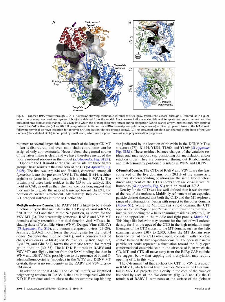
RNA-Dependent RNA Polymerase. The RdRp domains of RABVand VSV L have nearly coincident 3D structures (SI Appendix,Fig. S4), as expected from their amino acid sequences, which areidentical at 36.1% of the aligned positions. They have the familiarfinger-palm-thumb structure at their core, augmented by a sub-stantial N-terminal subdomain and by a C-terminal transition intothe capping domain (SI Appendix, Fig. S10). The catalytic site(GDN motif) is within a central hollow, connected to the exte-rior of the molecule by four channels. Comparison with double-stranded RNA virus polymerases, for which structures of initiatingand elongating complexes are known (19, 20), has allowed as-signment of these channels to the four molecular-passage func-tions required for RdRp activity: template entry, template exit,nucleotide substrate entry, and product exit (Fig. 5) (21). In theinitiation-competent configuration of RABV L that we describehere, the product exit channel is occluded by a retractable primingloop from CAP (Fig. 5B, discussed below), while the putativetemplate exit channel appears “closed” by a grouping of smallloops. The exiting template RNA could push these loops asideduring elongation (Fig. 5C), as shown for template exiting frominfluenza virus polymerase (22). A substantially more openchannel is present in VSV L (7).The region with weakest conservation, both of sequence and of
structure, surrounds the template entrance channel, the positionat which the template must separate from N and thread throughthe catalytic site. Evidence that virus-specific features of L mayfacilitate this process comes from the observation that RABV Lcannot transcribe RNAs from the VSV nucleocapsid, even whenthe RNP-binding region of P is replaced by that of VSV (8). TheN-terminal 28 residues of RABV L are disordered, as are theinitial 34 residues of VSV L. Their adjacency to the templateentrance channel suggests a role in nucleocapsid engagement and
Fig. 3. Association of RABV P1–91 with L. (A, Left) RABV L-P1–91 as in Fig. 2C. Bubbled letters indicate the regions shown in greater detail in i–iv on theRight. (A, Right) (i) Unassigned residues of P at the top of the CTD with the 3.3-Å map in black mesh. (ii–iv) Indicated residues of L (black) and P (purple) instick representation. (B) CTDs of RABV and VSV L; P is in plum for RABV and light pink for VSV. Black arrows indicate the C-terminal residues of each L. (C )Superposition of RABV L (colored cartoon, as in Fig. 2) and VSV L (gray cartoon). (Left) Different paths taken by RABV P (plum, surface) and VSV P (lightpink, surface) through L. (Right) RABV P (plum, surface) is partially coincident with the VSV C-tail (red, surface) in the interdomain cavity of L, while VSV Poccupies far less of the same cavity.
2102 | www.pnas.org/cgi/doi/10.1073/pnas.1918809117 Horwitz et al.
Dow
nloa
ded
by g
uest
on
Mar
ch 1
6, 2
020

template insertion; the low isoelectric point of the disorderedpeptide further suggests that it might facilitate separation ofthe template RNA from N.
Capping Domain. The RABV L capping enzyme catalyzes theformation of GTP-capped pre-mRNA by a mechanism that dif-fers from eukaryotic capping. The nascent RNA transcript with a5′ triphosphate is first covalently linked to a catalytic histidineresidue (H1241) in a reaction that leads to a monophosphateRNA-L intermediate. This linkage is subsequently thought to beattacked by a GTP molecule, resulting in addition of GDP to themonophosphate-RNA to form a GTP-capped pre-mRNA (23).Several residues in both RABV and VSV CAP that affect capaddition have been identified, including the GxxT and HR motifsbroadly conserved throughout Mononegavirales (1, 24). The his-tidine in the latter motif is the site of covalent attachment. Whilewe initially supposed that the GxxT motif would support thesubstrate GTP, the precise role of the motif in capping is unclear,as mutation of the threonine, essential for VSV capping (25), isless critical for capping by RSV L (26). The respective CAP do-mains are extremely similar (SI Appendix, Figs. S4 and S11), al-though there are small differences in the positioning of certainloops, including those in or near the active site (SI Appendix, Fig.
S11A). The two sites that coordinate structural zinc ions in VSV Lare also present in RABV L (SI Appendix, Fig. S11B). Each siteincludes two pairs of residues separated by approximately 200residues in the polypeptide chain, thereby joining separateregions within the CAP domain.The GxxT motif leads into the ∼20-residue-long priming loop,
which extends from CAP toward the RdRp active site (SI Ap-pendix, Fig. S11C), as we also found for VSV L (1). Relatively poordensity in both maps indicates conformational variability. The statecaptured in this structure is evidently a preinitiation or initiationconformation, as elongation after priming requires that the prim-ing loop withdraw from the RdRp catalytic cavity to make roomfor nascent product to reach the CAP active site and extend up-ward into the MT domain (Fig. 5). The catalytic cavity of CAPappears to be large enough to accommodate a retracted primingloop (Fig. 5B), together with a short segment of nascent transcript,even without displacing CAP from RdRp.
Connector Domain. The RABV CD has no known enzymaticfunction and probably has a largely organizational role. The CDof RABV L consists of eight helices; long linkers at either endconnect it to the CAP and MT domains (SI Appendix, Fig. S12).Whereas the CAP-CD linker is sufficiently ordered to assign
Fig. 4. Analysis of negatively stained L-GFP-P1–91 and L-P dimers. (A, Left) Two-dimensional class averages of negatively stained L-GFP-P1–91 and matchedprojections of the RABV L atomic model with GFP position approximated (SI Appendix, Figs. S5 and S6). (A, Right) GFP trace from a 3D reconstruction ofnegatively stained RABV L-GFP-P1–91 (SI Appendix, Fig. S6) superimposed on the RABV L atomic model. (B) Two-dimensional classes of negatively stained RABVL-PFL dimers and matched projections of the RABV L atomic model (SI Appendix, Figs. S7–S9), as described in legend to SI Appendix, Fig. S7. Square boxes in Aand B are 336 Å on a side. (C) Minimal distances between Cα atoms of either P87 (C:C) or the N terminus of the P segment bound at the CTD apex (N:N) for eachprojection-matched dimer. (D) Approximate locations of P1 and P91 with respect to L determined from the projection-matching analysis.
Horwitz et al. PNAS | January 28, 2020 | vol. 117 | no. 4 | 2103
MICRO
BIOLO
GY
Dow
nloa
ded
by g
uest
on
Mar
ch 1
6, 2
020
rotamers to several larger side-chains, much of the longer CD-MTlinker is disordered, and even main-chain coordinates can beassigned only approximately. Nevertheless, the general courseof the latter linker is clear, and we have therefore included thepoorly ordered residues in the model (SI Appendix, Fig. S12A).Opposite the HR motif at the CAP active site are three tightly
grouped basic resides in the final helix of the CD (SI Appendix, Fig.S12B). The first two, Arg1610 and His1611, conserved among allLyssavirus L, are also present in VSV L. The third, R1614, is eitherarginine or lysine in all lyssaviruses; it is a lysine in VSV L. Theproximity of these basic residues in the CD to the catalytic HRmotif in CAP, as well as their chemical composition, suggest thatthey may help guide the nascent transcript toward His1241, theposition of covalent attachment. Alternatively, they could directGTP-capped mRNAs into the MT active site.
Methyltransferase Domain. The RABV MT is likely to be a dual-function enzyme that methylates the GTP cap of viral mRNAs,first at the 2′-O and then at the N-7 position, as shown for theVSV MT (3). The structurally conserved RABV and VSV MTdomains closely resemble other dual-function viral MTases, in-cluding those of West Nile Virus (WNV), Dengue Virus (DENV)(SI Appendix, Fig. S13), and human metapneumovirus (27–29).A shared GxGxG motif forms the binding site for the methyldonor, S-adenosylmethionine (SAM), and a conserved set ofcharged residues (K-D-K-E; RABV residues Lys1685, Asp1797,Lys1829, and Glu1867) forms the catalytic tetrad for methylgroup addition (30–33). The K-D-K-E tetrads in RABV andVSV MTs are slightly further from the SAM-binding site than inWNV and DENV MTs, possibly due to the presence of bound S-adenosylhomocysteine (modeled) in the WNV and DENV MTcrystals; there is no such density in the RABV and VSV L cryo-EM maps.In addition to the K-D-K-E and GxGxG motifs, we identified
neighboring residues in RABV L that are interspersed with theK-D-K-E residues and are close to the presumptive cap-binding
site [indicated by the location of ribavirin in the DENV MTasestructure (27)]: R1674, Y1831, T1860, and Y1869 (SI Appendix,Fig. S13B). These residues balance charges of the catalytic res-idues and may support cap positioning for methylation and/orreaction order. They are conserved throughout Rhabdoviridaeand match similarly positioned residues in WNV and DENV.
C-Terminal Domain. The CTDs of RABV and VSV L are the leastconserved of the five domains; only 20.1% of the amino acidresidues at corresponding positions are the same. Nonetheless,direct alignment of the CTDs shows they are close structuralhomologs (SI Appendix, Fig. S3) with an rmsd of 3.7 Å.Density for the CTD was less well defined than it was for most
of the rest of the molecule. Multibody refinement of an expandedparticle dataset showed that both the CTD and the MT explore arange of conformations, flexing with respect to the other domains(Movie S1). While the MT flexes as a rigid domain, the CTDappears to have “open” and “closed” conformations that wouldinvolve remodeling the α helix spanning residues 2,092 to 2,105(see the upper left in the middle and right panels, Movie S1).This hinge-like behavior may account for the lack of well-ordereddensity for P at the apex of the CTD in the high-resolution map.Elements of the CTD closest to the MT domain, such as the helixspanning residues 2,019 to 2,033, follow the MT domain awayfrom the rest of the CTD when open, consistent with the closecontact between the two sequential domains. The open state in ourparticle set could represent a fluctuation toward the fully openconformational ensemble seen in the absence of P, in which theCD, MT, and CTD all move away from the RdRp-CAP module.We suggest below that capping and methylation may requireopening of L in this way.The C-terminal tail that anchors the CTD in VSV L is absent
in RABV L, which has 24 fewer residues. Whereas the C-terminaltail in VSV L-P projects into a cavity in the core of the complexbounded by each of the five domains (Fig. 3 B and C), the Cterminus of RABV L terminates at the surface of the globular
Fig. 5. Proposed RNA transit through L. (A–C) Cutaways showing continuous internal cavities (gray, translucent surface) through L (colored, as in Fig. 2C)when the priming loop residues (green ribbon) are deleted from the model. Black arrows indicate nucleotide and template entrance channels and thepresumed RNA product exit channel. (B) Cavity into which the priming loop may retract during elongation (white dashed arrow). Nascent RNA may continuetoward the CAP active site (HR motif) following internal initiation for mRNA transcription (solid orange arrow) or directly upward toward the MT domainfollowing terminal de novo initiation for genomic RNA replication (dashed orange arrow). (C) The presumed template exit channel at the back of the CAPdomain (black dashed circle) is occupied by small loops, which we propose move aside as polymerization progresses.
2104 | www.pnas.org/cgi/doi/10.1073/pnas.1918809117 Horwitz et al.
Dow
nloa
ded
by g
uest
on
Mar
ch 1
6, 2
020
CTD (Fig. 3B). A segment of P that penetrates the gap betweenCTD and CD, and somewhat closer packing of the adjacentdomains, together appear to compensate for the “missing”C-terminalarm (Fig. 3C).
Priming, Initiation, Elongation, and Capping. The structure describedhere, together with those of VSV L-P at 3.0 Å resolution (7) and oftwo recently published pneumovirus L-P complexes (5, 6), suggestsa mechanism for switching between replication and transcription,with alternative priming configurations and alternative sites forproduct exit.The “priming loop” (residues 1,170 to 1,186 in the CAP do-
main) projects into the RdRp catalytic cavity, closing off a channelthat connects it with the catalytic cavity of the CAP domain. Modelbuilding in the homologous VSV L-P complex shows that an ini-tiating nucleotide would stack on a tryptophan (W1167), corre-sponding to residueW1180 in RABV L, at the tip of the projectingloop (7). Mutation of this residue compromises end initiation, butnot internal initiation or capping (4). Elongation beyond formationof the initial dinucleotide requires that the priming loop retractinto the CAP catalytic cavity. The recent pneumovirus L-P struc-tures show just such a retracted configuration (5, 6). A nascenttranscript can then pass across the retracted loop. Moreover,analysis of cavities in the L-P complex (Fig. 5) shows that, afterpriming loop retraction, a continuous tunnel leads from the con-nected catalytic cavities of RdRp and CAP to a likely exit site forthe full-length replication products (antigenomic and genomicRNA). Proximity of this putative exit site to the N-terminal end ofP would then allow prompt delivery of N protein, bound near theN terminus of P, coating and protecting the emerging replicationproduct (or uncapped leader transcript).The N-protein delivery pathway suggested above depends on
the site at the apex of the CTD that anchors the N-terminalsegment of P. That site is a shallow pocket, which accommo-dates just four or five residues. In the VSV L-P complex, a tyrosineside-chain packs against the base of the pocket; mutation of thattyrosine to small residues severely impairs viral growth, but doesnot affect RdRp activity (7). We have suggested that failure toanchor P in that site may impede replication (but not transcrip-tion) by interfering with efficient delivery of N to the product. Thatfunction might explain the conservation of the position of the site,but not its chemistry.Priming for internal initiation and capping and priming for end
initiation may depend on different configurations and differentmechanisms for supporting the priming GTP. Internal initiation isinsensitive to mutation of Trp1180 (4), suggesting that the primingloop remains retracted after termination of the leader transcriptand that support for the priming GTP comes from some othersource, such as the 3′ end of the preceding transcript, still base-paired with the template (see a related, early proposal in ref. 34),or from some aspect of the posttermination conformation of thetemplate-engaged RdRp domain. Each of the products of internalinitiation has a 5′-AACA sequence and forms a covalent attach-ment with His1241. A noteworthy characteristic of the “closed”initiation competent structure seen here and for VSV L is theabsence of an obvious GTP-binding site in the capping domain.We suggest that the retracted priming loop and elongated nascenttranscript might create such a site, perhaps linked to the covalentattachment of the 5′ end of the transcript to His1241. As long asthe 5′ attachment to His1241 is present, elongation will produce aloop that will fill the CAP catalytic cavity and force the smaller do-mains (CD, MT, CTD) to swing outward (SI Appendix, Fig. S14)—asthey do in the absence of P (Fig. 1). This transition to a moreopen complex will allow GTP to diffuse into the CAP active-sitecavity (if it is not already there), and it will also expose themethyltransferase catalytic site more completely than in theclosed structure. These steps may account for the observation that
capping of VSV mRNA occurs only after 31 nucleotides havebeen transcribed (35).The scheme proposed here provides a simple mechanism for
switching between replication mode, in which full-length productacquires an N-protein coat, and transcription mode, in which anmRNA product acquires a 5′ cap. It also provides an evolu-tionary rationale for the distinctive capping mechanism (poly-ribonucleotide transferase rather than guanylyl transferase)found in NNS viruses. Retraction of the priming loop creates acontinuous cavity shared by the CAP and RdRp catalytic sites.Covalent attachment of the 5′ end of the transcript then allowsongoing RNA polymerization to fill this cavity with productstrand, generating the force needed to release the CD-MT-CTD module. Tests of this model will require structures oftranscribing intermediates.
Materials and MethodsProtein Expression and Purification. We expressed 6xHis-tagged recombinantRABV SAD-B19 L alone or with variants of RABV SAD-B19 P in SF9 or SF21insect cells from baculovirus vectors constructed using the pFastBac-Dual re-combination system (Thermo Fisher) as described previously (8). The N-terminalfragments of RABV SAD-B19 P, P11–50, and P1–91 were expressed in Escherichiacoli and purified as previously described (8). The P variants GFP-P1–91, PFL, andPΔOD with StrepII affinity tags were coexpressed with L in insect cells andcopurified with L. For GFP-P1–91, with an N-terminal StrepII tag, L-GFP-P1–91complexes were purified first on nickel and then on Strep-Tactin resin (GEHealthcare). For PFL, with a C-terminal StrepII tag, L-PFL complexes were puri-fied first on Strep-Tactin and then on nickel resin. For PΔOD, with a hemag-glutinin (HA) epitope tag followed by an internal HRV 3C protease cleavagesite in place of the deleted oligomerization domain of P (P residues 92 to 131),as well as a C-terminal StrepII tag, L-PΔOD complexes were immobilized onStrep-Tactin resin and then cleaved from the resin in the presence of HRV 3Cprotease to yield purified complexes of L-P1–91. The nine-residue HA tag, notused for purification, remained attached to P1–91 following proteolytic cleav-age. For cryo-EM specimens, these L-PΔOD–derived L-P1–91 complexes werefurther purified on a Superdex 200 Increase column, and the peak fractionswere concentrated to 0.3 mg/mL immediately before grid preparation. Foreach grid, 3 μL of sample were applied to glow-discharged, C-Flat 400-meshcopper grids coated with 40-nm-thick holey carbon (1.2/1.3 spacing) andplunge-frozen in liquid ethane in a CP3 cryo-dipper.
Electron Microscopy. For the medium-resolution initial reconstruction, imagesof RABV L-P1–91 were recorded on a Tecnai F20 electron microscope (FEI)operated at 200 kV, using UCSF Image4 (Yuemin Li, University of California,San Francisco) to collect movies on a K2 Summit direct detector (Gatan) insuperresolution mode with dose fractionation. For each 8-s exposure, wecollected 32 frames at 250 ms each with a total electron dose of 72 e/Å2.Collection was performed at a nominal magnification of 29,000×, with acalibrated pixel size of 1.28 Å/pixel. Movie frames were gain-subtracted,Fourier-binned 2×, and motion-corrected using MotionCor2 with 5 × 5patch correction.
For the high-resolution dataset, images were recorded on a Tecnai F30Polara electron microscope (FEI) operated at 300 kV, using SerialEM to collectmovies on a K2 Summit direct detector (Gatan) in superresolution mode withdose fractionation. For each 8-s exposure, we collected 32 frames at 250 mseach with a total electron dose of 72 e/Å2. Collection was performed at anominal magnification of 31,000× with a calibrated physical pixel size of1.234 Å/pixel.
Image Processing. Micrograph movie frames were gain-subtracted, Fourier-binned to physical pixel size, and motion-corrected using MotionCor2 with5 × 5 patch correction. Micrograph CTF coefficients were calculated usingCTFFIND4 (36), and particle images were CTF-corrected as needed by theprocessing software used.
For the medium-resolution reconstruction, a single dataset was collectedfrom one of four identical grids prepared in parallel from a single proteinsample. We picked 43,000 particles by hand from 316 motion-correctedmovies using EMAN2.1 (37). A de novo 3D reference was obtained usingthe ab-initio procedure in cisTEM (11) following preliminary 2D classification,and the hand was subsequently flipped to yield a reference bearing strongresemblance to VSV L-P. Final 2D and 3D classifications using the flippedreference were then carried out in RELION 2.1 (38), yielding a 6.7 Å (medium-resolution) map from 23,000 particles.
Horwitz et al. PNAS | January 28, 2020 | vol. 117 | no. 4 | 2105
MICRO
BIOLO
GY
Dow
nloa
ded
by g
uest
on
Mar
ch 1
6, 2
020
For the high-resolution reconstruction, we collected a total of 10,949 moviesfrom three datasets using the remaining three grids from the initial set of four.Selected 20-Å low-pass-filtered projections of the 6.7-Å map were used asparticle-picking templates for autopicking in RELION (38). Autopicked particleson each micrograph were manually cleaned of junk, contamination, noise,drift, and otherwise bad or damaged particles using SamViewer, an interactiveimage analysis program written in wxPython by Maofu Liao, Harvard MedicalSchool. From 5.3 M remaining particles, coarse 2D classification at a high-resolution cutoff of 25 Å was performed in RELION (38) to remove addi-tional poor particles, and classes representing dominant views were separatedfrom the remaining good classes, as described in SI Appendix, Fig. S2. Three-dimensional classification was performed on 1.47 M nondominant view parti-cles, and the best class of six was selected, containing 649 k particles. Theseparticles were added to the separated 408 k dominant view particles; a finalround of 3D classification then yielded a total of 963 k particles from the bestclass of three. This particle set was then subjected to 3D refinement, per-particle CTF refinement, and Bayesian polishing in RELION 3.0 (13), and theresulting stack was imported into cisTEM (11) along with corresponding Eulerangles and offsets for further refinement, as described in SI Appendix, Figs. S1and S2). The anisotropic 3D reconstruction from refinement in RELION 3.0 (13)was used as a starting reference for manual refinement in cisTEM (11), andparticles were removed from the reconstruction at each refinement iterationusing the SCORE parameter. We quickly removed the worst-scoring 50% ofparticles. Because dominant view particles had higher scores (due to anisotropyof the map caused by a preferred orientation), we applied an angular binningscript to cap the number of particles per 4° × 4° bin, keeping those with thehighest scores. Further manual refinement using these remaining 177 kparticles with flattened angular distribution led to reduced anisotropy andimproved map quality, with the best 25% of particles by score giving thehighest-resolution map. A final refinement using this set of 44,500 particlesat an alignment resolution cutoff of 4.5 Å yielded a reasonably isotropic map(despite a majority of particles having the preferred orientation) with an av-erage resolution of 3.3 Å. The Electron Microscopy Data Bank (EMDB) accessionnumber for the deposited map is EMD-20753.
Model Building. To generate the atomicmodel of RABV L, we first replaced thesequence of VSV L with RABV L in the model of VSV L (PDB ID 6U1X) (7),adding simple loops where discontinuities in the alignment occurred. We fitthese coordinates as a single rigid body into the 3.3-Å RABV L-P1–91 densitymap and then divided the coordinates into the five structural domains(RdRp, CAP, CD, MT, and CTD) and refit each domain as a rigid body. Wethen performed iterative real-space refinement using PHENIX (39, 40) atincreasing resolution, from 40 to 5 Å. The resulting model was then used asan input model for ROSETTA (17). To improve loop fitting, we also inputlibraries of 3- and 9-mer peptide structures corresponding to all 3- and 9-amino acid sequences contained in the 2,127-residue RABV L sequence (41).From 5,000 simulations, we selected a model with very good map-to-modelagreement and with visibly strong fitting of secondary structure elementsthroughout. We then performed several rounds of manual adjustment inCoot, alternating with real-space refinement in PHENIX, using secondarystructure restraints throughout and Ramachandran restraints in the finalrounds. The final model was validated using PHENIX and MolProbity (SIAppendix, Table S1) and deposited in the Protein Data Bank under PDBID 6UEB.
Projection Matching. Molecules of RABV L in complex with GFP-P1–91 werevisualized by negative-stain EM at ∼0.01 mg/mL total protein in the presenceof 0.7% uranyl formate. Twenty-eight 2D class averages of picked particlesshowed clear density for the GFP moiety at the N terminus of P (Fig. 4A andSI Appendix, Figs. S5 and S6). We aligned the 2D class averages with obvious
GFP density to a single reference to standardize their orientation in the imageplane and applied a circular noise mask to obscure the GFP signal as much aspossible without obstructing signal from L. To determine the viewing angle ofthe RABV L atomic model corresponding to each 2D class, we projected thehigh-resolution RABV L-P1–91 cryo-EM map over all viewing angles, incre-mented every ∼7.2° for a total of 800 projections. Projections were low-pass-filtered to 20 Å and then scaled and clipped to match the pixel and box size ofthe 2D classes, and a circular noise mask was applied to obscure the clippingedges. Cross-correlation coefficients between projections and 2D classes wereobtained using e2simmx.py from EMAN2 (37), allowing for rotation andtranslation only. The top projection match for each 2D class is shown in Fig. 4and SI Appendix, Fig. S5, with GFP modeled as a best approximation.
L-PFL dimers, representing about 25% of L species on micrographs of neg-atively stained, purified RABV L/PFL, were picked by hand and subjected to 2Dclass averaging using EMAN2.1 (37). From 29 high-quality 2D class averages ofdimers, we extracted each of the 58 dimeric monomer averages as individualparticles (hereafter, DMAPs). We applied a circular noise mask to each DMAPto obscure excess signal from the paired DMAPs present in each particle image.We then projected the high-resolution RABV L-P1–91 cryo-EM map over allviewing angles, incremented every ∼7.2° for a total of 800 projections. Pro-jections were low-pass-filtered to 20 Å and then clipped and scaled to matchthe box and pixel size of the negative-stain EM DMAPs, and a circular noisemask was applied to obscure the clipping edges. Cross-correlation coefficientsbetween projections and DMAPs were obtained using e2simmx.py, allowingfor rotation and translation only. The resulting matrix was then normalizedwithin each DMAP over all projections, from which we constructed sphericalheat maps in the coordinate system of the RABV L-P1–91 cryo-EM map to fa-cilitate visualization of peaks reflecting high cross-correlation scores. For abouthalf of the DMAPs, a single peak predominated, and application of the angularcoordinates from the peak to the atomic model of RABV L-P1–91 returned anobvious match to the corresponding DMAP. Whenmore than one high-scoringpeak was observed, the best apparent visual match was selected. Of the 58DMAP:projection comparisons, only 6 were matched to a secondary peak,and 5 could not be reliably matched. In total, we successfully matched 24of the 29 DMAP pairs. Using PyMOL (Schrödinger, LLC), we then appliedthe angular coordinates from each match to an atomic model of RABV L-P1–91 for each DMAP in a pair and approximated the relationship betweenthe two modeled DMAPs using only two dimensions (X and Y), as sug-gested by the original 2D class average. We then measured the minimumdistance between C-alpha atoms of either P87 or the mapped, but un-assigned, N-terminal-most residue of P in each modeled pair by translatingone of the models along the z axis. The results of these projection-matching analyses are summarized in Fig. 4B and illustrated in detail inSI Appendix, Fig. S8.
Visualization of Internal Cavities.We used the program VOIDOO (42) to probeinternal cavities, substrate entry, and product exit channels of the RABVL-P1–91 complex. We used a probe radius of 1.8 Å to calculate a probe-occupiedvolume.
Data Availability. Data for EM 3D reconstruction have been deposited in theElectron Microscopy Data Bank (accession no. EMD-20753), and data forcoordinates fit to 3D reconstruction have been deposited in the Protein DataBank (PDB ID 6UEB).
ACKNOWLEDGMENTS. We thank Louis-Marie Bloyet for discussions of Mono-negavirus biology and advice on L-protein preparation. J.A.H. is an AmgenFellow of the Life Sciences Research Foundation. The work was supported byNIH Grant R37 AI059371 (to S.P.J.W.) and Grant R01 CA13202 (to S.C.H.). S.C.H.is an Investigator in the Howard Hughes Medical Institute.
1. B. Liang et al., Structure of the L protein of vesicular stomatitis virus from electroncryomicroscopy. Cell 162, 314–327 (2015).
2. T. Ogino, A. K. Banerjee, Formation of guanosine(5′)tetraphospho(5′)adenosine capstructure by an unconventional mRNA capping enzyme of vesicular stomatitis virus. J.Virol. 82, 7729–7734 (2008).
3. A. A. Rahmeh, J. Li, P. J. Kranzusch, S. P. Whelan, Ribose 2′-O methylation of thevesicular stomatitis virus mRNA cap precedes and facilitates subsequent guanine-N-7methylation by the large polymerase protein. J. Virol. 83, 11043–11050 (2009).
4. M. Ogino, N. Gupta, T. J. Green, T. Ogino, A dual-functional priming-capping loop ofrhabdoviral RNA polymerases directs terminal de novo initiation and capping in-termediate formation. Nucleic Acids Res. 47, 299–309 (2019).
5. M. S. A. Gilman et al., Structure of the respiratory syncytial virus polymerase complex.Cell 179, 193–204.e14 (2019).
6. J. Pan et al., Structure of the human metapneumovirus polymerase phosphoproteincomplex. Nature, 10.1038/s41586-019-1759-1 (2019).
7. S. Jenni et al., Structure of the vesicular stomatitis virus L protein in complex with itsphosphoprotein cofactor. Cell Rep., 10.1016/j.celrep.2019.12.024 (2020).
8. B. Morin, B. Liang, E. Gardner, R. A. Ross, S. P. J. Whelan, An in vitro RNA synthesisassay for rabies virus defines ribonucleoprotein interactions critical for polymeraseactivity. J. Virol. 91, e01508-16 (2017).
9. A. A. Rahmeh et al., Molecular architecture of the vesicular stomatitis virus RNApolymerase. Proc. Natl. Acad. Sci. U.S.A. 107, 20075–20080 (2010).
10. A. A. Rahmeh et al., Critical phosphoprotein elements that regulate polymerase architectureand function in vesicular stomatitis virus. Proc. Natl. Acad. Sci. U.S.A. 109, 14628–14633 (2012).
11. T. Grant, A. Rohou, N. Grigorieff, cisTEM, user-friendly software for single-particleimage processing. eLife 7, e35383 (2018).
12. S. H. Scheres, RELION: Implementation of a Bayesian approach to cryo-EM structuredetermination. J. Struct. Biol. 180, 519–530 (2012).
13. J. Zivanov et al., New tools for automated high-resolution cryo-EM structure deter-mination in RELION-3. eLife 7, e42166 (2018).
2106 | www.pnas.org/cgi/doi/10.1073/pnas.1918809117 Horwitz et al.
Dow
nloa
ded
by g
uest
on
Mar
ch 1
6, 2
020

14. T. Nakane, D. Kimanius, E. Lindahl, S. H. Scheres, Characterisation of molecular mo-tions in cryo-EM single-particle data by multi-body refinement in RELION. eLife 7,e36861 (2018).
15. C. A. Rohl, C. E. Strauss, D. Chivian, D. Baker, Modeling structurally variable regions inhomologous proteins with Rosetta. Proteins 55, 656–677 (2004).
16. C. A. Rohl, C. E. Strauss, K. M. Misura, D. Baker, Protein structure prediction usingRosetta. Methods Enzymol. 383, 66–93 (2004).
17. Y. Song et al., High-resolution comparative modeling with RosettaCM. Structure 21,1735–1742 (2013).
18. I. Ivanov, T. Crépin, M. Jamin, R. W. Ruigrok, Structure of the dimerization domain ofthe rabies virus phosphoprotein. J. Virol. 84, 3707–3710 (2010).
19. X. Lu et al., Mechanism for coordinated RNA packaging and genome replication byrotavirus polymerase VP1. Structure 16, 1678–1688 (2008).
20. Y. Tao, D. L. Farsetta, M. L. Nibert, S. C. Harrison, RNA synthesis in a cage: Structuralstudies of reovirus polymerase lambda3. Cell 111, 733–745 (2002).
21. J. Reguera, P. Gerlach, S. Cusack, Towards a structural understanding of RNA synthesisby negative strand RNA viral polymerases. Curr. Opin. Struct. Biol. 36, 75–84 (2016).
22. T. Kouba, P. Drncová, S. Cusack, Structural snapshots of actively transcribing influenzapolymerase. Nat. Struct. Mol. Biol. 26, 460–470 (2019).
23. T. Ogino, T. J. Green, Transcriptional control and mRNA capping by the GDP poly-ribonucleotidyltransferase domain of the rabies virus large protein. Viruses 11, E504(2019).
24. J. Neubauer, M. Ogino, T. J. Green, T. Ogino, Signature motifs of GDP poly-ribonucleotidyltransferase, a non-segmented negative strand RNA viral mRNA cap-ping enzyme, domain in the L protein are required for covalent enzyme-pRNAintermediate formation. Nucleic Acids Res. 44, 330–341 (2016).
25. J. Li, A. Rahmeh, M. Morelli, S. P. Whelan, A conserved motif in region v of the largepolymerase proteins of nonsegmented negative-sense RNA viruses that is essential formRNA capping. J. Virol. 82, 775–784 (2008).
26. M. R. Braun et al., RNA elongation by respiratory syncytial virus polymerase is cali-brated by conserved region V. PLoS Pathog. 13, e1006803 (2017).
27. D. Benarroch et al., A structural basis for the inhibition of the NS5 dengue virus mRNA2′-O-methyltransferase domain by ribavirin 5′-triphosphate. J. Biol. Chem. 279,35638–35643 (2004).
28. Y. Zhou et al., Structure and function of flavivirus NS5 methyltransferase. J. Virol. 81,3891–3903 (2007).
29. G. C. Paesen et al., X-ray structure and activities of an essential mononegavirales L-protein domain. Nat. Commun. 6, 8749 (2015).
30. J. Li, A. Rahmeh, V. Brusic, S. P. Whelan, Opposing effects of inhibiting cap additionand cap methylation on polyadenylation during vesicular stomatitis virus mRNAsynthesis. J. Virol. 83, 1930–1940 (2009).
31. J. Li, J. S. Chorba, S. P. Whelan, Vesicular stomatitis viruses resistant to the methylaseinhibitor sinefungin upregulate RNA synthesis and reveal mutations that affectmRNA cap methylation. J. Virol. 81, 4104–4115 (2007).
32. J. Li, J. T. Wang, S. P. Whelan, A unique strategy for mRNA cap methylation used byvesicular stomatitis virus. Proc. Natl. Acad. Sci. U.S.A. 103, 8493–8498 (2006).
33. J. Li, E. C. Fontaine-Rodriguez, S. P. Whelan, Amino acid residues within conserveddomain VI of the vesicular stomatitis virus large polymerase protein essential formRNA cap methyltransferase activity. J. Virol. 79, 13373–13384 (2005).
34. S. Shuman, A proposed mechanism of mRNA synthesis and capping by vesicular sto-matitis virus. Virology 227, 1–6 (1997).
35. G. Tekes, A. A. Rahmeh, S. P. Whelan, A freeze frame view of vesicular stomatitis virustranscription defines a minimal length of RNA for 5′ processing. PLoS Pathog. 7,e1002073 (2011).
36. A. Rohou, N. Grigorieff, CTFFIND4: Fast and accurate defocus estimation from elec-tron micrographs. J. Struct. Biol. 192, 216–221 (2015).
37. S. J. Ludtke, Single-particle refinement and variability analysis in EMAN2.1. MethodsEnzymol. 579, 159–189 (2016).
38. D. Kimanius, B. O. Forsberg, S. H. Scheres, E. Lindahl, Accelerated cryo-EM structuredetermination with parallelisation using GPUs in RELION-2. eLife 5, e18722 (2016).
39. P. V. Afonine et al., Real-space refinement in PHENIX for cryo-EM and crystallography.Acta Crystallogr. D Struct. Biol. 74, 531–544 (2018).
40. P. D. Adams et al., PHENIX: A comprehensive Python-based system for macromolec-ular structure solution. Acta Crystallogr. D Biol. Crystallogr. 66, 213–221 (2010).
41. D. E. Kim, D. Chivian, D. Baker, Protein structure prediction and analysis using theRobetta server. Nucleic Acids Res. 32, W526–W531 (2004).
42. G. J. Kleywegt, T. A. Jones, Detection, delineation, measurement and display ofcavities in macromolecular structures. Acta Crystallogr. D Biol. Crystallogr. 50, 178–185 (1994).
Horwitz et al. PNAS | January 28, 2020 | vol. 117 | no. 4 | 2107
MICRO
BIOLO
GY
Dow
nloa
ded
by g
uest
on
Mar
ch 1
6, 2
020