Structure-FunctionalCharacterizationofCytochromeP450 ... · 5-amino acid sequence fragment MAKKT-...
20
Structure-Functional Characterization of Cytochrome P450 Sterol 14-Demethylase (CYP51B) from Aspergillus fumigatus and Molecular Basis for the Development of Antifungal Drugs * Received for publication, July 7, 2015, and in revised form, August 6, 2015 Published, JBC Papers in Press, August 12, 2015, DOI 10.1074/jbc.M115.677310 Tatiana Y. Hargrove ‡ , Zdzislaw Wawrzak § , David C. Lamb ¶ , F. Peter Guengerich ‡ , and Galina I. Lepesheva ‡1 From the ‡ Department of Biochemistry, Vanderbilt University School of Medicine, Nashville, Tennessee 37232, the § Synchrotron Research Center, Life Science Collaborative Access Team, Northwestern University, Argonne, Illinois 60439, ¶ Swansea University, Swansea, Wales SA2 8PP, United Kingdom, and the Center for Structural Biology, Vanderbilt University, Nashville, Tennessee 37232 Background: The fungus Aspergillus fumigatus causes human diseases that are treated with CYP51 azole inhibitors. Results: We report crystal structures of A. fumigatus CYP51B complexes with two inhibitors, voriconazole and VNI. Conclusion: The structures reveal fungus-specific features not observed in CYP51 enzymes from other biological kingdoms. Significance: This molecular insight facilitates rational design of novel antifungals without inhibition of human enzymes. Aspergillus fumigatus is the opportunistic fungal pathogen that predominantly affects the immunocompromised popula- tion and causes 600,000 deaths/year. The cytochrome P450 51 (CYP51) inhibitor voriconazole is currently the drug of choice, yet the treatment efficiency remains low, calling for rational development of more efficient agents. A. fumigatus has two CYP51 genes, CYP51A and CYP51B, which share 59% amino acid sequence identity. CYP51B is expressed constitutively, whereas gene CYP51A is reported to be inducible. We expressed, purified, and characterized A. fumigatus CYP51B, including determination of its substrate preferences, catalytic parameters, inhibition, and x-ray structure in complexes with voriconazole and the experimental inhibitor (R)-N-(1-(2,4- dichlorophenyl)-2-(1H-imidazol-1-yl)ethyl)-4-(5-phenyl-1,3,4- oxadiazol-2-yl)benzamide (VNI). The enzyme demethylated its natural substrate eburicol and the plant CYP51 substrate obtusifoliol at steady-state rates of 17 and 16 min 1 , respec- tively, but did not metabolize lanosterol, and the topical anti- fungal drug miconazole was the strongest inhibitor that we identified. The x-ray crystal structures displayed high overall similarity of A. fumigatus CYP51B to CYP51 orthologs from other biological kingdoms but revealed phylum-specific differ- ences relevant to enzyme catalysis and inhibition. The complex with voriconazole provides an explanation for the potency of this relatively small molecule, whereas the complex with VNI outlines a direction for further enhancement of the efficiency of this new inhibitory scaffold to treat humans afflicted with fila- mentous fungal infections. Fungal diseases represent an increasing global health burden and presently affect 1.2 billion people. The severity of these diseases ranges from topical superficial infections of skin, hair, and nails, which are relatively straightforward to cure (e.g. ath- lete’s foot, ringworm of the scalp) to invasive infections that kill 1.5–2.0 million people annually, surpassing the mortality from tuberculosis or malaria (1). Aspergillus, Candida, and Crypto- coccus are the three fungal genera responsible for most of this mortality, the incidence of which is growing steadily, particu- larly in clinically ill and immunocompromised populations (1– 6). Aspergillus is a genus of soil-dwelling saprophytic molds (filamentous fungi), consisting of 200 species. These molds are found throughout the world and are the most common type of fungi in the environment (see the Aspergillus Web- site). About 16 species of Aspergillus are known to cause disease in humans, Aspergillus fumigatus being responsible for 90% (7, 8). In immunocompetent patients, A. fumigatus can be involved in chronic pulmonary aspergillosis, also known as aspergilloma or “fungal ball,” which is a gradually destructive disease in the lung and often associated with tuberculosis, pulmonary emphysema, and sarcoidosis (3 million people are estimated to be affected). A. fumigatus is also a ubiquitous aeroallergen. Severe asthma related to fun- gal sensitization affects up to 12 million people worldwide, and 100,000 people die from asthma annually. About 5 mil- lion people have allergic bronchopulmonary aspergillosis, whereas 12 million people are afflicted with fungal rhino- sinusitis (9, 10). In immunocompromised individuals (can- cer chemotherapy patients, those on steroids, solid organ and bone marrow transplant recipients, HIV/AIDS patients, and many others), A. fumigatus often manifests as invasive * This work was supported, in whole or in part, by National Institutes of Health Grant R01 GM067871 (to G. I. L.). Vanderbilt University is a member institu- tion of the Life Science Collaborative Team at Sector 21 of the Advanced Photon Source (APS) (Argonne, IL). Use of the APS at Argonne National Laboratory was supported by the United States Department of Energy, Office of Science, Office of Basic Energy Sciences, under Contract DE-AC02- 06CH11357. The authors declare that they have no conflicts of interest with the contents of this article. The atomic coordinates and structure factors (codes 4UYL and 4UYM) have been deposited in the Protein Data Bank (http://wwpdb.org/). 1 To whom correspondence should be addressed: Dept. of Biochemistry, Van- derbilt University School of Medicine, 622 Robinson Research Bldg., 2200 Pierce Ave., Nashville, TN 37232-0146. Tel.: 615-343-1373; Fax: 615-322- 4349; E-mail: [email protected]. crossmark THE JOURNAL OF BIOLOGICAL CHEMISTRY VOL. 290, NO. 39, pp. 23916 –23934, September 25, 2015 © 2015 by The American Society for Biochemistry and Molecular Biology, Inc. Published in the U.S.A. 23916 JOURNAL OF BIOLOGICAL CHEMISTRY VOLUME 290 • NUMBER 39 • SEPTEMBER 25, 2015 by guest on December 23, 2019 http://www.jbc.org/ Downloaded from
Transcript of Structure-FunctionalCharacterizationofCytochromeP450 ... · 5-amino acid sequence fragment MAKKT-...

Structure-Functional Characterization of Cytochrome P450Sterol 14�-Demethylase (CYP51B) from Aspergillus fumigatusand Molecular Basis for the Development of AntifungalDrugs*
Received for publication, July 7, 2015, and in revised form, August 6, 2015 Published, JBC Papers in Press, August 12, 2015, DOI 10.1074/jbc.M115.677310
Tatiana Y. Hargrove‡, Zdzislaw Wawrzak§, David C. Lamb¶, F. Peter Guengerich‡, and Galina I. Lepesheva‡�1
From the ‡Department of Biochemistry, Vanderbilt University School of Medicine, Nashville, Tennessee 37232, the §SynchrotronResearch Center, Life Science Collaborative Access Team, Northwestern University, Argonne, Illinois 60439, ¶Swansea University,Swansea, Wales SA2 8PP, United Kingdom, and the �Center for Structural Biology, Vanderbilt University,Nashville, Tennessee 37232
Background: The fungus Aspergillus fumigatus causes human diseases that are treated with CYP51 azole inhibitors.Results: We report crystal structures of A. fumigatus CYP51B complexes with two inhibitors, voriconazole and VNI.Conclusion: The structures reveal fungus-specific features not observed in CYP51 enzymes from other biological kingdoms.Significance: This molecular insight facilitates rational design of novel antifungals without inhibition of human enzymes.
Aspergillus fumigatus is the opportunistic fungal pathogenthat predominantly affects the immunocompromised popula-tion and causes 600,000 deaths/year. The cytochrome P450 51(CYP51) inhibitor voriconazole is currently the drug of choice,yet the treatment efficiency remains low, calling for rationaldevelopment of more efficient agents. A. fumigatus has twoCYP51 genes, CYP51A and CYP51B, which share 59% aminoacid sequence identity. CYP51B is expressed constitutively,whereas gene CYP51A is reported to be inducible. Weexpressed, purified, and characterized A. fumigatus CYP51B,including determination of its substrate preferences, catalyticparameters, inhibition, and x-ray structure in complexes withvoriconazole and the experimental inhibitor (R)-N-(1-(2,4-dichlorophenyl)-2-(1H-imidazol-1-yl)ethyl)-4-(5-phenyl-1,3,4-oxadiazol-2-yl)benzamide (VNI). The enzyme demethylated itsnatural substrate eburicol and the plant CYP51 substrateobtusifoliol at steady-state rates of 17 and 16 min�1, respec-tively, but did not metabolize lanosterol, and the topical anti-fungal drug miconazole was the strongest inhibitor that weidentified. The x-ray crystal structures displayed high overallsimilarity of A. fumigatus CYP51B to CYP51 orthologs fromother biological kingdoms but revealed phylum-specific differ-ences relevant to enzyme catalysis and inhibition. The complexwith voriconazole provides an explanation for the potency ofthis relatively small molecule, whereas the complex with VNI
outlines a direction for further enhancement of the efficiency ofthis new inhibitory scaffold to treat humans afflicted with fila-mentous fungal infections.
Fungal diseases represent an increasing global health burdenand presently affect �1.2 billion people. The severity of thesediseases ranges from topical superficial infections of skin, hair,and nails, which are relatively straightforward to cure (e.g. ath-lete’s foot, ringworm of the scalp) to invasive infections that kill1.5–2.0 million people annually, surpassing the mortality fromtuberculosis or malaria (1). Aspergillus, Candida, and Crypto-coccus are the three fungal genera responsible for most of thismortality, the incidence of which is growing steadily, particu-larly in clinically ill and immunocompromised populations(1– 6).
Aspergillus is a genus of soil-dwelling saprophytic molds(filamentous fungi), consisting of �200 species. These moldsare found throughout the world and are the most commontype of fungi in the environment (see the Aspergillus Web-site). About 16 species of Aspergillus are known to causedisease in humans, Aspergillus fumigatus being responsiblefor �90% (7, 8). In immunocompetent patients, A. fumigatuscan be involved in chronic pulmonary aspergillosis, alsoknown as aspergilloma or “fungal ball,” which is a graduallydestructive disease in the lung and often associated withtuberculosis, pulmonary emphysema, and sarcoidosis (�3million people are estimated to be affected). A. fumigatus isalso a ubiquitous aeroallergen. Severe asthma related to fun-gal sensitization affects up to 12 million people worldwide,and 100,000 people die from asthma annually. About 5 mil-lion people have allergic bronchopulmonary aspergillosis,whereas �12 million people are afflicted with fungal rhino-sinusitis (9, 10). In immunocompromised individuals (can-cer chemotherapy patients, those on steroids, solid organand bone marrow transplant recipients, HIV/AIDS patients,and many others), A. fumigatus often manifests as invasive
* This work was supported, in whole or in part, by National Institutes of HealthGrant R01 GM067871 (to G. I. L.). Vanderbilt University is a member institu-tion of the Life Science Collaborative Team at Sector 21 of the AdvancedPhoton Source (APS) (Argonne, IL). Use of the APS at Argonne NationalLaboratory was supported by the United States Department of Energy,Office of Science, Office of Basic Energy Sciences, under Contract DE-AC02-06CH11357. The authors declare that they have no conflicts of interestwith the contents of this article.
The atomic coordinates and structure factors (codes 4UYL and 4UYM) have beendeposited in the Protein Data Bank (http://wwpdb.org/).
1 To whom correspondence should be addressed: Dept. of Biochemistry, Van-derbilt University School of Medicine, 622 Robinson Research Bldg., 2200Pierce Ave., Nashville, TN 37232-0146. Tel.: 615-343-1373; Fax: 615-322-4349; E-mail: [email protected].
crossmarkTHE JOURNAL OF BIOLOGICAL CHEMISTRY VOL. 290, NO. 39, pp. 23916 –23934, September 25, 2015
© 2015 by The American Society for Biochemistry and Molecular Biology, Inc. Published in the U.S.A.
23916 JOURNAL OF BIOLOGICAL CHEMISTRY VOLUME 290 • NUMBER 39 • SEPTEMBER 25, 2015
by guest on Decem
ber 23, 2019http://w
ww
.jbc.org/D
ownloaded from

aspergillosis, the most dangerous form of infection, whichspreads to multiple organs, is difficult to treat, and leads to�600,000 deaths annually. The treatment options are stillvery scarce. Overall, invasive aspergillosis has a �50% mor-tality rate if diagnosed and treated early, but if diagnosis ismissed or delayed, then it is nearly 100% fatal (9).
Voriconazole remains the agent of choice for treatment (1),although the success rate is not particularly high, and theadverse side effects (visual disturbances, skin rushes, hepato-toxicity, vomiting, abdominal pain, etc.) require permanenttherapeutic drug monitoring (11, 12). Other medications usedclinically include itraconazole, posaconazole, amphotericin B,or caspofungin and micafungin (combination therapy), butaspergillosis is known to be insensitive to fluconazole (see theAspergillus Website).
Voriconazole, posaconazole, itraconazole, and fluconazole(Fig. 1A) are inhibitors of sterol 14�-demethylase (CYP51, EC1.14.13.70), the cytochrome P450 (CYP)2 enzyme that catalyzesthe three-step reaction of oxidative removal of the 14�-methylgroup from cyclized sterol precursors (Fig. 1, B and C) (13). TheCYP51 reaction is required for sterol biosynthesis and therefore
is conserved across biological kingdoms (14). Fungal CYP51enzymes participate in the biosynthesis of ergosterol, which isan indispensable component of fungal membranes. Ergosteroldepletion affects both the structure of the membrane and sev-eral of its functions. Ergosterol also has a hormone-like (“spark-ing”) role in fungal cells, which stimulates growth and prolifer-ation. This function, however, may be disrupted only whenergosterol depletion is virtually complete (15). Azole-, pyri-dine-, or pyrimidine-based compounds block ergosterol bio-synthesis and cause accumulation of the 14�-methylated pre-cursors (16, 17). These drugs act as competitive CYP51inhibitors that occupy the P450 active site, preventing substratebinding and oxidation. The basic heterocyclic nitrogen coordi-nates to the P450 heme iron (Fig. 1A), sharing its lone pair ofelectrons and blocking binding of molecular oxygen, whereasthe non-ligated portion of the inhibitor molecule forms multi-ple contacts with the protein moiety, shaping the protein-li-gand surface interface that largely defines the strength of theinhibition (18, 19).
Because fungal CYP51 enzymes are very hydrophobic mem-brane-bound proteins (a feature that complicates their han-dling and assay in vitro), data on structure-function relation-ships and inhibition are limited and, when they exist, oftencontroversial (20). As a result, although azoles represent theleading class of antimycotic agents both in medicine and agri-culture (15, 21), each new compound has to be discovered anddeveloped empirically by monitoring its action on fungal cellgrowth. Thus, 1200 fluconazole analogs were synthesized and
2 The abbreviations used are: CYP or P450, cytochrome P450; CYP51, ste-rol 14�-demethylase; CPR, NADPH-cytochrome P450 reductase; SRS,substrate recognition sequence; VNI, (R)-N-(1-(2,4-dichlorophenyl)-2-(1H-imidazol-1-yl)ethyl)-4-(5-phenyl-1,3,4-oxadiazol-2-yl)benzamide; VFV,(R)-N-(1-(3,4�-difluorobiphenyl-4-yl)-2-(1H-imidazol-1-yl)ethyl)-4-(5-phe-nyl-1,3,4-oxadiazol-2-yl)benzamide; MCP, 14�-methylenecyclopropyl-�7-24,25-dihydrolanosterol; HPCD, hydroxypropyl-�-cyclodextrin; NTA, nitri-lotriacetic acid.
FIGURE 1. CYP51 inhibitors and catalytic reaction. A, structural formulas of the marketed drugs, 1,2,4-triazoles voriconazole (Vor), fluconazole (Fluc), andposaconazole (Poz), and an experimental CYP51 inhibitor, the 1,3-imidazole VNI. The heme-coordinating nitrogen atoms are circled. B, 14�-demethylation ofeburicol. The reaction involves three consecutive P450 catalytic cycles, each consuming two electrons (provided by CPR, plus two protons) to reduce thecatalytic heme iron and activate the molecular oxygen, resulting in the insertion of one oxygen atom into the substrate and reduction of the other oxygen atomto water. In this reaction, the sterol 14�-methyl group is first converted into the 14�-alcohol and then into the 14�-aldehyde (CHO-) intermediate and finallyreleased as formic acid. C, HPLC profile of sterols extracted after a 5-min reaction of A. fumigatus CYP51B with eburicol (37 °C; P450, CPR, and eburicolconcentrations were 0.5, 1, and 25 �M, respectively).
Structures of A. fumigatus CYP51B
SEPTEMBER 25, 2015 • VOLUME 290 • NUMBER 39 JOURNAL OF BIOLOGICAL CHEMISTRY 23917
by guest on Decem
ber 23, 2019http://w
ww
.jbc.org/D
ownloaded from

tested at Pfizer before voriconazole was discovered, an ineffi-cient and costly process (1).
Drug resistance (both intrinsic and acquired) representsanother serious problem. Different mechanisms have been pro-posed, including activation of azole efflux pumps, CYP51 muta-tions/CYP51 gene overexpression, and the combination ofpumps and P450 (reviewed in Refs. 22 and 23). Also, it has beensuggested that A. fumigatus resistance to clinically used azolescan be acquired through long time use in the environment (24).Yeast, human, and other vertebrate genomes contain only oneCYP51 gene; however, A. fumigatus and some other filamen-tous ascomycetes (25) have two CYP51 paralogs (CYP51A andCYP51B; Fig. 2), each present as a single copy (26) (chromo-somes 4 and 7, respectively). Although both genes had beensuggested to be active in A. fumigatus, it was later reported thatthe gene CYP51B encodes the enzyme primarily responsible forsterol 14�-demethylation. The CYP51B gene is expressed con-stitutively and found in all sequenced filamentous fungi,whereas the CYP51A gene appears in some fungal lineages (22,25). The presence of two CYP51 genes implies a possibility forfaster sterol biosynthesis in A. fumigatus as one of the reasonsfor high resistance of the pathogen to treatment.
We report the expression and purification of A. fumigatusCYP51B and its substrate preferences, catalytic parameters,inhibition, and x-ray structures in complexes with voriconazoleand (R)-N-(1-(2,4-dichlorophenyl)-2-(1H-imidazol-1-yl)ethyl)-4-(5-phenyl-1,3,4-oxadiazol-2-yl)benzamide (VNI), an experi-mental inhibitor of protozoan CYP51 enzymes. Overall,A. fumigatus CYP51B has a strong requirement for the C24-methylene group in sterols (eburicol and obtusifoliol) and has arelatively high catalytic efficiency but is rather unstable, partic-ularly in the reduced ligand-free form. The x-ray structuresconfirm high three-dimensional similarity as the molecularbasis for the CYP51 catalytic conservation across phyla, butsome structural features appear to be fungus-specific andtherefore potentially useful in antifungal drug developmentinvolving rational structure-based design of novel drugs.
Experimental Procedures
Chemicals—VNI was synthesized by the Chemical SynthesisCore Facility (Vanderbilt Institute of Chemical Biology) (27).Voriconazole, ketoconazole, itraconazole, and posaconazolewere purchased from Santa Cruz Biotechnology, Inc. (Dallas,TX); fluconazole, clotrimazole, and miconazole were from ICNBiomedicals. Hydroxypropyl-�-cyclodextrin (HPCD) was pur-chased from Cyclodextrin Technology Development (Gaines-ville, FL). DEAE- and CM-Sepharose were from GE Healthcare,and Ni2�-nitrilotriacetate (NTA)-agarose was purchased fromQiagen.
Proteins—Trypanosoma brucei and Candida albicans CYP51and T. brucei and rat NADPH-cytochrome P450 reductase(CPR) were expressed in Escherichia coli and purified asdescribed previously (28). The A. fumigatus CYP51B gene(strain Af293), commercially synthesized by GeneCust Europe(Dudelange, Luxembourg) in pUC57 (20), was a gift from Dr. D.Kelly (University of Swansea). The gene encoding the full-length CYP51B protein (UniProt protein accession numberQ96W81, 524 amino acid residues plus the His6 tag at the C
terminus, 59,800 Da) was used for functional studies, includingligand binding, enzymatic activity, and inhibition. For crystal-lization purposes, the protein was truncated (477 amino acidresidues, 54,000 Da) as follows. The 49-amino acid membraneanchor sequence at the N terminus (up to the conserved CYP51proline Pro50 in A. fumigatus CYP51B) was replaced with the5-amino acid sequence fragment MAKKT- (29), and 6 aminoacid residues at the C terminus (-ESATKA) were removed,using the upstream primer 5�-CGCATATGGCTAAGAAAA-CCCCGCCGGTTGTTTTTCATTGGTTCCCG-3� and thedownstream primer 5�-CGCAAGCTTCTAGTGATGGTGA-TGGTGATGACGTTTTTCGAACTCGACG-3�. The PCR in-cluded 50 ng of genomic DNA, 1 �M each of the forward andreverse primers, and 0.5 �l of FailSafe PCR enzyme (EpicenterBiotechnologies, Madison, WI) in a final volume of 25 �l. Fail-Safe PCR 2� Premix E (25 �l) was added, and amplification wascarried out by denaturation at 95 °C for 2 min, followed by 28cycles of denaturation at 95 °C for 60 s, annealing at 52 °C for30 s, and extension at 72 °C for 140 s. Terminal extension for 2min at 72 °C completed the reaction. The products werepurified from an agarose gel and subcloned into the pGEM-TEasy vector (Promega). The correctness of the insert wasconfirmed by nucleotide sequence analysis. For protein expres-sion, both the full-length and truncated CYP51B genes weresubcloned into the pCW expression vector using the NdeI (5�)and HindIII (3�) sites.
Expression of A. fumigatus CYP51B—E. coli HMS174 (DE3)(Novagen) competent cells were transformed with 100 ng of theA. fumigatus CYP51B plasmid (see above). A single colony ofbacteria was used to inoculate 5 ml of LB medium containing0.1 mg/ml ampicillin. An overnight culture was incubated at37 °C and 250 rpm for 16 h (C25KC incubator, New BrunswickScientific, Edison, NJ) and then diluted into 500 ml of TerrificBroth medium supplemented with 100 mM potassium phos-phate buffer (pH 7.2) containing 0.1 mg/ml ampicillin and 125�l of trace element salt solution (30). After incubation at 37 °Cand 250 rpm for 5 h, the flasks were cooled to 26 °C, and thecultures were supplemented with 1 mM �-aminolevulinic acid, 1mM isopropyl 1-thio-�-D-galactopyranoside, and 0.05 mg/mlampicillin (final concentrations) and incubated for 42 h at 26 °Cand 180 rpm. The bacterial cells were harvested by centrifuga-tion at 2851 � g for 20 min (Allegra X-15R centrifuge, BeckmanCoulter). The pellet was resuspended in 100 mM Tris acetatebuffer (pH 7.2) containing 0.5 mM EDTA and 250 mM sucrose.Lysozyme (1.25 mg/ml) was added, and the suspension wasplaced on ice for 10 min and then centrifuged at 4 °C and2100 � g for 20 min to collect the pellet.
Purification of A. fumigatus CYP51B—All purification stepswere done at 4 °C, and all buffers contained 0.1 mM phenyl-methylsulfonyl fluoride and 0.1 mM dithiothreitol, which wereadded fresh daily. The pellet was homogenized in 50 mM potas-sium phosphate buffer (pH 7.2) containing 100 mM NaCl, 0.5mM EDTA, 10% glycerol (v/v), and 0.2% Triton X-100 (v/v)(20% glycerol (v/v) and 0.4% Triton X-100 (v/v) in the case ofthe full-length protein). The suspension was sonicated on ice(Sonic Dismembrator model 500, Fisher) and stirred at 4 °C for4 h. The solubilized protein was separated from the insolublematerial by centrifugation at 82,000 � g for 40 min (Optima
Structures of A. fumigatus CYP51B
23918 JOURNAL OF BIOLOGICAL CHEMISTRY VOLUME 290 • NUMBER 39 • SEPTEMBER 25, 2015
by guest on Decem
ber 23, 2019http://w
ww
.jbc.org/D
ownloaded from
L-80 ultracentrifuge, Beckman). The supernatant was frozen inliquid nitrogen and stored at �80 °C.
For enzymatic experiments, full-length A. fumigatus CYP51Bwas purified by affinity chromatography using Ni2�-NTA-aga-
rose, followed by dialysis. The supernatant was thawed andapplied to the Ni2�-NTA column equilibrated with 50 mM
potassium phosphate buffer (pH 7.2) containing 100 mM NaCl,20% glycerol (v/v), 5 mM imidazole, and 0.4% Triton X-100
FIGURE 2. Sequence alignment of CYP51 proteins from A. fumigatus (A.fuB and A.fuA), human, and a protozoan pathogen, T. brucei (T.bru). Thealignment was generated in ClustalW and processed in ESPript to add secondary structure information on A.fuB (top) and T.bru (bottom), using molecules A ofProtein Data Bank files 4UYL and 3GW9, respectively. The amino acid sequence identity between A.fuB and A.fuA is 59%. Within the Aspergillus genus, CYP51Bidentities range from 78% (Aspergillus niger) to 99% (Aspergillus fischerianus), whereas CYP51A identities range from 69% (Aspergillus nidulans) to 96% (A. fis-cherianus) (not shown). The identities between A.fuB versus human and A.fuB versus T.bru CYP51 amino acids are 33 and 23%, respectively. The alignmentshows that, regardless of low amino acid sequence identity, the length and location of the secondary structural elements in A.fuB and T.bru CYP51s match verywell, except for the FG arm segment, which is longer in T.bru (in green), and the additional �-bundle in A.fuB (strands �5-1 and �5-2, in blue); this segmentappears to be specific to fungal CYP51 and so far has not been seen in the structures of CYPs from any other families. The CYP51-specific helical turn in the SRS1region (�B) is shown in brown. The heme-coordinated cysteine and the conserved CYP51-specific histidine (proton delivery, helix I) are colored in yellow; thefive residues bonded with porphyrin propionates are marked with asterisks.
Structures of A. fumigatus CYP51B
SEPTEMBER 25, 2015 • VOLUME 290 • NUMBER 39 JOURNAL OF BIOLOGICAL CHEMISTRY 23919
by guest on Decem
ber 23, 2019http://w
ww
.jbc.org/D
ownloaded from

(v/v). The column was washed with 10 bed volumes of equili-bration buffer and then with 50 bed volumes of 50 mM potas-sium phosphate buffer (pH 7.2) containing 500 mM NaCl, 20%glycerol (v/v), 20 mM imidazole, and 0.2% Triton X-100 (v/v).The protein was eluted with the same buffer containing 150 mM
imidazole, concentrated to about 50 �M, dialyzed against 50 mM
potassium phosphate buffer (pH 7.2) containing 500 mM NaCl,20% glycerol (v/v), 0.1 mM EDTA, and 0.2% Triton X-100 (v/v)to remove imidazole, aliquoted, frozen in liquid nitrogen, andstored at �80 °C until use. The yield was about 100 nmol/literof culture.
For crystallographic experiments, the truncated A. fumiga-tus CYP51B was purified in three steps, including anionexchange chromatography on DEAE-Sepharose, affinity chro-matography on Ni2�-NTA-agarose, and cation exchange chro-matography on CM-Sepharose. The thawed supernatant wasdiluted 2-fold with 50 mM potassium phosphate buffer (pH 7.2)containing 100 mM NaCl, 10% glycerol (v/v), and 5 mM imidaz-ole and applied to columns of DEAE-Sepharose and Ni2�-NTA-agarose linked in tandem, equilibrated with 50 mM potas-sium phosphate buffer (pH 7.2) containing 100 mM NaCl, 10%glycerol (v/v), 5 mM imidazole, and 0.1% Triton X-100 (v/v).The P450 did not bind to the DEAE column and was concen-trated on the Ni2�-NTA column; the columns were discon-nected, and the (NTA-) bound protein was washed with 10 bedvolumes of equilibration buffer and then with 50 mM potassiumphosphate buffer (pH 7.2) containing 500 mM NaCl, 10% glyc-erol (v/v), and 10 mM imidazole until the Triton X-100 waseliminated (as judged by A280 measurements). The P450 waseluted with a linear gradient of imidazole (20 –150 mM), and thefractions with a spectrophotometric index (A425/A280) � 1 werepooled and concentrated using an Amicon Ultra 50 K (Milli-pore) concentration device to a volume of 2– 4 ml. At this stage,A. fumigatus CYP51B was co-purified with an inhibitor (VNI orvoriconazole) as follows. The concentrated protein was diluted10-fold with 20 mM potassium phosphate buffer (pH 7.2) con-taining 10% glycerol (v/v), 0.1 mM EDTA, and 10 �M inhibitor(CM-buffer), incubated for 30 min, and applied to a CM-Sep-harose column (5-ml bed volume) equilibrated with CM-buffercontaining 50 mM NaCl. The column was washed with 5 bedvolumes of equilibration buffer and then 40 bed volumes ofCM-buffer with an increasing linear gradient of NaCl (50 –200mM). The protein was eluted with CM-buffer containing 350mM NaCl, pooled, concentrated to about 500 �M using an Ami-con Ultra 50 K concentration device, aliquoted, frozen in liquidnitrogen, and stored at �80 °C until use. The yield was between200 and 300 nmol/liter of culture. The purity was verified bySDS-PAGE.
Spectroscopic Measurements and Ligand Binding Assays—UV-visible absorption spectra were recorded using a dual-beam Shimadzu UV-2401PC spectrophotometer in 50 mM
potassium phosphate buffer (pH 7.2) containing 10% glyc-erol (v/v) and 0.1% Triton X-100 (v/v). P450 concentrationswere estimated from the Soret band intensity using �417 117 mM�1 cm�1 for the low spin ferric form of the protein or��450 – 490 91 mM�1 cm�1 for the reduced carbon monox-ide difference spectra (30, 31). The spin state of the P450samples was estimated from the absolute absorbance spectra
as the ratio (�A393 � A470/�A418 � A470), the values 0.4 and2.0 corresponding to 100% low spin and 100% high spin iron,respectively (32).
To record the CO-binding spectra, chemical reduction wascarried out with Na2S2O4, either in the presence of a 5-foldmolar excess of substrate (two cuvettes, Na2S2O4 added beforeCO was introduced into the sample cuvette) or in the absence ofsubstrate (single cuvette, Na2S2O4 added after CO was intro-duced into the cuvette; e.g. Fig. 3A). Enzymatic reduction withNADPH was performed at a 1:2:10 molar ratio (P450/CPR/sub-
FIGURE 3. Spectral characteristics of A. fumigatus CYP51B. A, ligand-freefull-length protein. Shown are the absolute absorbance spectrum of the ferric(Fe3�) state (Soret band maximum at 418 nm) and the difference absorbancespectrum of the reduced CO-bound state (Soret band maximum at 448 nm).The P450 concentration was 3.3 �M, and the ratio (�A393 � A470)/�A418 �A470) was 0.40. B, truncated protein co-purified with VNI and used for crystal-lization. Shown is the absolute absorbance spectrum; spectrophotometricindex A425/A280 1.3. The P450 concentration was 2.8 �M. Inset, 12% (w/v)SDS-PAGE electrophoretogram. Left lane, rainbow marker; middle and rightlanes, P450 after Ni2�-NTA-agarose and CM-Sepharose chromatography,respectively (54,000 Da).
Structures of A. fumigatus CYP51B
23920 JOURNAL OF BIOLOGICAL CHEMISTRY VOLUME 290 • NUMBER 39 • SEPTEMBER 25, 2015
by guest on Decem
ber 23, 2019http://w
ww
.jbc.org/D
ownloaded from

strate, two cuvettes, 100-fold molar excess of NADPH addedbefore CO was passed through the sample cuvette).
Substrate binding was monitored as a “Type I” spectralresponse reflecting low to high spin transition of the ferric P450heme iron as a result of displacement of the heme-coordinatedwater molecule (blue shift in the Soret band maximum from418 to 393 nm) (33). Various aliquots of sterols (dissolved in45% (w/v) HPCD) (28) were added to the sample cuvette (1-cmoptical path length), and the same volume of HPCD was addedto the reference cuvette. The P450 concentration in theseexperiments was 5 �M. The apparent dissociation constants ofthe enzyme-substrate complex (Kd) were calculated in Prismversion 6 (GraphPad Software, La Jolla, CA) by fitting the datafor the substrate-induced absorbance changes in the differencespectra �(A390 � A420) versus substrate concentration to a one-site total binding equation (binding-saturation). Correctionwas made for the equilibrium with HPCD by fitting to twoequations, the equilibrium expressions for binding of the ste-rols to both the P450 and HPCD (34, 35), in the programDynaFit (36), with the assumption that the Kd value for choles-terol is similar to those of the sterols examined here.
Titration with azoles was carried out at 1 �M P450 concen-tration in 5-cm optical path length cuvettes, with inhibitorbinding being monitored as a “Type II” spectral responsereflecting coordination of the heterocyclic nitrogen to the P450heme iron (red shift in the Soret band maximum from 418 to421– 427 nm) (27, 33). Difference spectra were generated byrecording the P450 absorbance in a sample cuvette versus theabsorbance in a reference cuvette, both containing the sameamount of the protein. Aliquots of azoles (dissolved in(CH3)2SO) were added to the sample cuvette in the concentra-tion range 0.1–2.0 �M, with each titration step being 0.1 �M. Ateach step, the corresponding volume of (CH3)2SO was added tothe reference cuvette. The apparent dissociation constants ofthe enzyme-ligand complex (Kd) were calculated in GraphPadPrism version 6 by fitting the data for the ligand-induced absor-bance changes in the difference spectra �(Amax � Amin) versusligand concentration to quadratic Equation 1 (tight bindingligands) (18),
�A ��Amax/2E���L E Kd� � ��L E Kd�2 � 4LE�0.5�
(Eq. 1)
where L and E represent the total concentrations of ligand andenzyme used for the titration, respectively.
Reconstitution of Catalytic Activity, Kinetic Analysis, andCYP51 Inhibition Assays—The standard reaction mixture (28)contained 0.5 �M A. fumigatus CYP51B and 1.0 �M rat CPR,100 �M L-�-1,2-dilauroyl-sn-glycerophosphocholine, 0.4mg/ml isocitrate dehydrogenase, and 25 mM sodium isocitratein 50 mM potassium phosphate buffer (pH 7.2) containing 10%glycerol (v/v) and 0.1% Triton X-100 (v/v). After the addition ofthe radiolabeled (3-3H) sterol substrates (eburicol, lanosterol,obtusifoliol, or C4-norlanosterol, �4,000 dpm/nmol; dissolvedin 45% (w/v) HPCD) (37), the mixture was preincubated for 30 sat 37 °C in a shaking water bath, and the reaction was initiatedby the addition of 100 �M NADPH and stopped by extraction ofthe sterols with 5 ml of ethyl acetate. The extracted sterols were
dried, dissolved in CH3OH, and analyzed by a reversed-phaseHPLC system (Waters) equipped with a �-RAM detector(INUS Systems, Inc.) using a NovaPak octadecylsilane (C18)column (particle size 4 �m, 3.9 � 150 mm) and a linear gradientH2O/CH3CN/CH3OH (1.0:4.5:4.5, v/v/v) (solvent A) toCH3OH (solvent B), increasing from 0 to 100% B for 30 min at aflow rate of 1.0 ml/min. Time course experiments were carriedout at 50 �M concentrations of sterol substrates. For steady-state kinetic analysis, the reactions were run for 60 s at 37 °C,and the sterol concentration range was 6 –50 �M. Michaelis-Menten parameters were calculated using GraphPad Prism,with the reaction rates (nmol of product formed/nmol of P450/min) being plotted against total substrate concentration. As inthe Kd work, correction was made for the equilibrium withHPCD by fitting to two equations (34, 35) in the programDynaFit (36) (see above). The inhibitory potencies of VNI, VFV,and antifungal azoles on A. fumigatus CYP51 activity werecompared on the basis of decreases in substrate conversion in60-min reactions (29, 38, 39) at a substrate/enzyme/inhibitormolar ratio of 50:1:2 (18, 40), with the reaction temperaturebeing decreased to 28 °C.
Crystallization, Data Collection, Structure Determination,and Analysis—The initial screening of crystallization condi-tions was performed using Hampton Research crystallizationkits. The crystals were obtained by the hanging drop vapor dif-fusion technique. Crystals of A. fumigatus CYP51B with VNIwere grown at 18 °C. Equal volumes of complex solution prein-cubated with 24.5 mM n-octyl-�-D-glucoside and 5.8 mM
tris(carboxyethyl)phosphine were mixed with mother liquor(15% PEG 4000 (w/v) and 0.2 M lithium acetate (pH 7.4)and equilibrated against the reservoir solution. Crystals ofA. fumigatus CYP51B with voriconazole were grown at 16 °C bymixing equal volumes of the P450-voriconazole complex solu-tion preincubated with 11.5 mM cyclohexylpentanoyl-N-hy-droxyethylglucamide (Anagrade) and 5.8 mM tris(carboxyeth-yl)phosphine with 15% PEG 3350 (w/v) and 0.2 M lithiumacetate (pH 7.5). In both cases, crystals appeared after severaldays and were cryoprotected by soaking them in mother liquorwith 40% glycerol (v/v) and flash-cooled in liquid nitrogen. Alldata were collected on the 21-ID-F beamline of the Life Sci-ences Collaborative Access Team at the Advanced PhotonSource, Argonne National Laboratory (Argonne, IL) at a wave-length of 0.9787 Å and using a MAR225 CCD detector.
The diffraction images were integrated using Mosflm (41)and scaled with Aimless (CCP4 Program Suite version 6.3.0)(42) in the hexagonal P31 space group to maximum resolutionsof 2.81 Å (VNI) and 2.55 Å (voriconazole). Solvent content wasestimated with the Matthews probability calculator in theCCP4 suite (42). Both structures were determined by molecularreplacement. The structure of the A. fumigatus CYP51B-VNIcomplex was solved in Phenix (43) using a poly-Ala chain ofligand-free T. brucei CYP51 (3G1Q) to calculate the initialphases and model building with autobuild. Iterative models ofthe protein-inhibitor complexes were then built with Coot (44)and refined with Refmac5 in the CCP4 suite (42). The structureof the A. fumigatus CYP51B-voriconazole complex was deter-mined by molecular replacement in Phaser (45), using a com-plex of A. fumigatus CYP51B with VNI as the search model, and
Structures of A. fumigatus CYP51B
SEPTEMBER 25, 2015 • VOLUME 290 • NUMBER 39 JOURNAL OF BIOLOGICAL CHEMISTRY 23921
by guest on Decem
ber 23, 2019http://w
ww
.jbc.org/D
ownloaded from
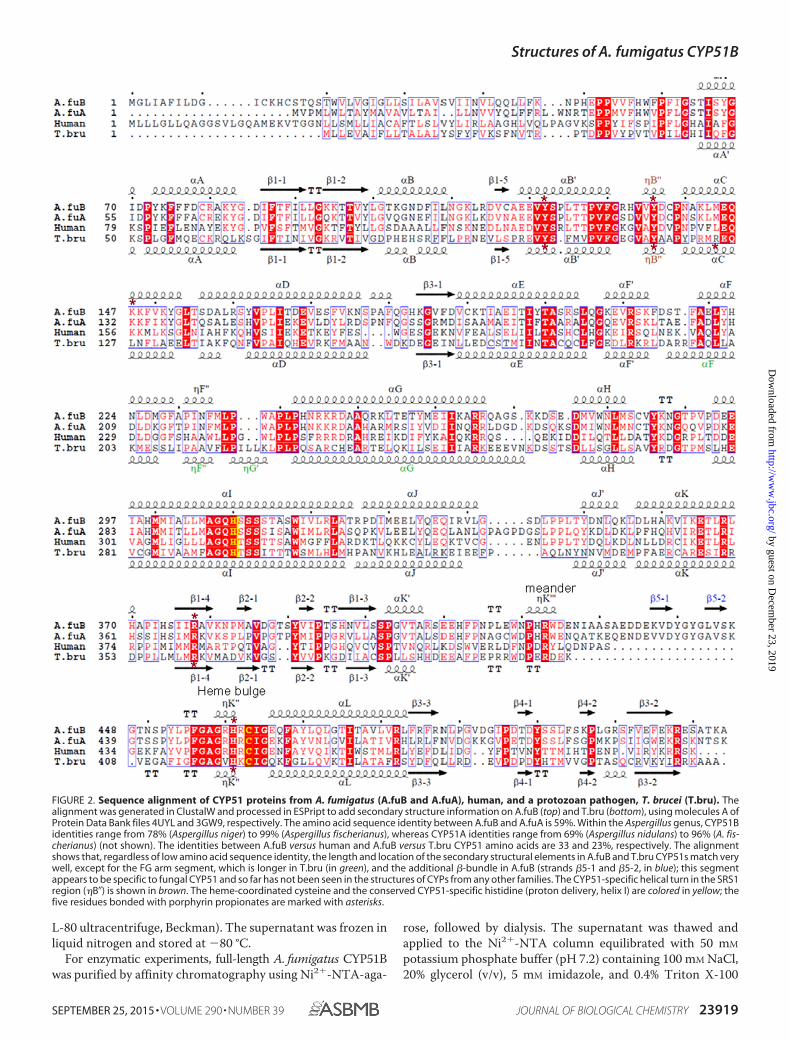
refined in Refmac5. Voriconazole was inserted in Coot. Datacollection and refinement statistics are shown in Table 1. Bothstructures have two monomers in the asymmetric unit. Theprotein chain was seen from Lys50 (KT- in the N-terminalMAKKT- sequence) to His519 (C-terminal His tag). The elec-tron density for both VNI and voriconazole (the ligand ProteinData Bank ID VOR) was well defined, showing a single orienta-tion of the inhibitor molecules within the enzyme binding cav-ity and full occupancy. (The 2Fo � Fc electron density maps forthe inhibitor-containing area are shown in Figs. 11 and 12; seebelow.) The coordinates and structure factors have been depos-ited in the Protein Data Bank under accession codes 4UYL(A. fumigatus CYP51B-VNI) and 4UYM (A. fumigatus CYP51-voriconazole). The accession codes of other CYP51 structuresdiscussed in this work are 3G1Q (ligand-free T. brucei), 3GW9(T. brucei-VNI), 3K1O (T. cruzi-posaconazole), and 3LD6(human-ketoconazole); the Protein Data Bank code of CYP46-voriconazole is 3MDT.
Structure superpositions were done in LSQkab of the CCP4suite. The substrate was positioned in the active sites of thesuperimposed CYP51 enzymes to adopt an orientation similarto that of 14�-methylenecyclopropyl-�7-24,25-dihydrolanos-terol (MCP) in complex with T. brucei CYP51 (Protein DataBank code 3P99) (46) using Coot. Molecular volumes and sur-face areas were calculated in Accelrys Discovery Studio Visual-izer verison 2.5 (probe radius 1.4 Å). Figures were prepared withAccelrys and Chimera. A molecular model of A. fumigatusCYP51A was built in Modeler (CCP4 suite).
A. fumigatus Cellular Growth Inhibition Assays—A. fumiga-tus strain af293 (47) was maintained on Sabouraud dextroseagar at 37 °C. Sensitivity tests were carried out using RPMI 1640medium (Sigma-Aldrich) buffered with 165 mM MOPS, pH 7.0.
The spore inoculum was prepared in sterile phosphate-buff-ered saline containing 0.05% Tween 80 (v/v), and dilutions inRPMI 1640 were made to 2 � 103/ml. Treatments with VNI,itraconazole, and voriconazole were done in triplicate for 48 hat 37 °C (150 rpm), and the minimal inhibitory concentrationwas defined as the concentration of drug that produced nogrowth, as described previously (48). (CH3)2SO was used in thecontrol experiments. Each independent experiment wasrepeated in triplicate.
Results
Spectral Characterization—Similar to other CYP51orthologs (28, 30, 37), A. fumigatus CYP51B was obtained in theligand-free ferric low spin form after purification from E. coli(Fig. 3A). The heme iron was readily reduced by sodium dithio-nite, and the complex with CO was formed very rapidly, with anabsorbance maximum at 448 nm and no presence of (inactive)cytochrome P420. The ferrous form, however, was highlyunstable and, particularly in the ligand-free state, displayed arapid loss of heme absorbance. The binding of CO or anotherligand stabilized the hemoprotein (both ferrous and ferricforms); therefore, for crystallization purposes, the last step ofpurification was performed in the presence of an azole inhibi-tor, which afforded a stable spectrophotometric index (A425/A280) of �1.3 (Fig. 3A).
Enzymatic reduction of A. fumigatus CYP51B in the pres-ence of the natural fungal CYP51 substrate eburicol was at least50-fold faster and more efficient than when the protein wasreduced in the ligand-free form (Fig. 4). Titration of A. fumiga-tus CYP51B with the substrate eburicol produced a character-istic Type I spectral response, with the peak, isosbestic point,and trough in the difference spectra at 390, 408, and 420 nm,
TABLE 1Data collection and refinement statistics
Complex A. fumigatus CYP51B-VNI A. fumigatus CYP51B- voriconazole
Data collectionWavelength (Å) 0.9787 0.9787Space group P31 P31Cell dimensions
a, b, c (Å) 110.498, 110.498, 90.479 109.193, 109.193, 90.221�, �, � (degrees) 90.00, 90.00, 120.00 90.00, 90.00, 120.00
Molecules per asymmetric unit 2 (A/B) 2 (A/B)Solvent content (%) 57.6 57.2Resolution (last shell) (Å) 30–2.81 (2.91–2.81) 30–2.55 (2.64–2.55)No. of reflections (last shell) 28,394 (1526) 36,885 (1939)Rmerge (last shell) 0.047 (0.627) 0.051 (0.615)I/ (last shell) 17.5 (2.1) 18.9 (2.1)Completeness (last shell) (%) 99 (100) 99.8 (100)Redundancy (last shell) 4.8 (4.4) 3.9 (4.2)
RefinementRwork 0.237 0.209Rfree 0.279 0.239Root mean square deviations from ideal geometry
Bond lengths (Å) 0.002 0.001Bond angles (degrees) 1.21 1.03
Ramachandran plotResidues in favorable/allowed regions (%) 96.6/100 96.9/100Outliers (%) 0.0 0.3
Wilson B-factor (Å) 85.6 68.3No. of atoms (mean B-factor (Å)) 7790 (92.8) 7803 (78.6)No. of residues per molecule (A/B)
Protein (mean B-factor (Å)) 470 (83.0)/470 (105.1) 470 (77.0)/470 (81.7)Heme (mean B-factor (Å)) 1 (66.7)/1 (90.1) 1 (56.0)/1 (65.1)Ligand (mean B-factor (Å)) VNI 1 (83.5)/1(91.1) Vor 1 (63.8)/1 (77.1)Water (mean B-factor (Å)) 128 (77.4) 163 (71.4)
Protein Data Bank code 4UYL 4UYM
Structures of A. fumigatus CYP51B
23922 JOURNAL OF BIOLOGICAL CHEMISTRY VOLUME 290 • NUMBER 39 • SEPTEMBER 25, 2015
by guest on Decem
ber 23, 2019http://w
ww
.jbc.org/D
ownloaded from

respectively, and an �18% low to high spin transition in theheme iron (Fig. 5A). Titration with lanosterol (the major natu-ral substrate of mammalian and yeast CYP51 enzymes) did not
produce any spectral changes (Fig. 5B). The C4-monomethy-lated lanosterol analog C4-norlanosterol (a natural CYP51 sub-strate in T. brucei and Leishmania (30)) also did not induce any
FIGURE 4. Time course of enzymatic reduction (detected as the CO complex) of A. fumigatus CYP51B in the presence and in the absence of substrate(1.5 �M P450, 3.0 �M CPR, 150 �M NADPH). Optical path length was 1 cm. Insets, difference CO-binding spectra, �t 2 min.
FIGURE 5. Spectral responses of A. fumigatus CYP51B to the addition of sterols. A, eburicol. B, lanosterol. C, obtusifoliol. Absolute (top) and difference(bottom) absorbance spectra are shown. S, sample; R, reference. P450 concentration was 5 �M, and optical path length was 1 cm. The titration curves (obtainedusing Equation 1) are shown in the insets, and the Kd values are corrected for cyclodextrin binding. The C24-methylene group appears to be required for theproper binding of sterol substrates to A. fumigatus CYP51B.
Structures of A. fumigatus CYP51B
SEPTEMBER 25, 2015 • VOLUME 290 • NUMBER 39 JOURNAL OF BIOLOGICAL CHEMISTRY 23923
by guest on Decem
ber 23, 2019http://w
ww
.jbc.org/D
ownloaded from

spectral response (data not shown), whereas the response to theplant CYP51 substrate obtusifoliol (�14% low to high spintransition in the heme iron) was quite similar to the response toeburicol, with the apparent Kd being 1.7 versus 1.3 �M (Fig. 5C),respectively, thus implying that in order to be productivelybound to A. fumigatus CYP51B, the sterol substrates must con-tain a methylene group at the C24 position and/or not aC24C25 double bond. Finally, titration of A. fumigatusCYP51B with azoles induced typical Type II spectral responses.Two are shown in Fig. 6 as examples, with the apparent Kdvalues for VNI and voriconazole being �200 and 60 nM,respectively.
Catalytic Activity, Steady-state Kinetic Parameters, SubstratePreferences, and Inhibition—Preliminary time course studies(Fig. 7A) indicated that, under the standard CYP51 reactionconditions (0.5 �M P450, 1 �M CPR, 37 °C), A. fumigatusCYP51B had a relatively high initial turnover rate but wasunstable and rapidly inactivated (no P450 could be detected inthe reaction mixture after a 10-min reaction). Steady-statekinetic parameters were measured using 60-s reactions to min-imize the influence of P450 inactivation (Fig. 7B). Under theseconditions, A. fumigatus CYP51B displayed a slight substratepreference toward eburicol over obtusifoliol (1.8-fold highercatalytic efficiency, due to the lower Km value (Table 2)). Longerreaction times and various reaction temperatures were
employed with 14�-demethylation of lanosterol and C4-nor-lanosterol, but no conversion of these C24-demethyl sterolswas observed.
To compare the potencies of different inhibitors ofA. fumigatus CYP51B, the reaction temperature was decreasedto 28 °C to attenuate the rate of enzyme denaturation. We useda 60-min reaction (instead of a 1–5-min reaction) because alonger reaction time affords higher sensitivity in these assays(27, 38, 39). The initial molar ratio of enzyme/inhibitor/eburi-col was 1:2:50, and under these conditions, �40% of the originalamount of P450 was still detected in the CO complex of thecontrol sample after a 60-min reaction (results not shown).Among all of the clinical antifungal azoles and experimentalCYP51 inhibitors tested (Fig. 8), the topical antifungal drugmiconazole was the only compound that caused complete inhi-bition of the A. fumigatus CYP51 activity. Voriconazole (6%substrate conversion), posaconazole (10%), and itraconazole(16%) were followed by VNI (20%), whereas VFV (presently themost potent inhibitor of protozoan CYP51 enzymes (49)) had31% substrate conversion. The weakest inhibitory effect onA. fumigatus CYP51B was observed with fluconazole, inaccordance with the lack of activity of this drug in cellularexperiments with A. fumigatus and against aspergillosis in vivo(see the Aspergillus Website).
FIGURE 6. Spectral response of A. fumigatus CYP51B to the addition of the heme-coordinating ligands VNI and voriconazole. Absolute (top) anddifference (bottom) absorbance spectra are shown. The P450 concentration was 1.0 �M, and the optical path length was 5 cm. The titration curves (obtainedusing Equation 1) are shown in the inset.
Structures of A. fumigatus CYP51B
23924 JOURNAL OF BIOLOGICAL CHEMISTRY VOLUME 290 • NUMBER 39 • SEPTEMBER 25, 2015
by guest on Decem
ber 23, 2019http://w
ww
.jbc.org/D
ownloaded from

Crystal Structures of CYP51B—In both complexes (with VNIand with voriconazole), A. fumigatus CYP51B crystallized inthe same trigonal space group (P31). The asymmetric unit con-sisted of two monomers that are related via a non-crystallo-graphic 180° rotation axis and are positioned in such a way thatthe most hydrophobic, membrane-bound fragments of themolecule (the mouth of the CYP51 substrate access channel:helix A�, FG loop, and the �4-hairpin), are facing each other(Fig. 9A). The monomers exhibit high structural similarity (Fig.9B), with the root mean square deviation for the C� atom posi-tions being 0.51 0.04 and 0.55 0.07 Å between the proteinsin the VNI- and voriconazole-bound complexes, respectively(molecular breathing) and 0.73 0.06 Å between the two com-plexes (ligand accommodation). This result supports the view(14) that structural rigidity of the active site cavity, previously
observed in various complexes of protozoan CYP51 orthologs(18, 29, 30, 39, 40, 46), is a general feature of the CYP51 family.
Overall, A. fumigatus CYP51 displayed the typical P450 fold(an upside-down triangular prism from the distal view (Fig. 9B))with the heme prosthetic group positioned between helices Iand L and the iron coordinated to Cys463 on the proximal side ofthe porphyrin plane. The structure has an �/�/coil ratio of60:10:30 and consists of 22 helices (12 main ones plus 10 addi-tional helices (denoted with a prime)) and five �-bundles(instead of four) formed by 14 �-strands. Because of the addi-tional �5-bundle, the molecular volume of A. fumigatus CYP51(63,500 300 Å3) is slightly higher than the volumes of theprotozoan (61,300 400 Å3) and human (61,900 500 Å3)CYP51 orthologs.
The active side cavity is formed by the distal surface of theporphyrin macrocycle, helix B and B�C loop (Val121–Tyr136,substrate recognition sequence 1 (SRS1)) (50), helix C (Gln146–Val150), helix F (Pro231–Met235, SRS2), the N-terminal portionof helix I (Met300–Ser311, SRS4), the K�1– 4 loop (Pro372–Arg378, SRS5), and the �4-hairpin (Leu503–Ser505, SRS6) (Fig.10A). SRS1 represents the major CYP51 substrate binding area.It carries the family signature 1 (13), the conserved or phylum-specific residues that are essential for enzyme function (37, 51,52). In the T. brucei CYP51 complex with the substrate analogMCP, this area of the protein (shown as a yellow ribbon in Fig.10A) was found to encircle the whole skeleton of the sterolmolecule from its �-side (46). Helix C represents an SRS thatthus far is unique for CYP51 (30, 46); SRS4 and SRS5 are slightlyshifted (toward the protein N terminus) relative to their canon-ical locations in the sequences of other CYP families; and theP450 SRS3 (the residues preceding helix G) does not appear tobe able to directly interact with a ligand because, as in otherCYP51 structures, this region is shielded from the active sitecavity by the B� helix.
As with the CYP51 structures from other biological king-doms, A. fumigatus CYP51B has five residues that can providehydrogen bonds to the heme propionates (Fig. 10B). Arg378 andHis461 are conserved in most CYP families, and Lys147 corre-sponds to Lys156 in the human CYP51 structure and is con-served in fungi and vertebrates, whereas in the protozoanCYP51 orthologs, the same role is played by the phylum-spe-cific arginine that is located one turn upstream in the C helix
FIGURE 7. Enzymatic activity of A. fumigatus CYP51B. A, time course of substrate conversion at 37 °C (0.5 �M P450, 1.0 �M CPR, and 25 �M eburicol), shownin comparison with C. albicans CYP51. B, steady-state kinetics of A. fumigatus CYP51B with its natural substrate eburicol and obtusifoliol (a substrate for plantCYP51 enzymes). The P450 concentration was 0.5 �M (37 °C, 60-s reaction). The experiments were performed in triplicate, and results are presented as means S.E. (error bars).
TABLE 2Steady-state kinetic parameters for 14�-demethylation of eburicoland obtusifoliol by A. fumigatus CYP51B
SterolMichaelis-Menten parameters
kcat Km
a kcat/Km
min�1 �M �M�1 min�1
Eburicol 45 3 1.8 0.4 25 5Lanosterol —b
Obtusifoliol 52 5 3.7 0.8 14 4C4-norlanosterol —
a Km values are corrected for HPCD binding.b —, the limit of detection was �0.05 min�1.
FIGURE 8. Inhibitory effects of azoles on the activity of A. fumigatusCYP51B. The incubation time was 60 min (at 28 °C). The molar enzyme/inhib-itor/substrate ratio was 1:2:50, with 0.5 �M P450. The experiments were per-formed in triplicate, and results are presented as means S.E. (error bars).
Structures of A. fumigatus CYP51B
SEPTEMBER 25, 2015 • VOLUME 290 • NUMBER 39 JOURNAL OF BIOLOGICAL CHEMISTRY 23925
by guest on Decem
ber 23, 2019http://w
ww
.jbc.org/D
ownloaded from

(Arg124 in T. brucei CYP51). Tyr122 is invariant across thewhole CYP51 family. In the ligand-free and sterol-bound struc-tures, it forms the hydrogen bond with the heme ring A propi-onate, but binding of strong inhibitors often disrupts thishydrogen bond (29, 39, 49). The hydrogen bond between Tyr136
(phenylalanine in plant CYP51, tyrosine in all other phyla) wasalso found disrupted in several CYP51-inhibitor complexes (29,40, 53).
His310 (Fig. 10C) is the invariant SRS4 residue (CYP51 signa-ture 2 (-qHtSs-) (13)). It precedes the “conserved P450 threo-
nine” (Ser311 in both A. fumigatus CYP51 sequences (see alsoFig. 2)) and, being hydrogen-bonded with Asp226, lines the sur-face of a small channel (see Fig. 3 in Ref. 39) that is directedtoward the water-soluble exterior of the protein. In thesequences of P450s from all other families, this position isalways occupied by an acidic residue (aspartic or glutamic acid(13)). The exact mechanism of proton delivery in CYP51 hasnot been clarified, but the conservation of the His310/Asp226 saltbridge across the structures of CYP51s from different biologicalkingdoms strongly suggests that it is likely to be similar to the
FIGURE 9. Overall view of the A. fumigatus CYP51 structures. A, view along the rotation axis that runs from top to bottom and relates two VNI (pink)-A. fumigatus CYP51B complexes that comprise the asymmetric unit. The secondary structural elements forming the mouth of the substrate access channel(helices A� and F and the �4-hairpin) are marked. B, superimposition of four molecules of A. fumigatus CYP51B. Shown is a distal P450 view; x/y/z: 65/67/45 Å.Molecule A of the VNI complex is shown in a ribbon representation; the backbones of the other three molecules are presented as wires. VNI and voriconazole aredeleted for clarity. In both panels, the protein backbone is presented in rainbow coloring from blue (N terminus) to red (C terminus). The heme is shown as a stickmodel, and the iron is depicted as an orange sphere. C, stereo view of B.
FIGURE 10. Family-specific structural features of A. fumigatus CYP51B. A, the active site cavity. Eburicol (green) was modeled in a position similar to thesubstrate analog MCP as described under “Experimental Procedures.” The protein backbone is colored by secondary structure (helices are red, �-strands areblue, loops are gray, and turns are green). The corresponding SRS1 area in T. brucei CYP51 is shown with a light yellow ribbon. The B� helical turn, helix C, and the�5-bundle are marked. B, heme support from the protein moiety. Hydrogen bonds are shown as green dashes. C, proton transfer route. His310 is conservedacross the CYP51 family and in all known CYP51 structures forms a hydrogen bond with an acidic residue (aspartate or glutamate) of the F helix. The length ofthe His310-Asp227 hydrogen bond in the four A. fumigatus CYP51B molecules is 2.8 0.1 Å (mean S.E.). Ser311 corresponds to the conserved P450 threonine.
Structures of A. fumigatus CYP51B
23926 JOURNAL OF BIOLOGICAL CHEMISTRY VOLUME 290 • NUMBER 39 • SEPTEMBER 25, 2015
by guest on Decem
ber 23, 2019http://w
ww
.jbc.org/D
ownloaded from

mechanism described for CYP101 (P450cam), where it wasreported to be switched on upon the formation of the P450-electron donor complex, which disrupts the corresponding saltbridge (between Asp251 (I helix) and Lys178 (F helix) (54)) andthus activates the proton delivery. The charge of this salt bridgepair is reversed in the CYP51 family.
Complex with Voriconazole—Fluconazole is not efficient intreating A. fumigatus infections (see the Aspergillus Website),and for many years, it remained enigmatic why its close deriv-ative voriconazole (see Fig. 1), also a rather small molecule(molecular volume 399 Å3 versus 348 Å3 for fluconazole (29)),has much higher potency. The structure of the voriconazole-A. fumigatus CYP51B complex suggests that the reason may bethe formation of the hydrogen bonds between the 5-fluoropy-rimidine ring of voriconazole (versus the substantially smallertriazole ring of fluconazole) and A. fumigatus CYP51 Tyr122
(Fig. 11A), the residue hydrogen-bonded with the heme ring Apropionate. In addition to these hydrogen bonds with Tyr122
and the coordination of the N4-triazole nitrogen to the hemeiron (the length of the coordination bond is 2.1 Å), voriconazoleforms van der Waals interactions (distance �4.5 Å) with 14more amino acid residues of A. fumigatus CYP51B (Fig. 11A).The contacting residues are mainly from the B� helix/B�C loop(Tyr122, Leu125, Thr126, Phe130, Val135, and Tyr136); Phe229 isfrom the F helix; Ala303, Ala307, and Ser311 are from helix I; andIle373, Ser375, and Ile376 are from the K helix/�1-4 loop and �1-4strand, whereas Leu503 and Phe504 are from the �4-hairpin.Interestingly, the conformation of voriconazole in the activesite of A. fumigatus CYP51 (Fig. 11, B and C) is very differentfrom the conformation that the drug adopts within the activesite of CYP46 (55), providing an additional example of the
importance of experimental target-based structural informa-tion in the process of target-driven drug discovery.
Complex with VNI—The strong electron density within theactive site of the A. fumigatus CYP51B co-crystallized with VNI(Fig. 12A) unambiguously demonstrates that the position ofthis inhibitor in the fungal CYP51 binding cavity is also quitedifferent from its position in the complex with the protozoanCYP51 ortholog (Fig. 12B). The observed differences (i) validatethe importance of the hydrogen bond network around the VNIcarboxamide fragment for its high antiprotozoan potency andselectivity (27, 39, 49, 56), (ii) explain its weaker effect on theA. fumigatus CYP51 activity (see Fig. 8), and (iii) outline apotential strategy for further enhancement of the VNI scaffoldantifungal activity. Thus, the structure suggests that the lack ofthe hydrogen bond network between the protein and the inhib-itor may result from the altered orientation of the VNI three-ring arm, which in turn repositions (shifts down as it is seen inFig. 12B) the dichlorinated �-phenyl ring of the inhibitor.Because the VNI derivative VFV contains the longer, �-biphe-nyl arm here (shown in Fig. 8), it is unlikely to acquire the VNIposition in A. fumigatus CYP51B because its two-ring armshould not fit into the C-helix-directed portion of the enzymebinding cavity (not shown). This might be the reason why VFV,which to date is our most potent antiprotozoan drug candidate(49), is a weaker inhibitor of A. fumigatus CYP51B than VNI(Fig. 8).
Although no direct hydrogen bonds are formed with the pro-tein, VNI makes van der Waals contacts (distance �4.5 Å) with19 amino acid residues, including Tyr122, which is pushed bythe inhibitor too far away from the heme ring A propionate (4.3Å) to interact with its oxygen (Fig. 12C). The length of the
FIGURE 11. Voriconazole binding mode. A, view of the A. fumigatus CYP51B active site illustrating interactions of the hemoprotein with the inhibitor. Theresidues located within van der Waals contacts (�4.5 Å) with voriconazole are depicted as wire models and labeled; the carbon atoms are colored in gray; andthree reference secondary structural elements are shown as semitransparent blue ribbon. The carbon atoms of voriconazole and the heme (stick representations)are blue and orange, respectively. The hydrogen bonds are shown as green dashed lines. B, 2Fo � Fc omit electron density map of the active site area aroundvoriconazole contoured at 1.5 . C, superimposition of voriconazole complexes with A. fumigatus CYP51B and with CYP46 (Protein Data Bank code 3MDT). Theheme and the drug are shown in a stick representation; the carbon atoms are blue and gray in A. fumigatus CYP51B and CYP46, respectively.
Structures of A. fumigatus CYP51B
SEPTEMBER 25, 2015 • VOLUME 290 • NUMBER 39 JOURNAL OF BIOLOGICAL CHEMISTRY 23927
by guest on Decem
ber 23, 2019http://w
ww
.jbc.org/D
ownloaded from

coordination bond between the VNI N3 nitrogen and the hemeiron is 2.0 Å. Interestingly, in the complex with VNI, some rear-rangements in the side chain positioning of A. fumigatusCYP51B can be seen, especially around the substrate accesschannel (Fig. 13), when compared with the voriconazole-boundstructure. Thus, the basic His310 shifts closer to the VNI car-boxamide oxygen (the distance is 3.7 Å versus 4.5 Å in the com-plex with voriconazole), whereas the hydrophobic Phe504
moves about 5 Å away from this polar area of the inhibitor.Most relocations, however, occur around the substrate channelentrance. Particularly interesting is the movement of Phe234,the aromatic ring of which turns about 70° and moves towardthe aromatic ring of VNI to form face-to-face (sandwich) �-pstacking interactions (�4 Å), which are likely to be important instabilizing both the position of the inhibitor and the entry intothe enzyme substrate access channel. The side chains of Leu92
(the residue from the turn between the �1-1 and �1-2), Met235
(helix F�), and Leu503 (the �4-hairpin) must also shift towardthe distal aromatic ring of VNI �3, 6, and 4 Å, respectively, toaccomplish this change (Fig. 13).
A. fumigatus Cellular Growth Inhibition Assays—Thepotency of VNI against A. fumigatus cells was found to be com-parable with the effects of clinical antifungals, with the minimal
inhibitory concentration values for itraconazole, voriconazole,and VNI being 0.5, 0.7, and 0.5 �g/ml, respectively.
Discussion
When overlaid with the structures of its protozoan andmammalian orthologs, A. fumigatus CYP51 displays remarka-ble similarity in the spatial organization of the protein back-bone. Although its amino acid sequence identity to T. bruceiand human sterol 14�-demethylases is only 23 and 33%, respec-tively, the average root mean square deviation values for the C�atom positions are only 1.8 and 1.4 Å (Fig. 14A). The residuesthat are conserved across the whole CYP51 family also super-impose very well; those located within the substrate bindingcavity are shown in Fig. 14B. Thus, altogether, the fungal struc-tures strongly support the proposal (14) that sterol 14�-demeth-ylases from different biological kingdoms have preserved theirconserved metabolic roles by maintaining high similarity at thesecondary and tertiary structural levels, variations in their sub-strate preferences, catalytic parameters, and susceptibility toinhibition being fine tuned (as experimentally established forthe protozoan CYP51 enzymes (30, 37, 57) by the phylum-spe-cific residues that line the interior of the CYP51 binding cavity,thus defining the local topology of the active site.
FIGURE 12. VNI binding mode. A, 2Fo � Fc omit electron density map of the active site area around VNI contoured at 1.3 . B, superimposition of the VNIcomplexes with A. fumigatus CYP51B and T. brucei CYP51 (Protein Data Bank code 3GW9). The heme and VNI are shown in a stick representation; the carbonatoms are magenta and gray in the A. fumigatus and T. brucei enzymes, respectively. The hydrogen bonds are presented as green dashed lines. Two segmentsof the T. brucei CYP51 molecule that are connected by the hydrogen bond network with VNI (helices B� and I) are outlined as gray ribbons. C, view of theA. fumigatus CYP51B active site illustrating its interactions with VNI. The residues located within van der Waals distances (�4.5 Å) with VNI are depicted as wiremodels and labeled, and the carbon atoms are colored in gray. Three reference secondary structural elements are seen as a semitransparent pink ribbon. Thecarbon atoms of VNI and the heme (stick representations) are colored magenta and orange, respectively.
Structures of A. fumigatus CYP51B
23928 JOURNAL OF BIOLOGICAL CHEMISTRY VOLUME 290 • NUMBER 39 • SEPTEMBER 25, 2015
by guest on Decem
ber 23, 2019http://w
ww
.jbc.org/D
ownloaded from

In addition to the topological differences inside the bindingcavity, which must be taken into account in CYP51 structure-based drug development, the A. fumigatus CYP51B structuresreveal two very specific features, not seen among the CYP51enzymes from other phyla, which are of functional importance.
Substrate Entrance—A. fumigatus CYP51 has a relativelyshorter FG arm (Fig. 15A). A different position of the FG loopalters the shape of the enzyme substrate access channel, so thatthe ligand entry site looks more like a long cleft extended to theupper P450 surface with a “bridge” over it (Fig. 15B, 1 and 2)than like a small round opening, which in protozoan and
human CYP51 enzymes is only seen from the distal side of themolecule (Fig. 15B, 3 and 4). The bridge in A. fumigatusCYP51B is formed by the hydrophobic interactions betweenPhe61 (helix A�) or Leu92 (the �1-1/�1-2 turn) and Met235 (helixF). In the complex with voriconazole, the bridge appears to bemore flexible (about to adopt an open conformation, Fig. 15B,1), whereas the �-� stacking interactions between the aromaticrings of VNI and Phe234 of A. fumigatus CYP51 (shown in Fig.13) keep it in the closed state (Fig. 15B, 2). In both complexes,the ligands can be seen from both the distal and the upper viewof the fungal P450 molecule. This relatively wider opening of
FIGURE 13. VNI-induced rearrangements in A. fumigatus CYP51B (semitransparent pink structure, except for Phe234 and VNI, which are colored inmagenta). Superimposition with the voriconazole co-structure (semitransparent blue) is shown. The �-� stacking interactions between VNI and Phe234 aredepicted as black dashed lines; the distances are marked. The hydrogen bonds are green. Helices A� and F and the �4-hairpin are the elements forming theentrance into the CYP51 substrate access channel. The heme is seen as a gray sphere model. The other 4 residues in the superimposed complex withvoriconazole (Leu92, Met235, His310, and Phe504), whose side chain locations differ substantially, are shown as semitransparent blue lines; the directions of therearrangements are indicated with gray arrows.
FIGURE 14. Superimposed structures of A. fumigatus (4UYL, pink), T. brucei (3GW9, blue), and human (3LD6, yellow) CYP51 enzymes. A, distal view. Theprotein backbone is presented as a semitransparent ribbon, and the area of the substrate-binding cavity is within the black square. B, enlarged view of thebinding cavity. The heme and the residues invariant in all known CYP51 enzymes (�300 sequences) are depicted in a stick representation with T. brucei CYP51numbering (see also Fig. 2).
Structures of A. fumigatus CYP51B
SEPTEMBER 25, 2015 • VOLUME 290 • NUMBER 39 JOURNAL OF BIOLOGICAL CHEMISTRY 23929
by guest on Decem
ber 23, 2019http://w
ww
.jbc.org/D
ownloaded from

the substrate entry may well be the reason why A. fumigatus isnot very sensitive to the small inhibitors, except for voricona-zole (which forms hydrogen bonds with Tyr122), and “longer”structures (e.g. posaconazole) are required to restrict themotions of the channel entry and therefore strengthen the inhi-bition. Most important, this structural feature of A. fumigatusCYP51 implies that the FG loop fragment is likely to play agating role in all CYP51 enzymes, opening briefly for the sub-strate (ligand) to enter the active site and closing when thecomplex acquires energetically optimal conformation.
�5-Bundle on the Proximal P450 Surface—Multiplesequence alignment of the CYP51 family proteins from differ-ent biological kingdoms reveals a 15–25-amino acid residue-long insert that is present in all fungal CYP51 sequencesbetween the “meander” and the heme binding segment (alsoknown as the heme bulge) (Fig. 16). In the A. fumigatus CYP51Bstructures, this post-meander insert forms an additional�-bundle (�5), which consists of two adjacent antiparallel�-strands (5-1 and 5-2 in Fig. 16A) and buries the heme bulgesegment inside the protein globule, shielding it from the prox-imal surface. The function of the �5-bundle remains unclear,and, to our knowledge, no analogous structure has beenobserved so far in any other P450 families.
We hypothesize that the �5-bundle may somehow beinvolved in regulation of the electron transfer process by influ-encing the interaction of the fungal CYP51 with the P450reductase, modulating the heme environment (58, 59). Theproximal surface of the P450 molecule, including the hemebulge area, is known to be involved in the electrostatic interac-tion with the negatively charged surface of the electron donorprotein (54, 60, 61), and therefore, it must be electropositive(blue in the insets in Fig. 16A). Due to the �5-bundle, in thefungal CYP51 structures, the proximal surface appears to beelectronegative (Fig. 16A, right inset, the �5-bundle region iscircled). This discrepancy, however, can be easily resolved if thebase of the �5-bundle is highly flexible (residues 427– 432 and450 – 454 (see Fig. 2) have the highest B-factor values in all fourA. fumigatus CYP51B molecules) and the �5-bundle is likely tomove aside upon CYP51-reductase complex formation. Suchmotion would expose the CYP51 heme bulge segment and alterthe number of heme-contacting residues. Our structural anal-ysis supports such a possibility, particularly because the�5-bundle pushes the upper portion of the heme bulge segmentslightly away from the porphyrin ring plane, so that the numberof heme-contacting residues in the fungal CYP51 structure islower (i.e. 19 versus 24 in the protozoan CYP51 ortholog). Thisresult, in turn, correlates with the experimentally observed ele-vated propensity of A. fumigatus CYP51B to form a destabilizedferrous species (results not shown).
FIGURE 15. Substrate entry. A, superimposition of A. fumigatus (4UYL, col-ored according to secondary structure as in Fig. 10) with T. brucei CYP51(3SW9, gray; the FG-arm is yellow). The distal view is shown. The length of theFG arm in A. fumigatus CYP51B is shorter (by �5 Å) than it is in the T. bruceienzyme. B, surface representation of CYP51s from different phyla. 1, voricona-zole-bound A. fumigatus CYP51; 2, VNI-bound A. fumigatus CYP51; 3, keto-conazole-bound human CYP51; 4, MCP-bound T. brucei CYP51. The ligandsare shown as spheres with green carbon atoms. There is no opening in theupper surface of the human or protozoan CYP51 structure.
Structures of A. fumigatus CYP51B
23930 JOURNAL OF BIOLOGICAL CHEMISTRY VOLUME 290 • NUMBER 39 • SEPTEMBER 25, 2015
by guest on Decem
ber 23, 2019http://w
ww
.jbc.org/D
ownloaded from

FIGURE 16. Fungi-specific postmeander insert (�5-bundle). A, proximal view of the CYP51 molecule, colored by secondary structure as in Fig. 10. Insets, electrostaticpotential mapped onto the proximal surface. Red, negative charge; blue, positive charge. The �5-area is circled. B, a fragment of multiple CYP51 sequence alignment.
FIGURE 17. A. fumigatus CYP51B and CYP51A may differ in their substrate preferences. A, substrate binding cavities in A. fumigatus CYP51B and T. brucei CYP51.A. fumigatus CYP51B has more space in the area holding the distal portion of the sterol aliphatic arm (circled). B, a fragment of multiple sequence alignment of CYP51sA and B from filamentous fungi. A.fum, A. fumigatus; A.flav, Aspergillus flavus; A.lent, Aspergillus lentulus; A.clav, Aspergillus clavatus; N.fisch, Neosartorya fischeri; T.tons,Trichophyton tonsurans; F.gram, Fusarium graminearum; V.inaec, Venturia inaequalis; U.necator, Uncinula necator; C.neoform, Cryptococcus neoformans; U.maydis, Usti-lago maydis. C, location of Thr285 (yellow), which aligns with Ala303 in A. fumigatus CYP51B (blue) in the molecular model of A. fumigatus CYP51A.
Structures of A. fumigatus CYP51B
SEPTEMBER 25, 2015 • VOLUME 290 • NUMBER 39 JOURNAL OF BIOLOGICAL CHEMISTRY 23931
by guest on Decem
ber 23, 2019http://w
ww
.jbc.org/D
ownloaded from

The question of why A. fumigatus CYP51B does not oxidizeC24-demethyl sterols (e.g. lanosterol) remains open. The struc-tures suggest that it might be because of the relatively largervolume of this enzyme binding cavity, particularly in the areaholding the sterol aliphatic arm (Fig. 17). If this is the case, themodel of A. fumigatus CYP51A implies that this CYP51enzyme might oxidize lanosterol, due to a single amino aciddifference in the I-helix (Thr285 versus Ala303; Fig. 17B). Morestructure-functional characterization of A. fumigatus CYP51Ais desirable before drawing any conclusions regarding bothenzyme substrate preferences and CYP51A and CYP51B sus-ceptibility to inhibition. The experiments on A. fumigatusCYP51A expression/purification in the P450 form are currentlyin progress.
The stabilizing rearrangements in the region of theA. fumigatus CYP51B substrate entrance that we observedupon binding of VNI suggest that minor modifications aroundthe aromatic ring of the VNI long arm might significantlyincrease its potency (and selectivity) as a fungal CYP51 inhibi-tor. This would be of special interest because of the VNI scaf-fold advantages (i.e. low toxicity, excellent cellular permeabil-ity, and weak influence on human drug-metabolizing P450s)and favorable phamacokinetics and tissue distribution (27, 49,62).
To summarize, the A. fumigatus CYP51B structures supporthigh overall three-dimensional similarity as the molecular basisfor the CYP51 family catalytic conservation across phyla. Theyalso reveal a few interesting differences, which we believe to beimportant for better understanding CYP51 family structure/function in general; to establish some phylum-specific features;and to help in distinguishing species-related peculiarities andtheir possible relevance to enzyme catalysis, inhibition, andstructure-based design of more effective antifungal agents.
Author Contributions—G. I. L. conceived and coordinated the study.T. Y. H., Z. W., D. C. L., and G. I. L. performed the experiments.G. I. L. and F. P. G. analyzed the data and wrote the paper. Allauthors reviewed the results and approved the final version of themanuscript.
Acknowledgments—We thank Dr. Diane Kelly (University of Swan-sea) for the A. fumigatus CYP51 cDNA, Dr. Sandeep Goyal for theDynaFit calculations, and K. Trisler for assistance in preparation ofthe manuscript.
References1. Denning, D. W., and Bromley, M. J. (2015) How to bolster the antifungal
pipeline. Science 347, 1414 –14162. Shapiro, R. S., Robbins, N., and Cowen, L. E. (2011) Regulatory circuitry
governing fungal development, drug resistance, and disease. Microbiol.Mol. Biol. Rev. 75, 213–267
3. Gullo, A. (2009) Invasive fungal infections. Drugs 69, 65–734. Xie, J. L., Polvi, E. J., Shekhar-Guturja, T., and Cowen, L. E. (2014) Eluci-
dating drug resistance in human fungal pathogens. Future Microbiol. 9,523–542
5. Angarone, M. (2014) Fungal infections in cancer patients. Cancer Treat.Res. 161, 129 –155
6. Chen, S. C. A., Playford, E. G., and Sorrell, T. C. (2010) Antifungal therapyin invasive fungal infections. Cur. Opin. Pharmacol. 10, 522–530
7. Hope, W. W., Walsh, T. J., and Denning, D. W. (2005) The invasive and
saprophytic syndromes due to Aspergillus spp. Med. Mycol. 43,S207–S238
8. Latge, J.-P. (1999) Aspergillus fumigatus and aspergillosis. Clin. Microbiol.Rev. 12, 310 –350
9. Brown, G. D., Denning, D. W., Gow, N. A. R., Levitz, S. M., Netea, M. G.,and White, T. C. (2012) Hidden killers: human fungal infections. Sci.Trans. Med. 4, 165rv113
10. Denning, D. W., Pleuvry, A., and Cole, D. C. (2013) Global burden ofallergic bronchopulmonary aspergillosis with asthma and its complicationchronic pulmonary aspergillosis in adults. Med. Mycol. 51, 361–370
11. Johnson, L. B., and Kauffman, C. A. (2003) Voriconazole: a new triazoleantifungal agent. Clin. Infect. Dis. 36, 630 – 637
12. Ashbee, H. R., Barnes, R. A., Johnson, E. M., Richardson, M. D., Gorton, R.,and Hope, W. W. (2014) Therapeutic drug monitoring (TDM) of antifun-gal agents: guidelines from the British Society for Medical Mycology. J.Antimicrob. Chemother. 69, 1162–1176
13. Lepesheva, G. I., and Waterman, M. R. (2007) Sterol 14�-demethylasecytochrome P450 (CYP51), a P450 in all biological kingdoms. Biochim.Biophys. Acta 1770, 467– 477
14. Lepesheva, G. I., and Waterman, M. R. (2011) Structural basis for conser-vation in the CYP51 family. Biochim. Biophys. Acta 1814, 88 –93
15. Sheehan, D. J., Hitchcock, C. A., and Sibley, C. M. (1999) Current andemerging azole antifungal agents. Clin. Microbiol. Rev. 12, 40 –79
16. Van den Bossche, H., Willemsens, G., Cools, W., Cornelissen, F., Lauwers,W. F., and van Cutsem, J. M. (1980) In vitro and in vivo effects of theantimycotic drug ketoconazole on sterol synthesis. Antimicrob. AgentsChemother. 17, 922–928
17. Van den Bossche, H. (ed) (1988) Mode of Action of Pyridine, Pyrimidineand Azole Antifungals, pp. 79 –119, Ellis Horwood, Chichester, UK
18. Hargrove, T. Y., Wawrzak, Z., Alexander, P. W., Chaplin, J. H., Keenan, M.,Charman, S. A., Perez, C. J., Waterman, M. R., Chatelain, E., and Lepe-sheva, G. I. (2013) Complexes of Trypanosoma cruzi sterol 14�-demeth-ylase (CYP51) with two pyridine-based drug candidates for Chagas dis-ease: structural basis for pathogen selectivity. J. Biol. Chem. 288,31602–31615
19. Correia, M. A., and Ortiz de Montellano, P. R. (2005) in Cytochrome P450:Structure, Mechanism, and Biochemistry, 3rd Ed. (Ortiz de Montellano,P. R., ed) pp. 246 –322, Kluwer Academic/Plenum Publishers, New York
20. Warrilow, A. G. S., Melo, N., Martel, C. M., Parker, J. E., Nes, W. D., Kelly,S. L., and Kelly, D. E. (2010) Expression, purification, and characterizationof Aspergillus fumigatus sterol 14-� demethylase (CYP51) isoenzymes Aand B. Antimicrob. Agents Chemother. 54, 4225– 4234
21. Heeres, J., Meerpoel, L., and Lewi, P. (2010) Conazoles. Molecules 15,4129 – 4188
22. Fan, J., Urban, M., Parker, J. E., Brewer, H. C., Kelly, S. L., Hammond-Kosack, K. E., Fraaije, B. A., Liu, X., and Cools, H. J. (2013) Characteriza-tion of the sterol 14�-demethylases of Fusarium graminearum identifies anovel genus-specific CYP51 function. New Phytol. 198, 821– 835
23. Lupetti, A., Danesi, R., Campa, M., Del Tacca, M., and Kelly, S. (2002)Molecular basis of resistance to azole antifungals. Trends Mol. Med. 8,76 – 81
24. van der Linden, J. W. M., Camps, S. M. T., Kampinga, G. A., Arends,J. P. A., Debets-Ossenkopp, Y. J., Haas, P. J. A., Rijnders, B. J. A., Kuijper,E. J., van Tiel, F. H., Varga, J., Karawajczyk, A., Zoll, J., Melchers, W. J. G.,and Verweij, P. E. (2013) Aspergillosis due to voriconazole highly resistantAspergillus fumigatus and recovery of genetically related resistant isolatesfrom domiciles. Clin. Infect. Dis. 57, 513–520
25. Hawkins, N. J., Cools, H. J., Sierotzki, H., Shaw, M. W., Knogge, W., Kelly,S. L., Kelly, D. E., and Fraaije, B. A. (2014) Paralog re-emergence: a novel,historically contingent mechanism in the evolution of antimicrobial resis-tance. Mol. Biol. Evol. 31, 1793–1802
26. Mellado, E., Diaz-Guerra, T. M., Cuenca-Estrella, M., and Rodriguez-Tudela, J. L. (2001) Identification of two different 14�-sterol demethylase-related genes (cyp51A and cyp51B) in Aspergillus fumigatus and otherAspergillus species. J. Clin. Microbiol. 39, 2431–2438
27. Hargrove, T. Y., Kim, K., de Nazare Correia Soeiro, M., da Silva, C. F.,Batista, D. D., Batista, M. M., Yazlovitskaya, E. M., Waterman, M. R.,Sulikowski, G. A., and Lepesheva, G. I. (2012) CYP51 structures and struc-
Structures of A. fumigatus CYP51B
23932 JOURNAL OF BIOLOGICAL CHEMISTRY VOLUME 290 • NUMBER 39 • SEPTEMBER 25, 2015
by guest on Decem
ber 23, 2019http://w
ww
.jbc.org/D
ownloaded from

ture-based development of novel, pathogen-specific inhibitory scaffolds.Int. J. Parasitol. Drugs Drug Resist. 2, 178 –186
28. Lepesheva, G. I., Nes, W. D., Zhou, W., Hill, G. C., and Waterman, M. R.(2004) CYP51 from Trypanosoma brucei is obtusifoliol-specific. Biochem-istry 43, 10789 –10799
29. Lepesheva, G. I., Hargrove, T. Y., Anderson, S., Kleshchenko, Y., Furtak,V., Wawrzak, Z., Villalta, F., and Waterman, M. R. (2010) Structural in-sights into inhibition of sterol 14�-demethylase in the human pathogenTrypanosoma cruzi. J. Biol. Chem. 285, 25582–25590
30. Hargrove, T. Y., Wawrzak, Z., Liu, J., Nes, W. D., Waterman, M. R., andLepesheva, G. I. (2011) Substrate preferences and catalytic parametersdetermined by structural characteristics of sterol 14�-demethylase(CYP51) from Leishmania infantum. J. Biol. Chem. 286, 26838 –26848
31. Omura, T., and Sato, R. (1964) The carbon monoxide-binding pigment ofliver microsomes. I. Evidence for its hemoprotein nature. J. Biol. Chem.239, 2370 –2378
32. Lepesheva, G. I., Strushkevich, N. V., and Usanov, S. A. (1999) Conforma-tional dynamics and molecular interaction reactions of recombinant cy-tochrome P450scc (CYP11A1) detected by fluorescence energy transfer.Biochim. Biophys. Acta 1434, 31– 43
33. Schenkman, J. B., Remmer, H., and Estabrook, R. W. (1967) Spectral stud-ies of drug interaction with hepatic microsomal cytochrome. Mol. Phar-macol. 3, 113–123
34. Shinkyo, R., and Guengerich, F. P. (2011) Cytochrome P450 7A1 choles-terol 7�-hydroxylation: individual reaction steps in the catalytic cycle andrate-limiting ferric iron reduction. J. Biol. Chem. 286, 4632– 4643
35. Mast, N., and Pikuleva, I. A. (2005) A simple and rapid method to measurecholesterol binding to P450s and other proteins. J. Lipid Res. 46,1561–1568
36. Kuzmic, P. (1996) Program DYNAFIT for the analysis of enzyme kineticdata: application to HIV proteinase. Anal. Biochem. 237, 260 –273
37. Lepesheva, G. I., Zaitseva, N. G., Nes, W. D., Zhou, W., Arase, M., Liu, J.,Hill, G. C., and Waterman, M. R. (2006) CYP51 from Trypanosoma cruzi:a phyla-specific residue in the B� helix defines substrate preferences ofsterol 14�-demethylase. J. Biol. Chem. 281, 3577–3585
38. Lepesheva, G. I., Ott, R. D., Hargrove, T. Y., Kleshchenko, Y. Y., Schuster,I., Nes, W. D., Hill, G. C., Villalta, F., and Waterman, M. R. (2007) Sterol14�-demethylase as a potential target for antitrypanosomal therapy: en-zyme inhibition and parasite cell growth. Chem. Biol. 14, 1283–1293
39. Lepesheva, G. I., Park, H. W., Hargrove, T. Y., Vanhollebeke, B., Wawrzak,Z., Harp, J. M., Sundaramoorthy, M., Nes, W. D., Pays, E., Chaudhuri, M.,Villalta, F., and Waterman, M. R. (2010) Crystal structures of Trypano-soma brucei sterol 14�-demethylase and implications for selective treat-ment of human infections. J. Biol. Chem. 285, 1773–1780
40. Friggeri, L., Hargrove, T. Y., Rachakonda, G., Williams, A. D., Wawrzak,Z., Di Santo, R., De Vita, D., Waterman, M. R., Tortorella, S., Villalta, F.,and Lepesheva, G. I. (2014) Structural basis for rational design of inhibi-tors targeting Trypanosoma cruzi sterol 14�-demethylase: two regions ofthe enzyme molecule potentiate its inhibition. J. Med. Chem. 57,6704 – 6717
41. Leslie, A. (2006) The integration of macromolecular diffraction data. ActaCrystallogr. D Biol. Crystallogr. 62, 48 –57
42. Collaborative Computational Project, Number 4 (1994) The CCP4 suite:programs for protein crystallography. Acta Crystallogr. D Biol. Crystallogr.50, 760 –763
43. Adams, P. D., Afonine, P. V., Bunkoczi, G., Chen, V. B., Davis, I. W., Echols,N., Headd, J. J., Hung, L.-W., Kapral, G. J., Grosse-Kunstleve, R. W., Mc-Coy, A. J., Moriarty, N. W., Oeffner, R., Read, R. J., Richardson, D. C.,Richardson, J. S., Terwilliger, T. C., and Zwart, P. H. (2010) PHENIX: acomprehensive Python-based system for macromolecular structure solu-tion. Acta Crystallogr. D Biol. Crystallogr. 66, 213–221
44. Emsley, P., Lohkamp, B., Scott, W. G., and Cowtan, K. (2010) Features anddevelopment of Coot. Acta Crystallogr. D Biol. Crystallogr. 66, 486 –501
45. McCoy, A. J., Grosse-Kunstleve, R. W., Adams, P. D., Winn, M. D., Sto-roni, L. C., and Read, R. J. (2007) Phaser crystallographic software. J. Appl.Crystallogr. 40, 658 – 674
46. Hargrove, T. Y., Wawrzak, Z., Liu, J., Waterman, M. R., Nes, W. D., andLepesheva, G. I. (2012) Structural complex of sterol 14�-demethylase
(CYP51) with 14�-methylenecyclopropyl-�7-24, 25-dihydrolanosterol. J.Lipid Res. 53, 311–320
47. Nierman, W. C., Pain, A., Anderson, M. J., Wortman, J. R., Kim, H. S.,Arroyo, J., Berriman, M., Abe, K., Archer, D. B., Bermejo, C., Bennett, J.,Bowyer, P., Chen, D., Collins, M., Coulsen, R., Davies, R., Dyer, P.S., Farman, M., Fedorova, N., Fedorova, N., Feldblyum, T. V., Fischer, R.,Fosker, N., Fraser, A., Garcıa, J. L., Garcıa, M. J., Goble, A., Goldman, G. H.,Gomi, K., Griffith-Jones, S., Gwilliam, R., Haas, B., Haas, H., Harris, D.,Horiuchi, H., Huang, J., Humphray, S., Jimenez, J., Keller, N., Khouri,H., Kitamoto, K., Kobayashi, T., Konzack, S., Kulkarni, R., Kumagai, T.,Lafon, A., Latge, J.-P., Li, W., Lord, A., Lu, C., Majoros, W. H., May, G. S.,Miller, B. L., Mohamoud, Y., Molina, M., Monod, M., Mouyna, I., Mulli-gan, S., Murphy, L., O’Neil, S., Paulsen, I., Penalva, M. A., Pertea, M., Price,C., Pritchard, B. L., Quail, M. A., Rabbinowitsch, E., Rawlins, N., Rajan-dream, M.-A., Reichard, U., Renauld, H., Robson, G. D., de Cordoba, S. R.,Rodriguez-Pena, J. M., Ronning, C. M., Rutter, S., Salzberg, S. L., Sanchez,M., Sanchez-Ferrero, J. C., Saunders, D., Seeger, K., Squares, R., Squares,S., Takeuchi, M., Tekaia, F., Turner, G., de Aldana, C. R. V., Weidman, J.,White, O., Woodward, J., Yu, J.-H., Fraser, C., Galagan, J. E., Asai, K.,Machida, M., Hall, N., Barrell, B., and Denning, D. W. (2005) Genomicsequence of the pathogenic and allergenic filamentous fungus Aspergillusfumigatus. Nature 438, 1151–1156
48. Denning, D. W., Venkateswarlu, K., Oakley, K. L., Anderson, M. J., Man-ning, N. J., Stevens, D. A., Warnock, D. W., and Kelly, S. L. (1997) Itra-conazole resistance in Aspergillus fumigatus. Antimicrob. Agents Che-mother. 41, 1364 –1368
49. Lepesheva, G. I., Hargrove, T. Y., Rachakonda, G., Wawrzak, Z., Pomel, S.,Cojean, S., Nde, P. N., Nes, W. D., Locuson, C. W., Calcutt, M. W., Wa-terman, M. R., Daniels, J. S., Loiseau, P. M., and Villalta, F. (2015) VFV asa new effective CYP51 structure-derived drug candidate for Chagas dis-ease and visceral leishmaniasis. J. Infect. Dis. 10.1093/infdis/jiv228
50. Gotoh, O. (1992) Substrate recognition sites in cytochrome P450 family 2(CYP2) proteins inferred from comparative analyses of amino acid andcoding nucleotide sequences. J. Biol. Chem. 267, 83–90
51. Lepesheva, G. I., Virus, C., and Waterman, M. R. (2003) Conservation inthe CYP51 family: role of the B� helix/BC loop and helices F and G inenzymatic function. Biochemistry 42, 9091–9101
52. Lepesheva, G. I., Seliskar, M., Knutson, C. G., Stourman, N. V., Rozman,D., and Waterman, M. R. (2007) Conformational dynamics in the F/Gsegment of CYP51 from Mycobacterium tuberculosis monitored by FRET.Arch. Biochem. Biophys. 464, 221–227
53. Andriani, G., Amata, E., Beatty, J., Clements, Z., Coffey, B. J., Courte-manche, G., Devine, W., Erath, J., Juda, C. E., Wawrzak, Z., Wood, J. T.,Lepesheva, G. I., Rodriguez, A., and Pollastri, M. P. (2013) Antit-rypanosomal lead discovery: identification of a ligand-efficient inhibi-tor of Trypanosoma cruzi CYP51 and parasite growth. J. Med. Chem.56, 2556 –2567
54. Tripathi, S., Li, H., and Poulos, T. L. (2013) Structural basis for effectorcontrol and redox partner recognition in cytochrome P450. Science 340,1227–1230
55. Mast, N., Zheng, W., Stout, C. D., and Pikuleva, I. A. (2013) Antifungalazoles: structural insights into undesired tight binding to cholesterol-me-tabolizing CYP46A1. Mol. Pharmacol. 84, 86 –94
56. Villalta, F., Dobish, M. C., Nde, P. N., Kleshchenko, Y. Y., Hargrove, T. Y.,Johnson, C. A., Waterman, M. R., Johnston, J. N., and Lepesheva, G. I.(2013) VNI cures acute and chronic experimental Chagas disease. J. Infect.Dis. 208, 504 –511
57. Cherkesova, T. S., Hargrove, T. Y., Vanrell, M. C., Ges, I., Usanov, S. A.,Romano, P. S., and Lepesheva, G. I. (2014) Sequence variation in CYP51Afrom the Y strain of Trypanosoma cruzi alters its sensitivity to inhibition.FEBS Lett. 588, 3878 –3885
58. Papadopoulou, N. D., Mewies, M., McLean, K. J., Seward, H. E., Svis-tunenko, D. A., Munro, A. W., and Raven, E. L. (2005) Redox and spectro-scopic properties of human indoleamine 2,3-dioxygenase and a His303Alavariant: implications for catalysis. Biochemistry 44, 14318 –14328
59. Efimov, I., Parkin, G., Millett, E. S., Glenday, J., Chan, C. K., Weedon, H.,Randhawa, H., Basran, J., and Raven, E. L. (2014) A simple method for thedetermination of reduction potentials in heme proteins. FEBS Lett. 588,
Structures of A. fumigatus CYP51B
SEPTEMBER 25, 2015 • VOLUME 290 • NUMBER 39 JOURNAL OF BIOLOGICAL CHEMISTRY 23933
by guest on Decem
ber 23, 2019http://w
ww
.jbc.org/D
ownloaded from

701–70460. Wang, M., Roberts, D. L., Paschke, R., Shea, T. M., Masters, B. S. S., and
Kim, J.-J. P. (1997) Three-dimensional structure of NADPH-cytochromeP450 reductase: prototype for FMN- and FAD-containing enzymes. Proc.Natl. Acad. Sci. U.S.A. 94, 8411– 8416
61. Strushkevich, N., MacKenzie, F., Cherkesova, T., Grabovec, I., Usanov, S.,and Park, H.-W. (2011) Structural basis for pregnenolone biosynthesis bythe mitochondrial monooxygenase system. Proc. Natl. Acad. Sci. U.S.A.
108, 10139 –1014362. Soeiro Mde, N., de Souza, E. M., da Silva, C. F., Batista Dda, G., Batista,
M. M., Pavao, B. P., Araujo, J. S., Aiub, C. A., da Silva, P. B., Lionel, J., Britto,C., Kim, K., Sulikowski, G., Hargrove, T. Y., Waterman, M. R., and Lepe-sheva, G. I. (2013) In vitro and in vivo studies of the antiparasitic activity ofsterol 14�-demethylase (CYP51) inhibitor VNI against drug-resistantstrains of Trypanosoma cruzi. Antimicrob. Agents Chemother. 57,4151– 4163
Structures of A. fumigatus CYP51B
23934 JOURNAL OF BIOLOGICAL CHEMISTRY VOLUME 290 • NUMBER 39 • SEPTEMBER 25, 2015
by guest on Decem
ber 23, 2019http://w
ww
.jbc.org/D
ownloaded from

Galina I. LepeshevaTatiana Y. Hargrove, Zdzislaw Wawrzak, David C. Lamb, F. Peter Guengerich and
Development of Antifungal Drugs and Molecular Basis for theAspergillus fumigatus-Demethylase (CYP51B) from
αStructure-Functional Characterization of Cytochrome P450 Sterol 14
doi: 10.1074/jbc.M115.677310 originally published online August 12, 20152015, 290:23916-23934.J. Biol. Chem.
10.1074/jbc.M115.677310Access the most updated version of this article at doi:
Alerts:
When a correction for this article is posted•
When this article is cited•
to choose from all of JBC's e-mail alertsClick here
http://www.jbc.org/content/290/39/23916.full.html#ref-list-1
This article cites 60 references, 24 of which can be accessed free at
by guest on Decem
ber 23, 2019http://w
ww
.jbc.org/D
ownloaded from