Structure and function of vav
9
Cell. Signal. Vol. 8, No. 8, pp. 545-553, 1996 ISSN 0898-6568/96 $15.00 Copyright © 1996 Elsevier Science Inc. PlI S0898-6568(96)00118-0 ELSEVIER Structure and Function of Vav Francisco Romero and Siegmund Fischer* INSTITUT COCHIN DE GI~NI~TIQUE MOLt~CULAIRE, U363 INSERM, H()P1TAL COCHIN, 27 RUE ou FAUBOURG SAINT JACQUES, 75014-PARIS, FRANCE ABSTRACT. The proto-oncogene vav is expressed solely in cells of hematopoietic origin regardless of their dif- ferentiation lineage. However, recently an homologue of vav, which is widely expressed (vav2) has been identi- fied. Vav is a complicated and interesting molecule that contains a number of structural features found in proteins inw3lved in cell signaling. Vav has a leucine-rich region, a leucine-zipper, a calponin homology domain, an acidic domain, a Dbl-homology domain, a pleckstrin homology domain, a cysteine-rich domain, two Src homology 3 domains, with a proline-rich region in the amino-SH3 domain, and finally an Src homology 2 domain. These domains have been implicated in protein-protein interactions and strongly suggest that vav is involved in signal- ing events, vav is also rapidly and transiently tyrosine phosphorylated through the activation of multiple recep- tors on hematopoietic cells. Furthermore, vav is tyrosine phosphorylated upon the activation of several cytokines and growths factors. Recently, the generation of mice vav -/ showed that vav has an essential role in prolifera- tion/activation of T and B cells. The purpose of this review is to summarize the current knowledge on vav and to evaluate the roles of vav in cellular functions. Copyright © 1996 Elsevier Science Inc. CELL SIONAL 8;8:545-- 553, 1996. KEY WORDS. Signal transduction, vav, SH2, SH3 and GEF. INTRODUCTION The activation of receptors by their ligands on the cell sur- face is followed by the formation of signalling complexes which relay the activation inside the cell. The identifica- tion of these complexes is basic to the comprehension of normal and abnormal cell proliferation and differentiation. Upon activation of many cell surface receptors, a number of proteins become tyrosine phosphorylated (Tyr(P)). These include the cytoplasmic domain of the activated receptor, several cytoplasmic proteins with known enzymatic func- tion (like phospholipase C-y (PLC~/), phosphatidylinositol 3-kinase (PI3K), Ras-GTPase activating protein (Ras-GAP) or diverse tyrosine kinases) as well as some molecules with no precise function. Signalling complexes are built through protein-protein interactions. These interactions involve the binding of SH2 (Src homology 2) domain-containing pro- teins to Tyr(P) motives on activated receptors and/or on soluble Tyr(P) proteins. Transducing complexes are also formed by the interaction of proteins with specific proline sequences to proteins containing SH3 (Src homology 3) do- mains. Interactions mediated by SH2 and/or SH3 domains are well known for their ability to form multimolecular trans- ducing complexes [1, 2]. But they are certainly not the only ones. Recently, an alternative phosphotyrosine (PTyr) bind- ing domain has been characterized, the PTB (PTyr binding) domain [3]. Whereas the SH2 domain binds to specific se- quences carboxy-terminal to the FTyr, the PTB domain rec- *Author to whom all correspondence should be sent. Received 8 June 1996; and accepted 1 July 1996. ognizes amino acids localized amino-terminal to the PTyr. A new SH3-1ike binding module has been described, the WW domain. This domain requires a preliminary minimal core consensus of X-Pro-Pro-X-Tyr for binding [4]. Between the proteins rapidly and transiently Tyr(P) is the protooncogene p95 w~. Vav is expressed exclusively in cells of hematopoietic origin and its primary structure (Fig. 1) reveals the absence of an ATP binding and catalytic do- main for kinase activity. Therefore vav is not a tyrosine ki- nase but a substrate for FTKs (protein tyrosine kinases). Vav also has a number of conserved domains found in molecules involved in protein-protein interactions and in signalling events. They were instrumental in the initial hypothesis of vav involvement in hematopoietic cell signalling [5, 6]. The molecular mechanisms responsible for vav activation are mostly unknown. For a few hematopoietic cell receptors vav has been shown to coimmunoprecipitate with the acti- vated receptor. It is plausible that vav is Tyr(P) by the acti- vated receptor through kinases interacting with the cyto- plasmic domain of the receptor and this will be developed further. Vav is also phosphorylated on serine/threonine but the protein kinase/s responsible are unknown. The purpose of this review is to focus on recent developments in the field of vav. EXPRESSION OF Vav Oncogenic vav was isolated in the course of gene transfer assays aimed at the detection of transforming genes in hu- man carcinoma suspected of having a chemical etiology.
-
Upload
francisco-romero -
Category
Documents
-
view
213 -
download
0
Transcript of Structure and function of vav

Cell. Signal. Vol. 8, No. 8, pp. 545-553, 1996 ISSN 0898-6568/96 $15.00 Copyright © 1996 Elsevier Science Inc. PlI S0898-6568(96)00118-0
ELSEVIER
Structure and Function of Vav
Francisco Romero and Siegmund Fischer* INSTITUT COCHIN DE GI~NI~TIQUE MOLt~CULAIRE, U363 INSERM,
H()P1TAL COCHIN, 27 RUE ou FAUBOURG SAINT JACQUES, 75014-PARIS, FRANCE
ABSTRACT. The proto-oncogene vav is expressed solely in cells of hematopoietic origin regardless of their dif- ferentiation lineage. However, recently an homologue of vav, which is widely expressed (vav2) has been identi- fied. Vav is a complicated and interesting molecule that contains a number of structural features found in proteins inw3lved in cell signaling. Vav has a leucine-rich region, a leucine-zipper, a calponin homology domain, an acidic domain, a Dbl-homology domain, a pleckstrin homology domain, a cysteine-rich domain, two Src homology 3 domains, with a proline-rich region in the amino-SH3 domain, and finally an Src homology 2 domain. These domains have been implicated in protein-protein interactions and strongly suggest that vav is involved in signal- ing events, vav is also rapidly and transiently tyrosine phosphorylated through the activation of multiple recep- tors on hematopoietic cells. Furthermore, vav is tyrosine phosphorylated upon the activation of several cytokines and growths factors. Recently, the generation of mice vav -/ showed that vav has an essential role in prolifera- tion/activation of T and B cells. The purpose of this review is to summarize the current knowledge on vav and to evaluate the roles of vav in cellular functions. Copyright © 1996 Elsevier Science Inc. CELL SIONAL 8;8:545-- 553, 1996.
KEY WORDS. Signal transduction, vav, SH2, SH3 and GEF.
I N T R O D U C T I O N
The activation of receptors by their ligands on the cell sur- face is followed by the formation of signalling complexes which relay the activation inside the cell. The identifica- tion of these complexes is basic to the comprehension of normal and abnormal cell proliferation and differentiation.
Upon activation of many cell surface receptors, a number of proteins become tyrosine phosphorylated (Tyr(P)). These include the cytoplasmic domain of the activated receptor, several cytoplasmic proteins with known enzymatic func- tion (like phospholipase C-y (PLC~/), phosphatidylinositol 3-kinase (PI3K), Ras-GTPase activating protein (Ras-GAP) or diverse tyrosine kinases) as well as some molecules with no precise function. Signalling complexes are built through protein-protein interactions. These interactions involve the binding of SH2 (Src homology 2) domain-containing pro- teins to Tyr(P) motives on activated receptors and/or on soluble Tyr(P) proteins. Transducing complexes are also formed by the interaction of proteins with specific proline sequences to proteins containing SH3 (Src homology 3) do- mains. Interactions mediated by SH2 and/or SH3 domains are well known for their ability to form multimolecular trans- ducing complexes [1, 2]. But they are certainly not the only ones. Recently, an alternative phosphotyrosine (PTyr) bind- ing domain has been characterized, the PTB (PTyr binding) domain [3]. Whereas the SH2 domain binds to specific se- quences carboxy-terminal to the FTyr, the PTB domain rec-
* Author to whom all correspondence should be sent. Received 8 June 1996; and accepted 1 July 1996.
ognizes amino acids localized amino-terminal to the PTyr. A new SH3-1ike binding module has been described, the WW domain. This domain requires a preliminary minimal core consensus of X-Pro-Pro-X-Tyr for binding [4].
Between the proteins rapidly and transiently Tyr(P) is the protooncogene p95 w~. V a v is expressed exclusively in cells of hematopoietic origin and its primary structure (Fig. 1) reveals the absence of an ATP binding and catalytic do- main for kinase activity. Therefore vav is not a tyrosine ki- nase but a substrate for FTKs (protein tyrosine kinases). V a v
also has a number of conserved domains found in molecules involved in protein-protein interactions and in signalling events. They were instrumental in the initial hypothesis of vav involvement in hematopoietic cell signalling [5, 6].
The molecular mechanisms responsible for vav activation are mostly unknown. For a few hematopoietic cell receptors vav has been shown to coimmunoprecipitate with the acti- vated receptor. It is plausible that vav is Tyr(P) by the acti- vated receptor through kinases interacting with the cyto- plasmic domain of the receptor and this will be developed further. V a v is also phosphorylated on serine/threonine but the protein kinase/s responsible are unknown. The purpose of this review is to focus on recent developments in the field of vav.
EXPRESSION OF Vav
Oncogenic vav was isolated in the course of gene transfer assays aimed at the detection of transforming genes in hu- man carcinoma suspected of having a chemical etiology.

546 F. Romero and S. Fischer
p95 v a v
Oncogenic form of Vav
1 ~Idill :[~ll ~ ' ( H ~ ~ ~ / p / / H ~ , r ich , ~SH3i ~SH3i
cH I PPPP
KTRELKKK KKDKLHRR Potential nuclear
localization signals
FIGURE 1. Structural domains of ray . The domains are at scale. The designation of each domain is explained in the text. The deletion of the HLH domain of vav, which gives rise to the oncogenic form of ray , is indicated.
O n c o - v a v was shown to have transforming activity by trans- fection into NIH3T3 fibroblasts which became tumorigenic in athymic nude mice [5]. Initial molecular characterization of the vav oncogene allowed the isolation of the corre- sponding cellular gene, the proto-oncogene vav which is ex- pressed only in hematopoietic cells [7, 8].
The expression of vav is temporally regulated during em- bryogenesis as determined by RNase protection assays [9]. V a v appears at mid gestation (El 1.5 to E14.5) when organo- genesis is important and its expression is restricted to the hematopoietic system (El5 in the liver and thymus). Later on, when liver is no longer a hematopoietic organ (E17.5), vav signals decrease. In advanced embryogenesis and adult- hood, vav transcripts are clearly seen in the bone marrow, lymph nodes, spleen and almost all hematopoietic cells. V a v
transcripts have been detected in a single non-hematopoi- etic tissue, the developing tooth bud, and its role in this tis- sue remains unknown [9].
Recently, a homologue of the proto-oncogene vav , vav2 ,
has been identified [10]. V a v and vav2 have over 50% iden- tical residues and, contrary to vav , vav2 is widely expressed, including hematopoietic cells, although at relatively low levels. V a v 2 is localized on human chromosome 9q34, whereas vav maps to the p12-p13.2 region of chromosome 19 [11]. Each of the signalling domains of vav are conserved in vav2 , therefore it is likely that vav2 may serve a similar role to vav in non-hematopoietic mammalian tissues [10].
S T R U C T U R A L D O M A I N S OF V a v
The nucleotide sequence of the cDNA clone of the human vav protooncogene predicts a protein of 845 amino acids which has a number of conserved domains. The predicted amino acid sequence of the mouse vav revealed a 96.5% of similarity with human vav and all of the relevant domains identified in the product of the human vav gene are also present in the mouse vav gene product. The following are the relevant structural features of vav (Fig. 1 and Table 1):
LEUCINE.RICH (HLH). V a v contains a helix-loop-helix (HLH) domain, but it lacks a basic region that is usually found adjacent to HLH domains in transcription factors. This domain is probably involved in protein heterodimer- ization with other HLH proteins and it may function as a negative regulator through formation of inactive hetero- dimers [7]. Loss of the HLH domain transforms the proto- vav into an oncogenic protein on fibroblast cell lines [7].
LEUCINE ZIPPER (LZ). g a y has a leucine zipper dimeriza- tion domain (Leu-X6-Leu-X6-Phe-X6-Leu) [7]. HLH and LZ domains have striking sequence similarity with a conserved region in the carboxy-terminus of members of the Myc family and with the steroid binding domain of nuclear receptors [6].
CALPONIN HOMOLOGY (CH). A calponin homology (CH) domain has been described in r a y and other signalling
TABLE 1. The amino acid positions of the domains of p95 ~. See the text for the meaning of each domain.
Domain Position (aa.) References
HLH 27-68 [71 LZ 72-93 [7] CH 3-115 112] Acidic 132-176 [5] DH 198434 [8] PH 397-508 [20] Cysteine-rich 529-567 [5[ N-SH3 603-659 [31] SH2 671-765 [30] C-SH3 787-841 [31] Proline-rich 607-610 [5, 36] NLS 487-494 576-583 [5] Tyr(P) 142, 160, 174, 209, 423, This paper
441,604, 745,826, 841 & 844.
Glycosylation 377-379, 510-512, [5] sites 641-643 & 687-689.
Target for PKA 440 [5]

Structure and Function of Vav 547
proteins l12]. Calponins are a family of proteins involved in the regulation of smooth muscle contraction. As two re- peats of CH domain have been detected in the actin-bind- ing region of alpha-actinin and related proteins, the authors propose that this domain is employed for association with filamentous actin and that this function correlates with their control of the Rho/Rac proteins, which are involved in the organization of cytoskeleton. The CH domain over- laps with the proposed HLH and LZ domains in vav pro- teins.
The amino-terminal region of vav also exhibits 25% of homology with CDC24 of Saccharomyces cerevisiae which participates with CDC42Sc in the organization of the cy- toskeleton for budding [8].
ACIDIC DOMAIN. In the amino-terminal region of vav
one finds a 45 amino acid long domain composed of mostly acidic (Glu or Asp) amino acid residues (23 residues) [5]. Acidic domains may be involved in protein-protein interac- tions [13].
DBL.HOMOLOGY (DH). A central domain of some 230 amino acids is similar to the product of the human dbl onco- gene, the yeast C D C 2 4 gene and the human bcr gene. Dbl is a GDP-GTP exchange factor for CDC42Hs, a member of the Rho/Rac subfamily. Yeast CDC24 and CDC42Sc are re- quired for polarity within the cytoplasm of budding yeast and for assembly of microtubules and filaments at the site of budding. The structural similarities suggest that vav may function as a GDP-GTP exchange factor for small G pro- teins and that its action may coordinate cytoplasmic archi- tecture with the cell cycle. Upon engagement of surface re- ceptors by antigen, associated tyrosine kinase becomes activated, vav is phosphorylated and the v a v . g u a n i n e nucle- otide exchange domain catalyzes the activation of an as yet unidentified small G protein-GTPase [14].
The homology between vav and other proteins possessing proven or putative GRF (guanine nucleotide releasing fac- tor) activity toward products of the r a s - r e l a t e d gene super- family suggested that vav may regulate guanine nucleotide exchange on rho-, rather than r a s - e n c o d e d proteins. How- ever, in T cells Gulbins et al. [15] showed that vav , itself, and not an associated protein, mediated the exchange ac- tivity of Ras. The activity was localized to a segment con- taining the DH domain and was regulated by TCR/CD3- coupled PTKs. In B cells, vav also possessed GDP/GTP exchange activity for Ras and this activity was increased by activation of IgM receptor [16]. In fibroblasts expressing p r o t o - v a v or oncogenic vav the exchange activity level of vav correlated with the activity of Ras and MAP kinases [17]. Contrary to these findings other reports showed that vav did not exhibit Ras GEF (guanine nucleotide exchange factor) activity in standard GDP/GTP exchange assays and that overexpressing vav did not increase the level of GTP- bound Ras protein [18[. It should also be recalled that over- expression of vav did not overcome the growth inhibitory activity of RasN 17, a mutant that blocks Ras signalling by inhibiting Ras GEFs. Furthermore, the morphology of vav-
transformed NIH3T3 cells is dramatically different from that induced by Ras and Ras CDC25, its well characterized GEF. In addition, neither vav nor Dbl induced transcrip- tional activation from Ras-response DNA elements and the transformation induced by vav or Dbl is not a consequence of Ras activation. Indeed vav- and Dbl-transformed cells showed well developed stress fibres and focal adhesions as observed in Rho-, but not in Ras-transformed NIH3T3 cells [19]. These results suggest that vav and Ras may mediate sig- nal transduction by distinct but interactive pathways.
PLECKSTRIN HOMOLOGY (PH). F a y contains a PH do- main, of approximately 100 amino acids, present in many kinases, isoforms of PLC, GTPases, GAPs and nucleotide- exchange factors. This domain may be involved in interac- tions with GTP-binding proteins, lipids and/or phosphoryl- ated serine/threonine residues [20-23].
CYSTEINE.RICH DOMAINS. Vav also possess a cysteine- rich domain that depicts two putative zinc-finger-like do- mains (Cys-X2-Cys-X13-Cys-X2-Cys and His-X2-Cys-Xc,-Cys- Xi-His). The amino-terminal conforms to the canonical pattern able to confer t r a n s - a c t i v a t i n g activity to the adeno- virus E1A protein, to yeast Gal4 and to certain steroid re- ceptors. These domains can promote binding to nucleic acids and/or to proteins [5, 7]. Further examination of the alignment of cysteine residues in this region revealed an al- ternative structure (Cys-X2-Cys-X~3-Cys-X2-Cys-X7-Cys-X6- Cys) which is reminiscent of the phorbol ester and of dia- cylglycerol binding domains of protein kinase C (PKC) and diacylglycerol kinase [6]. Replacement of some of the cys- teine and histidine residues completely abolished the trans- forming activity of vav oncogene. This shows that the cys- teine-rich domain is required for the biological function of vav.
Later, reports show that vav itself is a phorbol myriste ac- etate-binding and -responsive Ras GEF, that activation of its GEF activity depends on the cysteine-rich domain and that a PTK-independent receptor utilizes a diglyceride- mediated signalling pathway to activate vav in intact T cells [24, 25]. Thus, exchange activity o f v a v would be subject to dual, independent regulation by PTKs and by lipid second messengers. However, as mentioned, in part these findings have been questioned [18, 19[ and, for example, others showed that vav proteins do not bind phorbol esters [26].
SRC HOMOLOGY 3 (SH3) DOMAINS. The carboxy- terminal region of wzv has clear homology with SH3 and SH2 domains found, between others, in tyrosine kinases, in enzymes such PLCy, PI3K and Ras-GAP, in adaptor mole- cules such as Crk, Nck, Grb2 and Shc, and in substrates of tyrosine protein kinases [27-31].
SH3 domains have: about 50 amino acids and are known to bind proteins containing rather specific proline-rich se- quences. These domains have been implicated in the local- ization of proteins to specific sites within the cell. V a v has two SH3 domains and only one protein is known to interact with the N-SH3 (see below). On the contrary, several pro- teins associate with the C-SH3, like hnRNP K, Ku-70 or the focal adhesion protein zyxin (see below) [32-35].
548 F. Romero and S. Fischer
PROLINE.RICH REGION. gay has a proline-rich sequence [5, 36] in the N-SH3 domain. It was first suggested that v a v
binds to Grb2 by dimerization of SH3 domains of each of the partners [37]. In fact, our work shows that the interac- tion involves the C-SH3 domain of Grb2 and the proline- rich region of v a v [36].
SRC HOMOLOGY 2 (SH2) DOMAIN. g a y has a SH2 do- main that mediates high affinity interaction with tyrosine phosphorylated proteins [28, 38]. SH2 domains have approx- imately 100 amino acids and are involved in the regulation of kinase activity and substrate phosphorylation [14]. The binding specificity depends on the PTyr residue and the amino acid residues which lie carboxy-terminal to it [39-42].
Activation of EGF (epidermal growth factor) and PDGF (platelet-derived growth factor) receptors in cells express- ing v a v (NIH3T3 or embryonic human kidney cells) results in the association of v a v through the SH2 domain to the ac- tivated receptor [29, 38]. Moreover, after activation of IgM receptor in B lymphocytes, v a v associates with a 70 kDa Tyr(P) protein, Vap-1, through its SH2 domain [28]. Sev- eral PTyr proteins from TCR-activated T cells associate specifically with the v a v . S H 2 domain [43]. One of them is Zap-70, a protein tyrosine kinase homologous to p72 ~yk in B cells.
Recently it was shown that Jak2 kinase coimmunopre- cipitates with v a v in GM-CSF (granulocyte-macrophage colony-stimulating factor) treated cells and that Jak kinases associates in v i t ro with the SH2 domain of v a v [44]. Thus, in some conditions Jak kinases might phosphorylate v a v . Fi- nally the SH2 domain of v a v was shown to bind to Tyr(P) PTP1C in activated P815 cells [45]. This interaction is en- hanced by the two SH3 domains of v a v .
SITES OF PHOSPHORYLATION BY PTKS. According to our current knowledge on consensus phosphorylation sites by PTKs, there are eleven potential sites of tyrosine phos- phorylation in r a y (Table 1). We showed that Tyr(P) r a y
is able to interact with several SH2-proteins including Shc, PI3K and GAP. This suggests that v a v may be phospory- lated on more than one site. However, the site/s of tyrosine phosphorylation in v a v have not been determined in v i vo .
TARGET FOR PROTEIN KINASE A, NUCLEAR LOCALIZA- TION SIGNALS AND CONSENSUS GLYCOSYLATION SITES. V a v has a potential site for protein kinase A (PKA), two pu- tative nuclear localization signals (NLS) and four consensus glycosylation sites [5] (Table 1). V a v is phosphorylated on serine/threonine but we ignore if the nuclear localization and/or glycosylation sites are functional.
T R A N S F O R M I N G A C T I V I T Y OF Vav
The removal of the HLH domain from the proto-oncogene r a y leads to oncogenic activation. Indeed, NIH3T3 can be transformed by an amino-terminal deleted v a v whereas v a v
is not transforming [7]. Up to now, the role of activated v a v
(amino-terminal deleted molecule) has been studied in fi- broblasts, a cell which normally does not express v a v . It
would be of interest to perform similar experiments in transfected hematopoietic cells which carry the potential signalling proteins involved in v a v function.
The integrity of SH2 domains in several PTKs are impor- tant for their transforming ability. The SH2 domain of v a v
has structural features clearly different from others, like Src, Abl or Fps [46]. Point mutations of conserved amino acids in the SH2 domain of c-Src (Trp 148, Arg 155 and Gly 170) activates its transforming ability [47]. Similar muta- tions in v a v do not activate v a v transforming ability [48]. Most of the known SH2 domains carry a conserved Phe- Leu-Val-Arg-Glu-Ser motif (FLVRES). The mutation of Arg in this motif abolished transformation by c-Abl [49]. In v a v the corresponding Arg mutation does not modify v a v
transforming ability in NIH3T3 cells. However, substitu- tions of two of the four highly conserved residues in the SH2 domain of the v a v oncogene product, greatly reduced its transforming potential [48].
On the other hand, the mutation of some cysteine or his- tidine residues in the cysteine-rich region of the oncogenic v a v also abolishes its transforming activity [6].
Human v a v maps to chromosome 19 in a region of multi- pie karyotypic abnormalities in hematopoietic malignancies (p12-p13.2) [11]. It is possible that overexpression or mu- tated forms of v a v or others, may participate in transforma- tion of hematopoietic cells. To our knowledge systematic studies in patients with different types of leukemia have not been performed.
SUBCELLULAR LOCALIZATION.
Initial work using cytochemical aproaches, showed that v a v
was mainly localized in the cytoplasm [9]. This agrees with findings showing that v a v is rapidly Tyr(P) upon triggering a large array of surface receptors on hematopoietic cells [25, 28, 43, 44, 50-57]. Furthermore, the DH domain of v a v has 25% homology with a yeast cytoplasmic protein, CDC24 [8, 58], which is consistent with the idea for the cytoplasmic localization of v a v .
Nevertheless, some structural features of v a v suggested a possible nuclear localization of the protein. Indeed, r a y has two nuclear localization sites (Fig. 1), although their func- tionality has not been shown. V a v also has an acidic do- main, a leucine zipper motif and a helix-hoop-helix domain which are often present on nuclear factors.
Convincing evidence on the nuclear localization of v a v
came from activation of the prolactin (PRL) receptor by its ligand in T cells [25]. V a v was rapidly translocated to the nucleus after incubation of T cells with PRL, whereas in non-activated cells v a v was mainly detected in the cyto- plasm. However, by subcellular fractionation, immuno- flu- orescence, confocal and electron microscopy using two he- matopoietic cell lines (a megakaryocytic one (UT7) and a T lyrnphoma (Jurkat)), we showed that v a v localizes both in the cytoplasm and in the nucleus even in the absence of ac- tivation ([34] and unpublished results of F.R. and S.F.).
Furthermore, several reports involve v a v in cytoskeletal organization [8, 12, 35, 59]. However, the localization of v a v
Structure and Function of Vav
to the cytoskeleton or at the focal adhesion areas have not been reported.
PHOSPHORYLATION OF Vav
In almost every hematopoietic cell, upon triggering of a va- riety of surface receptors, vav becomes rapidly and tran- siently tyrosine phosphorylated (Tyr(P)). c-Kit by the steel factor, Flk-2, various cytokine receptors (like IL-2, IL-3 and IL-5, interferon cx, [3 and to, erythropoietin (Epo), thrombo- poietin, GM-CSF), growth factor receptors, like EGF, PDGF and insulin all induce rapid tyrosine phosphorylation of vav upon incubation with the cognate ligands [38, 44, 50, 53, 56, 60-62]. The FceRI on basophilic cells, several Fc portions of the IgG receptors, the CD28 receptor and the TCR/CD3 or CD2 activation on T cells, the IgM or CD19 receptors in B and the activation of lipopolysacharide re- ceptors on monocytes/macrophages [28, 29, 38, 52, 54, 55, 63], all induced rapid tyrosine phosphorylation of vav.
Vav immunoprecipitated from freshly isolated normal hu- man thymocytes is constitutively activated [64]. The find- ing that Tyr (P) -vav in the thymus microenvironment, in the absence of receptor triggering, suggests a role for vav in cell division and/or maturation in the thymus. It will be of interest to determine which are the tyrosine residues of ray
are phosphorylated in double positive and single positive thymocytes.
Upon lysis of activated T cells with mild detergents (like Brij 96 or CHAPS), Tyr(P) vav is coimmunoprecipitated with anti-CD3, whereas in Jurkat mutants lacking func- tional T C R / C D 3 vav is no longer phosphorylated [57].
Various receptors activating ray bind to and activate ki- nases of the Jak family. It is feasible that in some cells, acti- vated Jak kinases are responsible for tyrosine phosphoryla- tion of vav [65]. Indeed, tyrosine phosphorylation of vav
requires the proximal region of the activated Epo-receptor, which is the domain of Jak2 binding [51]. Furthermore, in the Epo stimulated cells, ray associates directly with the Tyr(P) Jak2 kinase, probably through the vav-SH2 domain, rather than being recruited into the receptor complex through a physical association with the receptor chains [44].
Vav is constitutively Tyr(P) in cells transfected with p210 ~'~l°*l. By transfecting myloid cells with a temperature sensitive bcr/abl mutant that has low kinase activity at non- permissive temperature (39°C), shifting to permissive tem- perature (33°C) shows that p210 bcT/~l kinase activity was re- sponsible for tyrosine phosphorylation of vav. It is possible that Bcr/Abl phosphorylates vav in chronic myelogenous leukemia (CML), and that in growth-factor-dependent cells, vav is phosphorylated by other PTKs [44].
It is clear that, in lymphoid cells, vav is Tyr(P) with ki- netics which are similar to the activation of Lck. A fraction of cellular vav was coimmunoprecipitated with Lck from Jurkat cells and recombinant Lck was able to phosphorylate vav [66]. On the other hand, in T cell mutants lck -l-, vav
was not phosphorylated [67], but a difficulty arises from the very low level of overall Tyr(P) proteins obtained from
549
/ck -j- cells which makes it somewhat difficult to interpret the absence of tyrosine phosphorylation of a specific protein.
Tyr(P) Zap-70 also binds, with reasonable affinity and specificity, to vav-SH2 fusion protein. The binding of Tyr(P) Zap-70 to vav-SH2 was blocked by a Zap-70 derived peptide (aa. 313-324 containing pTyr-Glu-Ser-Pro), whereas another Tyr(P)-peptide (aa. 488-499) derived from a do- main of Zap-70 unable to bind to vav-SH2 did not block vav-SH2/Zap-70 interaction [43]. This group and others have shown that the interaction of Zap-70/vav can be seen in intact cells by coimmunoprecipitation of Zap-70 with an anti-vav serum. Notably, they found a 10-fold increase of Zap- 70 coimmunoprecipitated with anti-vav from activated Jur- kat, and the amount of Zap-70 bound to vav was as high as 5% of total cellular Zap-70 [43]. Therefore, vav and Zap-70 probably interact in vivo through vav-SH2 domain binding to Tyr(P) Zap-70. However, the tyrosine phosphorylation of vav in CD3 stimulated Jurkat cells peaked around 0.5-1.0 min, whereas Zap-70 had a maximum tyrosine phosphoryla- tion later on, around 5-10 min. As the kinase activity of Zap-70 is modulated by tyrosine phosphorylation [68], this suggests quite strongly that vav is not phosphorylated by Zap-70 in vivo.
Recently, coimmunoprecipitation experiments demon- strated that Tec kinase binds vav upon Epo and interleu- kin-3 (IL-3) stimulations [69]. Tec kinase is a non-Src type cytoplasmic kinase predominantly expressed in hematopoi- etic cells and in liver, but little is known about its physio- logical functions. By in vitro binding assays it was shown that Epo and IL-3 induce the binding of vav to Tec kinase through Tec homology domains [69].
The available data, from in vitro experiments, utilizing a series of vav derived tyrosine-containing peptides and puri- fied spleen p72 syk, suggests that vav.tyrosine 174 is phos- phorylated by Syk [70]. To our knowledge there is no in vivo
data on tyrosine site/s phosphorylated on vav. Our in vitro
findings are in favour of the existence of multiple Tyr(P) sites on vav. Experiments from pervanadate treated Jurkat cells show that vav is immunoprecipitated with a variety of GST-SH2 proteins, like Crk, Nck, GAP and PLCy, which have different specificity for binding to Tyr(P) peptides. Al- though not a formal proof, this suggests that vav may be phosphorylated on multiple sites and therefore may bind to various SH2 containing proteins [54].
Vav is also constitutively phosphorylated on serine/threo- nine. Its role, if any, is still not known. However, recently during PRL stimulation of NB2 T-cells, an increase in vav
phosphorylation on serine/threonine residues was observed [25]. Interestingly, anti .vav immunoprecipitates have bound a serine/threonine kinase, which may be responsible for the phosphorylation of vav [43].
The role of different isoforms of CD45 in the activation of vav has recently been analysed (Onodera et al., submit- ted). CD45 forms a family of transmembrane PTPs (protein tyrosine phosphatases) which play a critical role in T cell activation [71]. Stable transfectants of the CD45(0) or the CD45(ABC), the smallest and largest of the CD45 isoforms
550 F. Romero and S. Fischer
respectively, in a C D 4 5 - / - T cell, showed that anti-CD3 stimulation induced much higher tyrosine phosphorylation of v a v in CD45(ABC) transfectant compared to CD45(0) cells. Interestingly, they coprecipitated with v a v a 76 kDa Tyr(P) protein (identified as SLP-76) which presented CD45 isoform-dependent tyrosine phosphorylation. They further show that SLP-76 binds to v a v - S H 2 domain with highest affinity in CD45(ABC) transfectant. Moreover, a phosphopeptide containing the v a v - S H 2 optimal binding sequence (pTyr-Glu-Ser-Pro), present in SLP-76, is able to disrupt this interaction (L. Tuosto and O. Acuto, personal communication). V a v / S L P - 7 6 association has also been ob- served after physiological stimulation of mouse T hybrido- mas with antigen/MHC. In addition, both v a v and SLP-76 constitutively associate with Grb2 through the C-SH3 of Grb2 [36, 37, 72]. Furthermore, the SH2 domain of SLP-76 may recruit additional molecules into a complex containing v a v . It will be of interest to determine the existence of these activation dependent complexes in intact cells.
An SH2 containing cytoplasmic PTP1C, predominantly expressed in hematopoietic cells, was immunoprecipitated with v a v from steel factor treated mastocytoma and splenic cells [45], suggesting that v a v may be dephosphorylated by PTP1C. They also showed that PTP1C associates with v a v
and the Grb2/Sos complex [45]. Interestingly, motheaten ( m e ) and viable motheaten ( m e v) mice, which have inactive phosphatase activity of PTP1C, present profound hemato- poietic dysregulation. The potential role of phosphorylated v a v , if any, is currently unknown (Panni e t al . submitted).
Function of Vav
The function of v a v has been studied by a number of differ- ent experimental approaches:
ANTISENSE NUCLEOTIDES. Initial results on the func- tion of v a v showed marked inhibition of hematopoietic cell differentiation upon transfection of mouse embryonic cells with a construct expressing v a v antisense RNA [73]. The authors concluded that v a v is required for early hematopoi- etic cell development, which was not sustained by v a v
knock-out experiments. Similarly, using antisense oligo- deoxynucleotides to disrupt expression of v a v in normal and malignant hematopoietic cells, marked inhibition of one progenitor growth, the BFU-E (burst-forming unit-eryth- roid) -derived colony formation and somewhat less the CFU (colony-forming unit) -mix progenitors, was found. Other hematopoietic cell precursors were not affected by v a v anti- sense oligodeoxynucleotides [74].
KNOCK.OUT EXPERIMENTS. From the functional alter- ations observed in v a v / - cells and animals we have a better comprehension of the role/s of v a v in hematopoiesis. Homo- zygous v a v - / - embryonic stem clones differentiate normally in culture and generate cells of erythroid, myeloid and mast cell lineages [75, 76]. However, v a v - / - animals died around implantation, suggesting a role for v a v in normal tropho- blast development and/or in the implantation processes of
the embryo [76]. Therefore, most of the v a v knock-out work used the rag -2 / complementation systems (recombinase activating gene-2 deficient mice) to obtain chimeric mice ( v a v - / - rag -2 - / - ) . The general picture emerging from the v a v
knock-out experiments shows, first, that v a v has a role in the expansion of both T and B cells and, second, that anti- gen-receptor mediated activation of T and B cells is drasti- cally reduced [77-79], whereas antigen-receptor indepen- dent activation (i.e., by PMA (phorbal myriste acetate) and IL-2) remained almost unaffected [79]. V a v also participates in the TCR-dependent maturation processes of the double positive (DP) lymphocytes into the single positive ones (SP) [781.
However, very recently, new v a v - / - animals were gener- ated and they are fertiles and normals in appearance (Tybu- lewicz, personal communication). If confirmed, it would in- dicate that v a v is not required for trophoblast development nor for implantation of the developing embryo.
OVEREXPRESSlON OF VAV IN HEMATOPOIETIC CELLS. The overexpression of v a v in T cells, which produced 5-10 times higher levels of v a v , induced significant activation of nuclear factors, like NFAT and NFIL-2A. Furthermore, v a v
synergized with TCR stimulation in inducing NFAT and NFIL-2A transcription [57]. In contrast, NFAT activation by a G-protein-coupled receptor is not modulated by v a v
overexpression. This suggests that the activation of specific transcriptional factors following the TCR/CD3 signalling pathway, involves v a v . Interestingly, an intact amino-termi- nal region of v a v is required for NFAT activation. As dis- cussed above, to produce transforming v a v in fibroblasts, the amino-terminal domain has to be deleted [6, 7]. Altogether, these findings clearly show that v a v is part of the TCR/CD3 signalling pathway.
PROTEINS INTERACTING WITH VAV. Recently, several partners of v a v have been described, suggesting that v a v
might be involved in diverse cellular functions. It was shown that v a v interacts with poly(rC)-specific
RNA-binding proteins (hnRNP K and a novel p45), sug- gesting that v a v may be involved in the regulation of splic- ing, in stabilization of RNA and/or in RNA export from the nucleus [32, 33]. hnRNP K has three proline-rich sequences, each of which interacts with the C-SH3 domain of v a v [32, 33, 80]. These regions are contiguous to a region which re- cruits an IL-1 responsive kinase, which phosphorylates hnRNP K in a poly(C) RNA dependent manner. Thereby hnRNP K can simultaneously engage proteins and nucleic acids [81]. A second poly(rC)-specific RNA-binding pro- tein of 45 kDa was also found to interact with the SH3- SH2-SH3 domains of v a v [33].
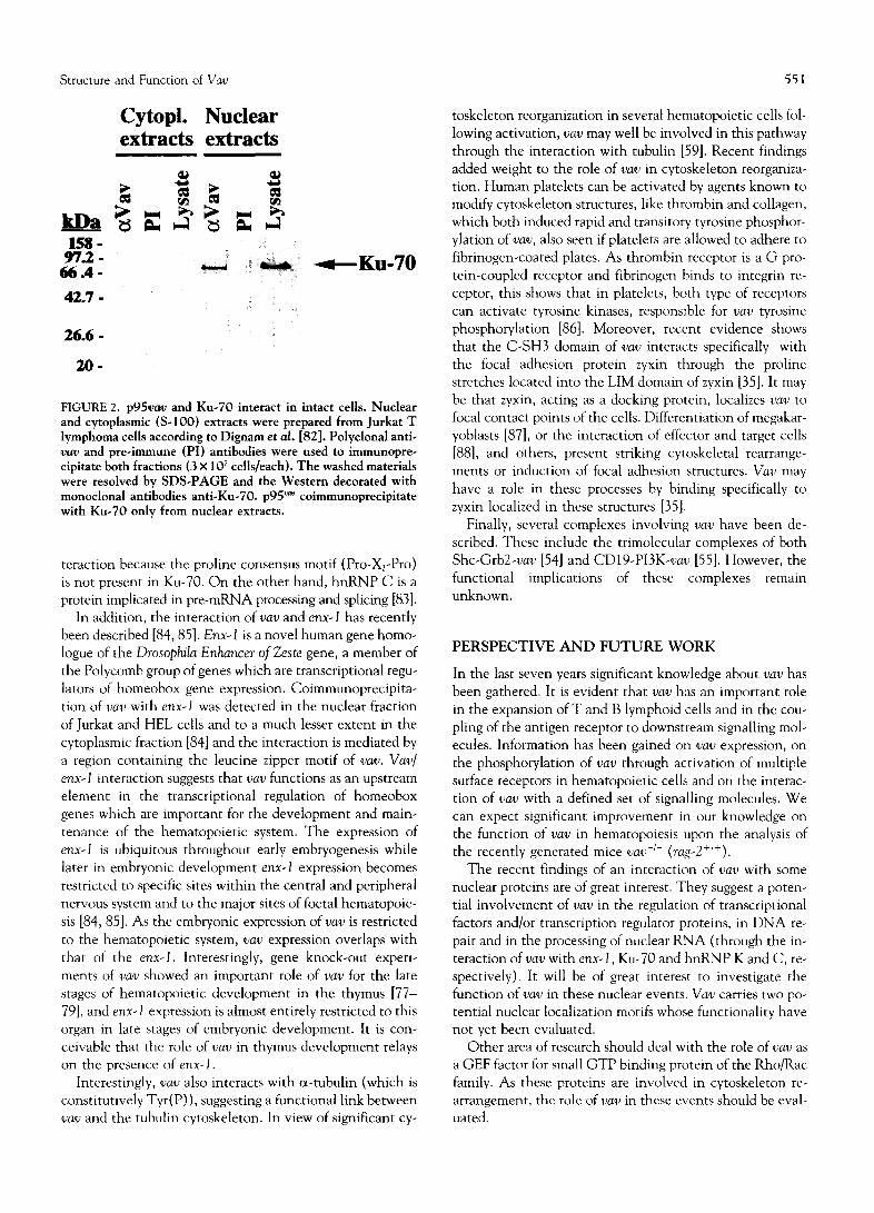
We have shown that the C-SH3 domain of v a v interacts with Ku-70 (Fig. 2) and hnRNP C, two proteins mainly present in the nucleus ([34] and unpublished results). Ku- 70 is the DNA-binding element of the DNA-dependent protein kinase which has been implicated in DNA repair, replication, recombination and transcriptional events [34]. This interaction may be a novel form of protein-protein in-
Structure and Function of Vav
Cytopl. Nuclear extracts extracts
1 5 8 - 9 7 . 2 -
6 6 . 4 -
42 .7 -
2 6 . 6 -
2 0 -
Ku-70
FIGURE 2. p 9 5 v a v and Ku-70 interact in intact cells. Nuclear and cytoplasmic (S-100) extracts were prepared from Jurkat T lymphoma cells according to Dignam et oil. [82]. Polyclonal anti- vav and pre-immune (PI) antibodies were used to immunopre- cipitate both fractions (3 x 10⁷ cells/each). The washed materials were resolved by SDS-PAGE and the Western decorated with monoclonal antibodies anti-Ku-70, p95 '~ coimmunoprecipitate with Ku-70 only from nuclear extracts.
teraction because the proline consensus motif (Pro-X2-Pro) is not present in Ku-70. On the other hand, hnRNP C is a protein implicated in pre-mRNA processing and splicing [83].
In addition, the interaction of vav and enx-1 has recently been described [84, 85]. E n x - I is a novel human gene homo- logue of the Drosophi la E n h a n c e r o f Zes te gene, a member of the Polycomb group of genes which are transcriptional regu- lators of homeobox gene expression. Coimmunoprecipita- tion of vav with enx-1 was detected in the nuclear fraction of Jurkat and HEL cells and to a much lesser extent in the cytoplasmic fraction [84] and the interaction is mediated by a region containing the leucine zipper motif of vav . V a v /
enx-1 interaction suggests that vav functions as an upstream element in the transcriptional regulation of homeobox genes which are important for the development and main- tenance of the hematopoietic system. The expression of e n x - I is ubiquitous throughout early embryogenesis while later in embryonic development enx-1 expression becomes restricted to specific sites within the central and peripheral nervous system and to the major sites of foetal hematopoie- sis [84, 85]. As the embryonic expression of vav is restricted to the hematopoietic system, vav expression overlaps with that of the enx -1 . Interestingly, gene knock-out experi- ments of vav showed an important role of vav for the late stages of hematopoietic development in the thymus [77- 79], and enx-1 expression is almost entirely restricted to this organ in late stages of embryonic development. It is con- ceivable that the role of vav in thymus development relays on the presence of enx -1 .
Interestingly, vav also interacts with cx-tubulin (which is constitutively Tyr(P)), suggesting a functional link between vav and the tubulin cytoskeleton. In view of significant cy-
551
toskeleton reorganization in several hematopoietic cells fol- lowing activation, vav may well be involved in this pathway through the interaction with tubulin [59]. Recent findings added weight to the role of vav in cytoskeleton reorganiza- tion. Human platelets can be activated by agents known to modify cytoskeleton structures, like thrombin and collagen, which both induced rapid and transitory tyrosine phosphor- ytation of vav , also seen if platelets are allowed to adhere to fibrinogen-coated plates. As thrombin receptor is a G pro- tein-coupled receptor and fibrinogen binds to integrin re- ceptor, this shows that in platelets, both type of receptors can activate tyrosine kinases, responsible for vav tyrosine phosphorylation [86]. Moreover, recent evidence shows that the C-SH3 domain of vav interacts specifically with the focal adhesion protein zyxin through the proline stretches located into the LIM domain of zyxin [35]. It may be that zyxin, acting as a docking protein, localizes vav to focal contact points of the cells. Differentiation of megakar- yoblasts [87], or the interaction of effector and target cells [88], and others, present striking cytoskeletal rearrange- ments or induction of focal adhesion structures. V a v may have a role in these processes by binding specifically to zyxin localized in these structures [35].
Finally, several complexes involving vav have been de- scribed. These include the trimolecular complexes of both S h c - G r b 2 - v a v [54] and C D 1 9 - P I 3 K - v a v [55]. However, the functional implications of these complexes remain unknown.
PERSPECTIVE AND F U TU RE WORK
In the last seven years significant knowledge about va v has been gathered. It is evident that vav has an important role in the expansion of T and B lymphoid cells and in the cou- pling of the antigen receptor to downstream signalling mol- ecules. Information has been gained on vav expression, on the phosphorylation of r a y through activation of multiple surface receptors in hematopoietic cells and on the interac- tion of r a y with a defined set of signalling molecules. We can expect significant improvement in our knowledge on the function of vav in hematopoiesis upon the analysis of the recently generated mice vav -I- (rag-2+l+).
The recent findings of an interaction of vav with some nuclear proteins are of great interest. They suggest a poten- tial involvement of vav in the regulation of transcriptional factors and/or transcription regulator proteins, in DNA re- pair and in the processing of nuclear RNA (through the in- teraction of vav with enx -1 , Ku-70 and hnRNP K and C, re- spectively). It will be of great interest to investigate the function of vav in these nuclear events. V a v carries two po- tential nuclear localization motifs whose functionality have not yet been evaluated.
Other area of research should deal with the role of va v as a GEF factor for small GTP binding protein of the Rho/Rac family. As these proteins are involved in cytoskeleton re- arrangement, the role of r a y in these events should be eval- uated.

552 F. Romero and S. Fischer
The PTK/s responsible for vav tyrosine phosphorylation in vivo have not been identified. It would be of interest to characterize the residues tyrosine phosphorylated in vav fol- lowing activation. Is there a single Tyr(P) or are there mul- tiple sites, varying with the stimuli, differentiation and others ?
The role of vav in human malignancy needs to be deter- mined. Are there hematological disorders with mutated or alternatively spliced vav or are there others where vav is ov- erexpressed?
As vav2 has conserved the critical domains of vav and is widely expressed, it will be of interest to examine whether growth factors alter its phosphorylation and to identify the SH2 domains protein interacting with vav2. It seems worth- while to investigate whether amino-terminal deleted vav2 has transforming properties for non-hematological cells.
We are grateful to several investigators for sending their work in press or in progress. We also thank Drs. Gisselbrecht and Dusanter-Fourt for critical reading of the manuscript, Dr. Saragosti for computer search and Association pour la Recherche sur le Cancer for financial help. F. R. was supported by an EEC postdoctoral fellowship (HCM and TMR programmes).
References 1. Mayer B. J. and Baltimore D. (1993) Trends Cell. Biol. 3,
8-13. 2. Pawson T. and Schlessinger J. (1993) Curr. Biol. 3,434-442. 3. Van Der Greer P. and Pawson T. (1995) Trends Biochem. Sci.
20, 277-280. 4. Bork P. and Sudol M. (1994) Trends Biochem. Sci. 19, 531-
533. 5. Katzav S., Martin-Zanca D. and Barbacid M. (1989) EMBO
J, 8, 2283-2290. 6. Coppola J., Bryant S., Koda T., Conway D. and Barbacid M.
(1991) Cell Growth Differ. 2, 95-105. 7. Katzav S., Cleveland J. L., Heslop H. E. and Pulido D. (1991)
Mol. Cell. Biol. 11, 1912-1920. 8. Adams J. M., Houston H., Allen J., Lints T. and Harvey R.
(1992) Oncogene 7, 611-618. 9. Bustelo X. R., Rubin S. D., Suen K. L., Carrasco D. and Bar-
bacid M. (1993) Cell Growth Differ. 4, 297-308. 10. Henske E. P., Short M. P., Jozwiak S., Bovey C. M., Ramlak-
han S., Haines J. L. and Kwiatkowski D. J. (1995) Ann. Hum. Genet. 59, 25-37.
11. Martinerie C., Cannizzaro L. A., Croce C. M., Huebner K., Katzav S. and Barbacid M. (1990) Hum. Genet. 86, 65-68.
12. Castresana J. and Saraste M. (1995) FEBS Lett. 374, 149- 151.
13. Sigler P. B. (1988) Nature 333, 210-211. 14. Hu P., Margolis B. and Schlessinger J. (1993) Bioessays 15,
179-183. 15. Gulbins E., Coggeshall K. M., Baier G., Katzav S., Burn P. and
Altman A. (1993) Science 260, 822-825. 16. Gulbins E., Langlet C., Baier G., Bonnefoy-Berard N., Her-
bert E., Altman A. and Coggeshall K. M. (1994) J. Immunol. 152, 2123-2129.
17. Gulbins E., Coggeshall K. M., Langlet C., Baier G., Bonnefoy- Berard N., Burn P., Wittinghofer A., Katzav S. and Altman A. (1994) Mol. Cell. Biol. 14, 906-913.
18. Bustelo X. R., Suen K. L., Leftheris K., Meyers C. A. and Bar- bacid M. (1994) Oncogene 9, 2405-2413.
19. Khosravi-Far R., Chrzanowska-Wodnicka M., Solski P. A., Eva A., Burridge K. and Der C. J. (1994) Mo/. Cell. Biol. 14, 6848-6857.
20. Musacchio A., Gibson T., Rice P., Thompson J. and Saraste M. (1993) Trends Biochem. Sci. 18, 343-348.
21. Harlan J. E., Hajduk P. J., Yoon H. S. and Fesik S. W. (1994) Nature 371, 168-170.
22. Touhara K., Inglese J., Pitcher J. A., Shaw G. and Lefkowitz R. J. (1994) J. Biol. Chem. 269, 10217-10220.
23. Yao L., Kawakami Y. and Kawakami T. (1994) Proc. Natl. Acad. Sci. U.S.A. 91, 9175-9179.
24. Gulbins E., Coggeshall K. M., Baier G., Telford D., Langlet C., Baier-Bitterlich G., Bonnefoy-Berard N., Burn P., Wittingofer A. and Altman A. (1994) Mo/. Cell. Biol. 14, 4749-4758.
25. Clevenger C. V., Ngo W., Sokol D. L., Luger S. M. and Gew- irtz A. M. (1995) J. Biol. Chem. 270, 13246-13253.
26. Kazanietz M. G., Bustelo X. R., Barbacid M., Kolch W., Mis- chak H., Wong G., Pettit G. R., Bruns J. D. and Blumberg P. M. (1994)J. Biol. Chem. 269, 11590-11594.
27. Koch C. A., Anderson D., Moran M. F., Ellis C. and Pawson T. (1991) Science 252, 668-674.
28. Bustelo X. R. and Barbacid M. (1992) Science 256, 1196- 1199.
29. Margolis B., Hu P., Katzav S., Li W., Oliver J. M., UUrich A., Weiss A. and Schlessinger J. (1992) Nature 356, 71-74.
30. Margolis B. (1992) Cell Growth Differ. 3, 73-80. 31. Cussac D., Frech M. and Chardin P. (1994) EMBO J. 13,
4011-4021. 32. Hobert O., Jallal B., Schlessinger J. and Ullrich A. (1994) J.
Biol. Chem. 269, 20225-20228. 33. Bustelo X. R., Suen K. L., Michael W. M., Dreyfuss G. and
Barbacid M. (1995) Mol. Cell. Biol. 15, 1324-1332. 34. Romero F., Dargemont C., Pozo F., Reeves W. H., Camonis
J., Gisselbrecht S. and Fischer S. (1996) Mol. Cell. Biol. 16, 37-44.
35. Hobert O., Schilling J. W., Beckerle M. C., Ullrich A. and Jallal B. (1996) Oncogene 12, 1577-1581.
36. Ramos-Morales F., Romero F., Schweighoffer F., Bismuth G., Camonis J., Tortolero M. and Fischer S. (1995) Oncogene 11, 1665-1669.
37. Ye Z. S. and Baltimore D. (1994) Proc. Natl. Acad. Sci. U.S.A. 91, 12629-12633.
38. Bustelo X. R., Ledbetter J. A. and Barbacid M. (1992) Nature 356, 68-71.
39. Overduin M., Mayer B., Rios C. B., Baltimore D. and Cow- burn D. (1992) Proc. Natl. Acad. Sci. U.S.A. 89, 11673- 11677.
40. Eck M. J., Shoelson S. E. and Harrison S. C. (1993) Nature 362, 87-91.
41. Songyang Z., Shoelson S. E., Chaudhuri M., Gish G., Pawson T., Haser W. G., King F., Roberts T., Ratnofsky S., Lechleider R. J., Neel B. G., Birge R. B., Fajardo J. E., Chou M. M., Ha- nafusa H., Schaffhausen B. and Cantley L. C. (1993) Cell 72, 767-778.
42. Waksman G., Shoelson S. E., Pant N., Cowburn D. and Kuri- yan J. (1993) Cell 72, 779-790.
43. Katzav S., Sutherland M., Packham G., Yi T., and Weiss A. (1994) J. Biol. Chem. 269, 32579-32585.
44. Matsuguchi T., Inhorn R. C., Carlesso N., Xu G., Druker B. and Griffin J. D. (1995) EMBO J. 14, 257-265.
45. Kon-Kozlowsky M., Pani G., Pawson T., and Siminovitch K. A. (1996) J. Biol. Chem. 271, 3856-3862.
46. Seidel-Dugan C., Meyer B. E., Thomas S. M. and Brugge J. S. (1992) Mol. Cell. Biol. 12, 1835-1845.
47. Hirai H. and Varmus H. E. (1990) Mol. Cell. Biol. 10, 1307- 1318.
48. Katzav S. (1993) Oncogene 8, 1757-1763.
Structure and Function of Vav 553
49. Mayer B. J., Jackson P. K., Van Etten R. A. and Baltimore D. (1992) Mol. Cell. Biol. 12, 609-618.
50. Alai M., Mui A. L., Cutler R. L., Bustelo X. R., Barbacid M. and Krystal G. (1992) J. Biol. Chem. 267, 18021-18025.
51. Miura O., Miura Y., Nakamura N., Quelle F. W., Witthuhn B. A., Ihle J. N. and Aoki N. (1994) Blood 84, 4135-4141.
52. Nunes J. A., Collette Y., Truneh A., Olive D. and Cantrell D. A. (1994)J. Exp. Med. 180, 1067-1076.
53. Platanias L. C. and Sweet M. E. (1994) J. Biol. Chem, 269, 3143-3146.
54. Ramos-Morales F., Druker B. J. and Fischer S. (1994) Onco- gene 9, 1917-1923.
55. Weng W. K., Jarvis L. and Le Bien T. W. (1994) J. Biol. Chem. 269, 32514-32521.
56. Uddin S., Katzav S., White M. F. and Platanias L. C. (1995) J. Biol. Chem. 270, 7712-7716.
57. Wu J., Katzav S. and Weiss A. (1995) Mol. Cell. Biol. 15, 4337-4346.
58. Galland F., Katzav S. and Birnbaum D. (1992) Oncogene 7, 585-597.
59. Huby R. D., Carlile G. W. and Ley S. C. (1995) J. Biol. Chem. 270, 30241-30244.
60. Miura O., D'Andrea A., Kabat D. and Ihle J. N. (1991) Mol. Cell. Biol. 11, 4895-4902.
61. Evans G. A., Howard O. M., Erwin R. and Farrar W. L. (1993) Biochem. J. 294, 339-342.
62. Sato S., Katagiri T., Takaki S., Kikuchi Y., Hitoshi Y., Yoneh- ara S., Tsukada S., Kitamura D., Watanabe T., Witte O. and Takatsu K. (1994) J. Exp. Med. 180, 2101-2111.
63. Dosil M., Wang S. and Lemischka I. R. (1993) Mol. Cell. Biol. 13, 6572-6585.
64. Gouy H., Debre P. and Bismuth G. (1995) Eur. J. Immunol. 25, 3030-3034.
65. Darnell J. E., Kerr I. M. and Stark G. R. (1994) Science 264, 1415-1421.
66. Gupta S., Weiss A., Kumar G., Wang S. and Nel A. (1994) J. Biol. Chem. 269, 17349-17357.
67. Straus D. B. and Weiss A. (1992) Cell 70, 585-593. 68. Chan A. C., Dalton M., Johnson R., Kong G. H., Wang T.,
Thoma R. and Kurosaki T. (1995) EMBO J. 14, 2499-2508.
69. Machide M., Mano H. and Todokoro K. (1995) Oncogene 11, 619-625.
70. Brunati A. M., Donella-Deana A., Ruzzene M., Marin O. and Pinna L. A. (1995) FEBS Lett. 367, 149-152.
71. Mustelin T. (1994) Immunity 1,351-356. 72. Motto D. G., Ross S. E., Wu J., Hendricks-Taylor L. R. and
Koretzky G. A. (1996)J. Exp. Med. 183, 1937-1943. 73. Wulf G. M., Adra C. N. and Lira B. (1993) EMBO J. 12,
5065-5074. 74. Luger S. M., Ratajczak J., Ratajczak M. Z., Kuczynski W. I.,
Dipaola R. S., Ngo W., Clevenger C. V. and Gerwirtz A. M. (1996) Blood 87, 1326-1334.
75. Zhang R., Tsai F. Y. and Orkin S. H. (1994) Proc. Natl. Acad. Sci. U.S.A. 91, 12755-12759.
76. Zmuidzinas A., Fischer K. D., Lira S. A., Forrester L., Bryant S., Bernstein A. and Barbacid M. (1995) EMBOJ. 14, 1-11.
77. Fischer K. D., Zmuldzinas A., Gardner S., Barbacid M., Berstein A. and Guidos C. (1995) Nature 374, 474-477.
78. Tarakhovsky A., Turner M., Schaal S., Mee P. J., Duddy L. P., Rajewsky K. and Tybulewicz V. L. (1995) Nature 374, 467 470.
79. Zhang R., Alt F. W., Davidson L., Orkin S. H. and Swat W. (1995) Nature 374, 470-473.
80. Van Seuningen I., Ostrowski J., Bustelo X. R., Sleath P. R. and Bomsztyk K. (1995) J. Biol. Chem. 270, 26976-26985.
81. Van Seuningen I., Ostrowski J. and Bomsztyk K. (1995) Bio- chemistry 34, 5644-5650.
82. Digman J. D., Lebovitz R. M. and Roeder R. G. (1983) Nucleic Acids Res. 11, 1475-1489.
83. Dreyfuss G., Matunis M. J., Pinol-Roma S. and Burd C. G. (1993) Annu. Rev. Biochem. 62, 289-321.
84. Hobert O., Jallal B. and Ullrich A. (1996) Mol. Cell. Biol. 16, 3066-3073.
85. Hobert O., Sures I., Ciossek T., Fuchs M. and Ullrich A. (1996) Hum. Genet. In press.
86. Cichowski K., Brugge J. S. and Brass L. F. (1996) J. Biol. Chem. 271, 7544--7550.
87. Hoffman R. (1989) Blood 74, 1196-12t2. 88. Podack E. R. and Kupfer A. (1991) Annu. Rev. Cell. Biol. 7,
479-504.