Structure and Function of Antigen Recognition Molecules · Antigen receptors of lymphocytes...
28
Structure and Function of Antigen Recognition Molecules Dr Allison Imrie [email protected] MICR2209 1
Transcript of Structure and Function of Antigen Recognition Molecules · Antigen receptors of lymphocytes...
Structure and Function of Antigen Recognition Molecules
Dr Allison Imrie
MICR2209
1
Synopsis: In this lecture we will examine the major receptors used by cells of the innate and adaptive immune response to detect antigen
Outcomes: You should be able to describe the major receptors that innate and adaptive immune systems cells use to recognize antigen, and the similarities and differences between the receptors used by these 2 arms of the immune system
2
Recognition of Microbes by Innate Immune System
• The components of innate immunity recognize structures that are shared by various classes of microbes and are not present on host cells • Each component of innate immunity may recognize many bacteria, or viruses, or fungi • Innate immune system receptors that detect microbes exhibit different patterns of expression and are expressed on phagocytes, dendritic cells, epithelial and endothelial cells, and also on lymphocytes
3
Recognition of Microbes by Innate Immune System
• The receptors of the innate immune system are encoded in the germline and are not produced by somatic recombination of genes (as are the receptors of the adaptive immune system – antibodies and the T cell receptor) • Specificity of innate immunity is therefore much less diverse than that of adaptive immunity: recognize about 103 ligands compared to > 109 for adaptive immune system • Receptors are nonclonally distributed – identical receptors are expressed on all cells of a certain type eg. all macrophages express identical TLR4; in contrast each T cell or B cell clone expresses a unique receptor for a specific antigen
4
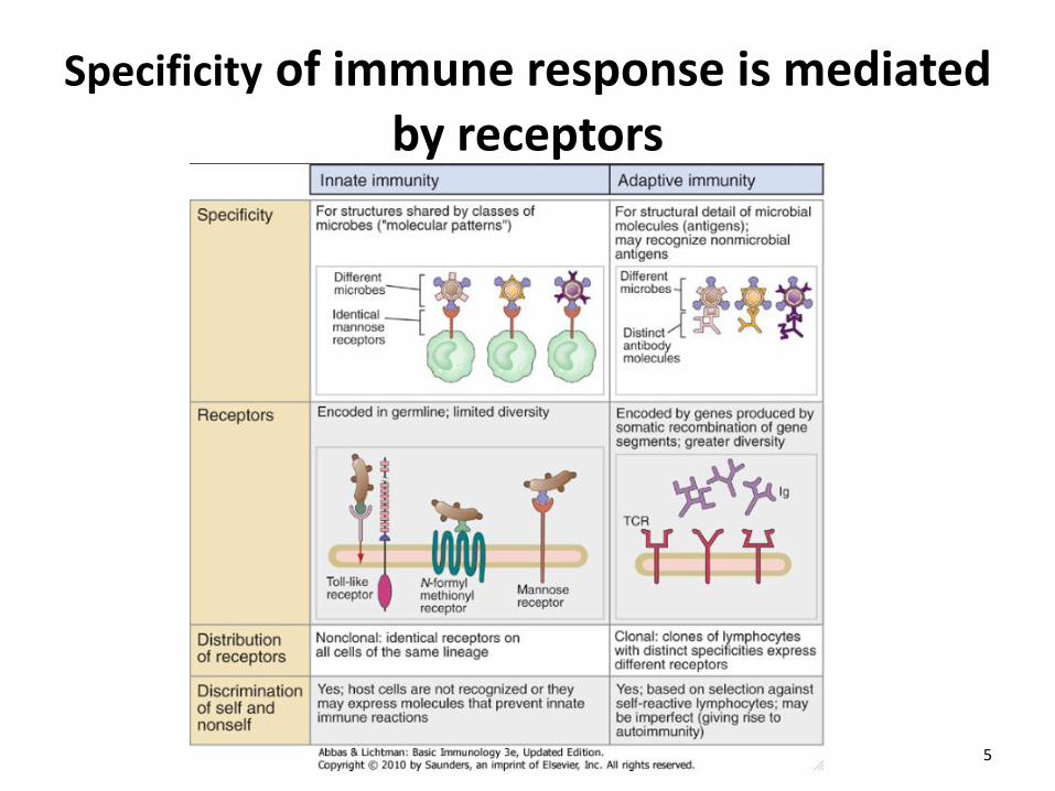
Specificity of immune response is mediated by receptors
5
Cellular Receptors for Microbes Toll-like receptors (TLRs)
• Differentially expressed on phagocytes (neutrophils and macrophages), dendritic cells, and many other cell types
• Expressed on cell surface or intracellularly on endosomal surface
• TLRs are specific for different components of microbes
• There are >12 TLRs identified so far; not all are present on mammalian cells
• Engagement of TLR by the appropriate microbial ligand activates transcription factors that stimulate expression of genes encoding cytokines, enzymes, and other proteins involved in antimicrobial functions: – NF-κΒ promotes expression of cytokines and endothelial
adhesion molecules
– IRF-3 stimulates production of Type I Interferons, cytokines that block viral replication
6
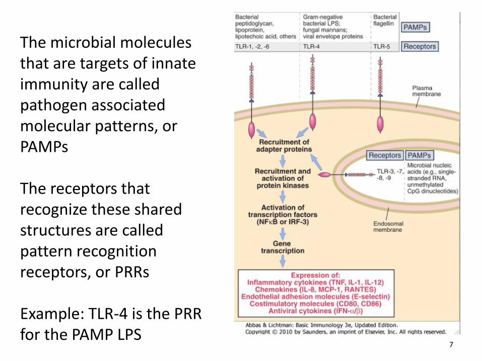
The microbial molecules that are targets of innate immunity are called pathogen associated molecular patterns, or PAMPs The receptors that recognize these shared structures are called pattern recognition receptors, or PRRs Example: TLR-4 is the PRR for the PAMP LPS
7
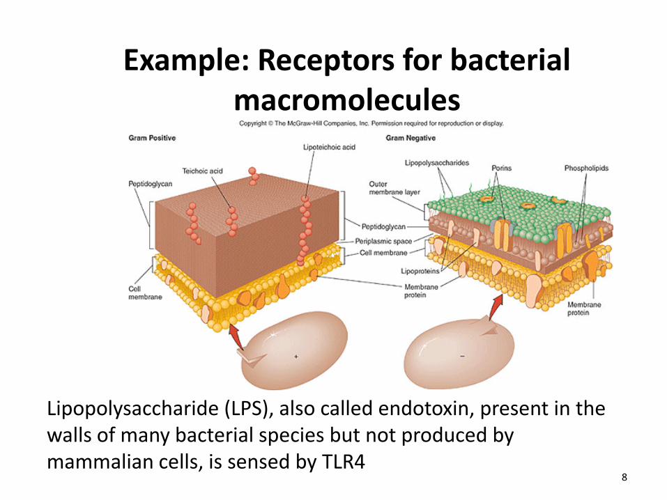
Example: Receptors for bacterial macromolecules
Lipopolysaccharide (LPS), also called endotoxin, present in the walls of many bacterial species but not produced by mammalian cells, is sensed by TLR4
8
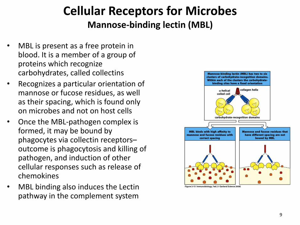
Cellular Receptors for Microbes Mannose-binding lectin (MBL)
• MBL is present as a free protein in
blood. It is a member of a group of proteins which recognize carbohydrates, called collectins
• Recognizes a particular orientation of mannose or fucose residues, as well as their spacing, which is found only on microbes and not on host cells
• Once the MBL-pathogen complex is formed, it may be bound by phagocytes via collectin receptors– outcome is phagocytosis and killing of pathogen, and induction of other cellular responses such as release of chemokines
• MBL binding also induces the Lectin pathway in the complement system
9
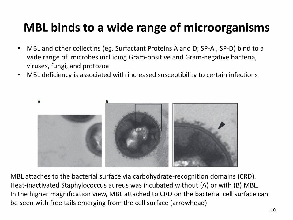
MBL binds to a wide range of microorganisms
MBL attaches to the bacterial surface via carbohydrate-recognition domains (CRD). Heat-inactivated Staphylococcus aureus was incubated without (A) or with (B) MBL. In the higher magnification view, MBL attached to CRD on the bacterial cell surface can be seen with free tails emerging from the cell surface (arrowhead)
• MBL and other collectins (eg. Surfactant Proteins A and D; SP-A , SP-D) bind to a wide range of microbes including Gram-positive and Gram-negative bacteria, viruses, fungi, and protozoa
• MBL deficiency is associated with increased susceptibility to certain infections
10
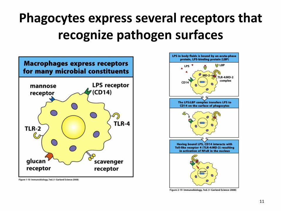
Phagocytes express several receptors that recognize pathogen surfaces
11
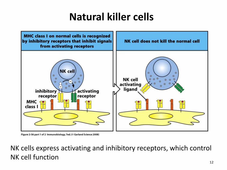
Natural killer cells
NK cells express activating and inhibitory receptors, which control NK cell function
12
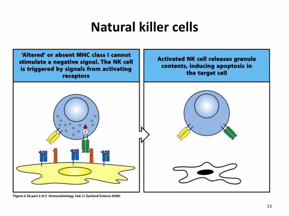
Natural killer cells
13
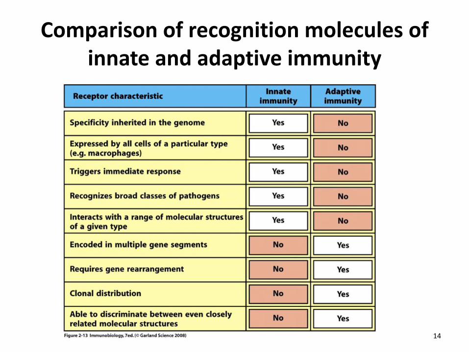
Comparison of recognition molecules of innate and adaptive immunity
14
Recognition of microbes by adaptive immune system
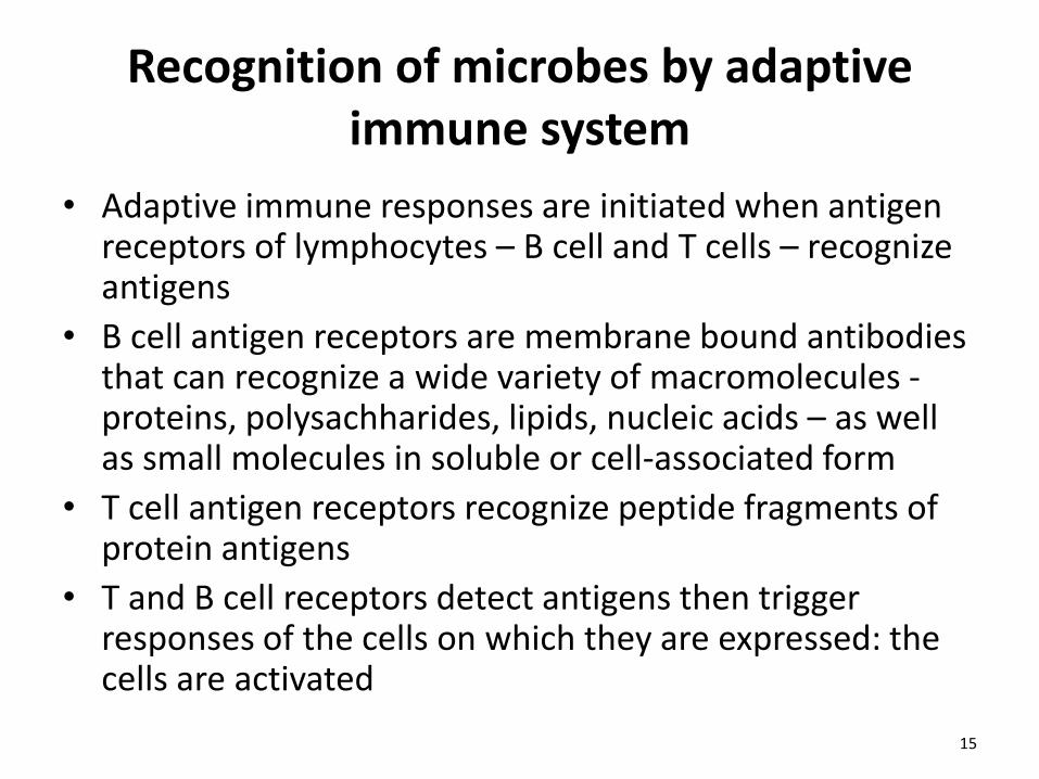
• Adaptive immune responses are initiated when antigen receptors of lymphocytes – B cell and T cells – recognize antigens
• B cell antigen receptors are membrane bound antibodies that can recognize a wide variety of macromolecules - proteins, polysachharides, lipids, nucleic acids – as well as small molecules in soluble or cell-associated form
• T cell antigen receptors recognize peptide fragments of protein antigens
• T and B cell receptors detect antigens then trigger responses of the cells on which they are expressed: the cells are activated
15
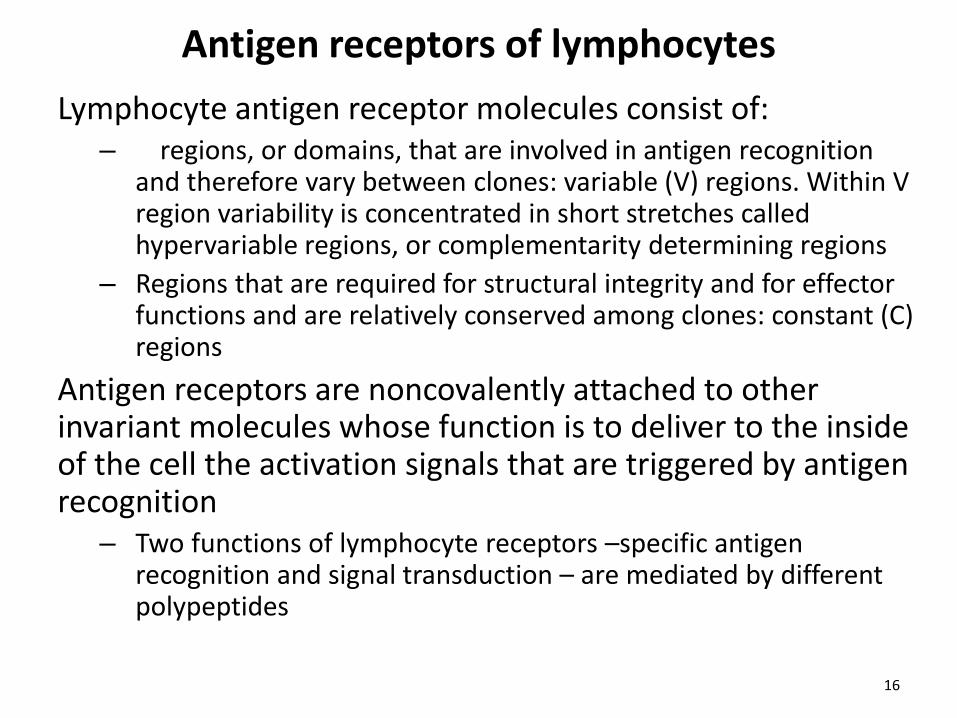
Antigen receptors of lymphocytes Lymphocyte antigen receptor molecules consist of:
– regions, or domains, that are involved in antigen recognition and therefore vary between clones: variable (V) regions. Within V region variability is concentrated in short stretches called hypervariable regions, or complementarity determining regions
– Regions that are required for structural integrity and for effector functions and are relatively conserved among clones: constant (C) regions
Antigen receptors are noncovalently attached to other invariant molecules whose function is to deliver to the inside of the cell the activation signals that are triggered by antigen recognition
– Two functions of lymphocyte receptors –specific antigen recognition and signal transduction – are mediated by different polypeptides
16
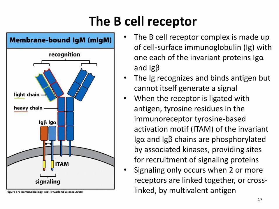
The B cell receptor • The B cell receptor complex is made up
of cell-surface immunoglobulin (Ig) with one each of the invariant proteins Igα and Igβ
• The Ig recognizes and binds antigen but cannot itself generate a signal
• When the receptor is ligated with antigen, tyrosine residues in the immunoreceptor tyrosine-based activation motif (ITAM) of the invariant Igα and Igβ chains are phosphorylated by associated kinases, providing sites for recruitment of signaling proteins
• Signaling only occurs when 2 or more receptors are linked together, or cross-linked, by multivalent antigen
17
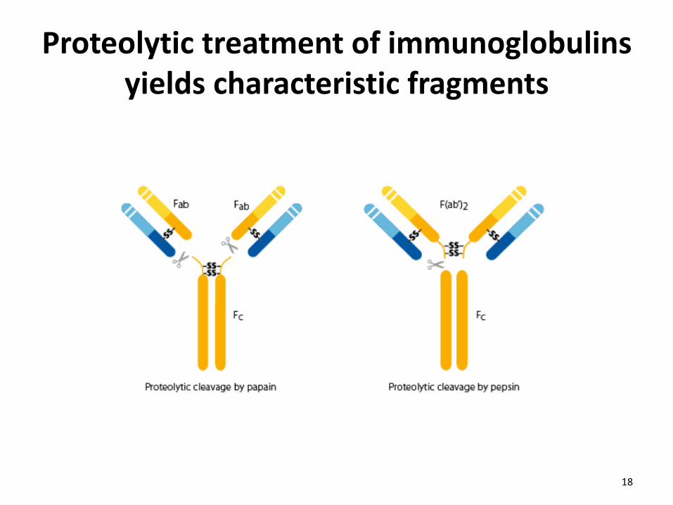
Proteolytic treatment of immunoglobulins yields characteristic fragments
18
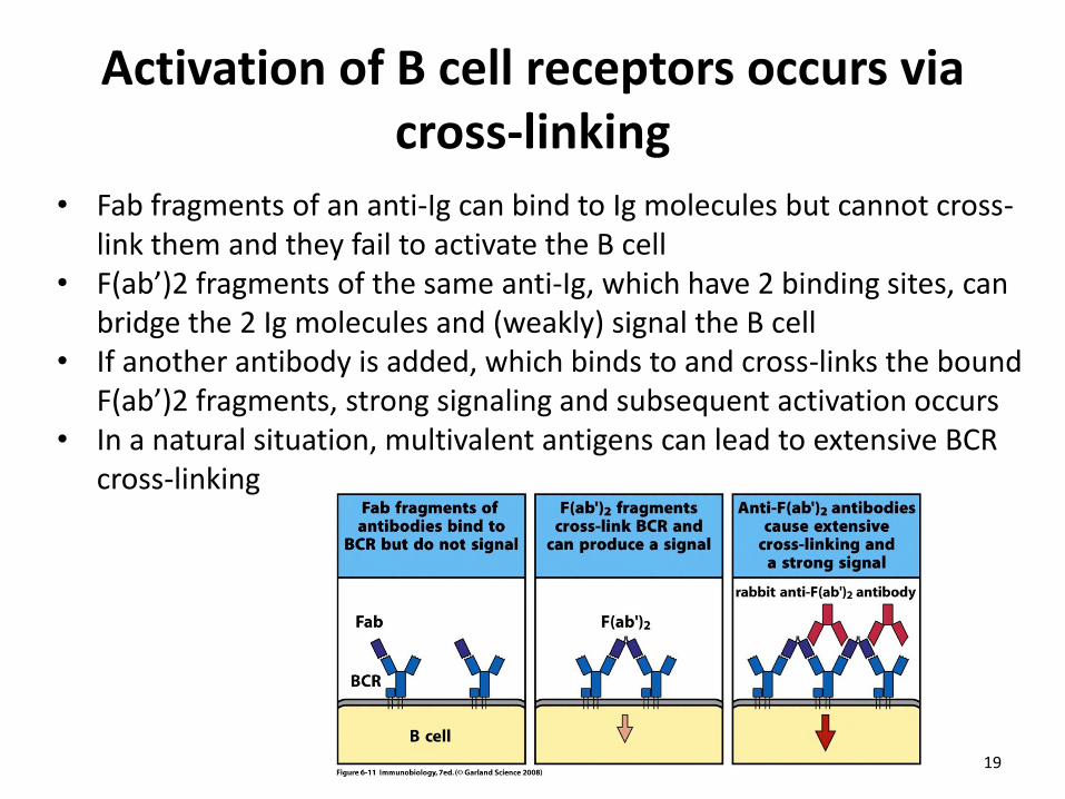
Activation of B cell receptors occurs via cross-linking
• Fab fragments of an anti-Ig can bind to Ig molecules but cannot cross-link them and they fail to activate the B cell
• F(ab’)2 fragments of the same anti-Ig, which have 2 binding sites, can bridge the 2 Ig molecules and (weakly) signal the B cell
• If another antibody is added, which binds to and cross-links the bound F(ab’)2 fragments, strong signaling and subsequent activation occurs
• In a natural situation, multivalent antigens can lead to extensive BCR cross-linking
19
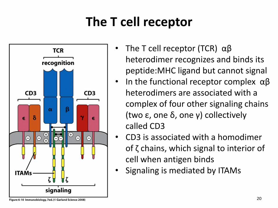
The T cell receptor
• The T cell receptor (TCR) αβ heterodimer recognizes and binds its peptide:MHC ligand but cannot signal
• In the functional receptor complex αβ heterodimers are associated with a complex of four other signaling chains (two ε, one δ, one γ) collectively called CD3
• CD3 is associated with a homodimer of ζ chains, which signal to interior of cell when antigen binds
• Signaling is mediated by ITAMs
20
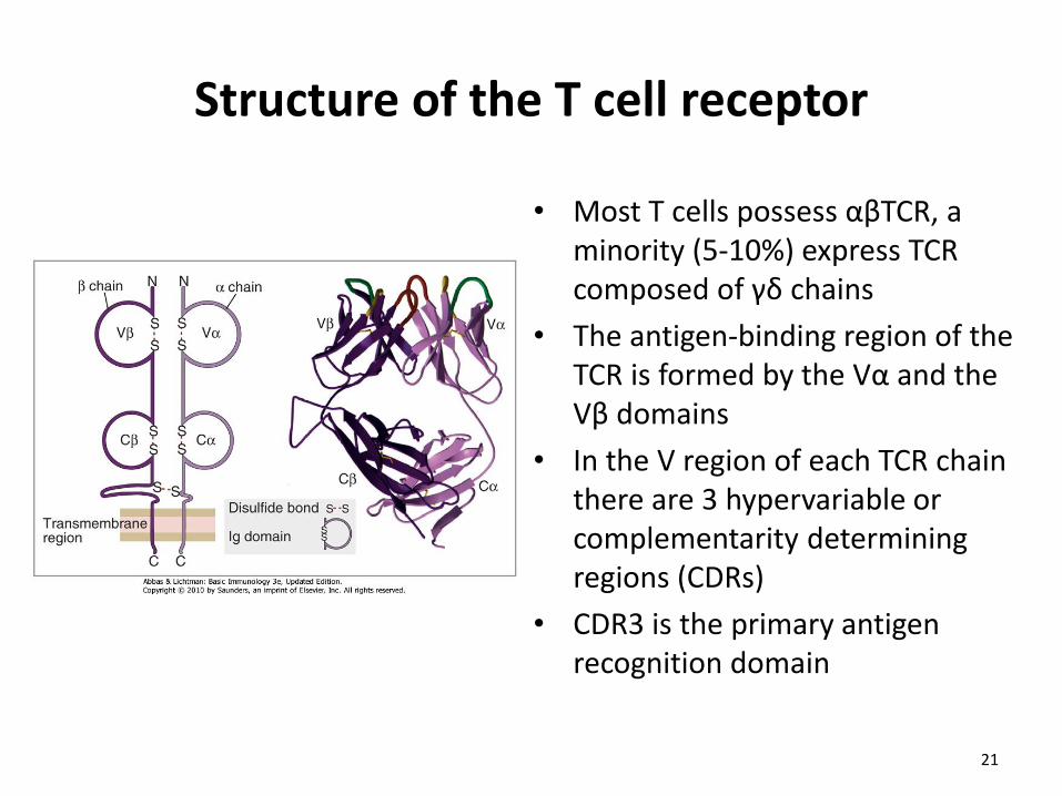
Structure of the T cell receptor
• Most T cells possess αβTCR, a minority (5-10%) express TCR composed of γδ chains
• The antigen-binding region of the TCR is formed by the Vα and the Vβ domains
• In the V region of each TCR chain there are 3 hypervariable or complementarity determining regions (CDRs)
• CDR3 is the primary antigen recognition domain
21
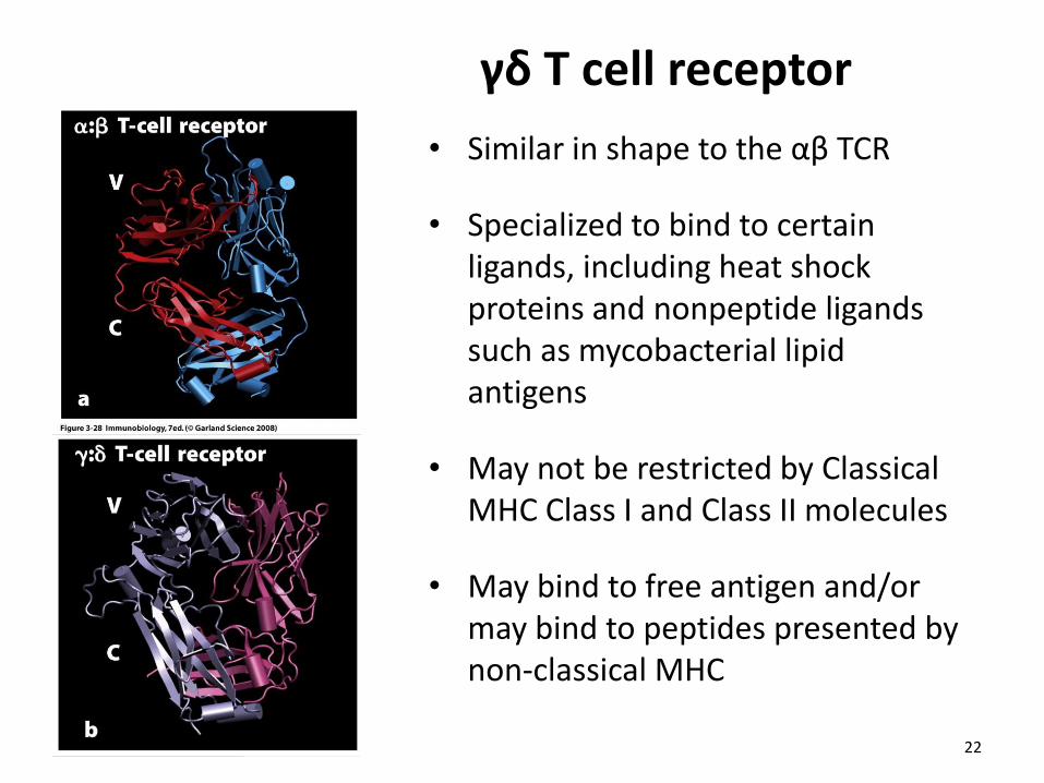
γδ T cell receptor
• Similar in shape to the αβ TCR
• Specialized to bind to certain ligands, including heat shock proteins and nonpeptide ligands such as mycobacterial lipid antigens
• May not be restricted by Classical MHC Class I and Class II molecules
• May bind to free antigen and/or may bind to peptides presented by non-classical MHC
22
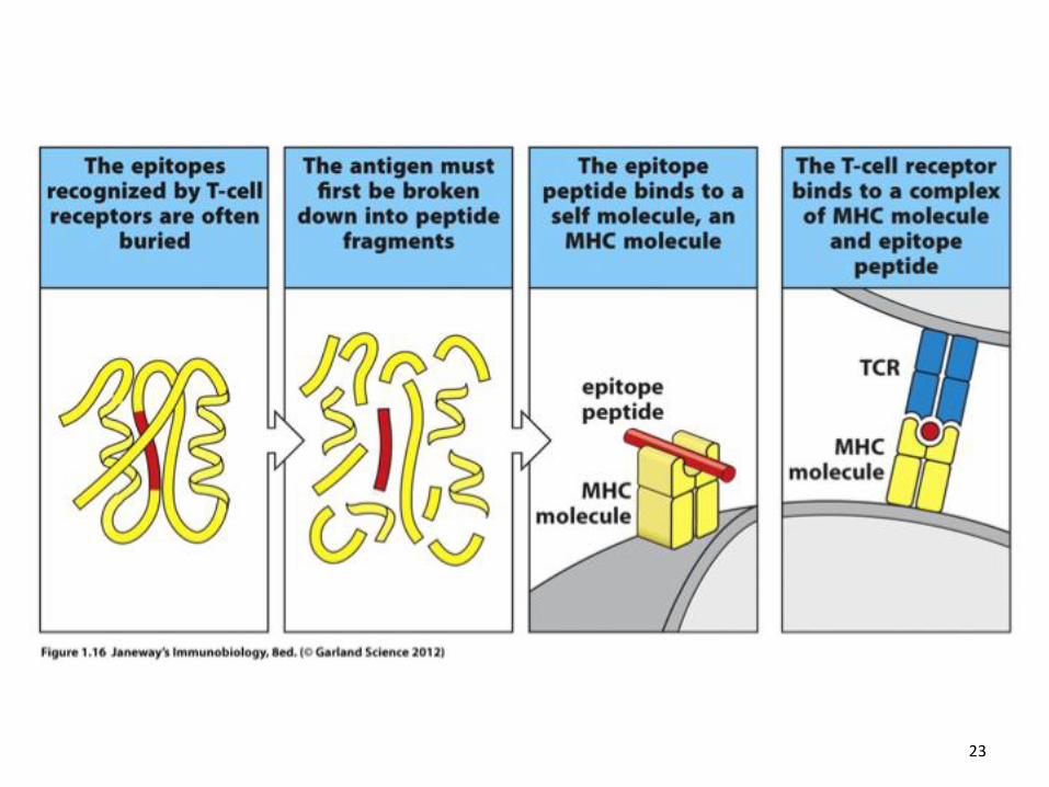
23
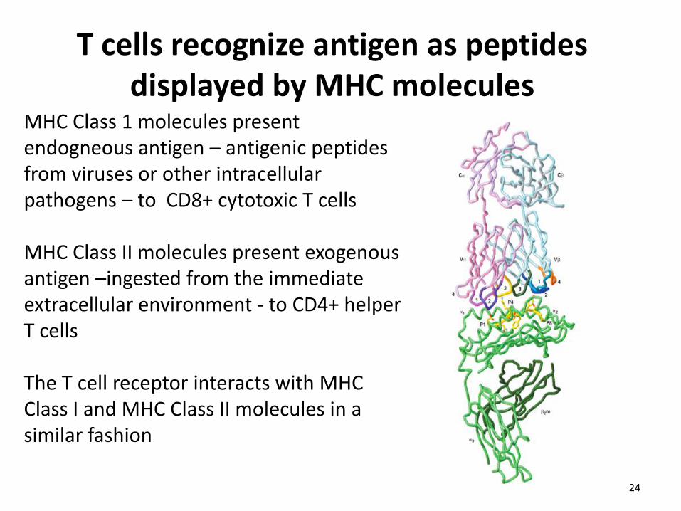
T cells recognize antigen as peptides displayed by MHC molecules
MHC Class 1 molecules present endogneous antigen – antigenic peptides from viruses or other intracellular pathogens – to CD8+ cytotoxic T cells MHC Class II molecules present exogenous antigen –ingested from the immediate extracellular environment - to CD4+ helper T cells The T cell receptor interacts with MHC Class I and MHC Class II molecules in a similar fashion
24
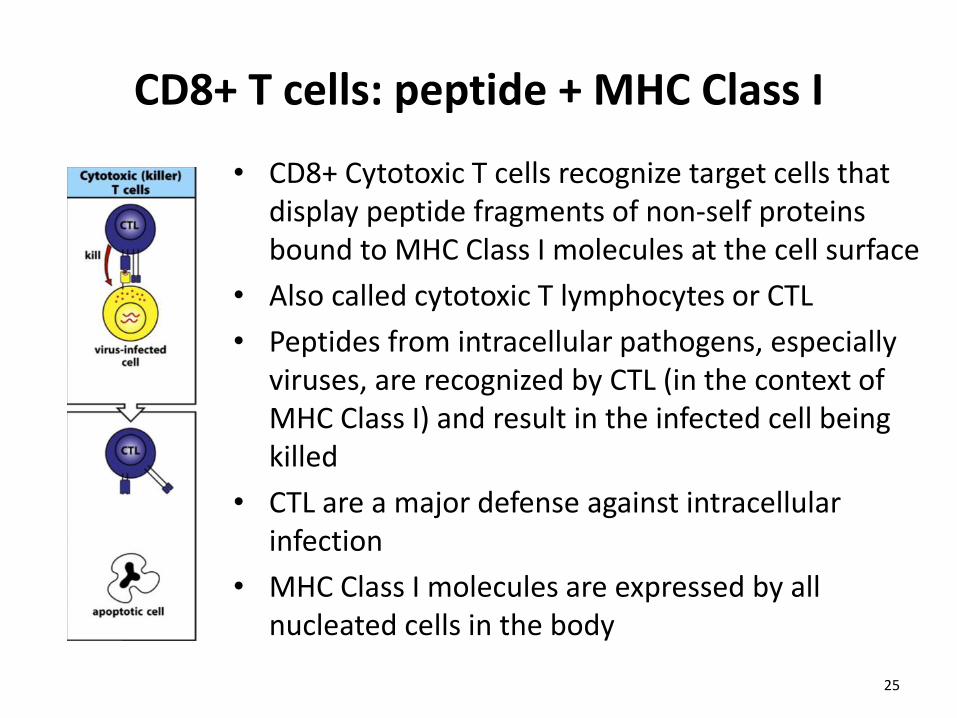
CD8+ T cells: peptide + MHC Class I
• CD8+ Cytotoxic T cells recognize target cells that display peptide fragments of non-self proteins bound to MHC Class I molecules at the cell surface
• Also called cytotoxic T lymphocytes or CTL
• Peptides from intracellular pathogens, especially viruses, are recognized by CTL (in the context of MHC Class I) and result in the infected cell being killed
• CTL are a major defense against intracellular infection
• MHC Class I molecules are expressed by all nucleated cells in the body
25
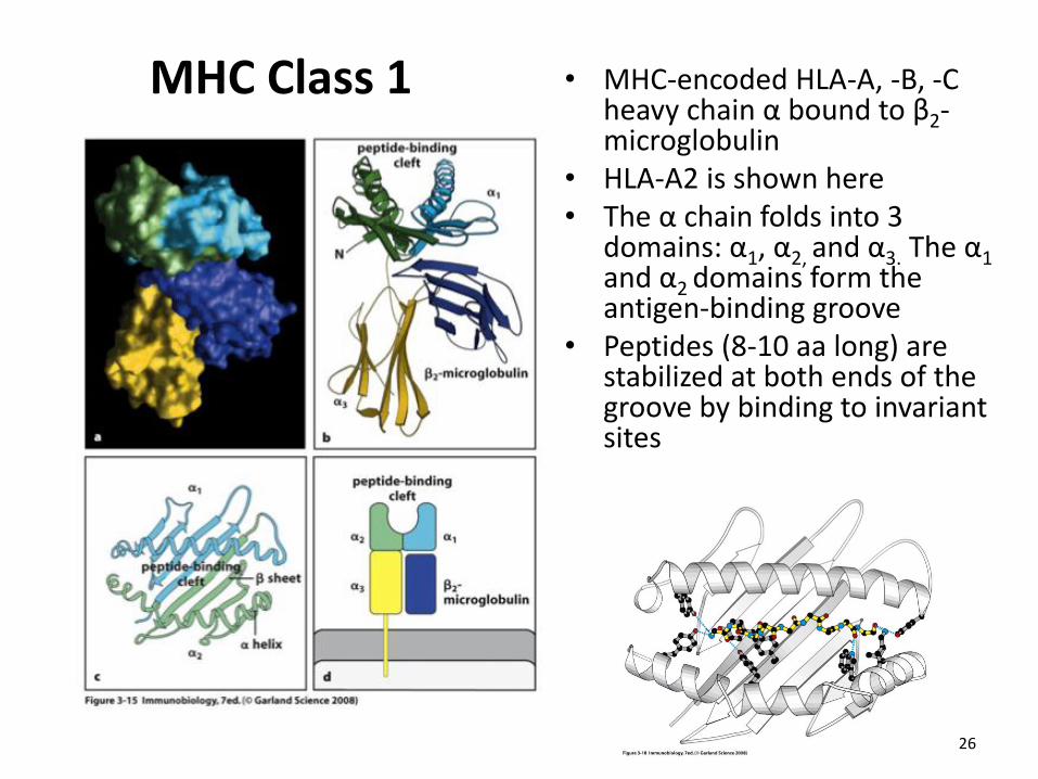
MHC Class 1 • MHC-encoded HLA-A, -B, -C heavy chain α bound to β2-microglobulin
• HLA-A2 is shown here • The α chain folds into 3
domains: α1, α2, and α3. The α1 and α2 domains form the antigen-binding groove
• Peptides (8-10 aa long) are stabilized at both ends of the groove by binding to invariant sites
26
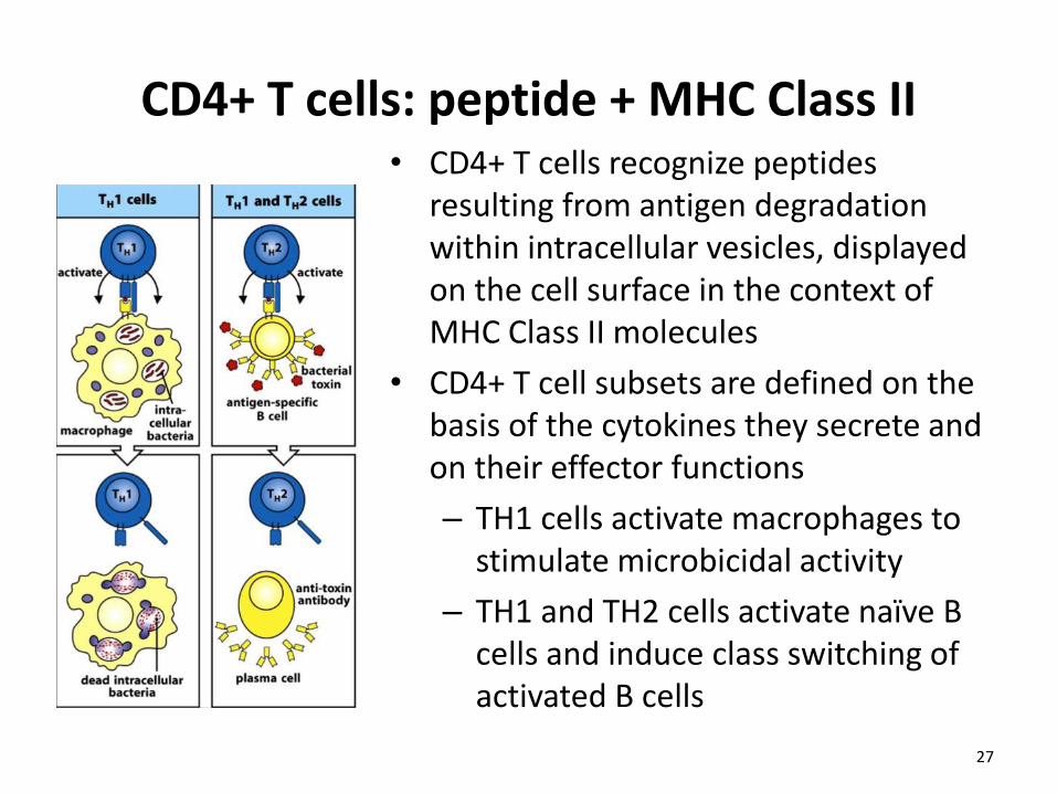
CD4+ T cells: peptide + MHC Class II • CD4+ T cells recognize peptides
resulting from antigen degradation within intracellular vesicles, displayed on the cell surface in the context of MHC Class II molecules
• CD4+ T cell subsets are defined on the basis of the cytokines they secrete and on their effector functions
– TH1 cells activate macrophages to stimulate microbicidal activity
– TH1 and TH2 cells activate naïve B cells and induce class switching of activated B cells
27
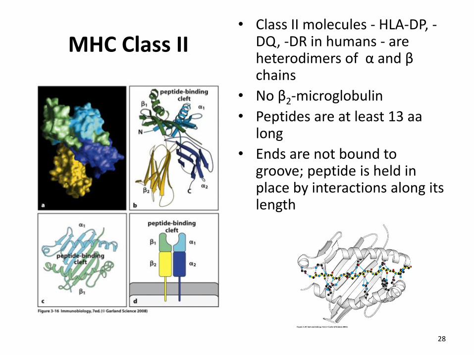
MHC Class II • Class II molecules - HLA-DP, -
DQ, -DR in humans - are heterodimers of α and β chains
• No β2-microglobulin
• Peptides are at least 13 aa long
• Ends are not bound to groove; peptide is held in place by interactions along its length
28