Structure and function of ant (Hymenoptera: Formicidae...
12
Myrmecological News 11 25-36 Vienna, August 2008 Structure and function of ant (Hymenoptera: Formicidae) brains: Strength in numbers Wulfila GRONENBERG Abstract This article reviews the brain of ants in the context of their behavior. Division of labor underlies the social lifestyle of ants; it results not only in behavioral specialization, but also in some adaptations of ant brains. The structure and function of major brain neuropils is described (visual and olfactory centers and central multi-sensory integrative brain compartments) together with some of their neurons. Unlike social vertebrates, which have larger brains and cerebral cortices than soli- tary species, ant brains are not bigger than those of solitary insects, but they are more specialized. The biological suc- cess of ants is probably not so much the result of an individual's brain as of the concerted action of a colony's hundreds or thousands of brains. Key words: Social brain, neuroanatomy, neurophysiology, olfaction, vision, review. Myrmecol. News 11: 25-36 (online 3 May 2008) ISSN 1994-4136 (print), ISSN 1997-3500 (online) Received 7 November 2007; revision received 29 December 2007; accepted 30 December 2007 Prof. Dr. Wulfila Gronenberg, University of Arizona, Arizona Research Laboratories – Division of Neurobiology, 611 Gould-Simpson Building, Tucson, Arizona 85721, USA. E-mail: [email protected] Brains reflect the ants' behavior and environment Ants are insects, and their brains conform to the general design of insect brains, just as human brains reflect their vertebrate, mammalian and primate origins (reviewed by STRIEDTER 2005). Before trying to understand the com- position and function of ant brains we have to briefly re- capitulate what is special about ants, and their behavior in particular. Sociality should affect not only the behavior of ants, but also the brains that generate and control the behavior. In particular, the brain composition might reflect the be- havioral specialization (division of labor), most notably be- tween sexuals and worker castes. Chemical (pheromones) and mechanical communication (vibration, touch) among nestmates are hallmarks of a social lifestyle, and one might expect these sensory modalities and their neural substrate to be well developed in ants. Ant workers of all subfamilies are wingless and even the alate sexuals are poor fliers com- pared to wasps and bees. This obviously shapes the mo- tor output (controlling leg instead of wing movements), but it also affects sensory input: flight relies predominantly on vision and also on information about air-currents, gravity and acceleration, all of which are much less important for terrestrial locomotion. Instead, the sense of touch will play a major role for ant locomotion. For foragers, it is essential not only to locate food re- sources, but also to find their way back home. Trails may be marked by pheromones, but many ants rely on visual navigation (and probably olfactory and magnetic cues), as has been particularly well studied in Cataglyphis desert ants by Rüdiger Wehner and his colleagues (WEHNER 2003). Besides the perception of stimuli, this requires learning and memory abilities allowing ants to remember odors (ROCES 1994, DUPUY & al. 2006) or directions and make use of landmarks. Other kinds of learning are important for nestmate recognition, and generally learning and memory are beneficial in novel or changing environments. One might therefore expect to find ant brains equipped with struc- tures and mechanisms that allow advanced learning and memory to take place. Among Hymenoptera, brain research has mainly fo- cused on honey bees (Apis mellifera), which have become a model system for insect behavioral brain research. The bee brain has been well and accurately described anato- mically more than a century ago by KENYON (1896), and all modern descriptions of ant brains refer to this stan- dard. Historically, ant brains have been repeatedly exa- mined and described since DUJARDIN (1850), but few stud- ies have compared brains of different ant species (e.g., BURLING-THOMPSON 1913, PANDAZIS 1930). Much of what is known about ant brains has been done on larger ants (mainly workers), in particular larger Camponotus spe- cies, because brains are easier to dissect in larger insects. Size is even more important for physiological studies, and this is the reason why there are very few attempts to elec- trically record activity from and response properties of nerve cells (neurons) in ants (JUST & GRONENBERG 1999, RAMON & GRONENBERG 2005, YAMAGATA & al. 2005, 2006). Only two studies have attempted to measure brain activity using recent, advanced optical recording techniques (calcium imaging) in ants (GALIZIA & al. 1999, ZUBE & al. 2008). This review will therefore focus mainly on func- tional brain anatomy. If the function of particular brain components has not been established in ants, it will be in- ferred by homology with other Hymenopterans or insects (e.g., the optic lobes process visual information in any known insect). Ant brains show all the characteristics and components one would expect to find in an insect brain (Fig. 1). The
Transcript of Structure and function of ant (Hymenoptera: Formicidae...

Myrmecological News 11 25-36 Vienna, August 2008
Structure and function of ant (Hymenoptera: Formicidae) brains: Strength in numbers
Wulfila GRONENBERG
Abstract
This article reviews the brain of ants in the context of their behavior. Division of labor underlies the social lifestyle of ants;it results not only in behavioral specialization, but also in some adaptations of ant brains. The structure and function ofmajor brain neuropils is described (visual and olfactory centers and central multi-sensory integrative brain compartments)together with some of their neurons. Unlike social vertebrates, which have larger brains and cerebral cortices than soli-tary species, ant brains are not bigger than those of solitary insects, but they are more specialized. The biological suc-cess of ants is probably not so much the result of an individual's brain as of the concerted action of a colony's hundredsor thousands of brains.
Key words: Social brain, neuroanatomy, neurophysiology, olfaction, vision, review.
Myrmecol. News 11: 25-36 (online 3 May 2008)ISSN 1994-4136 (print), ISSN 1997-3500 (online)
Received 7 November 2007; revision received 29 December 2007; accepted 30 December 2007
Prof. Dr. Wulfila Gronenberg, University of Arizona, Arizona Research Laboratories – Division of Neurobiology, 611Gould-Simpson Building, Tucson, Arizona 85721, USA. E-mail: [email protected]
Brains reflect the ants' behavior and environment
Ants are insects, and their brains conform to the generaldesign of insect brains, just as human brains reflect theirvertebrate, mammalian and primate origins (reviewed bySTRIEDTER 2005). Before trying to understand the com-position and function of ant brains we have to briefly re-capitulate what is special about ants, and their behavior inparticular.
Sociality should affect not only the behavior of ants,but also the brains that generate and control the behavior.In particular, the brain composition might reflect the be-havioral specialization (division of labor), most notably be-tween sexuals and worker castes. Chemical (pheromones)and mechanical communication (vibration, touch) amongnestmates are hallmarks of a social lifestyle, and one mightexpect these sensory modalities and their neural substrateto be well developed in ants. Ant workers of all subfamiliesare wingless and even the alate sexuals are poor fliers com-pared to wasps and bees. This obviously shapes the mo-tor output (controlling leg instead of wing movements), butit also affects sensory input: flight relies predominantly onvision and also on information about air-currents, gravityand acceleration, all of which are much less important forterrestrial locomotion. Instead, the sense of touch will playa major role for ant locomotion.
For foragers, it is essential not only to locate food re-sources, but also to find their way back home. Trails maybe marked by pheromones, but many ants rely on visualnavigation (and probably olfactory and magnetic cues), ashas been particularly well studied in Cataglyphis desert antsby Rüdiger Wehner and his colleagues (WEHNER 2003).Besides the perception of stimuli, this requires learningand memory abilities allowing ants to remember odors(ROCES 1994, DUPUY & al. 2006) or directions and makeuse of landmarks. Other kinds of learning are important for
nestmate recognition, and generally learning and memoryare beneficial in novel or changing environments. One mighttherefore expect to find ant brains equipped with struc-tures and mechanisms that allow advanced learning andmemory to take place.
Among Hymenoptera, brain research has mainly fo-cused on honey bees (Apis mellifera), which have becomea model system for insect behavioral brain research. Thebee brain has been well and accurately described anato-mically more than a century ago by KENYON (1896), andall modern descriptions of ant brains refer to this stan-dard. Historically, ant brains have been repeatedly exa-mined and described since DUJARDIN (1850), but few stud-ies have compared brains of different ant species (e.g.,BURLING-THOMPSON 1913, PANDAZIS 1930). Much of whatis known about ant brains has been done on larger ants(mainly workers), in particular larger Camponotus spe-cies, because brains are easier to dissect in larger insects.Size is even more important for physiological studies, andthis is the reason why there are very few attempts to elec-trically record activity from and response properties ofnerve cells (neurons) in ants (JUST & GRONENBERG 1999,RAMON & GRONENBERG 2005, YAMAGATA & al. 2005,2006). Only two studies have attempted to measure brainactivity using recent, advanced optical recording techniques(calcium imaging) in ants (GALIZIA & al. 1999, ZUBE & al.2008). This review will therefore focus mainly on func-tional brain anatomy. If the function of particular braincomponents has not been established in ants, it will be in-ferred by homology with other Hymenopterans or insects(e.g., the optic lobes process visual information in anyknown insect).
Ant brains show all the characteristics and componentsone would expect to find in an insect brain (Fig. 1). The

26
Fig. 1: Brain of a paperwasp Polistes dominulus (top) andworker of the ponerine ant Pachycondyla villosa (bottom);microtome sections (15 m) of osmium-stained material;antennal lobe (al), mushroom body calyx (ca) and lobes(mbl), central body (cb), lamina (la), medulla (me), lobula(lo), protocerebral lobe (pc), median ocellus (oc).
major brain compartments are labeled in Figure 1 for apaper wasp (Polistes dominulus) and a relatively basal ant(Pachycondyla villosa) and will be discussed below: theoptic lobes (visual centers) lamina, medulla and lobula (la-mina not shown for the ant in Figure 1 because it oftendetaches from the brain during dissection); the primary ol-factory center (antennal lobe), the protocerebral lobes, thecentral body, and the mushroom body, which comprisesthe calyx and the mushroom body lobes. In ants and otherholometabolous insects, the subesophageal ganglion (Fig.2a) is fused to the brain posteriorly. This ganglion is or-ganized like a "typical" insect ganglion (e.g., thoracicganglia) and is thus less complex than the brain properand differs little among insects; it will therefore not befurther considered in this review. While the sizes of thedifferent brain components differ in ants and closely re-lated vespid wasps (Fig. 1), the only structures that are ab-sent in ants are the ocelli and their associated neurons asmost ant workers do not have functional ocelli. Overall, thebrain of Pachycondyla (Fig. 1) is typical for ants, althoughthere are minor differences between subfamilies and be-tween species with different ecological adaptations (e.g.,visual predators vs. seed harvesters) and species (or castes)with small body or head size.
The most obvious difference between brains of waspsand ants is in the size of the optic lobes, which are verysmall in ants in general, although there are some excep-tions (see below). In contrast, the antennal lobes and theprotocerebrum and its components appear similar in sizecompared to the wasp brain (Fig. 1). There is also a pro-nounced difference in overall brain size across ants; how-ever, as brain size in all animals (JERISON 1973), includingants (WEHNER & al. 2007), generally correlates with bodysize, one would expect the ant to have a smaller brain thanthe larger paper wasp.
The sense of smell and olfactory processing
Insect antennae are appendages that carry many sensillathat sense mechanical stimuli (touch, vibrations) and, in al-most all advanced insects, and in ants in particular, odorstimuli. Other antennal sensilla perceive stimulus modali-ties different from "ordinary" odors: some respond to chem-ical stimuli that one would refer to as taste, e.g., salty, bit-ter or sweet substances; hygroreceptors respond to humi-dity, thermoreceptors signal temperature changes, and spe-cialized receptors inform the ant about the concentrationof carbon dioxide (KLEINEIDAM & al. 2000). All these sen-silla are hair-like structures or modified and derived fromhair-like structures and contain one or more sensory neu-rons. The majority of sensilla on the antenna contain ol-factory neurons that respond to particular subsets of odo-rants or chemical compounds.
Importantly, antennae are extremities that can be movedaround to scan a larger volume of air or to probe struc-tures, crevices, trails or other insects including nestmatesfor chemical and tactile cues. To perform antennal move-ments, antennae are equipped with sets of muscles insidethe head capsule and others inside the antenna's basal seg-ment, the scape. All of these muscles are controlled bymotor neurons that reside in a brain region behind (poste-rior to) the antennal lobe, referred to as dorsal lobe. Thispart of the brain also receives mechanosensory input fromthe antenna (EHMER & GRONENBERG 1997), hence anten-nal movement and the tactile input resulting from thatmovement can be integrated together by the dorsal lobe.
Each olfactory receptor neuron (c. 50,000 - 60,000 inFormica pratensis, GOLL 1967) in the antenna sends anaxon, a long nerve fiber, through the antennal nerve intothe antennal lobe of the brain, where the axons terminateon and interact with secondary neurons (interneurons). An-tennal lobes are particularly large in ants for two reasons:firstly, ants strongly rely on olfaction, more so than manyother insects (which have more elaborate visual behavi-ors); and secondly, pheromone communication is most ad-vanced and elaborate in ants compared to any other groupof social insects (let alone solitary insects). The antennallobes process both, "ordinary" odors and pheromones. Ol-factory axons terminate in the antennal lobe in globularstructures, called glomeruli (Fig. 2b). The sensory neuronssupplying the glomeruli can be traced by mass-filling theantennal nerve, which reveals all the glomeruli (Fig. 2b).
Homology with Drosophila suggests that each glome-rulus receives input from only one particular kind of sen-sory neuron (but many individual neurons of that kind)which in turn expresses one specific kind of odor receptormolecule at their receptive site in the antennal sensillum.Thus, one antennal lobe glomerulus might correspond toone particular kind of odorant receptor molecule and reco-gnize a subset of odor molecules out of an almost infinitenumber of chemical compounds (VOSSHALL & al. 2000;reviewed for insects and vertebrates by KORSCHING 2001).The sensitivity and specificity of olfactory sensory neuronsmay vary (DUMPERT 1972).
This arrangement between sensory neurons and anten-nal lobe glomeruli suggests that, in general, the numberof glomeruli in an insect correlates with the number ofodors that can be discriminated, or with the precision thatodors can be distinguished. While few studies have com-pared the number of glomeruli across taxa, it appears that

27
Fig. 2: Heads, brains and antennal lobes of workers of the formicine ant Camponotus rufipes (a, b) and the dacetine antsDaceton armigerum (c, d) and Strumigenys sp. (e, f); horizontal (a) and frontal (vertical) sections (b - f); reduced silver(a, d), osmium (f) and cobalt chloride stain (b). Areas boxed in a enlarged in b. Antennal lobe (al), antennal nerve (an),mushroom body calyx (ca), subesophageal ganglion (sog), directions anterior (ant), dorsal (do), and lateral (lat). Scale bars= 200 µm.
ants may have a particularly high number of glomeruli(c. 430 in Camponotus japonicus, NISHIKAWA & al. inpress; 415 ± 14 in Camponotus floridanus, ZUBE & al.2008; c. 200 in Formica pratensis, GOLL 1967). This com-pares to smaller numbers established for other insects: 43in the fruit fly Drosophila melanogaster (STOCKER 1994),
63 in the Sphinx moth Manduca sexta (ROSPARS & HILDE-BRAND 2000), 109 in the cockroach Blaberus craniifer(CHAMBILLE & al. 1980), and c. 160 in honey bee workers(FLANAGAN & MERCER 1989). Body size does not seemto be much correlated with the number of glomeruli: thesmall ant Strumigenys sp. (Figs. 2e, f) has considerably

28
Fig. 3: Olfactory projection neuron recorded from andstained in the brain of a carpenter ant (a). It receives in-put in a single large glomerulus (glo) in the antennal lobe(al) and projects (sends output) to the mushroom body ca-lyx (ca) and the lateral protocerebrum (lp). It responds tothe alarm pheromone component formic acid (stimulus dur-ation: blue bar) at higher (b) and lower concentrations (c),suggesting that the large glomerulus in (a) processes alarmpheromone information. Medulla (me); modified after YA-MAGATA & al. (2006).
more glomeruli (c. 220) than the larger (and socially moreadvanced) ant Daceton armigerum (c. 120 glomeruli, Figs.2c, d).
Information provided to the individual glomeruli fromthe odor receptor neurons on the antenna is integrated bylocal interneurons in the antennal lobe. These neurons arecalled "local" because their processes (input and outputbranches) are restricted to the antennal lobe. Many localinterneurons have inhibitory responses and are supposed tosharpen the contrast of the perceived odorants (HILDE-
BRAND & SHEPHERD 1997). Olfactory information pro-cessed by the antennal lobe is transferred to "higher" braincenters [the mushroom bodies (see below) and the lateralprotocerebrum] by projection neurons (Fig. 3), which inter-act with single or multiple glomeruli. Information aboutcomplex odor mixtures, concentrations and temporal prop-erties of odor plumes is assumed to be coded by the sim-ultaneous activity of many parallel projection neurons andthe correlation and synchronicity of their action potentials[the smallest units of information in nerve fibers, in a waycomparable to the binary digits (bits) in a computer line].Properties of olfactory projection neurons have been de-scribed in other insects (reviewed by HANSSON 2002) andrecently in carpenter ants (Fig. 3, YAMAGATA & al. 2006).
Much is known about pheromone communication inants regarding the behavior, the glands and the chemicalcompounds involved (reviewed in HÖLLDOBLER & WIL-SON 1990), but little research has been done on the neuralprocessing of pheromone information. In principle the per-ception and processing of pheromone information does notdiffer much from that of other odorants. Many insects havespecialized glomeruli that process the different componentsof sex pheromones; usually, the number of specializedglomeruli reflects the number of chemical compounds thatconstitute the pheromone blend (reviewed for insects byHANSSON 2002). This has recently also been shown for car-penter ants (NISHIKAWA & al. in press). Different casteshave different thresholds for particular odorants (LOPEZ-RIQUELME & al. 2006), which probably results from dif-ferences in the number of receptor neurons for particularodorants. However, it also results in differences in the cen-tral processing: a glomerulus that receives fewer input neu-rons from the antenna will be smaller than one that receivesmore input neurons. Thus two species of leafcutting antsthat differ in their sensitivity to particular trail pheromonecompounds also differ in the size of the respective as-sumed pheromone sensitive glomeruli (KLEINEIDAM & al.2005).
Nest odors are used as recognition cues for nestmatesand in that respect they are similar to pheromones. Theyare colony-specific complex mixtures of cuticular hydro-carbons, and like multi-component pheromones they haveto be decoded by the olfactory system. It is unlikely thatparticular neuronal specializations are required to analyzenest odors – the task does not seem much different fromanalyzing the complex bouquets of odorants emanatingfrom food items.
Ant vision: from nonexistent to highly acute
Ants in general are not particularly visual animals. Thisis reflected by the small size or even absence of eyes inmany ant species that rely on tactile and chemosensory or-ientation. The extinct primitive ant genus Sphecomyrmaand extant basal genera Nothomyrmecia and Myrmeciahave relatively large eyes, suggesting that these predatorsrely on vision, as do some more advanced genera in dif-ferent subfamilies (e.g., Harpegnathos, Gigantiops, Myr-moteras and Pseudomyrmecinae genera).
Like other insects, male and female ants have threeocelli (simple eyes, Figs. 4b, c), but most ant workers donot have ocelli (Fig. 4a, exceptions are pronouncedly visu-al ant genera such as Myrmecia, Harpegnathos and Gigan-tiops). More important are the compound eyes, which are
29
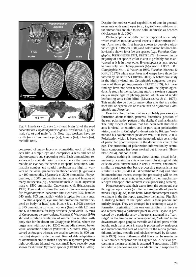
Fig. 4: Heads (a - c), eyes (d - f) and brain (g) of the seedharvester ant Pogonomyrmex rugosus: worker (a, d, g), fe-male (b, e) and male (c, f). Note that workers have noocelli (oc). Compound eye (ey), lamina (la), lobula (lo),medulla (me).
composed of many facets or ommatidia, each of whichacts like a simple eye and comprises a lens and set ofphotoreceptors and supporting cells. Each ommatidium re-solves only a single point in space, hence the more om-matidia an eye has, the better is its spatial resolution. Om-matidia number and spatial resolution are high in wor-kers of the visual predators mentioned above (Gigantiopsc. 4100 ommatidia, Myrmecia c. 3200 ommatidia, Harpe-gnathos, c. 1600 ommatidia) and in males and females ofmany ant species (e.g., Ectatomma male c. 1400, Mystriummale c. 1500 ommatidia, GRONENBERG & HÖLLDOBLER
1999). Figures 4d - f show the caste differences in eye sizefor Pogonomyrmex harvester ants (worker: 395, female:640, male: 803 ommatidia), which is typical for many ants.
Within a species, eye size and ommatidia number de-pend on body (or head) size. KLOTZ & al. (1992) describec. 375 ommatidia for small workers (head width 1.2 mm)and 658 ommatidia for large workers (head width 3.5 mm)of Camponotus pennsylvanicus. MENZEL & WEHNER (1970)showed similar correlation of ommatidia number withbody size for the desert ant Cataglyphis bicolor, where thelarger ants with more (c. 1200) ommatidia showed bettervisual orientation abilities (WEHNER & MENZEL 1969) andserved as foragers whereas the smaller workers (c. 600 om-matidia) stayed inside the nest. Remarkable adaptationsof ommatidial size to match a species' respective preferredlight conditions (diurnal vs. nocturnal) have recently beenshown for different Myrmecia species (GREINER & al. 2007).
Despite the modest visual capabilities of ants in general,even ants with small eyes (e.g., Leptothorax albipennis;60 ommatidia) are able to use bold landmarks as beacons(MCLEMAN & al. 2002).
Photoreceptors can differ in their spectral sensitivity,which enables most advanced insects to discriminate col-ors. Ants were the first insects shown to perceive ultra-violet light (LUBBOCK 1881) and color vision has been be-haviorally shown for a few ant species (e.g., Formica, Cata-glyphis, KIEPENHEUER 1971, KRETZ 1979). However, in themajority of ant species color vision is probably not as ad-vanced as it is in most other Hymenoptera as ants appearto have only two photopigments (Myrmecia: LIEKE 1981,Cataglyphis: MOTE & WEHNER 1980, Formica: MENZEL &KNAUT 1973) while most bees and wasps have three (re-viewed by BRISCOE & CHITTKA 2001). A behavioral studyin the highly visual ant Cataglyphis suggested the pre-sence of three photopigments (KRETZ 1979), but thesefindings have not been reconciled with the physiologicaldata. A study in the leafcutting ant Atta sexdens suggestsonly a single type of photopigment, which would renderleafcutting ants color blind (MARTINOYA & al. 1975).This might also be true for many other ants that are eithernocturnal or depend less on vision than do Myrmecia, Cata-glyphis and Formica.
Besides color, the brain of ants probably processes in-formation about motion, patterns, directions (position ofthe sun, polarization pattern of the skylight) and landmarks.The only aspect of vision that has been well studied inants is visual orientation, and in particular polarizationvision, mainly in Cataglyphis desert ants by Rüdiger Weh-ner and his collaborators (reviews: WEHNER 1994, 2003).Polarization vision is based on the polarization sensitivityof particular photoreceptors in the dorsal rim area of theeye. The processing of polarization information by centralbrain components has been worked out in locusts (HOM-BERG 2004), but not in ants.
Almost nothing is known about central visual infor-mation processing in ants – no neurophysiological dataexist on visual interneurons in ants. However, anatomicalsimilarity suggests that basic processing mechanisms aresimilar in ants (EHMER & GRONENBERG 2004) and otherholometabolous insects, except that processing will be lesssophisticated in most ants, as indicated by their much smal-ler eyes and optic lobes (central visual processing centers).
Photoreceptors send their axons from the compound eyethrough an optic nerve (or often a loose bundle of parallelnerves, Figs. 4g, 5a) to the brain. Most photoreceptor axonsterminate in the first optic ganglion (the lamina, Fig. 5a).A striking feature of the optic lobes is their precise andorderly design. They are arranged in a retinotopic way: in-formation originating from one ommatidium in the eyeand representing a particular point in visual space is pro-cessed by a particular array of neurons arranged in a "cart-ridge" in the lamina and a corresponding "column" in thedownstream optic ganglia medulla and distal parts of thelobula, such that a point in space is processed by analogousand interconnected sets of neurons in the retina (omma-tidium), lamina, medulla and lobula (reviewed by STRAUS-FELD 1989). Some of these parallel fibers can be seen in thesilver-stained preparations in Figures 5b, c. Visual pro-cessing in the insect lamina is assumed (STRAUSFELD 1989)to underlie phenomena such as adaptation in response to

30
Fig. 5: Brain and optic lobes of the harvester ant Pogono-myrmex rugosus. Males (a, b) have larger optic lobes thanworkers (c). Bodian stained sections show the regular (re-tinotopic) arrangement of fibers in particular in the outermedulla. Area boxed in (a) is enlarged in (b) and (c). An-tennal lobe (al), compound eye (ey), lamina (la), lobula (lo),medulla (me), mushroom body (mb).
changing light intensities, summation (which increases lightsensitivity but reduces spatial resolution), enhancement ofsignal-to-noise ratio, and lateral inhibition (which locallyenhances the contrast of the perceived image). Lamina neu-rons proceed to the medulla, where they contact many dif-ferent classes of local interneurons (STRAUSFELD 1976)as well as lateral (tangential) neurons that connect severalmedulla "columns" and thus process information acrosspoints in space. Both types of neurons can be seen in themedulla of male and female harvester ants (Figs. 5b, c). Oneof the many functions of the medulla is to process colorinformation. A second and parallel function of the medullaprobably is to extract motion information from the visualinput, in particular local or small-field motion events. How-ever, the large number of types of neurons comprising themedulla, as well as its size (it is the largest of the opticlobes) suggests that it is involved in many different andcomplex kinds of visual processing (reviewed by STRAUS-FELD 1989).
The retinotopic organization is rendered more coarselyin the third optic ganglion, the lobula, where each columnnow integrates information originating from several omma-tidia, or sampling points (STRAUSFELD 1989). In the lob-ula, wide-field neurons integrate motion information overlarge parts of the visual field. Such wide-field or panor-amic motion is important for an animal's movement controlin space and has been intensively studied in flies (reviewedby BORST & HAAG 2002). The lobula comprises large- andsmall-field neurons and probably represents both, color andmotion information, but otherwise little is known about thekinds of information processing performed in the lobula or
Fig. 6: (a) The mushroom bodies comprise the calyx (ca)and the mushroom body lobes and are composed of thou-sands of Kenyon cells (a single one shown in red) whichreceive input in the calyx. Their y-shaped axons project toboth, the vertical (vl) and medial lobe (ml). (b) The calyxis composed of cup-shaped neuropils (np, red) around whichthe small cell bodies (cb) of the Kenyon cells are arranged(green); part of the Kenyon cell body layer is removed toreveal the neuropil. (c) The calyx is usually subdividedinto a lip and a collar (co) region and most Kenyon cells re-ceive input in one or the other region. In addition, in ants aclass of Kenyon cells (blue) exists that probe both regions,lip and collar, with their dendrites. (d) The collar regionreceives visual input (green) originating from the medulla(me) and the lobula (lo) (input from lobula not shown). Ap-proximate areas boxed in (a) are enlarged in (b) and (c),respectively; antennal lobe (al). Scale bar = 250 µm.
the medulla. Output neurons from both, the medulla andthe lobula project to "optic foci" in the central brain and todescending interneurons which in turn project from thebrain to the thorax where they control movement (walk-ing; flight in alates) that are guided by visual input. Part ofthe visual information is also sent to the mushroom bod-ies, central brain structures involved in learning and mem-ory and other "advanced" neuronal processing, which willbe described in the next section.

31
Mushroom bodies: central control of advancedbehavior
The mushroom bodies are central brain structures found inalmost all insects (STRAUSFELD & al. 1998). They werefirst described from bees by Felix DUJARDIN (1850), whosuggested them to be involved in "intelligent" control ofbehavior (as opposed to instincts) and their size to corre-late with the degree of a species' social organization. Thissuggests that they should be important structures in theant brain. The idea that mushroom bodies may be involvedin learning and memory originates from lesion experimentsin the brains of wood ant (Formica) workers and the result-ing deficiency in negotiating a maze using olfactory cues(VOWLES 1964). Later, mushroom bodies have become keymodel systems in invertebrate learning and memory re-search, in particular in honeybees (ERBER & al. 1980) andin fruit flies (HEISENBERG & al. 1985). The mushroom bod-ies are particularly large in ant workers (Fig. 6a), their re-lative size (compared to the overall brain volume) beingmore than twice that found in honey bees (GRONENBERG
& HÖLLDOBLER 1999) whereas male ants have relativelysmall mushroom bodies (compare Figs. 7a, b). This cor-relates well with the general idea that worker ants rely onbehavioral plasticity (e.g., finding and remembering newfood sources or adapting to changing environments) whilemale behavior appears more "hard-wired" or pre-program-med. The larger mushroom bodies of ant workers assumed-ly endow them with increased learning and memory andother cognitive abilities.
Mushroom bodies are composed of many thousands(Fig. 6b) of particularly small neurons (globuli cells orKenyon cells; c. 130,000 in Camponotus rufipes, EHMER &GRONENBERG 2004). All these Kenyon cells have their den-drites in the calyx neuropil and send their axons in parallelto the vertical and medial lobes of the mushroom body(Fig. 6a). Most Kenyon cells have a y-shaped, bifurcatingaxon and together, these thousands of parallel axons shapethe mushroom body lobes (indicated by the sketch of aKenyon cell in Fig. 6a). Kenyon cells receive input at theirdendrites in the calyx (Fig. 6a), which comprises a so-called lip and a collar region (Fig. 6c). Kenyon cells eitherreceive olfactory input in the calyx' lip region (e.g., fromolfactory neurons such as shown in Fig. 3) or visual inputin the calyx' collar region (Fig. 6d, EHMER & GRONENBERG
2004). Some Kenyon cells have dendritic arborizations inboth calyx regions (lip and collar, Fig. 6c) and are there-fore assumed to combine visual and olfactory input infor-mation. Such "bimodal" Kenyon cells have not been foundin insects other than ants (EHMER & GRONENBERG 2004).
In the calyx, visual and olfactory input neurons termi-nate in presynaptic structures referred to as "boutons", a-round which postsynaptic (receiving) elements ("spines")are arranged. Together, these miniature output/input struc-tures form little spherical elements and give the calyx tis-sue a "microglomerular" texture in the light microscopicimage. These microglomeruli have diameters of 1 - 5 min different ant species (SEID & al. 2005, SEID & WEH-NER 2007). In the calyx' visual (collar) region, the vol-ume of individual microglomeruli and associated synapsesis only about 1/3 that of microglomeruli in the olfactory(lip) region (SEID & WEHNER 2007), which the authors(SEID & WEHNER 2007) interpret as resulting in a more
reliable transmission of olfactory information compared tovisual information.
The olfactory (Fig. 3) or visual input neurons (Fig. 6d)are referred to as extrinsic neurons because their cell bod-ies and major processes reside outside of the mushroombodies. Extrinsic neurons provide input to the Kenyon cellsor gather output from them, respectively. Besides inputto the calyx, other extrinsic neurons sample the lattice ofparallel Kenyon cell axons in the mushroom body lobeswith their comb or brush-like dendritic trees (or axonal ar-borizations in the case of input neurons), receiving simul-taneous input from (or sending output to) many hundreds ofKenyon cells. A hypothesis based on research in locustsand honeybees suggests that each Kenyon cell processeshighly specific information, e.g., responding only to a par-ticular stimulus combination and / or temporal coincidence,and even then only with a few action potentials ("sparsecoding", PEREZ-ORIVE & al. 2002, SZYSZKA & al. 2005).Mushroom body output neurons could therefore integratevery complex spatio-temporal sensory conditions, and elec-trophysiology in honeybees and cockroaches revealed com-plex response properties that often change over time andas a consequence of previous stimuli, hence representingphysiological mechanisms thought to underlie behavioralplasticity, learning and memory (MAUELSHAGEN 1993, LI
& STRAUSFELD 1999). Two recent studies show complexresponse properties for ant mushroom body neurons com-parable to those found in other insects, but only olfactorystimuli have been tested in those studies (YAMAGATA & al.2005, 2007).
Many ants who mainly rely on chemical and tactile cuesreceive mainly olfactory (and probably tactile) input totheir mushroom bodies while species that also rely on vi-sion for orientation purposes have larger visual input are-as in their mushroom body calyces (e.g., Formica woodants, Myrmecocustus honey ants or Cataglyphis desert ants,KÜHN-BÜHLMANN & WEHNER 2006). These genera areknown to use visual landmarks and other forms of visualorientation cues. This supports the idea that the mushroombodies play a central role in memory-based orientation abi-lities. This idea is also supported by findings that the mush-room body size, and in particular the calyx, increases inforagers when compared to nest workers of the same size.This has first been examined in honeybees (WITHERS & al.1993) and subsequently shown for Camponotus floridanus(GRONENBERG & al. 1996) and for Cataglyphis bicolor(KÜHN-BÜHLMANN & WEHNER 2006). While mushroombodies are a current "hot topic" in insect neuroscience andbeyond, and even though in ants the mushroom bodies arelarge, ants contribute little to this field as they are neithermolecular nor behavioral "model systems".
The central complex
The central complex comprises a group of unpaired struc-tures in the central brain of arthropods: the central body(upper and lower division; also called fan-shaped body andellipsoid body, respectively), the protocerebral bridge andthe paired noduli. The central body is composed of eight or16 subunits that are arranged in a fan-shaped fashion andinterconnected by a complex arrangement of chiasmata(STRAUSFELD 1975, LOESEL & al. 2002). Some research-ers suggest the central complex to be a major center forprocessing of polarized light information where the 16 sub-

32
units represent the spatial organization of polarizatione-vectors (based on evidence from locusts, HOMBERG 2004,HEINZE & HOMBERG 2007). Evidence from fruit fly mu-tants affecting the central complex led other researchers tosuggest that it is involved in limb coordination and walk-ing control (STRAUSFELD 1999, STRAUSS 2002). The cen-tral complex communicates with other central neuropils andis (anatomically) in a position to integrate all sensory mo-dalities and modulate ongoing motor commands sent "down"to the legs or wings. The fact that the central body com-prises sets of neurons each containing different neuromo-dulators (LOESEL & al. 2002) suggests that it may be in-volved in controlling and switching entire suites of behavi-ors. Little is known about the central complex of ants in par-ticular and it is likely that in ants it plays analogous rolesin organizing behavior as it does in locusts or fruit flies. Itlooks similar to the central complex of paper wasps andgeneral staining reveals eight major subdivisions of the up-per and lower division of the central body (Figs. 7c, d),which, upon closer inspection, appear to be subdivided intotwo parts, as is the case in most other insects. The centralbody is a structure found in all ants (including blind spe-cies such as in the genus Mystrium), hence visual proces-sing cannot be the sole function it serves. The central bodyis of similar relative size in those ant species where it hasbeen examined. In many ant species, it appears to be rela-tively larger in males than it is in workers (Figs. 7c, d), justthe opposite as is the case for mushroom bodies (Figs. 7a, b)or antennal lobes. As ant males in general are more "hardwired" or "pre-programmed" behaviorally, the large size ofmale central bodies would suggest that the central complexdoes not contribute to behavioral plasticity (in contrast tothe mushroom bodies) but instead supports some behavi-oral functions that do not require learning (such as switch-ing between behavioral contexts). Polarization vision or legcontrol would both fit this description.
Brain size and plasticity in social insects
A few common rules or notions come into play when con-sidering brain size in animals. These are generally based onvertebrates and are discussed in detail in reviews on brainevolution such as ROTH & DICKE (2005) or STRIEDTER
(2005). In related taxa, brain size correlates with body size(JERISON 1973). This appears to be generally true for ants,too, and has been shown for several species of Cataglyphisdesert ants (WEHNER & al. 2007). On average, larger antshave larger brains. However, like vertebrates, smaller in-sects have relatively larger brains when compared to theirbody or head size. This is evident when comparing a largeand small worker of the same colony of leaf-cutting antsAtta (Figs. 8b, c). In the large soldier, the brain is just a smallstructure in the center of the large head capsule that ismainly filled by mandible muscle (Fig. 8b). In contrast, inthe small minor worker (Fig. 8c), most of the head volumeis taken up by the brain, which is smaller in absolute termsthan that of the large soldier (Figs. 8e, f). This same re-lationship is also found across taxa where larger ants havemore muscle inside their larger heads.Brains can be miniaturized up to a certain limit, as in-dicated by some comparative brain volume data: large bum-blebee (head width c. 4.6 mm): c. 3 mm3, honeybee (headwidth c. 3.6 mm): c. 1 mm3 (MARES & al. 2005); carpenterants Camponotus (different species; head widths c. 1.3 -
Fig. 7: Brain and central body of a male (a, c) and worker(b, d) of Ectatomma ruidum. Note the overall smaller brain,larger optic lobes medulla (me) and lobula (lo), and largercentral body (cb) in males. Area boxed in (a, b) enlarged in(c, d), respectively. Mushroom body calyx (ca) and mediallobe (ml); central body's upper (cbu) and lower division(cbl).
2.4 mm): 0.15 - 0.03 mm3, seed harvester ant Pogono-myrmex rugosus worker (head widths c. 1.3 - 2.6 mm): c.0.08 mm3; Acantognathus (head width c. 0.6 mm): 0.01 mm3,Strumigenys (head width c. 0.36 mm): 0.004 mm3. As wehave seen (Figs. 2f, g), brain size can be reduced geome-trically, but not infinitely, leading to small brains with es-sentially the same composition as large brains. Space isseverely limited in small ants and brains probably requirea minimum size in order to not lose essential behavioralcapabilities or support a given behavioral repertoire.
Unlike solitary animals, in ants brain size cannot be con-clusively discussed at the species level, but has to take intoaccount the different castes and worker specialization.The most intuitive correlation is between brain size and ananimal's behavioral sophistication or repertoire, becausethis is the purpose of any brain: to generate and controlbehavior. Generally, males have smaller brains (Figs. 5, 7)than females or workers, and their behavioral repertoiresare reduced as males generally do not contribute much tomany of the colony's tasks and in particular do not forage.Workers generally have smaller optic lobes and larger an-tennal lobes than males (e.g., in the ponerine ant Harpe-gnathos saltator; HOYER & al. 2005). Likewise, Campo-notus japonicus workers have c. 430 antennal lobe glome-
33
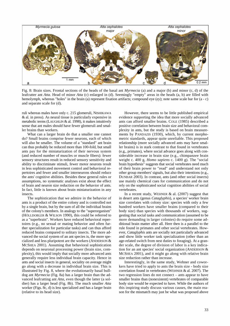
Fig. 8: Brain sizes. Frontal sections of the heads of the basal ant Myrmecia (a) and a major (b) and minor (c, d) of theleafcutter ant Atta. Head of minor Atta (c) enlarged in (d). Seemingly "empty" areas in the heads (a, b) are filled withhemolymph, whereas "holes" in the brain (a) represent fixation artifacts; compound eye (ey); note same scale bar for (a - c)and separate scale for (d).
ruli whereas males have only c. 215 glomeruli, NISHIKAWA
& al. in press). As neural tissue is particularly expensive inmetabolic terms (LAUGHLIN & al. 1998), it makes intuitivelysense that ant males should have fewer glomeruli and smal-ler brains than workers.
What can a larger brain do that a smaller one cannotdo? Small brains comprise fewer neurons, each of whichwill also be smaller. The volume of a "standard" ant braincan thus probably be reduced more than 100-fold, but smallants pay for the miniaturization of their nervous system(and reduced number of muscles or muscle fibers): fewersensory structures result in reduced sensory sensitivity andability to discriminate stimuli, fewer motor neurons resultin less sophisticated movement control and behavioral re-pertoires and fewer and smaller interneurons should reducethe ants' cognitive abilities. Besides these general rules orassumptions, no systematic analyses exist about the effectof brain and neuron size reduction on the behavior of ants.In fact, little is known about brain miniaturization in anyinsects.
The sophistication that we admire in the behavior ofants is a product of the entire colony and is controlled notby a single brain, but by the sum of all the individual brainsof the colony's members. In analogy to the "superorganism"(HÖLLDOBLER & WILSON 1990), this could be referred toas a "superbrain". Workers have reduced behavioral reper-toires (e.g., no sexual or mating behavior and often fur-ther specialization for particular tasks) and can thus affordreduced brains compared to solitary insects. The more ad-vanced the social system of an ant species is, the more spe-cialized and less pluripotent are the workers (ANDERSON &MCSHEA 2001). Assuming that behavioral sophisticationdepends on neuronal processing power (brain size, com-plexity), this would imply that socially more advanced antsgenerally require less individual brain capacity. Hence inants and social insects in general, sociality on average mightgo along with a decrease in individual brain size. This isillustrated by Fig. 8, where the evolutionarily basal bull-dog ant Myrmecia (Fig. 8a) has a larger brain than the ad-vanced leafcutting ant Atta, even though the latter (a sol-dier) has a larger head (Fig. 8b). The much smaller Attaworker (Figs. 8c, d) is less specialized and has a larger brain(relative to its head size).
However, there seems to be little published empiricalevidence supporting the idea that more socially advancedants can afford smaller brains. COLE (1985) described apositive correlation between brain size and behavioral com-plexity in ants, but the study is based on brain measure-ments by PANDAZIS (1930), which, by current morpho-metric standards, appear quite unreliable. This proposedrelationship (more socially advanced ants may have smal-ler brains) is in stark contrast to that found in vertebrates(e.g., primates), where social advance goes along with con-siderable increase in brain size (e.g., chimpanzee brainweight c. 400 g, Homo sapiens c. 1400 g). The "socialbrain hypothesis" suggests that social vertebrates need muchof their brain power to "read" and understand not onlyother group members' signals, but also their intentions (e.g.,DUNBAR 2003). In contrast, ants (and other social insects)use mainly chemical cues for communication and do notrely on the sophisticated social cognition abilities of socialvertebrates.
In a recent study, WEHNER & al. (2007) suggest thatin desert ants (genus Cataglyphis), a species' worker brainsize correlates with colony size: species with only a fewhundred workers have smaller brains (compared to theirbody size) than species with thousands of workers, sug-gesting that social tasks and communication (assumed to bemore demanding in larger colonies) do require some ad-ditional brain matter after all. Here then is a parallel to therule found in primates and other social vertebrates. How-ever, Cataglyphis ants are socially not particularly advancedand show little worker task specialization (other than anage-related switch from nest duties to foraging). At a gran-der scale, the degree of division of labor is a key indica-tion for an ant species' social organization (ANDERSON &MCSHEA 2001), and it might go along with relative brainsize reduction rather than increase.
Interestingly, in the same study, Wehner and cowor-kers have tried to apply to ants the brain size - body sizecorrelation found in vertebrates (WEHNER & al. 2007). Thetwo regression lines do not connect – ants appear to havesmaller brains than (nonexistent) vertebrates of comparablebody size would be expected to have. While the authors ofthis inspiring study discuss various causes, the main rea-son for the mismatch may just be what was proposed above

34
– that ants are social and the more socially advanced theyare, the smaller their brains might be. Perhaps one shouldrepeat the study (WEHNER & al. 2007) using flies, beetlesor locusts and one might conceivably find that the generaltrend found for vertebrates holds for (solitary) insects, too.
The reason for the proposed reduction of brain size inevolutionary terms is that nervous tissue is metabolicallyparticularly expensive to maintain (LAUGHLIN & al. 1998);hence, particular brain parts or neurons will be lost duringevolution if they are no longer required. The same is truefor an individual's life time, where neurons expand or arepruned and brains or brain components can shrink in vol-ume. This has been most prominently shown in male songbirds, where brain structures involved in song control shrinkafter the mating (singing) season and may expand in ad-vance of the next mating season (NOTTEBOHM 1981). Simi-lar brain plasticity has been shown in honeybees (WITHERS
& al. 1993) and in ants (GRONENBERG & al. 1996, KÜHN-BÜHLMANN & WEHNER 2006), in which the mushroombodies increase in size during the workers' transition fromworking inside the nest to becoming outside foragers, a taskthat is more demanding with respect to cognitive abilities.Besides experience, mushroom bodies also change in size inan age-dependent or "experience expectant" manner (FAHR-BACH & al. 1998). Complex experiences, such as are asso-ciated with foraging activity, probably give rise to the mo-dification, growth and addition of many synapses (pointsof information transfer between nerve cells), which in turnpresumably result in the observed changes of brain volume.
In contrast to this size increase, the entire brain, and theoptic lobes in particular, shrink in female ants once theyare mated (JULIAN & GRONENBERG 2002) or in substitutequeens (gamergates, GRONENBERG & LIEBIG 1999): virginfemales need a full behavioral repertoire during their mat-ing flights, but once they shed their wings and start livingunderground, the queens do not rely on vision any moreand can discard part of their "expensive" brain. Hence antshave large brains only when they need it and brain sizeadapts to the changing behavioral requirements during anant's life time. This brain plasticity may be more commonacross insects than originally thought, but it has mainlybeen studied in social insects.
Conclusion
We have seen that, like in other insects, ant brains are com-posed of sensory, motor and multimodal central compo-nents. What is special about ant brains is not an individualbrain component, but the overall composition of the brainswith a major emphasis on olfactory and pheromone pro-cessing and relatively less prominence of visual proces-sing. Another hallmark of ant brain composition is thelarge size of their mushroom bodies. Many ant foragersmay be specialized for finding and remembering new foodsources and efficient spatial navigation back to the nest,functions which assumedly are supported by the mushroombodies. In this context, it would be interesting to comparemushroom bodies in scouts and foragers that rely on mem-ory vs. ones that "simply" follow pheromone trails.
Unlike in vertebrates, there are no specific "social" braincomponents (except the increased capacity for processingpheromone information). The social life style and divi-sion of labor may have lead to potential brain reductionrather than increase: specialized workers do not need the
brain substrate required for sexual behaviors and othergeneral tasks. It is the interaction and communication ofmany small brains – through their vehicles, the individualant workers – from which emerges the complex behaviorof an ant colony, without a single master brain being incontrol. Brain size is further dynamically controlled bythe individual's need for neuronal processing power dur-ing its life time, hence no metabolic energy is wasted onunnecessary neural tissue. Ant brains thus appear well ad-apted at the individual and colony level. The social life styleof ants is based on inter-individual communication, and fu-ture research should try to establish how ant brains allowthe complex communication signals to be generated andanalyzed. Little is known about pheromone processing inant brains, and next to nothing about the generation, per-ception and analysis of touch and vibratory communica-tion signals by the brain.
Zusammenfassung
Dieser Artikel fasst den aktuellen Kenntnisstand zum Ge-hirn der Ameisen im Kontext mit ihrem Verhalten zusam-men. Arbeitsteilung ist die Basis der sozialen Lebensweisevon Ameisen; sie resultiert nicht nur in einer Spezialisie-rung des Verhaltens, sondern auch in einigen Anpassungender Ameisengehirne. Ich beschreibe hier die Struktur undFunktion der wichtigsten Neuropile (visuelle und olfakto-rische Zentren sowie zentrale multi-sensorische integrativeKompartimente des Gehirns) zusammen mit einigen ihrerNeuronen. Im Gegensatz zu sozialen Wirbeltieren, diegrößere Gehirne als solitäre Arten haben, sind die Gehirnevon Ameisen nicht größer als jene solitärer Insekten; siesind aber stärker spezialisiert. Der biologische Erfolg vonAmeisen ist wahrscheinlich weniger das Resultat der Ge-hirne von Einzelindividuen als das Resultat des Zusammen-spiels hunderter oder tausender Gehirne einer Kolonie.
References
ANDERSON, C. & MCSHEA, D.W. 2001: Individual versus socialcomplexity, with particular reference to ant colonies. – Bio-logical Reviews 76: 211-237.
BORST, A. & HAAG, J. 2002: Neural networks in the cockpit ofthe fly. – Journal of Comparative Physiology A 188: 419-437.
BRISCOE, A.D. & CHITTKA, L. 2001: The evolution of color vi-sion in insects. – Annual Review of Entomology 46: 471-510.
BURLING-THOMPSON, C. 1913: A comparative study of the brainsof three genera of ants, with special reference to the mush-room bodies. – Journal of Comparative Neurology 23: 515-572.
CHAMBILLE, I., MASSON, C. & ROSPARS, J.P. 1980: The deuto-cerebrum of the cockroach Blaberus craniifer BURM.: Spatialorganization of the sensory glomeruli. – Journal of Neurobio-logy 11: 135-157.
COLE, B.J. 1985: Size and behavior in ants: Constraints and com-plexity. – Proceedings of the National Academy of Sciencesof the United States of America 82: 8548-8551.
DUJARDIN, F. 1850: Mémoire sur le système nerveux des insectes.– Annales des Sciences Naturelles 14: 195-205.
DUMPERT, K. 1972: Alarmstoffrezeptoren auf der Antenne vonLasius fuliginosus (LATR.) (Hymenoptera Formicidae). – Zeit-schrift für Vergleichende Physiologie 76: 403-425.
DUNBAR, R.I.M. 2003: The social brain: mind, language, and so-ciety in evolutionary perspective. – Annual Review of Anthro-pology 32: 163-81.

35
DUPUY, F., SANDOZ, J.-C., GIURFA, M. & JOSENS, R. 2006: Indi-vidual olfactory learning in Camponotus ants. – Animal Be-haviour 72: 1081-1091.
EHMER, B. & GRONENBERG, W. 1997: Proprioceptors and fast an-tennal reflexes in the ant Odontomachus (Formicidae, Pone-rinae). – Cell and Tissue Research 290: 153-165.
EHMER, B. & GRONENBERG, W. 2004: Mushroom body volumesand visual interneurons in ants: comparison between sexes andcastes. – Journal of Comparative Neurology 469: 198-213.
ERBER, J., MASUHR, T. & MENZEL, R. 1980: Localization of short-term memory in the brain of the bee, Apis mellifera. – Physio-logical Entomology 5: 343-358.
FAHRBACH, S.E., MOORE, D., CAPALDI, E.A., FARRIS, S.M. &ROBINSON, G.E. 1998: Experience-expectant plasticity in themushroom bodies of the honeybee. – Learning and Memory5: 115-123.
FLANAGAN, D. & MERCER, A.R. 1989: An atlas and 3-D reconstruc-tion of the antennal lobes in the worker honey bee, Apis melli-fera L. (Hymenoptera: Apidae). – International Journal of InsectMorphology and Embryology 18: 145-159.
GALIZIA, C.G., MENZEL, R. & HÖLLDOBLER, B. 1999: Opticalimaging of odor-evoked glomerular activity patterns in the an-tennal lobes of the ant Camponotus rufipes. – Naturwissen-schaften 86: 533-537.
GOLL, W. 1967: Strukturuntersuchungen am Gehirn von Formica.– Zeitschrift für Morphologie und Ökologie der Tiere 59:143-210.
GREINER, B., NARENDRA, A., REID, S.F., DACKE, M., RIBI, W.A.& ZEIL, J. 2007: Eye structure correlates with distinct foraging-bout timing in primitive ants. – Current Biology 17: 879-880.
GRONENBERG, W., HEEREN, S. & HÖLLDOBLER, B. 1996: Age-dependent and task-related morphological changes in the brainand the mushroom bodies of the ant Camponotus floridanus.– Journal of Experimental Biology 199: 2011-2019.
GRONENBERG, W. & HÖLLDOBLER, B. 1999: Morphologic repre-sentation of visual and antennal information in the ant brain.– Journal of Comparative Neurology 412: 229-240.
GRONENBERG, W. & LIEBIG, J. 1999: Smaller brains and opticlobes in reproductive workers of the ant Harpegnathos. – Natur-wissenschaften 86: 343-345.
HANSSON, B.S. 2002: A bug's smell – research into insect olfac-tion. – Trends in Neuroscience 25: 270-274.
HEINZE, S. & HOMBERG, U. 2007: Maplike representation of cel-estial e-vector orientations in the brain of an insect. – Science315: 995-997.
HEISENBERG, M., BORST, A., WAGNER, S. & BYERS, D. 1985: Dro-sophila mushroom body mutants are deficient in olfactory learn-ing. – Journal of Neurogenetics 2: 1-30.
HILDEBRAND, J.G. & SHEPHERD, G.M. 1997: Mechanism of ol-factory discrimination: converging evidence for common princi-ples across phyla. – Annual Review of Neuroscience 20: 595-631.
HÖLLDOBLER, B. & WILSON, E.O. 1990: The ants. – Belknap Pressof Harvard University Press, Cambridge, MA, 732 pp.
HOMBERG, U. 2004: In search of the sky compass in the insectbrain. – Naturwissenschaften 91: 199-208.
HOYER, S.C., LIEBIG, J. & RÖSSLER, W. 2005: Biogenic aminesin the ponerine ant Harpegnathos saltator: serotonin and dop-amine immunoreactivity in the brain. – Arthropod Structure& Development 34: 429-440.
JERISON, H.J. 1973: Evolution of the brain and intelligence. – Aca-demic Press, New York, 267 pp.
JULIAN, G.E. & GRONENBERG, W. 2002: Smaller brains in queenants. – Brain, Behavior and Evolution 60: 152-164.
JUST, S. & GRONENBERG, W. 1999: The control of mandible move-ments in the ant Odontomachus. – Journal of Insect Physiol-ogy 45: 231-240.
KENYON, F.C. 1896: The brain of the bee. – Journal of Compara-tive Neurology 6: 133-210.
KIEPENHEUER, J. 1971: Farbunterscheidungsvermögen bei der ro-ten Waldameise Formica polyctena FOERSTER. – Zeitschriftfür Vergleichende Physiologie 75: 86-104.
KLEINEIDAM, C.J., OBERMAYER, M., HALBICH, W. & RÖSSLER,W. 2005: A macroglomerulus in the antennal lobe of leaf-cutting ant workers and its possible functional significance. –Chemical Senses 30: 383-392.
KLEINEIDAM, C., ROMANI, R., TAUTZ, J. & ISIDORO, N. 2000: Ul-trastructure and physiology of the CO2 sensitive sensillum am-pullaceum in the leaf-cutting ant Atta sexdens. – ArthropodStructure & Development 29: 43-55.
KLOTZ, J.H., REID, B.L. & GORDON, W.C. 1992: Variation of om-matidia number as a function of worker size in Camponotuspennsylvanicus (DEGEER) (Hymenoptera: Formicidae). – In-sectes Sociaux 39: 233-236.
KORSCHING, S.I. 2001: Odor maps in the brain: Spatial aspectsof odor representation in sensory surface and olfactory bulb.– Cellular and Molecular Life Sciences 58: 520-530.
KRETZ, R. 1979: A behavioral analysis of color vision on the antCataglyphis bicolor (Hymenoptera: Formicidae). – Journal ofComparative Physiology A 131: 217-233.
KÜHN-BÜHLMANN, S. & WEHNER, R. 2006: Age-dependent andtask-related volume changes in the mushroom bodies of visu-ally guided desert ants, Cataglyphis bicolor. – Journal of Neu-robiology 66: 511-521.
LAUGHLIN, S.B., DE RUYTER VAN STEVENINCK, R.R. & ANDER-SON, J.C. 1998: The metabolic cost of neural information. –Natural Neuroscience 1: 36-41.
LI, Y. & STRAUSFELD, N.J. 1999: Multimodal efferent and re-current neurons in the medial lobes of cockroach mushroombodies. – Journal of Comparative Neurology 409: 647-663.
LIEKE, E. 1981: Graded and discrete receptor potentials in the com-pound eye of the Australian bulldog-ant (Myrmecia gulosa).– Biological Cybernetics 40: 151-56.
LOESEL, R., NÄSSEL, D.R. & STRAUSFELD, N.J. 2002: Commondesign in a unique midline neuropil in the brains of arthropods.– Arthropod Structure & Development 31: 77-91.
LOPEZ-RIQUELME, G.O., MALO, E., CRUZ-LOPEZ, L. & FANJUL-MOLES, M.L. 2006: Antennal olfactory sensitivity in responseto task-related odours of three castes of the ant Atta mexicana(Hymenoptera: Formicidae). – Physiological Entomology 31:353-360.
LUBBOCK, SIR J. 1881: Observations on ants, bees, and wasps.IX. Color of flowers as an attraction to bees: Experiments andconsiderations thereon. – Journal of the Linnean Society Lon-don (Zoology) 16: 110-112.
MARES, S., ASH, L. & GRONENBERG, W. 2005: Brain allometryin bumblebee and honey bee workers. – Brain, Behavior andEvolution 66: 50-61.
MARTINOYA, C., BLOCH, S., VENTURA, D. & PUGLIA, N.M. 1975:Spectral efficiency as measured by ERG in the ant (Atta sex-dens rubrospinosa). – Journal of Comparative Physiology 104:205-210.
MAUELSHAGEN, J. 1993: Neural correlates of olfactory learningparadigms in an identified neuron in the honeybee brain. –Journal of Neurophysiology 69: 609-625.
MCLEMAN, M.A., PRATT, S.C. & FRANKS, N.R. 2002: Navigationusing visual landmarks by the ant Leptothorax albipennis. –Insectes Sociaux 49: 203-208.

36
MENZEL, R. & KNAUT, R. 1973: Pigment movement during lightand chromatic adaptation in the retinula cells of Formica poly-ctena (Hymenoptera, Formicidae). – Journal of ComparativePhysiology 86: 125-38.
MENZEL, R. & WEHNER, R. 1970: Augenstrukturen bei verschie-dengrossen Arbeiterinnen von Cataglyphis bicolor FABR. (For-micidae, Hymenoptera). – Zeitschrift für Vergleichende Physio-logie 68: 446-449.
MOTE, M.I. & WEHNER, R. 1980: Functional characteristics ofphotoreceptors in the compound eye and ocellus of the desertant, Cataglyphis bicolor. – Journal of Comparative Physiology137: 63-71.
NISHIKAWA, M., NISHINO, H., MISAKA, Y., KUBOTA, M., TSUJI, E.,SATOJI, Y., OZAKI, M. & YOKOHARI, F. in press: Sexual Di-morphism in the Antennal Lobe Structure of the Ant, Campo-notus japonicus. – Zoological Science (Japan).
NOTTEBOHM, F. 1981: A brain for all seasons: cyclical anatomicalchanges in song control nuclei of the canary brain. – Science214: 1368-1370.
PANDAZIS, G. 1930: Über die relative Ausbildung der Gehirn-zentren bei biologisch verschiedenen Ameisenarten. – Zeit-schrift für Morphologie und Ökologie der Tiere 18: 114-169.
PEREZ-ORIVE, J., MAZOR, O., TURNER, G.C., CASSENAER, S.,WILSON, R.I. & LAURENT, G. 2002: Oscillations and sparsen-ing of odor representations in the mushroom body. – Science297: 359-365.
RAMON, F. & GRONENBERG, W. 2005: Electrical potentials indi-cate stimulus expectancy in the brains of ants and bees. – Cel-lular and Molecular Neurobiology 25: 313-327.
ROCES, F. 1994: Odour learning and decision-making during foodcollection in the leaf-cutting ant Acromyrmex lundi. – Insec-tes Sociaux 41: 235-239.
ROSPARS, J.P. & HILDEBRAND, J.G. 2000: Sexually dimorphic andisomorphic glomeruli in the antennal lobe of the Sphinx mothManduca sexta. – Chemical Senses 25: 119-129.
ROTH, G. & DICKE, U. 2005: Evolution of the brain and intellig-ence. – Trends in Cognitive Sciences 9: 250-257.
SEID, M.A., HARRIS, K.M. & TRANIELLO, J.F.A. 2005: Age-relatedchanges in the number and structure of synapses in the lip re-gion of the mushroom bodies in the ant Pheidole dentata. – Jour-nal of Comparative Neurology 488: 269-277.
SEID, M.A. & WEHNER, R. 2007: Ultrastructure and synaptic differ-ences of the boutons of the projection neurons between the lipand collar regions of the mushroom bodies in the ant, Cata-glyphis albicans. – Journal of Comparative Neurology 507:1102-1108.
STOCKER, R.F. 1994: The organization of the chemosensory sys-tem in Drosophila melanogaster: a review. – Cell and TissueResearch 275: 3-26.
STRAUSFELD, N.J. 1976: Atlas of an insect brain. – Springer, Hei-delberg, 145 pp.
STRAUSFELD, N.J. 1989: Beneath the compound eye: neuroana-tomical analysis and physiological correlates in the study ofinsect vision. In: STAVENGA, D.G. & HARDIE, R.C. (Eds.): Fa-cets of vision. – Springer, Heidelberg, New York, pp. 318-359.
STRAUSFELD, N.J. 1999: A brain region in insects that super-vises walking. – Progress in Brain Research 123: 273-284.
STRAUSFELD, N.J., HANSEN, L., LI, Y., GOMEZ, R.S. & ITO, K.1998: Evolution, discovery, and interpretation of arthropodmushroom bodies. – Learning and Memory 5: 11-37.
STRAUSS, R. 2002: The central complex and the genetic dissec-tion of locomotor behaviour. – Current Opinion in Neurobi-ology 12: 633-638.
STRIEDTER, G.F. 2005: Principles of brain evolution. – Sinauer,Sunderland, MA, 280 pp.
SZYSZKA, P., DITZEN, M., GALKIN, A., GALIZIA, C.G. & MEN-ZEL, R. 2005: Sparsening and temporal sharpening of olfac-tory representations in the honeybee mushroom bodies. – Jour-nal of Neurophysiology 94: 3303-3313.
VOSSHALL, L.B., WONG, A.M. & AXEL, R. 2000: An olfactory sen-sory map in the fly brain. – Cell 102: 147-159.
VOWLES, D.M. 1964: Olfactory learning and brain lesions in thewood ant (Formica rufa). – Journal of Comparative Physiol-ogy and Psychology 58: 105-111.
WEHNER, R. 1994: The polarization-vision project: championingorganismic biology. – Fortschritte der Zoologie 59: 103-143.
WEHNER, R. 2003: Desert ant navigation: How miniature brainssolve complex tasks. – Journal of Comparative Physiology A189: 579-588.
WEHNER, R., FUKUSHI, T. & ISLER, K. 2007: On being small: Brainallometry in ants. – Brain, Behavior and Evolution 69: 220-228.
WEHNER, R. & MENZEL, R. 1969: Homing in the ant Cataglyphisbicolor. – Science 164: 192-194.
WITHERS, G.S., FAHRBACH, S.E. & ROBINSON, G.E. 1993: Selec-tive neuroanatomical plasticity and division of labour in thehoneybee. – Nature 364: 238-240.
YAMAGATA, N., FUJIWARA-TSUJII, N., YAMAOKA, R. & MIZUNAMI,M. 2005: Pheromone communication and the mushroom bodyof the ant, Camponotus obscuripes (Hymenoptera: Formicidae).– Naturwissenschaften 92: 532-536.
YAMAGATA, N., NISHINO, H. & MIZUNAMI, M. 2006: Pheromone-sensitive glomeruli in the primary olfactory centre of ants. –Proceedings of the Royal Society B 273: 2219-2225.
YAMAGATA, N., NISHINO, H. & MIZUNAMI, M. 2007: Neural path-ways for the processing of alarm pheromone in the ant brain.– Journal of Comparative Neurology 505: 424-442.
ZUBE, C., KLEINEIDAM, C.J., KIRSCHNER, S., NEEF, J. & RÖSS-LER, W. 2008: Organization of the olfactory pathway and odorprocessing in the antennal lobe of the ant Camponotus flori-danus. – Journal of Comparative Neurology 506: 425-441.