Structural identification and cytotoxic activity of a polysaccharide from the fruits of Lagenaria...
6
Note Structural identification and cytotoxic activity of a polysaccharide from the fruits of Lagenaria siceraria (Lau) Kaushik Ghosh a , Krishnendu Chandra a , Arnab K. Ojha a , Siddik Sarkar b , Syed S. Islam a, * a Department of Chemistry and Chemical Technology, Vidyasagar University, Midnapore 721 102, West Bengal, India b School of Medical Science and Technology, Indian Institute of Technology, Kharagpur 721 302, West Bengal, India article info Article history: Received 17 October 2008 Received in revised form 12 January 2009 Accepted 13 January 2009 Available online 19 January 2009 Keywords: Lagenaria siceraria Polysaccharide Structure NMR spectroscopy Adenocarcinoma cell line (MCF-7) abstract A water-soluble polysaccharide, isolated from fruiting bodies of Lagenaria siceraria, is composed of methyl-a-D-galacturonate, 3-O-acetyl methyl-a-D-galacturonate, and b-D-galactose in a ratio of nearly 1:1:1. Compositional analysis, methylation analysis, periodate oxidation, and NMR studies ( 1 H, 13 C, 2D- COSY, TOCSY, NOESY, HMQC, and HMBC) revealed the presence of the following repeating unit in the polysaccharide: This polysaccharide showed cytotoxic activity in vitro against human breast adenocarcinoma cell line (MCF-7). Ó 2009 Elsevier Ltd. All rights reserved. Bottle gourd is an excellent fruit in nature having all the essen- tial constituents required for normal and sound health. It cures pain, ulcers, fever, asthma, and other bronchial disorders. 1 The fruit extract of Lagenaria siceraria possesses cardioprotective 2 as well as anti-inflammatory 3 effects. It is also used as a purgative and a diuretic. 4,5 The antihepatotoxic activity 6 of different fractions of the ethanolic extract of fruit was evaluated and showed significant activity in a dose of 250 mg/kg. Modern phytochemical screening methods showed the presence of triterpenoid, cucurbitacins B, D, G, H 7–9 and flavone C-glycosides 10 in fruits. The seeds of L. siceraria are used in the treatment of headache and pain 1,11 and a novel ribosome inactivating protein, lagenin isolated from it reported to show antitumor and anti-HIV activities. 12 Detailed structural works on polysaccharide isolated from the stem of L. siceraria were carried out and reported 13 by our group in Carbohydrate Research. Here, in this case, a different polysaccha- ride from the fruits (pepos) of L. siceraria was isolated from hot water extract followed by acetic acid treatment and gel filtration. We are reporting herein the detailed structural studies and cyto- toxic activity of this polysaccharide on breast adenocarcinoma cell line (MCF-7). The molecular weight of the polysaccharide was estimated as 78,000 Da from a calibration curve prepared with standard dex- trans. 14 The pure polysaccharide has ½a 25 D +11.6 (c 0.68, water). Pa- per chromatographic analysis 15 of the hydrolyzed product showed the presence of galacturonic acid and galactose only. The GLC anal- ysis of the alditol acetates of the sugars showed the presence of galactose and carboxyl-reduced polysaccharide on hydrolysis fol- lowed by GLC examination of the corresponding alditol acetates which also showed the presence of galactose. The absolute config- urations of the sugars were determined by the method of Gerwig et al. 16 taking intact and carboxyl-reduced polysaccharide. The polysaccharide was methylated by the Ciucanu and Kerek meth- od 17 and then hydrolyzed. The alditol acetates of methylated prod- uct were analyzed by GLC–MS analysis and showed the presence of 1,4,5-tri-O-acetyl-2,3,6-tri-O-methyl-galactitol only. This result indicates that either (1?4)-galactopyranosyl or (1?5)-galactofur- anosyl moiety may be present in the polysaccharide. The carboxyl- reduced polysaccharide 18 was methylated, and alditol acetates of methylated sugars were identified by GLC–MS analysis, which showed the presence of 1,4,5-tri-O-acetyl-2,3,6-tri-O-methyl- galactitol and 1,2,5-tri-O-acetyl-3,4,6-tri-O-methyl-galactitol in a molar ratio of nearly 2:1. This result indicates that (1?4)-linked galactopyranose or (1?5)-galactofuranose, (1?4) and (1?2)- linked galacturonic acid may be present in the polysaccharide. Then, a periodate oxidation experiment was carried out with this polysaccharide. The periodate-oxidized, reduced material was 0008-6215/$ - see front matter Ó 2009 Elsevier Ltd. All rights reserved. doi:10.1016/j.carres.2009.01.012 * Corresponding author. Tel.: +91 03222 276558x437, +91 9932629971 (M), fax: +91 03222 275329. E-mail address: [email protected] (S.S. Islam). Carbohydrate Research 344 (2009) 693–698 Contents lists available at ScienceDirect Carbohydrate Research journal homepage: www.elsevier.com/locate/carres
-
Upload
kaushik-ghosh -
Category
Documents
-
view
226 -
download
5
Transcript of Structural identification and cytotoxic activity of a polysaccharide from the fruits of Lagenaria...
Carbohydrate Research 344 (2009) 693–698
Contents lists available at ScienceDirect
Carbohydrate Research
journal homepage: www.elsevier .com/locate /carres
Note
Structural identification and cytotoxic activity of a polysaccharide from thefruits of Lagenaria siceraria (Lau)
Kaushik Ghosh a, Krishnendu Chandra a, Arnab K. Ojha a, Siddik Sarkar b, Syed S. Islam a,*
a Department of Chemistry and Chemical Technology, Vidyasagar University, Midnapore 721 102, West Bengal, Indiab School of Medical Science and Technology, Indian Institute of Technology, Kharagpur 721 302, West Bengal, India
a r t i c l e i n f o a b s t r a c t
Article history:Received 17 October 2008Received in revised form 12 January 2009Accepted 13 January 2009Available online 19 January 2009
Keywords:Lagenaria sicerariaPolysaccharideStructureNMR spectroscopyAdenocarcinoma cell line (MCF-7)
0008-6215/$ - see front matter � 2009 Elsevier Ltd. Adoi:10.1016/j.carres.2009.01.012
* Corresponding author. Tel.: +91 03222 276558x43+91 03222 275329.
E-mail address: [email protected] (S.S. Islam
A water-soluble polysaccharide, isolated from fruiting bodies of Lagenaria siceraria, is composed ofmethyl-a-D-galacturonate, 3-O-acetyl methyl-a-D-galacturonate, and b-D-galactose in a ratio of nearly1:1:1. Compositional analysis, methylation analysis, periodate oxidation, and NMR studies (1H, 13C, 2D-COSY, TOCSY, NOESY, HMQC, and HMBC) revealed the presence of the following repeating unit in thepolysaccharide:
This polysaccharide showed cytotoxic activity in vitro against human breast adenocarcinoma
cell line (MCF-7).� 2009 Elsevier Ltd. All rights reserved.
Bottle gourd is an excellent fruit in nature having all the essen-tial constituents required for normal and sound health. It curespain, ulcers, fever, asthma, and other bronchial disorders.1 The fruitextract of Lagenaria siceraria possesses cardioprotective2 as well asanti-inflammatory3 effects. It is also used as a purgative and adiuretic.4,5 The antihepatotoxic activity6 of different fractions ofthe ethanolic extract of fruit was evaluated and showed significantactivity in a dose of 250 mg/kg. Modern phytochemical screeningmethods showed the presence of triterpenoid, cucurbitacins B, D,G, H7–9 and flavone C-glycosides10 in fruits. The seeds of L. sicerariaare used in the treatment of headache and pain1,11 and a novelribosome inactivating protein, lagenin isolated from it reportedto show antitumor and anti-HIV activities.12
Detailed structural works on polysaccharide isolated from thestem of L. siceraria were carried out and reported13 by our groupin Carbohydrate Research. Here, in this case, a different polysaccha-ride from the fruits (pepos) of L. siceraria was isolated from hotwater extract followed by acetic acid treatment and gel filtration.We are reporting herein the detailed structural studies and cyto-toxic activity of this polysaccharide on breast adenocarcinoma cellline (MCF-7).
ll rights reserved.
7, +91 9932629971 (M), fax:
).
The molecular weight of the polysaccharide was estimated as�78,000 Da from a calibration curve prepared with standard dex-trans.14 The pure polysaccharide has ½a�25
D +11.6 (c 0.68, water). Pa-per chromatographic analysis15 of the hydrolyzed product showedthe presence of galacturonic acid and galactose only. The GLC anal-ysis of the alditol acetates of the sugars showed the presence ofgalactose and carboxyl-reduced polysaccharide on hydrolysis fol-lowed by GLC examination of the corresponding alditol acetateswhich also showed the presence of galactose. The absolute config-urations of the sugars were determined by the method of Gerwiget al.16 taking intact and carboxyl-reduced polysaccharide. Thepolysaccharide was methylated by the Ciucanu and Kerek meth-od17 and then hydrolyzed. The alditol acetates of methylated prod-uct were analyzed by GLC–MS analysis and showed the presence of1,4,5-tri-O-acetyl-2,3,6-tri-O-methyl-galactitol only. This resultindicates that either (1?4)-galactopyranosyl or (1?5)-galactofur-anosyl moiety may be present in the polysaccharide. The carboxyl-reduced polysaccharide18 was methylated, and alditol acetates ofmethylated sugars were identified by GLC–MS analysis, whichshowed the presence of 1,4,5-tri-O-acetyl-2,3,6-tri-O-methyl-galactitol and 1,2,5-tri-O-acetyl-3,4,6-tri-O-methyl-galactitol in amolar ratio of nearly 2:1. This result indicates that (1?4)-linkedgalactopyranose or (1?5)-galactofuranose, (1?4) and (1?2)-linked galacturonic acid may be present in the polysaccharide.Then, a periodate oxidation experiment was carried out with thispolysaccharide. The periodate-oxidized, reduced material was
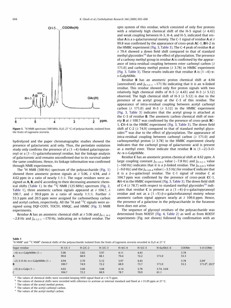
Figure 1. 1H NMR spectrum (500 MHz, D2O, 27 �C) of polysaccharide, isolated fromthe fruits of Lagenaria siceraria.
694 K. Ghosh et al. / Carbohydrate Research 344 (2009) 693–698
hydrolyzed and the paper chromatographic studies showed thepresence of galacturonic acid only. Thus, the periodate oxidationstudy only confirms the presence of a (1?4)-linked galactopyran-osyl or a (1?5)-galactofuranosyl residue, but the linkage analysisof galacturonic acid remains unconfirmed due to its survival underthe same conditions. Hence, its linkage information was confirmedthrough NMR experiments.
The 1H NMR (500 Hz) spectrum of the polysaccharide (Fig. 1)showed three anomeric proton signals at d 5.06, d 4.94, and d4.62 ppm in a ratio of nearly 1:1:1. The sugar residues were as-signed as A, B, and C according to their decreasing anomeric chem-ical shifts (Table 1). In the 13C NMR (125 MHz) spectrum (Fig. 2,Table 1), three anomeric carbon signals appeared at d 104.7, d100.7, and d 99.8 ppm in a ratio of nearly 1:1:1. Further d53.3 ppm and 20.5 ppm were assigned for carbomethoxy carbonand acetyl carbon, respectively. All the 1H and 13C signals were as-signed using DQF-COSY, TOCSY, HMQC, and HMBC (Fig. 3) NMRexperiments.
Residue A has an anomeric chemical shift at d 5.06 and JH-1, H-2
�2.8 Hz and JH-1,C-1 �170 Hz, indicating an a-linked residue. The
Table 11H NMRa and 13C NMRb chemical shifts of the polysaccharide isolated from the fruits of L
Sugar residue H-1/C-1 H-2/C-2 H-3/C-3
?4)-a-D-GalpA6Me-(1? 5.06 3.82 3.97A 99.8 68.9 68.1
?2)-3-O-Ac-a-D-GalpA6Me-(1? 4.94 3.70 5.12B 100.7 74.9 72.2
?4)-b-D-Galp-(1? 4.62 3.66 3.68C 104.7 72.2 68.9
a The values of chemical shifts were recorded keeping HOD signal fixed at d 4.73 ppmb The values of chemical shifts were recorded with reference to acetone as internal stc The values of the acetyl methyl proton.d The values of the acetyl carbonyl carbon.e The values of the acetyl methyl carbon.
spin system of this residue, which consisted of only five protonswith a relatively high chemical shift of the H-5 signal (d 4.43)and weak coupling between H-3, H-4, and H-5, indicated that res-idue A is a D-galacturonosyl moiety. The C-1 signal of residue A at d99.8 was confirmed by the appearance of cross-peak AC-1, BH-2 inthe HMBC experiment (Fig. 3, Table 3). The C-4 peak of residue A atd 79.4 showed a down field shift compared to that of standardmethyl glycosides19 due to the effect of glycosylation. The presenceof a carboxy methyl group in residue A is confirmed by the appear-ance of intra-residual coupling between ester carbonyl carbon (d171.0) and carboxy methyl proton (d 3.78) in HMBC experiment(Fig. 3, Table 3). These results indicate that residue A is (1?4)-a-D-GalpA6Me.
Residue B has an anomeric proton chemical shift at 4.94(unresolved) and JH-1,C-1 �171 Hz indicating that it is an a-linkedresidue. This residue showed only five proton signals with tworelatively high chemical shifts of H-5 (d 4.43) and H-3 (d 5.12)observed. The high chemical shift of H-3 (d 5.12) is due to thepresence of an acetyl group at the C-3 of this residue. Theappearance of intra-residual coupling between acetyl carbonylcarbon (d 171.0) and H-3 (d 5.12) in the HMBC experiment(Fig. 3, Table 3) indicates that the acetyl group is attached atthe C-3 of residue B. The anomeric carbon chemical shift of moi-ety B at d 100.7 was confirmed by the presence of cross-peak BC-1, CH-4 in the HMBC experiment (Fig. 3, Table 3). The down fieldshift of C-2 (d 74.9) compared to that of standard methyl glyco-sides19 was due to the effect of glycosylation. The appearance ofintra-residual coupling between carbonyl carbon (d 171.0) andcarboxymethyl proton (d 3.78) in the HMBC experiment clearlyindicates that the carboxyl group of galacturonic acid is presentas a methyl ester. These indicate that residue B is (1?2)-3-O-Ac-a-D-GalpA6Me.
Residue C has an anomeric proton chemical shift at 4.62 ppm. Alarge coupling constant JH-1,H-2 value (�7.8 Hz) and JH-1,C-1 value(�160 Hz) indicates that it is a b-linked residue. The JH-2,H-3 value(�9.0 Hz) and the JH-3,H-4 value (�3.5 Hz) for residue C indicate thatit is a b-D-galactosyl residue. The C-1 signal of residue C at104.7 ppm was confirmed by the presence of cross-peak CC-1,AH-4 in the HMBC experiment (Fig. 3, Table 3). The down field shiftof C-4 (d 78.7) with respect to standard methyl glycosides19 indi-cates that residue C is present as a (1?4)-b-D-galactopyranosylresidue and not as a (1?5)-b-D-galactofuranosyl moiety whoseanomeric carbon signal appears nearly at d 109.6 ppm. Hence,the presence of a galactose in the polysaccharide in the furanoseform does not arise.
The sequence of glycosyl residues of the polysaccharide wasdetermined from NOESY (Fig. 4, Table 2) as well as from ROESYexperiments (Fig. not shown) followed by confirmation with an
agenaria siceraria recorded in D2O at 27 �C
H-4/C-4 H-5/C-5 H-6a/6b/C-6 COOMe 3-O-COMe
4.11 4.43 3.7879.4 72.2 171.0 53.3
3.97 4.43 3.78 2.04c
68.9 73.7 171.0 53.3 171.0d, 20.5e
4.14 3.78 3.74, 3.6478.7 70.9 61.1
.andard and fixed at d 31.05 ppm at 27 �C.

Figure 2. 13C NMR (125 MHz, D2O, 27 �C) spectrum of polysaccharide, isolated from the fruits of Lagenaria siceraria.
Figure 3. HMBC spectrum of polysaccharide, isolated from the fruits of Lagenaria siceraria. The delay time in the HMBC experiment was 80 ms.
Table 2NOE data for the polysaccharide isolated from the fruits of Lagenaria siceraria
Glycosyl residue Anomeric proton NOE contact to proton
dH dH Residue, atom
?4)-a-D-GalpA6Me-(1? 5.06 4.94 BH-1A 3.70 BH-2
4.43 AH-53.97 AH-34.11 AH-4
?2)-3-O-Ac-a-D-GalpA6Me-(1? 4.94 5.06 AH-1B 3.70 BH-2
3.97 BH-44.14 CH-44.43 BH-5
?4)-b-D-Galp-(1? 4.62 3.66 CH-2C 3.68 CH-3
4.14 CH-44.11 AH-4
K. Ghosh et al. / Carbohydrate Research 344 (2009) 693–698 695

Table 3The significant 3JH,C connectivities observed in an HMBC spectrum for the anomeric protons/carbons of the sugar residues of the polysaccharide isolated from the fruits ofLagenaria siceraria
Residue Sugar linkage H-1/C-1 Observed connectivities
dH/dC Residue Atom
A ?4)-a-D-GalpA6Me-(1? 5.06 74.9 B C-299.8 3.70 B H-2
B ?2)-3-O-Ac-a-D-GalpA6Me-(1? 4.94 78.7 C C-4100.7 4.14 C H-4
C ?4)-b-D-Galp-(1? 4.62 79.4 A C-4104.7 4.11 A H-4
3.68 C H-3
COOMe (dH) Observed connectivities
dC Residue Atom
A ?4)-a-D-GalpA6Me-(1? 3.78 171.0 A C-6
B ?4)-3-O-Ac-a-D-GalpA6Me-(1? 3.78 171.0 B C-6
3-O-COMe (dC) Observed connectivities
dH Residue Atom
B ?4)-3-O-Ac-a-D-GalpA6Me-(1 171.0 5.12 B H-3
696 K. Ghosh et al. / Carbohydrate Research 344 (2009) 693–698
HMBC experiment. From inter-residue NOE contacts (Fig. 4, Table2), the following sequences were established:
The sequence was further confirmed by the HMBC experiment(Fig. 3, Table 3). Cross-peaks were found between H-1 of residueA (d 5.06) and C-2 of residue B (AH-1, BC-2); C-1 of residue A (d
Figure 4. NOESY spectrum of polysaccharide, isolated from the fru
99.8) and H-2 of residue B (AC-1, BH-2). Similarly, cross-peakswere found between H-1 of residue B (d 4.94) and C-4 of residueC (BH-1, CC-4); C-1 of residue B (d 100.7) and H-4 of residue C(BC-1, CH-4). Cross-peaks were also observed between H-1 of res-idue C (d 4.62) and C-4 of residue A (CH-1, AC-4); C-1 of residue C(d 104.7) and H-4 of residue A (CC-1, AH-4) along with intra-resid-ual coupling between C-1 of residue C with its own H-3 (CC-1, CH-3). Other intra-residual interactions (CH-3, CC-5) and (CC-3, CH-5)were observed. Another two intra-residual couplings between thecarboxymethyl proton (d 3.78) and the ester carbonyl carbon (d171.0) of residue A and residue B were observed. Intra-residualcoupling between acetyl carbonyl carbon (d 171.0) and H-3 (d5.12) atom of residue B [OAc (carbonyl carbon), BH-3], and alsocross-peak between acetyl carbonyl carbon (d 171.0) and acetylmethyl proton (d 2.04) of residue B [OAc (carbonyl carbon), OAc(methyl proton)] were observed.
Therefore, on the basis of the above results, the following trisac-charide-repeating unit in the polysaccharide is assigned
its of Lagenaria siceraria. The NOESY mixing time was 300 ms.
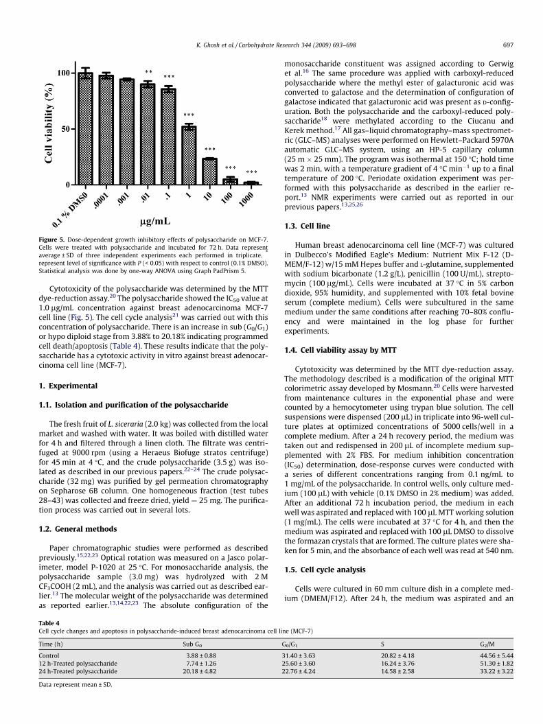
Figure 5. Dose-dependent growth inhibitory effects of polysaccharide on MCF-7.Cells were treated with polysaccharide and incubated for 72 h. Data representaverage ± SD of three independent experiments each performed in triplicate. *
represent level of significance with P (< 0.05) with respect to control (0.1% DMSO).Statistical analysis was done by one-way ANOVA using Graph PadPrism 5.
K. Ghosh et al. / Carbohydrate Research 344 (2009) 693–698 697
Cytotoxicity of the polysaccharide was determined by the MTTdye-reduction assay.20 The polysaccharide showed the IC50 value at1.0 lg/mL concentration against breast adenocarcinoma MCF-7cell line (Fig. 5). The cell cycle analysis21 was carried out with thisconcentration of polysaccharide. There is an increase in sub (G0/G1)or hypo diploid stage from 3.88% to 20.18% indicating programmedcell death/apoptosis (Table 4). These results indicate that the poly-saccharide has a cytotoxic activity in vitro against breast adenocar-cinoma cell line (MCF-7).
1. Experimental
1.1. Isolation and purification of the polysaccharide
The fresh fruit of L. siceraria (2.0 kg) was collected from the localmarket and washed with water. It was boiled with distilled waterfor 4 h and filtered through a linen cloth. The filtrate was centri-fuged at 9000 rpm (using a Heraeus Biofuge stratos centrifuge)for 45 min at 4 �C, and the crude polysaccharide (3.5 g) was iso-lated as described in our previous papers.22–24 The crude polysac-charide (32 mg) was purified by gel permeation chromatographyon Sepharose 6B column. One homogeneous fraction (test tubes28–43) was collected and freeze dried, yield — 25 mg. The purifica-tion process was carried out in several lots.
1.2. General methods
Paper chromatographic studies were performed as describedpreviously.15,22,23 Optical rotation was measured on a Jasco polar-imeter, model P-1020 at 25 �C. For monosaccharide analysis, thepolysaccharide sample (3.0 mg) was hydrolyzed with 2 MCF3COOH (2 mL), and the analysis was carried out as described ear-lier.13 The molecular weight of the polysaccharide was determinedas reported earlier.13,14,22,23 The absolute configuration of the
Table 4Cell cycle changes and apoptosis in polysaccharide-induced breast adenocarcinoma cell li
Time (h) Sub G0 G
Control 3.88 ± 0.88 312 h-Treated polysaccharide 7.74 ± 1.26 224 h-Treated polysaccharide 20.18 ± 4.82 2
Data represent mean ± SD.
monosaccharide constituent was assigned according to Gerwiget al.16 The same procedure was applied with carboxyl-reducedpolysaccharide where the methyl ester of galacturonic acid wasconverted to galactose and the determination of configuration ofgalactose indicated that galacturonic acid was present as D-config-uration. Both the polysaccharide and the carboxyl-reduced poly-saccharide18 were methylated according to the Ciucanu andKerek method.17 All gas–liquid chromatography–mass spectromet-ric (GLC–MS) analyses were performed on Hewlett–Packard 5970Aautomatic GLC–MS system, using an HP-5 capillary column(25 m � 25 mm). The program was isothermal at 150 �C; hold timewas 2 min, with a temperature gradient of 4 �C min�1 up to a finaltemperature of 200 �C. Periodate oxidation experiment was per-formed with this polysaccharide as described in the earlier re-port.13 NMR experiments were carried out as reported in ourprevious papers.13,25,26
1.3. Cell line
Human breast adenocarcinoma cell line (MCF-7) was culturedin Dulbecco’s Modified Eagle’s Medium: Nutrient Mix F-12 (D-MEM/F-12) w/15 mM Hepes buffer and L-glutamine, supplementedwith sodium bicarbonate (1.2 g/L), penicillin (100 U/mL), strepto-mycin (100 lg/mL). Cells were incubated at 37 �C in 5% carbondioxide, 95% humidity, and supplemented with 10% fetal bovineserum (complete medium). Cells were subcultured in the samemedium under the same conditions after reaching 70–80% conflu-ency and were maintained in the log phase for furtherexperiments.
1.4. Cell viability assay by MTT
Cytotoxicity was determined by the MTT dye-reduction assay.The methodology described is a modification of the original MTTcolorimetric assay developed by Mosmann.20 Cells were harvestedfrom maintenance cultures in the exponential phase and werecounted by a hemocytometer using trypan blue solution. The cellsuspensions were dispensed (200 lL) in triplicate into 96-well cul-ture plates at optimized concentrations of 5000 cells/well in acomplete medium. After a 24 h recovery period, the medium wastaken out and redispensed in 200 lL of incomplete medium sup-plemented with 2% FBS. For medium inhibition concentration(IC50) determination, dose-response curves were conducted witha series of different concentrations ranging from 0.1 ng/mL to1 mg/mL of the polysaccharide. In control wells, only culture med-ium (100 lL) with vehicle (0.1% DMSO in 2% medium) was added.After an additional 72 h incubation period, the medium in eachwell was aspirated and replaced with 100 lL MTT working solution(1 mg/mL). The cells were incubated at 37 �C for 4 h, and then themedium was aspirated and replaced with 100 lL DMSO to dissolvethe formazan crystals that are formed. The culture plates were sha-ken for 5 min, and the absorbance of each well was read at 540 nm.
1.5. Cell cycle analysis
Cells were cultured in 60 mm culture dish in a complete med-ium (DMEM/F12). After 24 h, the medium was aspirated and an
ne (MCF-7)
0/G1 S G2/M
1.40 ± 3.63 20.82 ± 4.18 44.56 ± 5.445.60 ± 3.60 16.24 ± 3.76 51.30 ± 1.822.76 ± 4.24 14.58 ± 2.58 33.22 ± 3.22

698 K. Ghosh et al. / Carbohydrate Research 344 (2009) 693–698
incomplete medium with 5% FBS was added. Time response curvefor polysaccharide (1.0 lg/mL) was studied for 12 and 24 h alongwith control. The cells were harvested and fixed in 70% ethanol,maintained at �20 �C overnight for resolving sub G0 stage. Then,the cells were washed with ice cold PBS (pH 7.4) and resuspendedin 1 mL of PI Master mixture (40 lg/mL PI, 100 lg/mL RNaseA inPBS), followed by incubation at 37 �C for 1 h. Apoptotic cells weredetermined by their hypochromic sub-diploid staining profiles.The distribution of cells in the different cell cycle phases was ana-lyzed from the DNA histogram using Becton–Dickinson FACS Cali-ber flow cytometer and CELL QUEST software.21
Acknowledgments
The authors are grateful to Professor S. Roy, Director, IICB, Dr. A.K. Sen (Jr.), IICB and Dr. S. Lahiri, IACS, Kolkatta, for providinginstrumental facilities. Mr. Barun Majumder of Bose Institute, Kolk-atta, is acknowledged for preparing the NMR spectra. DST, Govt ofIndia, is acknowledged for sanctioning a project (Ref. No.: SR/S1/OC-52/2006 dated 19/02/2007). The authors (K.C., A.K.O.) acknowl-edge the UGC for offering junior research fellowship.
References
1. Rahman, A. S. H. Nat. Prod. Rad. 2003, 2, 249–250.2. Deshpande, J. R.; Mishra, M. R.; Meghre, V. S.; Wadodkar, S. G.; Dorle, A. K. Nat.
Prod. Rad. 2007, 6, 127–130.3. Ghule, B. V.; Ghante, M. H.; Upaganlawar, A. B.; Yeole, P. G. Phcog. Mag. 2006, 2,
232–238.
4. Shivarajan, V. V.; Balachandra, I. Ayurvedic Drugs and their Plant Source; Oxfordand IBH: New Delhi, 1996. pp 176–177.
5. Kirtikar, K. R. Indian Medicinal Plants; Oriental Enterprises: Dehradun, India,2001. pp 722–723.
6. Shirwaikar, A.; Sreenivasan, K. K. Ind. J. Pharm. Sci. 1996, 58, 197–202.7. Duke, J. A. Handbook of Phytochemical and Constituents of Grass Herbs and Other
Economic Plants; CRC Press: Boco Raton, 1999. pp 98–119.8. Evans, W. C. Treese and Evans Pharmacognosy; Balliere: Tindall, 1996. pp 388–
433.9. Sonja, S.; Hermann, S. Phytochem. Anal. 2000, 11, 121.
10. Baranowska, M. K.; Cisowski, W. J. Chromatogr., A 1994, 675, 240–243.11. Chopra, R. N.; Chopra, I. C.; Verma, B. S. Supplement to Glossary of Indian
Medicinal Plants; CSIR: New Delhi, 1992. p 51.12. Wang, H. X.; Ng, T. B. Life Sci. 2000, 67, 2631–2638.13. Ghosh, K.; Chandra, K.; Roy, K. S.; Mondal, S.; Maiti, D.; Das, D.; Ojha, K. A.;
Islam, S. S. Carbohydr. Res. 2008, 343, 341–349.14. Hara, C.; Kiho, T.; Tanaka, Y.; Ukai, S. Carbohydr. Res. 1982, 110, 77–87.15. Hoffman, J.; Lindberg, B.; Svensson, S. Acta Chem. Scand. 1972, 26, 661–666.16. Gerwig, G. J.; Kamerling, J. P.; Vliegenthart, J. F. G. Carbohydr. Res. 1978, 62,
349–357.17. Ciucanu, I.; Kerek, F. Carbohydr. Res. 1984, 131, 209–217.18. Maness, N. O.; Ryan, J. D.; Mort, A. J. Anal. Biochem. 1990, 185, 346–352.19. Agarwal, P. K. Phytochemistry 1992, 31, 3307–3330.20. Mosmann, T. J. Immunol. Method 1983, 65, 55–59.21. Evans, D. L.; Bishop, G. R.; Jaso-Friedmann, L. Method Cell Sci. 2000, 22, 225–231.22. Ghosh, K.; Chandra, K.; Roy, K. S.; Mondal, S.; Maiti, D.; Das, D.; Ojha, K. A.;
Islam, S. S. Carbohydr. Res. 2008, 343, 1071–1078.23. Ghosh, K.; Chandra, K.; Ojha, K. A.; Islam, S. S. Carbohydr. Res. 2008, 343, 2834–
2840.24. York, W. S.; Darvill, A. K.; McNeil, M.; Stevenson, T. T.; Albersheim, P. Methods
Enzymol. 1985, 118, 33–40.25. Dueñas Chasco, M. T.; Rodriguez-Carvajal, M. A.; Mateo, P. T.; Franko-
Rodriguez, G.; Espartero, J. L.; Iribas, A. I.; Gil-Serrano, A. M. Carbohydr. Res.1997, 303, 453–458.
26. Hård, K.; Zadelhoff, G. V.; Moonen, P.; Kamerling, J. P.; Vliegenthart, J. F. G. Eur.J. Biochem. 1992, 209, 895–915.