Structural elucidation of three antioxidative …€¦ · Structural elucidation of three...
9
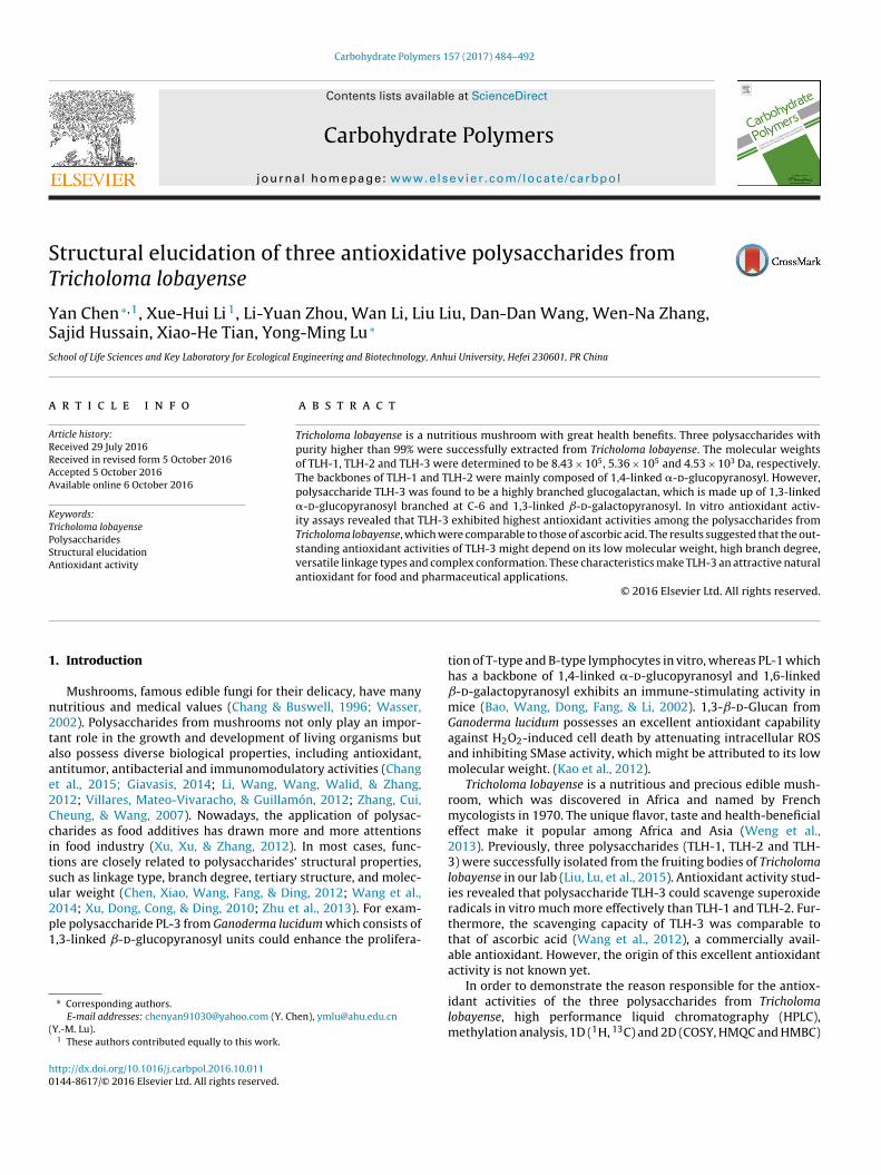
Carbohydrate Polymers 157 (2017) 484–492 Contents lists available at ScienceDirect Carbohydrate Polymers journal homepage: www.elsevier.com/locate/carbpol Structural elucidation of three antioxidative polysaccharides from Tricholoma lobayense Yan Chen ∗,1 , Xue-Hui Li 1 , Li-Yuan Zhou, Wan Li, Liu Liu, Dan-Dan Wang, Wen-Na Zhang, Sajid Hussain, Xiao-He Tian, Yong-Ming Lu ∗ School of Life Sciences and Key Laboratory for Ecological Engineering and Biotechnology, Anhui University, Hefei 230601, PR China article info Article history: Received 29 July 2016 Received in revised form 5 October 2016 Accepted 5 October 2016 Available online 6 October 2016 Keywords: Tricholoma lobayense Polysaccharides Structural elucidation Antioxidant activity abstract Tricholoma lobayense is a nutritious mushroom with great health benefits. Three polysaccharides with purity higher than 99% were successfully extracted from Tricholoma lobayense. The molecular weights of TLH-1, TLH-2 and TLH-3 were determined to be 8.43 × 10 5 , 5.36 × 10 5 and 4.53 × 10 3 Da, respectively. The backbones of TLH-1 and TLH-2 were mainly composed of 1,4-linked -d-glucopyranosyl. However, polysaccharide TLH-3 was found to be a highly branched glucogalactan, which is made up of 1,3-linked -d-glucopyranosyl branched at C-6 and 1,3-linked ˇ-d-galactopyranosyl. In vitro antioxidant activ- ity assays revealed that TLH-3 exhibited highest antioxidant activities among the polysaccharides from Tricholoma lobayense, which were comparable to those of ascorbic acid. The results suggested that the out- standing antioxidant activities of TLH-3 might depend on its low molecular weight, high branch degree, versatile linkage types and complex conformation. These characteristics make TLH-3 an attractive natural antioxidant for food and pharmaceutical applications. © 2016 Elsevier Ltd. All rights reserved. 1. Introduction Mushrooms, famous edible fungi for their delicacy, have many nutritious and medical values (Chang & Buswell, 1996; Wasser, 2002). Polysaccharides from mushrooms not only play an impor- tant role in the growth and development of living organisms but also possess diverse biological properties, including antioxidant, antitumor, antibacterial and immunomodulatory activities (Chang et al., 2015; Giavasis, 2014; Li, Wang, Wang, Walid, & Zhang, 2012; Villares, Mateo-Vivaracho, & Guillamón, 2012; Zhang, Cui, Cheung, & Wang, 2007). Nowadays, the application of polysac- charides as food additives has drawn more and more attentions in food industry (Xu, Xu, & Zhang, 2012). In most cases, func- tions are closely related to polysaccharides’ structural properties, such as linkage type, branch degree, tertiary structure, and molec- ular weight (Chen, Xiao, Wang, Fang, & Ding, 2012; Wang et al., 2014; Xu, Dong, Cong, & Ding, 2010; Zhu et al., 2013). For exam- ple polysaccharide PL-3 from Ganoderma lucidum which consists of 1,3-linked ˇ-d-glucopyranosyl units could enhance the prolifera- ∗ Corresponding authors. E-mail addresses: [email protected] (Y. Chen), [email protected] (Y.-M. Lu). 1 These authors contributed equally to this work. tion of T-type and B-type lymphocytes in vitro, whereas PL-1 which has a backbone of 1,4-linked -d-glucopyranosyl and 1,6-linked ˇ-d-galactopyranosyl exhibits an immune-stimulating activity in mice (Bao, Wang, Dong, Fang, & Li, 2002). 1,3-ˇ-d-Glucan from Ganoderma lucidum possesses an excellent antioxidant capability against H 2 O 2 -induced cell death by attenuating intracellular ROS and inhibiting SMase activity, which might be attributed to its low molecular weight. (Kao et al., 2012). Tricholoma lobayense is a nutritious and precious edible mush- room, which was discovered in Africa and named by French mycologists in 1970. The unique flavor, taste and health-beneficial effect make it popular among Africa and Asia (Weng et al., 2013). Previously, three polysaccharides (TLH-1, TLH-2 and TLH- 3) were successfully isolated from the fruiting bodies of Tricholoma lobayense in our lab (Liu, Lu, et al., 2015). Antioxidant activity stud- ies revealed that polysaccharide TLH-3 could scavenge superoxide radicals in vitro much more effectively than TLH-1 and TLH-2. Fur- thermore, the scavenging capacity of TLH-3 was comparable to that of ascorbic acid (Wang et al., 2012), a commercially avail- able antioxidant. However, the origin of this excellent antioxidant activity is not known yet. In order to demonstrate the reason responsible for the antiox- idant activities of the three polysaccharides from Tricholoma lobayense, high performance liquid chromatography (HPLC), methylation analysis, 1D ( 1 H, 13 C) and 2D (COSY, HMQC and HMBC) http://dx.doi.org/10.1016/j.carbpol.2016.10.011 0144-8617/© 2016 Elsevier Ltd. All rights reserved.
Transcript of Structural elucidation of three antioxidative …€¦ · Structural elucidation of three...
ST
YSS
a
ARRAA
KTPSA
1
n2taae2Ccitsu2p1
(
h0
Carbohydrate Polymers 157 (2017) 484–492
Contents lists available at ScienceDirect
Carbohydrate Polymers
journa l homepage: www.e lsev ier .com/ locate /carbpol
tructural elucidation of three antioxidative polysaccharides fromricholoma lobayense
an Chen ∗,1, Xue-Hui Li 1, Li-Yuan Zhou, Wan Li, Liu Liu, Dan-Dan Wang, Wen-Na Zhang,ajid Hussain, Xiao-He Tian, Yong-Ming Lu ∗
chool of Life Sciences and Key Laboratory for Ecological Engineering and Biotechnology, Anhui University, Hefei 230601, PR China
r t i c l e i n f o
rticle history:eceived 29 July 2016eceived in revised form 5 October 2016ccepted 5 October 2016vailable online 6 October 2016
eywords:
a b s t r a c t
Tricholoma lobayense is a nutritious mushroom with great health benefits. Three polysaccharides withpurity higher than 99% were successfully extracted from Tricholoma lobayense. The molecular weightsof TLH-1, TLH-2 and TLH-3 were determined to be 8.43 × 10⁵, 5.36 × 10⁵ and 4.53 × 10³ Da, respectively.The backbones of TLH-1 and TLH-2 were mainly composed of 1,4-linked �-d-glucopyranosyl. However,polysaccharide TLH-3 was found to be a highly branched glucogalactan, which is made up of 1,3-linked�-d-glucopyranosyl branched at C-6 and 1,3-linked ˇ-d-galactopyranosyl. In vitro antioxidant activ-
richoloma lobayenseolysaccharidestructural elucidationntioxidant activity
ity assays revealed that TLH-3 exhibited highest antioxidant activities among the polysaccharides fromTricholoma lobayense, which were comparable to those of ascorbic acid. The results suggested that the out-standing antioxidant activities of TLH-3 might depend on its low molecular weight, high branch degree,versatile linkage types and complex conformation. These characteristics make TLH-3 an attractive naturalantioxidant for food and pharmaceutical applications.
© 2016 Elsevier Ltd. All rights reserved.
. Introduction
Mushrooms, famous edible fungi for their delicacy, have manyutritious and medical values (Chang & Buswell, 1996; Wasser,002). Polysaccharides from mushrooms not only play an impor-ant role in the growth and development of living organisms butlso possess diverse biological properties, including antioxidant,ntitumor, antibacterial and immunomodulatory activities (Changt al., 2015; Giavasis, 2014; Li, Wang, Wang, Walid, & Zhang,012; Villares, Mateo-Vivaracho, & Guillamón, 2012; Zhang, Cui,heung, & Wang, 2007). Nowadays, the application of polysac-harides as food additives has drawn more and more attentionsn food industry (Xu, Xu, & Zhang, 2012). In most cases, func-ions are closely related to polysaccharides’ structural properties,uch as linkage type, branch degree, tertiary structure, and molec-lar weight (Chen, Xiao, Wang, Fang, & Ding, 2012; Wang et al.,
014; Xu, Dong, Cong, & Ding, 2010; Zhu et al., 2013). For exam-le polysaccharide PL-3 from Ganoderma lucidum which consists of,3-linked ˇ-d-glucopyranosyl units could enhance the prolifera-∗ Corresponding authors.E-mail addresses: [email protected] (Y. Chen), [email protected]
Y.-M. Lu).1 These authors contributed equally to this work.
ttp://dx.doi.org/10.1016/j.carbpol.2016.10.011144-8617/© 2016 Elsevier Ltd. All rights reserved.
tion of T-type and B-type lymphocytes in vitro, whereas PL-1 whichhas a backbone of 1,4-linked �-d-glucopyranosyl and 1,6-linkedˇ-d-galactopyranosyl exhibits an immune-stimulating activity inmice (Bao, Wang, Dong, Fang, & Li, 2002). 1,3-ˇ-d-Glucan fromGanoderma lucidum possesses an excellent antioxidant capabilityagainst H2O2-induced cell death by attenuating intracellular ROSand inhibiting SMase activity, which might be attributed to its lowmolecular weight. (Kao et al., 2012).
Tricholoma lobayense is a nutritious and precious edible mush-room, which was discovered in Africa and named by Frenchmycologists in 1970. The unique flavor, taste and health-beneficialeffect make it popular among Africa and Asia (Weng et al.,2013). Previously, three polysaccharides (TLH-1, TLH-2 and TLH-3) were successfully isolated from the fruiting bodies of Tricholomalobayense in our lab (Liu, Lu, et al., 2015). Antioxidant activity stud-ies revealed that polysaccharide TLH-3 could scavenge superoxideradicals in vitro much more effectively than TLH-1 and TLH-2. Fur-thermore, the scavenging capacity of TLH-3 was comparable tothat of ascorbic acid (Wang et al., 2012), a commercially avail-able antioxidant. However, the origin of this excellent antioxidantactivity is not known yet.
In order to demonstrate the reason responsible for the antiox-idant activities of the three polysaccharides from Tricholomalobayense, high performance liquid chromatography (HPLC),methylation analysis, 1D (1H, 13C) and 2D (COSY, HMQC and HMBC)

e Poly
NScwias
2
2
bppuawfc9nog
2
dns(cTi1
ps1&ptptr
2
amwXn(flpp(1
Y. Chen et al. / Carbohydrat
MR spectroscopy were used for the structure characterization.tructure features including physicochemical properties, monosac-haride composition, glycosidic bonds and chain conformationsere elucidated. Furthermore, antioxidant activities were exam-
ned by DPPH, superoxide, hydroxyl and ABTS radicals scavengingssays. The results were analyzed and compared to clarify thetructure-activity relationship.
. Materials and methods
.1. Materials and reagents
Tricholoma lobayense was purchased from Panji Shenshan edi-le fungus cooperative (Huainan, Anhui Province, China). Crudeolysaccharides were extracted from Tricholoma lobayense by high-ressure homogenization. Polysaccharide TLH-1, TLH-2 and TLH-3sed in this study were obtained by precipitation with 60% ethanolnd ultrafiltration according to the method of Liu, Lu, et al., (2015)ith some modifications. Deuterium oxide (D2O) was purchased
rom Sigma Chemical Co. (St. Louis, MO, USA). Seven monosac-haride standards (Rhamnose 99%, Mannose 99%, Glucuronic acid8%, Galacturonic acid 97%, Glucose 99.5%, Galactose 99%, Arabi-ose 98%) were purchased from Aladdin Industrial Corporation. Allther chemicals and solvents used in this study were of analyticalrade.
.2. Physical and chemical properties analysis
The purity and molecular weights of three polysaccharides wereetermined by HPLC on an Agilent 1260 system (Agilent Tech-ologies, Palo Alto, CA, USA) equipped with an evaporative lightcattering detector (ELSD) and a TSK gel G4000 PWXL column300 × 7.8 mm, Tosoh Corp, Tokyo, Japan). The linear regression wasalibrated with T-series dextran standards (T-3, T-10, T-40, T-100,-200, T-500 and T-1000). Sample solution (2 mg/mL, 10 �L) was
njected and eluted with distilled water at 30 ◦C with a flow rate of.0 mL/min.
Content of the total sugars, proteins and uronic acids in the threeurified fractions were determined colorimetrically using phenol-ulfuric acid (Masuko et al., 2005), Bradford method (Bradford,976) and meta-hydroxydiphenyl-sulfuric acid (Blumenkrantz
Asboe-Hansen, 1973), respectively. The IR spectra of theolysaccharides were determined using Fourier Transform IR Spec-rophotometry (FT-IR, Nicolet 380, Thermo, USA). The purifiedolysaccharides (1 mg) were ground with KBr powder (100 mg) andhen pressed into pellets for FT-IR measurement in the frequencyange of 4000–400 cm−1.
.3. Monosaccharide composition analysis
Each polysaccharide (10 mg) was hydrolyzed by trifluoroaceticcid (TFA, 2 mol/L), followed by complexing with 1-phenyl-3-ethyl-5-pyrazolone (PMP) (0.5 mol/L). The resulting productsere then analyzed by HPLC equipped with a ZORBAX EclipseDB-C18 column (150 × 4.6 mm, particle size 5 �m, Agilent Tech-ologies, CA, USA) and a UV detection at 245 nm. Sample solution10 �L) was injected and eluted with distilled water at 25 ◦C with aow rate of 1.0 mL/min. The mobile phase A was a mixture of phos-
hate buffer (50 mmol/L)-acetonitrile (85:15, v/v) and the mobilehase B was a mixture of phosphate buffer (50 mmol/L)-acetonitrile60:40, v/v). The analysis was carried out with a gradient elution of5-23-15% phase B from 0 to 20–35 min.mers 157 (2017) 484–492 485
2.4. Methylation and GC–MS analysis
The sample (20 mg) was methylated with methyl iodide threetimes (Liu & Sun, 2011; Yan, Yin, Zhang, Yang, & Yu, 2013). Thenpartially methylated products were hydrolyzed with TFA for 6 hat 100 ◦C, followed by reduction with NaBH4 and acetylation withacetic anhydride. The partially methylated alditol acetates wereanalyzed by GC–MS (Agilent 7890A/5975C, USA) equipped witha HP-5 capillary column (30 m × 0.32 mm, 0.50 �m, Agilent Tech-nologies, CA, USA) programmed from 120 (keep for 1 min) to 240 ◦C(keep for 6.5 min) at 10 ◦C per min. The degree of branching (DB)could be calculated with the following equation (Wu et al., 2014):
DB = NT + NBNT + NB + NL
(1)
where NT, NB and NL are the total contents of the terminal residues,branched residues, and backbone residues, respectively.
2.5. NMR analysis
The NMR spectra of TLH-1, TLH-2 and TLH-3 were obtainedby an AVANCE-600 NMR spectrometer (Bruker Inc., Rheinstetten,Germany). 40 mg of the dried sample was dissolved in deuteriumoxide (D2O, 0.5 mL) at room temperature for 3 h before NMR anal-ysis. The 1H (600Mz) and 13C (150Mz) NMR spectra were recordedat 50 ◦C. Then, the 2D NMR spectra including 1H/1H correlationexperiments (COSY), Heteronuclear Multiple-Quantum Coherence(HMQC) and Heteronuclear Multiple Bond Correlation (HMBC)were recorded.
2.6. In vitro antioxidant activities analysis
2.6.1. DPPH radical scavenging activityTwo milliliters of aqueous solution of TLH-1, TLH-2 and TLH-
3 (50, 100, 200, 300, 400 and 500 �g/mL) were mixed withDPPH-methanol solution (2 mL, 0.13 mM). The mixture was shakenvigorously and left to stand in test tubes covered in aluminum foilfor 30 min at 25 ◦C. Then the absorbance at 517 nm was measured(Jing et al., 2015). Ascorbic acid was used as positive control. TheDPPH radical scavenging activity (RSA) was calculated according tothe following equation:
DPPH RSA (%) =(
1 − A2 − A1
A0
)× 100 (2)
where A0 is the absorbance value of DPPH-methanol solution (2 mL)plus methanol (2 mL). A1 is the absorbance value of the mixtureof methanol (2 mL) plus samples (2 mL) with different concentra-tions. A2 is the absorbance value of DPPH-methanol solution (2 mL)plus samples (2 mL) with different concentrations. All assays werecarried out in triplicate.
2.6.2. Superoxide radical scavenging activity0.5 mL of phosphate buffer (50 mM, pH 8.3) and 0.4 mL of aque-
ous samples (50, 100, 200, 300, 400 and 500 �g/mL) were mixedand stored for 20 min at 25 ◦C. Then 0.1 mL of pyrogallol (3 mM)preheated to 25 ◦C was added. The absorbance value of the mixturewas measured at 325 nm every 30 s for 5 min (Zhu & Wu, 2009).Ascorbic acid was used as positive control. Superoxide RSA wascalculated according to the following equation:
Superoxide anion RSA (%) = �A2 − �A1
�A0× 100 (3)
where �A1 is the difference of absorbance value per 30 s for differ-ent concentrations of samples. �A0 is the difference of absorbancevalue per 30 s without samples. All assays were carried out in trip-licate.
4 te Poly
2
s3ltftc
H
wsdw
2
epcCww3daw
A
wooo
2
cwP
3
3
saptssT
3
watww
86 Y. Chen et al. / Carbohydra
.6.3. Hydroxyl radical scavenging activityMixtures containing 1 mL of FeSO4 (6 mM), 1 mL of ethanol-
alicylic acid (6 mM) and 1 mL of aqueous sample (50, 100, 200,00, 400 and 500 �g/mL) were incubated in test tubes, shaken and
eft to stand for 10 min. Then, 1 mL of H2O2 (2.4 mM) was added andhe test tubes were covered in aluminum foil. After storing at 37 ◦Cor 30 min, the presence of hydroxyl radical was detected by moni-oring the absorbance at 510 nm. Ascorbic acid was used as positiveontrol. Hydroxyl RSA was calculated by the following equation:
ydroxyl RSA (%) =(
1 − A2 − A1
A0
)× 100 (4)
here A0 is the absorbance value of the solution without theamples. A2 is the absorbance value of the samples solution withifferent concentrations. A1 is the absorbance value of the solutionithout ethanol-salicylic acid.
.6.4. Total antioxidant capacityAfter reacting aqueous ABTS (2, 2′-Azinobis-(3-
thylbenzthiazoline-6-sulphonate)) solution with potassiumersulfate (2.45 mM) at room temperature for 14 h, ABTS radicalation (ABTS
•+) was obtained according to the literature (Barahona,handía, Encinas, Matsuhiro, & Zúniga, 2011). The ABTS
•+ solutionas diluted with PBS (pH 7.0) until the absorbance at 734 nmas 0.70. Then 10 �L of polysaccharide samples (50, 100, 200,
00, 400 and 500 �g/mL) in PBS was mixed with 200 �L of theiluted ABTS
•+ solution and the absorbance value was measuredt 734 nm. Ascorbic acid was used as positive control. ABTS RSAas calculated by the following equation:
BTS RSA (%) =(
1 − Aj − Ai
A0
)× 100 (5)
here A0 is the absorbance value of the ABTS•+ solution with 10 �L
f PBS. Aj is the absorbance value of the ABTS•+ solution in presence
f samples with different concentrations. Ai is the absorbance valuef the samples solution in the absence of ABTS
•+.
.7. Statistical analysis
The data were mean ± standard deviation (SD) of three repli-ates except for cell tests of four replicates. Statistical significanceas determined by the one-way analysis of variance (ANOVA), and
value less than 0.05 was considered statistically significant.
. Results
.1. Antioxidant activities of TLH-1, TLH-2 and TLH-3
The antioxidant activities of TLH-1, TLH-2 and TLH-3 were pre-ented in Fig. 1 and IC50 values were listed in Table S1. Fourssays were used to evaluate the antioxidant capacities of the threeolysaccharides. The results indicated that with increasing concen-ration of polysaccharides, the scavenging activities towards DPPH,uperoxide, hydroxyl and ABTS radicals were also enhanced. Thecavenging abilities of TLH-1 and TLH-2 were similar, but those ofLH-3 were much higher and similar to those of ascorbic acid.
.2. Physical and chemical properties of TLH-1, TLH-2 and TLH-3
The content of total saccharides of TLH-1, TLH-2 and TLH-3ere 99.7%, 99.6% and 99.3%, respectively. The content of uronic
cids for TLH-1, TLH-2 and TLH-3 were 0, 3.01% and 6.44%, respec-ively. As shown in Fig. S1a-c, only one symmetrical sharp peakas found, which indicated that all the obtained polysaccharidesere homogeneous. According to the calibration curve of standards
mers 157 (2017) 484–492
(log MW = −0.350t + 7.449, R2 = 0.997), the molecular weights ofTLH-1, TLH-2 and TLH-3 were calculated to be 8.43 × 10⁵, 5.36 × 10⁵
and 4.53 × 10³ Da, respectively.Fourier transform infrared spectroscopy has been broadly used
for the characterization of polysaccharides. The FT-IR spectra ofTLH-1, TLH-2 and TLH-3 were depicted in Fig. S1d. Five typicalsignals were clearly presented at nearly 3394, 2931, 1643, 1413and 1037 cm−1 for all the three polysaccharides, which could beassigned for the stretching vibration of O H and C H, the bendingvibration of O H and C H, and a pyranose form of sugars, respec-tively. The bands at about 850 cm−1 and 890 cm−1 for the threepolysaccharides suggested a � configuration and a anomeric con-figuration (Calonje, García Mendoza, & Novaes-Ledieu, 1996; Wang,Zhang, Zhang, & Chen, 2008). These results indicated that all thethree components were polysaccharides.
3.3. Monosaccharide composition of TLH-1, TLH-2 and TLH-3
As shown in Fig. 2, fraction TLH-1 was composed of rhamnose,mannose, glucose, galactose and arabinose with the molar ratio of0.02: 1.00: 3.22: 0.01: 0.28, which could be denoted as a glucan.However, fraction TLH-2 and TLH-3 were made up of rhamnose,mannose, glucuronic acid, galacturonic acid, glucose, galactose andarabinose, while the molar ratio was 0.02: 1.00: 0.05: 0.04: 3.80:0.03: 0.28 and 0.47: 1.00: 0.13: 0.10: 9.84: 6.18: 0.93, respectively.The monosaccharide composition of TLH-2 was similar to TLH-3,whereas the molar ratio of monosaccharide was very close to TLH-1. Thus TLH-2 could also be mainly regarded as a glucan. In contrastTLH-3, made up of seven monosaccharides with higher molar ratiothan TLH-2, could be noted as a glucogalactan (Liu, Wen, Kan, & Jin,2015; Zhu, Han, Sun, Wang, & Yang, 2012).
3.4. Linkage types of TLH-1, TLH-2 and TLH-3
To determine the linkage types, TLH-1, TLH-2 and TLH-3 weremethylated and converted into the corresponding alditol acetatesfor further GC–MS analysis. As shown in Table 1 and Fig. S2, themajor derivative from TLH-1 and TLH-2 was both 2, 3, 6-tri-O-methyl glucitolpyranosyl with a molar ratio of 38.96 and 26.54,respectively. The DB values of TLH-1 and TLH-2 were calculatedto be 0.27 and 0.23, implying they were both small-branchedpolysaccharides. However, the major derivatives from TLH-3 werecomposed of 2, 4-Me2-Glcp, 2,3,4,6-Me4-Glcp, 2,3,6-Me3-Glcp, and2,4,6-Me3-Galp. The DB value of TLH-3 was 0.74, suggesting ahighly-branched structure. These results were substantially con-sistent with the results of monosaccharide composition analysismentioned above.
3.5. NMR study of TLH-1
Four dominant peaks were found at ı 5.37, 5.08, 5.05 and4.99 ppm in the anomeric region of 1H NMR spectrum of TLH-1 (Fig.S3a), indicating that these glucosyl residues were �-glycosidicallylinked except the residue presented at ı 4.99 ppm. The chemicalshift of H-2 can be assigned from the COSY spectrum based on theprinciple that H-2 correlates with H-1 (Fig. S3b). Other hydrogensignals (H-3 to H-6) were also assigned by the same analogy. Thecorresponding anomeric carbons were identified from cross peaksin the HMQC spectrum at ı 102.06, 101.11, 104.15 and 100.54 ppm(Fig. S3c). All the 1H and 13C chemical shifts for TLH-1 were listed inTable 2. According to literature data (Cao et al., 2006; Chen, Zhang,
Chen, & Cheung, 2014; Chen et al., 2011; Golovchenko, Khramova,Ovodova, Shashkov, & Ovodov, 2012; Kang et al., 2011; Yin et al.,2010), these cross peaks were assigned to 1,4-linked �-d-Glcp(A), T-�-l-Rhap (D), 1,3,6-linked �-d-Manp (C) and T-ˇ-d-Glcp (B),
Y. Chen et al. / Carbohydrate Polymers 157 (2017) 484–492 487
Fig. 1. Scavenging effects of TLH-1, TLH-2, TLH-3 and Vc at different concentrations on DPPH radicals (a), superoxide radicals (b), hydroxyl radicals (c) and ABTS radicals (d).Each value represents the mean ± standard deviation (SD) (n = 3). Vc stands for ascorbic acid as positive control.
Table 1GC–MS analysis of methylated TLH-1, TLH-2 and TLH-3.
Peak No. Saccharide derivative Deduced linkage Molar ratio
TLH-1 TLH-2 TLH-3
1 2,3,5-Me3-Araf T-Araf-(1→ 1.14 0.83 0.762 2, 4-Me2-Glcp →3,6)-Glcp-(1→ 1.11 0.99 8.533 2,3,6- Me3-Glcp →4)-Glcp-(1→ 38.96 26.54 3.084 2,3,4,6-Me4-Glcp T-Glcp-(1→ 5.83 4.08 4.845 2,4,6-Me3-Galp →3)-Galp-(1→ 0.93 0.58 2.856 2,4- Me2-Manp →3,6)-Manp-(1→ 5.17 2.61 1.587 2,4,6-Me2-Manp →3)-Manp-(1→ 1.00 1.00 ND8 2,3,4,6-Me4-Rhap T-Rhap(1→ 1.56 ND 1.009 3,4- Me2-Rhap →2)-Rhap(1→ ND 0.32 NDT: Terminal. ND: Not detected.
Table 2Summary of 1H and 13C Chemical Shifts for TLH-1.
Glycosidic linkage H1/C1 H2/C2 H3/C3 H4/C4 H5/C5 H6, H’6/C6
A →4)-�-d-Glcp-(1→ 5.37102.06
3.5974.36
3.7271.25
3.8675.12
3.9169.13
3.75, 3.8962.87
B T-ˇ-d-Glcp-(1→ 4.99100.54
3.8474.78
3.6374.17
3.9971.61
3.7475.22
3.81, 3.9263.02
C →3,6)-�-d-Manp-(1→ 5.05104.15
3.6465.26
3.6769.13
3.7970.90
3.5274.24
3.9273.55
D T-�-l-Rhap-(1→ 5.08101.11
4.1271.24
3.9270.61
3.7472.89
3.8069.01
1.3518.19

488 Y. Chen et al. / Carbohydrate Polymers 157 (2017) 484–492
F LH-3
1 ose, a
rnoria
gSr�ccp�
3
T4HtGGtntrr1Gto
ig. 2. Chromatograms of monosaccharide compositions of TLH-1 (a), TLH-2 (b), T-rhamnose, 2-mannose, 3-glucuronic acid, 4-galacturonic acid, 5-glucose, 6-galact
espectively. However, the signals of sugar residues such as arabi-ose and galactose were not clearly detected in the NMR spectrumf TLH-1, which was probably due to the low contents of theseesidues. No typical signal was observed for uronic acid, which wasn agreement with the results of meta-hydroxydiphenyl-sulfuriccid treatment.
HMBC experiment was carried out to confirm the sequence oflucosyl residues in the polysaccharide. The HMBC spectrum (Fig.3d) indicated that the H-1 signals of 1,4-linked �-d-Glcp cor-elated with C-4 of 1,4-linked �-d-Glcp and C-6 of 1,3,6-linked-d-Manp. It was also observed that H-1 of 1,3,6-linked �-d-Manporrelated with its C-3 and C-6. H-1 of T-�-l-Rhap and T-ˇ-d-Glcporrelated with C-3 of 1,3,6-linked �-d-Manp. Reversely, the crosseak of C-1 of 1,4-linked �-d-Glcp correlated with H-4 of 1,4-linked-d-Glcp and so on.
.6. NMR study of TLH-2
All the 1H and 13C chemical shifts for TLH-2 were listed inable 3. Three dominant peaks were found at ı 5.24/102.06,.91/104.15 and 4.84/100.54 ppm in the anomeric region of theMQC spectrum of TLH-2 (Fig. S4d). These peaks could be assigned
o 1,4-linked �-d-Glcp (A), 1,3,6-linked ˇ-d-Manp (Cı) and T-ˇ-d-lcp (B), respectively (Cao et al., 2006; Chen et al., 2011, 2014;olovchenko et al., 2012; Kang et al., 2011; Yin et al., 2010). Due
o the low content of rhamnose, arabinose, and galactose, the sig-als of these residues were not detected. The cross peaks of H-1o C-6 were clearly observed in the HMBC spectrum (Fig. S4e). Theesults indicated that the H-1 signals of 1,4-linked �-d-Glcp cor-elated with the C-4 of 1,4-linked �-d-Glcp and C-6 position of
,3,6-linked ˇ-d-Manp. H-1 of 1,3,6-linked ˇ-d-Manp and T-ˇ-d-lcp correlated with the C-3 of 1,3,6-linked ˇ-d-Manp. Reversely,he cross peak of C-1 of 1,4-linked �-d-Glcp correlated with the H-4f 1,4-linked �-d-Glcp and so on.
(c) and seven monosaccharide standards (d) (The asterisk indicates solvent peak.nd 7-arabinose).
3.7. NMR study of TLH-3
As shown in Fig. S5a-d, TLH-3 also contains 1,4-linked �-d-Glcp(A), T-ˇ-d-Glcp (B) and 1,3,6-linked ˇ-d-Manp (Cı). The chemicalshifts of these residues were identical to those of TLH-1 and TLH-2. Adominant peak at ı 4.92/101.11 ppm was observed in the anomericregion of the HMQC spectrum of TLH-3. Signals of H-2, H-3, H-4,H-5 and H-6 of this residue can be assigned at ı 4.10, 3.80, 3.61,3.77 and 1.75 ppm. According to literature data, this residue canbe identified as T-ˇ-l-Rhap (Dı). In the 1HNMR spectrum (Fig. S5a),the anomeric hydrogen signal at ı 5.07 ppm should be assignedto �-glycosidically linked residue. The corresponding carbon sig-nal can be confirmed by the HMQC spectrum (Fig. S5d) to be at ı94.45 ppm in 13C NMR spectrum (Fig. S5b). According to the resultsof methylation analysis and literature data, this residue can be iden-tified as 1,3,6-linked �-d-Glcp (E). From the COSY spectrum (Fig.S5c), H-2 can be assigned at ı 3.38 ppm which correlated with H-1.H-3, H-4, H-5, and H-6 can be assigned to the shifts at ı 3.55, 3.41,3.86 and 3.45 ppm. The corresponding chemical shifts for carbonsof 1,3,6-linked �-d-Glcp can be assigned from the HMQC spectrum(Fig. S5d) and the results were listed in Table 4. The assignmentswere consistent with the literature data (Das et al., 2009; L. Habibi,Heyraud, Mahrouz, & Vignon, 2004; Jing et al., 2014; Sun et al.,2012; Wu et al., 2014; Xu et al., 2010; Yan et al., 2013; Zhao, Kan,Li, & Chen, 2005; Zhu et al., 2013).
As shown in Fig. S5d, the H-1/C-1 signal at ı 4.37/104.51 ppm canbe assigned to 1,3-linked ˇ-d-Galp (F), which was consistent withliterature data (Golovchenko et al., 2012; Kang et al., 2011; Liu, Lu,et al., 2015; Yin et al., 2010). The assignment of H-2 (ı 3.28 ppm)could be confirmed by its correlation with H-1 in the COSY spec-trum (Fig. S5c). The chemical shift of H-3 can be found at ı 3.42 ppm
by its correlation with H-2. Accordingly, the chemical shifts of H-4,H-5 and H-6 can also be found at ı 3.93, 3.75, and 3.86 ppm, respec-tively. The results were listed in Table 4 and completely consistent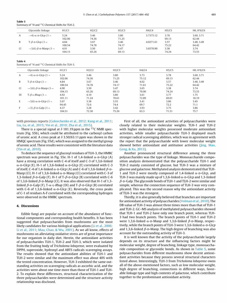
Y. Chen et al. / Carbohydrate Polymers 157 (2017) 484–492 489
Table 3Summary of 1H and 13C Chemical Shifts for TLH-2.
Glycosidic linkage H1/C1 H2/C2 H3/C3 H4/C4 H5/C5 H6, H’6/C6
A →4)-�-d-Glcp-(1→ 5.24102.06
3.4674.36
3.8071.25
3.7375.12 3.7869.13
3.60, 3.7162.44
B T-ˇ-d-Glcp-(1→ 4.84100.54
3.6774.78
3.4674.17
4.0271.61 3.5775.22
3.40, 3.4964.42
Cı →3,6)-ˇ-d-Manp-(1→ 4.91104.15
3.5065.26
3.4769.13
3.6570.90 3.3874.24
3.7473.55
Table 4Summary of 1H and 13C Chemical Shifts for TLH-3.
Glycosidic linkage H1/C1 H2/C2 H3/C3 H4/C4 H5/C5 H6, H’6/C6
A →4)-�-d-Glcp-(1→ 5.24102.06
3.4674.36
3.8071.25
3.7375.12
3.7869.13
3.60, 3.7162.44
B T-ˇ-d-Glcp-(1→ 4.84100.54
3.6774.78
3.4674.17
4.0271.61
3.5775.22
3.40, 3.4964.42
Cı →3,6)-ˇ-d-Manp-(1→ 4.90104.15
3.5065.26
3.4769.13
3.6570.90
3.3874.24
3.7473.55
Dı T-ˇ-l-Rhap-(1→ 4.92101.11
4.1071.24
3.8070.61
3.6172.89
3.7769.01
1.7518.19
E →3,6)-�-d-Glcp-(1→ 5.07 3.38 3.5574.4
3.41 3.86 3.45
3.4275.26
wL
toHo(
sh�oM1olwow
4
tslLmfofDTTtsa2tr
94.45 72.6F →3)-ˇ-d-Galp-(1→ 4.37
104.513.2872.30
ith previous reports (Golovchenko et al., 2012; Kang et al., 2011;iu, Lu, et al., 2015; Yin et al., 2010; Zha et al., 2015).
There is a special signal at ı 181.19 ppm in the 13C NMR spec-rum (Fig. S5b), which could be attributed to the carboxyl carbonf uronic acid. A cross peak at ı 3.50/63.11 ppm was shown in theMQC spectrum (Fig. S5d), which was assigned to the methyl groupf uronic acid. These results were consistent with the literature dataYin et al., 2010).
To deduce the sequence of glucosyl residues of TLH-3, the HMBCpectrum was present in Fig. S5e. H-1 of 1,4-linked �-d-Glcp (A)ave a strong correlation with C-4 of itself and C-3 of 1,3,6-linked-d-Glcp (E). H-1 of 1,3,6-linked �-d-Glcp (E) correlated with C-3f itself, C-4 of 1,4-linked �-d-Glcp (A) and C-6 of 1,3,6-linked ˇ-d-anp (Cı). H-1 of 1,3,6-linked �-d-Manp (Cı) correlated with C-3 of
,3-linked ˇ-d-Galp (F). H-1 of T-ˇ-d-Glcp (B) correlated with C-6f 1,3,6-linked ˇ-d-Manp (Cı). It was also observed that H-1 of 1,3-
inked ˇ-d-Galp (F), T-�-l-Rhap (Dı) and T-ˇ-d-Glcp (B) correlatedith C-6 of 1,3,6-linked �-d-Glcp (E). Reversely, the cross peaks
f C-1 of residues A-F correlated with the corresponding hydrogenere observed in the HMBC spectrum.
. Discussions
Edible fungi are popular on account of the abundance of func-ional components and corresponding health benefits. It has beenuggested that polysaccharides from mushrooms are the mostikely candidates for various biological applications (Li et al., 2006;i et al., 2011; Mau, Chao, & Wu, 2001). As we all know, effects ofushrooms on alleviating oxidative stress are of great importance
or our organism in daily diet. Herein, the antioxidant activitiesf polysaccharides TLH-1, TLH-2 and TLH-3, which were isolatedrom the fruiting body of Tricholoma lobayense, were evaluated byPPH, superoxide, hydroxyl and ABTS radicals scavenging assays.he results showed that the antioxidant abilities of TLH-1 andLH-2 were similar and the maximum effect was about 40% withhe tested concentration. However, TLH-3 exhibited the same out-tanding activities on scavenging radicals as ascorbic acid, and the
ctivities were about one time more than those of TLH-1 and TLH-. To explain these differences, structural characterization of thehree polysaccharides were determined and the structure-activityelationship was disclosed.69.7 72.2 71.13.9363.3
3.7571.51
3.8675.7
First of all, the antioxidant activities of polysaccharides wereclosely related to their molecular weights. TLH-1 and TLH-2with higher molecular weights possessed moderate antioxidantactivities, while smaller polysaccharide TLH-3 displayed muchstronger radical scavenging abilities, which was in agreement withthe report that the polysaccharide with lower molecular weightshowed better antioxidant and antitumor activities (Jing, Mao,Geng, & Xu, 2013).
Another pronounced structural difference among the threepolysaccharides was the type of linkage. Monosaccharide compo-sition analysis demonstrated that the polysaccharide TLH-1 andTLH-2 mainly consisted of glucose, but TLH-3 was a mixture ofglucose and galactose. Methylated analysis demonstrated that TLH-1 and TLH-2 were mostly composed of 1,4-linked �-d-Glcp, andTLH-3 was mainly made up of 1,3,6-linked �-d-Glcp and 1,3-linkedˇ-d-Galp. The glycoside bonds of TLH-1 and TLH-2 were similar andsimple, whereas the connection sequence of TLH-3 was very com-plicated. This was the second reason why the antioxidant activityof TLH-3 was the strongest.
Thirdly, it is also generally believed that branches are importantfor antioxidant activity of polysaccharides (Volman et al., 2010). TheDB value of TLH-3 was almost three times more than that of TLH-1and TLH-2. GC–MS analysis of methylated polysaccharides showedthat TLH-1 and TLH-2 have only one branch point, whereas TLH-3 had two branch points. The branch points of TLH-1 and TLH-2were 1,3,6-linked �-d-Manp and 1,3,6-linked ˇ-d-Manp, respec-tively, while the branch points of TLH-3 were 1,3,6-linked �-d-Glcpand 1,3,6-linked ˇ-d-Manp. The high degree of branching was alsoaccount for the outstanding activity of TLH-3.
It is well known that the activity of the polysaccharide largelydepends on its structure and the influencing factors might bemolecular weight, degree of branching, linkage type, monosaccha-ride composition or glycosidic bonds. As shown in Table 5, somepolysaccharides from different mushrooms show decent antioxi-dant activities because they possess several structural characterslisted above. Interestingly, TLH-3 from Tricholoma lobayense ownsall of the above-mentioned factors, such as low molecular weight,
high degree of branching, connections in different ways, favor-able linkage type and high contents of galactose, which contributetogether to the predominant antioxidant activity.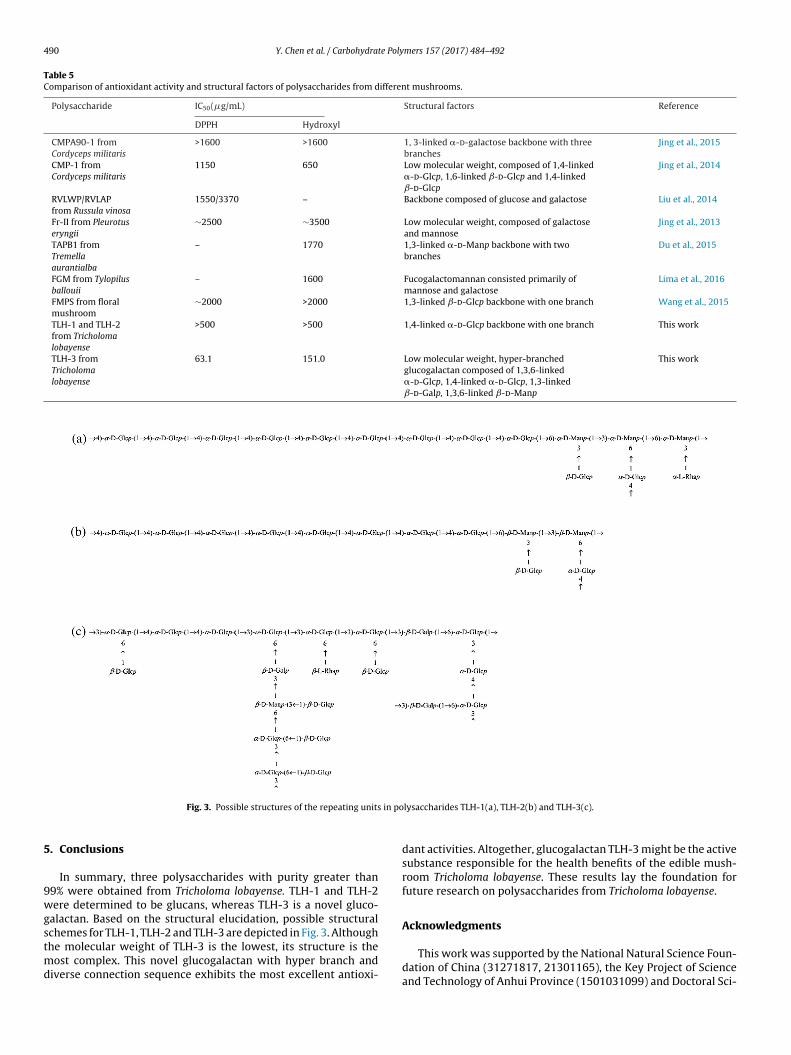
490 Y. Chen et al. / Carbohydrate Polymers 157 (2017) 484–492
Table 5Comparison of antioxidant activity and structural factors of polysaccharides from different mushrooms.
Polysaccharide IC50(�g/mL) Structural factors Reference
DPPH Hydroxyl
CMPA90-1 fromCordyceps militaris
>1600 >1600 1, 3-linked �-d-galactose backbone with threebranches
Jing et al., 2015
CMP-1 fromCordyceps militaris
1150 650 Low molecular weight, composed of 1,4-linked�-d-Glcp, 1,6-linked ˇ-d-Glcp and 1,4-linkedˇ-d-Glcp
Jing et al., 2014
RVLWP/RVLAPfrom Russula vinosa
1550/3370 – Backbone composed of glucose and galactose Liu et al., 2014
Fr-II from Pleurotuseryngii
∼2500 ∼3500 Low molecular weight, composed of galactoseand mannose
Jing et al., 2013
TAPB1 fromTremellaaurantialba
– 1770 1,3-linked �-d-Manp backbone with twobranches
Du et al., 2015
FGM from Tylopilusballouii
– 1600 Fucogalactomannan consisted primarily ofmannose and galactose
Lima et al., 2016
FMPS from floralmushroom
∼2000 >2000 1,3-linked ˇ-d-Glcp backbone with one branch Wang et al., 2015
TLH-1 and TLH-2from Tricholomalobayense
>500 >500 1,4-linked �-d-Glcp backbone with one branch This work
TLH-3 fromTricholomalobayense
63.1 151.0 Low molecular weight, hyper-branchedglucogalactan composed of 1,3,6-linked�-d-Glcp, 1,4-linked �-d-Glcp, 1,3-linkedˇ-d-Galp, 1,3,6-linked ˇ-d-Manp
This work
in po
5
9wgstmd
Fig. 3. Possible structures of the repeating units
. Conclusions
In summary, three polysaccharides with purity greater than9% were obtained from Tricholoma lobayense. TLH-1 and TLH-2ere determined to be glucans, whereas TLH-3 is a novel gluco-
alactan. Based on the structural elucidation, possible structural
chemes for TLH-1, TLH-2 and TLH-3 are depicted in Fig. 3. Althoughhe molecular weight of TLH-3 is the lowest, its structure is theost complex. This novel glucogalactan with hyper branch andiverse connection sequence exhibits the most excellent antioxi-
lysaccharides TLH-1(a), TLH-2(b) and TLH-3(c).
dant activities. Altogether, glucogalactan TLH-3 might be the activesubstance responsible for the health benefits of the edible mush-room Tricholoma lobayense. These results lay the foundation forfuture research on polysaccharides from Tricholoma lobayense.
Acknowledgments
This work was supported by the National Natural Science Foun-dation of China (31271817, 21301165), the Key Project of Scienceand Technology of Anhui Province (1501031099) and Doctoral Sci-
e Poly
eta
A
t0
R
B
B
B
B
C
C
C
C
C
C
C
D
D
G
G
H
L
J
J
J
K
K
Y. Chen et al. / Carbohydrat
ntific Research Foundation of Anhui University. The authors thankhe testing center of University of Science and Technology of Chinand Hefei University of Technology for NMR measurements.
ppendix A. Supplementary data
Supplementary data associated with this article can be found, inhe online version, at http://dx.doi.org/10.1016/j.carbpol.2016.10.11.
eferences
ao, X.-F., Wang, X.-S., Dong, Q., Fang, J.-N., & Li, X.-Y. (2002). Structural features ofimmunologically active polysaccharides from Ganoderma lucidum.Phytochemistry, 59, 175–181.
arahona, T., Chandía, N. P., Encinas, M. V., Matsuhiro, B., & Zúniga, E. A. (2011).Antioxidant capacity of sulfated polysaccharides from seaweeds. A kineticapproach. Food Hydrocolloids, 25, 529–535.
lumenkrantz, N., & Asboe-Hansen, G. (1973). New method for quantitativedetermination of uronic acids. Analytical Biochemistry, 54, 484–489.
radford, M. M. (1976). A rapid and sensitive method for the quantitation ofmicrogram quantities of protein utilizing the principle of protein-dye binding.Analytical Biochemistry, 72, 248–254.
alonje, M., García Mendoza, C., & Novaes-Ledieu, M. (1996). New contributions tothe wall polysaccharide structure of vegetative mycelium and fruit body cellwalls of Agaricus bisporus. Microbiologia (Madrid, Spain), 12, 599–606.
ao, W., Li, X. Q., Liu, L., Wang, M., Fan, H. T., Li, C., et al. (2006). Structural analysisof water-soluble glucans from the root of Angelica sinensis (Oliv.) Diels.Carbohydrate Research, 341, 1870–1877.
hang, S., & Buswell, J. (1996). Mushroom nutriceuticals. World Journal ofMicrobiology and Biotechnology, 12, 473–476.
hang, C. J., Lin, C. S., Lu, C. C., Martel, J., Ko, Y. F., Ojcius, D. M., et al. (2015).Ganoderma lucidum reduces obesity in mice by modulating the compositionof the gut microbiota. Nature Communications, 6, 7489.
hen, X., Cao, D., Zhou, L., Jin, H., Dong, Q., Yao, J., et al. (2011). Structure of apolysaccharide from Gastrodia elata Bl.: and oligosaccharides prepared thereofwith anti-pancreatic cancer cell growth activities. Carbohydrate Polymers, 86,1300–1305.
hen, X., Xiao, F., Wang, Y., Fang, J., & Ding, K. (2012). Structure-activityrelationship study of WSS25 derivatives with anti-angiogenesis effects.Glycoconjugate Journal, 29, 389–398.
hen, L., Zhang, B.-B., Chen, J.-L., & Cheung, P. C. K. (2014). Cell wall structure ofmushroom sclerotium (Pleurotus tuber-regium): Part 2: Fine structure of anovel alkali-soluble hyper-branched cell wall polysaccharide. FoodHydrocolloids, 38, 48–55.
as, D., Mondal, S., Roy, S. K., Maiti, D., Bhunia, B., Maiti, T. K., et al. (2009). Isolationand characterization of a heteropolysaccharide from the corm ofAmorphophallus campanulatus. Carbohydrate Research, 344, 2581–2585.
u, X., Zhang, Y., Mu, H., Lv, Z., Yang, Y., & Zhang, J. (2015). Structural elucidationand antioxidant activity of a novel polysaccharide (TAPB1) from Tremellaaurantialba. Food Hydrocolloids, 43, 459–464.
iavasis, I. (2014). Bioactive fungal polysaccharides as potential functionalingredients in food and nutraceuticals. Current Opinion in Biotechnology, 26,162–173.
olovchenko, V. V., Khramova, D. S., Ovodova, R. G., Shashkov, A. S., & Ovodov, Y. S.(2012). Structure of pectic polysaccharides isolated from onion Allium cepa L:Using a simulated gastric medium and their effect on intestinal absorption.Food Chemistry, 134, 1813–1822.
abibi, Y., Heyraud, A., Mahrouz, M., & Vignon, M. R. (2004). Structural features ofpectic polysaccharides from the skin of Opuntia ficus-indica prickly pear fruits.Carbohydrate Research, 339, 1119–1127.
iu, J., Wen, X. Y., Kan, J., & Jin, C. H. (2015). Structural characterization of twowater-soluble polysaccharides from black soybean (Glycine max (L.) Merr.).Journal of Agricultural and Food Chemistry, 63, 225–234.
ing, X., Mao, D., Geng, L., & Xu, C. (2013). Medium optimization, molecularcharacterization: And bioactivity of exopolysaccharides from Pleurotus eryngii.Archives of Microbiology, 195, 749–757.
ing, Y., Cui, X., Chen, Z., Huang, L., Song, L., Liu, T., et al. (2014). Elucidation andbiological activities of a new polysaccharide from cultured Cordyceps militaris.Carbohydrate Polymers, 102, 288–296.
ing, Y., Zhu, J., Liu, T., Bi, S., Hu, X., Chen, Z., et al. (2015). Structuralcharacterization and biological activities of a novel polysaccharide fromcultured Cordyceps militaris and its sulfated derivative. Journal of Agriculturaland Food Chemistry, 63, 3464–3471.
ang, J., Cui, S. W., Phillips, G. O., Chen, J., Guo, Q., & Wang, Q. (2011). New studieson gum ghatti (Anogeissus latifolia) part II. Structure characterization of an
arabinogalactan from the gum by 1D, 2D NMR spectroscopy and methylationanalysis. Food Hydrocolloids, 25, 1991–1998.ao, P. F., Wang, S. H., Hung, W. T., Liao, Y. H., Lin, C. M., & Yang, W. B. (2012).Structural characterization and antioxidative activity oflow-molecular-weights beta-1, 3-glucan from the residue of extracted
mers 157 (2017) 484–492 491
Ganoderma lucidum fruiting bodies. Journal of Biomedicine and Biotechnology,2012, 673764.
Liu, L., Lu, Y., Li, X., Zhou, L., Yang, D., Wang, L., et al. (2015). A novel process forisolation and purification of the bioactive polysaccharide TLH-3′ fromTricholoma lobayense. Process Biochemistry, 50, 1146–1151.
Li, S., Zhang, G., Zeng, Q., Huang, Z., Wang, Y., Dong, T., et al. (2006). Hypoglycemicactivity of polysaccharide, with antioxidation: Isolated from culturedCordyceps mycelia. Phytomedicine, 13, 428–433.
Li, H., Xu, J., Liu, Y., Ai, S., Qin, F., Li, Z., et al. (2011). Antioxidant andmoisture-retention activities of the polysaccharide from Nostoc commune.Carbohydrate Polymers, 83, 1821–1827.
Li, X., Wang, Z., Wang, L., Walid, E., & Zhang, H. (2012). In vitro antioxidant andanti-proliferation activities of polysaccharides from various extracts ofdifferent mushrooms. International Journal of Molecular Sciences, 13,5801–5817.
Lima, A. T. M., Santos, M. N., de Souza, L. A. R., Pinheiro, T. S., Paiva, A. A. O., Dore, C.M. P. G., et al. (2016). Chemical characteristics of a heteropolysaccharide fromTylopilus ballouii mushroom and its antioxidant and anti-inflammatoryactivities. Carbohydrate Polymers, 144, 400–409.
Liu, J., & Sun, Y. (2011). Structural analysis of an alkali-extractable andwater-soluble polysaccharide (ABP-AW1) from the fruiting bodies of Agaricusblazei Murill. Carbohydrate Polymers, 86, 429–432.
Liu, Q., Tian, G., Yan, H., Geng, X., Cao, Q., Wang, H., et al. (2014). Characterization ofpolysaccharides with antioxidant and hepatoprotective activities from thewild edible mushroom Russula vinosa Lindblad. Journal of Agricultural and FoodChemistry, 62, 8858–8866.
Masuko, T., Minami, A., Iwasaki, N., Majima, T., Nishimura, S., & Lee, Y. C. (2005).Carbohydrate analysis by a phenol-sulfuric acid method in microplate format.Analytical Biochemistry, 339, 69–72.
Mau, J.-L., Chao, G.-R., & Wu, K.-T. (2001). Antioxidant properties of methanolicextracts from several ear mushrooms. Journal of Agricultural and FoodChemistry, 49, 5461–5467.
Sun, L., Peng, X., Sun, P., Shi, J., Yuan, X., Zhu, J., et al. (2012). Structuralcharacterization and immunostimulatory activity of a novel linearalpha-(1 → 6)-D-glucan isolated from Panax ginseng C. A. Meyer.Glycoconjugate Journal, 29, 357–364.
Villares, A., Mateo-Vivaracho, L., & Guillamón, E. (2012). Structural features andhealthy properties of polysaccharides occurring in mushrooms. Agriculture, 2,452–471.
Volman, J. J., Helsper, J. P., Wei, S., Baars, J. J., van Griensven, L. J., Sonnenberg, A. S.,et al. (2010). Effects of mushroom-derived beta-glucan-rich polysaccharideextracts on nitric oxide production by bone marrow-derived macrophages andnuclear factor-kappaB transactivation in Caco-2 reporter cells: Can effects beexplained by structure? Molecular Nutrition & Food Research, 54, 268–276.
Wang, L., Zhang, H., Zhang, X., & Chen, Z. (2008). Purification and identification of anovel heteropolysaccharide RBPS2a with anti-complementary activity fromdefatted rice bran. Food Chemistry, 110, 150–155.
Wang, C., Chen, Y., Hu, M., Ding, J., Xu, C., & Wang, R. (2012). In vitro antioxidantactivities of the polysaccharides from Tricholoma lobayense. InternationalJournal of Biological Macromolecules, 50, 534–539.
Wang, K.-P., Wang, J., Li, Q., Zhang, Q.-L., You, R.-X., Cheng, Y., et al. (2014).Structural differences and conformational characterization of five bioactivepolysaccharides from Lentinus edodes. Food Research International, 62, 223–232.
Wang, J.-H., Xu, J.-L., Zhang, J.-C., Liu, Y., Sun, H.-J., & Zha, X. (2015). Physicochemicalproperties and antioxidant activities of polysaccharide from floral mushroomcultivated in Huangshan Mountain. Carbohydrate Polymers, 131, 240–247.
Wasser, S. P. (2002). Medicinal mushrooms as a source of antitumor andimmunomodulating polysaccharides. Applied Microbiology and Biotechnology,60, 258–274.
Weng, B., Lei, J., Jiang, Z., Zhong, Z., Xu, G., & Ye, J. (2013). Substituting wheat branwith Chamaecrista Rotundifolia hay powder in the substrate of TricholomaLobayense culture: Substrate weight loss dynamics and mass transformationratios. Scientia Horticulturae, 155, 105–110.
Wu, D. T., Meng, L. Z., Wang, L. Y., Lv, G. P., Cheong, K. L., Hu, D. J., et al. (2014).Chain conformation and immunomodulatory activity of a hyperbranchedpolysaccharide from Cordyceps sinensis. Carbohydrate Polymers, 110, 405–414.
Xu, Y., Dong, Q., Qiu, H., Cong, R., & Ding, K. (2010). Structural characterization ofan arabinogalactan from Platycodon grandiflorum roots and antiangiogenicactivity of its sulfated derivative. Biomacromolecules, 11, 2558–2566.
Xu, S., Xu, X., & Zhang, L. (2012). Branching structure and chain conformation ofwater-soluble glucan extracted from Auricularia auricula-judae. Journal ofAgricultural and Food Chemistry, 60, 3498–3506.
Yan, C., Yin, Y., Zhang, D., Yang, W., & Yu, R. (2013). Structural characterization andin vitro antitumor activity of a novel polysaccharide from Taxus yunnanensis.Carbohydrate Polymers, 96, 389–395.
Yin, Y., Yu, R., Yang, W., Yuan, F., Yan, C., & Song, L. (2010). Structuralcharacterization and anti-tumor activity of a novel heteropolysaccharideisolated from Taxus yunnanensis. Carbohydrate Polymers, 82, 543–548.
Zha, X. Q., Xue, L., Zhang, H. L., Asghar, M. N., Pan, L. H., Liu, J., et al. (2015).Molecular mechanism of a new Laminaria japonica polysaccharide on thesuppression of macrophage foam cell formation via regulating cellular lipid
metabolism and suppressing cellular inflammation. Molecular Nutrition & FoodResearch, 59, 2008–2021.Zhang, M., Cui, S. W., Cheung, P. C. K., & Wang, Q. (2007). Antitumorpolysaccharides from mushrooms: A review on their isolation process:
4 te Poly
Z
Z
87, 2725–2729.
92 Y. Chen et al. / Carbohydra
Structural characteristics and antitumor activity. Trends in Food Science &Technology, 18, 4–19.
hao, G., Kan, J., Li, Z., & Chen, Z. (2005). Characterization and immunostimulatory
activity of an (1 → 6)-�-D-glucan from the root of Ipomoea batatas.International Immunopharmacology, 5, 1436–1445.hu, J., & Wu, M. (2009). Characterization and free radical scavenging activity ofrapeseed meal polysaccharides WPS-1 and APS-2. Journal of Agricultural andFood Chemistry, 57, 812–819.
mers 157 (2017) 484–492
Zhu, W., Han, B., Sun, Y., Wang, Z., & Yang, X. (2012). Immunoregulatory effects of aglucogalactan from the root of Panax quinquefolium L. Carbohydrate Polymers,
Zhu, Q., Jiang, Y., Lin, S., Wen, L., Wu, D., Zhao, M., et al. (2013). Structuralidentification of (1 → 6)-�-D-glucan, a key responsible for the health benefitsof longan, and evaluation of anticancer activity. Biomacromolecules, 14,1999–2003.