Structural dissection of Ebola virus and its assembly determinants ...

Structural determinants for membrane associationand dynamic organization of the hepatitis C virusNS3-4A complexVolker Brass†‡, Jan Martin Berke†‡§, Roland Montserret‡¶, Hubert E. Blum†, Francois Penin¶�, and Darius Moradpour†§�
†Department of Medicine II, University of Freiburg, D-79106 Freiburg, Germany; §Division of Gastroenterology and Hepatology, Centre HospitalierUniversitaire Vaudois, University of Lausanne, CH-1011 Lausanne, Switzerland; and ¶Institut de Biologie et Chimie des Proteines, UMR 5086,Centre National de la Recherche Scientifique, Universite de Lyon, IFR 128 BioSciences Gerland-Lyon Sud, F-69397 Lyon, France
Communicated by Charles M. Rice, The Rockefeller University, New York, NY, July 29, 2008 (received for review April 6, 2008)
Hepatitis C virus (HCV) NS3-4A is a membrane-associated multi-functional protein harboring serine protease and RNA helicaseactivities. It is an essential component of the HCV replicationcomplex and a prime target for antiviral intervention. Here, weshow that membrane association and structural organization ofHCV NS3-4A are ensured in a cooperative manner by two mem-brane-binding determinants. We demonstrate that the N-terminal21 amino acids of NS4A form a transmembrane �-helix that may beinvolved in intramembrane protein–protein interactions importantfor the assembly of a functional replication complex. In addition,we demonstrate that amphipathic helix �0, formed by NS3 residues12–23, serves as a second essential determinant for membraneassociation of NS3-4A, allowing proper positioning of the serineprotease active site on the membrane. These results allowed us topropose a dynamic model for the membrane association, process-ing, and structural organization of NS3-4A on the membrane. Thismodel has implications for the functional architecture of the HCVreplication complex, proteolytic targeting of host factors, and drugdesign.
HCV � NMR � replication complex � serine protease � nonstructural protein
Hepatitis C virus (HCV) infection is a major cause of chronichepatitis, liver cirrhosis, and hepatocellular carcinoma
worldwide. HCV contains a 9.6-kb positive-strand RNA genomeencoding a polyprotein precursor of �3,000 amino acids, whichis co- and post-translationally processed by cellular and viralproteases to yield the structural and nonstructural proteins(reviewed in ref. 1). Nonstructural protein 3 (NS3) is a multi-functional protein, with a serine protease located in the N-terminal one-third (amino acids 1–180) and an RNA helicase inthe C-terminal two-thirds (amino acids 181–631). The proteasedomain adopts a chymotrypsin-like fold with two �-barrelsubdomains (reviewed in ref. 2). The catalytic triad is formed byHis 57, Asp 81, and Ser 139. The 54-aa NS4A polypeptidefunctions as a cofactor for the NS3 serine protease. Its centralportion comprises a �-strand that is incorporated into theN-terminal �-barrel of NS3 (3–5) whereas the hydrophobicN-terminal segment is required for membrane association (6)and the C-terminal acidic domain was recently shown to mod-ulate HCV RNA replication (7).
NS3-4A has emerged as a prime target for antiviral interven-tion (reviewed in ref. 8). In addition, it has recently been shownthat the NS3-4A protease cleaves, and thereby inactivates, twocrucial adaptor proteins in innate immune sensing, namely Trif(9) and Cardif (10) (also known as MAVS, IPS-1, and VISA),thereby blocking IFN production. Thus, the NS3-4A complexplays an essential role in HCV replication and pathogenesis.
As in all positive-strand RNA viruses investigated thus far, theHCV nonstructural proteins form a membrane-associated rep-lication complex. Here, we examined the determinants formembrane association of the NS3-4A complex through biochem-ical assays, site-directed mutagenesis, and CD and NMR struc-
tural analyses. We demonstrate that the N-terminal 21 aminoacids of NS4A form a transmembrane �-helix required forintegral membrane association of the NS3-4A complex. More-over, we demonstrate that NS3 helix �0 represents a secondessential determinant for membrane association, allowingproper positioning of the serine protease active site on themembrane. These results allowed us to propose a dynamic modelfor the membrane association, processing, and structural orga-nization of NS3-4A on the membrane.
ResultsThe N-Terminal Segment of NS4A Forms a Transmembrane �-Helix.The amino acid repertoire derived from ClustalW alignment of26 reference sequences representative of all major HCV geno-types and subtypes revealed that the N-terminal predictedmembrane segment (amino acids 1–20) and the C-terminalacidic domain (amino acids 40–54) are highly conserved (Fig.1A). By contrast, the central portion of NS4A, including the NS3cofactor (amino acids 21–32) and kink (amino acids 33–39)segments, appears much more variable. However, this apparentvariability is limited at most positions because the observedresidues exhibit similar physico-chemical properties.
We and others have previously shown that NS4A mediatesmembrane association of the NS3-4A complex (6). A panel ofNS4A deletion constructs fused to the GFP was prepared todetermine the segment required for membrane association. Asshown in Fig. 1B, constructs bearing 21 or more N-terminalamino acid residues of NS4A showed a membrane-associatedfluorescence pattern. By contrast, the other constructs showeda diffuse fluorescence pattern. These observations were con-firmed by membrane flotation analyses (data not illustrated).Taken together, these data indicate that the N-terminal 21 aminoacids of NS4A mediate membrane association.
Next, an NST consensus motif for N-linked glycosylation,followed by a 14-aa spacer sequence, was added to the Nterminus of NS4A to determine its membrane topology (gt-NS4A-GFP, Fig. 1C). A construct harboring Gln instead of Asnwithin the glycosylation acceptor site served as control (gtmut-NS4A-GFP). As shown in Fig. 1C, an additional �3-kDa bandwas observed for construct gt-NS4A-GFP, indicating that the
Author contributions: V.B., J.M.B., R.M., F.P., and D.M. designed research; V.B., J.M.B., R.M.,F.P., and D.M. performed research; V.B., J.M.B., R.M., H.E.B., F.P., and D.M. analyzed data;and H.E.B., F.P., and D.M. wrote the paper.
The authors declare no conflict of interest.
Data deposition: The atomic coordinates of have been deposited in the BioMagResBank(BMRB) (accession nos. 15580 (NS4A[1-22]*) and 15582 (NS3[10-24]).
‡V.B., J.M.B., and R.M. contributed equally to this work.
�Towhomcorrespondencemaybeaddressed.E-mail: [email protected]@chuv.ch.
This article contains supporting information online at www.pnas.org/cgi/content/full/0807298105/DCSupplemental.
© 2008 by The National Academy of Sciences of the USA
www.pnas.org�cgi�doi�10.1073�pnas.0807298105 PNAS � September 23, 2008 � vol. 105 � no. 38 � 14545–14550
MED
ICA
LSC
IEN
CES

added Asn residue was glycosylated by oligosaccharyltransferasepresent in the endoplasmic reticulum (ER) lumen. Digestionwith both endoglycosidase H and N-glycosidase F led to disap-pearance of this band, with the remaining band migrating at thesame position as gtmut-NS4A-GFP. These results indicate thatthe N-terminal segment of NS4A traverses the phospholipidbilayer as a transmembrane segment and that, given the en-doglycosidase H-sensitive nature of the transferred carbohydratemoiety, NS4A is retained in the ER. The transmembranetopology of the N-terminal segment of NS4A was confirmed byinsertion of a glycosylation acceptor site in the context of afunctional NS3-4A complex [supporting information (SI) Fig.S1]. As expected, studies involving a proteolytically inactiveNS3-4A mutant and differential membrane extraction and flo-tation analyses indicated that processing between NS3 and NS4Ais required for integral membrane association of the NS3-4Acomplex (Fig. S1 and data not illustrated).
The structure of the transmembrane segment of NS4A wasexamined by CD and NMR by using a synthetic peptide withKKGG and GGKK solubilization tags at the N- and C-terminalends, respectively, designated as NS4A[1-22]*. After solubiliza-tion in various membrane mimetics NS4A[1-22]* exhibited CDspectra typical for an �-helix (Fig. S2). For NMR, the peptidewas in 50% TFE-d2. An overview of nuclear Overhauser en-hancement (NOE) connectivities and the deviation of 1H�chemical shifts from random coil values are shown in Fig. S3.These analyses clearly showed that the main body of the peptide(Thr 2–Thr 20) forms an �-helix. On the basis of the NOE-
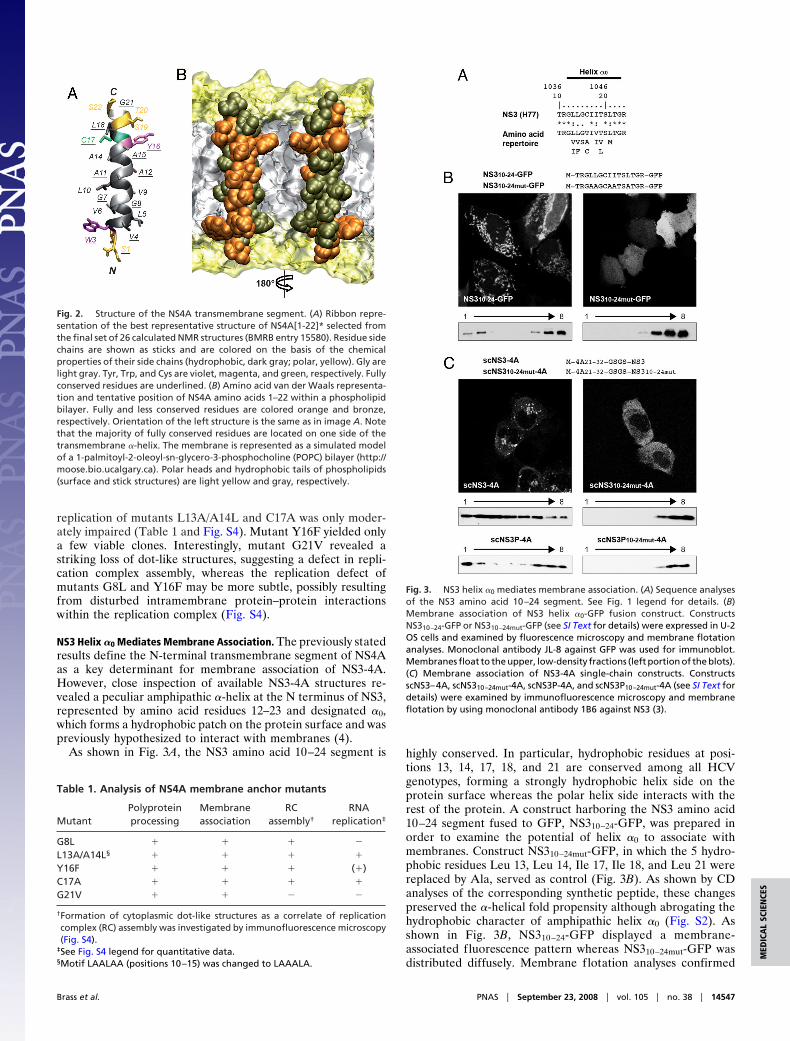
derived interproton distance constraints, a structural model ofNS4A[1-22]* was calculated that fully satisfied the experimentalNMR data (Fig. 2A and Table S1). The NS4A transmembrane�-helix displays several remarkable features, including an un-usually high number of small Gly and Ala residues, an absolutelyconserved side (Fig. 2B, residues highlighted in orange), andtypical interface residues (Trp 3 and Tyr 16) at both ends. Asdiscussed below, the numerous well-conserved small amino acidresidues may play a role in the post-translational membraneinsertion process and their conservation on one side of the helixmay point to intramembrane protein–protein interactions.
Mutational Analysis of the NS4A Membrane Anchor. The high degreeof amino acid sequence conservation within the NS4A trans-membrane segment suggests that it may have additional func-tions apart from serving as membrane anchor. To furtherexplore this idea, we performed site-directed mutagenesis ofselected conserved amino acid residues and examined thepolyprotein processing, membrane association, and RNA repli-cation of these mutants. In addition, the formation of cytoplas-mic dot-like structures as a correlate of replication complexassembly (1) was investigated by immunofluorescence micros-copy. Mutations were designed to preserve the �-helical fold ofthe NS4A transmembrane segment. All mutants were properlyprocessed and membrane-associated when expressed in thecontext of the HCV polyprotein (data not illustrated). Whenanalyzed in the context of selectable subgenomic replicons,mutants G8L and G21V did not yield any colonies whereas RNA
Fig. 1. Identification of the membrane segment of NS4A. (A) Amino acid sequence analyses. The NS4A sequence from the HCV H77 consensus clone (GenBankaccession no. AF009606) is shown at the top. Amino acids are numbered with respect to NS4A and the HCV polyprotein (top row). The amino acid repertoire of26 reference sequences representative of all major HCV genotypes and subtypes (http://euhcvdb.ibcp.fr; ref. 21) is given below, with the observed amino acidslisted in decreasing order of frequency and the similarity index according to ClustalW convention (asterisk, invariant; colon, highly similar; dot, similar). (B)Subcellular localization of NS4A-GFP fusion constructs. U-2 OS cells transiently transfected with pCMVNS4A-GFP (NS4A) or C- and N-terminal NS4A deletionconstructs fused to GFP (see SI Text for details) were examined by fluorescence microscopy. (C) Membrane topology of NS4A. Lysates of U-2 OS cells transfectedwith pCMVgt-NS4A-GFP or pCMVgtmut-NS4A-GFP (see SI Text for details) were digested with endoglycosidase H (Endo H) or N-glycosidase F (N-glyc F), followedby 15% SDS/PAGE and immunoblot with monoclonal antibody JL-8 against GFP (Clontech). The position of glycosylated gt-NS4A-GFP is indicated by an asterisk.
14546 � www.pnas.org�cgi�doi�10.1073�pnas.0807298105 Brass et al.
replication of mutants L13A/A14L and C17A was only moder-ately impaired (Table 1 and Fig. S4). Mutant Y16F yielded onlya few viable clones. Interestingly, mutant G21V revealed astriking loss of dot-like structures, suggesting a defect in repli-cation complex assembly, whereas the replication defect ofmutants G8L and Y16F may be more subtle, possibly resultingfrom disturbed intramembrane protein–protein interactionswithin the replication complex (Fig. S4).
NS3 Helix �0 Mediates Membrane Association. The previously statedresults define the N-terminal transmembrane segment of NS4Aas a key determinant for membrane association of NS3-4A.However, close inspection of available NS3-4A structures re-vealed a peculiar amphipathic �-helix at the N terminus of NS3,represented by amino acid residues 12–23 and designated �0,which forms a hydrophobic patch on the protein surface and waspreviously hypothesized to interact with membranes (4).
As shown in Fig. 3A, the NS3 amino acid 10–24 segment is highly conserved. In particular, hydrophobic residues at posi-tions 13, 14, 17, 18, and 21 are conserved among all HCVgenotypes, forming a strongly hydrophobic helix side on theprotein surface whereas the polar helix side interacts with therest of the protein. A construct harboring the NS3 amino acid10–24 segment fused to GFP, NS310–24-GFP, was prepared inorder to examine the potential of helix �0 to associate withmembranes. Construct NS310–24mut-GFP, in which the 5 hydro-phobic residues Leu 13, Leu 14, Ile 17, Ile 18, and Leu 21 werereplaced by Ala, served as control (Fig. 3B). As shown by CDanalyses of the corresponding synthetic peptide, these changespreserved the �-helical fold propensity although abrogating thehydrophobic character of amphipathic helix �0 (Fig. S2). Asshown in Fig. 3B, NS310–24-GFP displayed a membrane-associated fluorescence pattern whereas NS310–24mut-GFP wasdistributed diffusely. Membrane flotation analyses confirmed
Fig. 2. Structure of the NS4A transmembrane segment. (A) Ribbon repre-sentation of the best representative structure of NS4A[1-22]* selected fromthe final set of 26 calculated NMR structures (BMRB entry 15580). Residue sidechains are shown as sticks and are colored on the basis of the chemicalproperties of their side chains (hydrophobic, dark gray; polar, yellow). Gly arelight gray. Tyr, Trp, and Cys are violet, magenta, and green, respectively. Fullyconserved residues are underlined. (B) Amino acid van der Waals representa-tion and tentative position of NS4A amino acids 1–22 within a phospholipidbilayer. Fully and less conserved residues are colored orange and bronze,respectively. Orientation of the left structure is the same as in image A. Notethat the majority of fully conserved residues are located on one side of thetransmembrane �-helix. The membrane is represented as a simulated modelof a 1-palmitoyl-2-oleoyl-sn-glycero-3-phosphocholine (POPC) bilayer (http://moose.bio.ucalgary.ca). Polar heads and hydrophobic tails of phospholipids(surface and stick structures) are light yellow and gray, respectively.
Table 1. Analysis of NS4A membrane anchor mutants
MutantPolyproteinprocessing
Membraneassociation
RCassembly†
RNAreplication‡
G8L � � � �
L13A/A14L§ � � � �
Y16F � � � (�)C17A � � � �
G21V � � � �
†Formation of cytoplasmic dot-like structures as a correlate of replicationcomplex (RC) assembly was investigated by immunofluorescence microscopy(Fig. S4).
‡See Fig. S4 legend for quantitative data.§Motif LAALAA (positions 10–15) was changed to LAAALA.
Fig. 3. NS3 helix �0 mediates membrane association. (A) Sequence analysesof the NS3 amino acid 10–24 segment. See Fig. 1 legend for details. (B)Membrane association of NS3 helix �0-GFP fusion construct. ConstructsNS310–24-GFP or NS310–24mut-GFP (see SI Text for details) were expressed in U-2OS cells and examined by fluorescence microscopy and membrane flotationanalyses. Monoclonal antibody JL-8 against GFP was used for immunoblot.Membranes float to the upper, low-density fractions (left portion of the blots).(C) Membrane association of NS3-4A single-chain constructs. ConstructsscNS3–4A, scNS310–24mut-4A, scNS3P-4A, and scNS3P10–24mut-4A (see SI Text fordetails) were examined by immunofluorescence microscopy and membraneflotation by using monoclonal antibody 1B6 against NS3 (3).
Brass et al. PNAS � September 23, 2008 � vol. 105 � no. 38 � 14547
MED
ICA
LSC
IEN
CES

that NS310–24-GFP, but not NS310–24mut-GFP, associates withmembranes (Fig. 3B, bottom).
To explore the role of NS3 helix �0 in the membrane associ-ation of the NS3-4A complex, single-chain (sc) constructs wereprepared in which the central portion of NS4A was fused tothe N terminus of either full-length NS3 (scNS3–4A) (5) orthe protease domain alone (scNS3P-4A). ConstructsscNS310–24mut-4A and scNS3P10–24mut-4A harbor the Ala substi-tutions described above. As shown in Fig. 3C, scNS3–4A dis-played a membrane-associated staining pattern with formationof granular structures in the cytoplasm. By contrast, diffusestaining with sparing of the nucleus was observed forscNS310–24mut-4A. These results were confirmed by membraneflotation analyses (Fig. 3C). Similar observations were made forscNS3P-4A and scNS3P10–24mut-4A (Fig. 3C, bottom). The broaddistribution of scNS3–4A and, to a lesser extent, scNS3P-4A inthe density gradient may be explained by the formation ofmicellar protein aggregates, mainly because of the hydrophobicLeu and Ile residues in helix �0.
Structural Analyses of NS3 Helix �0 and Implications for the MembraneTopology of NS3-4A. The lipophilic properties and structure ofNS3 helix �0 were examined by CD and NMR in membrane-mimetic media by using a synthetic peptide representing the NS3amino acid 10–24 segment, NS3[10–24] (Fig. S2). This peptideis unfolded in water but folds into an �-helix in all membranemimetics tested, indicating the lipophilic properties of helix �0.NMR analyses of NS3[10–24] in 50% TFE-d2 and 100 mMSDS-d25 yielded well-resolved spectra that allowed completesequential attribution and, together with the 1H� and 13C�chemical shift variation (Fig. S3), revealed an amphipathic�-helix between Leu 13 and Thr 22 (Fig. 4A). Importantly, thestructure resolved by NMR in membrane-mimetic media wasperfectly superposable to the structure of helix �0 as present inthe X-ray structure of NS3-4A (Fig. 4B). Taken together, the CDand NMR data indicate that amphipathic helix �0 likely foldsupon interaction with the membrane interface. Importantly, thepositioning of amphipathic helix �0 in an in-plane topology at themembrane interface, with the hydrophobic residues orientedtoward the hydrophobic membrane core, and of the transmem-brane �-helix of NS4A dictate the topology of the NS3 serineprotease domain on the membrane (Fig. 4 C and D).
DiscussionHere, we show that membrane association of NS3-4A is con-ferred by two determinants, amphipathic helix �0, formed by NS3residues 12–23, which interacts in-plane with the membraneinterface, and the N-terminal 21 amino acids of NS4A, whichform a transmembrane �-helix. These observations and availablestructural data allowed us to propose a dynamic model for themembrane association, processing, and structural organizationof NS3-4A on the membrane (Fig. 5; video available at http://www.ibcp.fr/en/gallery/49/gallery.php).
In the HCV polyprotein context, translation of NS3 likelyoccurs at the membrane (Fig. 5, step 1). Induced folding ofamphipathic helix �0 upon interaction with the membraneinterface may therefore represent a cotranslational event, fol-lowed by folding of the protease and helicase domains (Fig. 5,step 2). The zinc molecule likely plays a central role in the foldingof the C-terminal �-barrel subdomain of the NS3 serine protease(11). The folding of uncleaved NS4A is not defined at this stage.As its central portion is required for cleavage at the NS3/NS4Asite (12), it is expected that this segment interacts with theprotease domain before cleavage. However, tight incorporationof the central NS4A �-strand into the N-terminal �-barrelsubdomain of the protease would induce its final folding and, assubsequently discussed, would break the protease–helicase in-teraction. Thus, a low affinity interaction with NS3 serine
protease �-strands A1 and A0 (see ref. 2 for the nomenclature ofNS3-4A secondary structure elements) may be postulated at thispoint. At this stage, the in-plane membrane association of helix�0 does not impose any constraints on the positioning of NS3,which could thus participate in NS2–NS3 processing. Forwardmovement of NS3 could bring the hydrophobic N-terminalsegment of NS4A into close contact with the membrane, therebyfacilitating its post-translational insertion into the membraneafter processing at the NS3/NS4A site (Fig. 5, step 3). Finalincorporation of the central segment of NS4A induces thecofolding of composite �-sheet A0(NS3)-D1�(NS4A)-A1(NS3)within the N-terminal �-barrel which in turn stabilizes theinteraction of helix �0 with the NS3 serine protease. Thiscomplete folding and membrane association by amphipathichelix �0 and the transmembrane segment of NS4A lock theprotease in a strictly defined position onto the membrane (Fig.5, step 4). As shown in the brackets, the hydrophilic helicasedomain would be immersed into the membrane at this stage inthe NS3-4A cis-cleavage conformation (5). Hence, the helicasedomain has to move away from the protease in the finalmembrane-associated stage through a rotation of the linkersegment connecting the two domains (Fig. 5, step 5). As aconsequence, the helicase domain is free to interact with othercomponents of the HCV replicase. Moreover, one should pos-tulate a second conformation of the NS3-4A complex, with theprotease and helicase domains interacting by contacts differentfrom the ones identified in the cis-cleavage structure. Indeed, therecently reported crystal structure of NS3 from the related
Fig. 4. Structural analyses of NS3 helix �0. (A) Ribbon representation of thebest representative structure of NS3[10–24] selected from the final set of 26calculated NMR structures in 100 mM SDS-d25 (BMRB entry 15582). Residueside chains are colored as in Fig. 2A. (B) Comparison of NS3 amino acid 10–24structures obtained by NMR (red) and by X-ray crystallography (ref. 5; PDBentry 1CU1; green and cyan). The two structures were superimposed fromresidues 12–23. Left and right images correspond to axial and perpendicularviews. (C and D) Model of the membrane-associated NS3 serine proteasedomain complexed with NS4A (perpendicular and axial views relative to NS3helix �0). This model was constructed by using the coordinates reported by Yaoet al. (ref. 5; PDB entry 1CU1) and the structure of NS4A[1-22]* reported in Fig.2. The NS3 serine protease domain is cyan, with side chain atoms of thecatalytic triad (His 57, Asp 81, and Ser 139) highlighted as purple spheres. NS3helix �0 is green and the five hydrophobic residues are represented by sticksand balls. The N-terminal transmembrane (amino acids 1–20) and central(amino acids 21–32) segments of NS4A are orange and light orange, respec-tively. The membrane is represented as a simulated model of POPC bilayer (seeFig. 2 legend for details).
14548 � www.pnas.org�cgi�doi�10.1073�pnas.0807298105 Brass et al.

Dengue virus shows a relative orientation between the proteaseand helicase domains drastically different from the one knownfor the HCV NS3-4A cis-cleavage structure, resulting in anelongated shape of the molecule (13). Interestingly, a similarconformation on the membrane has recently been proposed forthe NS2B-3 serine protease of the related flaviviruses (14, 15).In this case, hydrophobic residues within an N-terminal hairpinloop of NS3 are spatially homologous to the hydrophobicresidues of helix �0, suggesting convergent evolution of themembrane binding elements.
Our model suggests a possible interaction between NS3 helix�0 and the transmembrane �-helix of NS4A around NS4A aminoacids 19–22. This may explain the absolute conservation of Gly21 and the dramatic phenotype of NS4A mutant G21V. As afurther consequence of the positioning of helix �0 at the mem-brane interface, NS3 loop 38–40 is expected to contact themembrane interface, resulting in tripod-like positioning of theNS3-4A complex on the membrane surface (Fig. 4 C and D).
This positioning has implications for polyprotein processing byNS3-4A. The first trans-cleavage occurs rapidly and withoutabsolute requirement for NS4A (12) at the NS5A/NS5B site. Asthis site is likely not located at the membrane surface, it isconceivable that cleavage already occurs before full incorpora-tion of the central NS4A segment into the NS3 serine protease
and its locking onto the membrane (Fig. 5, step 3). In thisscenario, however, the helicase domain has to already moveaway from the protease to accommodate trans-cleavage be-tween NS5A and NS5B. The resulting NS4A-5A precursor iscleaved first between NS4A and NS4B, yielding in a relativelystable NS4B-5A intermediate, and finally between NS4B andNS5A. As the NS4A/NS4B and NS4B/NS5A cleavage sites arepredicted to be located at the membrane surface, it is likelythat trans-cleavage at these sites is performed by the NS3-4Aprotease in its final membrane-associated conformation (Fig.5, step 5).
Another important consequence of our model relates to theproteolytic targeting of host factors. In this context, strictpositioning of the protease active site with respect to themembrane confers a high degree of selectivity to potentialcellular trans-cleavage substrates. Indeed, NS3-4A cleavage ofthe RIG-I adaptor Cardif at Cys 508 occurs very close to itsC-terminal transmembrane segment (amino acids 514–535),resulting in displacement from the outer mitochondrial mem-brane and inactivation of Cardif (10, 16). Of note, we havepreviously shown that a minor proportion of NS3-4A localizes tomitochondria (6).
Our results show that NS3 helix �0 is a key determinant in themembrane association process of NS3-4A. Accordingly, mutant
Fig. 5. Mechanistic model of the membrane association process of NS3-4A. See Discussion for comments. The cytosolic side of the membrane bilayer is on thetop. (1) N-terminal portion of NS3 (amino acids 13–23 highlighted in green) before folding of helix �0 (constructed by using the NS3 serine protease structurewithout the central segment of NS4A solved by X-ray crystallography (ref. 24; PDB entry 1A1Q). (2) Membrane association of uncleaved NS3-NS4A by NS3 helix�0. The NS4A structure is undefined at this step. It is represented as sticks with N-terminal segment 1–20, central segment 21–32, and the beginning of theC-terminal segment (amino acids 33–40) in medium, light, and dark orange, respectively. The NS3 structure was constructed by using: (i) The crystal structureof scNS3–4A (ref. 5; PDB entry 1CU1), where the C terminus of the helicase domain (silver) lies within the active site of the serine protease domain (cyan); (ii) TheNS3 serine protease structure without the central segment of NS4A solved by NMR (ref. 25; PDB entry 1BT7) for the N-terminal �-barrel subdomain; and (iii) TheNMR structure of NS3[10–24] comprising helix �0 (this study). Well folded structures (helix �0, C-terminal �-barrel subdomain, and helicase domain) arerepresented as ribbon diagrams whereas the less stable or unfolded structures are represented as sticks (NS3 segment 1–9 and N-terminal �-barrel subdomain).Side-chain atoms of the catalytic triad (His 57, Asp 81, and Ser 139) are highlighted as purple spheres. (3) Cleavage at the NS3/NS4A site allows membrane insertionof the N-terminal segment of NS4A, resulting in a transmembrane �-helix. (4) Cofolding of the central segment 21–32 of NS4A into the N-terminal �-barrelsubdomain stabilizes the structure of the serine protease, which is locked onto the membrane by NS3 helix �0 and the NS4A transmembrane �-helix. Note thatthe hydrophilic helicase domain would be partially immersed into the membrane in the NS3-NS4A cis-cleavage conformation (5) (model in brackets). (5) Finaltopology of the NS3-4A complex on the membrane.
Brass et al. PNAS � September 23, 2008 � vol. 105 � no. 38 � 14549
MED
ICA
LSC
IEN
CES

constructs harboring Ala substitutions of the five hydrophobicresidues in helix �0 showed major defects in the associationbetween NS3 and NS4A, polyprotein processing, and cleavage ofCardif (data not illustrated). Interestingly, helix �0 overlaps withan important HLA-A2-restricted cytotoxic T lymphocyteepitope (NS3 amino acids 12–21) (17). The central role of helix�0 in NS3-4A function likely explains the conservation of thisepitope which may render it an attractive candidate for immu-notherapeutic interventions.
We also report here that certain NS4A membrane anchormutants show a striking discordance between preserved polypro-tein processing and membrane association on the one hand andRNA replication on the other hand. We have previously re-ported similar observations for the membrane anchors of HCVNS5A (18) and NS5B (19), suggesting that these segments haveadditional functions and are likely involved in protein–proteininteractions essential for the assembly of a functional replicationcomplex. Mutant G8L is particularly noteworthy in this regard,as glycines are frequently involved in transmembrane helix–helixinteractions (20). In addition, the high degree of amino acidconservation within the NS4A transmembrane segment may berelated to its unusual mechanism of post-translational mem-brane insertion.
In conclusion, we demonstrate that membrane association ofHCV NS3-4A is conferred by two structural determinants, NS3amphipathic helix �0 and the transmembrane segment of NS4A.On the basis of these results we propose a dynamic molecularmodel in which the sequential membrane association of bothdeterminants plays an active role in the processing and structuralorganization of NS3-4A and its final topology on the membrane.This model has implications for the functional architecture of the
HCV replication complex, proteolytic targeting of host factors,and drug design.
Materials and MethodsSequence Analyses. Sequence analyses were performed by using the EuropeanHepatitis C Virus Database (http://euhcvdb.ibcp.fr; ref. 21).
Expression Constructs. HCV sequences were derived from the HCV H77 con-sensus (22) and Con1 clones (23). Details are reported in SI Text and Table S2.
Immunofluorescence Microscopy, Membrane Flotation, and Immunoblot. Im-munofluorescence microscopy, membrane flotation, and immunoblot wereperformed as described (18).
Peptide Synthesis and Purification. Peptides NS3[10–24] (TRGLLGCIITSLTGR),NS3[10–24]mut (TRGAAGCAATSATGR), and NS4A[1-22]* (KKGGSTWVLVG-GVLAALAAYCLSTGSGGKK) were synthesized by Clonestar Biotech and puri-fied by RP-HPLC (purity �98%).
Structure Determination by CD and NMR. CD, NMR spectroscopy, NMR-derivedconstraints and structure calculation, and molecular modeling and structurerepresentation were performed by standard approaches as described in SIText.
ACKNOWLEDGMENTS. We thank Raffaele de Francesco for discussion andcritical review of the manuscript; Elke Bieck and Anja Wahl for excellenttechnical assistance; Charles M. Rice, Ralf Bartenschlager, and Jan AlbertHellings for reagents; Christophe Combet for bioinformatics supports; andEmmanuel Bettler for video preparation. This work was supported by theSwiss National Science Foundation (3100A0–107831/1), the Swiss CancerLeague (OCS-01762–08-2005), the Leenaards Foundation, the Deutsche For-schungsgemeinschaft (Mo 799/1–3 and Br 3440/2–1), the Bundesministeriumfur Bildung und Forschung (01 KI 9951), the European Commission (LSHM-CT-2004–503359, VIRGIL), and the French Centre National de la Recherche Sci-entifique and Agence Nationale de Recherches sur le SIDA et les HepatitesVirales.
1. Moradpour D, Penin F, Rice CM (2007) Replication of hepatitis C virus. Nat RevMicrobiol 5:453–463.
2. De Francesco R, Steinkuhler C (2000) Structure and function of the hepatitis C virusNS3-NS4A serine protease. Curr Top Microbiol Immunol 242:149–169.
3. Kim JL, et al. (1996) Crystal structure of the hepatitis C virus NS3 protease domaincomplexed with a synthetic NS4A cofactor peptide. Cell 87:343–355.
4. Yan Y, et al. (1998) Complex of NS3 protease and NS4A peptide of BK strain of hepatitisC virus: A 2.2 Å resolution structure in a hexagonal crystal form. Protein Sci 7:837–847.
5. Yao N, et al. (1999) Molecular views of viral polyprotein processing revealed by thecrystal structure of the hepatitis C virus bifunctional protease-helicase. Structure FoldDes 7:1353–1363.
6. Wolk B, et al. (2000) Subcellular localization, stability and trans-cleavage competenceof the hepatitis C virus NS3–NS4A complex expressed in tetracycline-regulated celllines. J Virol 74:2293–2304.
7. Lindenbach BD, et al. (2007) The C-terminus of hepatitis C virus NS4A encodes anelectrostatic switch that regulates NS5A hyperphosphorylation and viral replication.J Virol 81:8905–8918.
8. De Francesco R, Carfi A (2007) Advances in the development of new therapeutic agentstargeting the NS3-4A serine protease or the NS5B RNA-dependent RNA polymerase ofthe hepatitis C virus. Adv Drug Deliv Rev 59:1242–1262.
9. Li K, et al. (2005) Immune evasion by hepatitis C virus NS3/4A protease-mediated cleavageof the Toll-like receptor 3 adaptor protein TRIF. Proc Natl Acad Sci USA 102:2992–2997.
10. Meylan E, et al. (2005) Cardif is an adaptor protein in the RIG-I antiviral pathway andis targeted by hepatitis C virus. Nature 437:1167–1172.
11. Urbani A, et al. (1998) The metal binding site of the hepatitis C virus NS3 protease. Aspectroscopic investigation. J Biol Chem 273:18760–18769.
12. Tomei L, et al. (1996) A central hydrophobic domain of the hepatitis C virus NS4Aprotein is necessary and sufficient for the activation of the NS3 protease. J Gen Virol77:1065–1070.
13. Luo D, et al. (2008) Crystal structure of the NS3 protease-helicase from Dengue virus.J Virol 82:173–183.
14. Aleshin AE, Shiryaev SA, Strongin AY, Liddington RC (2007) Structural evidence forregulation and specificity of flaviviral proteases and evolution of the Flaviviridae fold.Protein Sci 16:795–806.
15. Chernov AV, et al. (2008) The two-component NS2B–NS3 proteinase repressesDNA unwinding activity of the West Nile virus NS3 helicase. J Biol Chem25:17270 –17278.
16. Li XD, et al. (2005) Hepatitis C virus protease NS3/4A cleaves mitochondrial antiviralsignaling protein off the mitochondria to evade innate immunity. Proc Natl Acad SciUSA 102:17717–17722.
17. Fournillier A, et al. (2007) An accelerated vaccine schedule with a poly-antigenichepatitis C virus MVA-based candidate vaccine induces potent, long lasting, and in vivocross-reactive T cell responses. Vaccine 25:7339–7353.
18. Penin F, et al. (2004) Structure and function of the membrane anchor domain ofhepatitis C virus nonstructural protein 5A. J Biol Chem 279:40835–40843.
19. Moradpour D, et al. (2004) Membrane association of the RNA-dependent RNA poly-merase is essential for hepatitis C virus RNA replication. J Virol 78:13278–13284.
20. Senes A, Ubarretxena-Belandia I, Engelman DM (2001) The C�—H�O hydrogen bond:A determinant of stability and specificity in transmembrane helix interactions. ProcNatl Acad Sci USA 98:9056–9061.
21. Combet C, et al. (2007) euHCVdb: The European hepatitis C virus database. NucleicAcids Res 35:D363–366.
22. Kolykhalov AA, et al. (1997) Transmission of hepatitis C by intrahepatic inoculationwith transcribed RNA. Science 277:570–574.
23. Lohmann V, et al. (1999) Replication of subgenomic hepatitis C virus RNAs in ahepatoma cell line. Science 285:110–113.
24. Love RA, et al. (1996) The crystal structure of hepatitis C virus NS3 proteinase revealsa trypsin-like fold and a structural zinc binding site. Cell 87:331–342.
25. Barbato G, et al. (1999) The solution structure of the N-terminal proteinase domain ofthe hepatitis C virus (HCV) NS3 protein provides new insights into its activation andcatalytic mechanism. J Mol Biol 289:371–384.
14550 � www.pnas.org�cgi�doi�10.1073�pnas.0807298105 Brass et al.