Structural bioinformatics...
68
Structural bioinformatics KFC/STBI What is structural bioinformatics? Karel Berka 1
Transcript of Structural bioinformatics...

Structural bioinformatics
KFC/STBI
What is structural bioinformatics?
Karel Berka
1

Requirements
• Project: • Structure analysis, docking, comparison of
proteins, prediction of properties from structure, ...
• 1(max. 2) page-long report with
– Hypothesis
– Brief Methodology
– Conclusions
ev. Wikipedia – fill up of pages about structural
bioinformatics
• Exam: • Project-like Questions – problem + discussion
about its possible resolution from you side 2

Content
• Structural bioinformatics, Biomolecules, Structural hierarchy
• Structure determination (X-Ray,NMR,EM), Structure file formats
• Structural databases (PDB, CATH, SCOP, Drugbank)
• Vizualization of structure, structural alignment
• Structure prediction, CASP
• Function prediction
• Binding prediction – protein-ligand and protein-protein docking
• Challenges of structural bioinformatics - membrane proteins,
nucleic acids, protein-protein interactions prediction
• …??
3

Bioinformatics
(Molecular) bio – informatics: bioinformatics is conceptualising biology in terms of molecules (in the sense of physical chemistry) and applying "informatics techniques" (derived from disciplines such as applied maths, computer science and statistics) to understand and organise the data and information associated with these molecules, on a large scale.
In short, bioinformatics is a management information system for molecular biology and has many practical applications.
Oxford English Dictionary 4
Structural bioinformatics
Use of structure
• Databases, classification – proteins, NA, drugs
• Patterns – Active sites, allosteric sites, ...
• Prediction – structure, function, active site, channels…
• Docking – Fitting of small molecules into the active site
-> in silico drug design
• Simulations – What if…
5
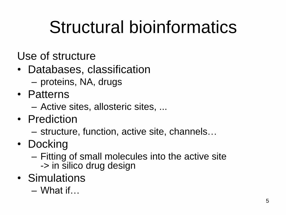
Problems of structural bioinformatics
• Structural data are hard to work with: – Nonlinear
– Imprecise from experiment (resolution of structure)
– 3D representation (3D search)
– Visualization is not trivial
– More conserved than sequence data (genomics)
– Structural genomics prepare structures without annotation
– Most structures are water soluble globular proteins (most drug targets are membrane proteins)
6

Challenges
• Target selection – Large structures are resource intensive, maybe just
one domain might be enough
• Structure methods – XRay – crystalisation is not easy
– NMR – size problem
– EM – not with atomistic detail
• Validation and Annotation
• Databases
• Correlation of structural data with experimental data
7
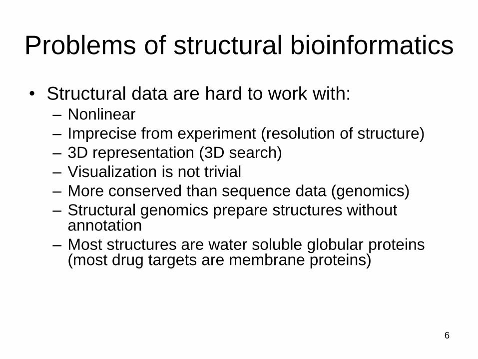
Example 1 : Prediction of protein structure
• Tertiary structure
– Fold recognition
– Homolog modelling
• Structural alignment
– ab initio modelling
• Function prediction
– active sites, channels
8
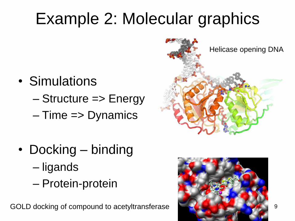
Example 2: Molecular graphics
• Simulations
– Structure => Energy
– Time => Dynamics
• Docking – binding
– ligands
– Protein-protein
Helicase opening DNA
GOLD docking of compound to acetyltransferase
9
• Coordinate systems
- XYZ (cartesian)
- Inner coordinates (bond lengths, bond
angles, torsion angles)
- object representation (secondary structure)
• Structure comparison:
RMSD – root mean square distance
Structure Description
10
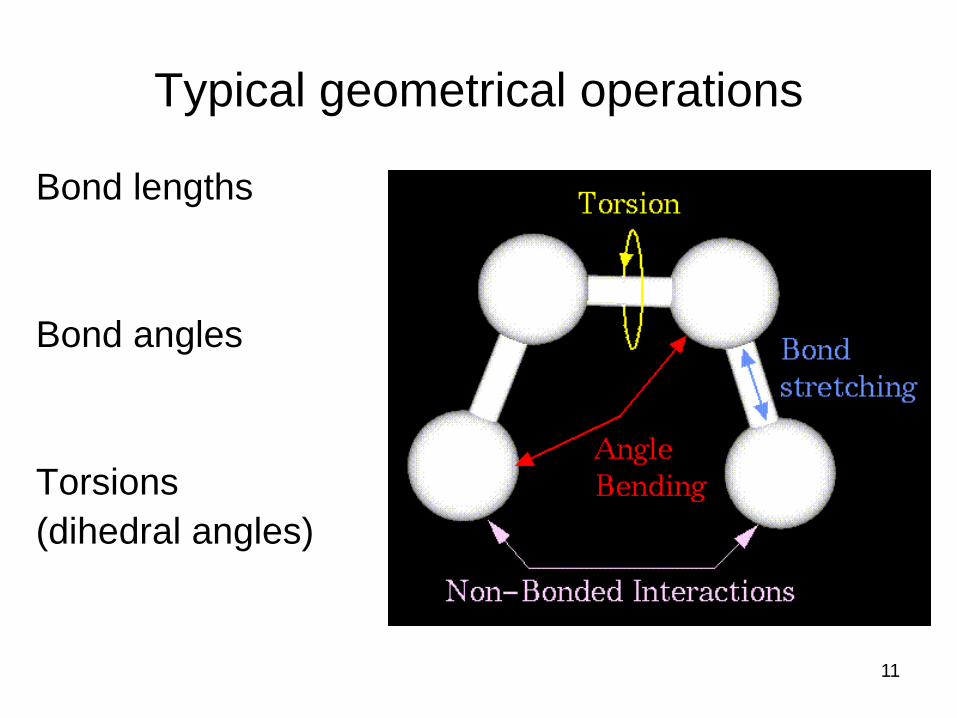
Typical geometrical operations
Bond lengths
Bond angles
Torsions
(dihedral angles)
11
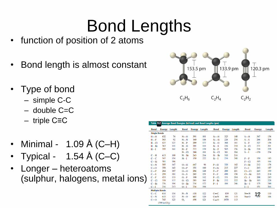
Bond Lengths • function of position of 2 atoms
• Bond length is almost constant
• Type of bond – simple C-C
– double C=C
– triple C≡C
• Minimal - 1.09 Å (C–H)
• Typical - 1.54 Å (C–C)
• Longer – heteroatoms (sulphur, halogens, metal ions)
12
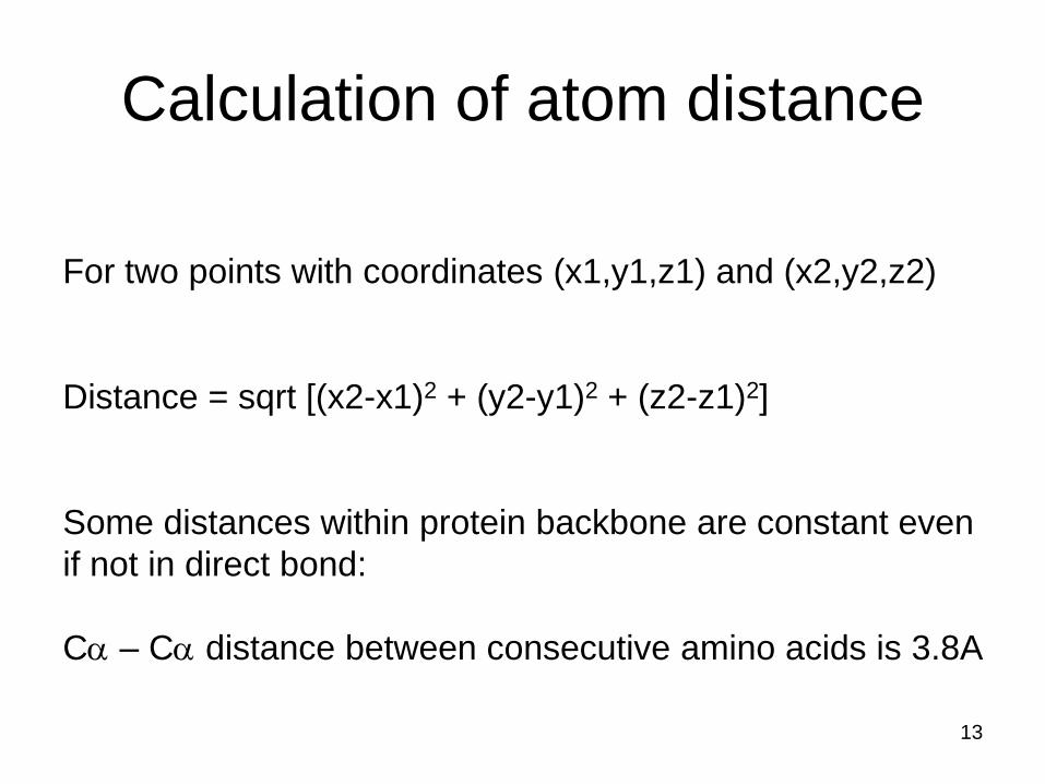
For two points with coordinates (x1,y1,z1) and (x2,y2,z2)
Distance = sqrt [(x2-x1)2 + (y2-y1)2 + (z2-z1)2]
Some distances within protein backbone are constant even
if not in direct bond:
Ca – Ca distance between consecutive amino acids is 3.8A
Calculation of atom distance
13

• function of position of 3 atoms
- Almost constant for given
combination of type of atoms
• Depend on atom type and
number of electrons in bonding
• Interval from 90 to 180
Bond Angles
14

A
B
C
x
y Q
X.Y = |X|.|Y|.cos (Q)
Q = arccos (X.Y/|X|.|Y|)
Arccosin of angle between two vectors BA and BC
Calculation of bonding angle
15

• function of position
of 4 atoms
- Quite variable (0
to 360°)
- its change change
conformations
Dihedral Angle
16

A
B C
D
A
B
C
D
f
vision
Dihedral Angle
17
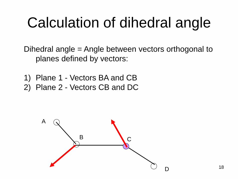
Dihedral angle = Angle between vectors orthogonal to
planes defined by vectors:
1) Plane 1 - Vectors BA and CB
2) Plane 2 - Vectors CB and DC
A
B C
D
Calculation of dihedral angle
18
Ca
N
C
O
N
N Ca
Ca
O
O
C
C
w
y
f
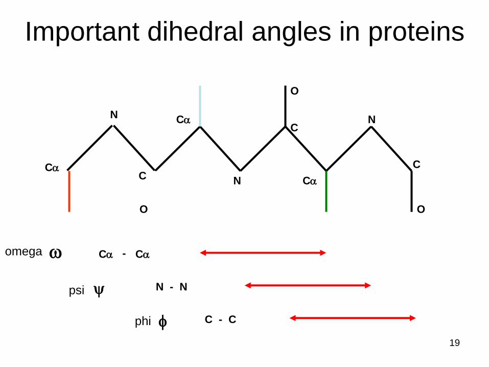
Ca Ca
N - N
C - C
-
Important dihedral angles in proteins
omega
phi
psi
19
Ca
N
C
O
N
N Ca
Ca
O
O
C
C
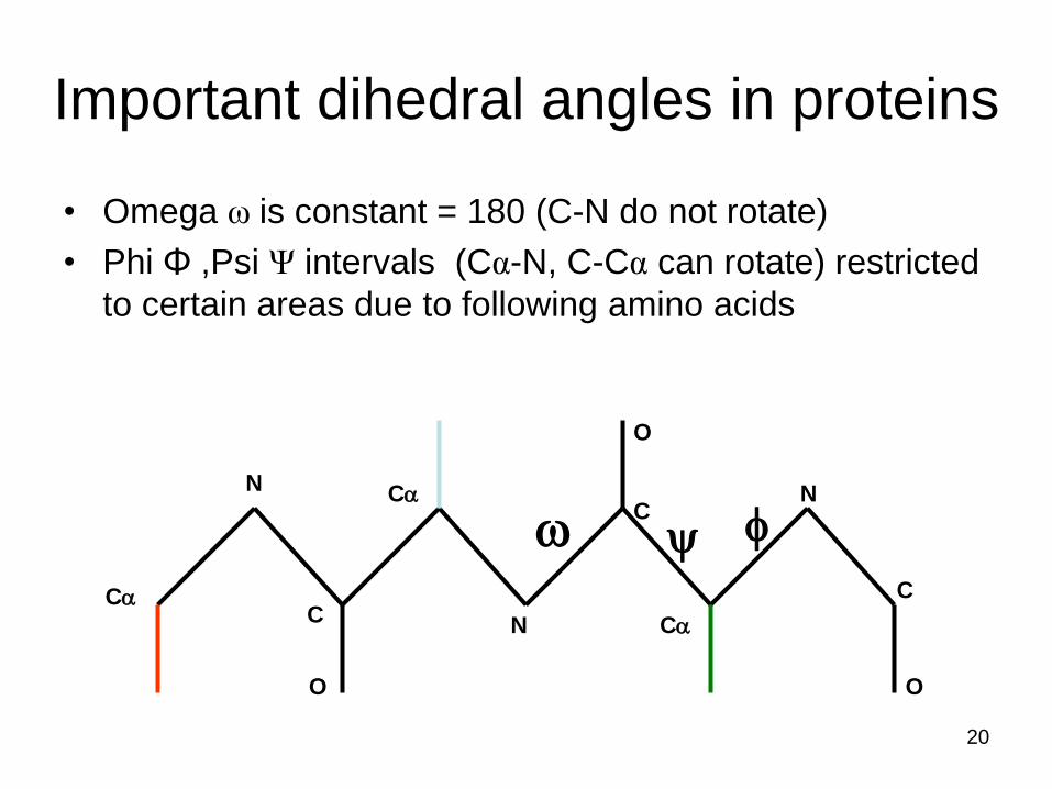
Important dihedral angles in proteins
• Omega ω is constant = 180 (C-N do not rotate)
• Phi Φ ,Psi Ψ intervals (Cα-N, C-Cα can rotate) restricted
to certain areas due to following amino acids
w y f
20

Ramachandran plot
• Typical values of dihedral angles define
individual secondary structure elements:
– α-helix phi = - 57, psi = - 47
– 3-10 helix phi = - 49, psi = - 26
– Parallel β-sheet phi = - 119, psi = 113
– Antiparallel β-sheet phi = - 139, psi = 135
21

Ramachandran plot
22

Other Coordinate Systems
Cartesian coordinates are orthogonal (x,y,z)
-> used most often
If bond lengths and bond angles are constant -> reduction of coordinates -> only dihedral angles =>
Inner coordinates
If some part of structure can be defined by “rigid” structural element -> solid objects =>
Object-based coordinates
23

3 peptide units = 12 atoms = 36 coordinates OR 6 dihedral angles
3 sidechains = 12 atoms = 36 souřadnic OR 5 dihedral angles
72 cartesians versus 11 inner
Advantages of Inner Coordinates
24

Disadvantages of Inner Coordinates
Some calculations are more difficult
Atom-atom distance
Closest atoms toward a point in space
Hard comparison of independent objects (two molecules)
Nonlinear relationships between coordinates => problem for optimizations and simulations
25
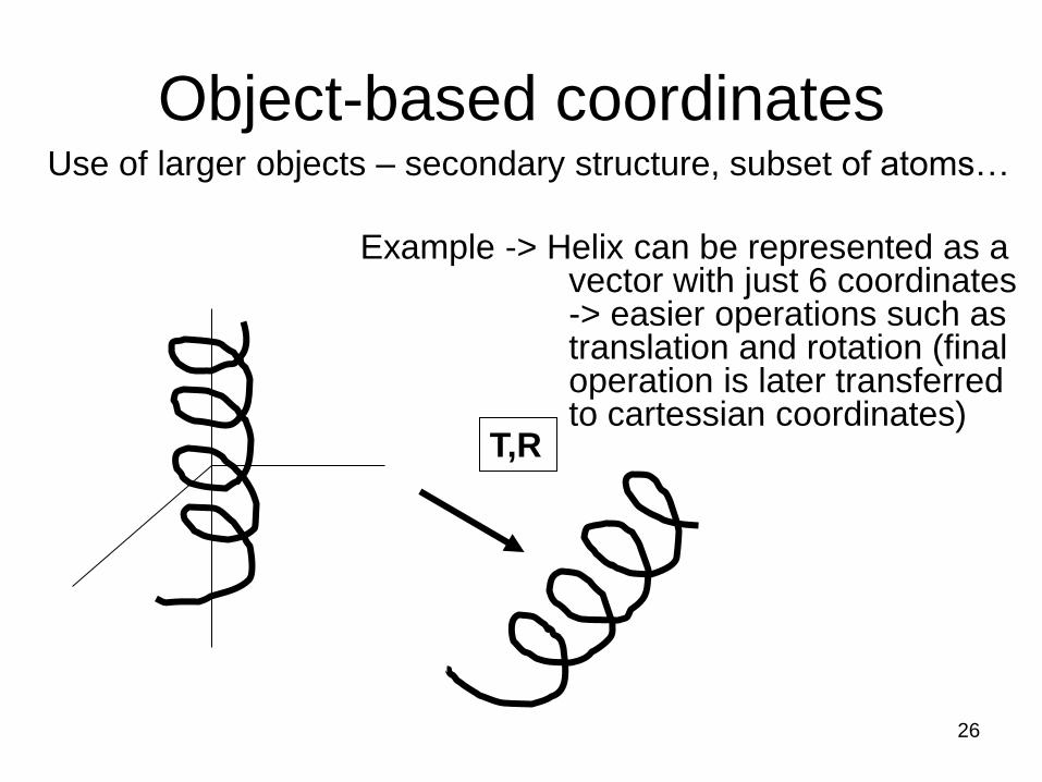
Object-based coordinates Use of larger objects – secondary structure, subset of atoms…
Example -> Helix can be represented as a vector with just 6 coordinates -> easier operations such as translation and rotation (final operation is later transferred to cartessian coordinates)
T,R
26

Structure Comparison
For comparison of two structures A and B we need:
1. Which atom from A corresponds to which atom from B
=> alignment
2. Atom localization
=> PDB files
3. Comparison criteria
RMSD, energy
27

RMSD = S d2
i
N
N – number of atoms
di – distance of two atoms with index i from A and B
RMSD = Root Mean Square Deviation
• Atoms from A and B are taken as equivalent
• Superposition and calculation of differences in distance
• If are structures identical -> RMSD = 0
• With more differences between structures -> RMSD
increses
28

Structure Comparison
To find minimal RMSD
29
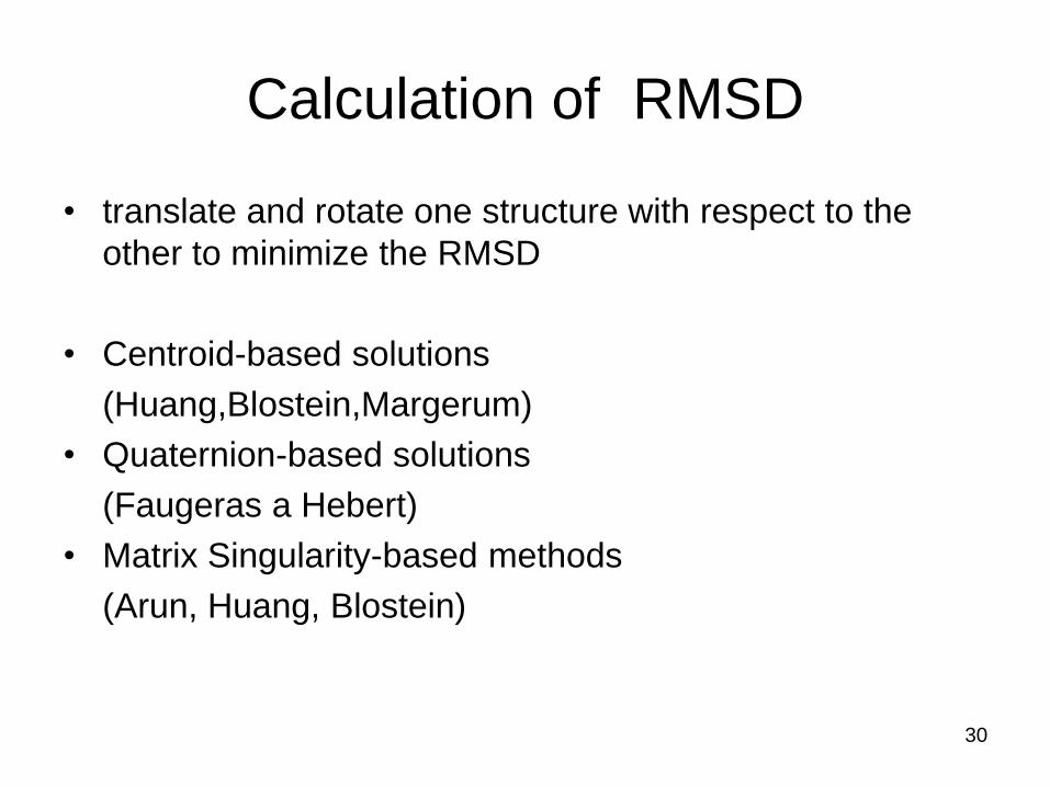
Calculation of RMSD
• translate and rotate one structure with respect to the
other to minimize the RMSD
• Centroid-based solutions
(Huang,Blostein,Margerum)
• Quaternion-based solutions
(Faugeras a Hebert)
• Matrix Singularity-based methods
(Arun, Huang, Blostein)
30

Arun algorithm
• Matrices of pi’ = R.pi + T + Ni – pi – 3x1 column matrix of positions
– R – rotation matrix
– T – translation vector 3x1 column matrix
– N – noise vector
• 1) Translation over centroids
• 2) Singular value decomposition of matrix to obtain rotation
• Arun algorithm is optimal, universal and not iterative
31
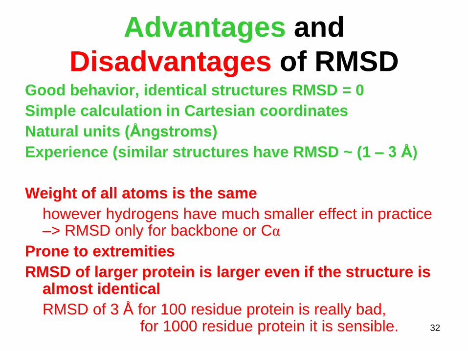
Advantages and
Disadvantages of RMSD Good behavior, identical structures RMSD = 0
Simple calculation in Cartesian coordinates
Natural units (Ångstroms)
Experience (similar structures have RMSD ~ (1 – 3 Å)
Weight of all atoms is the same
however hydrogens have much smaller effect in practice –> RMSD only for backbone or Cα
Prone to extremities
RMSD of larger protein is larger even if the structure is almost identical
RMSD of 3 Å for 100 residue protein is really bad, for 1000 residue protein it is sensible. 32

Other measures
• global distance test (GDT)
– largest set of amino acid residues' Cα atoms in the model
structure falling within a defined distance cutoff of their
position in the experimental structure.
Used in structure prediction assessment (CASP)
• template modeling score (TM-score)
– difference between two structures
by a score between (0,1]
– TM-score = 1 - perfect match between two structures
– TM-score > 0.5 assume roughly the same fold
– TM-score < 0.20 - randomly chosen unrelated proteins
Used in structure prediction assessment (CASP) 33

Biomolecules • proteins
• NA – DNA, RNA
• lipids
• polysaccharides
• Small molecules (hormones, drugs)
34
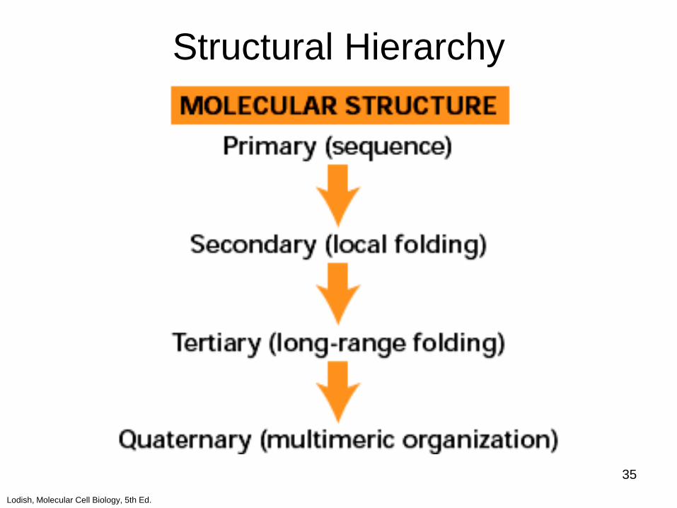
Lodish, Molecular Cell Biology, 5th Ed.
Structural Hierarchy
35

Proteins • Amino acids
• Backbone and Sidechains
• Primary structure – sequence of amino acids
• Secondary structure – Local structural patterns
• Tertiary structure – Domain Fold
• Quarternary structure – Multichain organization
http://cs.wikipedia.org/wiki/Soubor:ProteinStructures.png
36

Amino acids
37
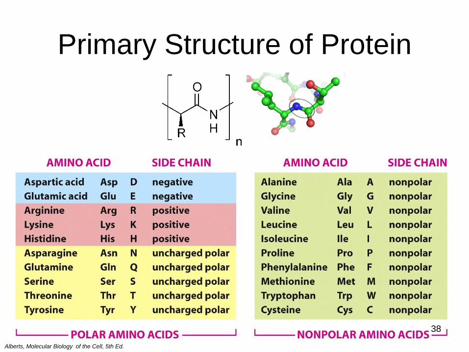
Primary Structure of Protein
Alberts, Molecular Biology of the Cell, 5th Ed.
38

Secondary structure of Proteins •Local folding
•Secondary structure depends on amino
acid sequence
– a-helix
– 3-10 helix
– β-sheet
– β-turn, loop
39

Ramachandran plot
40

PROCHECK summary for 1aaq
PROCHECK statistics
Ramachandran Plot statistics No. of residues %-tage
------ ------
Most favoured regions [A,B,L] 146 92.4% Additional allowed regions [a,b,l,p] 12 7.6%
Generously allowed regions [~a,~b,~l,~p] 0 0.0%
Disallowed regions [XX] 0 0.0%
---- ------
Non-glycine and non-proline residues 158 100.0%
End-residues (excl. Gly and Pro) 2
Glycine residues 26
Proline residues 12
----
Total number of residues 198
41
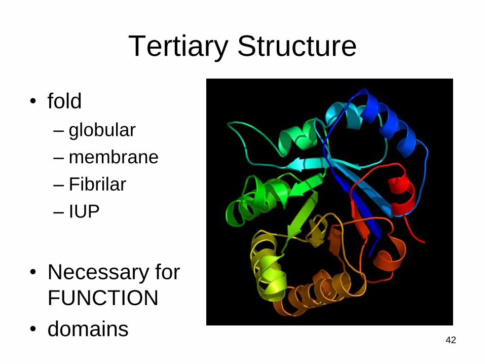
Tertiary Structure
• fold
– globular
– membrane
– Fibrilar
– IUP
• Necessary for
FUNCTION
• domains 42

Cuff A L et al. Nucl. Acids Res. 2011;39:D420-D426
© The Author(s) 2010. Published by Oxford University Press.
The distribution of all non-homologous structures (2386) within CATH v3.3 Classes: pink (mainly α), yellow (mainly β), green (αβ) brown (little secondary structure).
Proportion of structures within any given architecture (inner circle) Fold group (outer circle).
‘CATHerine wheels’.
43

Petsko, Ringe – Protein structure and function
Quarternary Structure
• asociace více řetězců: – Kooperativita
(asociace zesílí vazebné vlastnosti)
hemoglobin
– Kolokalizace funkce (každá podjednotka dělá něco
jiného)
tryptophansyntáza
– Kombinace podjednotek (přizpůsobování)
imunoglobuliny
– Skládání větších struktur (podjednotky uspořádávají
procesem self-assembly)
aktin,
virové kapsidy
44

Nucleic Acids (NA)
• Primary structure – sequence of NA basis in chains
• Secondary structure – set of interactions between nucleic basis
• Tertiary structure – 3D localization of atoms
• Quarternary structure – Higher organization levels
• DNA in chromatin
• Interaction of RNA units in ribosome or spliceosome.
45
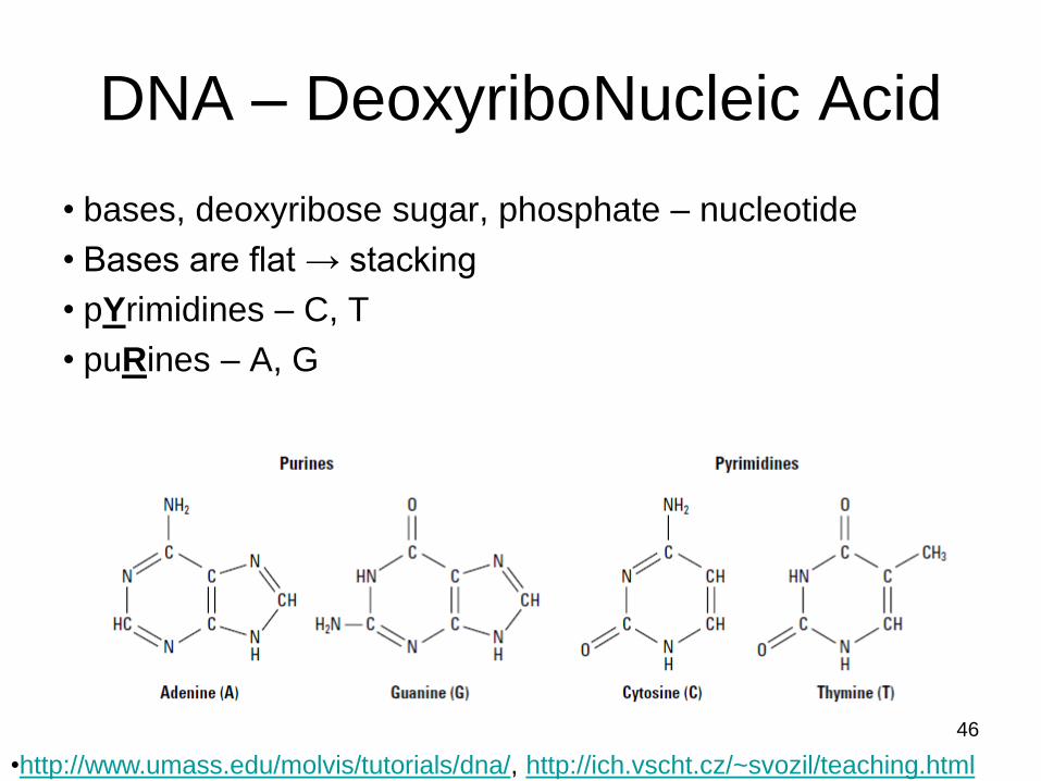
DNA – DeoxyriboNucleic Acid
• bases, deoxyribose sugar, phosphate – nucleotide
• Bases are flat → stacking
• pYrimidines – C, T
• puRines – A, G
•http://www.umass.edu/molvis/tutorials/dna/, http://ich.vscht.cz/~svozil/teaching.html
46

O3‘
O5‘
C3‘
C5‘
base
sugar
Nucleoside
47

Nucleotide
•nucleosides are interconnected by
phospohodiester bond
•nucleotide monophosphate
nucleoside
48
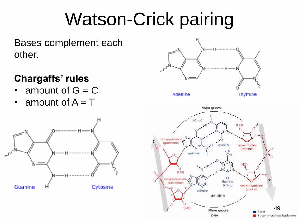
Bases complement each
other.
Chargaffs’ rules
• amount of G = C
• amount of A = T
Watson-Crick pairing
49

Párování
50

DNA backbone
5’ – end
3’ – end
51
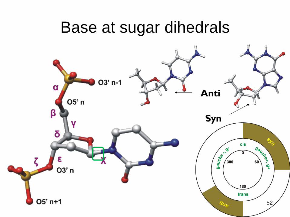
Base at sugar dihedrals
Anti
Syn
52

Sugar conformation
orientation with respect to C5’
• same side – endo
• opposite side – exo
53
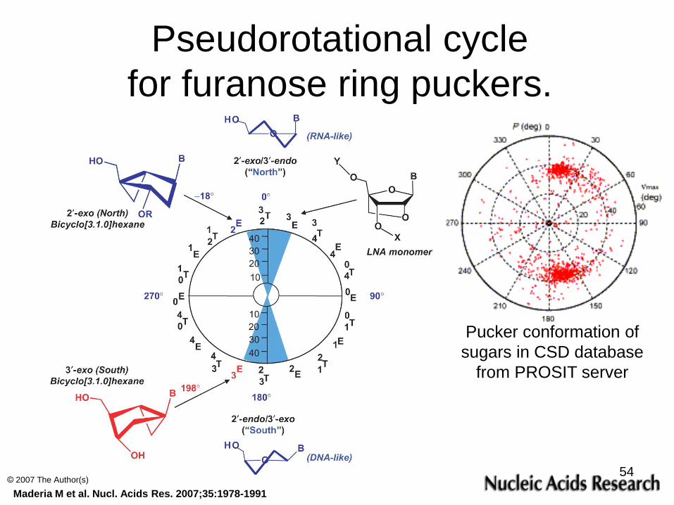
Maderia M et al. Nucl. Acids Res. 2007;35:1978-1991
© 2007 The Author(s)
Pseudorotational cycle
for furanose ring puckers.
Pucker conformation of
sugars in CSD database
from PROSIT server
54

AATCGCTA
TTAGCGAT
5’
3’
3’
5’
antiparallel
DNA Double helix
55
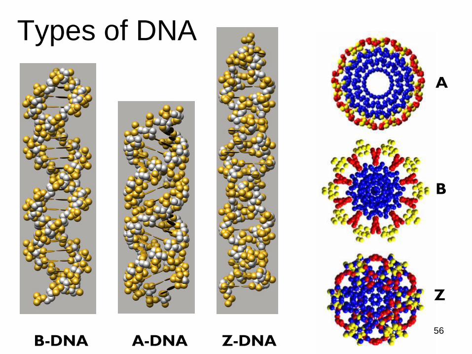
B-DNA A-DNA Z-DNA
B
A
Z
Types of DNA
56
Biological role of different DNAs
• B-DNA – canonical DNA
– predominant
• A-DNA – Conditions of lower humidity, common in crystallographic
experiments. However, they’re artificial.
– In vivo – local conformations induced e.g. by interaction with proteins.
• Z-DNA – No definite biological significance found up to now.
– It is commonly believed to provide torsional strain relief (supercoiling) while DNA transcription occurs.
– The potential to form a Z-DNA structure also correlates with regions of active transcription.
57

Different sets of DNA
• nuclear DNA
– cell’s nucleus
– majority of functions cell carries out
– sequencing the genome – scientists mean nuclear DNA
• mitochondrial DNA
– mtDNA
– circular, in human very short (17 kbp) with 37 genes (controling
cellular metabolism)
– all mtDNA comes from mom
• chloroplast DNA
– cpDNA
– circular and fairly large (120 – 160 kbp), with only 120 genes
– inheritance is either maternal, or paternal 58

RNA - ribonucleic acid
hammerhead
ribozyme 2GOZ
primární struktura
sekundární struktura
terciární
struktura
59
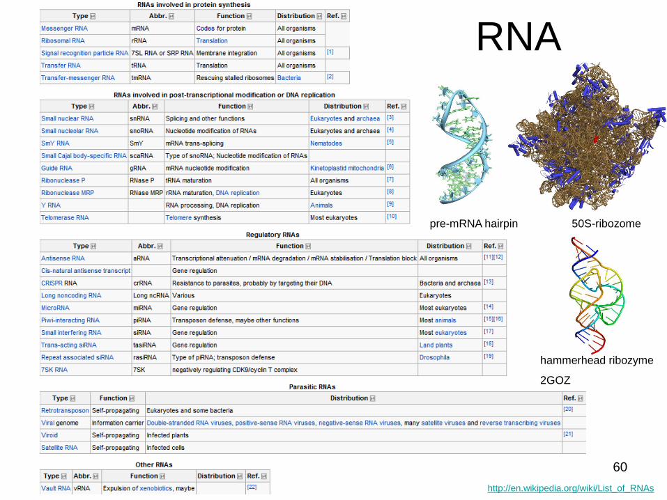
RNA
http://en.wikipedia.org/wiki/List_of_RNAs
pre-mRNA hairpin 50S-ribozome
hammerhead ribozyme
2GOZ
60

RNA
N. B. Leontis, E. Westhof, RNA (2001), 7:499-512
61

RNA sekundární struktura
N. B. Leontis, E. Westhof, RNA (2001), 7:499-512
62

RNA Representations
Mokdad A , Leontis N B Bioinformatics 2006;22:2168-2170
63
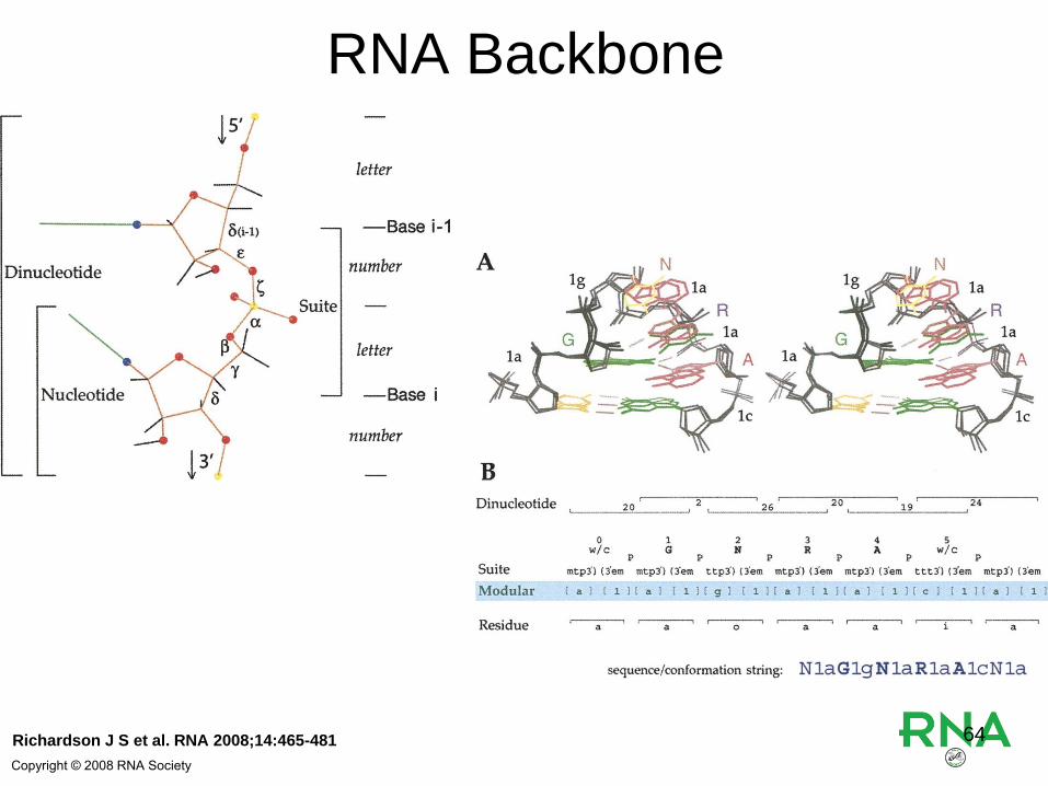
Richardson J S et al. RNA 2008;14:465-481
Copyright © 2008 RNA Society
RNA Backbone
64

Hsiao C et al. Nucl. Acids Res. 2006;34:1481-1491
RNA Tetraloop Family Tree.
65
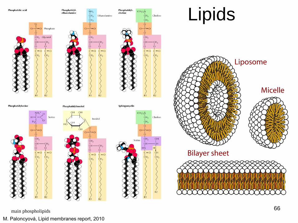
Lipids
main phospholipids
M. Paloncyová, Lipid membranes report, 2010
66

Polysaccharides • role:
– Energy storage
– Molecular recognition
• Harder to read in
sequences than NA or
proteins
• Quite often on
extracellular proteins
glycogen 67

Small molecules
• NTP
– Cell energy transporter (ATP)
– Basic stones for NA
• Messengers, Agonists, antagonists
– (cAMP, xenobiotics)
caffeine ibuprofen 68