Structural and Kinetic Properties of Lumazine Synthase ... neu sortiert/Klinke et al JMB...
17
Structural and Kinetic Properties of Lumazine Synthase Isoenzymes in the Order Rhizobiales Sebastián Klinke 1 , Vanesa Zylberman 1 , Hernán R. Bonomi 1 , Ilka Haase 2 , Beatriz G. Guimarães 3 , Bradford C. Braden 4 , Adelbert Bacher 5 , Markus Fischer 2 and Fernando A. Goldbaum 1 ⁎ 1 Fundación Instituto Leloir, IIBBA-CONICET, C1405BWE, Buenos Aires, Argentina 2 Institut für Biochemie und Lebensmittelchemie, Universität Hamburg, D-20146 Hamburg, Germany 3 Laboratório Nacional de Luz Síncrotron, Caixa Postal 6192, CEP 13084-971, Campinas, SP, Brazil 4 Department of Natural Sciences, Bowie State University, Bowie, MD 20715-9465, USA 5 Lehrstuhl für Organische Chemie und Biochemie, Technische Universität München, D-85747 Garching, Germany Received 4 July 2007; received in revised form 6 August 2007; accepted 9 August 2007 6,7-Dimethyl-8-ribityllumazine synthase (lumazine synthase; LS) catalyzes the penultimate step in the biosynthesis of riboflavin in plants and microorganisms. This protein is known to exhibit different quaternary assemblies between species, existing as free pentamers, decamers (dimers of pentamers) and icosahedrally arranged dodecamers of pentamers. A phylogenetic analysis on eubacterial, fungal and plant LSs allowed us to classify them into two categories: Type I LSs (pentameric or icosahedral) and Type II LSs (decameric). The Rhizobiales represent an order of α-proteobacteria that includes, among others, the genera Mesorhizobium, Agrobacterium and Brucella. Here, we present structural and kinetic studies on several LSs from Rhizobiales. Interestingly, Mesorhizobium and Brucella encode both a Type-I LS and a Type-II LS called RibH1 and RibH2, respectively. We show that Type II LSs appear to be almost inactive, whereas Type I LSs present a highly variable catalytic activity according to the genus. Additionally, we have solved four RibH1/RibH2 crystallographic structures from the genera Mesorhizobium and Brucella. The relationship between the active-site architecture and catalytic properties in these isoenzymes is discussed, and a model that describes the enzymatic behavior is proposed. Furthermore, sequence alignment studies allowed us to extend our results to the genus Agrobacterium. Our results suggest that the selective pressure controlling the riboflavin pathway favored the evolution of catalysts with low reaction rates, since the excess of flavins in the intracellular pool in Rhizobiales could act as a negative factor when these bacteria are exposed to oxidative or nitrosative stress. © 2007 Elsevier Ltd. All rights reserved. Edited by M. Guss Keywords: 6,7-dimethyl-8-ribityllumazine synthase; X-ray crystallography; riboflavin biosynthesis; order Rhizobiales; enzymology *Corresponding author. E-mail addresses: [email protected]; [email protected]; [email protected]; [email protected]; [email protected]; [email protected]; [email protected]; [email protected]; [email protected] . Abbreviations used: LS, 6,7-dimethyl-8-ribityllumazine synthase; NRP, 5-nitro-6-ribitylamino-2,4(1H,3H)- pyrimidinedione; PEG, polyethylene glycol; RibH1-Bab, structure of Brucella abortus RibH1 enzyme; RibH1-Bab-NRP, structure of B. abortus RibH1 bound to the substrate analogue inhibitor NRP; RibH1-Bme-NRP, structure of Brucella melitensis RibH1 bound to NRP; RibH2-Mlo-NRP, structure of Mesorhizobium loti RibH2 bound to NRP. ARTICLE IN PRESS YJMBI-59689; No. of pages: 17; 4C: 4, 6, 7, 10, 11 RGR doi:10.1016/j.jmb.2007.08.021 J. Mol. Biol. (2007) xx, xxx–xxx 0022-2836/$ - see front matter © 2007 Elsevier Ltd. All rights reserved. Please cite this article as: Klinke, S. et al., Structural and Kinetic Properties of Lumazine Synthase Isoenzymes in the Order Rhizobiales, J. Mol. Biol. (2007), doi:10.1016/j.jmb.2007.08.021
Transcript of Structural and Kinetic Properties of Lumazine Synthase ... neu sortiert/Klinke et al JMB...

ARTICLE IN PRESSYJMBI-59689; No. of pages: 17; 4C: 4, 6, 7, 10, 11RGR
doi:10.1016/j.jmb.2007.08.021 J. Mol. Biol. (2007) xx, xxx–xxx
Structural and Kinetic Properties of Lumazine SynthaseIsoenzymes in the Order Rhizobiales
Sebastián Klinke1, Vanesa Zylberman1, Hernán R. Bonomi1,Ilka Haase2, Beatriz G. Guimarães3, Bradford C. Braden4,Adelbert Bacher5, Markus Fischer2 and Fernando A. Goldbaum1⁎
1Fundación Instituto Leloir,IIBBA-CONICET, C1405BWE,Buenos Aires, Argentina2Institut für Biochemie undLebensmittelchemie, UniversitätHamburg, D-20146 Hamburg,Germany3Laboratório Nacional de LuzSíncrotron, Caixa Postal 6192,CEP 13084-971, Campinas, SP,Brazil4Department of NaturalSciences, Bowie StateUniversity, Bowie,MD 20715-9465, USA5Lehrstuhl für OrganischeChemie und Biochemie,Technische UniversitätMünchen, D-85747 Garching,Germany
Received 4 July 2007;received in revised form6 August 2007;accepted 9 August 2007
*Corresponding author. E-mail [email protected];[email protected] used: LS, 6,7-dimet
pyrimidinedione; PEG, polyethylenestructure of B. abortus RibH1 boundmelitensis RibH1 bound to NRP; Rib
0022-2836/$ - see front matter © 2007 E
Please cite this article as: Klinke, S.Rhizobiales, J. Mol. Biol. (2007), doi:10.
6,7-Dimethyl-8-ribityllumazine synthase (lumazine synthase; LS) catalyzesthe penultimate step in the biosynthesis of riboflavin in plants andmicroorganisms. This protein is known to exhibit different quaternaryassemblies between species, existing as free pentamers, decamers (dimers ofpentamers) and icosahedrally arranged dodecamers of pentamers. Aphylogenetic analysis on eubacterial, fungal and plant LSs allowed us toclassify them into two categories: Type I LSs (pentameric or icosahedral)and Type II LSs (decameric).The Rhizobiales represent an order of α-proteobacteria that includes,
among others, the genera Mesorhizobium, Agrobacterium and Brucella. Here,we present structural and kinetic studies on several LSs from Rhizobiales.Interestingly, Mesorhizobium and Brucella encode both a Type-I LS and aType-II LS called RibH1 and RibH2, respectively. We show that Type II LSsappear to be almost inactive, whereas Type I LSs present a highly variablecatalytic activity according to the genus. Additionally, we have solved fourRibH1/RibH2 crystallographic structures from the genera Mesorhizobiumand Brucella. The relationship between the active-site architecture andcatalytic properties in these isoenzymes is discussed, and a model thatdescribes the enzymatic behavior is proposed. Furthermore, sequencealignment studies allowed us to extend our results to the genusAgrobacterium. Our results suggest that the selective pressure controllingthe riboflavin pathway favored the evolution of catalysts with low reactionrates, since the excess of flavins in the intracellular pool in Rhizobiales couldact as a negative factor when these bacteria are exposed to oxidative ornitrosative stress.
© 2007 Elsevier Ltd. All rights reserved.
Keywords: 6,7-dimethyl-8-ribityllumazine synthase; X-ray crystallography;riboflavin biosynthesis; order Rhizobiales; enzymology
Edited by M. Gussesses: [email protected]; [email protected]; [email protected];[email protected]; [email protected]; [email protected];g.de; [email protected] synthase; NRP, 5-nitro-6-ribitylamino-2,4(1H,3H)-glycol; RibH1-Bab, structure of Brucella abortus RibH1 enzyme; RibH1-Bab-NRP,to the substrate analogue inhibitor NRP; RibH1-Bme-NRP, structure of BrucellaH2-Mlo-NRP, structure of Mesorhizobium loti RibH2 bound to NRP.
lsevier Ltd. All rights reserved.
et al., Structural and Kinetic Properties of Lumazine Synthase Isoenzymes in the Order1016/j.jmb.2007.08.021

2 Lumazine Synthase Isoenzymes in Rhizobiales
ARTICLE IN PRESSRGR
Introduction
Riboflavin (vitamin B2) is the precursor of flavinmononucleotide and flavin adenine dinucleotide—two key cofactors involved in a wide variety ofredox processes. This vitamin is biosynthesized inplants, fungi and certain microorganisms. In con-trast, animals lack the necessary enzymes to pro-duce riboflavin and, thus, they must acquire it fromdietary sources.The biosynthetic pathway of riboflavin has been
studied in some detail (Fig. 1).1,2 We focus ourattention on its penultimate step, which is catalyzedby the enzyme 6,7-dimethyl-8-ribityllumazinesynthase (lumazine synthase; LS).The structures of LSs from several species have
been studied by X-ray crystallography, electronmicroscopy and small-angle X-ray scattering.3–10
Although all of them present a characteristicpentameric moiety as a basic building block, diffe-rent degrees of quaternary association are seen forthe molecules in solution according to the species.The protein exists as free pentamers, decamers(dimers of pentamers) and icosahedrons (dodeca-mers of pentamers). Additionally, some LSs canform large capsids with unknown molecular struc-tures under certain circumstances.11
The Rhizobiales represent an order of α-proteo-bacteria that includes, among others, the generaMesorhizobium (which forms nitrogen-fixing sym-biosis with legumes), Agrobacterium (a plant patho-gen that transforms cells, integrating the transferredDNA into the plant genome) and Brucella (an animaland human intracellular pathogen). The mainBrucella species and their hosts are Brucella abortus(cattle), Brucella melitensis (sheep and goats) and
Fig. 1. Reactions involved in the riboflavin biosynthesis padione; 2, 3,4-dihydroxy-2-butanone 4-phosphate; 3, 6,7-dimetbutanone 4-phosphate synthase; LS, 6,7-dimethyl-8-ribityllumRS, riboflavin synthase (dismutation reaction).
Please cite this article as: Klinke, S. et al., Structural and KineticRhizobiales, J. Mol. Biol. (2007), doi:10.1016/j.jmb.2007.08.021
Brucella suis (hogs). Genomic sequences for thesethree closely related species have been recentlydetermined.12–14Rhizobiales bear a riboflavin biosynthetic pathway
that presents LS isoenzymes. We have demonstratedthat Brucella spp. express two proteins with sequencesimilarity to LS, namely, RibH1 and RibH2. Theenzyme RibH2 is assembled as a decamer in solu-tion,15 shows only residual activity as LS and mayhave evolved for a different, yet undescribed func-tion.16 Next, we have demonstrated that B. abortusRibH1 is pentameric in solution and also shows verylow LS catalytic activity.17 The existence of twoenzymes for the same putative function in a singlemicroorganism had prompted us to investigate thisoccurrence in other species and to perform anexhaustive phylogenetic analysis of the LS family.17
This analysis allowed us to postulate the existence oftwo types of eubacterial, fungal and plant LS: Type ILSs, the great majority of LSs studied to date, andType II LSs, which are present only in someeubacteria that also have a Type-I LS. Brucella spp.RibH2 is a Type-II LS, whereas its homolog RibH1belongs to the Type-I branch. Additionally, Mesorhi-zobium loti also encodes its own RibH1 and RibH2enzymes, which were assigned as part of the Type-Iand Type-II LSs, respectively (Table 1).17 In contrast,Agrobacterium tumefaciens only encodes a Type-I LSthat is called RibH.Herein, we complement these findings by pre-
senting the crystallographic structure of RibH1 fromB. abortus and B. melitensis. Minor differences areseen in the primary structure of RibH1 in the threesequenced Brucella species, whereas RibH2 bears a100% sequence identity among them; thus, we willrefer to it as Brucella spp. RibH2 throughout this
thway. 1, 5-Amino-6-ribitylamino-2,4(1H,3H)-pyrimidine-hyl-8-ribityllumazine; 4, riboflavin; BPS, 3,4-dihydroxy-2-azine synthase (reaction highlighted on gray background);
Properties of Lumazine Synthase Isoenzymes in the Order

Table 1. Main attributes of LSs in Rhizobiales
Attributes
Rhizobiales
Brucella Mesorhizobium
Type I LS Type II LS Type I LS Type II LS
Gene RibH1 RibH2 RibH1 RibH2Localization Operon Isolated Operon IsolatedQuaternary
structurePentameric Decameric Studied here
Enzymaticactivity
Very low Very low
3Lumazine Synthase Isoenzymes in Rhizobiales
ARTICLE IN PRESSRGR
work. The three-dimensional (3D) models weresolved either free or bound to 5-nitro-6-ribityla-mino-2,4(1H,3H)-pyrimidinedione (NRP), a struc-tural analogue of substrate 1 (see Figs. 1 and 3). Athorough comparison of these structures with that ofBrucella spp. RibH2 and other LSs is presented.We also describe the catalytic and structural
properties of M. loti RibH1 and RibH2 proteins. All3D structures solved in this work allowed us toperform a detailed analysis regarding LS catalyticactivity and structure in Brucella and Mesorhizobiumand to extend our results to the Agrobacterium genus,thus covering an important fraction of Rhizobiales.In this sense, we present a model that may explainwhy RibH1 from M. loti behaves catalytically likepreviously reported LSs, whereas its RibH2 proteinand both RibH1 and RibH2 from Brucella are almostinactive as LSs.
Results and Discussion
Overall analysis and quality of crystallographicstructures
We have solved four structures by X-ray crystallo-graphy in this work: B. abortus RibH1 (i) unliganded(RibH1-Bab) and (ii) bound to the substrate analo-gue inhibitor NRP (RibH1-Bab-NRP), and thecomplexes between NRP and (iii) B. melitensisRibH1 (RibH1-Bme-NRP) and (iv) M. loti RibH2(RibH2-Mlo-NRP). All structures were solved by the
Table 2. Refinement statistics of the final crystallographic mo
Statistics RibH1-Bab RibH1-
Resolution limits (Å) 30.0–2.22 33.5R-factor 0.242 0.Rfree 0.287 0.Nonhydrogen protein atoms 5479 5Nonhydrogen ligand atoms – 1Solvent molecules 181 2Nonhydrogen ion atoms 5R.m.s.d. bond lengths (Å) 0.006 0.R.m.s.d. bond angles (°) 1.15 1Average B-factor (Å2) 44.3 3
Ramachandran plotMost favored (%) 90.4 9Additional allowed (%) 8.8 5Generously allowed (%) 0.8 0Disallowed (%) –
Please cite this article as: Klinke, S. et al., Structural and KinetiRhizobiales, J. Mol. Biol. (2007), doi:10.1016/j.jmb.2007.08.021
molecular-replacement procedure as described inMaterials and Methods. Good stereochemistry wasobserved in all cases, with N90% of the residueslying in the most favored region of their Ramachan-dran plots (Table 2). A single pentamer was found inthe asymmetric units of RibH1-Bab, RibH1-Bab-NRP and RibH1-Bme-NRP, whereas two pentamericmotifs are present in RibH2-Mlo-NRP in a head-to-head conformation to form a decamer. Therefore, acomplete molecule was found in the asymmetricunits of all structures, as will be discussed later. In allcases, there is lack of electron density in the first 5–11residues at the N-termini. It is known that thisregion is highly flexible and, apart from icosahedralLSs in which they form a β strand, namely, β1,4,7,9
these residues either are absent or show differentconformations in the electron density of all non-icosahedral LS crystallographic structures availableto date. The final 2Fo−Fc Fourier maps are consistentwith the resolution and are continuous, with theexception of RibH1-Bab and RibH1-Bab-NRP, inwhich there is very weak electron density at the loop85–91. In addition, RibH2-Mlo-NRP Gly42 residuefrom chain G is clearly disordered, being part of theG40–G44 loop that is in direct contact with aneighboring molecule in the crystal. RibH2-Mlo-NRP also lacks electron density for the last one totwo C-terminal residues in each chain, as seen in theBrucella spp. RibH2 structure.16 In the complexmodels, clear electron density for the ligand NRPwas found in all active sites, which allowed itscorrect fitting both in position and in conformation.Additionally, several residues present poor ormissing side-chain electron density in all structuresand were modeled as alanine. In most cases, theycorrespond to exposed polar or charged residues.Crystallographic self-rotation functions calculatedwith CNS18 show clear peaks at κ near 72°, sup-porting the pentameric architecture observed in thestructures. It is important to note that the first cyclesof refinement were done by applying restrainednoncrystallographic symmetry to the models, con-sidering each single chain as an equivalent region.During the last cycles, noncrystallographic symme-try restraints were not applied to let all monomers
dels
Bab-NRP RibH1-Bme-NRP RibH2-Mlo-NRP
–2.30 48.8–2.70 50.0–2.53217 0.210 0.213241 0.271 0.281463 5480 11,21905 105 21019 87 2196 6 45006 0.007 0.007.21 1.24 1.414.1 32.7 26.6
4.1 92.9 93.7.5 6.3 5.9.3 0.8 0.2– – 0.2
c Properties of Lumazine Synthase Isoenzymes in the Order

4 Lumazine Synthase Isoenzymes in Rhizobiales
ARTICLE IN PRESSRGR
refine independently. The final average Cα root-mean-square deviations (r.m.s.d.), taking into account eachchain individually, are 0.37 Å (RibH1-Bab), 0.22 Å(RibH1-Bab-NRP), 0.26 Å (RibH1-Bme-NRP) and0.40 Å (RibH2-Mlo-NRP). As these values lie insidethe experimental error for theworking resolutions,wecan confirm again that all structures presented hereobey a strict 5-fold symmetry. From a formal point ofview and considering the symmetry elementsobserved in the models, pentameric RibH1-Bab,RibH1-Bab-NRP and RibH1-Bme-NRP belong to thesymmetry point group C5, whereas decamericRibH2-Mlo-NRP belongs to the D5 group. Detailedrefinement statistics are shown in Table 2.
Description of RibH1-Bab structure
RibH1-Bab folds as a stable homopentamer, aspresented in a previous work by our group.17 Here,we further study this enzyme by describing its 3Dcrystallographic structure at 2.22 Å. Static light-scattering (SLS) experiments were also performed tocomplement the original data. In this sense, anaverage molecular weight (Mr) of 80,100 wasdetermined (corresponding to 4.8 monomers), whichis in good agreement with the 84,000 value calculatedfrom its sequence.The RibH1-Bab 16.8-kDa monomer structure
closely resembles that of Type I eubacterial LSs.17
Briefly, it consists of a 157-residue single domain thatis composed of a central β sheet with four parallelstrands, namely, β3β2β4β5. A total of five helicessurround this β sheet: two on one side (α2α3) andthree on the other (α1α4α5). Figure 2 shows theoverall structure of RibH1-Bab, as well as itsmonomer architecture. α-Helices and β-strands are
Fig. 2. RibH1-Bab structure. (a) Top view along the 5-fold ndifferent color. (b) Monomer architecture within the pentameLS nomenclature.19 Polypeptide N- and C-termini are also ma
Please cite this article as: Klinke, S. et al., Structural and KineticRhizobiales, J. Mol. Biol. (2007), doi:10.1016/j.jmb.2007.08.021
successively alternated along the polypeptide, giv-ing rise to an overall β2α1β3α2β4α3β5α4α5 topo-logy.19 As mentioned before, the strand β1 isobserved only in icosahedral LSs, being part of thecentral β sheet together with four strands of anadjacent subunit. The secondary structure elementsin RibH1-Bab superimpose very well on thosepresent in other LSs of known structure, with theexception of Brucella spp. RibH2, which will bediscussed later. In contrast, there are clear differencesin the arrangement of several loops that connectneighboring helices and strands. For example, theloop that joins α1 with β3 in RibH1-Bab is theshortest among all LSs described to date, comprisingonly the residues Gly42 and Ala43. A 3D alignmentof all known family members (for an extensiveanalysis of sequence alignments, see Zylberman etal.17) shows the presence of one to four extra residuesin that loop (Mycobacterium tuberculosis and Schizo-saccharomyces pombe LS, respectively).3,5
Additionally, RibH1-Bab differs both in sequenceand in conformation at the β hairpin that links α4with α5, which is formed by four residues (Arg132,Glu133, Asp134 and Lys135) and is exposed to thesolvent. Icosahedral LSs of known structure presentthere a highly conserved motif G(T/G)K(A/H)GNthat is involved in a series of contacts with residuesin helices α1 and α4 from a neighboring pentamer,thus stabilizing the capsid arrangement.5 Insertionsat this loop are known to be a determinant for thedivergence in the quaternary assembly of thisenzyme. In this sense, one to four extra residuesare present in all known pentameric LSs. Forexample, Saccharomyces cerevisiae LS bears the inser-tion IDEA after the first glycine residue of the kink.6
This gives rise to steric clashes both with its N-
oncrystallographic axis, with each monomer depicted in ar. Secondary structure elements are shown after B. subtilisrked.
Properties of Lumazine Synthase Isoenzymes in the Order

5Lumazine Synthase Isoenzymes in Rhizobiales
ARTICLE IN PRESSRGR
terminus and with residues in helix α4 when mo-deled as if the pentamers were arranged followingan icosahedral symmetry similar to Bacillus subtilisLS. A comparable situation takes place in penta-meric Magnaporthe grisea LS,4 where the insertion VIwould generate conflicts with N-terminal residues ifthe enzyme were icosahedrally assembled. In con-trast to the observations mentioned before, RibH1-Bab is the first LS of known crystallographicstructure that is arranged as a pentamer and bearsno insertions in this loop. We will discuss below thereasons of this behavior. First, it is important to notethat the structure of this β hairpin is unique amongall known LSs, with residues Arg132–Lys135 beingessentially nonsuperimposable on those present inthe rest of the structures. There are basically tworeasons for this: the occurrence of an arginineresidue at position 131 and the ionic nature of theamino acids forming the loop. Residue 131 is the lastmember of helix α4 and is invariably glycine in alldescribed LSs, with the exception of the Brucella spp.RibH2 enzyme (Asp) that is not considered in ouranalysis because it belongs to a different type of LS,as discussed later. The conformation of Arg131 inRibH1-Bab, with average torsion angles of (ϕ,ψ)=(−58°, 135°), clearly differs from that of the lessconstrained Gly131 in other structures (e.g., theaverage torsion angles are 72° and 22°, respectively,in S. cerevisiae LS). This generates a striking devia-tion of the main-chain path that affects the whole βhairpin. Additionally, the fact that all its residues arecharged opens the chance to establish salt bridgesthat may stabilize the structure and conformation ofthe loop. Indeed, there are two key salt bridges:Arg131–Glu133 and Arg132–Asp32. These interac-tions are complemented with three hydrogen bondsthat confer additional structural stability: Glu133-N–Glu133-Oε1, Asp134-N–Arg131-O and Lys135-N–Val128-O.We propose that the main reason why RibH1-Bab
folds as a pentamer and is unable to adopt a higheroligomerization state lies in this group of distinctivesalt bridges. When RibH1-Bab pentamers arearranged following B. subtilis LS icosahedral archi-tecture, no steric clashes are observed between themain-chain atoms of the neighboring pentamericmoieties in the putative icosahedral structure.Nevertheless, severe collisions are seen in theα4–α5 β hairpin when all atoms are taken intoaccount. Explicitly, Glu133 side chain would clashwith that of Phe22 and the main chain of Asp24,and Arg131 side chain would sterically interferewith the main and side chains of Tyr23. The latterobservation is valid for all five chains of theenzyme. Although these clashes might be avoidableat first sight by shifting the side-chain orientation inthese residues, this would result in the break of thehighly stabilizing Arg131–Glu133 salt bridge.Minor changes in the main-chain conformationdue to this strict side-chain rearrangement mightalso disrupt the rest of the key interactionsmentioned before, affecting the stability of thewhole particle.
Please cite this article as: Klinke, S. et al., Structural and KinetiRhizobiales, J. Mol. Biol. (2007), doi:10.1016/j.jmb.2007.08.021
It is also important to note that the occurrence of aproline among the first 10 residues in all knownpentameric LSs has been proposed to be a majordeterminant of their quaternary assembly.4,6 Indeed,the conformational restrictions caused by its pre-sence have been shown to affect the orientation ofthe N-terminus, which becomes unable to associateas the fifth strand of the β sheet (as seen inicosahedral LSs) and also leads to steric clasheswhen the pentameric particles that present electrondensity in this region are modeled as icosahedrons.RibH1-Bab is the first LS of known pentamericstructure that lacks a proline residue in that zone. Incontrast, a proline is found at position 13 just beforestrand β2 begins. To date, its influence on thequaternary structure of the enzyme is unknown.Unfortunately, electron density is absent for the first10–11 residues in RibH1-Bab, which impedes aproper comparison with other LSs.In conclusion, although we propose the loss of
putative salt bridges as the main reason for the lackof an icosahedral arrangement, the N-terminusconformation effects cannot be ruled out as a furtherelement that defines the pentameric quaternaryassociation for RibH1-Bab.
Studies on the active site of RibH1-Bab
Pentameric assembly of LS subunits is of vitalimportance for catalytic activity, since the active siteof this enzyme is located in all cases at the interfacebetween adjacent monomers, involving residuesfrom both chains.19 RibH1-Bab, as well as alldocumented LSs, assembles a pentamer in whichthe five α3 helices interact with each other and facethe center of the particle, generating a superhelicalleft-handed coiled coil around the 5-fold axis (seeFig. 2a).The topologically equivalent active sites of LS (5 in
the case of pentamers and 60 in the case of ico-sahedral particles) are deep clefts with high expo-sure to the solvent and specific binding sites for bothsubstrates involved in the reaction. In order toprovide a better understanding of the RibH1-Babactive site, in terms of both ligand recognition andarchitecture, we have crystallized and solved thestructure of this protein bound to the substrateanalogue inhibitor NRP (RibH1-Bab-NRP).The RibH1-Bab active site resembles that of the
previous LSs studied. One monomer contributesresidues from three different regions: Phe22 andTyr23 (loop after β2), Gly52–Glu55 (loop after β3)and Thr82–Val94 (part of β4 and α3 and the loopthat joins them). Additionally, the neighboringmonomer participates with residues Asn115′–Leu118′ (strand β5) and the side chains of His129′(helix α4), Lys135′ (β hairpin connecting α4 and α5),and Lys137′ and Phe140′ (helix α5).The ligand NRP bears a single chemical modifica-
tion with respect to the natural pyrimidinedionesubstrate 1, that being the presence of a nitro groupinstead of an amino linked to its aromatic ring (seeFig. 3). This inhibitor binds to the active site of
c Properties of Lumazine Synthase Isoenzymes in the Order

Fig. 3. Schematic representation of the hydrophilicinteractions between NRP and LS in the complexstructures RibH2-Mlo-NRP (residue atoms in blue) andRibH1-Bab-NRP and RibH1-Bme-NRP (residue atoms inred). The contact with residue Lys137′ is only valid inRibH1-Bab-NRP. Numbers near the ligand atoms corre-spond to the nomenclature used in the Protein Data Bankstructures.
Fig. 4. Superposition of the active sites of the complexstructures RibH1-Bab-NRP (green) and S. pombe LS-NRP(yellow). The substrate analogue inhibitor is drawn withslim lines. The interaction between the phosphate groupand the side chain of residue Arg133′ (both from the S.pombe enzyme) is depicted with dotted lines. Note thattwo neighboring subunits are involved in the recognitionof NRP and the phosphate ion.
6 Lumazine Synthase Isoenzymes in Rhizobiales
ARTICLE IN PRESSRGR
B. abortus RibH1 in a way similar to that observed inseveral complexed LSs described before.3–7,20
Ligand recognition is carried out by a series ofhydrophobic and hydrophilic interactions withresidues of two neighboring subunits. Polar contactscomprise hydrogen bonds between atoms O2, N3and O4 from NRP and the main-chain amides andcarbonyl groups of the enzyme. The ribityl moiety ofthe ligand is also hydrogen-bonded to several main-chain atoms, to the Lys137′ amino group and to thecarboxyl Oε1 and Oε2 atoms of residue Glu55, whichis highly conserved among LSs. Water moleculeslinking atom O2 in NRP and main-chain groups ofthe protein are also observed. Figure 3 summarizesthe polar interactions mentioned before.A strong hydrophobic environment at both sides
of the aromatic ring of NRP complements itsrecognition by the enzyme. The main contact isperformed by the residue Phe22, whose side chainadopts a parallel conformation with respect to thepyrimidine ring of the ligand with a separation of3.5 Å. This coplanar π–π interaction is always foundin complexed LSs and is of key importance, withposition 22 being occupied either by a Phe residue(icosahedral LSs) or by a Trp residue (pentamericLSs) in almost all sequences known to date.Surprisingly, RibH1-Bab is the first LS of known3D structure that assembles as a pentamer andpresents a phenylalanine residue at this position. Inaddition to Phe22, aromatic side chains of Phe140′and Tyr23 give rise to an “aromatic shield” on top ofthe ligand. Below its pyrimidine ring, side chains ofresidues Ala53, Leu54, Val83 and Val94 complementthe nonpolar environment at the substrate 1-bindingsite. A comparison between RibH1-Bab and RibH1-Bab-NRP models reveals no major differences inthe overall structure of the enzyme. The average Cα
r.m.s.d., taking into account each monomer sepa-rately, is only 0.51 Å.The substrate 2-binding site comprises the region
that is most exposed to the solvent in the active siteof LS. Due to the fact that this compound isrelatively unstable,21 there are no crystallographic
Please cite this article as: Klinke, S. et al., Structural and KineticRhizobiales, J. Mol. Biol. (2007), doi:10.1016/j.jmb.2007.08.021
structures available to fully describe its specificrecognition. Nevertheless, several models of LScomplexed either with analogues bearing phospho-nated alkyl moieties or with inorganic phosphatepresent in their mother liquors allow the precisebinding site of the phosphate group of substrate 2(see Fig. 1) to be determined. This group is usuallylinked to amide nitrogen atoms of residues presentin the loop that joins β4 with α3. However, its maincontact takes place by means of a salt bridge with anarginine or a histidine residue. A basic amino acid inthis position (position 129 in RibH1-Bab numbering)is highly conserved among LSs, with N90% of theknown sequences bearing an arginine. RibH1-Babpresents a histidine residue in that position, withclear electron density on the Fourier map. The factthat the mother liquor both in RibH1-Bab and inRibH1-Bab-NRP contains an appreciable amount ofcalcium chloride (0.2 M) has made it impossible toadd inorganic phosphate to the crystallizationcondition. As such, no electron density is seen atthe phosphate-binding site. However, a 3D super-position of RibH1-Bab-NRP and S. pombe LS com-plexed with the same ligand5 (average Cα r.m.s.d.=1.42 Å) shows that residue His129 is located andoriented in a proper conformation to bind thephosphate group of substrate 2 at the active site(Fig. 4). The presence of intact binding sites for bothsubstrates of the reaction is a main attribute of Type Ieubacterial, fungal and plant LSs, in contrast to TypeII LSs, as will be discussed later in this work.
Properties of Lumazine Synthase Isoenzymes in the Order

7Lumazine Synthase Isoenzymes in Rhizobiales
ARTICLE IN PRESSRGR
Structural comparison between LSs RibH1 andRibH2 from B. abortus
The determination of the 3D structure of RibH1-Bab opens a new perspective to understanding thisfamily of enzymes, as this is the first time that twoLSs encoded by a single organism are fully describedin terms of their crystallographic structures. Beforegenomes of B. abortus, B. melitensis and B. suis weresequenced,12–14 the existence of RibH1 was un-known. On the other hand, RibH2 had been dis-covered before and had been thought to be the actualLS of Brucella spp. and, thus, the enzyme responsiblefor that function in the cell. However, a relativelylow LS catalytic activity was observed,22 and cleardifferences were noted in the crystallographicstructure of RibH2 in comparison with previousknown LSs,15,23 even before RibH1 was discovered.This led to the hypothesis that RibH2 might performa different function in the cell, although conserving aresidual LS activity.16 Recently, an exhaustive phylo-genetic analysis17 allowed us to classify RibH2 as amember of a new distinctive family of LSs—that ofthe Type-II eubacterial LSs.Figure 5 shows a superposition of the monomers
of RibH1 and RibH2 from B. abortus. Despite the lowsequence identity (only 21%), both polypeptidessuperimpose well, with an average Cα r.m.s.d. of1.50 Å for 128 Cα aligned. Nevertheless, cleardifferences are seen in two regions. Helix α2 is oneturn longer and deviates away from the central βsheet in RibH1-Bab (see Fig. 5b). Additionally, theregion that begins at the end of strand β5 andfinishes at the β hairpin diverges in both models. Asshown in Fig. 5a, RibH2 presents a longer strand β5,followed by a pronounced loop that places helix α4about 30° shifted with respect to the one in RibH1.This shift, along with a special residue insertion
Fig. 5. Superposition of the monomers from RibH1 (greencode 1DI023) from B. abortus. Key elements of the structures aretopology of both polypeptides and the differences in the β5–αα2 helix in RibH1-Bab.
Please cite this article as: Klinke, S. et al., Structural and KinetiRhizobiales, J. Mol. Biol. (2007), doi:10.1016/j.jmb.2007.08.021
where all the other known LSs present a β hairpin,leads to an undistorted 32-residue-long α4 helix thatextends up to the C-terminus of the RibH2 poly-peptide. This difference has been proposed to be adeterminant for the novel decameric arrangement ofRibH2 in Brucella spp.16 Additionally, this strikingvariation in both the secondary and the tertiarystructures of the polypeptide chain leads to theabsence of a positively charged residue (like His129as seen, e.g., in RibH1) that is able to contact thephosphate group of substrate 2 in RibH2. Thus, thepresence of a distorted binding site for this substratehas been proposed as the main reason for the verylow catalytic activity observed in RibH2.16
Finally, the fact that both RibH1 and RibH2 havealso been solved bound to the ligand NRP allows thepossibility to compare its recognition between them.In this sense, all contacts are very well conserved,with the exception of Lys137′-Nζ-NRP-O11 inRibH1-Bab-NRP. In the RibH2 complex, its relatedresidue (Lys135′) places its side chain N6 Å awayfrom the ribityl moiety of the inhibitor.
Crystallographic and enzymatic studies onRibH1 from B. melitensis
As noted before, all sequenced Brucella species,namely, B. abortus, B. melitensis and B. suis, encode aRibH2 enzyme with a 100% sequence identity. Onthe other hand, minor but important differences areobserved in RibH1 between these three members.For example, B. melitensis RibH1 presents a singledifference in a key residue of the active site withrespect to the B. abortus enzyme, which is theoccurrence of an arginine residue at position 129instead of a histidine (see Fig. 6). Next, B. suis RibH1also shows a second amino acid exchange atposition 2, bearing an aspartate residue instead of
; this work) and RibH2 (red; Protein Data Bank accessionmarked. (a) Front view emphasizing the α/β/α sandwich4 region. (b) Left view highlighting a longer and displaced
c Properties of Lumazine Synthase Isoenzymes in the Order

Fig. 6. Amino acid sequence alignment of Type I RibH1 from M. loti, B. abortus and B. melitensis, and of Type I RibHfrom A. tumefaciens. Identical residues are marked with dark-gray background, whereas light gray is used for conservedsubstitutions with respect to B. abortus and B. melitensis RibH1. Elements of secondary structure are presented below thesequences, and residues belonging to the active site are marked with stars. Differences at the active site residue 129 (afterB. abortus and B. melitensis numbering) are highlighted on a black background.
8 Lumazine Synthase Isoenzymes in Rhizobiales
ARTICLE IN PRESSRGR
a glutamate that is observed in the other twosequences. Since this region is highly flexible, thelatter conservative change is thought not to affect theproperties of this enzyme at all. However, thedivergence at residue 129 may be of importance interms of structure and catalytic activity, since themajority of LSs bear an arginine residue in thisposition, as stated before. For this reason, wehave crystallized and solved the 3D structure ofB. melitensis RibH1 bound to the inhibitor NRP(RibH1-Bme-NRP).The enzyme is a homopentamer as expected.
SLS experiments yielded an average Mr of 75,300,which corresponds to 4.5 monomers, in reasonableconcordance with the calculated Mr of 84,000. TheRibH1-Bme-NRP structure clearly resembles thatof RibH1-Bab-NRP. The average Cα r.m.s.d. is verylow—0.40 Å when isolated monomers are con-sidered and 0.53 Å for the whole pentamers. In cont-
Table 3. Enzymatic parameters and quaternary structure for
Enzyme
Km (μM)
kcat (s−1)
kca
Substrate 1 Substrate 2 Substr
RibH1 B. abortus 90±16 125±10 0.005±0.001 56±RibH1 B. melitensis 4±2 225±40 0.003±0.001 700±RibH1 M. loti 2.5±1.0 15±4 0.040±0.003 16,000±RibH2 Brucella spp. 10 4000 0.006±0.001 60RibH2 M. loti 20±10 450±100 b3×10−4 b1
Please cite this article as: Klinke, S. et al., Structural and KineticRhizobiales, J. Mol. Biol. (2007), doi:10.1016/j.jmb.2007.08.021
rast to RibH1-Bab and RibH1-Bab-NRP, there is aclear and definite electron density in the loop 85–91in the B. melitensis enzyme, but the reason for thisbehavior remains unclear. When the active sites ofRibH1-Bme-NRP and S. pombe LS bound to thesame ligand are superimposed as in Fig. 4, both keyarginine residues (residue 129 in B. melitensis andresidue 133 in S. pombe) overlap almost perfectly.This is a sign of an intact phosphate-binding site inRibH1-Bme-NRP. The inhibitor shows the samecontact pattern as in RibH1-Bab-NRP but lacks thesalt bridge between Lys137′ and the ribityl side chainof NRP (see Fig. 3). As noted above, the samesituation takes place in RibH2.Table 3 shows the catalytic parameters of RibH1
from B. melitensis, as well as other enzymes studiedin this work. The influence of the His129→Argmutation on the catalytic activity of RibH1 fromboth Brucella species will be discussed later.
all enzymes studied in this work
t/Km (M−1 s−1)
SourceQuaternarystructure
3D structureavailable (source)ate 1 Substrate 2
16 40±8 17 Pentameric Yes (this study)630 12±7 This study Pentameric Yes (this study)7500 2700±900 This study Pentameric No
0 1.5 16 Decameric Yes (16,23)4 b0.7 This study Decameric Yes (this study)
Properties of Lumazine Synthase Isoenzymes in the Order
9Lumazine Synthase Isoenzymes in Rhizobiales
ARTICLE IN PRESSRGR
Quaternary structure and enzymatic analysis ofRibH1 and RibH2 from M. loti
M. loti is an α2-proteobacterium that is phylogen-etically related to Brucella, whose main function isrelated to nitrogen fixation in plants. Despite theirtwo unrelated hosts,M. loti also encodes both RibH1and RibH2 enzymes in its genome. We have clonedand expressed both proteins in order to comparetheir quaternary structure and catalytic parameterswith those from Brucella.SLS experiments yielded an average Mr of 85,500
for RibH1, in excellent agreement with a theoreticalpentameric Mr of 86,000. Next, SLS experiments onRibH2 showed an average Mr of 158,500 (corre-sponding to 9.2 monomers), in good concordancewith the calculated decameric Mr of 173,000. Inconclusion, both M. loti RibH1 and RibH2 presentthe same quaternary arrangement as in Brucella.Nevertheless, these results are not surprising, sinceinterspecies sequence identity is 68% for RibH1 and61% for RibH2. Figures 6 and 7 show the sequencealignment of RibH1 and RibH2 in both genera,respectively.Table 3 also presents the enzymatic parameters of
both LSs from M. loti. As we will discuss later, allthese results are in fair agreement with the Type-I/Type-II LS phylogenetic analysis proposed in ourprevious work.17
Three-dimensional structure analysis on RibH2from M. loti
In this study, we also solved the structure of RibH2from M. loti bound to NRP (RibH2-Mlo-NRP) tocompare it with the closely related Brucella spp.RibH2 model. As SLS experiments anticipated, thecrystallographic structure of the M. loti protein isdecameric, being assembled as a head-to-head-
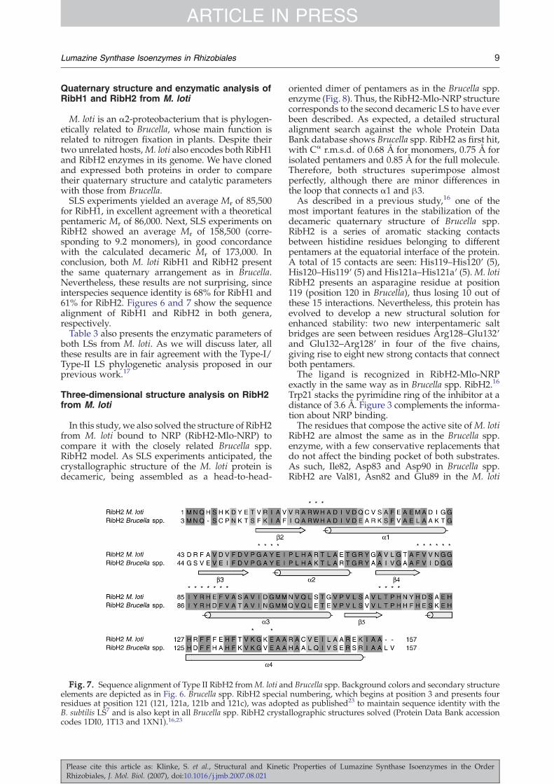
Fig. 7. Sequence alignment of Type II RibH2 fromM. loti anelements are depicted as in Fig. 6. Brucella spp. RibH2 specialresidues at position 121 (121, 121a, 121b and 121c), was adopB. subtilis LS7 and is also kept in all Brucella spp. RibH2 crystacodes 1DI0, 1T13 and 1XN1).16,23
Please cite this article as: Klinke, S. et al., Structural and KinetiRhizobiales, J. Mol. Biol. (2007), doi:10.1016/j.jmb.2007.08.021
oriented dimer of pentamers as in the Brucella spp.enzyme (Fig. 8). Thus, the RibH2-Mlo-NRP structurecorresponds to the second decameric LS to have everbeen described. As expected, a detailed structuralalignment search against the whole Protein DataBank database shows Brucella spp. RibH2 as first hit,with Cα r.m.s.d. of 0.68 Å for monomers, 0.75 Å forisolated pentamers and 0.85 Å for the full molecule.Therefore, both structures superimpose almostperfectly, although there are minor differences inthe loop that connects α1 and β3.As described in a previous study,16 one of the
most important features in the stabilization of thedecameric quaternary structure of Brucella spp.RibH2 is a series of aromatic stacking contactsbetween histidine residues belonging to differentpentamers at the equatorial interface of the protein.A total of 15 contacts are seen: His119–His120′ (5),His120–His119′ (5) and His121a–His121a′ (5).M. lotiRibH2 presents an asparagine residue at position119 (position 120 in Brucella), thus losing 10 out ofthese 15 interactions. Nevertheless, this protein hasevolved to develop a new structural solution forenhanced stability: two new interpentameric saltbridges are seen between residues Arg128–Glu132′and Glu132–Arg128′ in four of the five chains,giving rise to eight new strong contacts that connectboth pentamers.The ligand is recognized in RibH2-Mlo-NRP
exactly in the same way as in Brucella spp. RibH2.16
Trp21 stacks the pyrimidine ring of the inhibitor at adistance of 3.6 Å. Figure 3 complements the informa-tion about NRP binding.The residues that compose the active site ofM. loti
RibH2 are almost the same as in the Brucella spp.enzyme, with a few conservative replacements thatdo not affect the binding pocket of both substrates.As such, Ile82, Asp83 and Asp90 in Brucella spp.RibH2 are Val81, Asn82 and Glu89 in the M. loti
d Brucella spp. Background colors and secondary structurenumbering, which begins at position 3 and presents fourted as published23 to maintain sequence identity with thellographic structures solved (Protein Data Bank accession
c Properties of Lumazine Synthase Isoenzymes in the Order

Fig. 8. RibH2-Mlo-NRP structure. Each pentamer is drawn in a different color, and the inhibitor molecules (10) arerepresented as space-filling models in yellow. (a) View along the 5-fold axis. (b) Front view highlighting the pentamer–pentamer interface and the overall spool shape of the enzyme.
10 Lumazine Synthase Isoenzymes in Rhizobiales
ARTICLE IN PRESSRGR
enzyme, respectively. In order to study the bindingsite of substrate 2, RibH2-Mlo-NRP was crystallizedin the presence of 20 mM inorganic phosphatecoming from the final dialysis buffer. Weak electrondensity was found in the last cycles of refinement innine of the active sites at the putative binding pocketof the phosphate group of substrate 2. For thisreason, these peaks were assigned as phosphate ionsin the final model. Their positions and binding modeare similar to those found in the Brucella spp. RibH2enzyme.
Influence of the active-site architecture oncatalytic activity: focus on the special Brucellacase
To conclude, we analyze in close detail theenzymatic parameters presented in Table 3. Thevalue of the kinetic constant Km of substrate 1resembles that of the previously studied LSs in bothM. loti and Brucella enzymes because its binding siteis very well conserved among all types of LSs ofknown structure, as noted before. Nevertheless,there is a clear divergence in the case of B. abortusRibH1 (90 μM). This value is about 1 order ofmagnitude higher than that usually observed (i.e.,8.6 μMinB. subtilis LS and 5.0 μMin S. pombeLS).24,25The RibH1-Bab-NRP structure does not show anyspecial feature that could explain this fact; moreover,its related RibH1-Bme-NRP structure (which onlydiffers at Arg129, a residue that does not contactNRP) presents a “standard” Km of 4 μM.With respect to the Km of substrate 2, both RibH2
present the highest values seen for LSs. The evolu-tion of Type II LSs to assemble as decamers via anextended α4-helix architecture affects its binding siteas noted above. There is lack of a proper salt bridgeinteractionwith anArg or aHis residue. On the other
Please cite this article as: Klinke, S. et al., Structural and KineticRhizobiales, J. Mol. Biol. (2007), doi:10.1016/j.jmb.2007.08.021
side,M. loti RibH1 shows a Km of 15 μM, which is inthe same order as those seen in Type I and archaealLSs (e.g., 55 μM in Type I B. subtilis LS and 52 μM inarchaeal Methanococcus jannaschii LS).24,26 Althoughits 3D structure has not been determined yet, keyposition 134 (which corresponds to position 129 in B.abortus andB. melitensisRibH1; see Fig. 6) presents anarginine residue that is believed to be responsible forsubstrate 2 binding. Next, both B. abortus andB. melitensis RibH1 show a lower Km with respectto Type II LSs, due to the presence of His129 andArg129, respectively. However, both Km values areslightly higher than in the rest of the Type-I andarchaeal LSs investigated. It is interesting to note thatwe had expected a lower Km value for B. melitensisRibH1 in comparison with the B. abortus enzyme. Atfirst sight, the presence of its arginine residue, whichpresents a greater net positive charge than a histidineand a longer side chain, would have been able tobind the phosphate group of substrate 2 with astronger affinity.Finally, remarkable results regarding the turnover
number kcat in RibH1 and RibH2 were observed inboth genera studied. Brucella spp. and M. loti RibH2clearly present lower values than Type I and archaealLSs (i.e., 0.056 s−1 in B. subtilis and 0.023 s−1 inM. jannaschii).24,26 Moreover, the M. loti enzyme isalmost inactive. The partially distorted active-sitearchitecture of Type II LSs is the basis of this behavior,as discussed earlier. On the other hand,M. loti RibH1presents catalytic efficiencies (kcat/Km) for bothsubstrates that are a few orders of magnitude greaterthan those seen in all enzymes in Table 3. Indeed, wecan affirm that it behaves as an “ordinary” Type-I LS,with catalytic parameters in the same order ofmagnitude as those seen in other active LSs.Surprisingly, both B. abortus and B. melitensis
RibH1 are enzymes with an unexpected low kcat,
Properties of Lumazine Synthase Isoenzymes in the Order

11Lumazine Synthase Isoenzymes in Rhizobiales
ARTICLE IN PRESSRGR
which is even smaller than that of RibH2 in the samebacteria. In order to arrive at a possible explanationfor this unusual performance, we focused our ana-lysis on a special region of the active site (residues129–137) and compared it with all known LSs. Thereare three key residues whose side chain pointstowards the cleft: Arg/His129, Lys137 and Lys135(Fig. 9b). The role of residue 129 has already beenanalyzed. Next, residue Lys137 has been proposedby Zhang et al. to be a major determinant in thecorrect positioning and binding of a phosphonatedintermediate compound during the reaction.20 Thisstudy was performed on Aquifex aeolicus LS, whoseLys135 residue superimposes almost perfectly onBrucellaRibH1 Lys137. This latter residue, a histidinein some sequences, is always linked via a salt bridgeto an aspartate or a glutamate in its vicinity in all LSsstudied (Glu138 in Fig. 9a and c). This pair has beenproposed to reorientate during the reaction course in
Fig. 9. Focus on the influence of positive residues on the actribitylamino-2,4(1H,3H)-pyrimidinedione (NORP) and inorgbetween the charged groups are presented. The side chain of Palso drawn. (c and d) Electrostatic surface analysis of the stru
Please cite this article as: Klinke, S. et al., Structural and KinetiRhizobiales, J. Mol. Biol. (2007), doi:10.1016/j.jmb.2007.08.021
a cooperative manner to “pull” this phosphonatedintermediate to afford the following product (a Schiffbase) in a correct cis configuration. It is important tonote that all Brucella RibH1 enzymes lack thecounterpart of Lys137, as Phe140, rather than a Gluor an Asp, is found in this position (see Fig. 9b). Theabsence of this important pairmay affect the catalyticactivity of this enzyme.However,we cannot proposethis as the main reason for the low kcat in Brucella,sinceM. lotiRibH1,which catalyzes the reactionwithmuch better performance, presents the same aro-matic residue (Phe145) instead of an acidic one.On the other hand, residue Lys135 is unique for
Brucella RibH1 among all known LSs. It is part of itsdistinctive β hairpin; thus, a 3D superposition withall LSs of known structure shows no basic residuesin that particular region of the active site. Theoccurrence of three positive amino acids close toeach other (see Fig. 9b and d) may generate severe
ive site of LS. (a) A. aeolicus LS complexed with 5-nitroso-6-anic phosphate. (b) RibH1-Bme-NRP. Average distanceshe140 (a residue infrequently seen in LSs at this position) isctures shown above.
c Properties of Lumazine Synthase Isoenzymes in the Order

12 Lumazine Synthase Isoenzymes in Rhizobiales
ARTICLE IN PRESSRGR
charge repulsions during the course of the reactionthat may lead to a reduced catalytic activity inBrucella. This observation is in agreement with theabsence of a basic residue in this particular positionin M. loti RibH1, which is a glycine (Gly140). Thepositive environment generated by Lys135 may alsobe responsible for the slightly higher Km valuesobserved for substrate 2 in both B. abortus andB. melitensis RibH1 enzymes.Then, taking into account the sequence alignment
in Fig. 6, we predict A. tumefaciens RibH to be an LSof very low catalytic activity, since it presents thesame positive triad Arg–Lys–Lys as in B. melitensisRibH1, with a 76% sequence identity.It is important to note that the only remarkable
difference in the active site of RibH2 from Brucellaspp. and M. loti is the absence of His/Arg129, since,like almost all LSs, they bear the distinctive basic–acidic pair Lys135–Glu138 (after Brucella spp.numbering; see Fig. 7) and they do not present thepositive triad mentioned before. This supports againthe idea that the very low catalytic activities of theRibH2 enzymes may lie in the absence of a basicresidue that binds substrate 2.To conclude, we discard any negative influence of
other residues of the active site on the catalyticparameters, particularly the region near His90(according to Brucella RibH1 numbering, whichhas been proposed to act as a proton-transferagent during the reaction), since sequence identitybetween A. tumefaciens RibH andM. loti and BrucellaRibH1 enzymes is quite good there, with a fewconservative substitutions that most likely do notaffect the structure of the catalytic cleft on these LSs.
Conclusions
The four crystallographic structures and thekinetic and biophysical studies presented here arewell in line with the phylogenetic analysis and theType-I/Type-II LS family classification we hadproposed in our previous study.17 For the firsttime, two RibH1–RibH2 pairs were characterizedboth in structure and in catalytic properties.In summary, the structural elements that dictate
the different quaternary structures in LSs fromRhizobiales are quite distinct from those describedpreviously in other species. In this sense, pentamericRibH1 from the genus Brucella shows a character-istically smaller and charged loop at the region thatlinks α4 with α5, which may preclude the assemblyof this protein into an icosahedral particle. Next, wehave demonstrated that both RibH2 from Brucellaspp. andM. loti form decameric assemblies based onthe absence of the abovementioned loop and theoccurrence of an undistorted, 32-residue-long helixα4. In the RibH2 pentameric moieties, the N-terminiof the five α4 helices and the loops that connect themwith the contiguous β5 strands make a protuberantsurface that fits tightly into the neighboring penta-mer to form a decameric particle. Although thegeneral assembly of the equatorial interface is very
Please cite this article as: Klinke, S. et al., Structural and KineticRhizobiales, J. Mol. Biol. (2007), doi:10.1016/j.jmb.2007.08.021
similar in both cases studied, M. loti RibH2 shows anoteworthy variation in the nature of the interpen-tameric contacts, as salt bridges stabilize the deca-meric structure rather the than aromatic stackinginteractions between His residues that are observedin Brucella spp. RibH2.Regarding the enzymatic properties, we have
shown that Mesorhizobium bears a fully active LS(RibH1) and an almost inactive LS (RibH2), whereasin Brucella, both enzymes have very low catalyticactivity, although for different reasons. On this basis,the unique RibH to Agrobacterium is thereforeproposed to also exhibit a very low catalytic activity.This unusual scenario suggests that the selectivepressure controlling this pathway favored the evolu-tion of catalysts with low reaction rates. In fact,riboflavin is required in small amounts only, andexcess production could unnecessarily deplete theprecursor pools, namely, Guanosine triphosphateand ribulose 5′-phosphate. Moreover, an excess offlavins in the intracellular pool in Rhizobiales couldact as a negative factor when these bacteria areexposed to oxidative or nitrosative stress. In thissense, Rhizobiales regulate iron concentration bysensing the physiological consequence of metalavailability, rather than its concentration per se, andthus provide for more flexible regulation.27,28The question of which selective pressures could
have prevented the loss of the RibH2 gene or couldhave favored its more recent acquisition by hor-izontal gene transfer is still open to answer.Interestingly, the Type-II LS is an immunodominantantigen of B. abortus, and knockout mutants for thisprotein have shown a marked decrease in survivaland virulence (Marchesini et al., unpublishedresults). As in the case of Brucella, M. loti RibH2has almost no activity as LS and would be regulatedby a riboswitch that senses flavin mononucleotide,29
suggesting that the Type-II LSs may have evolvedinto very poor catalysts or, alternatively, to harbor anew, as-yet-unknown function.
Materials and Methods
Materials
NRP was obtained as described by published pro-cedures.30 Genomic DNAs of B. melitensis andM. lotiwerekindly provided by Dr. Angeles Zorreguieta (FundaciónInstituto Leloir) and Dr. Rodolfo Ugalde (IIB-UNSAM),respectively.
Gene cloning
B. abortus RibH1 gene was cloned into the expressionplasmid pT7-7 as described.17 B. melitensis RibH1 gene, aswell as M. loti RibH1 and RibH2 genes, was amplified byPCR using their respective chromosomal DNAs astemplate and the oligonucleotides presented in Table 4as primers. The three amplicons were digested with NdeIand BamHI and were then ligated into the plasmidpET11a (Novagen, Madison, WI), which had been treated
Properties of Lumazine Synthase Isoenzymes in the Order

Table 4. Oligonucleotides used for PCR amplification of B. melitensis RibH1 and M. loti RibH1 and RibH2 genes
Designation Sequence Restriction sites
RibH1 B. melitensis F: 5′ ATAATAATACATATGGAGTTTCTCATGTCCAAGCA 3′ NdeI (CA*TATG)R: 5′ TATTATTATAAGGATCCTTATCAGGCTCCGAATTTTTTG 3′ BamHI (G*GATCC)
RibH1 M. loti F: 5′ ATAATAATACATATGGCTGGTATATCCCAACACGGC 3′ NdeIR: 5′ TATTATTATAAGGATCCTTATCACGATCGGGCTCCCAAT 3′ BamHI
RibH2 M. loti F: 5′ ATAATAATACATATGAATCAGCATTCCCACAAAGACTAT 3′ NdeIR: 5′ TATTATTATAAGGATCCTTATCATGCCGCGATCTTTTC 3′ BamHI
F, oligonucleotide used as forward primer; R, oligonucleotide used as reverse primer.
13Lumazine Synthase Isoenzymes in Rhizobiales
ARTICLE IN PRESSRGR
with the same restriction enzymes. All constructions werechecked by DNA sequencing following the method ofSanger et al.31 Escherichia coli BL21(DE3) competent cells(Stratagene, La Jolla, CA) were transformed with theresulting ligation mixtures.
Protein expression
In all cases, recombinant cells were grown at 37 °Covernight in 25 ml of LB medium containing 100 μg/mlampicillin, with agitation (250 rpm). These preparationswere diluted to 500 ml and grown to an absorbance(600 nm) of 1.0. At this time, the cultures were induced bythe addition of isopropyl-β-D-thiogalactopyranoside to afinal concentration of 1 mM and incubated further for 4 hat 37 °C, with agitation (250 rpm). The bacteria werecentrifuged at 10,000g for 8 min at 4 °C.
Protein purification
After expression, different strategies were followedaccording to the particular protein. B. abortus, B. melitensisand M. loti RibH1 bacterial pellets were suspended andsonicated in a solution containing 50 mM sodium/potassium phosphate (pH 7.0) and 5 mM EDTA (bufferA). Preparations were centrifuged at 25,000g for 20 min at4 °C, and the insoluble fractions were discarded. Cyto-plasm fractions were dialyzed overnight against buffer Aand then purified by anion-exchange chromatography ina fast protein liquid chromatography apparatus using aQ-Sepharose column (all columns by GE Healthcare,Piscataway, NJ) equilibrated with buffer A. RibH1 frac-tions were eluted using a linear gradient of 0–1.5 Msodium chloride in buffer A (buffer B). The proteins werefurther purified by gel-filtration chromatography in aSuperdex 200 column with isocratic elution in phosphate-buffered saline containing 0.5 M sodium chloride. Theappropriate fractions were then dialyzed against buffer Aand loaded in a Mono-Q column equilibrated with thesame buffer. Highly purified RibH1 proteins were elutedwith a linear gradient of 0–1.5 M sodium chloride in bufferA (buffer B). It is important to note that three differentcolumns were needed in all cases to separate proteinsmostly from nonproteic contaminants. The final productswere then dialyzed against crystallization buffer [10 mMTris (pH 7.10) and 25 mM sodium chloride], concentratedby ultracentrifugation to ∼10 mg/ml with Centricon YM-10 concentrators (Millipore, Billerica, MA) and stored at−20 °C.For M. loti RibH2, the bacterial pellets were suspended
and sonicated in a solution containing 50 mM Tris (pH 8.0)and 5 mM EDTA. After centrifugation, the cytoplasmfraction was dialyzed overnight against 50 mM Tris (pH8.5; buffer C) and then loaded in a Q-Sepharose columnequilibrated with buffer C. RibH2 fractions were eluted
Please cite this article as: Klinke, S. et al., Structural and KinetiRhizobiales, J. Mol. Biol. (2007), doi:10.1016/j.jmb.2007.08.021
using a linear gradient of 0–1 M sodium chloride in bufferC (buffer D) and further purified by gel filtration (Super-dex 200 column) in a similar manner to the RibH1enzymes. Finally, RibH2 fractions were dialyzed againstbuffer C and loaded in a Mono-Q column equilibratedwith the same buffer. RibH2 fractions were eluted with alinear gradient of 0–1 M sodium chloride in buffer C(buffer D), dialyzed against crystallization buffer [20 mMsodium/potassium phosphate (pH 7.10) and 25 mMsodium chloride], concentrated to ∼10 mg/ml and storedat −20 °C.The purity of all protein preparations was checked by
15% SDS polyacrylamide gels and UV spectrophotometry.
Crystallization
All crystals were grown by means of the hanging-dropvapor-diffusion method at 19 °C. Round-shaped RibH1-Bab crystals of about 0.30 mm×0.20 mm×0.10 mm dimen-sion were obtained after several days with a mother liquorconsisting of 12% (wt/vol) polyethylene glycol (PEG)4000, 0.1 M Hepes (pH 7.7) and 0.2 M calcium chloride.RibH1-Bab-NRP crystals were grown with 18% (wt/vol)PEG 400, 0.1 MMes (pH 6.6) and 0.2 M calcium chloride inthe presence of a 5-fold molar excess of the substrateanalogue inhibitor NRP. Sharp-edged prisms of about0.30 mm×0.25 mm×0.10 mm dimension were found aftera week. RibH1-Bme-NRP crystals were grown in a similarmanner, with 22% (wt/vol) PEG 400, 0.1 M Mes (pH 6.3)and 0.2 M calcium chloride as mother liquor, yieldingcrystals that are comparable in shape and slightly reducedin size after a week. Finally, prism-shaped RibH2-Mlo-NRP crystals of 0.15 mm×0.15 mm×0.05 mm dimensionwere also obtained by cocrystallization with 16% (wt/vol)PEG 4000, 0.1 M ammonium sulfate and 15% isopropanolafter several days.
Diffraction data collection and processing
RibH1-Bab and RibH1-Bab-NRP datasets were collectedat the D03B-MX1 Beamline at the Laboratório Nacional deLuz Síncrotron (Campinas, Brazil)32 using a MarCCD 165detector (Mar USA, Evanston, IL). RibH1-Bme-NRP andRibH2-Mlo-NRP diffraction data were collected at theX9A Beamline at the National Synchrotron Light Source,Brookhaven National Laboratory (New York), also using aMarCCD 165 detector. Crystals were soaked in cryopro-tectant solutions consisting of a mother liquor supple-mented with 15% (wt/vol) PEG 400 (RibH1-Bab andRibH2-Mlo-NRP), 30% (wt/vol) PEG 400 (RibH1-Bab-NRP) and 12% (wt/vol) PEG 400 (RibH1-Bme-NRP), andflash-cooled in a 100-K nitrogen stream. A single crystalwas used for the collection of each dataset. X-raydiffraction datawere indexed and integratedwithMOSFLM,scaled using SCALA and converted to amplitudes with
c Properties of Lumazine Synthase Isoenzymes in the Order

14 Lumazine Synthase Isoenzymes in Rhizobiales
ARTICLE IN PRESSRGR
TRUNCATE. All these programs are part of the CCP4package.33 RibH1-Bab crystals diffracted to a maximumresolution of 2.22 Å and belonged to the monoclinic spacegroup C2. RibH1-Bab-NRP and RibH1-Bme-NRP crystalsdiffracted to 2.30 and 2.70 Å, respectively, in the trigonalspace group P3221. Finally, RibH2-Mlo-NRP crystalsdiffracted to 2.53 Å, belonging also to the space groupC2. In all cases, 5% of the measured reflections wereflagged for cross-validation purposes. Details on datacollection parameters and processing statistics are shownin Table 5.
Structure solution, model building and refinement
All structures were solved using the molecular-replace-ment method with the program AMoRe.34 The RibH1-Bab-NRP structure was solved using a pentameric block ofthe icosahedral LS from B. subtilis (Protein Data Bankaccession code 1RVV) as search model.7 To reduce bias,this model was modified by removing the first 50 residuesin each chain due to the low sequence identity betweenboth proteins in this region and by mutating allnonconserved residues to alanine. Five solutions werefound inside the asymmetric unit after rotation andtranslation steps (15–4.0 Å), which correspond to thesame unique solution rotated 72° from each othercoaxially to the 5-fold crystallographic axis of the protein.After checking for a consistent packing in the crystallattice, the oriented model was then subjected to rigidbody refinement in CNS18 (8–3.6 Å), considering eachchain as a separate entity. Next, a cycle of simulatedannealing refinement applying restrained noncrystallo-graphic symmetry was performed in CNS (8–3.2 Å),yielding a model with R-factor=0.463 and Rfree=0.479.Double- and single-difference Fourier maps built at thisstage showed clear electron density for several side chains,which were modeled using the program O.35 Further
Table 5. X-ray data collection parameters and processing sta
Parameters RibH1-Bab RibH1-
Data collectionNumber of frames 127 1Wavelength (Å) 1.438 1
Indexing and scalingCell parameters (Å)
a 124.10 9b 68.52 9c 90.65 17
Cell angles (°)α 90β 108.40γ 90 1
Space group C2 PResolution limit (Å) 2.22 2Number of unique reflections 31,466 40Multiplicitya 2.7 (2.6) 9.5I/σ 12.3 (2.6) 10.5Rmerge (%)b 4.1 (28.2) 5.6Completeness (%) 94.8 (88.7) 99.5Pentamers/asymmetric unit 1Solvent content (%) 43.0 5Matthews coefficient (Å3/Da) 2.2B-factor (Wilson plot; Å2) 43.1 3
a Values in parentheses correspond to the highest resolution shellBme-NRP, 2.85–2.70 Å; RibH2-Mlo-NRP, 2.66–2.53 Å.
b Rmerge=[Σi|Ii− ⟨I⟩|/ΣiIi].
Please cite this article as: Klinke, S. et al., Structural and KineticRhizobiales, J. Mol. Biol. (2007), doi:10.1016/j.jmb.2007.08.021
cycles of simulated annealing refinement using all reflec-tions gradually up to the diffraction limit (2.30 Å) yieldedmaps that allowed the reconstruction of the main chainbefore residue 50 and almost all side chains that had beenreplaced by alanine. A strong and well-defined electrondensity was found inside each active site, which corre-sponds to the bound substrate analogue inhibitor NRP.Coordinates of this ligand were obtained from the ProteinData Bank entry 1KYY (S. pombe LS complexed withNRP)5 and manually fit into the density, followed byseveral cycles of positional and isotropic B-factor refine-ment using the maximum-likelihood algorithm. In the lastcycles, noncrystallographic symmetry restraints wererelaxed, allowing each chain to refine independently.The model was completed with solvent molecules and afew calcium ions that were found near aspartate orglutamate residues, with most of them bridging neighbor-ing molecules.The final RibH1-Bab-NRP structure, without solvent
molecules and ligand coordinates, was successfully usedas search model for the resolution of RibH1-Bab andRibH1-Bme-NRP. In the latter complex, a special searchmodel was built, with residue 129 mutated to alanine. Therefinement and manual building procedures were, in bothcases, similar to the ones applied for RibH1-Bab-NRP.The RibH2-Mlo-NRP structure was solved using a
pentamer from Brucella spp. RibH2 as search model(Protein Data Bank entry 1T13),16 with all nonconservedresiduesmutated to alanine. A total of two pentamerswerefound in the asymmetric unit, facing each other in a head-to-head conformation as expected. The model was refinedand built in a similar manner to the other structures. In thefinal cycles of refinement, electron density was found inalmost all putative binding sites of the phosphate group ofsubstrate 2, which was interpreted as bound phosphateions coming from the crystallization buffer.In all structures, only reflections satisfying the condition
F≥2σ were included in the refinement. The topology and
tistics
Bab-NRP RibH1-Bme-NRP RibH2-Mlo-NRP
94 100 160.438 0.9794 0.9794
5.32 97.65 154.385.32 97.65 122.221.74 175.40 94.93
90 90 9090 90 125.4320 120 903221 P3221 C2.30 2.70 2.53,562 22,473 46,386(7.9) 3.9 (2.2) 3.4 (3.1)(2.2) 5.8 (2.2) 9.7 (2.9)(34.0) 10.0 (31.3) 7.4 (25.5)(97.0) 85.0 (82.3) 96.7 (89.2)1 1 23.7 56.8 41.32.7 2.9 2.19.7 54.6 39.4
: RibH1-Bab, 2.34–2.22 Å; RibH1-Bab-NRP, 2.42–2.30 Å; RibH1-
Properties of Lumazine Synthase Isoenzymes in the Order

15Lumazine Synthase Isoenzymes in Rhizobiales
ARTICLE IN PRESSRGR
parameter files needed for the refinement of the ligandNRP in the complex structures were obtained from theHIC-Up database.36 The final refinement statistics arefound in Table 2.
SLS measurements
The average molecular weight in solution of all proteinspresented in this work was calculated with a PrecisionDetectors PD2080 Static Light Scattering instrument(Precision Detectors, Bellingham, MA) linked to a fastprotein liquid chromatography apparatus and an LKB2142 differential refractometer (LKB, Bromma, Sweden).In each case, 500 μl of a ∼0.5-mg/ml protein solution wasinjected into a Superdex 200 column and eluted withphosphate-buffered saline containing 0.5 M sodiumchloride. Both signals corresponding to 90° light scattering(λ=682 nm) and the refractive index of the peaks elutedwere recorded and then processed with the Discovery32software supplied by Precision Detectors. Bovine serumalbumin (Mr=66,500) was used as standard for light-scattering detector calibration.
Determination of enzymatic parameters
LS enzymatic activity was determined as describedpreviously.21,24,37 Briefly, a series of mixtures containingthe enzyme, 100 mM potassium phosphate (pH 7.0), 5 mMEDTA and various concentrations of substrates 1 and 2were prepared. Generation of 6,7-dimethyl-8-ribitylluma-zine (product 3) was monitored photometrically atλ=410 nm and 37 °C. The resulting data were fitted tothe Michaelis–Menten equation or to the Hill equation.Due to the appreciable nonenzymatic generation ofproduct,38 reaction mixtures without enzyme were usedto correct the experimental data.
Sequence alignments
Sequences were aligned using the program ClustalW,version 1.83,39 at the European Bioinformatics Institutehomepage† (Figs. 6 and 7).
Evaluation of the models and graphicalrepresentation
All 3D structures presented herewere validatedwith theprograms PROCHECK40 and SFCHECK.41 Superpositionsand r.m.s.d. calculations were done with the ProteinStructure Comparison Service SSM at the EuropeanBioinformatics Institute.42 Figures were generated withMOLSCRIPT,43 Raster3D,44 Chimera45 and ChemDraw(CambridgeSoft, Cambridge, MA). Electrostatic surfacecalculations were performed with the program APBS.46
Protein Data Bank depositions
Atomic coordinates and structure factors were depos-ited in the RCSB Protein Data Bank under accession codes2F59 (RibH1-Bab-NRP), 2I0F (RibH1-Bab), 2O6H (RibH1-Bme-NRP) and 2OBX (RibH2-Mlo-NRP).
†www.ebi.ac.uk
Please cite this article as: Klinke, S. et al., Structural and KinetiRhizobiales, J. Mol. Biol. (2007), doi:10.1016/j.jmb.2007.08.021
Acknowledgements
This work was supported by a Howard HughesMedical Institute international grant to F.A.G. andby a grant from the Agencia Nacional de Promo-ción Científica y Tecnológica. We were also sup-ported by the Fonds der Chemischen Industrie, theHans-Fischer-Gesellschaft eV and the LaboratórioNacional de Luz Síncrotron (projects MX1-3342 andMX1-4420).M.F. and F.A.G. acknowledge Bundesministerium
für Bildung und Forschung's and Secretaría deCiencia y Tecnológia's support for exchange visitsbetween laboratories (project ARG 04/Z06). S.K.would also like to thank Dr. Robert M. Sweet and Dr.Zbigniew Dauter for their help during X-raydiffraction data collection at the Rapidata 2006course. The use of the National Synchrotron LightSource, Brookhaven National Laboratory, was sup-ported by the U.S. Department of Energy, Office ofScience, Office of Basic Energy Sciences, undercontract no. DE-AC02-98CH10886.
References
1. Fischer, M. & Bacher, A. (2005). Biosynthesis offlavocoenzymes. Nat. Prod. Rep. 22, 324–350.
2. Fischer, M. & Bacher, A. (2006). Biosynthesis ofvitamin B2 in plants. Physiol. Plant. 126, 304–318.
3. Morgunova, E., Meining, W., Illarionov, B., Haase, I.,Jin, G., Bacher, A. et al. (2005). Crystal structure oflumazine synthase from Mycobacterium tuberculosis asa target for rational drug design: binding mode of anew class of purinetrione inhibitors. Biochemistry, 44,2746–2758.
4. Persson, K., Schneider, G., Jordan, D. B., Viitanen, P. V.& Sandalova, T. (1999). Crystal structure analysis of apentameric fungal and an icosahedral plant lumazinesynthase reveals the structural basis for differences inassembly. Protein Sci. 8, 2355–2365.
5. Gerhardt, S., Haase, I., Steinbacher, S., Kaiser, J. T.,Cushman, M., Bacher, A. et al. (2002). The structuralbasis of riboflavin binding to Schizosaccharomyces pombe6,7-dimethyl-8-ribityllumazine synthase. J. Mol. Biol.318, 1317–1329.
6. Meining, W., Mörtl, S., Fischer, M., Cushman, M.,Bacher, A. & Ladenstein, R. (2000). The atomicstructure of pentameric lumazine synthase fromSaccharomyces cerevisiae at 1.85 Å resolution revealsthe binding mode of a phosphonate intermediateanalogue. J. Mol. Biol. 299, 181–197.
7. Ritsert, K., Huber, R., Turk, D., Ladenstein, R.,Schmidt-Bäse, K. & Bacher, A. (1995). Studies on thelumazine synthase/riboflavin synthase complex ofBacillus subtilis: crystal structure analysis of reconsti-tuted, icosahedral beta-subunit capsids with boundsubstrate analogue inhibitor at 2.4 Å resolution.J. Mol. Biol. 253, 151–167.
8. Zhang, X., Meining, W., Fischer, M., Bacher, A. &Ladenstein, R. (2001). X-ray structure analysis andcrystallographic refinement of lumazine synthasefrom the hyperthermophile Aquifex aeolicus at 1.6 Åresolution: determinants of thermostability revealedfrom structural comparisons. J. Mol. Biol. 306,1099–1114.
c Properties of Lumazine Synthase Isoenzymes in the Order

16 Lumazine Synthase Isoenzymes in Rhizobiales
ARTICLE IN PRESSRGR
9. Mortl, S., Fischer, M., Richter, G., Tack, J., Weinkauf, S.& Bacher, A. (1996). Biosynthesis of riboflavin.Lumazine synthase of Escherichia coli. J. Biol. Chem.271, 33201–33207.
10. Morgunova, E., Saller, S., Haase, I., Cushman, M.,Bacher, A., Fischer, M. & Ladenstein, R. (2007).Lumazine synthase from Candida albicans as an anti-fungal target enzyme: structural and biochemicalbasis for drug design. J. Biol. Chem. 282, 17231–17241.
11. Zhang, X., Konarev, P. V., Petoukhov, M. V., Svergun,D. I., Xing, L., Cheng, R. H. et al. (2006). Multipleassembly states of lumazine synthase: a model rela-ting catalytic function and molecular assembly. J. Mol.Biol. 362, 753–770.
12. DelVecchio, V. G., Kapatral, V., Redkar, R. J., Patra, G.,Mujer, C., Los, T. et al. (2002). The genome sequence ofthe facultative intracellular pathogen Brucella meliten-sis. Proc. Natl. Acad. Sci. USA, 99, 443–448.
13. Paulsen, I. T., Seshadri, R., Nelson, K. E., Eisen, J. A.,Heidelberg, J. F., Read, T. D. et al. (2002). The Brucellasuis genome reveals fundamental similarities betweenanimal and plant pathogens and symbionts. Proc. Natl.Acad. Sci. USA, 99, 13148–13153.
14. Halling, S. M., Peterson-Burch, B. D., Bricker, B. J.,Zuerner, R. L., Qing, Z., Li, L. L. et al. (2005).Completion of the genome sequence of Brucella abortusand comparison to the highly similar genomes ofBrucella melitensis and Brucella suis. J. Bacteriol. 187,2715–2726.
15. Zylberman, V., Craig, P. O., Klinke, S., Braden, B. C.,Cauerhff, A. & Goldbaum, F. A. (2004). High orderquaternary arrangement confers increased structuralstability to Brucella sp. lumazine synthase. J. Biol.Chem. 279, 8093–8101.
16. Klinke, S., Zylberman, V., Vega, D. R., Guimaraes,B. G., Braden, B. C. & Goldbaum, F. A. (2005).Crystallographic studies on decameric Brucella spp.Lumazine synthase: a novel quaternary arrangementevolved for a new function? J. Mol. Biol. 353, 124–137.
17. Zylberman, V., Klinke, S., Haase, I., Bacher, A.,Fischer, M. & Goldbaum, F. A. (2006). Evolution ofvitamin B2 biosynthesis: 6,7-dimethyl-8-ribitylluma-zine synthases of Brucella. J. Bacteriol. 188, 6135–6142.
18. Brunger, A. T., Adams, P. D., Clore, G. M., Delano,W. L., Gros, P., Grosse-Kunstleve, R. W. et al. (1998).Crystallography and NMR system (CNS): a newsoftware system for macromolecular structure deter-mination. Acta Crystallogr. Sect. D: Biol. Crystallogr.54, 905–921.
19. Ladenstein, R., Schneider, M., Huber, R., Bartunik,H. D., Wilson, K., Schott, K. & Bacher, A. (1988).Heavy riboflavin synthase from Bacillus subtilis.Crystal structure analysis of the icosahedral β60capsid at 3.3 Å resolution. J. Mol. Biol. 203, 1045–1070.
20. Zhang, X., Meining, W., Cushman, M., Haase, I.,Fischer, M., Bacher, A. & Ladenstein, R. (2003). Astructure-based model of the reaction catalyzed bylumazine synthase from Aquifex aeolicus. J. Mol. Biol.328, 167–182.
21. Kis, K., Volk, R. & Bacher, A. (1995). Biosynthesis ofriboflavin. Studies on the reaction mechanism of 6,7-dimethyl-8-ribityllumazine synthase. Biochemistry, 34,2883–2892.
22. Goldbaum, F. A., Velikovsky, C. A., Baldi, P. C., Mörtl,S., Bacher, A. & Fossati, C. A. (1999). The 18-kDacytoplasmic protein of Brucella species—an antigenuseful for diagnosis—is a lumazine synthase. J. Med.Microbiol. 48, 833–839.
23. Braden, B. C., Velikovsky, C. A., Cauerhff, A. A.,
Please cite this article as: Klinke, S. et al., Structural and KineticRhizobiales, J. Mol. Biol. (2007), doi:10.1016/j.jmb.2007.08.021
Polikarpov, I. & Goldbaum, F. A. (2000). Divergence inmacromolecular assembly: X-ray crystallographicstructure analysis of lumazine synthase from Brucellaabortus. J. Mol. Biol. 297, 1031–1036.
24. Fischer, M., Haase, I., Kis, K., Meining,W., Ladenstein,R., Cushman, M. et al. (2003). Enzyme catalysis viacontrol of activation entropy: site-directed mutagen-esis of 6,7-dimethyl-8-ribityllumazine synthase. J. Mol.Biol. 326, 783–793.
25. Fischer, M., Haase, I., Feicht, R., Richter, G., Gerhardt,S., Changeux, J. P. et al. (2002). Biosynthesis ofriboflavin: 6,7-dimethyl-8-ribityllumazine synthase ofSchizosaccharomyces pombe. Eur. J. Biochem. 269, 519–526.
26. Haase, I., Mortl, S., Kohler, P., Bacher, A. & Fischer, M.(2003). Biosynthesis of riboflavin in archaea. 6,7-Dimethyl-8-ribityllumazine synthase ofMethanococcusjannaschii. Eur. J. Biochem. 270, 1025–1032.
27. Rodionov, D. A., Gelfand, M. S., Todd, J. D., Curson,A. R. & Johnston, A. W. (2006). Computationalreconstruction of iron- and manganese-responsivetranscriptional networks in alpha-proteobacteria.PLoS Comput. Biol. 2, e163.
28. Johnston, A. W., Todd, J. D., Curson, A. R., Lei, S.,Nikolaidou-Katsaridou, N., Gelfand, M. S. & Rodio-nov, D. A. (2007). Living without Fur: the subtlety andcomplexity of iron-responsive gene regulation in thesymbiotic bacterium Rhizobium and other alpha-proteobacteria. Biometals, 20, 501–511.
29. Vitreschak, A. G., Rodionov, D. A., Mironov, A. A. &Gelfand, M. S. (2002). Regulation of riboflavinbiosynthesis and transport genes in bacteria bytranscriptional and translational attenuation. NucleicAcids Res. 30, 3141–3151.
30. Cresswell, R. M. & Wood, H. C. S. (1960). Thebiosynthesis of pteridines: Part I. The synthesis ofriboflavin. J. Chem. Soc., 4768–4775.
31. Sanger, F., Nicklen, S. & Coulson, A. R. (1977). DNAsequencing with chain-terminating inhibitors. Proc.Natl. Acad. Sci. USA, 74, 5463–5467.
32. Polikarpov, I., Oliva, G., Castellano, E. E., Garratt,R. C., Arruda, P., Leite, A. & Craievich, A. (1998).The protein crystallography beamline at LNLS, theBrazilian National Synchrotron Light source. Nucl.Instrum. Methods Phys. Res. Sect. A, 405, 159–164.
33. Collaborative Computational Project, Number 4(1994). The CCP4 suite: programs for protein crystal-lography. Acta Crystallogr. Sect. D: Biol. Crystallogr. 50,760–763.
34. Navaza, J. A. C. (1994). AMoRe: an automatedpackage for molecular replacement. Acta Crystallogr.Sect. A, 50, 157–163.
35. Jones, T. A., Zou, J. Y., Cowan, S. W. & Kjeldgaard, M.(1991). Improved methods for the building of proteinmodels in electron density maps and the location oferrors in these models. Acta Crystallogr. Sect. A, 47,110–119.
36. Kleywegt, G. J. & Jones, T. A. (1998). Databases inprotein crystallography. Acta Crystallogr. Sect. D: Biol.Crystallogr. 54, 1119–1131.
37. Kis, K. & Bacher, A. (1995). Substrate channeling in thelumazine synthase/riboflavin synthase complex ofBacillus subtilis. J. Biol. Chem. 270, 16788–16795.
38. Kis, K., Kugelbrey, K. & Bacher, A. (2001). Biosynth-esis of riboflavin. The reaction catalyzed by 6,7-dimethyl-8-ribityllumazine synthase can proceedwithout enzymatic catalysis under physiologicalconditions. J. Org. Chem. 66, 2555–2559.
39. Chenna, R., Sugawara, H., Koike, T., Lopez, R.,Gibson, T. J., Higgins, D. G. & Thompson, J. D.
Properties of Lumazine Synthase Isoenzymes in the Order

17Lumazine Synthase Isoenzymes in Rhizobiales
ARTICLE IN PRESSRGR
(2003). Multiple sequence alignment with the Clustalseries of programs. Nucleic Acids Res. 31, 3497–3500.
40. Laskowski, R. A., MacArthur, M. W., Moss, D. S. &Thornton, J. M. (1993). PROCHECK: a program tocheck the stereochemical quality of protein structures.J. Appl. Crystallogr. 26, 283–291.
41. Vagin, A. A., Richelle, J. & Wodak, S. J. (1999).SFCHECK: a unified set of procedure for evaluatingthe quality of macromolecular structure-factor dataand their agreement with atomic model. Acta Crystal-logr. Sect. D: Biol. Crystallogr. 55, 191–205.
42. Krissinel, E. & Henrick, K. (2004). Secondary-structurematching (SSM), a new tool for fast protein structurealignment in three dimensions. Acta Crystallogr. Sect.D: Biol. Crystallogr. 60, 2256–2268.
Please cite this article as: Klinke, S. et al., Structural and KinetiRhizobiales, J. Mol. Biol. (2007), doi:10.1016/j.jmb.2007.08.021
43. Kraulis, P. J. (1991). MOLSCRIPT: a program toproduce both detailed and schematic plots of proteinstructures. J. Appl. Crystallogr. 24, 946–950.
44. Merritt, E. A. & Bacon, D. J. (1997). Raster3D:photorealistic molecular graphics. Methods Enzymol.277, 505–524.
45. Pettersen, E. F., Goddard, T. D., Huang, C. C., Couch,G. S., Greenblatt, D. M., Meng, E. C. & Ferrin, T. E.(2004). UCSF Chimera—a visualization system forexploratory research and analysis. J. Comput. Chem. 25,1605–1612.
46. Baker, N. A., Sept, D., Joseph, S., Holst, M. J. &McCammon, J. A. (2001). Electrostatics of nanosys-tems: application to microtubules and the ribosome.Proc. Natl. Acad. Sci. USA, 98, 10037–10041.
c Properties of Lumazine Synthase Isoenzymes in the Order