Structural Analysis of the Essential Resuscitation ... · Gram-negative eubacteria. Among other as...
8
Structural Analysis of the Essential Resuscitation Promoting Factor YeaZ Suggests a Mechanism of Nucleotide Regulation through Dimer Reorganization Inci Aydin 1¤ , Yumiko Saijo-Hamano 2 , Keiichi Namba 2 , Connor Thomas 3 , Anna Roujeinikova 1 * 1 Department of Microbiology and Department of Biochemistry and Molecular Biology, Monash University, Clayton, Victoria, Australia, 2 Graduate School of Frontier Biosciences, Osaka University, Suita, Osaka, Japan, 3 School of Molecular and Biomedical Science, University of Adelaide, Adelaide, South Australia, Australia Abstract Background: The yeaZ gene product forms part of the conserved network YjeE/YeaZ/YgjD essential for the survival of many Gram-negative eubacteria. Among other as yet unidentified roles, YeaZ functions as a resuscitation promoting factor required for survival and resuscitation of cells in a viable but non-culturable (VBNC) state. Methodology/Principal Findings: In order to investigate in detail the structure/function relationship of this family of proteins we have performed X-ray crystallographic studies of Vibrio parahaemolyticus YeaZ. The YeaZ structure showed that it has a classic actin-like nucleotide-binding fold. Comparisons of this crystal structure to that of available homologues from E. coli, T. maritima and S. typhimurium revealed two distinctly different modes of dimer formation. In one form, prevalent in the absence of nucleotide, the putative nucleotide-binding site is incomplete, lacking a binding pocket for a nucleotide base. In the second form, residues from the second subunit complete the nucleotide-binding site. This suggests that the two dimer architectures observed in the crystal structures correspond to a free and a nucleotide-bound form of YeaZ. A multiple sequence alignment of YeaZ proteins from different bacteria allowed us to identify a large conserved hydrophobic patch on the protein surface that becomes exposed upon nucleotide-driven dimer re-arrangement. We hypothesize that the transition between two dimer architectures represents the transition between the ‘on’ and ‘off’ states of YeaZ. The effect of this transition is to alternately expose and bury a docking site for the partner protein YgjD. Conclusions/Significance: This paper provides the first structural insight into the putative mechanism of nucleotide regulation of YeaZ through dimer reorganization. Our analysis suggests that nucleotide binding to YeaZ may act as a regulator or switch that changes YeaZ shape, allowing it to switch partners between YjeE and YgjD. Citation: Aydin I, Saijo-Hamano Y, Namba K, Thomas C, Roujeinikova A (2011) Structural Analysis of the Essential Resuscitation Promoting Factor YeaZ Suggests a Mechanism of Nucleotide Regulation through Dimer Reorganization. PLoS ONE 6(8): e23245. doi:10.1371/journal.pone.0023245 Editor: Hendrik W. van Veen, University of Cambridge, United Kingdom Received April 9, 2011; Accepted July 11, 2011; Published August 17, 2011 Copyright: ß 2011 Aydin et al. This is an open-access article distributed under the terms of the Creative Commons Attribution License, which permits unrestricted use, distribution, and reproduction in any medium, provided the original author and source are credited. Funding: This work was supported by an Australian Research Council Research Fellowship to AR, DP1094619. The funders had no role in study design, data collection and analysis, decision to publish, or preparation of the manuscript. Competing Interests: The authors have declared that no competing interests exist. * E-mail: [email protected] ¤ Current address: Department of Infectious Diseases, Heidelberg University, Heidelberg, Germany Introduction The yeaZ gene product forms part of the conserved network YjeE/YeaZ/YgjD essential for survival of many eubacteria [1,2]. Studies in Salmonella [3] and V. parahaemolyticus (C. Thomas, personal communication, 19 November 2010) demonstrated that among other as yet unidentified roles, YeaZ functions as a resuscitation promoting factor required for cells to be able to survive in, and exit from, a VBNC state. Many pathogenic bacteria enter the VBNC state as a response to stress (e.g. starvation of nutrients, change in osmotic and oxygen concentrations or temperature) [4]. The marine enteropathogen V. parahaemolyticus, for example, enters a VBNC state at temperatures below 15uC, that correspond with typical winter seawater temperatures as well as temperatures used for storage of seafood, as a survival strategy [5]. VBNC cells exhibit antibiotic resistance and retain the ability to attach and persist in their environment. Exposing V. parahaemolyticus VBNC cells to a temperature upshift leads to resuscitation; the cells regain cultur- ability and renewed ability to cause infection. The requirement of YeaZ for persistence within, and exit from, the VBNC state suggests that it might be a new promising target for antimicrobial agents. Structural studies on YeaZ homologues from E. coli (EcYeaZ [6]), Thermotoga maritima (TmYeaZ [7]) and S. typhimurium (StYeaZ [8]) have revealed that these proteins adopt a two-lobed HSP70/ actin-like fold. Their structure is distinctly different from that of resuscitation promoting factors found in Gram-positive bacteria [8]. EcYeaZ has been shown to interact with the conserved essential proteins YjeE and YgjD, with YgjD being the preferred interaction partner [2]. YeaZ acted as a protease that specifically degrades YgjD in in vitro experiments [2] and it has been suggested that YeaZ can post-translationally regulate cellular pools of YgjD via proteolytic degradation. Complementation of the E. coli ygjD essentiality phenotype with orthologs from Bacillus subtilis required PLoS ONE | www.plosone.org 1 August 2011 | Volume 6 | Issue 8 | e23245
Transcript of Structural Analysis of the Essential Resuscitation ... · Gram-negative eubacteria. Among other as...
Structural Analysis of the Essential ResuscitationPromoting Factor YeaZ Suggests a Mechanism ofNucleotide Regulation through Dimer ReorganizationInci Aydin1¤, Yumiko Saijo-Hamano2, Keiichi Namba2, Connor Thomas3, Anna Roujeinikova1*
1 Department of Microbiology and Department of Biochemistry and Molecular Biology, Monash University, Clayton, Victoria, Australia, 2 Graduate School of Frontier
Biosciences, Osaka University, Suita, Osaka, Japan, 3 School of Molecular and Biomedical Science, University of Adelaide, Adelaide, South Australia, Australia
Abstract
Background: The yeaZ gene product forms part of the conserved network YjeE/YeaZ/YgjD essential for the survival of manyGram-negative eubacteria. Among other as yet unidentified roles, YeaZ functions as a resuscitation promoting factorrequired for survival and resuscitation of cells in a viable but non-culturable (VBNC) state.
Methodology/Principal Findings: In order to investigate in detail the structure/function relationship of this family ofproteins we have performed X-ray crystallographic studies of Vibrio parahaemolyticus YeaZ. The YeaZ structure showed thatit has a classic actin-like nucleotide-binding fold. Comparisons of this crystal structure to that of available homologues fromE. coli, T. maritima and S. typhimurium revealed two distinctly different modes of dimer formation. In one form, prevalent inthe absence of nucleotide, the putative nucleotide-binding site is incomplete, lacking a binding pocket for a nucleotidebase. In the second form, residues from the second subunit complete the nucleotide-binding site. This suggests that thetwo dimer architectures observed in the crystal structures correspond to a free and a nucleotide-bound form of YeaZ. Amultiple sequence alignment of YeaZ proteins from different bacteria allowed us to identify a large conserved hydrophobicpatch on the protein surface that becomes exposed upon nucleotide-driven dimer re-arrangement. We hypothesize thatthe transition between two dimer architectures represents the transition between the ‘on’ and ‘off’ states of YeaZ. The effectof this transition is to alternately expose and bury a docking site for the partner protein YgjD.
Conclusions/Significance: This paper provides the first structural insight into the putative mechanism of nucleotideregulation of YeaZ through dimer reorganization. Our analysis suggests that nucleotide binding to YeaZ may act as aregulator or switch that changes YeaZ shape, allowing it to switch partners between YjeE and YgjD.
Citation: Aydin I, Saijo-Hamano Y, Namba K, Thomas C, Roujeinikova A (2011) Structural Analysis of the Essential Resuscitation Promoting Factor YeaZ Suggests aMechanism of Nucleotide Regulation through Dimer Reorganization. PLoS ONE 6(8): e23245. doi:10.1371/journal.pone.0023245
Editor: Hendrik W. van Veen, University of Cambridge, United Kingdom
Received April 9, 2011; Accepted July 11, 2011; Published August 17, 2011
Copyright: � 2011 Aydin et al. This is an open-access article distributed under the terms of the Creative Commons Attribution License, which permitsunrestricted use, distribution, and reproduction in any medium, provided the original author and source are credited.
Funding: This work was supported by an Australian Research Council Research Fellowship to AR, DP1094619. The funders had no role in study design, datacollection and analysis, decision to publish, or preparation of the manuscript.
Competing Interests: The authors have declared that no competing interests exist.
* E-mail: [email protected]
¤ Current address: Department of Infectious Diseases, Heidelberg University, Heidelberg, Germany
Introduction
The yeaZ gene product forms part of the conserved network
YjeE/YeaZ/YgjD essential for survival of many eubacteria [1,2].
Studies in Salmonella [3] and V. parahaemolyticus (C. Thomas, personal
communication, 19 November 2010) demonstrated that among
other as yet unidentified roles, YeaZ functions as a resuscitation
promoting factor required for cells to be able to survive in, and exit
from, a VBNC state. Many pathogenic bacteria enter the VBNC
state as a response to stress (e.g. starvation of nutrients, change in
osmotic and oxygen concentrations or temperature) [4]. The
marine enteropathogen V. parahaemolyticus, for example, enters a
VBNC state at temperatures below 15uC, that correspond with
typical winter seawater temperatures as well as temperatures used
for storage of seafood, as a survival strategy [5]. VBNC cells exhibit
antibiotic resistance and retain the ability to attach and persist in
their environment. Exposing V. parahaemolyticus VBNC cells to a
temperature upshift leads to resuscitation; the cells regain cultur-
ability and renewed ability to cause infection. The requirement of
YeaZ for persistence within, and exit from, the VBNC state suggests
that it might be a new promising target for antimicrobial agents.
Structural studies on YeaZ homologues from E. coli (EcYeaZ
[6]), Thermotoga maritima (TmYeaZ [7]) and S. typhimurium (StYeaZ
[8]) have revealed that these proteins adopt a two-lobed HSP70/
actin-like fold. Their structure is distinctly different from that of
resuscitation promoting factors found in Gram-positive bacteria
[8].
EcYeaZ has been shown to interact with the conserved essential
proteins YjeE and YgjD, with YgjD being the preferred
interaction partner [2]. YeaZ acted as a protease that specifically
degrades YgjD in in vitro experiments [2] and it has been suggested
that YeaZ can post-translationally regulate cellular pools of YgjD
via proteolytic degradation. Complementation of the E. coli ygjD
essentiality phenotype with orthologs from Bacillus subtilis required
PLoS ONE | www.plosone.org 1 August 2011 | Volume 6 | Issue 8 | e23245
coexpression of the B. subtilis ygjD/yeaZ pair, indicating that the
YgjD function is dependent on YeaZ [9]. Although the
biochemical pathways dependent on YeaZ/YgjD/YjeE have yet
to be established, the requirement of YgjD for t6A biosynthesis in
E. coli suggests a functional link between this network and the
tRNA synthesis control system [9,10]. Purified E. coli YeaZ, YjeE
and YgjD are dimers in solution [2]. Although there is no detailed
experimental information about the mode of association between
YeaZ and YgjD or YeaZ and YjeE, the nucleotide-binding fold of
YeaZ suggests that its interactions and activity are likely to be
regulated by a nucleotide.
In this paper, we report the crystal structure of V. parahaemolyticus
YeaZ (VpYeaZ) to 3.1 A resolution. Comparisons of this crystal
structure to that of YeaZ from E. coli, T. maritima and S. typhimurium
reveals two distinctly different modes of dimerization, only one of
which has a complete putative nucleotide-binding site. Transitions
between the two forms expose a large conserved hydrophobic
region on the protein surface, suggesting a model in which
nucleotide binding to YeaZ is dependent on and regulated by
binding to its partner protein YgjD.
Results
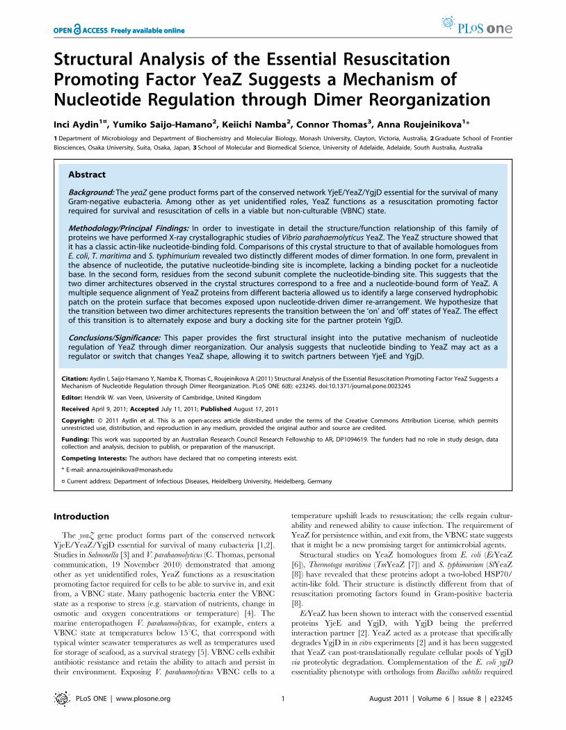
The Overall Fold of YeaZThe structure of VpYeaZ has been solved to 3.1 A resolution
revealing a two domain architecture with a duplicated bbbababasecondary structure (Figs. 1A,B). The domains I and II have a
common fold that comprises a five-stranded mixed b-sheet (order
32145; strand 2 is antiparallel to the rest) surrounded by three a-
helices. The folding pattern of the secondary structure elements is
identical to that of the core of actin-like nucleotide-binding
proteins (the superfamily referred to as ASKHA (acetate and sugar
kinases/Hsc70/actin) [12] or HALF (Hsp70/actin-like fold) in the
literature [13,14]. This suggests that, similar to other members of
this family, the cleft between domains I and II is likely to
accommodate a nucleotide-binding site.
The asymmetric unit contains four subunits, each comprising
residues 2–213. The N-terminal residue S1 is disordered. The 19 C-
terminal residues are missing, most likely having been proteolyti-
cally removed during crystallization (see Materials and Methods).
To investigate the boundaries of a stable core resistant to proteolysis,
Figure 1. A: Stereo diagram of the structure of the VpYeaZ monomer. Each element of the secondary structure is labeled. Domains I and IIand the putative nucleotide-binding cleft are identified. The figure was prepared using PyMOL [11]. B: The topology of the secondary structureelements. Residue numbers are indicated at the start and end of each secondary structure element. C: SDS-PAGE showing the time-course of VpYeaZdigestion by Glu-C protease. The positions of molecular mass markers are shown to the left. The arrow indicates a relatively stable C-terminallytruncated fragment. D: Amino acid sequence of VpYeaZ with Glu-C-sensitive site identified in this study shown by the arrow.doi:10.1371/journal.pone.0023245.g001
Putative Mechanism of YeaZ Dimer Regulation
PLoS ONE | www.plosone.org 2 August 2011 | Volume 6 | Issue 8 | e23245

we carried out digests of VpYeaZ with Glu-C protease which cleaves
at the C-terminal side of Glu residues. The time-course of
degradation, as monitored by SDS-PAGE (Fig. 1C) and Western
blotting analysis (data not shown), demonstrated accumulation of a
relatively stable truncated variant lacking C-terminal His-tag before
further degradation into shorter products. To determine the
proteolytic cleavage sites the truncated variant was isolated by
reverse-phase chromatography and its molecular mass was
measured by matrix-assisted laser desorption ionization-time-of-
flight mass spectrometry, yielding the value of 22,8176100 Da. N-
terminal sequencing confirmed that the N-terminus in the truncated
variant was intact. The truncated variant was identified as fragment
1–213 (Fig. 1D) with excellent agreement between the experimental
and the calculated (22,810 Da) mass values. This analysis suggested
that approximately 20 C-terminal residues of VpYeaZ are flexible
and accessible to protease and that the remainder has a compact
and stable fold. This conclusion is consistent with the observation
that the crystals were formed by fragment 1–213 rather than full-
length protein 1–232.
Structural comparisons to other members of the ASKHAfamily and characterization of the putative nucleotide-binding site
Comparison of the crystal structure of the VpYeaZ monomer
with that of homologous proteins from E. coli (PDB code 1okj [6]),
T. maritima (PDB code 2a6a [7]) and S. typhimurium (PDB code 2gel
[8]) showed that the four structures are closely similar, including
the relative orientation of the two domains (Fig. 2). The structures
of EcYeaZ and StYeaZ can be superimposed with that of VpYeaZ
over the 198 of 212 Ca atoms with an rmsd of 0.9 A, with the
three sequences showing 54% sequence identity over the aligned
amino acid residues. Good overlap of the structures is not
surprising, considering the high sequence identity. The structures
of VpYeaZ and TmYeaZ can be superimposed over 133 Ca atoms
with an rmsd of 1.3 A showing 24% sequence identity over the
aligned residues.
The distinct biological functions of functionally diverse proteins
from the ASKHA superfamily are thought to be mediated by
subdomains inserted at characteristic topological positions be-
tween b3 and a1, b4 and a2 and/or a2 and b5 of domain I, and
b39 and a19 of domain II [13]. These inserts play a role in
oligomerization, substrate and effector binding [12,13]. Remark-
ably, YeaZ appears to be unique amongst other members of
ASKHA superfamily in that it lacks these inserts; the lengths of
loops b3a1, b4a2, a2b5 and b39a19 in VpYeaZ do not exceed five
residues (Fig. 1B). Thus, the YeaZ structure represents the
minimal functional core of the ASKHA superfamily.
Despite similar core fold, the sequences of functionally diverse
proteins from the ASKHA superfamily show very little
conservation mainly characterized by the presence of one or
more glycine-rich or glycine-containing loops associated with
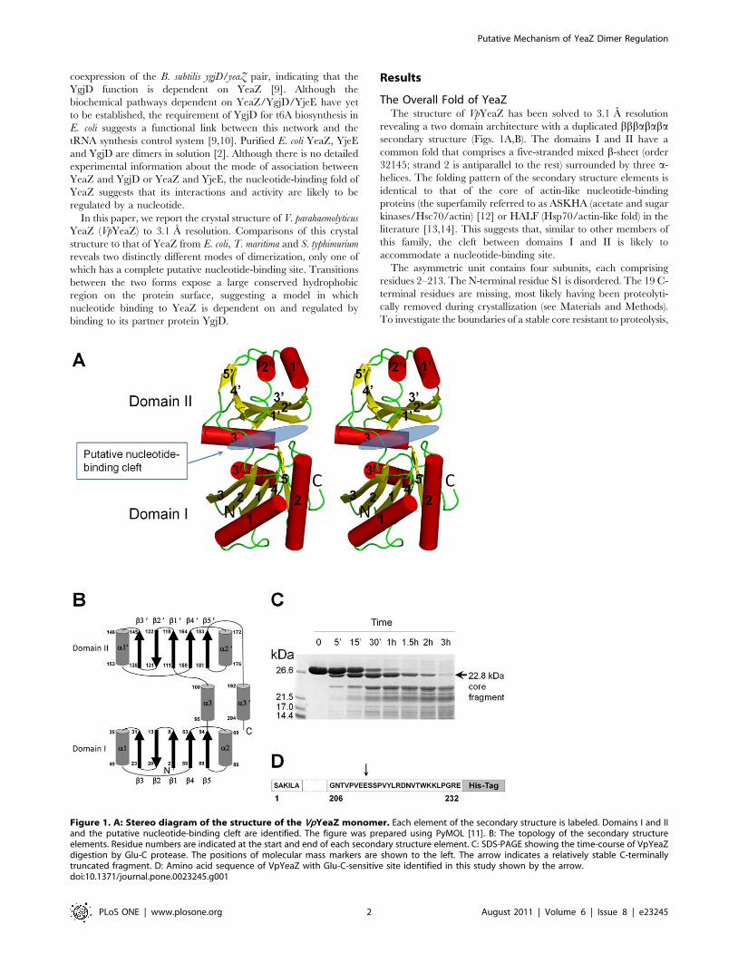
nucleotide binding [15]. The turn between b1 and b2 is thought
to play a critical role in nucleotide binding, with main chain
nitrogens of the turn interacting with the phosphates [13].
Structure and sequence comparisons showed that in YeaZ,
the glycine in the conserved motif DXG in loop b1b2 is
replaced by a small side-chain residue, such as alanine, valine or
serine (A10 in VpYeaZ), whilst the aspartate (D8 in VpYeaZ) is
strongly conserved (Fig. 3). The loop between b49 and a29
harbors a strongly conserved 165GXG motif (Figure 2B) that is
believed to be involved in binding of the nucleotide base moiety
[15]. YeaZ and a structurally similar yeast kinase-associated
endopeptidase 1 (Kae1) share a strongly conserved aspartate
D117 (VpYeaZ numbering) that is involved in binding of the
ribose moiety of the nucleotide in Kae1 [18,19]. Furthermore,
analysis of sequence conservation in YeaZ proteins from
different bacteria identified an absolutely conserved glycine-rich
motif 65GPGXXTGXR which is located in the cleft between
domains I and II and is therefore likely to be important for
recognition of the nucleotide phosphate moiety (Fig. 3). The
position of the aforementioned residues in the YeaZ structure is
shown in Fig. 2.
Two possible modes of YeaZ dimerization andimplications for nucleotide binding
We have earlier reported that VpYeaZ is mostly dimeric in
solution [20], in line with the previous gel-filtration studies on E.
coli YeaZ in solution [2]. Analysis of the packing of VpYeaZ
monomers in the crystal lattice identified an obvious dimer with 2-
fold symmetry and approximate dimensions of 30645680 A
(Fig. 4A). The subunits contact each other via domain I, which is
more conserved than domain II according to analysis of sequence
alignment (Fig. 3). The dimer interface is formed by residues from
helices a1 and a2 from both subunits, which form a 4-helical
bundle. Ten percent (1037 A2) of the subunit accessible surface
area is buried upon dimerization, which falls within the range
found for other dimeric proteins [21]. We therefore conclude that
Figure 2. The structural overlap between VpYeaZ (green),TmYeaZ (purple), EcYeaZ (red) and StYeaZ (black). The sidechains of the conserved and semi-conserved residues likely to beimplicated in nucleotide binding are shown for VpYeaZ.doi:10.1371/journal.pone.0023245.g002
Putative Mechanism of YeaZ Dimer Regulation
PLoS ONE | www.plosone.org 3 August 2011 | Volume 6 | Issue 8 | e23245
Putative Mechanism of YeaZ Dimer Regulation
PLoS ONE | www.plosone.org 4 August 2011 | Volume 6 | Issue 8 | e23245
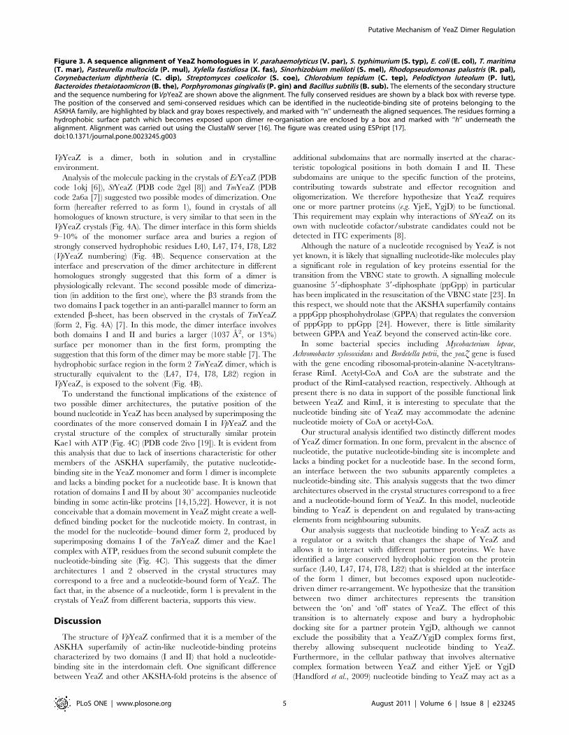
VpYeaZ is a dimer, both in solution and in crystalline
environment.
Analysis of the molecule packing in the crystals of EcYeaZ (PDB
code 1okj [6]), StYeaZ (PDB code 2gel [8]) and TmYeaZ (PDB
code 2a6a [7]) suggested two possible modes of dimerization. One
form (hereafter referred to as form 1), found in crystals of all
homologues of known structure, is very similar to that seen in the
VpYeaZ crystals (Fig. 4A). The dimer interface in this form shields
9–10% of the monomer surface area and buries a region of
strongly conserved hydrophobic residues L40, L47, I74, I78, L82
(VpYeaZ numbering) (Fig. 4B). Sequence conservation at the
interface and preservation of the dimer architecture in different
homologues strongly suggested that this form of a dimer is
physiologically relevant. The second possible mode of dimeriza-
tion (in addition to the first one), where the b3 strands from the
two domains I pack together in an anti-parallel manner to form an
extended b-sheet, has been observed in the crystals of TmYeaZ
(form 2, Fig. 4A) [7]. In this mode, the dimer interface involves
both domains I and II and buries a larger (1037 A2, or 13%)
surface per monomer than in the first form, prompting the
suggestion that this form of the dimer may be more stable [7]. The
hydrophobic surface region in the form 2 TmYeaZ dimer, which is
structurally equivalent to the (L47, I74, I78, L82) region in
VpYeaZ, is exposed to the solvent (Fig. 4B).
To understand the functional implications of the existence of
two possible dimer architectures, the putative position of the
bound nucleotide in YeaZ has been analysed by superimposing the
coordinates of the more conserved domain I in VpYeaZ and the
crystal structure of the complex of structurally similar protein
Kae1 with ATP (Fig. 4C) (PDB code 2ivo [19]). It is evident from
this analysis that due to lack of insertions characteristic for other
members of the ASKHA superfamily, the putative nucleotide-
binding site in the YeaZ monomer and form 1 dimer is incomplete
and lacks a binding pocket for a nucleotide base. It is known that
rotation of domains I and II by about 30u accompanies nucleotide
binding in some actin-like proteins [14,15,22]. However, it is not
conceivable that a domain movement in YeaZ might create a well-
defined binding pocket for the nucleotide moiety. In contrast, in
the model for the nucleotide–bound dimer form 2, produced by
superimposing domains I of the TmYeaZ dimer and the Kae1
complex with ATP, residues from the second subunit complete the
nucleotide-binding site (Fig. 4C). This suggests that the dimer
architectures 1 and 2 observed in the crystal structures may
correspond to a free and a nucleotide-bound form of YeaZ. The
fact that, in the absence of a nucleotide, form 1 is prevalent in the
crystals of YeaZ from different bacteria, supports this view.
Discussion
The structure of VpYeaZ confirmed that it is a member of the
ASKHA superfamily of actin-like nucleotide-binding proteins
characterized by two domains (I and II) that hold a nucleotide-
binding site in the interdomain cleft. One significant difference
between YeaZ and other AKSHA-fold proteins is the absence of
additional subdomains that are normally inserted at the charac-
teristic topological positions in both domain I and II. These
subdomains are unique to the specific function of the proteins,
contributing towards substrate and effector recognition and
oligomerization. We therefore hypothesize that YeaZ requires
one or more partner proteins (e.g. YjeE, YgjD) to be functional.
This requirement may explain why interactions of StYeaZ on its
own with nucleotide cofactor/substrate candidates could not be
detected in ITC experiments [8].
Although the nature of a nucleotide recognised by YeaZ is not
yet known, it is likely that signalling nucleotide-like molecules play
a significant role in regulation of key proteins essential for the
transition from the VBNC state to growth. A signalling molecule
guanosine 59-diphosphate 39-diphosphate (ppGpp) in particular
has been implicated in the resuscitation of the VBNC state [23]. In
this respect, we should note that the AKSHA superfamily contains
a pppGpp phosphohydrolase (GPPA) that regulates the conversion
of pppGpp to ppGpp [24]. However, there is little similarity
between GPPA and YeaZ beyond the conserved actin-like core.
In some bacterial species including Mycobacterium leprae,
Achromobacter xylosoxidans and Bordetella petrii, the yeaZ gene is fused
with the gene encoding ribosomal-protein-alanine N-acetyltrans-
ferase RimI. Acetyl-CoA and CoA are the substrate and the
product of the RimI-catalysed reaction, respectively. Although at
present there is no data in support of the possible functional link
between YeaZ and RimI, it is interesting to speculate that the
nucleotide binding site of YeaZ may accommodate the adenine
nucleotide moiety of CoA or acetyl-CoA.
Our structural analysis identified two distinctly different modes
of YeaZ dimer formation. In one form, prevalent in the absence of
nucleotide, the putative nucleotide-binding site is incomplete and
lacks a binding pocket for a nucleotide base. In the second form,
an interface between the two subunits apparently completes a
nucleotide-binding site. This analysis suggests that the two dimer
architectures observed in the crystal structures correspond to a free
and a nucleotide-bound form of YeaZ. In this model, nucleotide
binding to YeaZ is dependent on and regulated by trans-acting
elements from neighbouring subunits.
Our analysis suggests that nucleotide binding to YeaZ acts as
a regulator or a switch that changes the shape of YeaZ and
allows it to interact with different partner proteins. We have
identified a large conserved hydrophobic region on the protein
surface (L40, L47, I74, I78, L82) that is shielded at the interface
of the form 1 dimer, but becomes exposed upon nucleotide-
driven dimer re-arrangement. We hypothesize that the transition
between two dimer architectures represents the transition
between the ‘on’ and ‘off’ states of YeaZ. The effect of this
transition is to alternately expose and bury a hydrophobic
docking site for a partner protein YgjD, although we cannot
exclude the possibility that a YeaZ/YgjD complex forms first,
thereby allowing subsequent nucleotide binding to YeaZ.
Furthermore, in the cellular pathway that involves alternative
complex formation between YeaZ and either YjeE or YgjD
(Handford et al., 2009) nucleotide binding to YeaZ may act as a
Figure 3. A sequence alignment of YeaZ homologues in V. parahaemolyticus (V. par), S. typhimurium (S. typ), E. coli (E. col), T. maritima(T. mar), Pasteurella multocida (P. mul), Xylella fastidiosa (X. fas), Sinorhizobium meliloti (S. mel), Rhodopseudomonas palustris (R. pal),Corynebacterium diphtheria (C. dip), Streptomyces coelicolor (S. coe), Chlorobium tepidum (C. tep), Pelodictyon luteolum (P. lut),Bacteroides thetaiotaomicron (B. the), Porphyromonas gingivalis (P. gin) and Bacillus subtilis (B. sub). The elements of the secondary structureand the sequence numbering for VpYeaZ are shown above the alignment. The fully conserved residues are shown by a black box with reverse type.The position of the conserved and semi-conserved residues which can be identified in the nucleotide-binding site of proteins belonging to theASKHA family, are highlighted by black and gray boxes respectively, and marked with ‘‘n’’ underneath the aligned sequences. The residues forming ahydrophobic surface patch which becomes exposed upon dimer re-organisation are enclosed by a box and marked with ‘‘h’’ underneath thealignment. Alignment was carried out using the ClustalW server [16]. The figure was created using ESPript [17].doi:10.1371/journal.pone.0023245.g003
Putative Mechanism of YeaZ Dimer Regulation
PLoS ONE | www.plosone.org 5 August 2011 | Volume 6 | Issue 8 | e23245
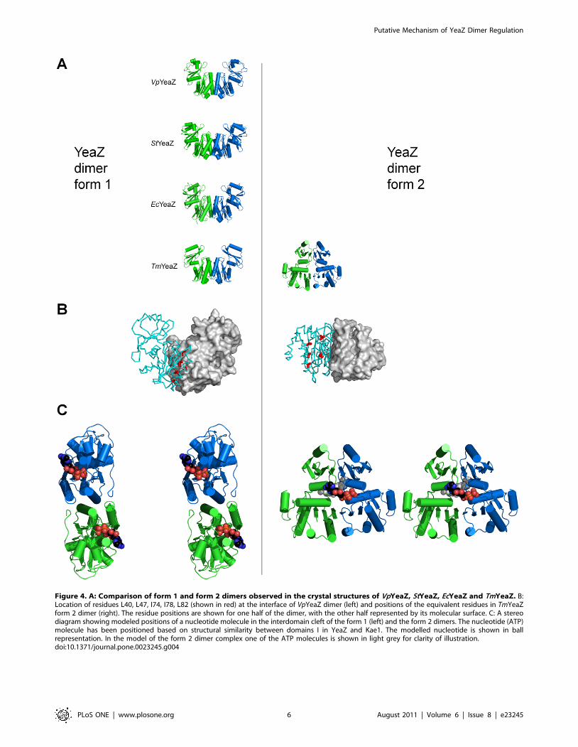
Figure 4. A: Comparison of form 1 and form 2 dimers observed in the crystal structures of VpYeaZ, StYeaZ, EcYeaZ and TmYeaZ. B:Location of residues L40, L47, I74, I78, L82 (shown in red) at the interface of VpYeaZ dimer (left) and positions of the equivalent residues in TmYeaZform 2 dimer (right). The residue positions are shown for one half of the dimer, with the other half represented by its molecular surface. C: A stereodiagram showing modeled positions of a nucleotide molecule in the interdomain cleft of the form 1 (left) and the form 2 dimers. The nucleotide (ATP)molecule has been positioned based on structural similarity between domains I in YeaZ and Kae1. The modelled nucleotide is shown in ballrepresentation. In the model of the form 2 dimer complex one of the ATP molecules is shown in light grey for clarity of illustration.doi:10.1371/journal.pone.0023245.g004
Putative Mechanism of YeaZ Dimer Regulation
PLoS ONE | www.plosone.org 6 August 2011 | Volume 6 | Issue 8 | e23245
regulator that allows YeaZ to switch partners between YjeE and
YgjD. The presented structural analysis provides a useful
foundation for more systematic mutagenesis and biochemical
studies with the aim to address this hypothesis.
Materials and Methods
Crystallization, Data Collection and StructureDetermination
Purification of YeaZ from V. parahaemolyticus strain NCTC
10884, crystallization, data collection and phasing by molecular
replacement have been previously described [20].
Limited Proteolysis of YeaZProteolysis of YeaZ was performed in 40 mM Tris/HCl buffer
pH 8.0 at 298 K. In a typical reaction Glu-C (V8 protease; Roche
Diagnostics) was added to a YeaZ solution at 1 mg/ml at a ratio of
protease to YeaZ of 1:300 (w/w). At various times 15 ml aliquots
were removed for electrophoretic and Western blotting analysis.
The reaction was stopped by adding SDS-PAGE loading dye,
incubating the sample at 368 K for 3 min and freezing it.
Isolation of Proteolytic Fragments0.4 mg of YeaZ was incubated with Glu-C as above for 1 h
(reaction volume 400 ml). The reaction was stopped by adding
400 ml of buffer A (0.065% trifluoroacetic acid (TFA), 2%
acetonitrile in water). The stable core fragment was isolated from
the mixture by reverse-phase chromatography on Resource RPC
3 ml column (GE Healthcare). Elution was achieved with a linear
gradient from buffer A to buffer B (0.05% TFA in 80%
acetonitrile) over 45 ml. Protein fragments were monitored in
the effluent at 280 nm and confirmed by SDS-PAGE analysis.
Matrix-assisted laser desorption ionization-time-of-flightmass spectrometry
1 ml of the purified stable core fragment was mixed with 1.0 ml
of 50% (v/v) acetonitrile, 0.3% (v/v) trifluoroacetic acid, 10 mg/
ml sinapinic acid, placed on the sample plate and allowed to dry.
Molecular mass was analyzed by a mass spectrometer (Voyager
DE/PRO, PerSeptive Biosystems) in linear mode.
N-terminal sequencing of the stable core fragmentProteins in SDS/polyacrylamide gel were transferred onto a
polyvinylidene difluoride (PVDF) membrane (Bio-Rad) with a
Hoefer transblotting apparatus and stained for 5 min with 0.025%
(w/v) Coomassie brilliant blue in 40% (v/v) methanol. After
destaining the PVDF membranes for 10 min with 50% (v/v)
methanol, the band of interest was cut out and subjected to N-
terminal amino acid sequence analysis (Center for Medical
Research and Education, Osaka university).
X-ray crystallographic refinement and analysisMolecular replacement with PHASER [25] revealed that the
asymmetric unit contains two YeaZ dimers that are related by a
non-crystallographic two-fold axis, thus forming a p222 tetramer.
The Fourier electron density maps inspected with the program
COOT [26] showed no density for the C-terminal residues
beyound E213 in all subunits in the asymmetric unit, suggesting
that 19 C-terminal residues may have been proteolytically
removed during crystallization. The fragment seen in the electron
density maps (S1-E213) and the stable core fragment found to be
resistant to proteolysis by Glu-C are identical. The VM and the
solvent content values calculated under the assumption that the C-
terminus is absent rather than disordered are 2.1 A3Da21 and
45%, respectively, which falls within the range observed for
protein crystals [27]. In contrast, the VM value calculated under
the assumption that there is one tetramer of full length YeaZ in the
asymmetric unit, is 1.9 A3Da21, corresponding to a solvent
content of approximately 36%. These values are at the low
extreme of the range observed by Matthews for protein crystals.
This confirmed that the crystals were formed by fragment 1–213
rather than full-length protein 1–232.
The model comprising residues 2–213 in each chain was built
through iterative cycles of re-building with COOT and tight-NCS
TLS refinement with Refmac [28,29]. NCS and TLS groups
corresponded to the four subunits in the asymmetric unit. Analysis
of the stereochemical quality of the models was accomplished using
MOLPROBITY [30]. Refinement statistics are given in Table 1.
Accession codesCoordinates and structure factors have been deposited to PDB
RCSB with the accession code 3R6M.
Acknowledgments
We thank Dr T. Caradoc-Davies at the Australian Synchrotron for
assistance with data collection.
Author Contributions
Conceived and designed the experiments: CT AR. Performed the
experiments: IA YS-H AR. Analyzed the data: IA YS-H AR. Contributed
reagents/materials/analysis tools: CT KN AR. Wrote the paper: IA KN
AR.
References
1. Msadek T (2009) Grasping at shadows: revealing the elusive nature of essential
genes. J Bacteriol 191: 4701–4704.
2. Handford JI, Ize B, Buchanan G, Butland GP, Greenblatt J, et al. (2009) Conserved
network of proteins essential for bacterial viability. J Bacteriol 191: 4732–4749.
Table 1. Refinement statistics.
Resolution range (A) 79–3.1
Reflections 12,912
Residues/atoms 848/6,156
aR-factor 0.241
bFree R-factor 0.294
Average residual B after TLS refinement (A2) 20.6
Bond-length deviation from ideality (A) 0.015
Bond-angle deviation from ideality (u) 1.7
cMolprobity scores
Ramachandran space (%)
Favored 84.0
Allowed 13.3
Outliers 2.7
aR~
X
h
Fobsj j{ Fcalcj jð Þj jX
h
Fobsj j.
bThe free R-factor was calculated on 5% of the data omitted at random.cReference [30].doi:10.1371/journal.pone.0023245.t001
Putative Mechanism of YeaZ Dimer Regulation
PLoS ONE | www.plosone.org 7 August 2011 | Volume 6 | Issue 8 | e23245
3. Panutdaporn N, Kawamoto K, Asakura H, Makino SI (2006) Resuscitation of
the viable but nonculturable state of Salmonella enterica serovar Oranienburg byrecombinant resuscitation-promoting factor derived from Salmonella typhimurium
strain LT2. Int J Food Microbiol 106: 241–247.
4. Oliver JD (2005) The viable but nonculturable state in bacteria. J Microbiol 43:93–100.
5. Oliver JD (2010) Recent findings on the viable but non culturable state inpathogenic bacteria. FEMS Microbiol Rev 34: 415–425.
6. Jeudy S, Stelter M, Coutard B, Kahn R, Abergel C (2005) Preliminary
crystallographic analysis of the Escherichia coli YeaZ protein using the anomaloussignal of a gadolinium derivative. Acta Cryst F61: 848–851.
7. Xu Q, McMullan D, Jaroszewski L, Krishna SS, Elsliger M-A, et al. (2010)Structure of an essential bacterial protein YeaZ (TM0874) from Thermotoga
maritima at 2.5 A resolution. Acta Cryst F66: 1230–1236.8. Nichols CE, Johnson C, Lockyer M, Charles IG, Lamb HK, et al. (2006)
Structural characterization of Salmonella typhimurium YeaZ, an M22 O-
sialoglycoprotein endopeptidase homolog. Proteins: Struct Funct Genet 64:111–123.
9. Yacoubi BE, Hatin I, Deutsch C, Kahveci T, Rousset J-P, et al. (2011) A role forthe universal Kae1/Qri7/YgjD (COG0533) family in tRNA modification.
EMBO J 30: 882–893.
10. Srinivasan M, Mehta P, Yu Y, Prugar E, Koonin EV, et al. (2010) The highlyconserved KEOPS/EKC complex is essential for a universal tRNA modifica-
tion, t6A. EMBO J 29: 1–9.11. Delano WL The PyMOL Molecular Graphics System: Version 0.90 (DeLano
Scientific Palo Alto CA).12. Buss KA, Cooper DR, Ingram-Smith C, Ferry JG, Sanders DA, et al. (2001)
Urkinase: structure of acetate kinase, a member of the ASKHA superfamily of
phosphotransferases. J Bacteriol 183: 680–686.13. Hurley JH (1996) The sugar kinase/heat shock protein 70/actin superfamily:
Implications of conserved structure for mechanism. Annu Rev Biophys Struct25: 137–162.
14. Kabsch W, Holmes KC (1995) The actin fold. FASEB J 9: 167–174.
15. Bork P, Sander C, Valencia A (1992) An ATPase domain common toprokaryotic cell cycle proteins, sugar kinases, actin and hsp70 heat shock
proteins. Proc Natl Acad Sci USA 89: 7290–7294.16. Thompson JD, Higgins DG, Gibson TJ (1997) CLUSTAL W: improving the
sensitivity of progressive multiple sequence alignment through sequenceweighting, position-specific gap penalties and weight matrix choice. Nucleic
Acids Res 22: 4673–4680.
17. Gouet P, Courcelle E, Stuart DI, Metoz F (1999) ESPript: analysis of multiple
sequence alignments in PostScript. Bioinformatics 15: 305–308.
18. Mao DY, Neculai D, Downey M, Orlicky S, Haffani YZ, et al. (2008) Atomic
structure of the KEOPS complex: an ancient protein kinase-containing
molecular machine. Mol Cell 32: 259–275.
19. Hecker A, Graille M, Madec E, Gadelle D, Le Cam E, et al. (2009) The
universal Kae1 protein and the associated Bud32 kinase (PRPK), a mysterious
protein couple probably essential for genome maintenance in Archaea and
Eukarya. Biochem Soc Trans 37: 29–35.
20. Aydin I, Dimitropoulos A, Chen SH, Thomas C, Roujeinikova A (2011)
Purification, crystallization and preliminary X-ray crystallographic analysis of
the putative Vibrio parahaemolyticus resuscitation promoting factor YeaZ. Acta
Cryst F67: 604–607.
21. Jones S, Thornton JM (1995) Protein–protein interfactions: A review of protein
dimer structures. Prog Biophys Mol Biol 63: 31–65.
22. Kristensen O, Laurberg M, Liljas A, Kastrup JS, Gajhede M (2004) Structural
characterization of the stringent response related exopolyphosphatase/guano-
sine pentaphosphate phosphohydrolase protein family. Biochemistry 43:
8894–8900.
23. Boaretti M, Lleo MM, Bonato B, Signoretto C, Canepari P (2003) Involvement
of rpoS in the survival of Escherichia coli in the viable but nonculturable state.
Environ Microbiol 5: 986–996.
24. Kristensen O, Ross B, Gajhede M (2008) Structure of the PPX/GPPA
phosphatase from Aquifex aeolicus in complex with the alarmone ppGpp. J Mol
Biol 375: 1469–1476.
25. McCoy AJ, Grosse-Kunstleve RW, Storoni LC, Read RJ (2005) Likelihood-
enhanced fast translation functions. Acta Cryst D61: 458–464.
26. Emsley P, Cowtan K (2004) Coot: model-building tools for molecular graphics.
Acta Crystallogr D60: 2126–2132.
27. Matthews BW (1968) Solvent content of protein crystals. J Mol Biol 33: 491–497.
28. Collaborative Computational Project Number 4 (1994) The CCP4 suite:
programs for protein crystallography. Acta Cryst D50: 760–763.
29. Winn MD, Isupov MN, Murshudov GN (2001) Use of tls parameters to model
anisotropic displacements in macromolecular refinement. Acta Crystallogr D57:
122–133.
30. Davis IW, Leaver-Fay A, Chen VB, Block JN, Kapral GJ, et al. (2007)
MolProbity: all-atom contacts and structure validation for proteins and nucleic
acids. Nucleic Acids Res 35: W375–W383.
Putative Mechanism of YeaZ Dimer Regulation
PLoS ONE | www.plosone.org 8 August 2011 | Volume 6 | Issue 8 | e23245