
Stromal fibroblasts from the interface zone of human breast
8
Research Article 3507 Introduction Breast cancer is a major health problem that affects the lives of millions of women worldwide each year. High-throughput genetic, epigenetic and gene-expression analyses have enhanced the understanding of the relationship of early neoplastic lesions to normal breast tissue, and distinct molecular alterations are observed in the tumor-stromal and myoepithelial cells during the transition from pre-invasive to invasive disease (Sgroi, 2010). Considering that most tumor burdens follow the activation of tumor microenvironment (TME) remodeling, the normal zone that is remote from the tumor load might yet be unaffected. Theoretically, remodeling of the TME could occur most rapidly in the interface zone, a unique functional and molecular region between the tumor invasion front and the normal zone (Kang et al., 2010). It has been reported that invasion markers in tumor tissues are most dynamic and active within the interface zone, where active cancer invasion or epithelial–mesenchymal transition (EMT) occurs (De Wever et al., 2008; Kang et al., 2010). Accumulating clinical and experimental evidences reveal that tumor development is intimately related to the complex interactions of the carcinomas with several distinct stromal cell types that together create the microenvironment of the cancer cells (Bhowmick et al., 2004; Mueller and Fusenig, 2004). The fibroblast is one of the most crucial components of the tumor microenvironment, which promotes the growth and invasion of cancer cells through the involvement in angiogenesis (Orimo et al., 2005), EMT (Radisky et al., 2005), progressive genetic instability, deregulation of anti-tumor immune responses (Djouad et al., 2003), and synthesis, deposition and remodeling of the extracellular matrix (ECM) (Bhowmick et al., 2004). Such fibroblasts, known as cancer-associated fibroblasts (CAFs), when mixed with oncogene-expressing mammary epithelial cells, promote faster tumor growth than fibroblasts extracted from cancer-free breast tissues (Orimo et al., 2005; Shekhar et al., 2001). CAFs also differ from normal fibroblasts in phenotypic properties, the expression of growth factors and ECM molecule synthesis (Lebret et al., 2007). These findings suggest that CAFs possess biological properties and functions distinct from those of normal fibroblasts. However, the functional contributions of fibroblasts located in the interface zone between the tumor invasion front and the remote normal zone to cancer cells remain poorly understood. In this report, we tested the hypothesis that fibroblasts isolated from tumor burden zone, interface zone and remote normal zone of breast tumor tissue contribute distinctive microenvironmental influences on breast cancer cells. We found that fibroblasts isolated from different tumor stromal zones differed with respect to their ability to induce EMT (measured as induction of vimentin and N-cadherin expression and suppression of E-cadherin expression), modulate expression of membrane-type 1 matrix- metalloproteinase (MT1-MMP), activate the ERK signaling cascade and promote cancer cell migration. Our results provide evidence for a direct correlation between the stromal zone of human mammary carcinomas from which fibroblasts were isolated and the ability of fibroblasts to promote breast cancer cell progression. Stromal fibroblasts from the interface zone of human breast carcinomas induce an epithelial–mesenchymal transition-like state in breast cancer cells in vitro Ming-Qing Gao 1,2 , Baek Gil Kim 1,2 , Suki Kang 2 , Yoon Pyo Choi 1,2 , Hangran Park 1,2 , Kyu Sub Kang 1,2 and Nam Hoon Cho 1,2, * 1 Brain Korea 21 Project for Medical Science, Yonsei University College of Medicine, Seoul, 120-752, Korea 2 Department of Pathology, Yonsei University College of Medicine, 134 Shinchon-dong, Seodaemum-gu, Seoul, 120-752, Korea *Author for correspondence ([email protected]) Accepted 1 July 2010 Journal of Cell Science 123, 3507-3514 © 2010. Published by The Company of Biologists Ltd doi:10.1242/jcs.072900 Summary Fibroblasts were extracted from tissue in tumor burden zones, distal normal zones and interface zones between tumor and normal tissue of human breast carcinomas, and the corresponding fibroblasts were designated as cancer-associated fibroblasts (CAFs), normal zone fibroblasts (NFs) and interface zone fibroblasts (INFs). The crosstalk between three types of fibroblasts and breast cancer cells was evaluated using an in vitro direct co-culture model. We found that INFs grew faster and expressed higher levels of fibroblast activation protein than did NFs and CAFs. Compared with CAFs and NFs, INFs grown with breast cancer cells were significantly more effective in inducing an epithelial-mesenchymal transition (EMT) in cancer cells, as indicated by induction of vimentin and N-cadherin and downregulation of E-cadherin. This EMT process was also accompanied by activation of extracellular signal-regulated kinase (ERK) and modulation of membrane-type 1 matrix metalloproteinase (MT1-MMP) expression. Additionally, INFs promoted breast cell migration to a larger extent compared with NFs and CAFs. Taken together, these findings indicate that INFs isolated from the tumor interface zone exhibited more robust biological modulatory activity than did NFs and CAFs isolated from normal and tumor zones of the same tumor tissue, suggesting that the interface zone of the tumor represents a dynamic region vital to tumor progression. Key words: Breast carcinoma, Epithelial-mesenchymal transition, Stromal fibroblasts, Tumor zones, Tumor microenvironment, Interface fibroblasts Journal of Cell Science
Transcript of Stromal fibroblasts from the interface zone of human breast

Research Article 3507
IntroductionBreast cancer is a major health problem that affects the lives ofmillions of women worldwide each year. High-throughput genetic,epigenetic and gene-expression analyses have enhanced theunderstanding of the relationship of early neoplastic lesions tonormal breast tissue, and distinct molecular alterations are observedin the tumor-stromal and myoepithelial cells during the transitionfrom pre-invasive to invasive disease (Sgroi, 2010). Consideringthat most tumor burdens follow the activation of tumormicroenvironment (TME) remodeling, the normal zone that isremote from the tumor load might yet be unaffected. Theoretically,remodeling of the TME could occur most rapidly in the interfacezone, a unique functional and molecular region between the tumorinvasion front and the normal zone (Kang et al., 2010). It has beenreported that invasion markers in tumor tissues are most dynamicand active within the interface zone, where active cancer invasionor epithelial–mesenchymal transition (EMT) occurs (De Wever etal., 2008; Kang et al., 2010).
Accumulating clinical and experimental evidences reveal thattumor development is intimately related to the complex interactionsof the carcinomas with several distinct stromal cell types thattogether create the microenvironment of the cancer cells(Bhowmick et al., 2004; Mueller and Fusenig, 2004). Thefibroblast is one of the most crucial components of the tumormicroenvironment, which promotes the growth and invasion ofcancer cells through the involvement in angiogenesis (Orimo etal., 2005), EMT (Radisky et al., 2005), progressive geneticinstability, deregulation of anti-tumor immune responses (Djouad
et al., 2003), and synthesis, deposition and remodeling of theextracellular matrix (ECM) (Bhowmick et al., 2004). Suchfibroblasts, known as cancer-associated fibroblasts (CAFs), whenmixed with oncogene-expressing mammary epithelial cells,promote faster tumor growth than fibroblasts extracted fromcancer-free breast tissues (Orimo et al., 2005; Shekhar et al.,2001). CAFs also differ from normal fibroblasts in phenotypicproperties, the expression of growth factors and ECM moleculesynthesis (Lebret et al., 2007). These findings suggest that CAFspossess biological properties and functions distinct from those ofnormal fibroblasts. However, the functional contributions offibroblasts located in the interface zone between the tumor invasionfront and the remote normal zone to cancer cells remain poorlyunderstood.
In this report, we tested the hypothesis that fibroblasts isolatedfrom tumor burden zone, interface zone and remote normal zoneof breast tumor tissue contribute distinctive microenvironmentalinfluences on breast cancer cells. We found that fibroblastsisolated from different tumor stromal zones differed with respectto their ability to induce EMT (measured as induction of vimentinand N-cadherin expression and suppression of E-cadherinexpression), modulate expression of membrane-type 1 matrix-metalloproteinase (MT1-MMP), activate the ERK signalingcascade and promote cancer cell migration. Our results provideevidence for a direct correlation between the stromal zone ofhuman mammary carcinomas from which fibroblasts were isolatedand the ability of fibroblasts to promote breast cancer cellprogression.
Stromal fibroblasts from the interface zone of humanbreast carcinomas induce an epithelial–mesenchymaltransition-like state in breast cancer cells in vitroMing-Qing Gao1,2, Baek Gil Kim1,2, Suki Kang2, Yoon Pyo Choi1,2, Hangran Park1,2, Kyu Sub Kang1,2
and Nam Hoon Cho1,2,*1Brain Korea 21 Project for Medical Science, Yonsei University College of Medicine, Seoul, 120-752, Korea2Department of Pathology, Yonsei University College of Medicine, 134 Shinchon-dong, Seodaemum-gu, Seoul, 120-752, Korea*Author for correspondence ([email protected])
Accepted 1 July 2010Journal of Cell Science 123, 3507-3514 © 2010. Published by The Company of Biologists Ltddoi:10.1242/jcs.072900
SummaryFibroblasts were extracted from tissue in tumor burden zones, distal normal zones and interface zones between tumor and normal tissueof human breast carcinomas, and the corresponding fibroblasts were designated as cancer-associated fibroblasts (CAFs), normal zonefibroblasts (NFs) and interface zone fibroblasts (INFs). The crosstalk between three types of fibroblasts and breast cancer cells wasevaluated using an in vitro direct co-culture model. We found that INFs grew faster and expressed higher levels of fibroblast activationprotein than did NFs and CAFs. Compared with CAFs and NFs, INFs grown with breast cancer cells were significantly more effectivein inducing an epithelial-mesenchymal transition (EMT) in cancer cells, as indicated by induction of vimentin and N-cadherin anddownregulation of E-cadherin. This EMT process was also accompanied by activation of extracellular signal-regulated kinase (ERK)and modulation of membrane-type 1 matrix metalloproteinase (MT1-MMP) expression. Additionally, INFs promoted breast cellmigration to a larger extent compared with NFs and CAFs. Taken together, these findings indicate that INFs isolated from the tumorinterface zone exhibited more robust biological modulatory activity than did NFs and CAFs isolated from normal and tumor zones ofthe same tumor tissue, suggesting that the interface zone of the tumor represents a dynamic region vital to tumor progression.
Key words: Breast carcinoma, Epithelial-mesenchymal transition, Stromal fibroblasts, Tumor zones, Tumor microenvironment, Interface fibroblasts
Jour
nal o
f Cel
l Sci
ence

ResultsComparison of primary fibroblasts isolated from differentzones of human breast tumor tissueBreast cancer tissues were acquired from six patients by macroscopicdissection after the following geographic mapping: (1) real tumorburden, termed the tumor zone, located at the epicenter of the tumortissue; (2) normal-like tissue located up to 5 mm from the invasivefront of the tumor, termed the interface zone; and (3) distal normaltissue located at least 10 mm from the tumor margin, termed thenormal zone (Fig. 1A). This concept is different from the traditionalconcept in which the tumor margin is defined by the edge of thetumor burden that faces the normal zone (Fig. 1B). For reproduciblespatial identification, small invasive ductal carcinomas with an arealess than 10 mm2 were included. Larger tumors (i.e. area>10 mm2),those with vague boundaries or cases other than infiltrating ductalcarcinomas that were not otherwise specified (NOS) were excluded.Regardless of marginal clearance, all cases included in the presentstudy were divided into a definite tumor burden zone, a clear normalfibrofatty region and an intervening fibrotic interface zone, all ofwhich were examined microscopically. The intervening interfacezone is characterized by a relatively more fibrotic zone with muchless fatty tissue than the surrounding normal zone.
The fibroblasts from normal, interface and tumor zones of thetissue were designated NFs, INFs and CAFs, respectively. In thecurrent study, a set of NF, INF and CAF populations weresuccessfully isolated from a patient and each type of fibroblastpossessed the basic fibroblast characteristics of an identical, long,spindle-shaped morphology, strong expression of fibroblastic markervimentin and being negative for epithelial markers such as cytokeratinand cytokeratin 5 (Fig. 2A). Subsequent experiments were carriedout using these cells passaged for 2–10 population doublings (PDs).
INFs cultured in vitro grew significantly faster than CAFs andNFs. This difference was confirmed using a Cell Counting Kit-8(Dojindo, Rockville, MD). After seeding at the same density (2000cells/well) in 96-well plates and culturing for 5 days, the numberof INFs was increased relative to NFs and CAFs (Fig. 2B).However, the apoptosis rates of the three fibroblast types, asanalyzed by FITC-Annexin V/PI staining and flow cytometry,were similar (Fig. 2C).
Additionally, fibroblast activation protein (FAP) was variablyexpressed in the isolated NFs, INFs and CAFs, with the highestexpression in INFs and the lowest expression in NFs (Fig. 2D). Asimilar pattern was found in tissue sections from which the
corresponding fibroblasts were derived (Fig. 2E). Furthermore, toassess the stability of FAP expression in fibroblasts cultured invitro, its expression was investigated at 2, 5 and 10 PDs of NFs,INFs and CAFs. Each type of fibroblast can maintain the phenotypiccharacteristic of FAP expression up to 10 PDs (Fig. 2D).
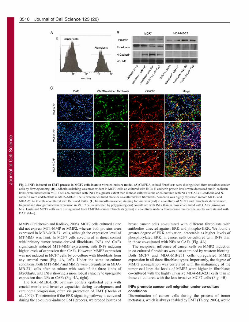
INFs induce an EMT state in MCF7 cells in an in vitroco-culture modelTo determine whether the fibroblasts isolated from normal, interfaceand tumor zones have different capacities to modulate tumorprogression, we cultured NFs, INFs and CAFs directly with MCF7breast cancer cells, allowing direct cell-cell interaction betweencancer cells and fibroblasts. After co-culture, cancer cells andCMFDA-stained (green) fluorescent fibroblasts were readilyseparated by fluorescence-activated cell sorting (Fig. 3A), allowingbreast cancer cells and fibroblasts to be individually assessed infollow-up experiments.
Downregulation of E-cadherin, a universal epithelial marker, isan early indication of EMT (Li et al., 2003; Zeisberg and Kalluri,2004). A western blot analysis of E-cadherin expression in breastcancer cells after co-culture with fibroblasts showed that E-cadherinprotein levels were decreased in MCF7 cells co-cultured with NFs,INFs and CAFs compared with those cultured alone. INFs exerteda more robust effect on this aspect of the EMT process than didNFs or CAFs (Fig. 3B, left). The observed decrease in E-cadherincoincided with the induction of N-cadherin and vimentin. Theinduction of vimentin expression in MCF7 cells co-cultured withfibroblasts was also confirmed by immunofluorescence staining.MCF7 cells co-cultured with INFs showed a much higherpercentage of vimentin-positive cells than did those co-culturedwith CAFs or NFs (Fig. 3C).
When the highly invasive MDA-MB-231 breast cancer line wasused, E-cadherin was not detectable by western blotting in cancercells cultured alone or co-cultured with fibroblasts. However, N-cadherin was faintly induced and vimentin was significantlyupregulated in MDA-MB-231 cells, and the extent of these changeswas greater in cells co-cultured with INFs than with NFs or CAFs(Fig. 3B, right).
MMPs and ERK are involved in the EMT process inducedby co-cultureCells that undergo EMT also undergo alterations in their interactionwith the surrounding ECM through increased expression of matrix
3508 Journal of Cell Science 123 (20)
Fig. 1. Collection of representative breast tumor tissue samples.(A)Tumor tissue was grossly divided into the tumor zone, interfacezone and normal zone as described in the text. The histologicalappearance was confirmed by hematoxylin and eosin (H&E)staining and a representative sample of tissue was collected fromeach region for subsequent fibroblast isolation. Microscopicfindings showed a normal zone dominantly composed ofintermingled fibrofatty tissue, an interface zone characterized byaltered fibroplasias, and a tumor zone dominated by an invasiveductal type of carcinoma within the desmoplastic stromal response.(B)The traditional tumor margin was simply established as theedge of the tumor burden facing the normal zone.
Jour
nal o
f Cel
l Sci
ence

3509In vitro induction of epithelial-mesenchymal transition
Fig. 2. Comparison of primary fibroblasts from each breast cancer tissue zone based on the new three-zone concept of tumor margins. (A)Normal zonefibroblasts (NFs), interface zone fibroblasts (INFs), cancer-associated fibroblasts (CAFs) and MCF7 cells in tissue sections were examined by phase-contrastmicroscopy and immunostaining for vimentin, cytokeratin 5 or a wide-spectrum cytokeratin; nuclei were stained with DAPI. The fibroblasts from each zone had anidentical, long, spindle-shaped morphology and exhibited vimentin expression, whereas they were negative for cytokeratin 5 and cytokeatin. By contrast, MCF7cells were negative for vimentin and positive for cytokeratin 5 and cytokeatin. (B)NFs, INFs and CAFs seeded at the same density (2000 cells/well) in 96-wellplates were assayed for cell viability after 5 days in culture using Cell Counting Kit-8. Cell numbers were calculated by reference to a standard curve obtainedunder the same experimental conditions. Fibroblasts from the interface zone exhibited significantly greater growth rates compared with NFs and CAFs (P<0.01).The data were derived from three independent experiments. (C)NFs, INFs and CAFs grown to confluence were routinely collected and apoptosis was measured byAnnexin/PI staining and flow cytometry analysis. The percentages of apoptotic cells were graphed and the data were derived from three independent experiments.A similar apoptosis rate was observed in each type of fibroblasts. (D)Fibroblast activation protein (FAP) expression was investigated by flow cytometry. Arepresentative histogram of analysis and mean fluorescence intensity of FAP-positive cells in each type of fibroblast at 2, 5 or 10 population doublings (PDs) isshown. FAP was significantly overexpressed in INFs compared with NFs and CAFs at each PD (P<0.01); furthermore, for each fibroblast type, FAP was stablyexpressed at each PD (*P>0.05 versus 5 PDs and 10 PDs, #P>0.05 versus 10 PDs). The data were derived from three independent experiments. (E)Breastcarcinoma sections were immunostained with anti-FAP antibody. FAP was stained specifically in fibroblasts, but this was only significant in the interface zone.Spindle cells are fibroblasts and the brown color indicates positive staining.
Jour
nal o
f Cel
l Sci
ence
MMPs (Orlichenko and Radisky, 2008). MCF7 cells cultured alonedid not express MT1-MMP or MMP2, whereas both proteins wereexpressed in MDA-MB-231 cells, although the expression level ofMT-MMP was faint. In MCF7 cells co-cultured in direct contactwith primary tumor stroma-derived fibroblasts, INFs and CAFssignificantly induced MT1-MMP expression, with INFs inducinghigher levels of expression than CAFs. However, MMP2 expressionwas not induced in MCF7 cells by co-culture with fibroblasts fromany stromal zone (Fig. 4A, left). Under the same co-cultureconditions, both MT1-MMP and MMP2 were upregulated in MDA-MB-231 cells after co-culture with each of the three kinds offibroblasts, with INFs showing a more robust capacity to upregulateexpression than NFs or CAFs (Fig. 4A, right).
The RAF-MEK-ERK pathway confers epithelial cells withcrucial motile and invasive capacities during development andcarcinoma progression, often via promotion of EMT (Doehn etal., 2009). To determine if the ERK signaling pathway is activatedduring the co-culture-induced EMT process, we probed lysates of
breast cancer cells co-cultured with different fibroblasts withantibodies directed against ERK and phospho-ERK. We found agreater degree of ERK activation, detectable as higher levels ofphosphorylated ERK, in cancer cells co-cultured with INFs thanin those co-cultured with NFs or CAFs (Fig. 4A).
The reciprocal influence of cancer cells on MMP2 inductionin co-cultured fibroblasts was also examined by western blotting.Both MCF7 and MDA-MB-231 cells upregulated MMP2expression in all three fibroblast types. Importantly, the degree ofMMP2 upregulation was correlated with the malignancy of thetumor cell line: the levels of MMP2 were higher in fibroblastsco-cultured with the highly invasive MDA-MB-231 cells than inthose co-cultured with the less-invasive MCF7 cells (Fig. 4B).
INFs promote cancer cell migration under co-cultureconditionsDissemination of cancer cells during the process of tumormetastasis, which is always enabled by EMT (Thiery, 2003), would
3510 Journal of Cell Science 123 (20)
Fig. 3. INFs induced an EMT process in MCF7 cells in an in vitro co-culture model. (A)CMFDA-stained fibroblasts were distinguished from unstained cancercells by flow cytometry. (B)Cadherin switching was most evident in MCF7 cells co-cultured with INFs. E-cadherin protein levels were decreased and N-cadherinlevels were increased in MCF7 cells co-cultured with INFs to a greater extent than in those cultured alone or co-cultured with NFs or CAFs. E-cadherin and N-cadherin were undetectable in MDA-MB-231 cells, whether cultured alone or co-cultured with fibroblasts. Vimentin was highly expressed in both MCF7 andMDA-MB-231 cells co-cultured with INFs and CAFs. (C)Immunofluorescence staining for vimentin (red) in co-cultures of MCF7 and fibroblasts showed morefrequent and stronger vimentin expression in MCF7 cells (indicated by polygon regions) co-cultured with INFs than in those co-cultured with CAFs (arrows) orNFs. Unstained MCF7 cells were distinguished from CMFDA-stained fibroblasts (green) in co-cultures under a fluorescence microscope; nuclei were stained withDAPI (blue).
Jour
nal o
f Cel
l Sci
ence

be expected to require enhanced migration ability. In light of theobservation that INFs induced an EMT state in co-cultured MCF7cells, we asked whether INFs promoted breast cancer cell migration.To examine this, we developed an in vitro transwell chemotaxisassay. We found a 2.1-fold and 1.7-fold increase in the number ofMCF7 cells when INF-conditioned medium was used in the lowerwell of the chamber compared with NF- or CAF-conditionedmedium. Similarly, 2.0-fold and 1.3-fold increases in the numberof MDA-MB-231 cells migrated into lower wells of the chamberwere observed when INF-conditioned medium was compared withNF- or CAF-conditioned medium (Fig. 5).
We also performed a scratch-recovery assay under direct co-culture conditions to investigate cell migration ability. Afterscratching, cells were allowed to recover and the capacity of cellsto migrate to fill the area devoid of cells was assessed. WhenMCF7 cells were co-cultured with each type of fibroblasts, phase-contrast microscopic observations showed that the size of thescratched area decreased and nearly closed within 12 hours.However, fluorescent photomicrographs revealed that only in theco-cultures of MCF7-INF, and not MCF7-NF or MCF7-CAF,CMFDA-stained MCF7 cells (green) were observed to migrateinto the scratched area (Fig. 6), indicating that, although all threetypes of fibroblasts migrated into the scratched area, only INFswere able to promote migratory behavior in the less-invasive MCF7
cells, although only to a low extent. Both phase-contrastmicroscopic observations and fluorescent photomicrographsshowed that by 12 hours almost all MDA-MB-231-INF woundswere closed, whereas the wounds were still open in MDA-MB-231-NF or MDA-MB-231-CAF, although there was an obviousreduction of the wound width (Fig. 6).
DiscussionThe results of the present study demonstrate potential differentialinteractions between fibroblasts from different tumor zones andbreast cancer cells. Human breast CAFs and INFs grown withcancer cells dramatically promoted cell migration and induced anEMT process in cancer cells. This effect was not detected or wasweaker when NFs were grown with cancer cells under the sameexperimental conditions. Importantly, INFs were more competentin promoting these changes in breast cancer cells than were CAFs.From these data, we conclude that INFs possess a greater capacityto interact with and modulate cancer cells in this human breast cellmodel system than do NFs or CAFs.
It should be noted that others have previously examinedfibroblast and epithelial interactions in tumor growth anddevelopment by co-inoculating normal fibroblasts or CAFs withtumorigenic epithelial cells, showing that these fibroblasts stimulatemammary epithelial cell growth, differentiation and tumorigenesisor induce EMT via several possible pathways (Lebret et al., 2007;Orimo et al., 2005). Notably, the co-culture models used in theseprevious studies were indirect co-culture systems using transwellsor conditional medium. Additionally, fibroblasts designated asnormal in previous publications referred to fibroblasts isolatedfrom human breast tissue from women undergoing reductionmammoplasty. By contrast, normal fibroblasts in the current studywere defined as those isolated from the normal region of the sametumor tissue as CAFs and INFs were obtained from. In addition,we have define an interface zone that corresponds to a regionlocated up to 5 mm from the invasive front of the tumor; this zonewas grossly estimated by an experienced anatomical pathologistand confirmed by hematoxylin and eosin (H&E) staining. To thebest of our knowledge, delineating normal, interface and tumorzones is a novel formulation that allows us to assess the direct
3511In vitro induction of epithelial-mesenchymal transition
Fig. 4. Effect of interaction between fibroblasts and breast cancer cells onthe expression of MT1-MMP, MMP2 and ERK in an in vitro co-culturesystem. After co-culture, both cancer cells and fibroblasts were isolated byFACS, which distinguished unstained MCF7 cells from the CMFDA-stainedfibroblasts. Each cell type was also cultured alone as a control. (A)Expressionof MT1-MMP, MMP2, total ERK and phospho-ERK in cancer cells. MT1-MMP was clearly induced in MCF7 cells co-cultured with INFs or CAFs,whereas MMP2 was undetectable in MCF7 cells cultured alone or withfibroblasts. Both MT1-MMP and MMP2 were induced in MDA-MB-231 cellsco-cultured with any type of fibroblast. Note that MT1-MMP and MMP2expression were most intense in MDA-MB-231 cells co-cultured with INFs.Both MCF7 and MDA-MB-231 cells co-cultured with INFs showed elevatedlevels of phosphorylated ERK. (B)MMP2 signals in fibroblasts co-culturedwith the invasive MDA-MB-231 cancer cell line were more intense than thosein fibroblasts co-cultured with the less-invasive MCF7 cell line.
Fig. 5. INF-conditioned medium promoted migration of breast cancercells. In vitro transwell chemotaxis assays were performed using conditionedmedium from each fibroblast type. The number of either MCF7 cells or MDA-MB-213 cells that migrated toward the lower wells of the transwell containingparticular fibroblast-conditioned medium was estimated using Cell CountingKit-8. INF-conditioned medium significantly increased the number of bothMCF7 and MDA-MB-231 cells that migrated toward lower wells of chamberscompared with NFs and CAFs. *P<0.01 versus NFs and CAFs.
Jour
nal o
f Cel
l Sci
ence

effects of the respective fibroblasts – NFs, INFs and CAFs – onEMT parameters and identify molecules involved in the EMTprocess.
Although NF, INF and CAF subpopulations were isolated fromwithin the same specimen and exhibited identical, long, spindle-shaped morphologies, they could still be distinguished on the basisof differences in growth rate and FAP expression. The growth ofsolid carcinoma is partly determined by the balance between cellproliferation and apoptosis of its cells (Holmgren et al., 1995). Odaet al. suggested that both proliferation and apoptosis should beconsidered when predicting the growth of renal cell carcinoma(Oda et al., 2003). Our data showed that the increased number ofINFs relative to NFs and CAFs on the basis of a cell-countingassay was not accompanied by either higher or lower apoptosis,indicating that this more rapid growth rate of INFs was mainlydetermined by the rapid proliferation rate. FAP is a cell-surfaceserine protease that is highly expressed in CAFs of human epithelialcarcinomas but not in normal fibroblasts, normal tissues or cancercells (Acharya et al., 2006; Park et al., 1999). A recent report alsoshowed that FAP was detectable in normal mammary fibroblastsby western blot analysis (Lebret et al., 2007). Here, FAP expressionwas detectable in NFs, INFs and CAFs, with the highest levelsobserved in INFs.
A significant finding of this study is that MCF7 cells weresusceptible to EMT transformations in the presence of INFs andCAFs, but not NFs, as indicated in part by the downregulation ofE-cadherin and induction of N-cadherin. This ‘cadherin switching’,
characterized by the loss of E-cadherin and the gain of N-cadherin,is a representative aspect of the EMT phenotype (Han et al., 1999;Tomita et al., 2000). The EMT marker vimentin was also inducedby INFs and CAFs but not by NFs. In each case, these changes(downregulation of E-cadherin and upregulation of N-cadherinand vimentin) were much more strongly induced by INFs thanCAFs. Additionally, although co-cultured NFs, INFs or CAFs hadno effect on the expression of E-cadherin or N-cadherin in thehighly invasive MDA-MB-231 breast cancer cell line, they didupregulate vimentin expression; similar to the observations inMCF7 cells, the effects of INFs were greater than those of CAFsand NFs. It is important to note that vimentin is not only an EMTmarker but is also a myoepithelial-cell-specific protein; therefore,induction of this protein alone is not definitive for EMT. Becausewe do not mean to imply that MCF7 cells transform into fibroblasts,but rather assume a fibroblast-like phenotype, we have used theterm ‘EMT-like state’ to describe the phenotypic changes inducedby INFs and CAFs.
ERK stimulates most forms of epithelial invasive motility suchas wound healing, EMT, malignant invasion and metastasis (Doehnet al., 2009; Horn et al., 2009). Our results showed that the EMTstate induced by INFs and CAFs in MCF7 cells was accompaniedby ERK activation. Although we showed that ERK was activatedin the EMT process, we did not address the mechanisms by whichERK promotes the motility and invasive capacities of epithelialcells. However, the regulation of MMPs by activated ERK hasbeen implicated in the invasive behavior of neuroblastoma cells
3512 Journal of Cell Science 123 (20)
Fig. 6. Effect of fibroblasts on cancer cell migrationin an in vitro direct co-culture system. The capacityof cells to migrate to fill a scratched area devoid ofcells was assessed in co-cultures of cancer cells withNFs, INFs or CAFs. Before co-culture, breast cancercells were pre-stained by CMFDA (green) todistinguish fibroblasts under a fluorescencemicroscope. For the co-cultures of MCF7 and eachtype of fibroblasts, phase-contrast microscopy showedthat the size of the scratched area almost closed within12 hours in all samples tested. However, fluorescencephotomicrographs revealed that CMFDA-stainedMCF7 cells only migrated into the scratched area ofMCF7-INF (arrows) and never migrated into thescratched area of MCF7-CAF or MCF7-NF. For theco-cultures of MDA-MB-231 and fibroblasts fromeach stroma zone, by 12 hours MDA-MB-231-INFwounds were almost completely closed, whereas thewounds were still open in MDA-MB-231-NF orMDA-MB-231-CAF wounds in which there was alsoan obvious reduction of the wound width as indicatedby both phase-contrast microscopy and fluorescencephotomicrographs.
Jour
nal o
f Cel
l Sci
ence

(Lakka et al., 2002) and it has been reported that MMP2 enzymeproduction is increased in fibroblasts cultured in the presence ofmedia conditioned by MCF7 cells (Singer et al., 2002). In thepresent study, using a direct co-culture system, we providedevidence that breast cancer cells upregulated MMP2 expression inall fibroblasts (NFs, INFs and CAFs) and showed that the degreeof MMP2 upregulation was positively correlated with themalignancy of the tumor cells. However, none of the three fibroblasttypes induced MMP2 expression in the less-invasive MCF7 cells,whereas each of these fibroblast types, but especially INFs, wascapable of up-regulating MMP2 expression in highly invasiveMDA-MD-231 cells. Interestingly, INFs upregulated the expressionof MT1-MMP, the only activator of soluble MMP2 (Strongin,2006) in both MCF7 and MDA-MB-231 cells. One possible reasonthat induction of MT1-MMP was not accompanied by MMP2upregulation in MCF7 cells co-cultured with fibroblasts (exceptpossibly indirectly by activating pro-MMP2 or other MMPs on thecell surface) is that MT1-MMP could directly degrade the ECM tocreate a path for cells to migrate (Itoh, 2006). Expression of MT1-MMP is reported to activate ERK, and ERK activation has beenshown to be essential for MT1-MMP-dependent cell migration(Gingras et al., 2001; Takino et al., 2004); however, further workis needed to verify the mechanism by which MT1-MMP activatesERK.
As expected, INFs were able to promote migration of bothMCF7 and MDA-MB-231 cells in a direct or indirect co-culturemodel to a larger extent when compared with NFs and CAFs,indicating that a factor, or factors, secreted or expressed by NFsand CAFs might be more highly secreted or expressed by INFs.Notably, the number of MDA-MB-231 cells that migrated towardslower wells of chambers was obviously higher than that of MCF7cells, and INFs also enhanced the migration ability of MDA-MB-231 cells to a larger extent compared with the migration abilityMCF7 obtained in a direct co-culture condition, which might bedetermined by their intrinsic properties. For instance, MCF7 is aless invasive cell line, whereas MDA-MB-231 is a highly invasivecell line.
In summary, our study demonstrated a direct role for INFs inbreast cancer progression through the induction of an EMT state,the regulation of MMPs and ERK and promotion of cell migration.Compared with NFs and CAFs, INFs exhibited an increasedpropensity to induce the EMT program in MCF7 cells. Our studyindicates that fibroblasts isolated from the interface zone of thetumor possessed more potent biomodulatory properties than NFsand CAFs isolated from normal and tumor zones, respectively.These results suggest that the interface region of the tumor, the siteof EMT, represents a dynamic zone that is vital to tumor progressionand local recurrence because of its robust biological activities.
Materials and MethodsIsolation of primary fibroblastsHuman breast tumor specimens were obtained from patients undergoing surgery atSeverance Hospital of the Yonsei University Health System, Korea. An experiencedanatomical pathologist grossly examined and obtained representative samples oftissue tumor from three zones: the tumor zone (tissue within the tumor boundary),an interface zone (adjacent tissue within 5 mm of the outer tumor boundary) and anormal zone (distal normal tissue at least 10 mm from the outer tumor boundary). Afraction of all tissues was fixed in formalin and embedded in paraffin for routinehistopathological analysis. The remainder was used to isolate primary fibroblasts. Indetail, fresh tissues obtained from different zones of the tumor tissue were cut intosmaller pieces, placed in digestion solution of Enzyme Cocktail (ISU ABXIS, Seoul,Korea) and incubated at 37°C in a humidified 5% CO2 incubator overnight. Digestedtissue was filtered through a 70 m cell strainer. The cells were suspended inmedium:Ficoll (3:2) and separated by differential centrifugation at 90 g for 2 minutes.
The supernatant containing fibroblasts was centrifuged at 485 g for 8 minutes,resuspended in DMEM/F12 supplemented with 10% fetal bovine serum (FBS) and100 IU/ml penicillin with 100 g/ml streptomycin (Gibco BRL, Grand island, NY)and cultured at 37°C in a humidified 5% CO2 environment. The fibroblastic natureof the isolated cells was confirmed by microscopic determination of morphologyportrait and immunofluorescence characterization using antibodies against vimentin(clone VI-10, diluted 1:1000, Abcam, Cambridge, UK), cytokeratin (clone AE/AE3,diluted 1:50, Dako, Glostrup, Denmark) and cytokeratin 5 (clone XM26, diluted1:50, Novocastra, Newcastle upon Tyne, UK).
Direct co-culture experimentsPrimary fibroblasts were directly co-cultured with human less-invasive MCF7 andhighly invasive MDA-MB-231 (Korean Cell Line Bank, Seoul, Korea) breastcarcinoma cells as described previously (Olumi et al., 1999). Briefly, adherentfibroblasts or breast cancer cells were stained by incubating for 45 minutes at 37°Cin serum-free DMEM/F12 containing 5 M CellTracker Green CMFDA (5-chloromethylfluorescein diacetate; Invitrogen, Eugene, OR), a green fluorescentdye. Subsequently, the dye solution was replaced with fresh, prewarmed mediumand the cells were incubated for an additional 2 hours at 37°C. The cells were thenwashed twice with phosphate-buffered saline (PBS) and unstained cancer cells orfibroblasts were seeded onto plates containing CMFDA-stained fibroblasts or cancercells, respectively. Finally, the co-cultures were incubated with minimum serummedium composed of DMEM/F12, 1% FBS and 100 IU/ml penicillin with 100 g/mlstreptomycin for 1 week. CMFDA-stained cells were easily distinguished fromunstained cells by fluorescent microscopy and flow cytometry, allowing independentmeasurements of each cell type.
ImmunofluorescenceCells cultured in 8-well glass chambers (BD Biosciences, Bedford, MA) were rinsedtwice with cold PBS, fixed in 4% paraformaldehyde for 20 minutes at roomtemperature and permeabilized with 0.2% Triton X-100 for 30 minutes on ice. Cellswere then washed twice and incubated with 2% BSA in PBS for 1 hour at roomtemperature to reduce nonspecific binding of primary antibodies. Next, 500 l of aprimary antibody solution was added into each chamber and slides were incubatedovernight at 4°C. After rinsing twice with PBS, cells were incubated withphycoerythrin (PE)-labeled secondary antibody for 1 hour at room temperaturefollowed by subsequent washings. Finally, the glass slide was separated from thechamber and cells were stained with a drop of DAPI (Invitrogen) for 30 minutes tovisualize nuclei and covered with a coverslip. Immunofluorescence was viewedusing an immunofluorescence microscope.
Flow cytometry and fluorescence-activated cell sorting (FACS)Detached cells were washed, resuspended in cold Hank’s balanced salt solution(HBSS) containing 2% heat-inactivated FBS and blocked for 10 minutes with FcRreagent. Next, an anti-fibroblast activation protein (FAP) primary antibody (cloneF11-24, Santa Cruz Biotechnology, Santa Cruz, CA) was added (1 g per 106 cells)and incubated for 30 minutes on ice in the dark. Thereafter, cells were washed twicewith PBS, incubated for 30 minutes on ice with PE-labeled secondary antibody andthen analyzed on a FACSCalibur Flow Cytometer (Becton Dickinson, San Jose,CA). Cells in co-cultures were segregated using an EPICS ALTRA Flow Cytometer(Beckman Coulter, Inc., Fullerton, CA).
Cell proliferation and apoptosis assayCell proliferation and apoptosis assays were performed using a Cell Counting Kit-8(Dojindo, Rockville, MD) and FITC Annexin V Apoptosis Detection Kit I (BDPharmingen, San Diego, CA). For proliferation assay, cells were plated in 96-wellplates and cultured in growth medium. At the indicated timepoints, the absorbanceof samples in triplicate wells was measured with a VersaMax Microplate Reader ata wavelength of 450 nm and cell numbers were calculated based on reference to astandard curve obtained under the same experimental conditions. For apoptosisanalysis, fibroblasts grown to confluence were routinely harvested and cells werethen analyzed using the FITC Annexin V/PI Apoptosis Detection Kit according tothe manufacturer’s protocol.
Western blotCells were lysed in PRO-PREP Protein Extraction Solution (iNtRON Biotechnology,Inc., Gyeonggi-do, Korea). Five to twenty micrograms of total protein from eachsample was resolved on a NuPAGE Novex 4-12% Bis-Tris Gel (Invitrogen, Carlsbad,CA) with MOPS running buffer and transferred to polyvinylidene difluoride (PVDF)membranes. The blots were then probed with antibodies against GAPDH (clone V-18, diluted 1:2000, Santa Cruz Biotechnology), E-cadherin (clone H-108, diluted1:200, Santa Cruz Biotechnology), vimentin (clone VI-10, diluted 1: 1000, Abcam),N-cadherin (clone H-63, diluted 1:1000, Santa Cruz Biotechnology), MMP2 (cloneA-Gel VC2, diluted 1:1000, Thermo Fisher Scientific Anatomical Pathology, Fremont,CA), MT1-MMP (clone H-72, diluted 1:500, Santa Cruz Biotechnology), p44/42MAPK (Erk1/2, diluted 1:1000, Cell Signaling Technology, Danvers, MA) orphospho-p44/42 MAPK (Erk1/2, Thr202/Tyr204, diluted 1:1000, Cell SignalingTechnology), followed by incubation with peroxidase-labeled secondary antibodies.
3513In vitro induction of epithelial-mesenchymal transition
Jour
nal o
f Cel
l Sci
ence

Immunoreactive proteins were visualized using an enhanced chemiluminescence(ECL) detection kit (Santa Cruz Biotechnology).
Wound healing assayBreast cancer cells grown to approximately 50% confluence were stained withCMFDA and then co-cultured with fibroblasts, as described above. Cells wereallowed to grow to 100% confluence and then simply wounded by making a singlescratch in the monolayer with a pipette tip. The medium was then replaced to removefloating cells and debris and cells were incubated for 12 hours to allow cells to growand close the wound. Photographs were taken at the same position of the wound.
Transwell chemotaxis assays in vitroConfluent fibroblasts were rinsed in PBS and fresh serum-free DMEM/F12 mediumwas added to cell culture dishes. Cells were cultured for another 2 days at 37°C with5% CO2. Then medium was collected from the dishes and used at a 9:1 ratio inaddition to fresh DMEM/F12 with 10% FBS as conditioned media after filtration inthe lower well of a 24-well chamber (Costar, NY); 5�103 breast cancer cells werethen seeded in 100 l serum-free DMEM/F12 medium in the upper well of aTranswell chamber with 8 m pore size filters. Transwell chambers were incubatedfor 1 week and the number of cells that migrated toward the lower chamber wasestimated using Cell Counting Kit-8.
This work was supported by the National Research Foundation ofKorea (NRF) grant funded by the Korean government (MEST) (No.20090079165; No. 20090078398; CNH).
ReferencesAcharya, P. S., Zukas, A., Chandan, V., Katzenstein, A. L. and Pure, E. (2006).
Fibroblast activation protein: a serine protease expressed at the remodeling interface inidiopathic pulmonary fibrosis. Hum. Pathol. 37, 352-360.
Bhowmick, N. A., Neilson, E. G. and Moses, H. L. (2004). Stromal fibroblasts in cancerinitiation and progression. Nature 432, 332-337.
De Wever, O., Pauwels, P., De Craene, B., Sabbah, M., Emami, S., Redeuilh, G.,Gespach, C., Bracke, M. and Berx, G. (2008). Molecular and pathological signaturesof epithelial-mesenchymal transitions at the cancer invasion front. Histochem. Cell Biol.130, 481-494.
Djouad, F., Plence, P., Bony, C., Tropel, P., Apparailly, F., Sany, J., Noel, D. andJorgensen, C. (2003). Immunosuppressive effect of mesenchymal stem cells favorstumor growth in allogeneic animals. Blood 102, 3837-3844.
Doehn, U., Hauge, C., Frank, S. R., Jensen, C. J., Duda, K., Nielsen, J. V., Cohen, M.S., Johansen, J. V., Winther, B. R., Lund, L. R. et al. (2009). RSK is a principaleffector of the RAS-ERK pathway for eliciting a coordinate promotile/invasive geneprogram and phenotype in epithelial cells. Mol. Cell 35, 511-522.
Gingras, D., Bousquet-Gagnon, N., Langlois, S., Lachambre, M. P., Annabi, B. andBeliveau, R. (2001). Activation of the extracellular signal-regulated protein kinase(ERK) cascade by membrane-type-1 matrix metalloproteinase (MT1-MMP). FEBSLett. 507, 231-236.
Han, A. C., Soler, A. P., Knudsen, K. A. and Salazar, H. (1999). Distinct cadherinprofiles in special variant carcinomas and other tumors of the breast. Hum. Pathol. 30,1035-1039.
Holmgren, L., O’Reilly, M. S. and Folkman, J. (1995). Dormancy of micrometastases:balanced proliferation and apoptosis in the presence of angiogenesis suppression. Nat.Med. 1, 149-153.
Horn, G., Gaziel, A., Wreschner, D. H., Smorodinsky, N. I. and Ehrlich, M. (2009).ERK and PI3K regulate different aspects of the epithelial to mesenchymal transition ofmammary tumor cells induced by truncated MUC1. Exp. Cell Res. 315, 1490-1504.
Itoh, Y. (2006). MT1-MMP: a key regulator of cell migration in tissue. IUBMB Life 58,589-596.
Kang, S., Shim, H. S., Lee, J. S., Kim, D. S., Kim, H. Y., Hong, S. H., Kim, P. S., Yoon,J. H. and Cho, N. H. (2010). Molecular proteomics imaging of tumor interfaces bymass spectrometry. J. Proteome Res. 9, 1157-1164.
Lakka, S. S., Jasti, S. L., Gondi, C., Boyd, D., Chandrasekar, N., Dinh, D. H.,Olivero, W. C., Gujrati, M. and Rao, J. S. (2002). Downregulation of MMP-9 inERK-mutated stable transfectants inhibits glioma invasion in vitro. Oncogene 21,5601-5608.
Lebret, S. C., Newgreen, D. F., Thompson, E. W. and Ackland, M. L. (2007). Inductionof epithelial to mesenchymal transition in PMC42-LA human breast carcinoma cells bycarcinoma-associated fibroblast secreted factors. Breast Cancer Res. 9, R19.
Li, Y., Yang, J., Dai, C., Wu, C. and Liu, Y. (2003). Role for integrin-linked kinase inmediating tubular epithelial to mesenchymal transition and renal interstitial fibrogenesis.J. Clin. Invest. 112, 503-516.
Mueller, M. M. and Fusenig, N. E. (2004). Friends or foes-bipolar effects of the tumourstroma in cancer. Nat. Rev. Cancer 4, 839-849.
Oda, T., Takahashi, A., Miyao, N., Yanase, M., Masumori, N., Itoh, N., Sato, M. A.,Kon, S. and Tsukamoto, T. (2003). Cell proliferation, apoptosis, angiogenesis andgrowth rate of incidentally found renal cell carcinoma. Int. J. Urol. 10, 13-18.
Olumi, A. F., Grossfeld, G. D., Hayward, S. W., Carroll, P. R., Tlsty, T. D. and Cunha,G. R. (1999). Carcinoma-associated fibroblasts direct tumor progression of initiatedhuman prostatic epithelium. Cancer Res. 59, 5002-5011.
Orimo, A., Gupta, P. B., Sgroi, D. C., Arenzana-Seisdedos, F., Delaunay, T., Naeem,R., Carey, V. J., Richardson, A. L. and Weinberg, R. A. (2005). Stromal fibroblastspresent in invasive human breast carcinomas promote tumor growth and angiogenesisthrough elevated SDF-1/CXCL12 secretion. Cell 121, 335-348.
Orlichenko, L. S. and Radisky, D. C. (2008). Matrix metalloproteinases stimulateepithelial-mesenchymal transition during tumor development. Clin. Exp. Metastasis 25,593-600.
Park, J. E., Lenter, M. C., Zimmermann, R. N., Garin-Chesa, P., Old, L. J. andRettig, W. J. (1999). Fibroblast activation protein, a dual specificity serine proteaseexpressed in reactive human tumor stromal fibroblasts. J. Biol. Chem. 274, 36505-36512.
Radisky, D. C., Levy, D. D., Littlepage, L. E., Liu, H., Nelson, C. M., Fata, J. E.,Leake, D., Godden, E. L., Albertson, D. G., Nieto, M. A. et al. (2005). Rac1b andreactive oxygen species mediate MMP-3-induced EMT and genomic instability. Nature436, 123-127.
Sgroi, D. C. (2010). Preinvasive breast cancer. Annu. Rev. Pathol. 5, 193-221.Shekhar, M. P., Werdell, J., Santner, S. J., Pauley, R. J. and Tait, L. (2001). Breast
stroma plays a dominant regulatory role in breast epithelial growth and differentiation:implications for tumor development and progression. Cancer Res. 61, 1320-1326.
Singer, C. F., Kronsteiner, N., Marton, E., Kubista, M., Cullen, K. J., Hirtenlehner,K., Seifert, M. and Kubista, E. (2002). MMP-2 and MMP-9 expression in breastcancer-derived human fibroblasts is differentially regulated by stromal-epithelialinteractions. Breast Cancer Res. Treat 72, 69-77.
Strongin, A. Y. (2006). Mislocalization and unconventional functions of cellular MMPsin cancer. Cancer Metastasis Rev. 25, 87-98.
Takino, T., Miyamori, H., Watanabe, Y., Yoshioka, K., Seiki, M. and Sato, H. (2004).Membrane type 1 matrix metalloproteinase regulates collagen-dependent mitogen-activated protein/extracellular signal-related kinase activation and cell migration. CancerRes. 64, 1044-1049.
Thiery, J. P. (2003). Epithelial-mesenchymal transitions in development and pathologies.Curr. Opin. Cell Biol. 15, 740-746.
Tomita, K., van Bokhoven, A., van Leenders, G. J., Ruijter, E. T., Jansen, C. F.,Bussemakers, M. J. and Schalken, J. A. (2000). Cadherin switching in human prostatecancer progression. Cancer Res. 60, 3650-3654.
Zeisberg, M. and Kalluri, R. (2004). The role of epithelial-to-mesenchymal transition inrenal fibrosis. J. Mol. Med. 82, 175-181.
3514 Journal of Cell Science 123 (20)
Jour
nal o
f Cel
l Sci
ence


![Cancer-associated fibroblasts in hepatocellular …...human carcinoma-associated stromal cells[13]. It was reported that even without exposure to cancer cells, the tumor promoting](https://static.fdocuments.in/doc/165x107/5f4766e071df1550f7486c8b/cancer-associated-fibroblasts-in-hepatocellular-human-carcinoma-associated-stromal.jpg)


