STROKE: The Cutting Edge: 2017 - ePublishing Login · STROKE: The Cutting Edge: 2017 iii ... East,...
18
i STROKE: The Cutting Edge: 2017
Transcript of STROKE: The Cutting Edge: 2017 - ePublishing Login · STROKE: The Cutting Edge: 2017 iii ... East,...

iSTROKE: The Cutting Edge: 2017

ii STROKE: The Cutting Edge: 2017
STROKETHE CUTTING EDGE
Copyright © 2017 AHC Media, a Relias Learning company
Reproduction, distribution, or translation without express written permission is strictly prohibited.
STROKE: The Cutting Edge is published by AHC Media, a Relias Learning company
Executive Editor: Leslie G. CoplinAHC Media Editorial Group Manager: Terrey L. Hatcher
Senior Accreditations Officer: Lee Landenberger
AHC Media, LLC111 Corning Road, Suite 250
Cary, NC 27518(800) 688-2421
ISBN: 978-1-941481-36-3
The articles within this publication were previously published in the following AHC Media publications:
Clinical Cardiology AlertED Management
Internal Medicine AlertNeurology Alert
OB/GYN Clinical Alert
iiiSTROKE: The Cutting Edge: 2017
ACCREDITATION INFORMATION
ACCREDITATION STATEMENTRelias Learning is accredited by the Accreditation Council for Continuing Medical Education to provide continuing medical education for physicians.
Relias Learning, LLC is accredited as a provider of continuing nursing education by the American Nurses Credentialing Center’s Commission on Accreditation. Provider approved by the California Board of Registered Nursing, Provider #CEP13791, for 8 Contact Hours.
CREDIT DESIGNATIONRelias Learning designates this enduring material for a maximum of 8.0 AMA PRA Category 1 Credits™. Physicians should claim only the credit commensurate with the extent of their participation in the activity.
This activity has been approved for 8.0 nursing contact hours using a 60-minute contact hour.
ACTIVITY OBJECTIVESAfter reading STROKE: The Cutting Edge: 2017, the participant will be able to:1. Discuss current scientific research and data regarding the diagnosis and treatment of stroke;2. Discuss the pathogenesis and treatment of stroke;3. Explain the basic science of brain function as it applies to stroke;4. Cite new information regarding new drugs for stroke and new uses for traditional drugs;5. Identify nonclinical issues of importance for health care providers who treat stroke patients.6. Discuss advances in neurointerventional treatment.
Physicians and nurses participate in this CME/CE activity by reading the articles, using the provided references for further research, and studying the relevant questions at the end of the book. Participants will then be directed to a website, where they will complete an online assessment to show what they’ve learned. They must score 100 on the assessment in order to complete the activity, but they are allowed to answer the questions multiple times if needed. After they have successfully completed the assessment, they will be directed to an online activity evaluation form. Once that is submitted, they will receive their credit letter.
TARGET AUDIENCE This activity is intended for for neurologists, emergency medicine physicians and nurses, Joint Commission-certified stroke center personnel, psychiatrists, and ENTs.
EXPIRATION DATEMarch 31, 2020
PHYSICIAN EDITORMatthew Fink, MDLouis and Gertrude Feil Professor in Clinical Neurology and Chairman, Department of Neurology, Weill Cornell Medical College; Neurologist-in-Chief, New York Presbyterian Hospital
PEER REVIEWER Amre M. Nouh, MD Director of the Stroke Center, Hartford Hospital; Assistant Professor and Associate Residency Program Director, Department of Neurology, University of Connecticut
NURSE PLANNERDonna Avanecean, MS, RN, FNP-C, CNRN, DNP-c Movement Disorders/DBS Program, Department of Neurosurgery, Yale University
FINANCIAL DISCLOSURESIn order to reveal any potential bias in this publication, and in accordance with Accreditation Council for Continuing Medical Education guidelines, Dr. Fink, Dr. Nouh, and Dr. Avanecean report no financial relationships relevant to this field of study.
Editorial and Production: Ms. Coplin, Ms. Hatcher, and Mr. Landenberger report no financial relationships relevant to this field of study.
Authors: Financial disclosure information for authors can be found at the beginning of their respective articles. This publication does not receive commercial support.

iv STROKE: The Cutting Edge: 2017
TABLE OF CONTENTS
Module 1: Prevention and Risk Factor Reduction (1 hour)Preventing 90% of Stroke with 10 Modifiable Risk Factors ................................................. 1
A New Risk Score for Stroke in Atrial Fibrillation ................................................................. 2
Effects of Aspirin on Risk of Early Recurrent Stroke After Transient Ischemic Attack and Ischemic Stroke .......................................................... 4
Headaches in the Elderly: A Non-specific Marker for Stroke Risk ....................................... 6
Migraine with Aura and Systemic Right-to-Left Shunt: Risk for Stroke? ............................. 8
Contraception, Migraines, and Stroke ............................................................................... 10
Arterial Ischemic Stroke Prevention and Risk Factor Management ................................... 13
Proceedings from the 2017 International Stroke Conference ........................................... 31
Stroke Alert: A Review of Current Stroke Literature.......................................................... 33
CME Test: Module 1 ........................................................................................................... 35
Module 2: Prehospital and Emergency Care (1 hour)Functional Outcomes in Acute Ischemic Stroke Patients Receiving Prehospital Thrombolysis in Mobile Stroke Units ............................................................... 38
New Mobile Stroke Unit Programs Aim to Improve Outcomes ........................................ 40
Proceedings from the 2017 International Stroke Conference ........................................... 44
Stroke Alert: A Review of Current Stroke Literature.......................................................... 45
CME Test: Module 2 ........................................................................................................... 46
Module 3: Neuroimaging for Stroke (1 hour)Blood-brain Barrier Breakdown in RCVS ............................................................................ 48
Proceedings from the 2017 International Stroke Conference ........................................... 50
CME Test: Module 3 ........................................................................................................... 51
Module 4: Large Vessel Ischemic Stroke Treatment (1 hour)Intravenous Glyburide to Reduce Brain Swelling in Large Hemispheric Infarction ........... 53
Proceedings from the 2017 International Stroke Conference ........................................... 55
Stroke Alert: A Review of Current Stroke Literature.......................................................... 57
CME Test: Module 4 ........................................................................................................... 59
Module 5: Acute Endovascular Treatment (1 hour)Proceedings from the 2017 International Stroke Conference ........................................... 60
A Review of Current Stroke Literature ............................................................................... 62
CME Test: Module 5 ........................................................................................................... 63

vSTROKE: The Cutting Edge | Module 1: Prevention and Risk Factor Reduction
TABLE OF CONTENTS
Module 6: In-hospital Management of Stroke (1 hour)RESCUEicp: A Trial of Decompressive Craniectomy for Traumatic Intracranial Hypertension ............................................................................. 65
Proceedings from the 2017 International Stroke Conference ........................................... 67
A Review of Current Stroke Literature ............................................................................... 69
CME Test: Module 6 ........................................................................................................... 71
Module 7: Aneurysms and AVMs (1 hour)Proceedings from the 2017 International Stroke Conference ........................................... 73
A Review of Current Stroke Literature ............................................................................... 74
CME Test: Module 7 ........................................................................................................... 75
Module 8: Stroke Rehabilitation (1 hour)Stroke: Rehabilitation and Recovery .................................................................................. 77
Proceedings from the 2017 International Stroke Conference ........................................... 87
CME Test: Module 8 .......................................................................................................... 88.

vi Module 1: Prevention and Risk Factor Reduction | STROKE: The Cutting Edge
STROKE: THE CUTTING EDGEModule 1: Prevention and Risk Factor Reduction
1STROKE: The Cutting Edge | Module 1: Prevention and Risk Factor Reduction
SYNOPSIS: Controlling 10 modifiable risk factors would prevent 90.7% of strokes, according to results from a 32-country study.
SOURCE: O’Donnell MJ, Chin SL, Rangarajan S, et al. Global and regional effects of modifiable risk factors associated with acute stroke in 32 countries. Lancet 2016; Jul 15 doi: http//dx.doi.org/10.1016/S0140-6736(16)30506-2. [Epub ahead of print].
The INTERSTROKE investigators from 32 countries in Asia, the Americas, Europe, Australia, the Middle East, and Africa conducted a case-control study of
patients suffering acute stroke compared with controls. Researchers compared 13,447 acute stroke cases (10,388 with ischemic stroke and 3,059 with hemorrhagic stroke) with 13,472 matched controls. The investigators identified 10 modifiable risk factors that may have prevented 90.7% of strokes.
The most important modifiable risk factor is hyperten-sion. Eliminating high blood pressure would reduce stroke risk by 48%. There were regional variations, with hyper-tension causing about 39% of strokes in North America, Australia, and Western Europe, and nearly 60% of strokes in Southeast Asia. The other modifiable risk factors, with obvious overlap in many patients are:
z Physical inactivity: 36% z Hyperlipidemia: 27% z Poor diet: 23% z Obesity: 19% z Smoking: 12% z Heart disease: 9% z Alcohol: 6% z Diabetes: 4% z Stress: 6%
CommentaryStroke is the third leading cause of death and a major
cause of disability, and as this study shows is highly pre-ventable. Unhealthy lifestyle leads to more than 90% of all strokes worldwide. The same could be said of heart disease and many cancers. Most instances of poor health and pre-mature demise are self-inflicted.
An argument can be made about personal responsibility in avoiding preventable chronic illness and events such as stroke. Individuals are in charge of their own lifestyle deci-sions. However, one also can make an argument that these risk factors are a public health problem, with culture and environment leading to poor health choices. In his book Fat Chance, Robert Lustig from the University of California, San Francisco makes this argument forcefully.1 Lustig points to the ill effects of added sugars and high fructose corn syr-up leading to obesity, type 2 diabetes, metabolic syndrome, and fatty liver, with the food industry largely to blame by hooking the population on these addictive sweets. The pub-lic health response has been lukewarm at best.
Science is coming together with medical practice to put more focus on modifiable risk factors in chronic disease and preventing major events such as stroke. Had this study been conducted 20 years ago, smoking would have been a much greater factor. We have the resources to become much healthier worldwide by adopting healthy lifestyles and protecting the environment. The public is becoming better educated in this area, and those businesses that sell unhealthy products are beginning to feel the effect. Physicians, especial-ly those in primary care, should be “captains” in the battle against the poor health choices that surround us every day.
Reference1. Lustig R. Fat Chance: Beating the Odds Against Sugar, Processed Food,
Obesity, and Disease. New York: Penguin Group; 2012.
ABSTRACT & COMMENTARY
Preventing 90% of Stroke with 10 Modifiable Risk Factors
By Joseph E. Scherger, MD, MPH, Vice President, Primary Care, Eisenhower Medical Center; Clinical Professor, Keck School of Medicine, University of Southern California.
Dr. Scherger reports no financial relationships relevant to this field of study.

13STROKE: The Cutting Edge | Module 1: Prevention and Risk Factor Reduction
Stroke is a common problem, affecting nearly 800,000 people annually in the United States.1 Domestically, it is the fifth most common cause of death and a leading
cause of significant long-term disability.1 Stroke costs the United States an estimated $34 billion each year.1 Given this extraordinary burden on the health of the American popula-tion, appropriate stroke prevention measures could dramati-cally improve our quality and length of life.
The widespread institution of screening, risk factor modi-fication, and treatment of known cardiac and cerebrovascu-lar disease is paramount to the health of our nation. The fol-lowing article discusses the best evidence-based practice for stroke diagnosis, prevention, and risk factor management. Where strong data are absent, usual practice and expert con-sensus are accepted and noted as such.
Stroke EpidemiologyThere are nearly 800,000 strokes in the United States each
year, with 130,000 associated deaths.1,2 Although stroke is the second leading cause of death worldwide, it is only the fifth most likely cause in the United States.1,2 Because of the major disability associated with stroke, the overall cost to this country is substantial. Up to 30% of patients who survive a stroke will require institutional care.2 Mortality from stroke varies according to ethnicity, with blacks twice as likely to suffer a stroke and more likely to die compared to whites.2 The “stroke belt” in the southeastern United States has the highest national stroke mortality.2 Stroke risk increases with age, but 34% of stroke affects individuals younger than 65 years of age.2 The overall stroke incidence is expected to rise in the near future as the population ages, despite recent advances in risk factor reduction, which has reduced the per capita incidence of strokes. There are gen-der differences in stroke rates as well.Women are 50% more likely than men to have a stroke, particularly due to a high incidence in white elderly females.2
Modifiable Stroke Risk FactorsHypertension is the most important modifiable stroke
risk factor. Multiple trials have shown the benefit of blood
pressure control, including many different agents such as angiotensin-converting enzyme inhibitors, angiotensin receptor blockers, calcium channel blockers, and diuretics.3 The choice of agent is less crucial than the successful lower-ing of blood pressure. In general, blood pressure treatment should be targeted to a normal blood pressure of < 140/90 mmHg.3 However, recent evidence suggests a more aggres-sive goal of systolic < 120 mmHg may be beneficial for the primary prevention of cardiovascular events.4 Although these data applied to a composite outcome of stroke, myocardial infarction, heart failure, and vascular death, they confirm that “pre-hypertension” (diastolic 80-90 and systolic 120-140) contributes to increased stroke risk.
Hypercholesterolemia is another important stroke risk fac-tor. Low-density liporprotein (LDL) cholesterol in patients with a prior stroke should be < 70 mg/dL. Cholesterol low-ering is particularly important in patients with a prior history of atherosclerotic disease, in which case the American Heart Association (AHA) recommends titrating statins to the maximum tolerated dosage (high-intensity statin) rather than specific LDL goals.5 Large cohorts of patients with prior myocardial infarction, such as the Heart Protection Study, have shown that control of lipids not only prevents future heart attack but specifically lowers stroke rates by about 25%.6
Obstructive sleep apnea (OSA) approximately doubles the risk of stroke when compared to controls, and stroke rates increase steadily with OSA severity.7 The mechanism by which OSA results in cerebral embolism is not entirely certain; however, it is known that sleep-disordered breath-ing increases the development of atrial fibrillation, a potent stroke risk factor.8 The treatment for OSA is quite effective. Continuous positive airway pressure (CPAP) therapy has been shown to be effective in reducing stroke risk to equal patients with no history of sleep-disordered breathing.7
Diabetes is known to confer excess risk of stroke inde-pendent of blood pressure.9 Specific stroke mechanisms, such as large artery atherosclerotic disease or small vessel ischemia, are associated with poor glucose control.9 Despite stroke being a known vascular complication of diabetes,
Michael P. Lerario, MD, Department of Neurology, Weill Cornell Medical College, New York; New York-Presbyterian/Queens Hospital, Flushing, NY
Alan Z. Segal, MD, Department of Neurology, Weill Cornell Medical College, New York
Drs. Lerario and Segal report no financial relationships relevant to this field of study.
Arterial Ischemic Stroke Prevention and Risk Factor Management

15STROKE: The Cutting Edge | Module 1: Prevention and Risk Factor Reduction
mimic stroke include seizure with postictal deficits, migraine aura, conversion disorder, encephalopathy from metabolic disturbances, intracranial tumors or infections, hyperten-sive encephalopathy, Bell’s palsy, transient global amnesia, spinal or nerve disorders, peripheral vertigo, and syncope. The discrimination of a true vascular event from a mimic is important in terms of treatment and follow-up. Whereas TIA is a known risk factor for subsequent stroke, the risk of future vascular events after the diagnosis of a mimic is negligible.23,24 The ABCD2 score may be helpful in making the diagnosis of a non-vascular event. Lower scores on this scale are associated with mimics, rather than true TIAs, and have a low likelihood of future stroke.24 Nevertheless, this differentiation between mimic and true stroke may not be as important in the acute setting, since it has been deemed safe to treat mimics with intravenous (IV) tissue plasminogen activator. The risk of symptomatic intracranial hemorrhage following IV thrombolysis has been demonstrated to be 1% or less.22,25
Stroke MechanismsCauses of Stroke. Ischemic stroke, the damage to neuro-
nal tissue as a result of reduced cerebral blood flow, can be a common endpoint due to many conditions. The main causes of stroke are due to either thrombosis or embolism of a cerebral artery. Thrombosis refers to a local occlusive process formed in situ within the artery. The site of ob-struction may occur either within a large intra- or extracra-nial artery (which is typically the result of atherosclerosis) or within a small penetrating artery (which typically is due to chronic vessel changes from hypertension).26,27 On the other hand, embolism refers to thrombus that travels from the site of formation and lodges within distal vessels. The source of embolism either may be a proximal artery, the heart, or paradoxically from the venous system in the case of a patent foramen ovale (PFO).26,27 Embolic strokes tend to cause symptoms that are abrupt and maximal at onset.26 Additionally, embolism often leads to cortical infarction in the cerebral surface of an arterial territory and is more likely to be associated with hemorrhagic conversion.28
TOAST Classification System. There have been many attempts to categorize mechanisms of stroke for clinical and research purposes. A commonly used classification schema for stroke subtype is known as the TOAST system.27 (See Table 2.) The TOAST system has inherent limitations, namely the large number of stroke patients who resultantly are classified as cryptogenic (i.e., having undetermined eti-ology). The TOAST investigators did not require aggressive diagnostic testing, by today’s standards, prior to categoriz-ing a patient as cryptogenic; for instance, transesophageal echocardiogram and extended arrhythmia monitoring were not required. Nevertheless, it is a useful tool for research purposes and for conceptualizing stroke mechanism in a simplified manner.
Lacunar Stroke. Lacunar stroke refers to the pathophys-iological, clinical, and radiographic findings observed in small vessel disease. A lacunar stroke is the result of arterial obstruction of a single deep, penetrating vessel that supplies the subcortical structures of the brain, such as the capsule, basal ganglia, thalamus, and paramedian brainstem.26,29 Such arterial obstruction is associated with the pathological changes occurring in response to chronic hypertension or diabetes, including microatheroma or lipohyalinosis.26 Lipo-hyalinosis refers to the degenerative change in small blood vessels due to the accumulation of lipid within the vessel wall.26
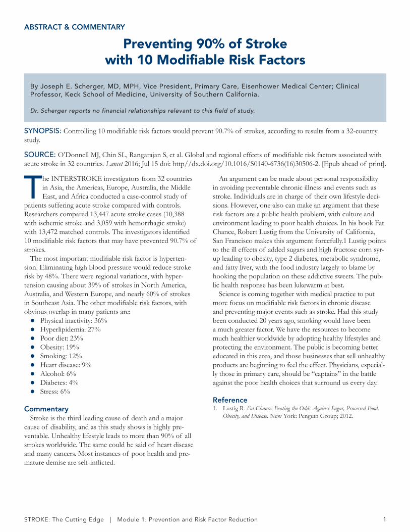
Since the affected arteries are small, the resultant strokes (known as lacunes) also are small. These irregular cavitary lesions typically are < 1.5 centimeters in diameter.30 Radio-graphically, lacunar strokes appear as small infarcts in typical subcortical structures, with MRI being more sensitive than CT for detecting these lesions.26 (See Figure 1.) The imaging findings of a small, deep infarct in the absence of other possible stroke etiologies is strongly supportive of a lacu-nar etiology. Larger areas of infarction, known as giant or super lacunes, may imply thrombosis or embolism of the proximal branch from which the penetrating artery arises; for instance, a lenticulostriate territory stroke could result from a nonocclusive embolus to the middle cerebral artery.26 Therefore, subcortical strokes > 1.5 centimeters in size may necessitate a diagnostic evaluation aimed at finding an em-bolic source of stroke, including echocardiography, teleme-try, and noninvasive angiography of the head and neck.
Lacunar infarcts clinically present as one of several classic syndromes, with the following being the most common: pure motor weakness, pure sensory loss, mixed sensorim-otor, ataxia hemiparesis, and dysarthria-clumsy hand syn-dromes.31 The symptoms of lacunar stroke often fluctuate over the acute course of the disease, hence the name “stut-
Table 2. TOAST Classification System of Stroke Subtypes
Stroke SubtypePrevalence of Stroke Subtype32
Large artery atherosclerosis 15.5%
Cardioembolism 27.8%
Small vessel occlusion 18.1%
Stroke of other determined etiology 4.2%
Stroke of undetermined etiologyTwo or more identified causesNegative evaluation
34.4%
16 Module 1: Prevention and Risk Factor Reduction | STROKE: The Cutting Edge
tering lacune.” Although lacunar strokes comprise nearly 20% of strokes,32 they have the lowest in-hospital mortality of the subtypes.33
Cardioembolic Stroke. Cerebral embolism occurs when particulate material from a proximal source travels through the arterial system to lodge within a downstream cerebral ar-tery. Emboli to the brain are most often composed of mural thrombi or platelet aggregates and most often travel to the anterior circulation (particularly the middle cerebral artery territory), given that these arteries accept the majority of ce-rebral blood flow.26 Although an embolic etiology accounts for up to 70% of stroke cases,26 probable or definite evi-dence of cardiac embolism is demonstrated in only 25-30% of ischemic strokes.32 In other instances, the source of em-bolus may be a proximal large artery, the venous circulation in the case of a PFO, or unknown (cryptogenic).27 A cardiac source is implied if the emboli result in bilateral infarcts, particularly if cortical and in multiple vascular territories.28 (See Figure 2.) For example, a potential cardioembolic etiolo-gy of stroke would be inferred from an MRI demonstrating simultaneously occurring left middle cerebral artery and right posterior cerebral artery acute transcortical infarctions. These emboli tend to be evanescent, and obstruction of an intracranial artery may recanalize spontaneously,34 likely
contributing to the higher rate of intracerebral hemorrhage with reperfusion associated with embolic strokes.28
There are many potential causes of cerebral cardioem-bolism. (See Table 3.) Whether a stroke can be attributed to a cardioembolic source requires the identification of a known cardiac risk factor for embolic stroke, as well as the exclusion of other etiologies, such as large artery atheroscle-rosis or lacunar infarct, based on dedicated neuroimaging.27 The probability of a stroke being attributed to an identified cardiac risk factor depends on how strongly that risk factor is associated with ischemic stroke.27 The most common high-risk sources of cardioembolism include valvular heart disease, the formation of an intracardiac thrombus (e.g., recent myocardial infarction or atrial fibrillation), ventricular or septal aneurysms, and cardiomyopathies. Three specific cardiac sources of stroke, including atrial fibrillation, PFO, and aortic arch atheroma, are discussed in depth below.
Atrial Fibrillation as a Stroke Risk Factor. Atrial fibril-lation is the most common sustained cardiac arrhythmia, occurring in 1-2% of the general population, and is even more prevalent in aging populations.35 Atrial fibrillation can lead to left atrial or atrial appendage thrombus formation, which can serve as a source of embolism. The arrhythmia is a very strong risk factor for stroke, increasing this risk by as
Figure 1. MRI Displaying Typical Imaging Characteristics of Acute Lacunar Infarctions
Note small, deep infarctions in the left paramedian pons (A) and right thalamus (B).
A B

27STROKE: The Cutting Edge | Module 1: Prevention and Risk Factor Reduction
monitoring on cardioembolic stroke recurrence due to the known superiority of anticoagulation to antiplatelets in me-dium- to high-risk atrial fibrillation patients. However, this has yet to be proven definitively. In the meantime, a standard approach is to monitor cryptogenic stroke patients for at least 30 days, while treating them with antiplatelet therapy, unless atrial fibrillation is discovered.107
In addition to extended arrhythmia monitoring, it has been suggested that additional advanced diagnostic tech-niques be used to uncover other potential stroke etiologies in cryptogenic patients who have otherwise unremarkable routine stroke evaluations.60 Such testing could include the measurement of D-dimer and screening for occult ma-lignancy to rule out cancer-related coagulopathy. Aortic pathology such as atherosclerosis also may embolize to the brain and is best visualized using transesophageal echocar-diography. Interatrial shunts, including PFO and atrial septal defects, should be tested for with bubble studies either using echocardiography or transcranial Doppler. Advanced, high-resolution vascular wall imaging, with or without contrast, also may detect arterial plaques with ulceration or other high-risk elements that could serve as a source of embolism.
ConclusionGiven the frequency of stroke occurrence, and the high
rates of resultant death and disability, an evidence-based prevention strategy is one of the largest weapons in a clinican’s armamentarium against stroke. With time, we are learning more about the importance of precise risk factor management, the role of statins for atherosclerotic disease, and the use of targeted antithrombotic therapy based on the underlying mechanism of stroke. Whereas large artery ath-erosclerotic and small vessel disease responds to antiplatelet agents, anticoagulation has been found to be superior in certain high-risk cardiac conditions, such as atrial fibrilla-tion. The recent expansion of oral anticoagulants to include factor Xa and direct thrombin inhibitors has significantly improved the overall risk/benefit profile of the medical therapy for patients with nonvalvular atrial fibrillation. Surgical options also vary by stroke subtype and mechanism. Although symptomatic cervical carotid stenosis is amena-ble to revascularization with stenting or endarterectomy, stenting of intracranial atherosclerosis has proven harmful in clinical trials. Patients with unexplained stroke after a standard diagnostic evaluation, particularly if young, should have advanced testing to evaluate for rarer causes of stroke. If a cryptogenic stroke appears embolic on neuroimaging, such patients may benefit from extended cardiac monitoring or empiric anticoagulation, although randomized trial data are needed to demonstrate improved clinical outcomes with such practice.
References1. Mozaffarian D, Benjamin EJ, Go AS, et al. Heart Disease and
Stroke Statistics—2015 Update: A report from the AmericanHeart Association. Circulation 2015;131:e29-322.
2. Mozaffarian D, Benjamin EJ, Go AS, et al. Heart Disease andStroke Statistics—2016 Update: A Report From the AmericanHeart Association. Circulation 2016;133:e38-e360.
3. Kernan WN, Ovbiagele B, Black HR, et al. Guidelines for theprevention of stroke in patients with stroke and transient isch-emic attack: a guideline for healthcare professionals from theAmerican Heart Association/American Stroke Association. Stroke2014;45:2160-2236.
4. Wright JT, Williamson JD, Whelton PK, et al. A randomized trialof intensive versus standard blood-pressure control. N Engl J Med2015;373:2103-2116.
5. Stone NJ, Robinson JG, Lichtenstein AH, et al. 2013 ACC/AHAguideline on the treatment of blood cholesterol to reduce athero-sclerotic cardiovascular risk in adults: A report of the AmericanCollege of Cardiology/American Heart Association Task Forceon Practice Guidelines. Circulation 2014;129(25 Suppl 2):S1-45.
6. Group HPSC. MRC/BHF Heart Protection Study of cholesterollowering with simvastatin in 20,536 high-risk individuals: A ran-domised placebo-controlled trial. Lancet 2002;360:7-22.
7. Yaggi HK, Concato J, Kernan WN, et al. Obstructive sleep apneaas a risk factor for stroke and death. N Engl J Med 2005;353:2034-2041.
8. Gami AS, Pressman G, Caples SM, et al. Association of atrialfibrillation and obstructive sleep apnea. Circulation 2004;110:364-367.
9. Barrett-Connor E, Khaw KT. Diabetes mellitus: An independentrisk factor for stroke? Am J Epidemiol 1988;128:116-123.
10. Kleindorfer D, Panagos P, Pancioli A, et al. Incidence and short-term prognosis of transient ischemic attack in a population-basedstudy. Stroke 2005;36:720-723.
11. Redgrave JN, Coutts SB, Schulz UG, et al. Systematic review ofassociations between the presence of acute ischemic lesions ondiffusion-weighted imaging and clinical predictors of early strokerisk after transient ischemic attack. Stroke 2007;38:1482-1488.
12. Easton JD, Saver JL, Albers GW, et al. Definition and evaluationof transient ischemic attack: a scientific statement for healthcareprofessionals from the American Heart Association/AmericanStroke Association Stroke Council; Council on CardiovascularSurgery and Anesthesia; Council on Cardiovascular Radiology andIntervention; Council on Cardiovascular Nursing; and the Inter-disciplinary Council on Peripheral Vascular Disease. The Ameri-can Academy of Neurology affirms the value of this statement asan educational tool for neurologists. Stroke 2009;40:2276-2293.
13. Prabhakaran S, Chong JY, Sacco RL. Impact of abnormal diffu-sion-weighted imaging results on short-term outcome followingtransient ischemic attack. Arch Neurol 2007;64:1105-1109.
14. Ovbiagele B, Kidwell CS, Saver JL. Epidemiological impact in theUnited States of a tissue-based definition of transient ischemicattack. Stroke 2003;34:919-924.
15. Johnston SC, Rothwell PM, Nguyen-Huynh MN, et al. Validationand refinement of scores to predict very early stroke risk aftertransient ischaemic attack. Lancet 2007;369:283-292.
16. Amarenco P, Lavallée PC, Labreuche J, et al. One-year risk ofstroke after transient ischemic attack or minor stroke. N Engl JMed 2016;374:1533-1542.
17. Olivot JM, Wolford C, Castle J, et al. Two aces: Transient ischemicattack work-up as outpatient assessment of clinical evaluation andsafety. Stroke 2011;42:1839-1843.

35STROKE: The Cutting Edge | Module 1: Prevention and Risk Factor Reduction
1. Which of the following has NOT been identified as a modifiable risk factor for stroke?a. Diabetesb. Vigorous exercise c. Poor dietd. Smoking
2. Which of the following is the greatest risk factor for causing acute stroke?a. Hypertension b. Smokingc. Obesityd. Diabetes
3. New research suggests which of the following tests may add significantly to the estimation of stroke risk in atrial fibrillation?a. NT-proBNPb. High-sensitivity troponinc. Both d. Neither
4. Aspirin administered orally after a transient ischemic attack:a. should be given at least five days after the onset
of symptoms.b. should be given as soon as possible after the
onset of symptoms. c. should be given at least three days after the onset
of symptoms.d. should never be given.
5. Which statement best describes headache and the elderly?a. Headaches are not indicators of disease risk.b. Headache prevalence decreases with age.c. Both migraine without and with aura increase
stroke risk.d. Tension-type headache is a rare cause of
headache.
6. The presence of a spontaneous right-to-left shunt in a migraineur in the general population is associated with an increased risk of which of the following?a. Silent posterior circulation infarctsb. White matter lesionsc. Symptomatic cerebral ischemiad. Persistence of migraine attacks over timee. Increased mean attack frequency
7. In the study by Champaloux et al, the risk ofstroke in women with migraine without aura using combined hormonal contraceptives and women with migraine without aura not using combined hormonal contraceptives was roughly equivalent.a. Trueb. False
8. Which of the following is not a modifiable stroke risk factor?a. Hypertensionb. Agec. Obstructive sleep apnead. Diabetes
9. Which is not a recent criticism of the ABCD2 score for stratifying stroke risk following transient ischemic attack?a. It does not include an assessment for high-risk
stroke mechanisms such as carotid stenosis or atrial fibrillation.
b. It overestimates stroke risk.c. It was not designed to triage patients from the
emergency department.d. It cannot be used to differentiate true stroke from
stroke mimic.
10. Which score on the CHA2DS2-VASc scale typically warrants anticoagulation for stroke prevention in atrial fibrillation patients at average bleeding risk?a. 0b. 1c. 2d. 3
11. Which diagnostic tests would be recommended for a patient suspected of having primary central nervous system vasculitis?a. Lumbar punctureb. Cerebral angiogramc. Brain biopsyd. All of the above
12. Evidence-based management for secondarystroke prevention in patients with intracranial atherosclerosis includes all of the following except:a. intracranial stenting.b. short-term dual antiplatelet therapy.c. strict blood pressure control.d. intensive LDL lowering with a statin medication.
CME/CE QUESTIONS
To earn credit for this module, log in to to take the post-test.
40 Module 2: Prehospital and Emergency Care | STROKE: The Cutting Edge
Everyone knows that shortening the time to treatment is key to improving outcomes in stroke patients. However, some experts believe that continued prog-
ress in this area requires a bold approach and new thinking on how care can be delivered optimally. For example, a number of medical centers are experimenting with the use of mobile stroke units (MSU), ambulance-like vehicles that are equipped with the necessary technology and expertise to bring brain-saving treatment to the patient.
Although the units are deployed in several states, including Texas, Ohio, Colorado, Tennessee, and New York, investiga-tors at these pioneering sites are experimenting with all the different parameters, everything from how, when, and where the units should be deployed to how the MSUs should inter-face ideally with hospitals and EDs. Further, some sites are experimenting with larger, more robust units equipped with the technology to perform more sophisticated diagnostics and potentially more care delivery in the prehospital envi-ronment.
Cut Through Red TapeIn October 2016, New York Presbyterian/Weill Cornell
Medical Center in New York became the first medical center on the East Coast to deploy an MSU. It’s a project that Mat-thew Fink, MD, the neurologist-in-chief of the Division of Stroke and Critical Care Neurology at the hospital, has been championing since 2013.
“The very first ambulance like this was developed in Ham-burg, Germany, in 2003,” Fink explains. “I heard about it on my trips there and was very intrigued because the European model of emergency medical service is different than the American model. In Europe, doctors often go out on the ambulances and they do more treatment in the field than we do.”
With good results observed in Europe from use of the MSUs, Fink concluded that his center should offer this capa-bility.
“We have worked very hard to initiate treatment of stroke as fast as possible, and we have done very well, but we have come to the point where we have done it as fast as we possibly can when patients [are brought] to the ED,” he
says. “The only way we can do it even faster is to do it in the field.”
Initially, stakeholders were hesitant to approve the project because of all the rules, regulations, and bureaucracy, but Fink persisted in pushing the concept.
“I was convinced that this was really the next phase in treating patients with acute stroke because if we could cut down the time to treatment, it was going to result in saving lives and increasing the number of people making a full recovery,” Fink notes. “To me, that was a really important goal.”
The turning point came in 2015 when a hospital board member bought into the MSU approach, convinced others on the merits of the idea, and offered to underwrite the project. “Then we were off and running,” Fink adds.
However, Fink notes that building the MSU and getting all the physicians on board with the innovation was the easy part. Much more difficult was the process of working through all the regulatory agencies and receiving appropriate approvals from both the state and the city.
“We had to integrate [the MSU approach] with the 911 EMS fire department system, which runs all of the am-bulances in New York City,” Fink explains. “We have the largest EMS system in the country.”
Integrate with EMSWith all the approvals finally in hand, the MSU began de-
ploying on Oct. 3, 2016, in what is phase one of the project. During this period, a neurologist is on board the MSU along with two paramedics and a CT technologist. Also on board is a portable CT scanner that can image a patient’s brain to determine if the patient is suffering a stroke.
When someone calls 911, the dispatcher will ask a few questions based on the Cincinnati Prehospital Stroke Scale. If the answers suggest that the probability of stroke is high, then the dispatcher will send both a basic life support unit and the MSU to the scene.
“We also monitor all the radio chatter on the EMS radio channel so if we hear of a case that we are not called for but we think it sounds like a stroke, then we will dispatch the stroke unit ourselves,” Fink explains. “We have what is called
By Dorothy Brooks, Editor, ED Management
Ms. Brooks reports no financial relationships relevant to this field of study.
New Mobile Stroke Unit Programs Aim to Improve Outcomes

57STROKE: The Cutting Edge | Module 4: Large Vessel Ischemic Stroke Treatment
Which Patients with TIA Are at High Risk for a Recurrent Cerebral Vascular Events?
SOURCE: Yaghi S, Rostanski SK, Boehme AK, et al. Imaging parameters and recurrent cerebral vascular events in patients with minor stroke or transient ischemic attack. JAMA Neu-rol 2016;73:572-578.
Recurrent cerebral vascular events (RCVEs) are one of the main determinants of outcome in patients after mi-
nor strokes and transient ischemic attacks (TIAs). The risk of recurrence is highest within 90 days and is particularly high in the first 48 hours. A number of scoring systems have been developed to attempt a prediction and stratify high-risk from low-risk patients. However, the scores have been lim-ited because they were derived from mostly non-neurologist diagnosed TIA samples and their applicability to patients seen by current neurology stroke teams is questionable. The objective of this study is to determine predictors of early recurrent cerebral vascular events among patients with TIA or minor stroke, defined as an NIHSS of 0 to 3. This ret-rospective cohort study was conducted at two tertiary care centers, Columbia University in New York, and Tulane Uni-versity in New Orleans, from 2010 until 2014. All patients were diagnosed with a TIA or minor stroke by a neurologist when they presented to the emergency department. The primary outcome was a recurrent neurological event un-explained by any other medical condition. Of 1,258 total patients, 71 had recurrent events. In a multivariate model of prediction for recurrent infarct, the significance predictors were 1) infarcts on neuroimaging (CT or diffusion-weighted MRI), with an odds ratio of 1.75, and 2) large vessel disease etiology, with an odds ratio of 6.69. When both predictors were present, there was a further increase in the risk of patients to have recurrent cerebral vascular events. When neither predictor was present, the rate of recurring events was extremely low (up to 2%). Patients who had recurrent events were less likely to be discharged to home.
Dual Antiplatelet Therapy Appears More Effective Than Single Therapy
SOURCE: Ge F, Lin H, Liu Y, et al. Duel antiplatelet therapy after stroke or transient ischemic attack – how long to treat? The duration of aspirin plus clopidogrel in stroke or transient isch-emic attack: A systematic review and meta-analysis. Eur J Neu-rol 2016;23:1051-1057.
The CHANCE study showed that the combination of aspirin and clopidogrel was superior to aspirin alone for
reducing the risk of stroke in the first 90 days after a TIA or minor ischemic stroke (N Engl J Med 2013;369:11-19). In its 2014 guidelines, the American Heart Association recom-mended that the combination of aspirin and clopidogrel can be initiated within 24 hours for a minor ischemic stroke or TIA and continued for 90 days. However, the CHANCE trial was performed in China with a discrete ethnic popula-tion, and it was not clear if the optimal duration of treat-ment should be 90 days or longer. In ischemic heart disease, treatment with dual antiplatelet therapy beyond one year is the standard of care in patients who have coronary stents, and this question has been unanswered in patients with transient ischemic attack or stroke. Therefore, the authors performed a comprehensive literature review and meta-anal-ysis, and identified nine randomized controlled trials that included 21,923 patients. In review of these trials, short-term dual antiplatelet therapy significantly reduced the risk of ischemic stroke recurrence by 41% and major vascular events by 30%, without an increased risk of intracranial hemorrhage. Prolonged treatment beyond 90 days reduced the risk of ischemic stroke recurrence by 12% and major vascular events by 10%. However, the risk of major bleeding and intracranial hemorrhage was increased in those patients treated for a longer term. Therefore, it appears that short-term dual antiplatelet therapy appears to be superior to prolonged treatment. However, this difference in outcome needs to be confirmed by further well-designed randomized clinical trials.
By Matthew E. Fink, MD, FAAN, Professor and Chairman, Department of Neurology, Weill Cornell Medical College; Neurologist-in-Chief, New York Presbyterian Hospital
Dr. Fink reports he is a retained consultant for Procter & Gamble and Pfizer.
STROKE ALERT
A Review of Current Stroke Literature

58 Module 4: Large Vessel Ischemic Stroke Treatment | STROKE: The Cutting Edge
1. Which of the following statements is true?a. Intravenous glyburide did not reduce edema
in large hemispheric infarction as measured by decreased midline shift.
b. Intravenous glyburide resulted in increased concentrations of metalloproteinase 9, a biomarker of brain edema.
c. Adverse reactions were more common in patients receiving intravenous glyburide compared to patients receiving placebo.
d. There was a strong trend towards poorer functional outcomes in patients receiving intravenous glyburide compared to patients receiving placebo.
e. None of the above
2. In secondary stroke prevention, a single-agent antiplatelet medication is just as effective in reducing recurrent stroke or TIA as dual antiplatelet treatments.a. Trueb. False
3. In patients who present with transient ischemic attack or minor stroke, the period of time withthe highest risk of recurrence is the first 48 hours following the initial event.a. Trueb. False
4. Following acute ischemic stroke, treatment witha single antiplatelet agent is just as efficacious as treatment with dual antiplatelet medications, and carries a lower risk of intracranial hemorrhage.a. Trueb. False
CME/CE QUESTIONS
To earn credit for this module, log in to ____________ to take the post-test.
60 Module 5: Acute Endovascular Treatment | STROKE: The Cutting Edge
Message from the Editor: This article is based on personal interactions as a participant at the International Stroke Con-ference in Houston, Feb. 22-24, 2017. All interpretations and opinions are exclusively those of the author.
General Anesthesia in Endovascular Reperfusion Stroke Trials
Bruce Campbell from the University of Calgary, repre-senting the HERMES collaboration, presented the effect of general anesthesia on outcomes in five endovascular reper-fusion stroke trials that were reported in 2015, using pooled data. A logistic regression model was developed and adjust-ed for variables, which included age, sex, stroke severity on the NIH stroke scale, time from symptom onset to random-ization, baseline ASPECT score, baseline site of occlusion, and whether the patient received alteplase prior to undergo-ing endovascular treatment. About 25% of patients received general anesthesia, and there were no significant differences in the baseline characteristics between the two groups. tPA was administered in 88% of the general anesthesia group and about 80% of the group that did not receive general anesthesia. Functional outcome was shown to be significant-ly better (mRS 0-2) in the group that did not receive general anesthesia, with an odds ratio of 2.62 in favor of no anes-thesia resulting in improved outcomes (statistically signifi-cant, P < 0.001). In patients who underwent thrombectomy, there was a 50% return to functional independence in those who were treated without general anesthesia, compared to only a 36% recovery to functional independence in those who were treated with general anesthesia. There was an improvement in terms of early recovery, return to normal function, and reduced mortality in patients who were treated with thrombectomy without general anesthesia, all statistical-ly significant. In conclusion, neurological outcomes are bet-ter in patients who were treated without general anesthesia, and there were more complications associated with general anesthesia, particularly an increased risk of pneumonia.
CT Perfusion ImagingIn a second presentation, Bruce Campbell presented data
from seven recent trials of endovascular therapy, which
included pool data from 1,764 patients, using CT perfusion imaging to define the ischemic core volume and determine the relationship of ischemic core volume to functional out-come following endovascular thrombectomy. Ischemic core was calculated by a fully automated computerized system from Phillips imaging. The ischemic core was defined as a reduction of cerebral blood flow > 30% of blood flow to normal brain. The endovascular group and the control group were well matched for baseline characteristics, includ-ing median NIHSS of 17, initial median ASPECTS score of 8, percent that received alteplase, and baseline median ischemic core volume of 10 mL in the endovascular group and 9 mL in the control group. In the control group that re-ceived IV tPA, there was a rapid drop-off in functional inde-pendence (mRS 0-2) as the ischemic core volume increased, from 50% recovery when there was a core volume close to 0 down to 10% as the ischemic core approached 70 mL. With endovascular therapy, the curve shifted upward, showing 70% functional recovery with an ischemic core of 0, and as much as 30% functional recovery with an ischemic core of 70 mL, and a straight-line relationship between those two points. The differences between the two groups were highly significant after adjustment for all other variables. If full reperfusion was accomplished, the curve shifted upward again, and functional independent outcome improved a step further. In patients who had ischemic cores > 70 mL, there was no statistically significant benefit or difference between treatment with intravenous tPA compared to endovascu-lar thrombectomy. In conclusion, ischemic core volume is correlated with worse outcome, and ischemic core volume > 70 mL does not seem to favor endovascular thrombectomy over intravenous thrombolysis.
Contact Aspiration of Intracranial Clot vs. Stent-retriever
Bertrand Lapergue, from Versailles, France, presented the results of the ASTER trial, comparing the addition of contact aspiration of intracranial clot vs. stent-retriever as the initial intervention for recanalization in patients with acute cerebral infarction. The investigators aimed to as-certain whether contact aspiration was more efficient as a
By Matthew E. Fink, MD, FAAN, Feil Professor and Chairman, Department of Neurology, and Assistant Dean of Clinical Affairs, Weill Cornell Medical College; Neurologist-in-Chief, New York Presbyterian Hospital
Dr. Fink reports he is a consultant for Procter & Gamble.
Proceedings from the 2017 International Stroke Conference

77STROKE: The Cutting Edge | Module 8: Stroke Rehabilitation
Rehabilitation is a critical component of stroke treatment, as most stroke survivors are left with significant neurolog-ical impairments and other sequelae, such as spasticity and pain. Approximately 40% of stroke patients are left with moderate functional impairment and 15%-30% with severe disability.1 Stroke rehabilitation aims to reverse these impair-ments to the extent possible, maximize functionality through the use of compensatory approaches, prevent complications, and manage comorbidities. This article reviews the basic principles of rehabilitation, current practices, and evidence supporting various aspects of stroke rehabilitation.
Role of Primary Care PhysicianWith millions of stroke survivors in the United States, pri-
mary care physicians often are faced with providing care to these individuals and, thus, need to be able to identify com-mon post-stroke rehabilitation issues. Post-stroke depression can occur at any time after stroke, and it is important that primary care physicians recognize this as a treatable and reversible condition, rather than a “natural” consequence of disability after stroke. Monitoring range of motion to iden-tify developing contracture and/or spasticity is important so that referral can be made to a rehabilitation physician if these occur. Lastly, addressing the stroke survivor’s mobility, ability to perform activities of daily living (ADL), and any deterioration in functional status are critical to making time-ly referrals to rehabilitation services when needed. When these issues are identified early, the primary care physician can prevent the progression of debilitating consequences by initiating treatment or referring to appropriate specialists for further evaluation and management.
RehabilitationFunctional improvement in rehabilitation is accom-
plished through a combination of neurological recovery and adoption of compensatory techniques and equipment. Improved functional independence may be attained by reducing impairment directly (i.e., via neural recovery using cerebral plasticity to overcome neuronal loss) or through compensation for impairment by using remaining physical
and cognitive abilities and strategies. Both recovery and compensation are crucial concepts in rehabilitation and key therapeutic approaches; interventions must be balanced to address patient goals and efficiently deploy the available rehabilitation resources.2
Over the last several decades, a significant amount of research has been conducted to further elucidate how best to provide comprehensive stroke rehabilitation. Currently, it is comprised of several key components, including assess-ment, goal setting, treatment of functional and psychosocial impairments, and prevention of complications. Patients are reassessed at regular intervals to evaluate progress, and treat-ment plans are adjusted accordingly. Ideally, rehabilitation begins immediately following stroke and often becomes a long-term element in the lives of these patients. Assessment and treatment should begin in the setting of acute hospi-talization, and depending on the needs of the individual, continued rehabilitation may transition to the appropriate inpatient or outpatient setting.
National and international guidelines have been developed to provide resources for health care providers with the latest evidence-based practices, such as the American Heart As-sociation (AHA) Guidelines published in 2016.3 The AHA grading system for level of evidence and strength of recom-mendation are referenced in this article where applicable.3 (See Table 1.) Adherence to stroke rehabilitation guidelines is associated with improved patient functional outcomes.4
In an effort by the Joint Commission and the AHA, certification has been developed using current guidelines and established standards of care to identify both Primary Stroke Centers and Comprehensive Stroke Centers. The rehabilitation requirements for Primary Stroke Center certi-fication include the ability to assess for rehabilitation needs and refer for appropriate post-acute care. Comprehensive Stroke Centers also must have a rehabilitation service led by a physician with expertise in neurorehabilitation, and the service must include therapists, nurses, and social workers with an expertise in addressing the rehabilitation needs of stroke patients.5,6 Complete guidelines can be found on the Joint Commission’s website.5
Joel Stein, MD, Simon Baruch Professor and Chair, Department of Rehabilitation and Regenerative Medicine, Columbia University College of Physicians and Surgeons; Professor and Chair, Department of Rehabilitation Medicine, Weill Cornell Medical College; and Physiatrist-in-Chief, NewYork-Presbyterian Hospital, New York
Dr. Stein reports he receives grant/research support from Tyromotion, GMBH and Myomo, Inc.; and he has developed online CME materials for QuantiaMD.
Stroke: Rehabilitation and Recovery

79STROKE: The Cutting Edge | Module 8: Stroke Rehabilitation
confirm this hypothesis for an upper limb exercise interven-tion.11 Overall, there is insufficient evidence to make specif-ic recommendations regarding the optimal level of intensity of rehabilitation services. Determining intensity level as well as duration of therapy is often greatly impacted by the mental and physical tolerance of the patient to participate in therapy, and therefore, programs must be individualized based on the multidisciplinary team’s assessment and plan of care.
Motor RehabilitationMany stroke patients suffer from muscle weakness and
impaired motor control, with resultant functional deficits. Multiple therapeutic methods have been developed to aid stroke patients with improving motor function. These methods typically involve repeated practice of movements as a foundation, and vary greatly from simple task-specif-ic training to more complex methods, using virtual reality or advanced robotics. Additional therapeutic options have emerged recently and are still currently being studied,
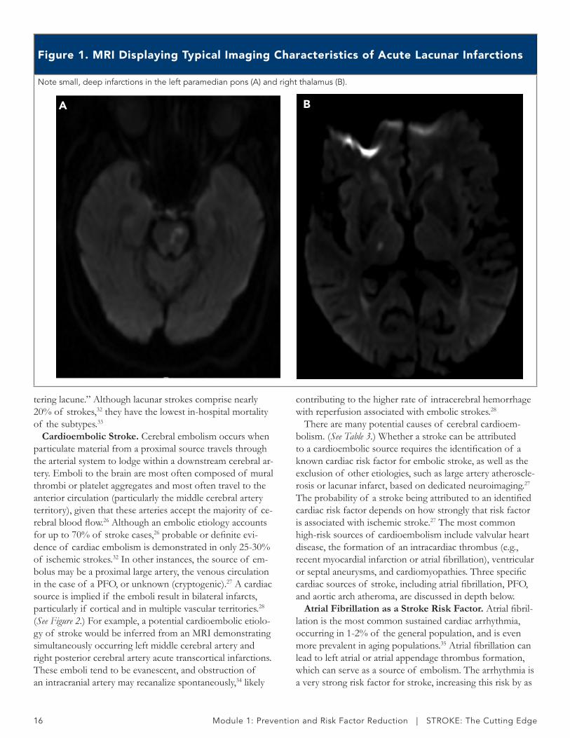
including pharmacotherapy to facilitate plasticity and the use of non-invasive brain stimulation. (See Figure 1.) Even if ultimately adopted, these novel therapies are likely to serve an adjunctive role to conventional physical and occupational therapy, rather than obviate the need for traditional forms of rehabilitation.
Conventional stroke rehabilitation includes repetitive task training; however, effects of specific interventions may gen-eralize poorly to related tasks. Emphasis should be placed on task- or context-specific training, which has been found effective in improving upper and lower extremity motor function.12
Constraint-induced movement therapy (CIMT) is another type of repetitive task training, involving forced use of the affected limb by “constraining” use of the non-paretic limb. A randomized trial found benefit in upper limb use after CIMT training, and it is considered a “reasonable” treat-ment for suitable stroke survivors (AHA Class IIa, Level of Evidence A).13 The durability of this benefit is unknown, however, and additional research is required to define opti-
Figure 1. Transcranial Magnetic Stimulation
This technique is used both as a tool for investigating brain physiology during stroke recovery, and also as a potential therapy to enhance motor recovery.
Photo courtesy of Dr. Joel Stein.