STROKE LECTURE By Arlyn M. Valencia, M.D. Associate Professo University Of Nevada School Of...
86
STROKE ARLYN M. VALENCIA , M.D. Associate Professor, UNSOM Diplomate, American Board Of Psychiatry & Neurology
-
Upload
arlyn-valencia-md -
Category
Health & Medicine
-
view
22.606 -
download
2
description
A detailed lecture on stroke epidemiology, pathophysiology, clinical presentation, latest treatment strategies.
Transcript of STROKE LECTURE By Arlyn M. Valencia, M.D. Associate Professo University Of Nevada School Of...
STROKEARLYN M. VALENCIA , M.D.Associate Professor, UNSOM
Diplomate, American Board Of Psychiatry & Neurology

LEARNING OBJECTIVES
To be able to define stroke, discuss its pathophysiology and risk factors
To emphasize early evaluation and management of stroke patients
To discuss the latest stroke treatment strategies
CASE STUDIES: To be able to analyze clinical situations, localize the stroke lesion, determine probable etiology

”THE BRAIN IS A VERY UNIQUE, HIGH-MAINTENANCE END- ORGAN. IT IS VERY DEPENDENT ON MOMENT-TO-MOMENT SUPPLY OF GLUCOSE AND OXYGEN TO SUSTAIN ITS HIGH-POWERED ACTIVITIES. IT IS VERY SENSITIVE TO THE SYSTEMIC STATE. ANY SEVERE MEDICAL INSULT, THEREBY, HAS TREMENDOUS IMPACT ON THE BRAIN METABOLISM. ANY MEDICAL EMERGENCY IS A NEUROLOGIC EMERGENCY!”
A. Valencia, M.D.

The biology of stroke
is such that each moment of ischemia and tissue injury increases the degree
of irreversible tissue
damage.

CEREBROVASCULAR ACCIDENT OR “BRAIN ATTACK”
Third leading cause of death 750, 000 cases/year Leading cause of significant
disability Cost: $40 billion/year
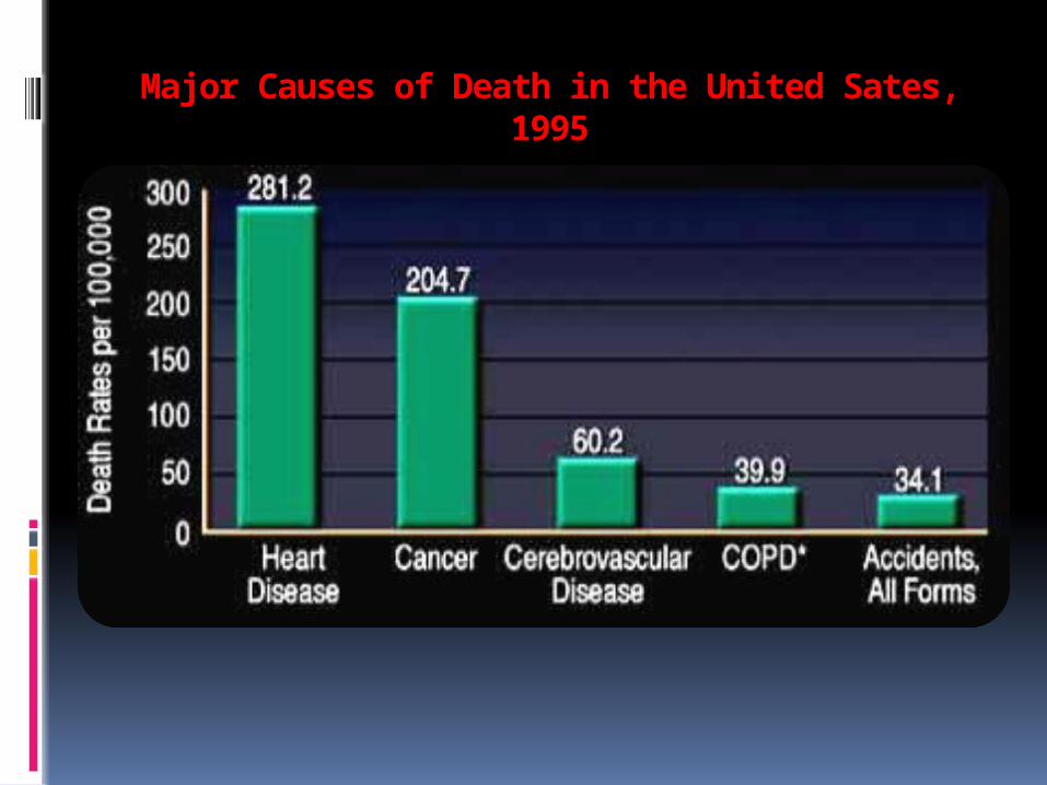
Major Causes of Death in the United Sates, 1995

Annual Economic Costs of Stroke (All Types) In The US

Death Rates for Stroke per 100,000 Population

Types of Stroke Ischemic, 80% - thrombosis, 50%
(small & large-vessel) - embolism, 30%
[now believed significantly higher]
Hemorrhagic, 20% - intracerebral (HTN
as risk) - subarachnoid
(aneurysm)
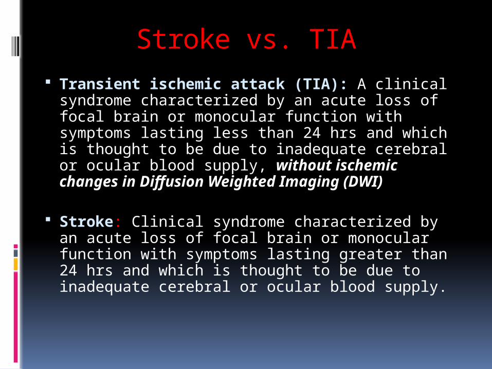
Stroke vs. TIA Transient ischemic attack (TIA): A clinical
syndrome characterized by an acute loss of focal brain or monocular function with symptoms lasting less than 24 hrs and which is thought to be due to inadequate cerebral or ocular blood supply, without ischemic changes in Diffusion Weighted Imaging (DWI)
Stroke: Clinical syndrome characterized by an acute loss of focal brain or monocular function with symptoms lasting greater than 24 hrs and which is thought to be due to inadequate cerebral or ocular blood supply.

Risk Factors for Stroke That Cannot Be Changed
Increased age Being male Race (e.g., African-Americans) Diabetes mellitus Prior stroke/transient ischemic
attacks Family history of stroke Asymptomatic carotid bruit

Up to approximately 30% of people who suffer transient attacks (TIAs) will develop a stroke within 5 years.
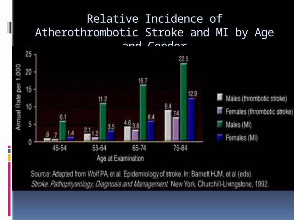
Relative Incidence of Atherothrombotic Stroke and MI by Age
and Gender


Death Rates for Stroke per 100,000 Population
Groups Defined by Race, Age, and Gender: 1993

Risk Factor For Stroke: TreatableMajor Hypertension Heart disease, esp. atrial fibrillation Cigarette smoking Transient ischemic attacks Dyslipidemia Physical inactivity Obesity
Less Well Documented Excessive alcohol intake / drug abuse Acute infection*

Alcohol Consumption as a Risk Factor for Stroke
Heavy alcohol consumption may increase risk of stroke by a number of mechanisms.
The reported effects of alcohol consumption on risk of ischemic stroke have been inconsistent.
A differential effect of alcohol consumption on stroke risk in men compared to women has been observed.

Alcohol Consumption as a Risk Factor for Stroke
Light and moderate alcohol use tend to raise levels of high-density-lipoprotein (HDL) -- the "good" lipoprotein.
Heavy drinking or binge drinking, is related to an increased incidence of stroke as a cause of death Light or moderate alcohol consumption, is related to a reduced risk of coronary heart disease.
There is positive, dose-related effect of alcohol consumption on risk of intracranial hemorrhage, both arachnoid and intracerebral .

Less Well Documented
Geography/climate Socieconomic factors

The Stroke Belt

Potential Genetic Risk Factors for Stroke
Apolipoprotein E4
Elevated homocysteine levels
Factor V mutation

ATHEROSCLEROSIS AND THROMBOSIS
Atherosclerosis: decades-long process; progression favored by hypercholesterolemia, HTN, cigarette smoking
•Fatty streak: yellowish discoloration on intimal surface of blood
•Focal plaques: eccentric thickening at bifurcations; addition of massive extracellular lipids that displaced normal cells and matrix
•Complicated fibrous plaques: central acellular area of lipid covered by a cap of smooth muscle cells and collagen
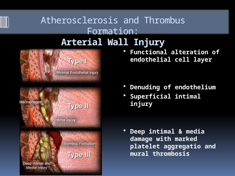
Atherosclerosis and Thrombus Formation:
Arterial Wall Injury Functional alteration of
endothelial cell layer
Denuding of endothelium
Superficial intimal injury
Deep intimal & media damage with marked platelet aggregatio and mural thrombosis

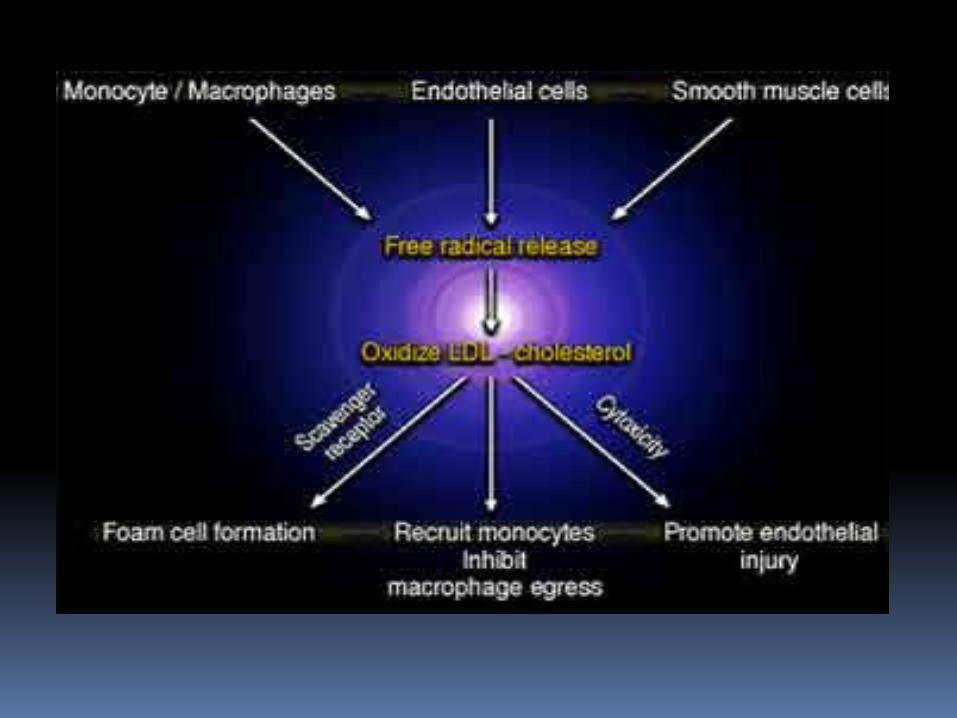
Role of Monocytes and T-Lymphocytes in the Transformation to Foam Cells

Oxidation of LDL-Cholesterol

Oxidized LDL-cholesterol Contributes To Atherogenesis In Three Other Ways:
It has cytotoxic properties that may promote endothelial injury;
It acts as a chemoattractant for circulating monocytes, leading to their increased accumulation with plaques; and
Inhibits egress of macrophages from plaques.

Smooth Muscle Cell Migration and Proliferation

Smooth Muscle Cell Migration and Proliferation
Along with macrophages, smooth-muscle cells proliferate in the intima during atherogenesis.
Smooth muscle cell layer makes up a substantial bulk of the atherosclerotic lesion, which may rise several millimeters above the surface of the surrounding intima

Role of Platelets
Platelet adhesion may
be promoted by type II injury and by toxic products
Platelets release growth factors that stimulate SM migration and proliferation and formation of “fibrointimal lesions” and the outside capsule of “fatty lesions

Plaque Fissuring and Formation of Platelet Thrombus
The vulnerability of such a structure to fissuring appears to be related to circumferential stress on the plaque cap in systole, as well as infiltration of the cap tissue with foam cells (with reduction of total collagen content and a concomitant fall in tensile strength)

Potential Outcomes of Plaque Fissuring Acute episodes of
transient ischemia and ischemic stroke (as well as myocardial infarction, unstable angina, as sudden death) may be precipitated by thrombosis on atherosclerotic plaques.

Thrombus Formation I -- Platelet Activation
On contact with collagen, platelets become activated, with platelet adhesion, secretion of platelet contents, and platelet aggregation at the site of injury. The activated platelet surface is an essential catalytic surface for several coagulation reactions that generate thrombin, a key factor in the coagulation sequence

Thrombus Formation II -- Platelet Activation and Blood
Flow

Thrombus Formation III -- Activation of Coagulation Cascade
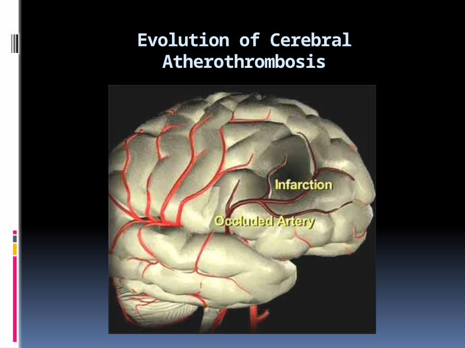
Evolution of Cerebral Atherothrombosis

Atherothrombotic occlusion of larger arteries
Embolism: Artery-to artrey, cardiogenic
Primary small vessel disease (lipohyalinosis)

Thromboembolism

Cardiogenic Emboli
Cardiogenic emboli lodge in the middle cerebral artery or its branches in 80% of cases, in the posterior cerebral artery or its branches 10% of the time, and in the vertebral artery or its branches in the remaining 10% of cases.

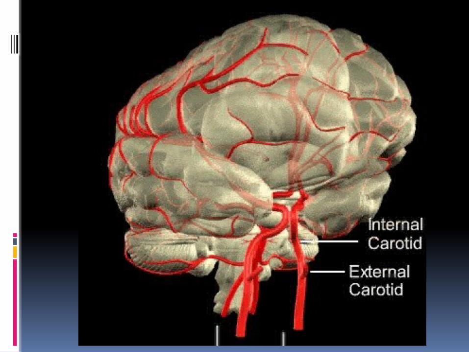
Major Blood Vessels Of The Brain
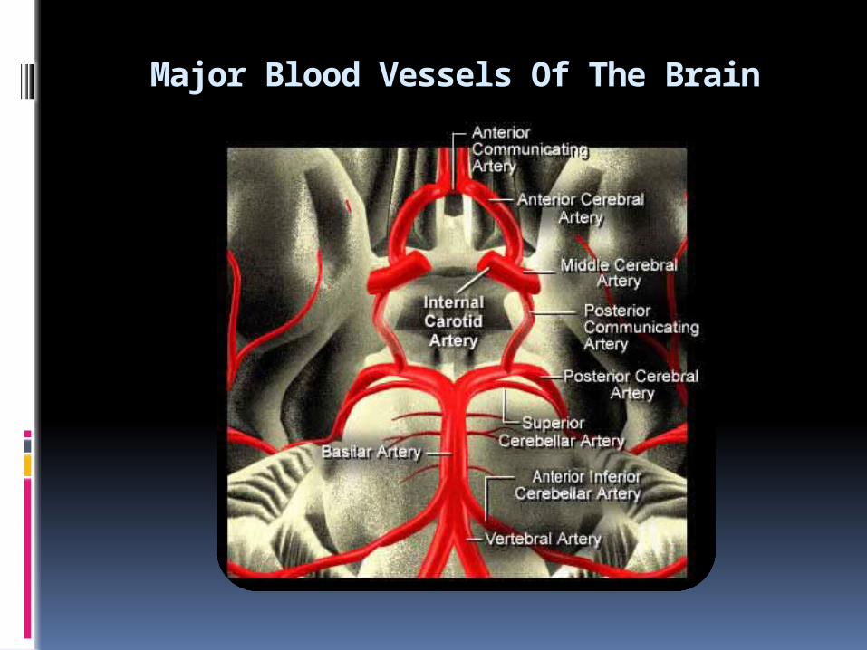
Major Blood Vessels Of The Brain
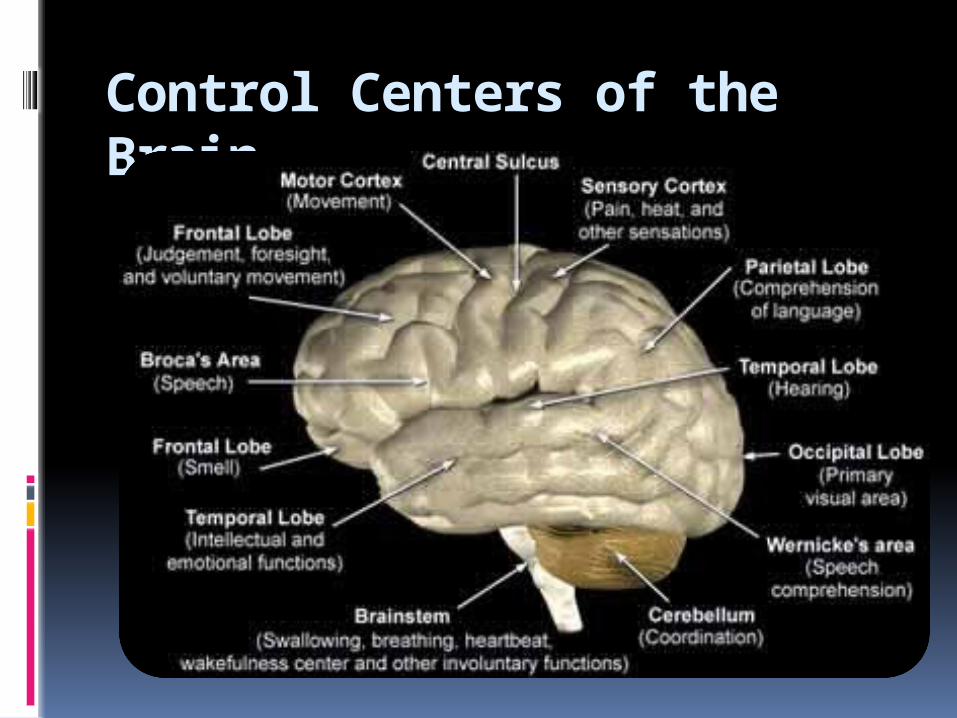
Control Centers of the Brain


Cellular Injury During IschemiaNeuronal Function: Importance of Oxygen and Glucose
The transient change in voltage induced by the action potential is determined by the concentration of ions on either side of the cell membrane. Maintaining these ionic gradients is an energy-consuming process that requires a constant supply of glucose and oxygen to the neuron.

Cellular Changes As Ischemia Progresses
The duration, severity, and location of focal cerebral ischemia determine the extent of brain function and thus the severity of stroke

Cellular Injury During IschemiaInadequate Energy Supply Lack of glucose and
oxygen deplete the cellular energy stores required to maintain electrical potentials and ion gradients.
The membrane that surrounds each affected neuron becomes "leaky," and the cell loses potassium and adenosine triphosphate (ATP), the tissue's medium for energy exchange

THE ISCHEMIC PENUMBRA


Stroke Warning Signs
Sudden weakness, paralysis, or numbness of the face, arm and the leg on one or both sides of the body
Loss of speech, or difficulty speaking or understanding speech
Dimness or loss of vision, particularly in only one eye
Unexplained dizziness (especially when associated with other neurologic symptoms), unsteadiness, or sudden falls
Sudden severe headache and/or loss of consciousness
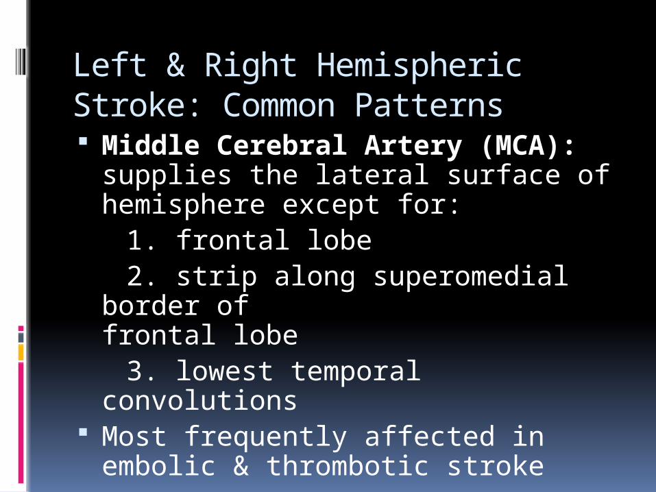
Left & Right Hemispheric Stroke: Common Patterns Middle Cerebral Artery (MCA):
supplies the lateral surface of hemisphere except for:
1. frontal lobe 2. strip along superomedial border of
frontal lobe 3. lowest temporal convolutions Most frequently affected in embolic &
thrombotic stroke

Left and Right Hemisphere Stroke: Common Patterns
Left (Dominant) Hemisphere Stroke: Common Pattern
Right (Non-dominant) Hemisphere Stroke: Common Pattern
Aphasia Right hemiparesis Right-sided sensory
loss Right visual field
defect Poor right conjugate
gaze Dysarthria Difficulty reading,
writing, or calculating
Neglect of left visual field
Extinction of left-sided stimuli
Left hemiparesis Left-sided sensory loss Left visual field defect Poor left conjugate gaze Dysarthria Spatial disorientation

Posterior Circulation (Vertebrobasilar Territory) Stroke
Ataxia, gait abnormalities Diplopia, oscillopsia, nystagmus,
dysconjugate eye movements Nausea & vomiting (center is in
area post-rema) Crossed hemiparesis,
hemisensory deficits Headache more common


Differential Diagnosis of Stroke
Craniocerebral / cervical traumaMeningitis/encephalitisIntracranial mass Tumor Subdural hematoma Seizure with persistent neurological signsMigraine with persistent neurological signsMetabolic Hyperglycemia Hypoglycemia Post-cardiac arrest ischemia Drug/narcotic overdose

AHA Stroke Council Recommended Assessment of a Person with Suspected Stroke
EMS should be instructed in the rapid recognition, evaluation, treatment and transport
Baseline assessment within minutes, CT scan ASAP; use National Institutes of Health Stroke Scale (NIHSS)

AHA Stroke Council Recommended Assessment of a Person with Suspected Stroke
EMS should be instructed in the rapid recognition, evaluation, treatment and transport
Baseline assessment within minutes, CT scan ASAP; use National Institutes of Health Stroke Scale (NIHSS)
Immediate evaluation of the following: 1. Airway 2. Vital signs 3. General medical assessment (including evidence of injury, cardiovascular abnormalities) 4. Neurological assessment (frequent)

EVALUATION AND WORK-UP
History and PE Computed Tomography (CT) scan
of the head 12-lead EKG, chest X-ray Complete blood count, PT, PTT Chemistries (sodium, phosphate,
glucose abnormalities may mimic stroke)
Urine and serum toxicology (drugs and alcohol)
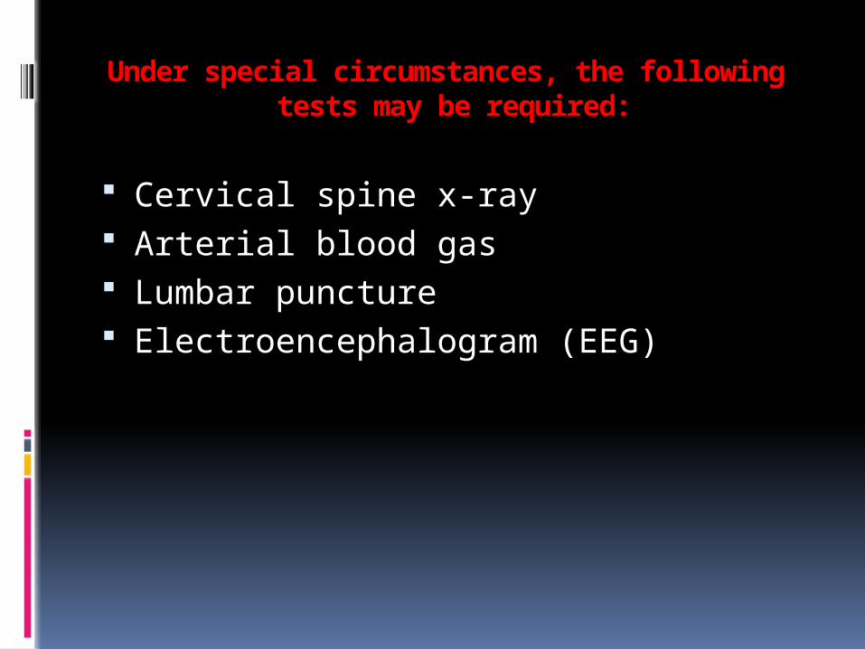
Under special circumstances, the following tests may be required:
Cervical spine x-ray Arterial blood gas Lumbar puncture Electroencephalogram (EEG)
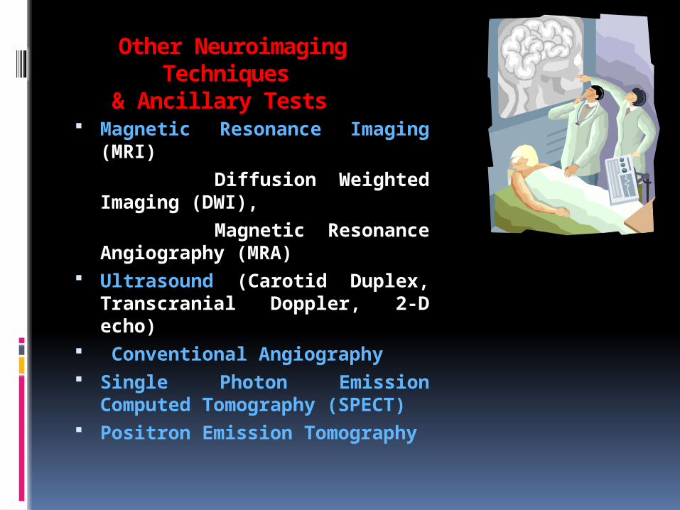
Other Neuroimaging Techniques
& Ancillary Tests Magnetic Resonance Imaging
(MRI) Diffusion Weighted Imaging
(DWI), Magnetic Resonance
Angiography (MRA) Ultrasound (Carotid Duplex,
Transcranial Doppler, 2-D echo) Conventional Angiography Single Photon Emission
Computed Tomography (SPECT) Positron Emission Tomography


Computed Tomography

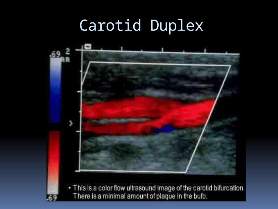
Carotid Duplex

Transcranial Doppler

Cerebral Angiography

Functional abnormality demonstrated with PET exceeds that seen with structural imaging techniques such as X-ray, CT, or MRI and is more representative depiction of the underlying functional state of the brain.

SPECT and Xenon Contrast CT
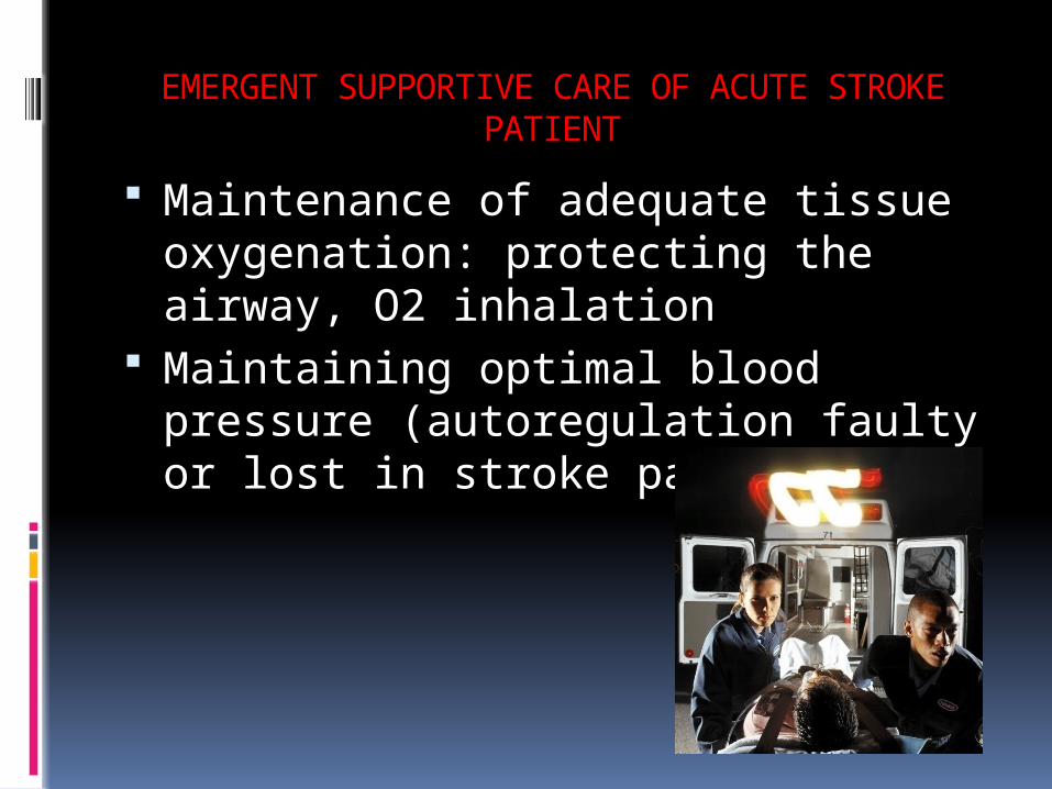
EMERGENT SUPPORTIVE CARE OF ACUTE STROKE PATIENT
Maintenance of adequate tissue oxygenation: protecting the airway, O2 inhalation
Maintaining optimal blood pressure (autoregulation faulty or lost in stroke patients)

STROKE MANAGEMENT

EMERGENT SUPPORTIVE CARE OF ACUTE STROKE PATIENT
Management of blood glucose abnormalities (hyperglycemia associated with poorer prognosis)
Management of fever and infections (ischemia worsened by hyperthermia, improved by hypothermia

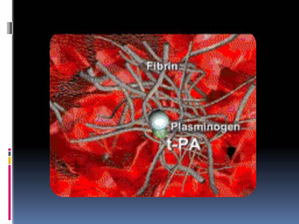
IV TPA (< 3hours) IA fibrinolysis (< 6 hours) IA MERCI retriever < 8 hours Endovascular temperature control


Acute Stroke Treatment
Intravenous recombinant tissue plasminogen activator (TPA): within 3 hours of stroke symptom onset
Intraarterial TPA: within 6 hours; MCA territory stroke by angiography

The Merci Retrieval System

Hypothermia For Acute Stroke:Intravascular Cooling
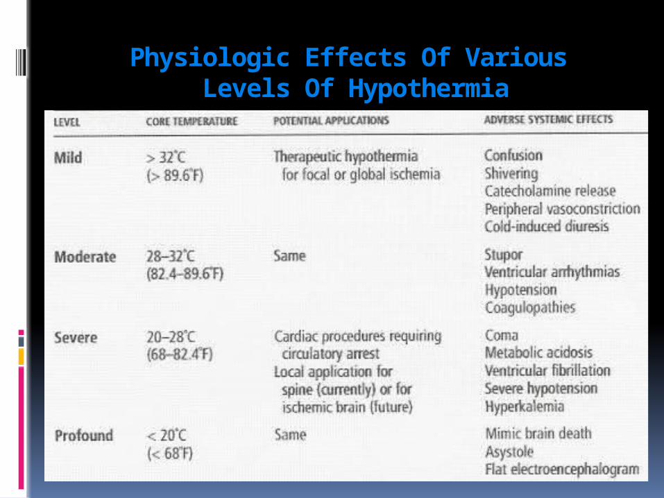
Physiologic Effects Of Various Levels Of Hypothermia

Known Factors That Cause Stroke Progression
Hypotension Hyperglycemia Hyperthermia Infection Cerebral hypoperfusion

Brain Edema

TREATMENT OF BRAIN SWELLING
Cerebral perfusion pressure =MAP-ICP
Fluid Restriction (1200 ml /day/m2) Controlled hyperventilation: 25 mm Hg Mannitol, 0.25 mg/kg IV over 20 minutes;
repeat PRN, serum osmolality maintained in the range of 300-320mOsm/l
Barbiturate coma, with ICP monitoring (subarachnoid bolt, IV catheter or Camino catheter): maintain CPP greater than 50 mmHg; pentobarbital serum level of 2-4 mg/dl
Surgery (wait 2 weeks)

Management of Cerebral Edema, Increased Intracranial Pressure and HydrocephalusBrain edema peaks at 3-5 daysTreatment includes: 1. hyperventilation (lower PCO2)
2. osmotic diuretics 3. drainage of CSF (ventriculostomy) 4. surgery (lobectomy)

Neuroprotective Agents
Several trials going on So far, trial on one free-radical
scavenger showed positive results Phase II trials have proven beneficial;
Phase III (human efficacy trials) non-benefial to negative
Common measures may “neuroprotect”
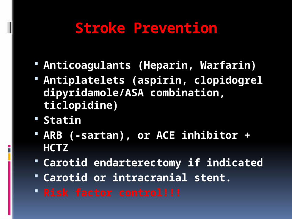
Stroke Prevention
Anticoagulants (Heparin, Warfarin) Antiplatelets (aspirin, clopidogrel
dipyridamole/ASA combination, ticlopidine)
Statin ARB (-sartan), or ACE inhibitor + HCTZ Carotid endarterectomy if indicated Carotid or intracranial stent. Risk factor control!!!

Concept of Stroke Teams & Stroke Units
“Time is brain” Stroke awareness Common mistakes
may lead to fatal consequences
“Boutique stroke neurology”: Patients will receive best care; length of stay shortened
THANK YOU!