STROKE - ePublishing Login The Cutting Edge (ISBN 978-1-934863-85-5) is published by AHC Media LLC....
19
Transcript of STROKE - ePublishing Login The Cutting Edge (ISBN 978-1-934863-85-5) is published by AHC Media LLC....

STROKETHE CUTTING EDGE
Relevant and Practical Information on Stroke Prevention, Emergency Treatment, and Rehabilitation
AHCMEDIA.COM | 404.262.7436

Copyright © 2016 AHC Media, Atlanta, GA. All rights reserved. Reproduction, distribution, or translation without express written permission is strictly prohibited.
STROKE: The Cutting Edge (ISBN 978-1-934863-85-5) is published by AHC Media LLC.
Executive Editor: Leslie CoplinAssociate Managing Editor: Jonathan Springston
AHC Media LLCOne Atlanta Plaza950 East Paces Ferry Road NE Suite 2850Atlanta, GA 30326(800) 688-2421 or (404) 262-7436
The articles within were previously published in the following AHC Media publications:
Clinical Cardiology Alert, Critical Care Alert, ED Legal Letter, Healthcare Risk Management, Neurology Alert, and Medical Ethics Advisor.

TABLE OF CONTENTS | ii
MODULE 1: STROKE UPDATES (2.0 CREDiTS)
Report of Clinical Trials Results from the 2016 International Stroke Conference ..........................
Stroke Alert: A Review of Current Stroke Literature ......................................................................
Optimal Antiplatelet Therapy for Secondary Prevention of Ischemic Stroke ................................
Every 5-minute Delay in Performing Endovascular Reperfusion Results in 1 out of 100 Patients Having a Worse Disability Outcome .......................................................
Intracerebral Hemorrhages Associated with Non-vitamin K Oral Anticoagulants Appear to Be Smaller than Those Associated with Warfarin ................................
Carotid Occlusion Rarely Develops from Asymptomatic Carotid Artery Stenosis.........................
Symptomatic Carotid Artery Stenosis Requires Urgent Revascularization ....................................
Left Atrial Dysfunction Without Fibrillation Increases Stroke Risk .................................................
Small Vessel Disease Often Accompanies Large Vessel Intracranial Atherosclerosis ....................
Loss of Consciousness at Onset of Aneurysmal SAH: Poor Prognosis ..........................................
Treatment of AVM-related Epilepsy ..............................................................................................
Module 1 Test: ..............................................................................................................................
MODULE 2: EMERGENCY MANAGEMENT (2.0 CREDiTS)
Isolated Dizziness and Vertigo Are Rarely Caused by Stroke Efficacy and Safety of Very Early Mobilization After Onset of Acute Stroke ...............................................
Vision Problems After Ischemic Stroke: Effects on Quality of Life .................................................
ICH May Clinically Mimic TIA ........................................................................................................
Mobile Stroke Units Bring Treatment to Patients, Potentially Improving Long-term Outcomes .................................................................................
Module 2 Test ...............................................................................................................................
MODULE 3: iSCHEMiC STROKE (2.0 CREDiTS)
Treatment of Acute Ischemic Stroke ..............................................................................................
Module 3 Test ...............................................................................................................................
MODULE 4: STROKE MiMiCS (2.0 CREDiTS)
Stroke Mimics: A Clinical Dilemma for the Emergency Physician .................................................
Module 4 Test ...............................................................................................................................

ACCREDiTATiON iNFORMATiON | iii
ACCREDITATION INFORMATION
ACCREDiTATiON STATEMENTAHC Media is accredited by the Accreditation Council for Continuing Medical Education to provide
continuing medical education for physicians.AHC Media is accredited as a provider of continuing nursing education by the American Nurses
Credentialing Center’s Commission on Accreditation.
CREDiT DESiGNATiONAHC Media designates this enduring material for a maximum of 8 AMA PRA Category 1 Credits.TM
Physicians and nurses should claim only the credit commensurate with the extent of their participation in the activity.
Approved by the American College of Emergency Physicians for a maximum of 8.00 hour(s) of ACEP Category I credit.
This activity has been approved for 8 nursing contact hours using a 60-minute contact hour.
Provider approved by the California Board of Registered Nursing, Provider #14749, for 8 Contact Hours.
To earn credit for this activity, follow these instructions:1. Read and study the activity, using the provided references for further research.2. Log on to AHCMedia.com and click on My Account. First-time users will have to register on the site
using the 8-digit subscriber number printed on the mailing label or invoice. Test questions for each chapter are included in separate tests.
3. Pass each online test with a score of 100%; you will be allowed to answer the questions as many times as needed to achieve a score of 100%.
4. After successfully completing the last section of the test, your browser will be automatically directed to the activity evaluation form, which you will submit online.
5. Once the completed evaluation is received, a credit letter will be e-mailed to you instantly.
EFFECTiVE DATESThis enduring Internet material is valid from May 1, 2016, to April 30, 2019.
ACTiViTY OBJECTiVESAt the conclusion of this activity, participants should be able to:1. Discuss current scientific research and data regarding the diagnosis and treatment of stroke;2. Discuss the pathogenesis and treatment of stroke;3. Explain the basic science of brain function as it applies to stroke;4. Cite new information regarding new drugs for stroke and new uses for traditional drugs;5. Identify nonclinical issues of importance for health care providers who treat stroke patients.6. Discuss advances in neurointerventional treatment.
TARGET AUDiENCE This CME/CE activity is intended for neurologists, emergency medicine physicians and nurses,
Joint Commission-certified stroke center personnel, psychiatrists, and ENTs.

ACCREDiTATiON iNFORMATiON | iV
PHYSiCiAN EDiTORS Matthew Fink, MD, Louis and Gertrude Feil Professor in Clinical Neurology and Chairman, Department of
Neurology, Weill Cornell Medical College; Neurologist-in-Chief, New York Presbyterian Hospital
NURSE PLANNERDonna Avanecean, MS, RN, FNP-C, CNRN, APRN, The Stroke Center at Hartford Hospital, Hartford, CT
PEER REViEWERAmre M. Nouh, MD, Director of the Stroke Center, Hartford Hospital; Assistant Professor and Associate
Residency Program Director, Department of Neurology, University of Connecticut
FiNANCiAL DiSCLOSURESIn order to reveal any potential bias in this publication, and in accordance with Accreditation Council
for Continuing Medical Education guidelines, Dr. Fink, Ms. Avanecean, and Dr. Nouh report no financial relationships relevant to this field of study.
Editorial and ProductionMs. Coplin and Mr. Springston report no financial relationships relevant to this field of study.
AuthorsFinancial disclosure information for authors can be found at the beginning of their respective articles. This
publication does not receive commercial support.
DiSCLAiMERThis is an educational publication designed to present scientific information and opinion to health profes-
sionals, to stimulate thought and further investigation. It does not provide advice regarding medical diagnosis or treatment for any individual case. It is not intended for use by the layman. Opinions expressed are not nec-essarily those of AHC Media or this publication. Mention of products or services does not constitute endorse-ment. Clinical, legal, tax, and other comments are offered for general guidance only; professional counsel should be sought for specific situations.

STROKE UPDATES | 1
MODULE 1STROKE UPDATES
2.0 CREDIT HOURS

STROKE UPDATES | 2
The FIND A FIB trial from Switzerland, is a randomized trial of 400 patients with isch-emic stroke aged 60 years or older, who
were not known to have atrial fibrillation and had normal sinus rhythm at the time of their stroke. One group had standard continuous monitoring of heart rhythm for 24 hours, and the second group had enhanced and prolonged monitoring with three 10-day Holter monitor sessions at the time of the stroke, at 3 months, and at 6 months after the stroke. Atrial fibrillation was found in 13.5% of patients in the group that underwent enhanced and prolonged monitoring and in 4.5% of the control group. Anticoagulation was started when atrial fibrillation was diagnosed, and after 1 year, there was a reduced rate of recurrent strokes in the extended monitoring group, compared to control (2.5% vs 4.5%).
The ARUBA trial, is a randomized trial of un-ruptured brain arteriovenous malformations, and 5-year results were presented. Two hundred twen-ty-eight patients were enrolled and randomized to medical management of symptoms or to medical management plus invasive management, which included surgery, endovascular embolization, or radiotherapy. Five-year follow-up showed that the death or stroke rate was 40.6% in the intervention group vs 10.8% in the medical group, with a highly significant difference. Stroke rate was 39.6% vs 9.2%. Medical treatment was found to reduce the risk of stroke or death by 78% compared to inter-vention.
The CREST trial, carotid endarterectomy vs stenting for treatment of carotid artery stenosis, presented its long-term results of carotid revascu-
larization endarterectomy (CEA) vs stenting (CAS). In this trial, 2502 patients were randomized and fol-lowed at 117 centers, following transient ischemic attack, or mild stroke, or asymptomatic with carotid stenosis. After randomization to either CEA or CAS, they were evaluated every 6 months and fol-lowed for a median of 10 years. The primary end-point, any cause of stroke, myocardial infarction, or death, was not significantly different after 10 years between the two groups. In conclusion, the differ-ence between CEA and CAS was not significant for post-procedural stroke.
The IRIS trial was designed to evaluate the ef-fects of pioglitazone, an insulin sensitizing drug, for secondary stroke prevention in patients with insulin resistance and a recent ischemic stroke or transient ischemic attack. Patients did not have overt diabetes, but had insulin resistance, and were 40 years of age or older. In this trial, 3876 patients were enrolled and the average time for follow-up was 4.8 years. The primary endpoint was fatal or nonfatal stroke or myocardial infarction. Trial results showed that fatal or nonfatal stroke or myocardial infarction occurred in 9% of the pioglitazone-treat-ed patients, and 11.8% of the placebo-controlled group, with a statistically significant difference and P value of 0.007. There were more adverse effects in the pioglitazone group, specifically bone frac-tures, then in the control group. The conclusion was that targeting insulin resistance with piogli-tazone resulted in reduction of secondary stroke and myocardial infarction, but there was a greater risk for fractures. n
[ABSTRACT & COMMENTARY]
Report of Clinical Trials Results from the International Stroke Conference 2016
By Matthew E. Fink, MDLouis and Gertrude Feil Professor in Clinical Neurology and Chairman, Department of Neurology, Weill Cornell Medical College; Neurologist-in-Chief, New York Presbyterian HospitalDr. Fink reports he is a consultant for Procter & Gamble and Pfizer.

STROKE UPDATES | 3
Optimal Antiplatelet Therapy for Secondary Prevention of Ischemic StrokeSource: Kim JT, et al. Different antiplatelet strategies in patients with new ischemic stroke while taking aspirin. Stroke 2016;47:128-134.
Patients presenting with acute ischemic stroke are often taking aspirin on a regular basis for
prevention of cardiovascular disease. The opti-mal antiplatelet therapy for secondary prevention has been uncertain in this setting. The authors of this study analyzed 1172 patients in a prospec-tive, multicenter stroke registry database from 14 hospitals in South Korea, selecting patients with acute non-cardioembolic stroke, who were taking aspirin for prevention of cardiovascular disease at the time of onset of stroke. These patients were then divided into three groups, 1) maintaining aspirin monotherapy (MA group = 212), 2) switch-ing aspirin to another antiplatelet agent (SA group = 246), and 3) adding another antiplatelet agent to aspirin (AA group = 714). The patients were then followed for 1 year, using a primary endpoint of a composite of all stroke, myocardial infarction, and vascular death. The results were analyzed in a Cox proportional hazards regression analysis. After 1 year of follow-up, compared to the aspirin only group, there was a reduction in the composite vas-cular event rate compared to the SA group (hazard ratio [HR], 0.50; P = 0.03) and in the AA group (HR, 0.40; P < 0.001). This study strongly suggests that compared with maintaining patients on aspirin alone, switching to a different antiplatelet agent, or adding a second antiplatelet agent to aspirin may be better in preventing subsequent vascular events in patients who experienced a new ischemic stroke while taking aspirin. n
Every 5-minute Delay in Performing Endovascular Reperfusion Results in 1 out of 100 Patients Having a Worse Disability OutcomeSource: Sheth SA, et al. Time to endovascular reperfusion and degree of disability in acute stroke. Ann Neurol 2015;78:584-593.
In the past year, multiple clinical trials have re-ported that intra-arterial endovascular reperfusion
with mechanical clot extraction, using the SOLI-TAIRE stent retriever device and others, results in better neurological outcomes than treating patients with intravenous thrombolysis alone with TPA. There is still uncertainty regarding the maxi-mum time window, and how important early inter-vention is as related to neurological recovery and long-term outcomes. The investigators used the combined databases of the SWIFT (Lancet 2012) and STAR (Stroke 2013) trials to identify patients treated with the SOLITAIRE device who achieved substantial reperfusion. They then ranked the 90-day modified Rankin scale outcomes for “time of onset to recanalization” (OTR) time intervals rang-ing from 180 min to 480 min.
Analysis of these data showed substantial time-related reductions in disability for the entire range of outcomes. A shorter OTR time was associated with an improved 90-day Rankin Scale outcome in all groups. The mean Rankin scores were 1.4 for the 120-240 min OTR group, 2.40 for the 241-360 min group, and 3.3 for the 361-660 min group (P < 0.001). There were no significant differences be-tween the groups in the incidence of intracerebral hemorrhage, mortality, or length of hospitalization. The predicted probability and confidence interval of good neurological outcome (mRS 0-2) at 90 days was a continuous variable inversely related to the time from symptom onset to recanaliza-tion. For every 15-min acceleration in the time to reperfusion, 34 per 1000 patients treated will have improved disability outcomes, which translates to 1 out of 100 patients improved, for every 5 minutes of reduced OTR time. n
Stroke Alert: A Review of Current Stroke LiteratureBy Matthew E. Fink, MDLouis and Gertrude Feil Professor in Clinical Neurology and Chairman, Department of Neurology, Weill Cornell Medical College; Neurologist-in-Chief, New York Presbyterian HospitalDr. Fink reports he is a consultant for Procter & Gamble and Pfizer.

STROKE UPDATES | 4
Intracerebral Hemorrhages Associated with Non-vitamin K Oral Anticoagulants Appear to Be Smaller than Those Associated with WarfarinSource: Wilson D, et al. Volume and functional outcome of intracerebral hemorrhage according to oral anticoagulant type. Neurology 2016;86:360-366.
Intracerebral hemorrhage is the most dangerous complication of treatment with oral anticoagu-
lants, and this complication carries a high mortality. Because of the increasing prevalence of atrial fibril-lation in the elderly population, and the increasing use of oral anticoagulants, the overall prevalence of intracerebral hemorrhage is increasing. In recent years, there has been a rapid transition of treat-ment from the use of warfarin to the non-vitamin K oral anticoagulants (NOAC – dabigatran, rivaroxa-ban, apixaban), and randomized trials comparing these agents with warfarin indicate a lower risk for intracerebral hemorrhage. However, it is not clear if the volume and severity of hemorrhages are differ-ent between these two classes of medications.
The investigators studied patients from a pro-spective registry in the United Kingdom of patients with anticoagulant-associated intracerebral hemor-rhages, and compared the size and clinical conse-quences of hemorrhages associated with warfarin and the NOAC agents. From a population of 344 anticoagulant-associated intracerebral hemor-rhages, 11 were related to NOAC treatment and 52 were related to warfarin treatment. The median size of hematomas in the NOAC group was 2.4 mL, compared to 8.9 mL for the warfarin group. In a linear regression analysis, use of warfarin and lobar location of the hematoma predicted a larger hematoma size. A multivariate linear regression to identify confounding variables, including sex, hypertension, previous ischemic stroke, and white matter disease, did not show any other significant variables. In addition, the warfarin-associated hem-orrhage group had a worse clinical outcome.
This is a small prospective observational study, but it does suggest that warfarin-associated in-tracerebral hemorrhages may be larger and have worse clinical outcomes then hemorrhages associ-ated with the newer anticoagulant agents. n
Carotid Occlusion Rarely Develops from Asymptomatic Carotid Artery StenosisSOURCE: Yang C, et al. Risk of stroke at the time of carotid occlusion. JAMA Neurol 2015; doi:10.1001/jamaneurol.2015.1843.
In recent years, there is increasing evidence that modern intensive medical therapy in most pa-
tients with asymptomatic carotid stenosis is effec-tive in stroke prevention, and few patients seem to benefit from carotid endarterectomy or carotid stenting. However, in the United States, 90% of carotid interventions are for asymptomatic stenosis, compared to Denmark, where the rate is 0%. The investigators conducted a retrospective analysis of data collected from patients at the stroke preven-tion clinic of Victoria Hospital, Ontario, Canada, from 1990 until 1995 using annual surveillance carotid ultrasound, and then compared them to a similar group followed from 1995 through 2014.
Among 3681 patients followed with annual ultrasound, 316 (8.6%) were asymptomatic before a carotid occlusion occurred during the observa-tion period. Most of the new occlusions (80%) oc-curred before 2002, when medical therapy was less intensive and the frequency of occlusion decreased in subsequent years. Only a single patient (0.3%) had a stroke at the time of carotid occlusion, and only three patients (0.9%) had ipsilateral stroke during follow-up. In reviewing survival analyses, neither severity of stenosis nor the presence of contralateral occlusion predicted the risk of stroke or TIA, fatal stroke, or death from other causes in > 3 years of follow-up from carotid occlusion. From this study, it appears that the risk of progression to carotid occlusion is well below the risk of carotid stenting or carotid endarterectomy, and intensive medical therapy appears to be preferred for the vast majority of patients with asymptomatic carotid artery stenosis. However, the CREST-2 trial, which is now underway and designed as a prospective, randomized trial, will be able to answer this ques-tion definitively. n

MODULE 2EMERGENCY MANAGEMENT
2.0 CREDIT HOURS

EMERGENCY MANAGEMENT | 10
Very early (i.e., within 24 hours) mobilization after stroke onset comprised of out-of-bed sitting, standing, and walking is thought
to contribute to the positive effects of stroke-unit care and is therefore recommended in many guidelines.1,2 However, positive evidence for recommending early mobilization has been quite limited.3 The biological rationale for early mobili-zation has centered around the clinical conclusions that bedrest negatively affects the musculoskeletal, cardiovascular, respiratory, and immune systems4 and might slow recovery, since immobility-related complications are common after stroke in patients who remain inactive.5 Finally, it has been argued that there is a narrow window for opportunity for brain plasticity to repair itself, and the optimum time for positive change is thought by many clini-cians to occur soon after the stroke occurs.7,8
The AVERT Trial Collaboration group was formed to perform a study investigating the rela-tive efficacy of a protocol intended to compare earlier-than-usual mobilization after a stroke with the usual care, which traditionally has begun 24 or more hours after the stroke.10 The researchers performed a parallel-group, single-blind, multi-center, international, randomized, controlled trial at 56 stroke units in five countries (Australia, New Zealand, Malaysia, Singapore, and the United King-dom)9 to determine if early mobilization would improve functional outcomes, reduce immobility-related complications, and accelerate walking re-covery at 3 months post-stroke occurrence with no
increase in neurological complications. A total of 2104 patients were enrolled and randomized to an early mobilization group and to a usual care group. The results demonstrated that very early mobiliza-tion after a stroke was associated with a significant reduction in the odds of a favorable outcome at 3 months after the stroke; however, the number of patients who died or had serious adverse events at 3 months after the stroke did not differ significantly between groups.
COMMENTARYThe results of this very large and complex clini-
cal trial comparing a high-dose, frequent mobi-lization protocol starting within 24 hours after a stroke appeared to be no better than “usual” care. However, careful analysis of the usual care proto-cols in the stroke care units will be required before drawing final conclusions, since components of early care intervention were already part of routine clinical care in many locations. The AVERT re-searchers indicated that they were going to further analyze the data and do a dose-response analysis to establish the effect of dose of rehabilitation on efficacy and safety outcomes. It should be noted that 26% of all patients were > 80 years of age and 24% of patients had received recombinant tissue plasminogen activator. Early mobilization did not reduce the incidence of immobility-related compli-cations or significantly accelerate walking recovery. In fact, it reduced the odds of a favorable outcome at 3 months after the stroke occurred.
The results of the AVERT trial are incomplete since the authors indicated that they are now undertaking a dose-response analysis to establish the effect of dose of rehabilitation on efficacy and safety outcomes and to establish a better under-standing of exactly which patients best respond to rehabilitative treatments, be they early or later, and which do not. It would appear that, for the time being, early mobilization would be appropriate and useful in carefully selected but not in all stroke patients. Until a deeper understanding evolves regarding which patients best respond to rehabili-
[ABSTRACT & COMMENTARY]
Efficacy and Safety of Very Early Mobilization After Onset of Acute Stroke
By Harold L. Karpman, MD, FACC, FACPClinical Professor of Medicine, UCLA School of MedicineDr. Karpman reports no financial relationships relevant to this field of study.
SYNOPSIS: The higher dose, very early (within 24 hours) mo-bilization protocol was associated with a reduction in the odds of a favorable outcome at 3 months after stroke occurrence.
SOURCE: Bernhardt J and the AVERT trial collaboration group. Efficacy and safety of very early mobilization within 24 hours of stroke onset (AVERT): A randomized controlled trial. Lancet 2015;386:46-55.

MODULE 3ISCHEMIC STROKE
2.0 CREDIT HOURS

EMERGENCY MANAGEMENT | 22
Time is brain. For every minute of prolonged ischemia without treatment, 1.9 million neurons are lost.1 Neural tissue’s exquisite
sensitivity to ischemia indicates the emergency nature of acute stroke care. The faster that defini-tive stroke treatment is administered following the onset of ischemia, the better the outcomes.2,3 Unfortunately, the majority of patients do not arrive to the hospital soon enough to receive emergency stroke treatment.4
Two FDA-approved therapies available for the treatment of acute ischemic stroke patients include intravenous tissue plasminogen activator (IV-tPA)5 and mechanical thrombectomy.6 Both of these treatments are recommended by the American Heart Association (AHA) and American Stroke As-sociation (ASA) to be administered only within dis-crete, narrow time windows. These windows limit effective treatment to only 6 hours from symptom onset,7 given the known diminishing net clinical benefit that occurs with the progression of time.3,8 Both IV thrombolysis and endovascular clot extrac-tion have been demonstrated to reduce medium-term disability at 3 or 6 months without an increase in mortality.3,7 Still, careful patient selection is the best tool to minimize the risks and maximize the benefits of acute stroke intervention.
Prehospital Considerations for Acute Stroke Care
Improving Prehospital Delays to Presentation. Al-though there are various reasons for delay to acute stroke treatment, prehospital issues continue to be responsible for the largest of these delays.9 Failure to promptly report to the emergency department (ED) when experiencing stroke symptoms is due to
multiple factors, including a general lack of aware-ness and inability to identify symptoms of stroke, self-presentation to the ED without activating the 911 emergency system, and difficulty obtaining the time the patient was last seen well prior to stroke onset.10 Therefore, patients at risk for stroke should be instructed thoroughly on the symptoms of stroke and the need to present immediately to an ED if they are experiencing such symptoms. Patients who are witnessed to have acute onset of facial asymmetry (F), arm weakness (A), or speech disturbance (S), have at least a 72% probability that they are experiencing a stroke and should be brought to emergent medical attention since time (T) is a factor.11 Public educational efforts, for instance the FAST campaign, have focused on teaching these stroke concepts through the use of mass media and have demonstrated sustained, subsequent improvement in prehospital delays.12 Such an educational tool is identical to the three-item screening questionnaire known as the Cincin-nati Prehospital Stroke Scale used by emergency medical professionals in prehospital stroke assess-ments to aid in the rapid identification of stroke. The Cincinnati Prehospital Stroke Scale has been equally reliable in use by the lay population,11 and therefore should be taught to patients who are at high risk for stroke and their families.
Wake-up Stroke and Extending Treatment Win-dows. Stroke treatment windows are dependent on obtaining a time the patient was last seen well. However, stroke onset times are often unreliable, particularly in the case of stroke upon awakening, which accounts for up to 28% of strokes.13 When deciding on treatment for wake-up stroke patients, it is the standard to assume that stroke onset was the time the patient went to bed normal, even though stroke onset frequency is known to be highest in the early morning.14 Unfortunately, more than 35% of patients presenting with stroke symp-toms upon awakening would be eligible for intra-venous thrombolysis had time not been a factor.13 Since wake-up stroke represents a large target for missed stroke therapy, there has been an emphasis on shifting treatment based on time windows to that of tissue windows using advanced neuroimag-ing. Perfusion scans, collateral scores on nonin-
Treatment of Acute Ischemic Stroke
By Michael P. Lerario, MD, and Alan Z. Segal, MDDr. Lerario is Assistant Professor of Clinical Neurology, Weill Cornell Medical College, New York; Attending Physician, New York–Presbyterian/Queens Hospital, Flushing, NY
Dr. Segal is Associate Professor of Clinical Neurology, Weill Cornell Medical College, New YorkDr. Lerario and Dr. Segal report no financial relationships relevant to this field of study.

iSCHEMiC STROKE | 23
vasive angiography, and evaluation of magnetic resonance imaging (MRI) sequence mismatches potentially may be employed to select patients for acute treatment who may otherwise be outside of conventional treatment windows that are based on time.15,16 Such imaging techniques can help differ-entiate ischemic penumbra from core infarct, and recognize which patients have completed stroke and which have salvageable brain tissue and there-by are amenable for salvage therapy within extend-ed time windows. Although these techniques have been adopted in some hospital protocols, they are still considered largely experimental in standard practice and are the basis of multiple ongoing clinical trials.17 Genetically engineered, mutant tPA, known as tenecteplase, may have several advan-tages over standard alteplase for stroke thromboly-sis, and has also been studied in extended time windows with encouraging results.18
Prehospital Thrombolysis. A novel method of expediting acute stroke treatment is to attempt prehospital thrombolysis. Germany has instituted and studied ambulance-based tPA administration in major cities with success in reducing onset to treatment times.19 Similar practices have begun to be established in the United States as well.20 The concept of bringing the tPA to the patient seems intuitive in its ability to hasten times to throm-bolysis; however, such methods are enacted at a significant expense in terms of equipment and staffing. Ambulances are required to have portable laboratory testing, computed tomography (CT) capabilities, and in-person or telemedicine ac-cess to a vascular neurologist and radiologist for efficient clinical evaluation and interpretation of the acquired studies. While widespread adoption of prehospital thrombolysis protocols is still years ahead of us, other methods to expedite stroke evaluation in the prehospital setting include the use of ambulance-based telemedicine21 or hospital pre-notification systems.22 These interventions can allow rapid, real-time diagnosis of stroke from a remote setting in the field, including confirmation of the stroke severity and onset time. In addition, improved communication between the transferring and receiving medical professionals will enhance the mobilization and coordination of the receiv-ing stroke team who can now be poised, awaiting the patient’s arrival at the hospital door, ready to administer treatment as applicable.
Systems of Care for Coordinated Stroke Treat-ment. Like any time-based therapy that requires coordination among multiple physician specialties,
Table 1: Emergency Department Diagnosit Evaluation for Acute Ischemic Stroke
Emergency Medical Services■n Confirm LKN time■n Assess airway and hemodynamics■n Initiate cardiac monitoring■n Determine fingerstick blood glucose and treat accordingly■n Triage and rapidly transport patient to the nearest, most
appropriate hospital ED■n Notify hospital ED of imminent stroke patient arrival
Emergency Room■n Confirm LKN time■n Rapid triage■n Rapid activation of stroke team■n Assess airway and hemodynamics■n Obtain history■n Assess for tPA contraindications■n Treat severe hypertension in conjunction with stroke team■n Make decision regarding tPA in conjunction with stroke
team
Nursing■n Assess vital signs■n Initiate cardiac monitoring■n Placement of 2 peripheral IVs■n Determine fingerstick blood glucose and treat accordingly■n Draw basic stroke laboratory studies*
■n 12-lead ECG
Stroke Team■n Confirm LKN time■n Obtain history■n Perform NIHSS■n Diagnose clinical stroke■n Head CT interpretation■n Assess for tPA contraindications■n Discuss risks and benefits of treatment with patient and
family■n Make decision regarding tPA
Radiology■n Head CT acquisition■n Head CT interpretation
Laboratory■n Expedited diagnostic testing of stroke laboratory studies*
Pharmacy■n Storage of tPA in ED■n Expedited mixing of tPA for stroke patients
*Stroke laboratory studies standardly include basic metabolic profile, complete blood count, coagulation studies (PT/INR and aPTT) and markers of cardiac ischemia. LKN = last known normal; NIHSS = National Institutes of Health Stroke Scale; CT = computed tomography; IV = intra-venous line; ED = emergency department
iSCHEMiC STROKE | 30
started as quickly as possible, as even small proce-dural delays (e.g., the need for general anesthesia) may worsen outcomes.67
Recent Endovascular Trials with Positive Results. Clinical equipoise regarding endovascular stroke care remained until 2015 when five trials with positive results were published within the span of 6 months. All five studies clearly demonstrated a clinical benefit of IAT over standard medical care (including IV thrombolysis) without an increase in mortality or sICH.56,60 MR CLEAN was the first of these trials to be published and prompted an early interim analysis of the other studies.6 The subsequent analyses from EXTEND-IA,45 ESCAPE,44 SWIFT PRIME,46 and REVASCAT47 confirmed the findings of MR CLEAN, that intra-arterial therapy is safe, is technically successful when utilizing the
newest stent-retriever devices, and results in sub-stantially improved functional outcomes. The AHA/ASA has since updated its guidelines to recom-mend IAT as the standard of care therapeutic op-tion for patients with demonstrated LVO and who present within 6 hours of symptom onset.7
Since MR CLEAN was the first to be published, fully completed its planned enrollment, and was designed to be the most pragmatic, it serves as the basis for comparison among the five trials. MR CLEAN was a multicenter, randomized trial of en-dovascular treatment for acute stroke patients with proximal arterial occlusions in the Netherlands.6 Five hundred patients were randomized to intra-arterial therapy, largely with stent-retreivers, and compared to standard medical care, in which 89% of control patients received IV thrombolysis. The
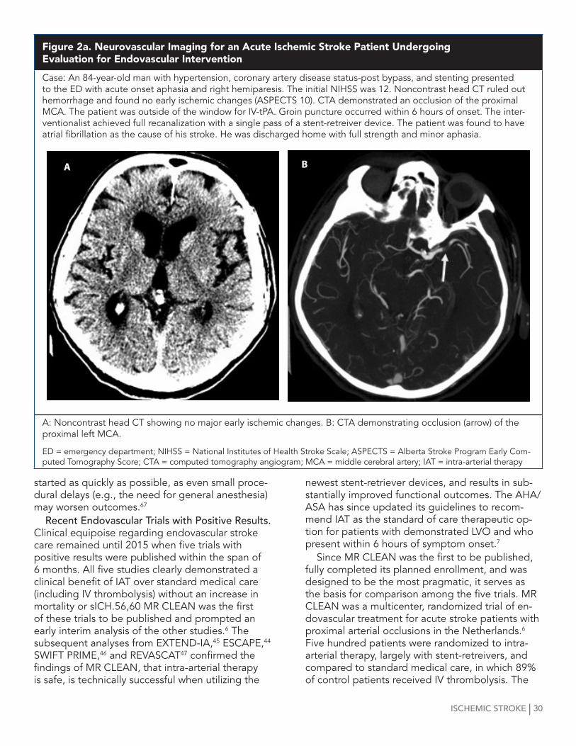
Figure 2a. Neurovascular Imaging for an Acute Ischemic Stroke Patient Undergoing Evaluation for Endovascular Intervention
Case: An 84-year-old man with hypertension, coronary artery disease status-post bypass, and stenting presented to the ED with acute onset aphasia and right hemiparesis. The initial NIHSS was 12. Noncontrast head CT ruled out hemorrhage and found no early ischemic changes (ASPECTS 10). CTA demonstrated an occlusion of the proximal MCA. The patient was outside of the window for IV-tPA. Groin puncture occurred within 6 hours of onset. The inter-ventionalist achieved full recanalization with a single pass of a stent-retreiver device. The patient was found to have atrial fibrillation as the cause of his stroke. He was discharged home with full strength and minor aphasia.
A: Noncontrast head CT showing no major early ischemic changes. B: CTA demonstrating occlusion (arrow) of the proximal left MCA.
ED = emergency department; NIHSS = National Institutes of Health Stroke Scale; ASPECTS = Alberta Stroke Program Early Com-puted Tomography Score; CTA = computed tomography angiogram; MCA = middle cerebral artery; IAT = intra-arterial therapy
A B

iSCHEMiC STROKE | 31
study included adult patients presenting within 6 hours of onset with an NIHSS > 2 and a confirmed large vessel occlusion within the anterior circulation (intracranial ICA, proximal MCA, or anterior cere-bral artery). Procedural success was relatively high compared to previous endovascular trials, with complete or near-complete recanalization being achieved in 59% of patients randomized to inter-vention. This was the first trial to demonstrate a benefit of endovascular therapy for acute ischemic stroke, with a 13.5% absolute increase in functional independence at 3 months and a number needed to treat of 3.4 for an improvement in disability with intervention. There were no differences observed in rates of symptomatic intracerebral hemorrhage (7.7% intervention vs 6.4% control) or 30-day mor-tality (18% each arm).
Despite these powerful results, MR CLEAN patients had the lowest rates of complete or near-complete revascularization (59%) and 3-month freedom from dependence (33%) out of all the en-dovascular trials published in 2015. This potential underestimation of the benefit of IAT may be due to the pragmatic nature of the trial, which allowed for the inclusion of cases with more complicated vascular anatomy. For instance, nearly one-third of patients in the intervention arm had an additional extracranial ICA occlusion, 13% of whom required a secondary stenting procedure. The complexity of these cases was further illustrated in the high rates of new stroke (5.6%) and embolization (9%) which occurred as a complication of the procedure in MR CLEAN.
Although there are notable differences in study inclusion and patient selection criteria between the recent endovascular trials, they all similarly compared mechanical thrombectomy, largely with stent-retriever devices, to standard medical care, which most often included IV thrombolysis. See Table 4 for a comparison of the major inclusion-ary criteria, patient characteristics, and outcomes between the landmark endovascular trials. The majority of patients in these randomized trials were treated within 6 hours, but some studies permit-ted a small number of patients to be randomized in later time windows.44,47 To select patients with a promising mismatch between core infarct and penumbra for study inclusion, REVASCAT allowed for the use of MRI,47 ESCAPE required patients to have good collateral flow on CTA,44 and SWIFT PRIME46 and EXTEND-IA45 employed CT perfusion imaging. MR CLEAN was the only study to enroll patients based only on ASPECTS score if an LVO
was confirmed on CTA.6 When considering all of the trials, it is appar-
ent that endovascular therapy can be performed efficiently (with symptom onset to revasculariza-tion times ranging from 200-269 minutes)44,47 and with technical success (with good reperfusion rates ranging from 59% to 88%).6,46 This procedural success results in lower rates of disability, with an absolute improvement in physical independence ranging from 13.5% to 31%,6,45 and a number needed to treat as low as 3.2.45 Furthermore, endo-vascular care is safe, with no consistent significant difference in rates of sICH or mortality, although the ESCAPE trial did report an 8.6% significant re-duction in mortality with IAT.44 Multiple meta-analy-ses have pooled data from the recent endovascular trials and confirmed the benefit of intervention on functional independence when compared to stan-dard medical care, with an odds ratio of 1.71.56,60 Given the strong benefit of IAT observed in these trials, mechanical thrombectomy of proximal arte-rial occlusions of the anterior circulation should be considered standard of care within 6 hours of symptom onset.7
Endovascular Revascularization of the Posterior Circulation
Acute basilar artery occlusion is frequently as-sociated with poor prognosis in terms of disability and mortality.68 Despite a typically devastating natural history, little is known about the most ef-fective therapeutic approach for basilar occlusion through randomized data.69 This is likely due to the rarity of the disease, which accounts for as little as 1% of ischemic strokes.70
Similar to acute stroke therapy in the anterior circulation, the goal of intervention in patients with vertebrobasilar occlusions is to increase the rate of recanalization, which has been linked to improved outcomes.71 Pooled analyses of nonrandomized data have shown that recanalization of an acute basilar occlusion results in a lower risk of death or dependency, with a number needed to treat of 3.72 This finding was present regardless of whether the patient was treated before or after 12 hours of symptom onset. Due to the poor prognosis ex-pected without recanalization, patients with basilar occlusion are often considered for IAT in extended time windows outside of those deemed acceptable for treatment of the anterior circulation. Although the guidelines for tPA administration in patients with basilar artery occlusion still limit IV therapy to

iSCHEMiC STROKE | 32
4.5 hours of stroke onset,26 thrombectomy of the basilar artery has been performed successfully as much as 24 hours following onset of symptoms.73 Nevertheless, outcomes are independently asso-ciated with time to treatment, and swift initiation of IAT for eligible patients is necessary to ensure the best treatment effect.74 Particularly of note, all of the recent endovascular trials did not include any patients with vertebrobasilar occlusions. More research into the intra-arterial treatment of these patients is therefore necessary, as potential benefit has been highly suggested by nonrandomized and anecdotal experience.
Summary and ConclusionsAcute stroke therapy requires efficient and
streamlined care to deliver early treatment to pa-tients and maximize outcomes. With each passing hour, ischemic tissue is further damaged and the risk of permanent physical dependence rises. The current time windows for effective treatment have been defined as 4.5 hours for IV thrombolysis and 6 hours for IAT in eligible patients with LVO of the anterior circulation.7,26 The optimal time window for endovascular therapy in patients with basilar artery occlusion remains uncertain because of a lack of randomized data. Nevertheless, it is suggested that these patients may be treated 12 or more hours after symptom onset due to the poor prog-nosis associated with untreated basilar occlusions.72 The use of advanced neuroimaging may be able to select patients for endovascular intervention out-side of conventional time windows if a large area of salvageable brain tissue can be demonstrated; however, such practice is still experimental and the subject of multiple ongoing clinical trials.
The ultimate goal of stroke therapy is to recana-lize the affected vessel, as higher rates of success-ful revascularization are linked to improvements in disability, whether the treatment is through IV thrombolysis or IAT. Although IV-tPA remains the mainstay of therapy for acute ischemic stroke, there are still many patients with LVOs and resul-tant severe stroke syndromes who are refractory to IV thrombolysis. Clot extraction using stent-re-triever devices is now the standard of care to treat such patients, and offers highly effective therapy in terms of improving disability from stroke. If a pa-tient with a suspected acute LVO is being treated in a hospital without endovascular capabilities, an immediate transfer should occur to transport the patient to a CSC for further evaluation. In these cases, IV-tPA should be used as a bridging therapy
for those patients subsequently planned for IAT, as long as no exclusionary criteria to fibrinolysis are present.
The profoundly positive results from the five endovascular trials published in 2015 mark an extremely encouraging year in the history of stroke care. Future work needs to be continued to further narrow times to treatment, increase revasculariza-tion rates, and expand the proportion of patients who are candidates for therapy. n
References1. Saver JL. Time is brain—quantified. Stroke 2006;37:263-
266.2. Fransen PS, Berkhemer OA, Lingsma HF, et al. Time
to reperfusion and treatment effect for acute isch-emic stroke: A randomized clinical trial. JAMA Neurol 2016;73:190-196.
3. Emberson J, Lees KR, Lyden P, et al. Effect of treatment delay, age, and stroke severity on the effects of intra-venous thrombolysis with alteplase for acute ischaemic stroke: A meta-analysis of individual patient data from randomised trials. Lancet 2014;384:1929-1935.
4. Tong D, Reeves MJ, Hernandez AF, et al. Times from symptom onset to hospital arrival in the Get with the Guidelines—Stroke Program 2002 to 2009: Temporal trends and implications. Stroke 2012;43:1912-1917.
5. Tissue plasminogen activator for acute ischemic stroke. The National Institute of Neurological Disorders and Stroke rt-PA Stroke Study Group. N Engl J Med 1995;333:1581-1587.
6. Berkhemer OA, Fransen PS, Beumer D, et al. A random-ized trial of intraarterial treatment for acute ischemic stroke. N Engl J Med 2015;372:11-20.
7. Powers WJ, Derdeyn CP, Biller J, et al. 2015 American Heart Association/American Stroke Association Focused Update of the 2013 Guidelines for the Early Management of Patients With Acute Ischemic Stroke Regarding Endovascular Treatment: A Guideline for Healthcare Professionals from the American Heart Association/American Stroke Association. Stroke 2015;46:3020-3035.
8. Fransen PS, Berkhemer OA, Lingsma HF, et al. Time to reperfusion and treatment effect for acute ischemic stroke: A randomized clinical trial. JAMA Neurol 2015:1-7.
9. Evenson KR, Foraker RE, Morris DL, Rosamond WD. A comprehensive review of prehospital and in-hospital delay times in acute stroke care. Int J Stroke 2009;4:187-199.
10. Mellon L, Doyle F, Williams D, et al. Patient behaviour at the time of stroke onset: A cross-sectional survey of patient response to stroke symptoms. Emerg Med J 2016; Jan. 18 doi: 10.1136/emermed-2015-204806. [Epub ahead of print].
11. Hurwitz AS, Brice JH, Overby BA, Evenson KR. Directed use of the Cincinnati Prehospital Stroke Scale by layper-sons. Prehosp Emerg Care 2005;9:292-296.
12. Wolters FJ, Paul NL, Li L, et al. Sustained impact of UK FAST-test public education on response to stroke:

CONTiNUiNG EDUCATiON TEST | 40
Continuing Education Test
CME/CE ObjectivesAt the conclusion of this activity, participants
should be able to:1. Discuss current scientific research and data re-
garding the diagnosis and treatment of stroke;2. Discuss the pathogenesis and treatment of
stroke;3. Explain the basic science of brain function as it
applies to stroke;4. Cite new information regarding new drugs for
stroke and new uses for traditional drugs;5. Identify nonclinical issues of importance for
health care providers who treat stroke patients.6. Discuss advances in neurointerventional
treatment.
CME/CE InstructionsTo earn credit for this activity, follow these
instructions:1. Read and study the activity, using the
provided references for further research.2. Log on to AHCMedia.com and click on My
Account. First-time users will have to register on the site using the 8-digit subscriber num-ber printed on the mailing label or invoice.
3. Once logged in, click on the “Tests Available to Take” tab and locate the Stroke Alert III tests. Test questions for each chapter are included in separate tests.
4. Pass each online test with a score of 100%; you will be allowed to answer the questions as many times as needed to achieve a score of 100%.
5. After successfully completing the last section of the test, your browser will be automatically directed to the activity evaluation form, which you will submit online.
6. Once the completed evaluation is received, a credit letter will be e-mailed to you instantly. You can also navigate to “Tests Taken + Credit Letters” and download the credit letter.
MODULE 1: STROKE UPDATES1. Prolonged cardiac rhythm monitoring increases
the rate of finding atrial fibrillation after ischemic stroke.a. True*b. False
2. in unruptured arteriovenous malformations of the brain, surgical intervention results in a lower rate of stroke or death than does medical management. a. True b. False*
3. After suffering from an ischemic stroke while taking aspirin, the best secondary prevention for cardiovascular disease would be the addition on a second antiplatelet agent. a. True* b. False
4. When treating acute ischemic stroke patients with endovascular clot extraction, the speed with which recanalization is accomplished is not a significant factor in determining the eventual neurological outcome.a. Trueb. False*
5. The NOAC anticoagulants have a lower rate of serious bleeding complications than warfarin. a. True*b. False
6. Carotid endarterectomy or carotid artery stenting results in less morbidity and mortality than does medical therapy for asymptomatic carotid artery stenosis.a. Trueb. False*