
STROKE CASE STUDIES IMPLICATIONS FOR EMS · Overview • Discuss current emergency department...
98
STROKE CASE STUDIES IMPLICATIONS FOR EMS Orin Eddy, MD, FACEP ED Stroke Champion Emergency Department Kaiser Redwood City October 29, 2015
Transcript of STROKE CASE STUDIES IMPLICATIONS FOR EMS · Overview • Discuss current emergency department...

STROKE CASE STUDIESIMPLICATIONS FOR EMSOrin Eddy, MD, FACEPED Stroke ChampionEmergency DepartmentKaiser Redwood CityOctober 29, 2015

Disclosures• none

Overview• Discuss current emergency department management of
acute stroke, including standard therapy, IV tPA and endovascular treatment
• Review the risks, benefits and alternatives of IV thrombolysis for stroke
• Review cases of stroke

Treatment Options for Stroke• Standard therapy• IV tPA• Endovascular therapy
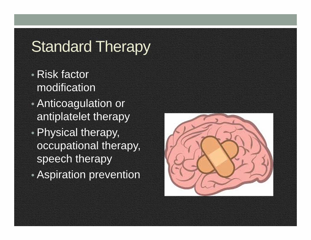
Standard Therapy
• Risk factor modification
• Anticoagulation or antiplatelet therapy
• Physical therapy, occupational therapy, speech therapy
• Aspiration prevention
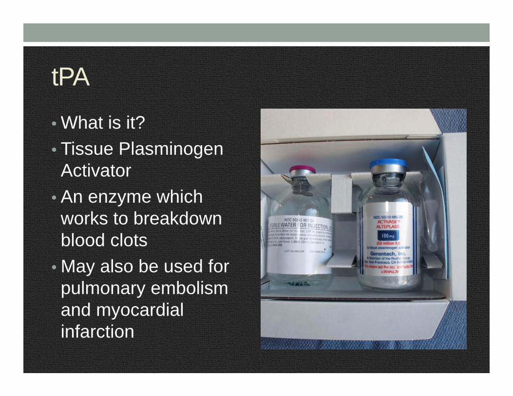
tPA
• What is it?• Tissue Plasminogen Activator
• An enzyme which works to breakdown blood clots
• May also be used for pulmonary embolism and myocardial infarction

tPA• Risks and Benefits
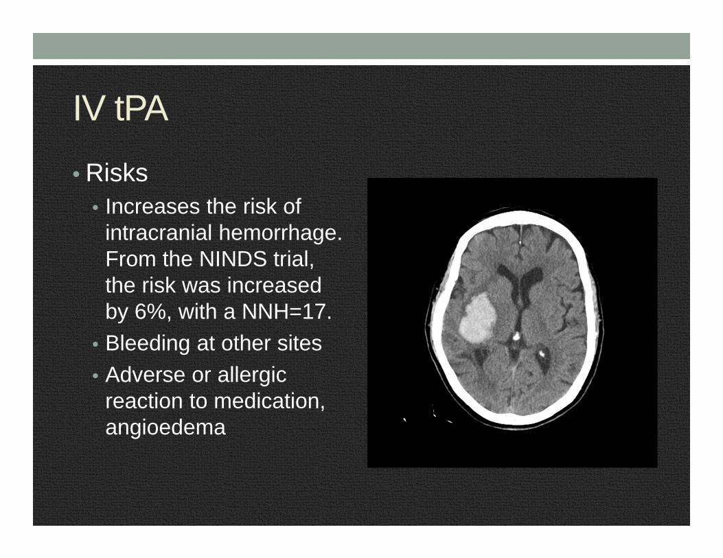
IV tPA
• Risks• Increases the risk of
intracranial hemorrhage. From the NINDS trial, the risk was increased by 6%, with a NNH=17.
• Bleeding at other sites• Adverse or allergic
reaction to medication, angioedema
IV tPA• Benefits
• More likely to have better functional outcome. Some patients will benefit, some will see no difference and a few will be worse off.
• Chance of significant improvement depends on how rapidly the drug can be given after onset of symptoms. For tPA given within 0-3 hours of onset, the NNT=8. For 3-4.5 hours, the NNT=14.

tPA indications• Age > 18 years• A significant neurologic deficit• Non contrast head CT demonstrates no ICH and no new
well-established infarct• Onset within 3 or 4.5 hours
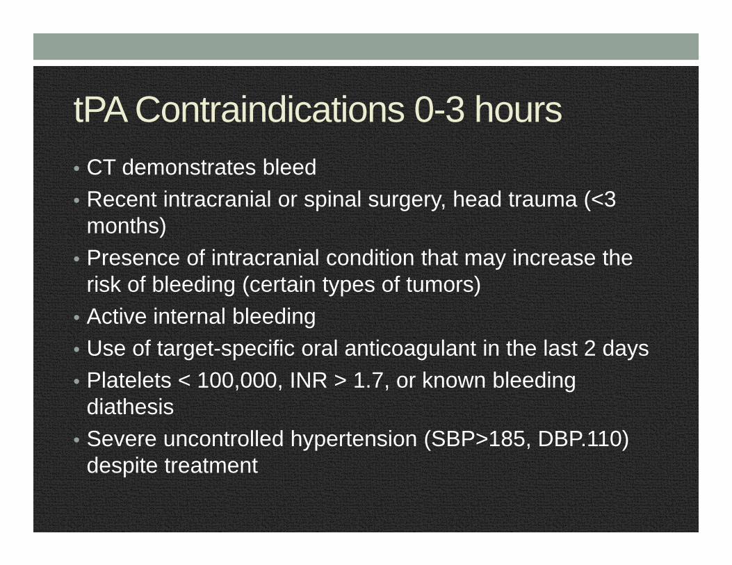
tPA Contraindications 0-3 hours• CT demonstrates bleed• Recent intracranial or spinal surgery, head trauma (<3
months)• Presence of intracranial condition that may increase the
risk of bleeding (certain types of tumors)• Active internal bleeding• Use of target-specific oral anticoagulant in the last 2 days• Platelets < 100,000, INR > 1.7, or known bleeding
diathesis• Severe uncontrolled hypertension (SBP>185, DBP.110)
despite treatment

tPA Contraindications 0-3 hours• Significant spontaneous improvement of deficit• Minor deficit (e.g. isolated sensory symptoms, limb ataxia)• Suspected subarachnoid hemorrhage• Recent myocardial infarction• GI/GU hemorrhage in the past 3 weeks• History of previous intracranial hemorrhage• Seizure at onset (if the deficit is felt to be post ictal)• Very severe neurologic deficit• Major early signs of infarct on CT (> 1/3 hemisphere)

tPA contraindications 3-4.5 hours• Same as 0-3 hour timeframe, plus:• Age > 80• History of prior stroke and DM• Any anticoagulant use (regardless of INR)• NIHSS > 25• CT findings involving > 1/3 MCA territory
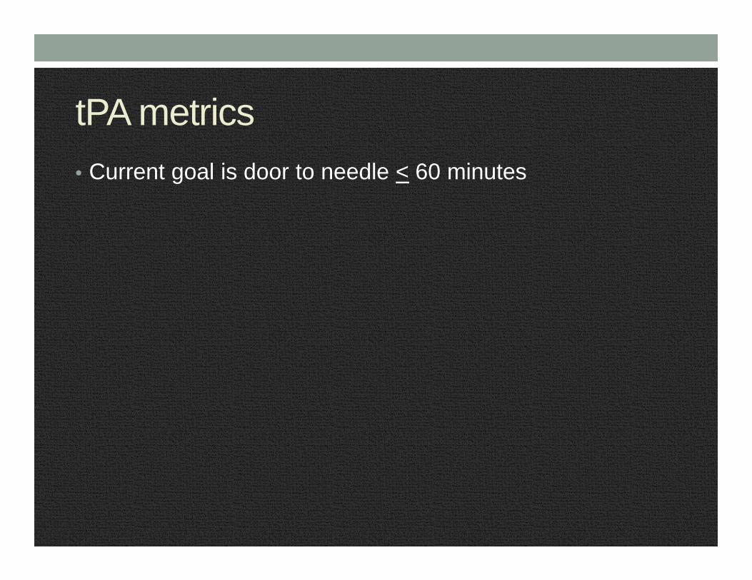
tPA metrics• Current goal is door to needle < 60 minutes
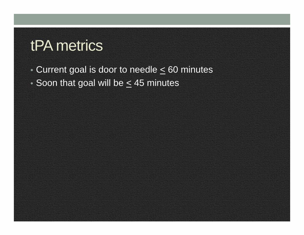
tPA metrics• Current goal is door to needle < 60 minutes• Soon that goal will be < 45 minutes

Endovascular Therapy• Interventions include
• Intra-arterial tPA• Thrombectomy• Angioplasty• Stenting
• May be helpful in select cases when the patient presents outside the tPA window
• Recent literature supports treatment of large vessel occlusions with tPA followed by endovascular intervention

Time = Brain
• Goal door to needle < 60 minutes
• Call stroke code within 10 minutes of ED arrival
• Door to CT read within 45 minutes
• Door to lab resulted 45 minutes

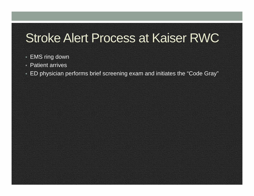
Stroke Alert Process at Kaiser RWC• EMS ring down

Stroke Alert Process at Kaiser RWC• EMS ring down• Patient arrives• ED physician performs brief screening exam and initiates the “Code Gray”
Stroke Alert Process at Kaiser RWC• EMS ring down• Patient arrives• ED physician performs brief screening exam and initiates the “Code Gray”
Stroke Alert Process at Kaiser RWC• EMS ring down• Patient arrives• ED physician performs brief screening exam and initiates the “Code Gray” • Weight obtained, labs drawn, CT notified, pharmacy called• Stat noncontrast CT followed by CT with contrast for angiogram

Stroke Alert Process at Kaiser RWC• EMS ring down• Patient arrives• ED physician performs brief screening exam and initiates the “Code Gray” • Weight obtained, labs drawn, CT notified, pharmacy called• Stat noncontrast CT followed by CT with contrast for angiogram• Radiology calls back with non-con CT head result• Consultation with neurology

Stroke Alert Process at Kaiser RWC• EMS ring down• Patient arrives• ED physician performs brief screening exam and initiates the “Code Gray” • Weight obtained, labs drawn, CT notified, pharmacy called• Stat noncontrast CT followed by CT with contrast for angiogram• Radiology calls back with non-con CT head result• Consultation with neurology• For appropriate patients, ED physician orders IV tPA
Stroke Alert Process at Kaiser RWC• EMS ring down• Patient arrives• ED physician performs brief screening exam and initiates the “Code Gray” • Weight obtained, labs drawn, CT notified, pharmacy called• Stat noncontrast CT followed by CT with contrast for angiogram• Radiology calls back with non-con CT head result• Consultation with neurology• For appropriate patients, ED physician orders IV tPA• CT angiogram resulted.
• For large vessel occlusion, next step is intervention. • If no large vessel occlusion, next step is admission.
• GOAL door-to-needle time<60 minutes
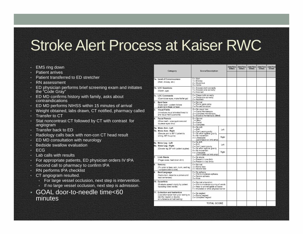
Stroke Alert Process at Kaiser RWC• EMS ring down• Patient arrives• Patient transferred to ED stretcher• RN assessment• ED physician performs brief screening exam and initiates
the “Code Gray” • ED MD confirms history with family, asks about
contraindications• ED MD performs NIHSS within 15 minutes of arrival• Weight obtained, labs drawn, CT notified, pharmacy called• Transfer to CT• Stat noncontrast CT followed by CT with contrast for
angiogram• Transfer back to ED• Radiology calls back with non-con CT head result• ED MD consultation with neurology• Bedside swallow evaluation• ECG• Lab calls with results• For appropriate patients, ED physician orders IV tPA• Second call to pharmacy to confirm tPA• RN performs tPA checklist• CT angiogram resulted.
• For large vessel occlusion, next step is intervention. • If no large vessel occlusion, next step is admission.
• GOAL door-to-needle time<60 minutes

Real Cases from the Kaiser RWC ED

Alpha 72-year-old left side weakness• June 2015 08:00• 72-year-old male BIBA from home at 8 am. C/o weakness
left arm, left leg noted upon awaking this morning. LKWT 23:00 last night.
• PMHx HTN, cholesterol, Bell’s Palsy• Blood sugar• Exam
• Vitals BP 179/89, HR 88, RR 16, T 36.6, O2 Sat 98% RA• Left facial droop, left arm weak, left leg weak, dysarthria• NIHSS = 11

Alpha 72-year-old left side weakness
• Labs unremarkable• CT demonstrates no acute intracranial finding. Dense appearance of the right MCA may reflect MCA occlusion.

Alpha 72-year-old left side weakness• Intervention?

Alpha 72-year-old left side weakness• Patient admitted, treated with aspirin and Plavix• At time of discharge, patient left with residual left facial
droop, weakness of left arm, able to ambulate with a cane• Patient discharged to SNF for rehab

Bravo 78-year-old left side weakness• May, 2015 19:41• 77-year-old man BIBA from home for left side weakness.
LKWT 17:30. • PMH HTN, GERD, CAD, hyperlipidemia• Blood glucose 91• Exam
• BP 200/110, P 81, R 20, T 36.9 C, SpO2 99% RA• Alert, oriented, left facial droop, left arm weak, left leg weak• NIHSS 12
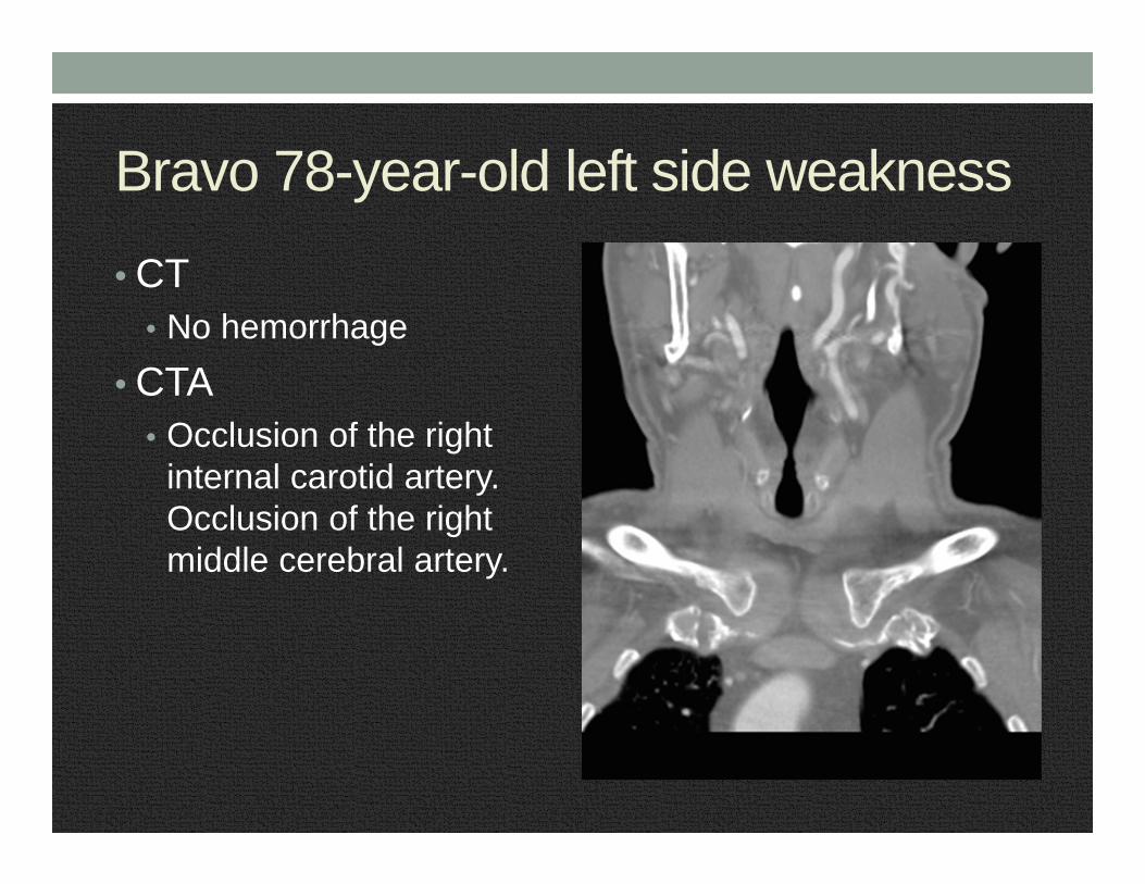
Bravo 78-year-old left side weakness
• CT • No hemorrhage
• CTA• Occlusion of the right
internal carotid artery. Occlusion of the right middle cerebral artery.

Bravo 78-year-old left side weakness• Time for tPA and intervention?
Bravo 78-year-old left side weakness• Time for tPA and intervention?• Remember the BP?
Bravo 78-year-old left side weakness• Time for tPA and intervention?• Remember the BP?• 200/110

Bravo 78-year-old left side weakness• Labetalol given, BP 165/81• tPA given• Transfer to NIR• Thrombectomy performed

Bravo 78-year-old left side weakness• Outcome • 6/2/2015 At time of discharge, ambulatory with a walker.
Discharged to SNF for rehab• 9/8/2015 Follow up, independent ADLs, walks without
assistive device

Bravo 78-year-old left side weakness• 17:30 LKWT• 19:41 arrival• 19:42 Code Gray called• 20:05 CT resulted• 20:18 tPA administered• Door to needle time 37 minutes• Treatment provided 2 hours 48 minutes after onset

Delta 64-year-old man with AMS• July 2015 7:55 am• 64-year-old man BIBA from home with altered mental
status. Last seen well 6:45 am. On EMS arrival, unconscious, minimally responsive, snoring respirations.
• PMH HTN• Blood glucose 107• Exam
• Vitals BP 173/102, P 77, R 14, Temp 37• Eyes closed, snoring respirations, not following commands,
withdraws to pain• NIHSS 26• GCS 6
Delta 64-year-old man with AMS• What needs to happen next?

Delta 64-year-old man with AMS• Code gray• Intubated for airway protection• Neurologist calls during intubation• CT, CTA

Delta 64-year-old man with AMS
• Non con head CT no bleed

Delta 64-year-old man with AMSDecision time

Delta 64-year-old man with AMS
Decision time • LKWT 06:45• CT result time 08:00

Delta 64-year-old man with AMS
Decision time • LKWT 06:45• CT result time 08:27• D/w neurology, give IV tPA
• CTA
Delta 64-year-old man with AMSCT angiogram result• Occlusive thrombus in the distal basilar artery and right
vertebral artery
Delta 64-year-old man with AMS• IV tPA administered• Then taken to neurointerventional lab
Delta 64-year-old man with AMS• Thrombectomy of the right vertebral artery
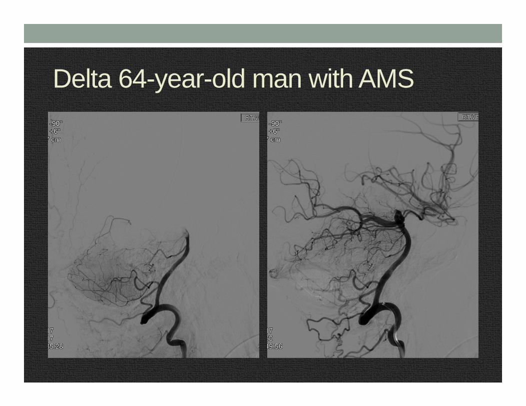
Delta 64-year-old man with AMS
Delta 64-year-old man with AMSOutcome• At time of discharge, no weakness or cognitive deficit• Discharged to home• One month later, doing well, living independently

Delta 64-year-old man with AMS• LKWT 06:45• Arrival to ED 07:55• tPA given 08:53• Door to Needle time 58 minutes• tPA provided 2 hours and 8 minutes after LKWT
Echo 31-year-old woman with slurred speech• Nov 2013 13:25• 31-year-old female presented to ED by private vehicle c/o
sudden onset slurred speech and blurry vision 55 minutes prior to arrival. Also c/o headache and neck pain x 1 week.
• PMH: HTN, migraine• Blood sugar: 76• Exam
• BP 185/130, P 78, R 19, T 37.1• Alert, oriented, slurred speech, aphasia, right side hemianopsia• NIHSS 4

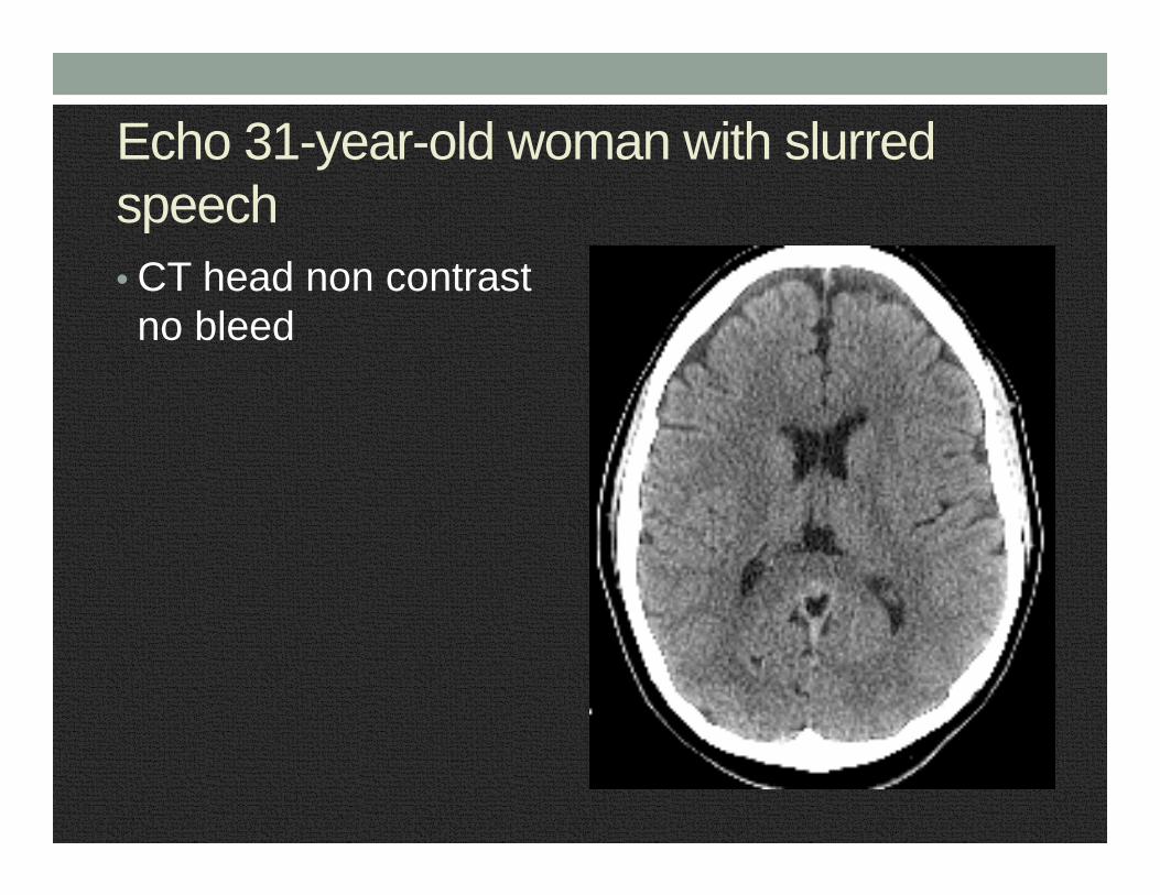
Echo 31-year-old woman with slurred speech• CT head non contrast no bleed
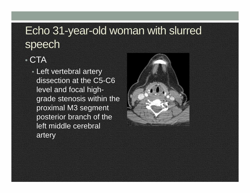
Echo 31-year-old woman with slurred speech• CTA
• Left vertebral artery dissection at the C5-C6 level and focal high-grade stenosis within the proximal M3 segment posterior branch of the left middle cerebral artery

Echo 31-year-old woman with slurred speech• When did the symptoms start?• NIHSS = 4• Hypertensive 185/130!• Dissection!!
Echo 31-year-old woman with slurred speech• Discussion with neurology• tPA given

Echo 31-year-old woman with slurred speech• Outcome
• Symptoms slowly resolved in the ICU• Anticoagulated with warfarin with enoxaparin bridge• No obvious reason for the dissection

Echo 31-year-old woman with slurred speech• LKWT 12:30• ED arrival 13:25• tPA given at 14:43• Door to needle 78 minutes• Treatment provided 2 hours and 13 minutes after onset of
symptoms
Foxtrot 79-year-old female AMS• September 2015 12:24• 79 yo female biba from home with altered mental status.
LKWT 9 am. At 9 am, she drove her car to see a friend, drove home, on return home crashed her car into the back of the garage. On EMS arrival, patient confused.
• PMH: stroke one year ago, DM, HTN• Blood sugar 204• Exam
• BP 156/60, HR 79, RR 20, T 35.7, O2 Sat 95% RA• Appears distressed, confused, left visual field cut, aphasic and
dysarthric, neglect• NIHSS 8

Foxtrot 79-year-old female AMS

Foxtrot 79-year-old female AMS• Code gray activated

Foxtrot 79-year-old female AMS• CT – old occipital infarct• CTA – Atherosclerosis. No significant arterial stenosis or
occlusion.

Foxtrot 79-year-old female AMS• Time for tPA?

Foxtrot 79-year-old female AMS09:00 Last known well time. However, patient drove her car home and arrived home at 12:0012:24 Patient arrives to ED, history is limited due to altered mental status. Known to have old stroke, how new are the deficits today? 12:40 CT resulted, old occipital infarct. Meanwhile, symptoms are waxing and waning. 13:00 Family confirms speech changes are new, vision changes are probably worse. Time is now 4 hours since LKWT. To give or not to give tPA? Neurology at bedside, explained risks/benefits to family, decision made to give tPA.
Foxtrot 79-year-old female AMSOutcome• Found to have atrial fibrillation, appropriate treatment
recommended• Almost complete resolution of symptoms• Discharged to home with home PT, OT
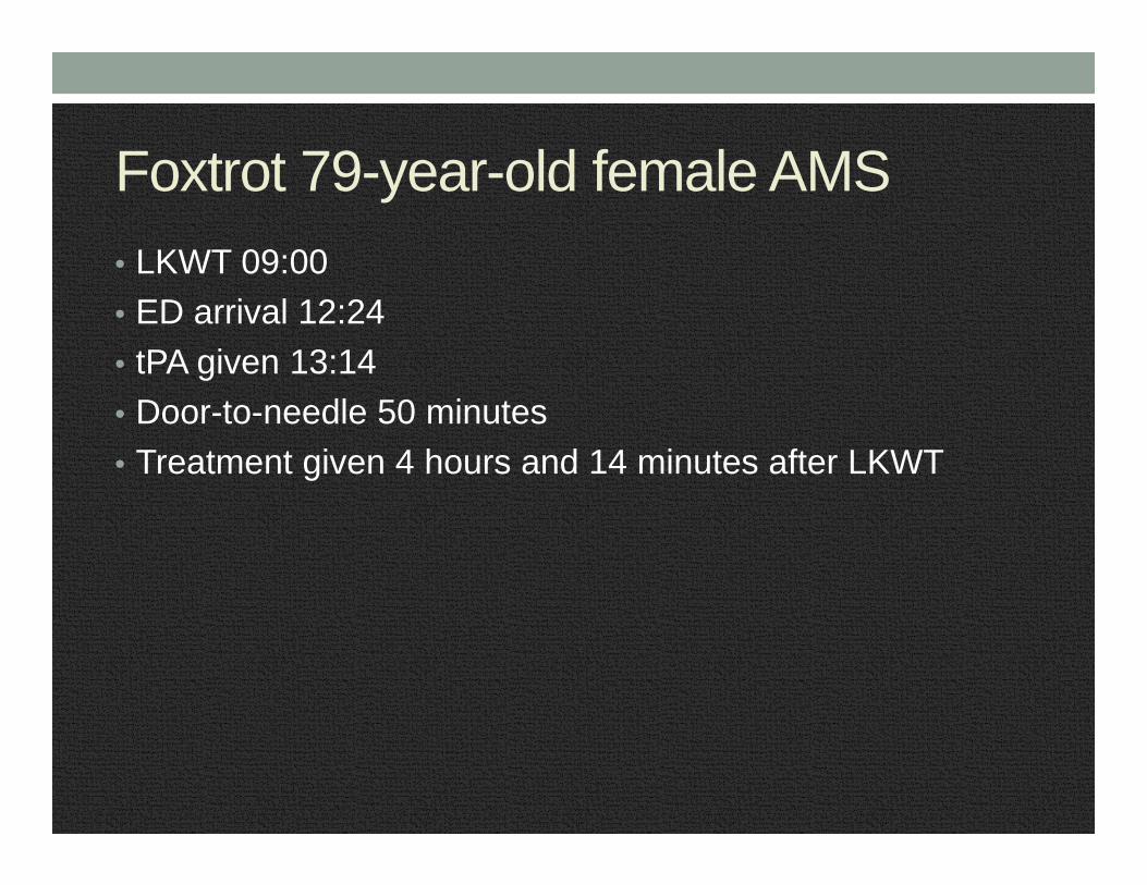
Foxtrot 79-year-old female AMS• LKWT 09:00• ED arrival 12:24• tPA given 13:14• Door-to-needle 50 minutes• Treatment given 4 hours and 14 minutes after LKWT

Kilo 52-year-old man with chest pain and left leg weakness • June 2013• 52-year-old man BIBA from home with acute onset chest
pain x 30 seconds, sharp and severe. Followed by acute onset left leg numbness and weakness.
• PMH: HTN, hyperlipidemia• Blood sugar 161• Exam
• BP 140/88 P 60 R 22 T 36.8• Ill-appearing, weak in the left leg• NIHSS 5 (weakness and loss of sensation left leg)
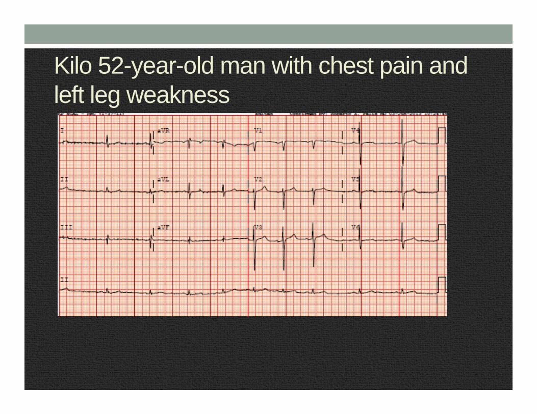
Kilo 52-year-old man with chest pain and left leg weakness

Kilo 52-year-old man with chest pain and left leg weakness
Noncontrast scan negative

Kilo 52-year-old man with chest pain and left leg weakness • LKWT 13:55• ED arrival 14:51• Results 15:35• Neurology recommends tPA barring any contraindications

Kilo 52-year-old man with chest pain and left leg weakness • t-PA ordered @ 15:18• Radiology calls back at 15:25 and notes the following:
• Type A/B dissection extending into the left common carotid artery with severe stenosis of the common carotid artery and complete occlusion of the left ICA distal to the bifurcation
• Dissection also involves left subclavian artery

Kilo 52-year-old man with chest pain and left leg weakness • t-PA NOT given (wasted)• CV surgery recommended nicardipine gtt, CT chest• CT chest/abd/pelvis - dissection extends down to the iliac
bifurcation• Transferred emergently to facility with CV surgery• Underwent emergent repair of Type A dissection• Flow re-established to viscera and lower extremities• Developed ischemic colitis and rhabdomyolysis• Underwent bilateral leg fasciotomies, then subtotal
colectomy and bilateral leg amputations• Died 2 days after event

Lima 88-year-old female• July 2015 8:28 am• 88 yo female BIBA from home with right side weakness.
LKWT 7:30 am. • PMH HTN, CHF• Blood glucose 112• Exam
• BP 120/45, P 54, T 36.4 C, O2 Sat 98% RA• Right side weakness, right side neglect, aphasic• NIHSS 25

Lima 88-year-old female• Code gray activated

Lima 88-year-old female• CT non contrast
• No hemorrhage
• CTA • Thrombosed left internal carotid. Left MCA is unopacified.
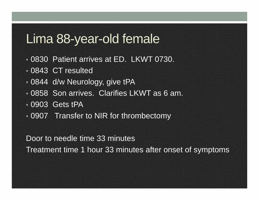
Lima 88-year-old female• 0830 Patient arrives at ED. LKWT 0730.• 0843 CT resulted • 0844 d/w Neurology, give tPA• 0858 Son arrives. Clarifies LKWT as 6 am. • 0903 Gets tPA• 0907 Transfer to NIR for thrombectomy
Door to needle time 33 minutesTreatment time 1 hour 33 minutes after onset of symptoms

Lima 88-year-old female
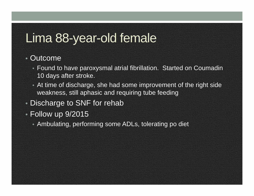
Lima 88-year-old female• Outcome
• Found to have paroxysmal atrial fibrillation. Started on Coumadin 10 days after stroke.
• At time of discharge, she had some improvement of the right side weakness, still aphasic and requiring tube feeding
• Discharge to SNF for rehab• Follow up 9/2015
• Ambulating, performing some ADLs, tolerating po diet

Quebec 91-year-old female left hemiparesis• February 2014• Elderly woman from home BIBA Code 3 with acute left
face/arm/left leg weakness with right eye deviation 50 minutes prior to arrival.
• Had stroke within past 3 months treated at RWC with aspirin/statins, discharged to home 1 mo. ago. Baseline A+Ox3.
• PMH: HLD, DM, stroke, CAD• Blood sugar: 192• Exam:
• Vitals 96.6 18 57 175/57 • ill-appearing• NIHSS: 35 (mostly for generalized unresponsiveness, flaccidity,
aphasia)

Quebec 91-year-old female left hemiparesis
NSR 58, no ischemic changesCBC, Chem 7, INR WNL

Quebec 91-year-old female left hemiparesis
CT head – negative for acute changesCTA not done due to IV contrast allergy noted in
HealthConnect

Quebec 91-year-old female left hemiparesis• Decision time
• LKWT 15:30• ED arrival 16:19• Results 16:57 – 1 hour 27 minutes after onset• Family states patient is Full Code

Quebec 91-year-old female left hemiparesisOutcome• t-PA NOT given for:
• Last stroke within the past 3 months• Large NIHSS score
• Neuroscience admitted patient and reviewed grave prognosis with family
• Because family was certain patient did not want to survive with “disability,” comfort measures initiated
• Patient died within 24 hours

Tango 60-year-old female• Oct 2015 11:55 am• 60 year old female left side weakness onset 8 am today.• PMHx HTN, thyroid disease• Blood sugar 90• Exam
• BP 156/82, HR 92, T 36.1, RR 20, O2 Sat 98% RA• Awake, alert, oriented x 3, left facial droop, left arm weak, left leg
weak• NIHSS 9

Tango 60-year-old female• Stroke symptoms presenting to the ED at 4 hours after
onset• Code Gray called, patient immediately taken to CT, labs
drawn, tPA ordered to expedite care
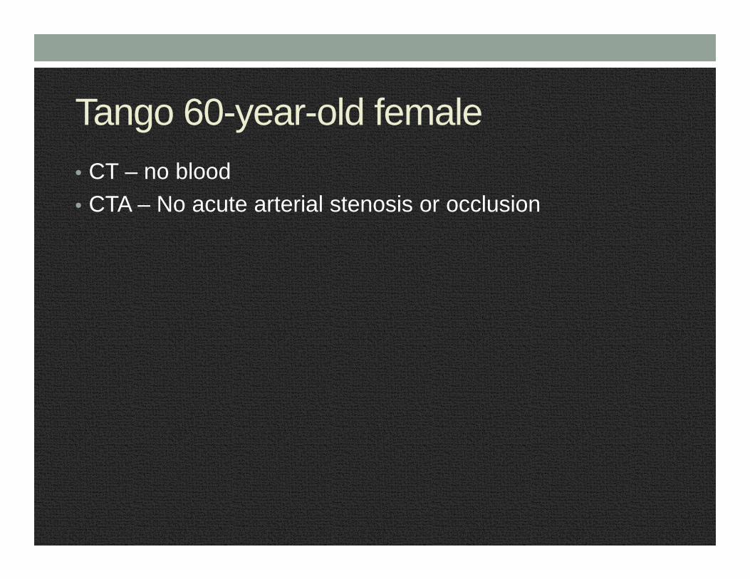
Tango 60-year-old female• CT – no blood• CTA – No acute arterial stenosis or occlusion
Tango 60-year-old female• Upon return from CT, patient reports she had similar
symptoms in the past which resolved completely and was diagnosed with a migraine
• Neurology advised no tPA• After observation, patient spontaneously improved.
Complete resolution of her neurologic symptoms. • tPA was wasted
Yankee 50-year-old man• March 2015 07:28• 50 yo male biba from home c/o right side weakness x 40
minutes. • PMH DM2• Exam
• BP 146/88 HR 72, T 36.6 C, RR 17, O2 Sat 98% RA• Right facial droop, right arm weak, right left weak, right
hemineglect, aphasia• NIHSS 9

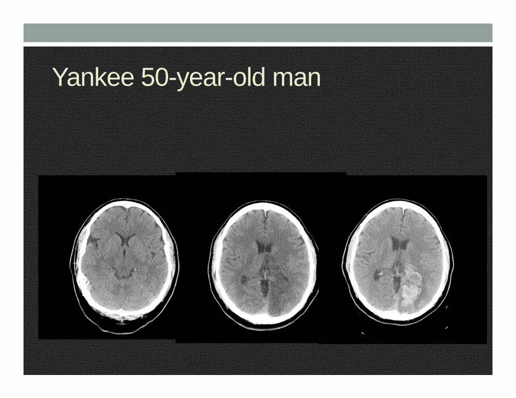
Yankee 50-year-old man• Left PCA and L vertebral artery thrombus
Yankee 50-year-old man• Treated with IV tPA• The thrombus is not amenable to intervention• Patient admitted to the ICU

Yankee 50-year-old man• 06:50 LKWT• 07:27 arrives ED• 08:03 tPA given• Door to needle time 36 minutes• Treatment 1 hour 13 minutes after onset of symptoms

Yankee 50-year-old manOutcome• Patient has mild improvement, still with speech difficulties,
memory problems and visual field cut. He is discharged to home on aspirin with family to provide 24-hour supervision. Outpatient OT.
• 2 weeks later develops severe headache. Presents to clinic. No new neuro deficits.
• CT demonstrates hemorrhage in the recent infarct area. • Patient is readmitted.
Yankee 50-year-old man
Yankee 50-year-old man• September 2015• Lives at home with wife and children, able to care for
himself and his children. Still with some cognitive difficulties and a visual field cut
• Back at work part time – IT at a big area tech company

Takeaway PointsTime is brain
Determine accurate LKWTTreat hypertension early
CT followed by CTA unless contraindicationBeware stroke mimics
tPA can improve outcomes


![Endovascular therapy in acute ischemic stroke: where we ...neurology.mcgill.ca/neurodocs/AHD 2010-2011... · standard of care in many stroke centers across the globe [18–20]. Endovascular](https://static.fdocuments.in/doc/165x107/5ea01a4853f7473169025d9d/endovascular-therapy-in-acute-ischemic-stroke-where-we-2010-2011-standard.jpg)


