1445-Zonulin and Its Regulation of Intestinal Barrier Function, Physiol Rev 2011

HAL Id: inserm-00793506http://www.hal.inserm.fr/inserm-00793506
Submitted on 22 Feb 2013
HAL is a multi-disciplinary open accessarchive for the deposit and dissemination of sci-entific research documents, whether they are pub-lished or not. The documents may come fromteaching and research institutions in France orabroad, or from public or private research centers.
L’archive ouverte pluridisciplinaire HAL, estdestinée au dépôt et à la diffusion de documentsscientifiques de niveau recherche, publiés ou non,émanant des établissements d’enseignement et derecherche français ou étrangers, des laboratoirespublics ou privés.
Stress neuromediators are key regulators of theintestinal barrier: Link to inflammation and cancer
Benjamin Ducarouge, Muriel Jacquier-Sarlin
To cite this version:Benjamin Ducarouge, Muriel Jacquier-Sarlin. Stress neuromediators are key regulators of the intesti-nal barrier: Link to inflammation and cancer: Stress neuromediators regulate the intestinal barrier.Trends in cell and molecular biology, Research Trends, 2011, 6, pp.59-88. <inserm-00793506>

Stress neuromediators are key regulators of the intestinal barrier: Link to inflammation and cancer
B. Ducarouge1, 2 and M.R. Jacquier-Sarlin1, 2*
From : 1 : Centre de Recherche Inserm U836, Grenoble Institut des Neurosciences. Equipe du Stress et des Interactions Neurodigestives, Site Santé BP 170 La Tronche F-38042 Grenoble Cedex 9, France ; 2 : Université de Grenoble, F-38000 Grenoble, France * To whom correspondence should be addressed: Centre de Recherche Inserm U836, Grenoble Institut des Neurosciences. Equipe du Stress et des Interactions Neurodigestives, Site Santé BP 170 La Tronche F-38042 Grenoble Cedex 9, France. Tel.: (+33) 476-56-52-50; Fax: (+33) 476-56-05-54; E-mail: [email protected] To be submited in: TRENDS IN CELL & MOLECULAR BIOLOGY Running title: Stress neuromediators regulate the intestinal barrier. Keywords: Stress, CRF, inflammation, cancer, intestinal epithelial barrier.

Abstract In the past year, the influence of psychosocial and environmental stressors in different pathogenesis received increased awareness. The brain is the master manager of the interpretation of what is stressful and of the physiological responses that are produced. Animals have developed conserved strategies to respond to stressful conditions, in particular, the secretion of stress-specific neuromediators which mediate protective and adaptative effects in the short run and yet can accelerate pathophysiology when they are over-produced or mis-managed. The Cortico-Releasing Factor (CRF) and their derived peptides are the majors stress neuromediators. Their localization has originally been described in the central nervous system where they play a pivotal role to activate the hypothalamic-pituitary-adrenal (HPA) axis and was recently extended to the periphery. While the peripheral effects of CRF signalling need to be more thoroughly investigated, it has been described to influence disease negatively, in particular in the intestine. The epithelial barrier is a crucial checkpoint to control body entrances. Prolonged exposure to stress can cause ultrastructural epithelial abnormalities and can increase barrier permeability, which favors luminal translocation, immune activation and thus induces inflammation. This review summarizes the present knowledge on the stress response and the effects of both acute and chronic stress to induce pathological damage to the intestine. We present the potential pathways involved, and the proposed mechanisms of action, mediating these effects. The CRF system is potentially useful as a diagnostic marker or a therapy target for inflammatory diseases and cancer.
Introduction In medical language the concept of “Stress” has been introduced by Hans Selye (1907-1982) [1]. In response to various stressors, living organisms have developed adaptative behaviors and coping strategies in order to maintain their homoeostasis. Stress is a complex process that involves the endocrine, immune and nervous systems. Altogether, they communicate by the production of mediators (hormones, cytokines, neuromediators) which target their specific receptors. “Cognitive stress” (from the central nervous system activity) such as psychological and emotional events is distinguished from the “non cognitive stress” induced by physical damage, infection or inflammation, although some “non cognitive stress” may be relayed by the nervous system especially via the vagal afferences (Figure 1). The stress is also characterized by various parameters such as duration, frequency (acute versus chronic stress) and intensity. Chronic or recurrent stress results in an increased demand of physiological systems (cardiac, immune, metabolic, hormonal…) that can lead to diseases and contribute to wear and tear on the body, a condition known as “allostatic load” [2]. According to the community of pathways used and processes generated, mediators and receptors used, stress of various nature leads to convergent effects. Indeed, whatever the nature of the stress, the main adaptative response is mediated by the hypothalamic-pituitary-adrenal (HPA) axis with a central role of the neurohormone, the corticotrophin-releasing factor (CRF). This stress response begins with the hypothalamic production of CRF which in turn, induces the production and release of adrenocorticotropic hormone (ACTH). Pituitary-derived ACTH stimulates adrenocortical production of glucocorticoids (GC) which counteract the effect of stressors, suppress the immune system, and attenuate the functional activity of HPA axis via feedback inhibition of the hypothalamic CRF expression [3, 4] (Figure 1). However, the brain is not the only centre of information and decisions: the interactions between the different systems also occur at a local level in some organs such as the skin, the heart and the digestive tract. They are particularly described in the brain-gut axis, which also contributes to manage the stress in the intestine using the same mediators and signalling pathways in reciprocity to the brain. Stress and pathologies In the past ten years, the influence of psychological and environmental stressors on pathogenesis such as obesity, metabolic syndrome, and type 2 diabetes, as well as pain and chronic fatigue syndromes, received increased awareness [5]. Furthermore, a large number of skin diseases, including atopic dermatitis and psoriasis, appear to be triggered or exacerbated by psychological stress [6]. They are also often associated with a perturbation of the cutaneous homeostatic permeability barrier [7]. Stress is recognized to participate in the development and/or aggravation of gastrointestinal (GI) disorders, such as inflammatory bowel diseases (IBD) and irritable bowel syndrome (IBS) despite differences in their etiologies [8-12]. IBD which includes Chron’s disease (CD) and ulcerative colitis (UC), consist of a measurable over-inflammatory response leading to gut damage [13]. Many in vitro and in vivo studies indicate that stress-related alterations of GI functions are mediated by both brain and peripheral CRF signalling pathways (for review see [14]). While the expression pattern of CRF receptors and ligands in the GI tract have been extensively described (for review see [15]), the

cellular and molecular mechanisms of their interactions are still poorly understood and principally focused on the neuronal CRF signalling influence on the immune response in the GI tract (for review [16]). Using data from clinical and basic research literature, the objectives of this review are to summarize evidence in support of a major role of stress and CRF signalling in the induction and progression of inflammatory intestinal disorders with a special emphasis on the intestinal barrier alteration. We also discuss the implication of CRF system in the colorectal cancer (CRC) initiation and progression. The CRF system: signalling and expression in the epithelium An overview of the CRF system In mammals, the CRF family (including urocortins, Ucn) is composed of 38 to 41aa peptides called CRF or urocortins (Ucn) such as Ucn1, Ucn2 and Ucn3. CRF and Ucn1 were first characterized for their ability to control ACTH secretion from anterior pituitary cells [3, 17], and hence play a pivotal role in the stress response by regulating the HPA axis. Later, DNA analysis has identified Ucn2 and Ucn3 by sequence homologies [18, 19]. Other orthologs have also been described, like the Urotensin1 [20] in fish or the sauvagine in frogs [21, 22]. Phylogenic analyses indicated that CRF-like peptides are well conserved through the evolution and derived from a common stem involved in osmo-regulation as an ancestral stress adaptation, neuro-endocine system [23-25]. Like many hormones, these peptides are derived from precursor proteins [26, 27] and their bioactivities are modulated by the secreted CRF-binding protein (CRF-BP) or soluble splice variants of their receptors [28-32] (Figure 2). These polypeptides exert their activities through the activation of two known class II G Protein Coupled Receptors (GPCR), CRF1 [33] and CRF2 [34]. While CRF receptors arise from the transcription of two different genes they share about 70% homology, but differ by their N-terminal ligand binding domains [35, 36]. CRF recognizes both receptors, but displays a higher affinity for CRF1. Ucn1 activates CRF1 and CRF2 with the same potency, whereas Ucn2 and Ucn3 exclusively bind to CRF2 [37, 38]. CRF receptors are subjected to additional modifications consisting of splicing regulation and glycosylation [39]. Several CRF1 splice variants classified from CRF1a to CRF1h were identified at the mRNA level but there is little knowledge about their protein expression and functionality within the different tissues. Except for CRF1a, these variants display low ligand affinity or are unable to induce intracellular signalling: they have been suggested to exert regulatory functions by titrating free ligands [40-43]. Recently, a novel functional isoform CRF1i has been identified in the BON cell line, which is also expressed in human ileum [44]. CRF2 undergoes three distinct functional forms: CRF2a, CRF2b and CRF2c, which consist of an alternative transcription start a, c versus b and alternative splicing between a and c, only detected in humans [45, 46]. This results in the modification of the N-terminus, involved in the ligands’ affinity [47]. Several dysfunctional CRF2 forms have also been described, in particular soluble truncated forms. First, they have been proposed as ligand scavengers which are in competition with membrane expressed receptors for free ligands [48]. However recent studies found that despite its correct translation, the variant of CRF2a is not secreted. This protein may regulate the CRF2 signalling by altering the transcription of full-length CRF2a mRNA [49, 50]. Similarly, a dominant-negative CRF2b splice variant has been described in mouse heart, to impair mCRF2b function by retaining its cellular location to the endoplasmic reticulum-golgi sites [51]. There is also some variability in the molecular weight observed by western blot analysis for these receptors depending on tissue, cell type and species. These differences are often due to the splicing, the antibodies used, or the glycosylation status of CRF receptors [52, 53] according to the five potential sites of N-glycosylation identified in the primary structure [39]. However CRF1 activity has been described to be regulated by inflammation [39] which also influences the glycosylation status of proteins [54]. Together these studies indicate that the CRF system is finely tuned by different regulatory pathways at the receptor and ligand levels. CRF signalling CRF signalling has been studied in many cell lines and tissues including the Central Nervous System (CNS) and the periphery. It appears that the binding of ligands give rise to structural arrangements of CRF receptors that increase the affinity of their third intracellular loop for the Gα subunits which become activated. CRF receptors are primarily coupled to Gαs and trigger cAMP formation via adenylyl cyclase activation [55] (Figure 2). However they could in few cases bind to Gαq, i, o, z and involve other signalling pathways like Phosho Lipase C (PLC) [56-59]. Gβγ subunits are also able to mobilize intracellular pathways like PI3K/Akt and

Ca2+ flux but their involvements in CRF signalling are poorly investigated. Thus, depending on the Gα subtype recruited and on the cell type, CRF receptors are able to transduce plethora of intracellular signalisations, such as Protein Kinase A, B, C (PKA, PKB, PKC), P42/p44, p38 Mitogen-Activated Protein Kinases (MAPK) Ca2+ flux, NOS activation Fas-ligand, and NFκb [60]. Recently novel downstream G protein-independent pathways have been described for the CRF receptors like Src which directly interacts with the endocytosed CRF receptors and takes part in the activation of ERK 1/2 [56, 61, 62]. Like other GPCR, CRF receptors expression and function are regulated by a desensitizing process. Moreover, the recruitment of G protein-coupled receptor kinase GRK3 and GRK6 by the activated receptor leads to the phosphorylation of its C-terminus and β-arrestin binding [40, 63]. All of these events are involved in the internalization of the receptor which can subsequently be degraded in the endo-lysosomal path, or recycling to the membrane. If both receptors are subjected to internalization after ligand binding, it seems that the time course of desensitization could be different depending on the cell model and the receptor sub-type [64, 65]. Sequences analyses also report multiple phosphorylation sites for either PKA or PKC, which can regulate CRF receptor function [40, 66]. However, the use of specific mutants, truncated forms or PKA inhibitors showed that these modifications are not required for the receptor internalization [65].
Distribution of CRFreceptors and peptides through the intestinal mucosa The CRF system was first characterized in the CNS for its implication in the brain response and adaptation to stress as well as in food intake [67, 68] and psychiatric disorders [69, 70]. Later, CRF receptors and their ligands have been identified in peripheral tissues like digestive tract (for review [15]), cardiovascular, immune, reproductive systems or skin [71]. There is a growing number of studies interested in CRF receptor expression and localization in the gut but the emerging features are often heterogeneous and incomplete. This can be explained by the CRF receptor isoforms, species and antibodies recognition, as mentioned previously [72]. It could also be due to the disparity in global and cell type expression between the different sections of the digestive tract [73]. According to our interest in the regulatory role of CRF system on the epithelial barrier, in this section, we will focus on what is known on epithelial cells (EC), lamina propria (LP) cells and on the enteric nervous system (ENS), at the small intestine and colonic levels (Figure 3). Indeed, the LP contains innate immune cells which participate in the primary response to intestinal homeostasis perturbations. Expression of CRF receptors and ligands in the small intestine In contrast to the colon, the expression and distribution of CRF receptors and their related peptides in the ileum have been poorly investigated in rodents and humans. In mouse, only one study has reported the expression of CRF1 mRNA and at low levels CRF2 mRNA in LP and EC in the ileum [74]. CRF2 mRNA was found in mucosa and at the base of villi in the duodenum [75]. Both CRF receptors have been principally detected in neuronal myenteric plexus (NMP) and neuronal submucous plexus (SNP) in guinea pigs and rats [73, 76, 77]. Their expression can also be observed in muscle layers of rats, however with opposite patterns depending on the region considered duodenal versus ileal. Lastly, CRF2 expression was found in enteric neurons and nerve fibers, in the duodenal glands [78] and in 5-HT negative EC in the villi [77]. Interestingly, the expression of CRF1 is complementary to that of CRF peptides suggesting that neurons are a potential target for CRF signalling [79]. CRF immunoreactivity was detected in MNP and SNP of the ileum and the duodenum in guinea-pigs as well as in rats [80]. CRF mRNA was also located at low levels in LP cells, Paneth cells and endocrine cells in the crypts of rat ileum, but not in enterocytes [80, 81]. Only one report describes CRF expression in LP and in a few EC of mouse ileum at both mRNA and protein levels [74]. The expression of Ucns in the ileum has been principally investigated in rat tissue. Ucn1 proteins and transcripts were found in both enteric plexuses [82, 83]. Ucn2 mRNA has been described in rat and to a lesser extent in mouse small intestine [73, 84]. In rat, Ucn2 was detected in the epithelium, LP and enteric plexuses. In human, Ucn2 was absent, while Ucn3 was detected in the muscularis mucosae [85]. Expression of CRF receptors and ligands in the colon Both CRF receptors have been detected at the gene and/or protein levels in human and rodent colons (Figure 3). Some reports mentioned that CRF1 is unexpressed in human IEC of the colon, neither at the mRNA

level on total purified EC, nor with tissue immunolocalization [86, 87]. Nevertheless, CRF1 transcripts have been detected in colonic sections from healthy adults and a CRF1 positive staining can be observed in LP cells (macrophages and mast cells), as well as in both enteric plexuses [88]. LP macrophages and mast cells from ileum and colon also express CRF2 receptors in humans [86, 89] and rats [73]. The mast cell leukaemia-derived cell line HMC-1 also expresses CRF1 and CRF2a specific mRNA isoforms and provides a useful model to study in vitro mastocyte-epithelial interactions [87, 90]. In rodents, CRF1 expression especially concerns neural plexuses. However regional differences were observed between species. In guinea pigs, the signal is more important in the SNP than in the MNP, whereas the opposite is observed in rats [78, 88, 91]. Non-neuronal expression of CRF1 has been reported in some LP cells and in IEC located at the base of rat colon crypts, which also contain stem cells [78]. Other experiments depict a protein expression in the whole crypts of rat proximal colons, which disappears distally, and mRNA is also described in the IEC of the mice ileum [74, 92, 93]. In ovines, CRF1 is expressed at the baso-lateral membrane in the IEC of the colon, during the foetus maturation but there are no data whether it’s still present in young and adults [72]. The presence of CRF2 in IEC, at a basal level is quite controversial in terms of expression and localization. CRF2a mARN or protein expression have been identified in human and rat IEC and in the epithelial derived carcinoma cell lines HT-29 or in the untransformed NCM460 [86, 89, 93-95]. However, in other studies, CRF2 has not been detected in rat, human or foetal ovine IEC [72, 73, 87, 92]. The CRF2 subcellular localization differs in EC, depending on the studies or species. While CRF2 immunostaining is apical in rats, localized in the luminal surface of the crypts, it seems to be localized on the baso-lateral membrane in humans [78, 95]. The real localization of this receptor is very important to understand its role in the direct regulation of epithelial barrier functions. Therefore, baso-lateral expression would signify that EC could be exposed to autocrine/paracrine activation, while apical expression would be restricted to autocrine stimulation or activation by luminal circulating ligands. Enterochromaffin cells, which are inserted within the epithelium, do not express CRF2 but CRF1 in humans and ovines [72, 89]. Both receptors have been identified in BON cells, a pancreatic carcinoid-derived human endocrine cell line, which shares functional similarities with intestinal enterochromaffin cells [96]. The quasi ubiquitous expression of the CRF receptors in these cell populations of the digestive wall reinforces the involvement of the CRF system in the immunomodulation response. The micro vasculature of the intestine plays an important role in the delivery of proinflammatory cytokines and the regulation of immune cells. There is a strong expression of CRF2 in endothelium and smooth muscles from the human colon vasculature [89, 97]. Identical patterns are found in rats with no CRF1 [78]. To date, CRF, Ucn1 and Ucn3 but not Ucn2 were detected in human colons. While CRF was found in the mucosa, in monocytes and IEC, mainly in enterochromaffin cells [98, 99], Ucn1 (protein and mRNA) was present in LP macrophages with a few amount in enterochromaffin cells [86, 100, 101]. Ucn3 expression is more heterogenous, since protein and mRNA were found in both enteric plexuses, smooth muscle, endothelium, enterochromaffin cells as well as in enteric glial cells [89, 102]. In rodents, there is no data available on Ucn3 expression, to date. Expression of CRF is predominantly neuronal with an immunoreactivity observed in both enteric plexuses, and in nerve fibers located in mucosal projections and submucosa ganglia and in the circular muscle layer [81, 88, 103]. Non-neuronal expression of CRF in rats was controversial, and was detected in enterochromaffin cells [81]. Expression of Ucn1 is higher than of CRF and was observed in rat enteric nervous plexuses and co-localized with CRF1 receptors [83, 91]. In contrast, mucosal expression of Ucn1 has been detected in very few cells [82]. Ucn2 protein was identified in mucosal and submucosal layers of rat colon in IEC, LP immune cells and enteric neurons [73, 84, 102]. Altogether these studies indicate that the intestine is a target for stress signalling. Most of the studies show that ligands are expressed in close proximity of the CRF receptors, indicating the existence of local autocrine/paracrine regulatory loops. However, stress ligands produced by enteric neurons could contribute to endocrine regulation. So whatever the nature of this regulation, different loops could be established (Figure 1). For example, neuronal activation following stress may regulate expression of stress-ligand and receptors in IEC and their function, directly according to the close proximity of enteric neurones and these cells [104] as well as indirectly after LP immune cell stimulation. Indeed, wallon et al. have shown that the release of CRF by oesinophils following the activation of muscarinic receptors can activate mast cells and leads to colonic mucosal barrier dysfunction in patients with UC or in eptithelial T84 cells [105]. Immune cells may also regulate CRF signalling on IEC, independently of neurones after infection. Finally, IEC may produce ligands which may regulate epithelial barrier function by an autocrine way. We found that rat IEC from the duodenum to the colon as well as human adenocarcima cells were able to produce CRF and Ucns (personal data).

Regulation of the CRF system by stress and inflammation Cumulative studies suggest that stress, whatever its origin, either interceptive (infection, inflammation) or exteroceptive (psychological or physical stress), modulates the expression of stress signalling molecules in the GI tract as it was observed for the brain. Regulation by stress During stressful conditions, brain perception is communicated to the periphery partially via the HPA axis that rapidly produces GC discharges into the blood. Promoters of CRF family genes contain Glucocorticoid Response Elements (GRE) which modulate their gene expression [48, 106-109]. However, GC regulate the expression of the CRF system compounds depending on cell type or location. For example, CRF and Ucn2 are differentially regulated by stress-related GC depending on the brain region [109]. Ligands and receptors are also submitted to regulation in different intestinal cell types, during stress and inflammation. The stress-induced regulation of intestinal CRF system could also be dependent on the nature of the stress inputs. Acute versus chronic stress could modify the GC response which depends on GC receptor signalling, rather than on mineralocorticoid receptors (MR) according to the GR expression profile of cells [110]. CRF2 is down-regulated by GC in the rat aortic smooth muscle A7r5, the skeletal muscle C2C12 for the β isoform or the HEK 293 cells transfected with the "a" promoter coupled to luciferase. Dexamethasone induces a decrease of CRF1 and CRF2b opposite to a CRF2a increase of the promoter activity in pancreatic islets or in the insulinoma cell line MIN6 [111]. Consequently, some CRF circuits could be shut down in some cell types or activated in others. O’ Malley et al.; have investigated the CRF receptor regulation during acute versus chronic stress or the association of both. Regulation appears to be different between proximal and distal sections of the rat colon, according to the nature of stressors [92]. Their experiments are very exhaustive in stress models but there is no information on the time course which is important according to the variation in CRF system expression during the inflammation process. In that way, our unpublished data show that acute restraint stress induces an increase in CRF2 expression which becomes significant in the proximal colon of rats, only after six hours post stress. Hence, if the level of CRF system components is unchanged at the early phase of stress, it doesn’t exclude that some modifications occur later. Lakshmanan et al. speculate that in ovines the increase of endogenous foetal GC and/or gestational stress contributes both to the down-regulation of the CRF2 and to the up-regulation of the CRF1 observed near the term and term foetuses [72]. CRF ligands are also differentially regulated by GC. Cortico-therapies reverse the Ucn1 up-regulation observed in the intestinal mucosa of UC patients [101], whereas CRF levels seem to be not regulated by the corticosterone and hence independent of the HPA axis [112]. When altered, the physiological state of the bowel and coping strategies will lead to inappropriate response to stress, as observed in maternal deprived (MD) rats and IBD or IBS patients [113-115]. Some IBD patients don’t respond to corticotherapy and in few cases, these treatments lead to worsening symptoms. Misregulations of the CRF system dependent on GC could explain these phenomenons. Also, CRF and Ucn genes contain CRE and GATA sequences that enhance promoter activity [116-118]. These sequences may participate to the tissue- and cell type-specific expression of CRF peptides [119]. Moreover, in human and rodents, the stress response and the associated HPA axis activation display sex related differences. Sex hormones like oestrogen and their receptors target the CRF promoter and up-regulate its activity [120]. Stress causes relapse after the remission of inflammation in IBD and animal colitis models [121-123], it also exacerbates flares in IBD [124]. Depending or not on stress, local modifications of the ratio ligands/receptors control the CRF signalling which participates to mucosal inflammation but the molecular mechanisms involved in this process are not well known. Finally, individual differences in CRF system compounds may explain differential susceptibility to stress. In this way, Wistar Kyoto and Sprague-Dawley, two strains of rats with diverse anxiety sensitivities, exhibit differential profiles of CRF1 and CRF2 receptor expression in their colon under basal conditions and following various stresses [93]. Regulation by inflammation CRF signalling molecules are increased in IBD biopsies versus healthy volunteers and in animal models of inflammation. Both Ucn2 and CRF2 expression were reported in IEC in regions with active colitis in UC and CD biopsies compared to healthy samples [95]. In UC biopsies, CRF was detected in monocytes, and Ucn1 in LP immune cells together with CRF1 and CRF2, but not in macrophages [99, 101]. Their expression correlates with the inflammatory stage of UC patients [101]. In this study, Ucn1 was also reported in mucosal cells such as

enterochromaffin cells. Interestingly, Ucn1 is absent in foetuses or neonates but appears in the LP inflammatory cells of pediatric subjects and increase in adults. Food intake and bacterial agent exposure after birth were supposed to contribute to Ucn1 expression and regulation [86]. Different models of animals developing inflammatory colitis have been described: 1) spontaneous colitis in animals with genetic manipulation, 2) colitis induced by the transfer of activated cells (T lymphocytes in nude mice, SCID), by chemical agents (indomethacin, 2.4.6-trinitrobenzenesulfonic acid, TNBS; dextran sulphate sodique, DSS; acetic acid), by bacterial products (peptidoglycan-polysaccharide, PG-PS) [125]. TNBS-induced colitis in rats is associated with a decrease in CRF2 expression in myenteric neurons and macrophages during the early phase (days1-3) of inflammation while its agonist Ucn2 mRNA is up-regulated in the early and late stages (days 12-15) of inflammation [73]. Our unpublished data depict an up-regulation of the CRF2 in the chronic DSS model of rat colitis that is coherent with the CRF2 overexpression observed in human colitis [95]. In these models, the development of colitis is influenced by the intestinal bacterial flora [126]. This suggested flora to mucosa regulation has been investigated in lipopolysaccharides (LPS) treated rats in which CRF mRNA and proteins are up-regulated in inflammatory cells, mesenchymal cells and myenteric plexus [112, 127]. Toll-Like Receptors (TLR) which are targets of LPS, participate for a bacteria-dependent colitogenic effect by internalizing and transporting pathogens from the lumen to the LP. The TLR-4 is strongly expressed in the colon and could then relay the inflammation-dependent increased expression of CRF and Ucn1 peptides [128]. The TLR-4 expression is increased in macrophages and IEC of CD patients and could favor the bacterial-induced inflammatory susceptibility [129]. Clostridium Difficile toxin A (C. Difficile Tx A)-induced inflammation in mice ileum leads to an early increased expression (within 1 hour) of CRF in subepithelial cells and both CRF receptors in LP cells and IEC [74]. Blocking of CRF1 led to decreased inflammation suggesting a pro-inflammatory role of CRF. In the same model, Ucn2, Ucn3 (but not Ucn1) and CRF2 mRNA levels were increased after a four hour treatment in mice [94] or rats [80], while in humans, exposure to C. Difficile Tx A was reported to increase the expression of CRF2 mRNA and protein levels in HT-29 cells and colonic xenografts [95]. Also, CRF receptors are sensitive to cytokines and inflammatory toxins but their regulation by endocytosis and degradation after ligand binding counteracts the observed levels. This hypothesis is supported by the observation that C. Difficile Tx A raises CRF1 levels in the ileum when CRF is down-regulated but not in control or Ucn2 silencing [80]. This differential modulation between ligand and receptor expression could be a regulatory response to stress signals. Commensal bacteria are beneficial or/and pathogenic, depending on strain, environment and localization. IBD patients, who are subjected to chronic colitis, present a modified enteric microflora composition (for review [130]), that could affect the basal expression of CRF system compounds depending on intestinal sections, and consequently enhances colitis susceptibility. Alternatively, mucosal inflammation can be considered on the immune cell side, where an inappropriate activation leads to pro-inflammatory cytokine secretion and participates in inflammatory flares in IBD. The CRF system could be regulated by inflammatory cytokines like IL1, IL6 or TNFα [112], which are differentially expressed depending on the colitis stage. This has been studied at the different phases of TNBS-induced colitis in rats [12, 73]. On day 1, at the early step of inflammation, the MPO peak matches with an Ucn2 up-regulation in mucosal macrophages, whereas Ucn1 and CRF2 are down-regulated. On day 6, at the late step of inflammation, Ucn1 and Ucn2 expressions are at their maximal levels with the TNFα peak. Up and down regulations may represent the multi-factorial influence of cytokine production. The cytokine profile is different in UC versus CD patients [131] and the respectively corresponding experimental models, DSS and TNBS colitis [132]. These divergences could explain some differences of both the immune activated population and the CRF system regulation pattern. Altogether, microflora and cytokines impact the basal level expression and localization of the CRF system according to the cell types and their location. Influence of CRF signalling in epithelial function : normal and pathological Intestinal epithelial homeostasis and function depend on various cell populations distributed on the different layers that constitute the GI tract. These cells participate in the control of intestinal secretory, motor and immune functions as well as epithelium permeability (for review [14]). Mucosal cells make up a complex network in which ENS, immunocytes, enterocytes and enterochomaffin cells, establish interaction circuits that are mobilized during stress and inflammation. This equilibrium of cell representations could also be affected in pathological conditions, as mastocytes, macrophages and lymphocytes are more vastly represented in the colon LP of IBD and IBS [133]. In this review, we will focus on epithelial barrier defects induced by CRF signalling. The intestinal homeostasis is maintained by the epithelial monolayer which separates immune cells from luminal contents. Thus the epithelium prevents unwanted solutes, micro-organisms, and antigens entering into the body. However, monocyte activation, epithelial cell-cell interaction weakness or down-regulation of

goblet cells induced-mucus secretion lead to a higher influx from the lumen that makes these two populations in contact (Figure 1). The GI epithelial lining consists of a monolayer of cells that are held together by circumferential intercellular junctions. Tight junctions (TJ) are composed of transmembrane proteins (claudins (CL), occludins and junctional adhesion molecules (JAM)), scaffold proteins like zona occludens (ZOs) that link the actin cytoskeleton, and intracellular regulatory molecules including kinases [134]. TJ proteins regulate the flux of water and solutes in the intercellular space, but also the movement of transmembrane proteins, thereby promoting apical-basal polarity [135]. Directly beneath TJ are the adherens junctions (AJ) which comprise E-cadherin and nectin connected to the actin cytoskeleton via α/β catenin and afadin respectively, and are regulated by 120ctn [136]. Rather than providing barrier function, AJ are thought to act as a dynamic connection between the actin contractile rings of adjacent cells [137]. The third group of cell-cell contacts, desmosomes, is structurally similar to AJ [138]. The transmembrane desmosomal cadherin, desmoglein and desmocollin, bind to the intercellular scaffold proteins plakoglobin and desmoplakin, which link the protein complexes to intermediate filaments to provide structural strength. In addition to form a physical barrier, EC participate in the innate defence mechanisms through the expression of a wide range of pattern-recognition receptors, such as TLRs and by the production of immunomodulatory molecules. By releasing 5-HT, enterochromaffin can cause the secretion of mucus from goblet cells [139], while Paneth cells produce defensins, crytidines and lysozymes which exert antibacterial activities [140]. The mucus protects from bacterial penetration by the formation of a coating laid at the epithelial surface [141]. Stress modulates the activity of neuroendocrine, immune and GI systems [142, 143]. Altered release of neuroendocrine factors, such as GC, vasoactive intestinal peptides, neurotensin, adrenomedullin, catecholamines or CRF and its related peptides, by stress, may disturb the intestinal cytokine balance and barrier integrity [10, 144, 145]. The impact of the CRF system on the intestinal epithelium could be considered at two different levels: On one hand, by targeting the immune system, which secondarily interacts with the epithelium through the production of cytokines, and on the other hand, by the mobilization of the enterocyte’s CRF receptors. According to the low expression of the enterocytic CRFergic system in basal conditions, this way has been less studied. However, considering the CRF ligand and receptor up-regulation that could occur under various conditions such as stress or inflammation, the EC aspect needs to be more thoroughly investigated. Stress and barrier dysfunction: role of CRF signalling While maintaining an effective barrier to harmful macromolecules and micro-organisms, enterocytes have also developed two mechanisms to control the selective permeability of the barrier. The management of ion selectivity, nutriments and solute occurs via the para-cellular route crossing between the epithelial cells, while large molecules such as antigens and immunoglobulins pass through epithelial cells via the trans-cellular route [146] (Figure 1). In humans, the effect of stress on mucosal barrier function has not been deeply investigated. The reason might be that it is difficult to evaluate the stress and to obtain intestinal biopsies of these patients without adding exogenous stress. However, some studies showed that acute stress leads to the reduction of water absorption, and sodium/chloride secretion, cooperatively with the luminal release of mast cell mediators in the colon [147, 148] and/or α-defensin in the jejunum [149]. Other studies reported increased small intestinal permeability in some patients with IBS compared to healthy controls [150, 151]. Futhermore colonic biopsies from IBS patients had increased para-cellular permeability [152] and a release in mast cell neuromediators [153]. Soluble mediators produced from cultured colonic biopsies of these IBS patients increased the permeability of human intestinal epithelial Caco-2 cells, a process correlated with a reduction of ZO-1 mRNA [152]. So far, the components that alter epithelial barrier in humans have not all been identified. However unpublished data of kiank et al., indicate that activation of peripheral CRF signalling contributes to defect in epithelial barrier by reducing the expression of TJ proteins and altering the expression of IFNγ and IL10 (Kiank et al., unpublished data). This effect seems to be related to the recruitment of CRF2 pathways by CRF1 activation. In rodents, both acute physical and chronic stresses increase the para- and trans-cellular permeability in colon. Studies, using various CRF antagonists, indicate that the modulation of colonic permeability seems to be CRF1 dependent [103, 154-160]. In rats, acute or chronic administration of peripheral CRF also leads to an increase in the colonic permeability by stimulating para-cellular transport, [154, 155, 161]. This process that appeared to be dependent on either CRF1 or CRF2 drives potential pro-inflammatory events [162]. Psychological stress induces eosinophils-derived CRF to activate mast cells, leading to epithelial barrier dysfunction [163]. As it might be expected, the exposure to chronic stress compared to a single exposure has more severe consequences on the intestinal function. The use of chronic stress (5-10 days of repeated exposure to stressors) is thought to reflect more accurately the daily stressors of humans. Indeed, the exposure to chronic water avoidance stress (WAS) caused longer lasting mucosal barrier defects than in acute stress, with enhanced

ultrastructural abnormalities in the epithelium, inflammation and mucus depletion [158, 164, 165]. Following acute stress, the endogenous CRF is responsible for the increased mucin secretion since IEC expressing CRF1 are partly goblet cells [10, 72, 154 , 166]. On the other hand, murine experiments advocated that under acute stress, provoked by high level acoustic stimuli, colonic permeability occurs, associated with mast cell degranulation and overproduction of interferon gamma (IFNγ). The colonic epithelial barrier was morphologically altered, mRNA encoding TJ proteins were reduced and colonocyte differentiation was altered [157]. Theses transient phenotypic changes in colonocytes are mediated by mast cell activation and IFNγ release [167]. Early life stress also generates long term impacts on the epithelial physiology by changing its cell composition and interactions. Maternal deprivation (MD) of rat pups induces alterations in the differentiation of IEC, resulting in a CRF2-dependent depletion of Paneth and goblet cells, concomitant to a CRF1-dependent hyperplasia of endocrine cells in the rat duodenum. These losses in the secretory epithelial cell lineage could contribute to the stress-associated epithelial barrier defects, disturb the mucosal function and promote subsequent exposure to sensitising antigen or bacterial infections. Similarly to MD, chronic administration of CRF in rats, increases enterochromaffin cells, while Ucn2 administration decreases Paneth and goblet cells [168]. The effect on goblet and Paneth cells but not on enteroendocrine cells does not exceed the duration of the renewal of cellular population, suggesting that the stem cell population was altered by a CRF/CRF1-dependant mechanism at a critical stage of the intestinal maturation. Intestinal crypts may contain both short-lived (days) as well as long-lived (months) multipotent progenitors that are able to differentiate in all epithelial cell fates [169]. Futhermore, in the rat colon, CRF receptors are both expressed in the basal third of the crypts on intestinal stem cells and could affect cell differentiation [78]. Finally, stress impairs rat ileal epithelial cell kinetics including proliferation, maturation and apoptosis. In adult rats, chronic stress reduced crypt length due to apoptosis, followed by an increase in cell proliferation to replace these damaged cells [170]. Hence, a reduced proportion of fully differentiated epithelial cells may produce a more permeable intestinal barrier. Various mechanisms are responsible for mucosal barrier dysfunction. A combination of local and extrinsic signals may be involved in mediating the effect of CRF on colonic epithelial function since CRF receptors were localized in the periphery as described previously. Here we will successively focus on the CRF system-dependent barrier dysfunctions at the neural, immunologic and epithelial level. The enteric nervous system (ENS) controls chloride secretion by acetylcholine (ACh) and 5-HT, through the secreto-motor reflex [171]. The stress susceptible Wistar-Kyoto strain of rats have a decreased ACh transferase activity in mucosal homogenates, compared to the parental strain which is less stress-suceptible, suggesting that ACh plays a role in these rats prior to restraint stress exposure [172]. Futhermore, atropine, a muscarinic receptor antagonist prevented the stress-induced increase in both mucin release following exposure to immobilization stress [166] and trans-cellular permeability [154, 173]. Then it has been proposed that MD-induced barrier dysfunction was dependent on CRF activation via CRF2 of enteric nerves to release ACh, which in turn activated EC to increase the permeability of the epithelium [174]. Acute stress also activates cholinergic pathways, to trigger exocrine pancreatic secretion. Trypsin released in these conditions may be responsible for colonic barrier alterations through activation of PAR2 [175]. Animals devoid of mastocytes (including Ws/Ws rats) do not develop gastrointestinal disorders following the exposure to stress [158, 164]. Hence, various studies have shown that stress-mediated changes in GI are mediated by mast cell activation and degranulation in the LP of mediators such as prostaglandins, proteases, histamine and 5-HT. In distal colon explants of rats, the administration of CRF induces a dose response increase in rat mast cell proteases (RMCP-II), which is responsible for entrerocyte down-regulation of occludin and ZO-1, and subsequent TJ opening [176]. These effects are reverted in presence of doxantrazole (a mast cell stabilizer) and significantly reduced in Ws/Ws rats. This seems to be mast cell and CRF2 dependent but the presence of neighboring cells that also express CRF receptors does not exclude other targets [10]. In vitro studies have shown that acute stress is able to induce ion-transport and abnormalities in para/trans-cellular permeability of rats [159, 173]. These effects mediated by peripheral CRF induce activation of mast cells, which is also blocked by doxantrazole [154, 155]. Mast cell mediators may affect epithelial permeability either directly eg tryptase acting through PAR2 receptors [177, 178], either by stimulating local immune system [167] or by the reduction of colonic mucus [179]. Chronic affections found in MD are responsible for a CRF-dependent production of the nerve growth factor (NGF) in mastocytes via CRF1, with a subsequent increase in para-cellular permeability [103, 180, 181]. Therefore, mast cells are important effectors of the intestinal responses to stress and inflammation, which include ion secretion abnormalities, increased permability and mucin release [10, 182]. CRF receptor expression and functionality have been described in mast cells in rat and human colonic mucosa, however in humans, only resident mast cells are concerned [10, 87]. NCM460 IEC stimulated by Ucn2, produce chemo-attractants for immunocytes, like IL8, which has been described to control intestinal permeability [95, 183].

Futhermore, the increased para-cellular flux of the epithelium can be explained by cell signalling that are activated directly on enterocytes. Therefore, CRF receptors coupled to adenyl cyclase and cAMP have been described to be responsible for the inter-cellular dissociation of IEC [184]. In addition to their canonical pathways, GPCRs as well as CRF receptors are able to activate the Src kinase by promoting its auto-phosphorylation on Y-418 [61] as observed in the HT-29 cell line (our unpublished data). Thus, by modulating the phosphorylation status of inter-cellular junction proteins Src activation could lead to the barrier opening [185]. Src kinase has also been implicated in trancytosis mechanisms, its activation leads to caveole formation and small molecules flux, from the apical to the basal pole, through a trans-cellular route. Both immobilization stress of rats and intravenous/intracerebral injection of CRF in conscious non stressed rats, lead to increased colonic mucosal levels of cyclooxygenase-2 (COX-2) mRNA and prostaglandin E2 (PGE2) secretion [166, 186]. However, RhoA-dependent COX-2 signalling has been shown to disrupt formation of AJ in HCA-7 cells [187], whereas PGE2 signalling mediates TJ disassembly through a mechanism that involves PKC and CL1 in human colorectal cancer Caco-2 cells [188]. Our results indicate that exposure of HT-29 cells to Ucn3 lead to AJ dissociation and increase cell motility, a process associated in part with RhoA activation (Ducarouge et al.; unpublished data). Together, these data suggest that stress-induced activation of both peripheral CRF receptors contributes to mucosal barrier dysfunction, with the recruitment of enteric nerves, mastocytes and the less investigated, IEC. Furthermore, timing and duration of the stressor are key elements determining the extent of the damage observed. Inflammation and barrier dysfunction: role of CRF signalling. Inflammation is a main component in the pathogenesis of IBD [13, 145] and there is increasing evidence that low grade pro-inflammatory processes may also have a role in the development of IBS, particularly in post-infectious IBS [10, 189, 190]. Psychological stress, including dismal life events and depression, triggers sympathetic activation and favors inflammatory reactions that increase the risk of relapse and/or exacerbation of IBD symptoms [9].Thus, a stress/inflammation relationship has been found in IBD patients, particularly for the stress in UC and the depressive symptoms in CD. Moreover, whether stress presents itself as a causative factor or a consequence of the IBD development remains questionable, since psychosocial stress might trigger or increase the inflammatory cascade through neuro-immunological interactions, whereas disease symptoms can themselves cause stress. It is already well-established that, in parallel to the indirect influences of the CRF system on the immune function through neuroendocrine activation of the HPA-axis, a direct pathway exists through immune tissue-derived local inflammatory actions. The CRF receptors are found in different immune cells, including macrophages, lymphocytes and mast cells and locally secreted CRF ligands are thought to act directly as autocrine/paracrine immuno-modulators [191]. The development of inflammatory processes could result from the increased passage of antigens and pathogens in the LP subsequent to a prolonged impairment of colonic epithelial secretion [192] or to an epithelial barrier dysfunction [193] (for review [194]). Both colitis and chronic psychological stress enhance bacterial translocation, which in turn exacerbates the course of colonic inflammation [195, 196]. During the mucosal inflammation, E. coli bacteria activate the TLR4 of immune cells, which enhance the local release of IFNγ and NO [197]. This exacerbates ileitis and thus causes local damages of tissues. In vitro study showed that in murine macrophages, the expression and transcription of TLR4 could be increased by stimulating CRF2 [198]. However, deficiency in the TLR4 signalling pathway leads to increased intestinal inflammation in animal models [199] and is associated with IBD pathology in humans [200]. Using a DSS-induced colitis model in mice, Chaniotou et al.; showed that CRF-deficient mice have a lower expression of TLR4 before the onset of inflammation and that inflammation is more severe in these animals [201]. In humans, it has been established that a protective pathway mediated by Paneth cells’s secretory activity is altered in IBD patients [202, 203]. Alterations in the expression of defensins could have deleterious effects on gut homeostasis and shift this balance toward inflammation. However, it is not clear whether defensin deficiency is implicated in the pathogenesis of IBD or is a symptom [204]. The possibility that stress may affect this innate defence mechanism is supported by recent data showing that psychological stress decreases the release of anti-microbacterial peptides in the skin and that MD stress or Ucn2 administration decreases Paneth cells [168]. IBD-associated inflammatory response in the gut also includes activation of the adaptive immune system. Both Th1 and Th2, T cells have been shown to cause chronic gut inflammation by releasing pro-inflammatory cytokines, with CD having a predominantly Th1 cytokine profile (IFNγ or TNFα) and UC having a Th2 cytokine profile (IL4 IL5, IL13) mediated by specialized cells such as natural killer (NK) T-cells. In addition to the Th1/Th2 theory, recent studies have unveiled in the pathogenesis of CD, the critical involvement of a third subset of effectors T helper cells, Th17 cells [13, 205]. These cells produce IL17, which promotes a local inflammatory response including a IL6 and IL8 release and neutrophil chemotaxis to remove microbes.

Under physiological conditions, the Th1/Th2 balance is equilibrated and regulatory T (Treg) cell driven responses, which include the release of IL10, TGFβ and IL4, counteract the Th1 mediated microbial and autoimmune actions [205, 206]. In several tissues, CRF signalling promotes the immune response: indeed, CRF stimulates the proliferation of human lymphocytes by increasing IL2 receptor expression and enhancing the production of IL1 and IL2 [207]. Tache and collaborators found that in mice treated with CRF1 agonist, the colon responds with increased TGFβ expression (a modulator of intestinal mast cell effector functions), while the ileum exhibits a dose-related IFNγ response indicating T cell and/or NK cell activation (Kiank et al.; unpublishend data). Finally, IBS patients who present high levels of psychological stress, exhibit a higher number of mucosal mast cells (in the jejunum and colon) as well as CD8+ T lymphocytes (in the colon) [133, 208, 209]. Similarly, chemically DNBS induced colitis in rodents can be reactivated by acute stress. Stress reduced colonic mucin, increased colon permeability and T lymphocytes mucosal infiltration [121, 210]. Stress susceptible reactivation required CD4+ lymphocytes. Furthermore, stress neuro-mediators like CRF can recruit and activate mast cells, neutrophils, oeisnophils mononuclear cells in the intestinal mucosa which can either cause tissue damage (pro-inflammatory actions) or have protective effects on intestinal homeostasis (anti-inflammatory)(for review [16]). To understand the role of the CRF system in the regulation of the intestinal homeostasis, some approaches were developed based on receptor and ligand inhibition by either genetic or chemical extinction. These works indicate that both CRF receptors play a role in the stress-induced inflammation but they may have a contrasting function since it was hypothesized that CRF1 acts as an anti-inflammatory by countering the effect of pro-inflammatory cytokines, while CRF2 signalling potentiates the inflammation. The establishment of knockout (KO) mice provided a useful tool for this purpose. However, there are some contradictory results possibly depending on the type of inflammation and since they are not conditional, they do not exclude central mediated effects on the animal behaviour, that impact the intestinal mucosa through the “brain-gut axis” [211-213]. Indeed, CRF2 KO is responsible for a reduced inflammation in C. Difficile Tx A-treated mice [94]. In contrast, in a DSS-induced colitis model, CRF1 KO mice showed decreased inflammation, while CRF2 KO mice displayed increased intestinal inflammation. This effect could also be obtained by antagonizing receptors in littermate mice [214]. This hypothesis is reinforced by contradictory data in which local injections of shRNA targeting CRF-receptors diminish TNBS-induced colitis suggesting that both receptors participate in the inflammation process [12]. Furthermore, IBD patients display a high level expression of Ucn2 and CRF2 in the colon [95]. Likewise, in the acute phase of inflammation, in a rat model of chemically-induced colitis, Ucn2 levels are increased in infiltrating cells whereas expression levels of CRF2 are decreased in myenteric neurons, suggesting a compensatory down-regulation [73]. Regulation of receptor expression by its ligands is a rather frequent homeostatic mechanism observed in endocrine/paracrine pathways. The pro-inflammatory role of CRF2 has also been demonstrated at the cellular level. In human NCM460 colonocytes, CRF2 activation by Ucn2 induces NF-κb signalling and a subsequent exaggerated release of IL8 and monocyte chemoattractant protein 1 (MCP-1) [94, 95]. In these cells, Ucn2 also induces the mitogen-activated protein (MAP) kinase which participates in cell differentiation, survival and apoptosis. The pro-inflammatory role of CRF ligands has also been demonstrated by KO or sh interference. In an experimental mouse model of C. Difficile Tx A induced intestinal inflammation, CRF KO animals developed a less severe inflammation [215]. In rats, CRF mRNA silencing but not UcnII dsRNA treatment abrogated both the inflammatory response and the increased CRF1 expression in inflamed tissue [80] suggesting an important role of CRF1 in the pro-inflammatory effect of CRF in rats. However, CRF receptors antagonists reduced the rise of TNFα and IL1β in C. Difficile Tx A-induced ileitis [74]. In murine TNBS-induced colitis, CRF KO reduced inflammation with a decline in the local IL1β upregulation [216]. Supporting a local pro-inflammatory role, CRF was shown to modulate secretion of cytokines and neuropeptides, as well as proliferation, chemotaxis and degranulation of purified macrophages and lymphocytes in vitro. Indeed, CRF/Ucns augments proinflammatory cytokine production (TNFα, IL1 and IL6) from macrophages in vitro and in LPS-induced endotoxin shock in mice. This induced the chemotaxis of mononuclear cells and macrophage activation, which are associated with a local release of oxidative mediators [74, 142, 198, 217]. Antalarmin administration inhibits CRF-induced local inflammation, suggesting the implication of CRF1 [218]. The pro-inflammatory effects of CRF are in contrast to the anti-inflammatory properties reported for Ucn1, which bind with higher affinity the same receptors. Treatment of endotoxemic animals with Ucn1 reduced several pro-inflammatory cytokines release, and also increased the levels of the anti-inflammatory IL10 [219]. Comparable results were obtained in TNBS-induced colitis with the induction of a clear anti-inflammatory cytokine profile that promoted Treg cell responses but reduced the Th1 after treatment with Ucn1 [220]. The contrasting results between CRF and Ucn1, may be attributed to their different distribution patterns. Whatever their role, clinical studies showed that the colonic mucosa of UC patients displays increased numbers of CRF positive enterochromaffin and macrophage cells and Ucn1 is upregulated in these cells in a positive correlation with the intensity of the disease [86, 101, 145]. Futhermore, multiple pathological conditions associated with chronic inflammation present high levels of CRF and/or Ucn1 in the affected tissues [221] including endometriosis, Hashimoto thyroiditis and rheumatoid arthritis, where they seem to act as pro-

inflammatory factors. In cardiomyocytes, Ucn1 induces an IL6 release in a time- and dose-dependent manner which is associated with the activation of ERK and p38 MAP kinases and the stimulation of NF-κb [222]. On the other hand, some studies show that CRF receptor signalling may also favor an anti-inflammatory process. Peripheral immune pro-inflammatory mediators such as IL1β, TNFα and IL6, stimulate the hypothalamic secretion of CRF which evokes adrenal GC release and activation of the sympathetic nervous system [143, 223]. Secondarily, the CRF-induced release of GC and cathecolamines displays an immunosuppressive effect by favoring anti-inflammatory responses and inhibiting innate and adaptative immune systems [224-226]. Furthermore, CRF stimulation of human monocyte-derived dendritic cells (DC) decreased the release of IL18, which is a pro-inflammatory mediator that promotes a Th1 shift [227]. During the early stage of inflammation, CRF, Ucn1 and Ucn2 can transiently inhibit the LPS-induced TNFα response in murine macrophages, via the induction of a COX-2/PGE2 pathway. However, it increases the TNFα transcription and release in late stages of inflammation [228]. These authors also previously showed that the CRF2 activation by low dose of ligands enhances macrophage apoptosis and thus promotes an anti-inflammatory response [229]. The differential modulation of inflammatory process by CRF peptides is time and dose dependent. It has been suggested that dysfunction in the epithelial barrier stimulates the mucosal immune system and may be the primary cause of IBD (reviewed in [230]). Permeability defects could conceivably be due to the pronounced apoptosis that occurs during inflammation processes. However it has been shown that IEC apoptosis alone is not sufficient for the entire deficit. Several studies have provided evidence for the perturbation of AJ or TJ in IBD, however the question of whether any altered expression of junctional molecules is a primary event in IBD mucosa, or a phenomenon secondary to the inflammatory process has yet to be clarified. It has been hypothesized that permeability defects might represent a primary disorder in CD, since intestinal permeability alterations have been observed not only in inflamed gut tissues but also in areas lacking any sign of macroscopic injury [231, 232]. Animal models mimicking CD such as the SAMP1/Yit mode, showed increased intestinal para-cellular permeability at an early stage of disease, prior to the onset of inflammation [233]. In UC, perturbations in permeability seem to be limited to the inflamed intestinal segment. However the debate still persists since animal studies support both tendencies [234, 235]. Various junctional molecules are affected by the actively inflamed status in IBD, in particular the expression of ZO-1, occludin, E-cadherin and desmoglein-2 [235]. E-cadherin mRNA transcripts were clearly expressed in actively inflamed mucosa of CD and UC, whereas the protein is less detected, suggesting a posttranscriptional regulation of barrier integrity as it was observed with cytokines and various growth factors (TGF, HGF, TNF) [236]. Transgenic animal models revealed the importance of E-cadherin in maintening the epithelial of barrier by showing that AJ proteins contributed to IBD-like processes [237] ([238] for review). Jankowski and co-wokers have demonstrated that deregulation of E- and P- cadherin correlates with the progression of human colitis [239]. Intestinal permeability is also directly regulated through alteration of TJ proteins [240]. Using non invasive techniques, various studies demonstrated an increased intestinal permeability in CD [241-244] which is most likely attributed to the actions of Th1 cytokines (TNFα and INFγ) that are characteristic of this disease [245]. Underlaying this increased permeability are disrupted TJ resulting in an up-regulation of pore-forming CL2 and a down-regulation and a redistribution of sealing CL3, CL4, CL5 and CL8 along the inflamed crypt epithelium, whilst absent or barely detectable in normal colon [246, 247]. In vitro models have demonstrated that TNFα can induce both apoptosis-independent disruption of epithelial barrier function via alteration of TJ and up-regulation of apoptosis in absence of changes in the expression of TJ proteins, suggesting that TNFα, may constitute a major link between a more leaky barrier and CD [248-250]. Furthermore, IFNγ can prime intestinal epithelial monolayers to respond to TNFα by disrupting TJ morphology and barrier function via myosin light chain (MLC) kinase up-regulation and MLC phosphorylation [245]. The mechanism by which IFNγ induces permeability changes is incompletetely understood. It has been associated to endocytosis of occludin, JAM-A and CL1 following activation of Rho GTPases [249]. In contrast to CD, increased intestinal permeability is not easily demonstrated in UC while ultrastructural evidence of inadequate TJ has been established without increasing bacterial translocation such as in CD patients. These alterations have been attributed to UC cytokine profiles IL1β, TNFα and IL13 which are able to alter TJ and permeability in cell cultures [251, 252]. IL13 impairs epithelial barrier function by affecting epithelial apoptosis, CL2 expression and restitution velocity [251]. Unlike CD patients, UC is characterized by a reduction of goblet cells and a diminished mucus barrier [253]. Interplay between Inflammation and cancer: role of the CRF system Even though CRC does not always develop after IBD, its high frequency in patients with IBD represents a paradigm for the connection between inflammation and cancer in terms of epidemiology and mechanistic studies in preclinical models (for review [254]). Although there is a very clear association between UC and an elevated risk for CRC, there has been some debate concerning CD patients. However, the increased risk of CRC in IBD patients seems correlate with the chronic inflammatory conditions in the intestinal mucosa,

in particular with the degree [255], duration [256, 257] and anatomical extent of colonic inflammation [258]. There is evidence that the regular use of anti-inflammatory medications can reduce the development of cancers in IBD patients, but these cancers are lymphomas and are not developed from intestinal epithelial cells [259, 260]. In animals models such as intraperitoneal injection of the carcinogen azoxymethane (AOM) followed by repeated cycles of DSS or mice lacking the gene for the IL10 cytokine, chronic inflammation also results in an increased frequency of intestinal tumors [261, 262]. However these data do not provide mechanistic insight into how inflammatory processes might contribute to cancer. Inflammation could contribute to carcinogenesis by: 1) enhancing levels of reactive oxygen species that have a mutagenic effect on DNA (tumor initiation) [263]; 2) activating pro-survival or anti-apoptotic pathways in EC (tumor promotion) and 3) generating an environnement in favor of substained growth, angiogenesis, migration and invasion of tumor cells (tumor progression and metastasis). Various components of the inflammatory environnement in IBD are key elements in the different steps of cancer [264]. Recent works have elucidated the role of various immune cells and mediators in all the steps of colon carcinogenesis with the dissection of some molecular pathways. The most significant findings are reviewed in [254, 265, 266]. The relationship between cancer and inflammation is not simple and cannot be reduced to the deleterious role of various inflammatory cells, mediators or signalling pathways in cancer. The inflammatory response also maintains physiological processes such as tissue homeostasis and repair after injury. Indeed, many molecules and pathways double-up, playing roles in homeostasis, tissue repair, and tumorigenesis. However, dedicated tissue injury and wounding supports tumor growth and neoplastic progression such making the two processes of tissue repair and tumoregenesis inseparable, in particular during chronis stress. (See review from [267]). In this review, we will focus on the role of CRF signalling in the regulation of cancer development. Our analysis will be extended to various cancers since there is little specific data concerning colon cancer. CRF receptors and ligands are expressed in many types of cancers and melanoma (for review [268]. However, a wild range screening of the CRF receptor expression by CRF autoradiography misses to identify the CRF receptors in different tumoral tissues like colorectal adenocarcinomas [269], while they were expressed in normal conditions suggesting that receptor loss may contribute to malignant transformation and/or tumor progression either as a causal or as a resulting effect. Similar conclusions have been drawn in prostate cancer characterized by an expression loss of CRF2 compared to benign tissues [270]. The comparison with high expressing tissues like endocrine tumors could dismiss less expressing tissues such as GI. However, CRF2 has been previously immuno-detected in HT-29 cells [94] and further in Caco2, SW620, SW1222 and HCT8 cells (Ducarouge et al. unpublished data), all adenocarcinoma cell lines which display differential metastatic properties. Finally, the distinct distribution and activities of the CRF system within the tumor or between normal tissues and tumors reinforce the hypothesis that CRF1 and/or CRF2 could modulate different aspects of cancer. A study performed on 51 untreated endometrial cancer patients as well as on normal surrounding tissues showed that in 61% of tumors specimens, CRF2 staining was diffuse in the cytoplasm while it was nuclear in normal endometrial glands [271]. The CRF2 cytoplasmic pattern was associated with a more advanced FIGO stage disease [271, 272]. As for CRF receptors, CRF and Ucn expressions have been extensively investigated in the GI tract, but their expression in colorectal cancers has not been the center of interest. Expression of CRF was first detected in various tumors of the GI by radioimmunoassay including one adenocarcinoma of the sigmoid colon [273]. One report indicated that CRF/Ucn peptides could inappropriately be secreted by several tumors [274, 275] and sometimes correlates with the aggressiveness of cancers [276]. It would therefore be interesting to determine the CRF peptide regulation in colorectal adenocarcinomas. Autocrine/paracrine actions of the CRF system have been suggested to be involved in the micro-environment control of the tumor and neighboring cells [277, 278]. In the tumor microenvironment, CRF is released by endothelial and immune cells and by the local neuronal innervations [279-281]. The non-tumoral cells could also be source of CRF ligands, which is influenced by stress and inflammation. The chronic inflammatory states may lead to environments that foster genomic lesions and tumor initiation. Disorganization of inter-epithelial junctions could participate in the infection process but also in the cellular dedifferentiation preceding carcinogenesis [282]. These epithelial alterations were more pronounced in UC tissues in which the development of malignancies is apparently more frequent than in CD tissues, suggesting that disturbances of junction-associated molecules are likely to be involved in carcinogenesis from IBD patients. In HT-29 cells, we found that exposure to Ucn3 contributes to the disorganization of AJ with an endocytosis of E-cadherin and a nuclear translocation of both β-catenins and p120ctn (Ducarouge et al.; unpsblished data). Apart from being cell-cell adhesion proteins, these catenins are also important signal transduction molecules that control proliferation and migration processes. The induction of Wnt signalling, mostly by affecting β-catenin, plays a critical role in both the maintenance of the steady-state proliferative compartment and tumorigenesis of tissue since it has been described as a hallmark of colon, breast, prostate and ovarian cancers, all of which express CRF system molecules [283, 284]. Using the 4T1 breast cancer cell line, Arranz et al. demonstrate that peripheral CRF modified the expression of SMAD2 and β-catenin, induced cell proliferation and increased the TGFβ action on proliferation, confirming its impact on TGFβ and the Wnt signalling [285]. Similarly, CRF

stimulates Neuro2a neublastoma cell line proliferation [286]. However, the effects exerted by CRF in cancer cells range from promotion of cell proliferation and angiogenesis versus cell apoptosis; all of these processes participating in the regulation of the tumor growth. Thus, CRF has been described to inhibit cell proliferation via CRF1 in the endometrial adenocarcinoma cell line Ishikawa [287], in human HaCat keratinocytes, in mouse immortalized keratinocytes and melanoma [288, 289], and in MCF7, an oestrogen dependent human breast cancer [290]. One study reports that Ucns could directly inhibit the proliferation and promote apoptosis of human small cell lung carcinoma via CRF2 activation [291] whereas both receptors don't affect the proliferation of the human gastric cancer cell line AGS [221]. Thus, tumor growth could also result from inhibition of apoptosis, which procures characteristics that participate in the chemotherapy escape and the survival of metastatic cells. Apoptosis is inhibited in the human gastric cancer cell line AGS after exposure to CRF, Ucn1 or Ucn2 [221]. Similar results were observed in the retinoblastoma cells treated by CRF via a PKA-mediated down-regulation of pro-caspase-3 cleavage and subsequent activation [292]. In contrast, CRF induces local immunosuppression by promoting apoptosis of cytotoxic T-cell via the production of Fas ligand in ovarian cancer cells [276]. In the mouse RM-1 prostate cancer cell line, CRF1 and CRF2 are expressed and exert opposite apoptotic roles. CRF reduces Bcl-2 expression while activating Bax-dependent caspase-9 and Ucn2 increases Bcl-2 expression and decreases Bax expression via a cAMP, AKT pathway [293]. The balance between proliferation and apoptosis could switch ON or OFF the tumor survival or death, depending on the cancer type and the nature of CRF peptides and the activated CRF receptors. Another way by which the CRF system may influence tumor growth is angiogenesis. It has been reported that Ucn2 inhibits the growth and vascularization of Lewis lung carcinoma cell tumors in vivo and in vitro [294] as well as in hepatocellular carcinoma [295]. Furthermore, the CRF2 is strongly expressed in blood vessels [89] that are neo-generated in growing solid tumors [296]. In many tumors, the neo-angiogenesis is affected by the CRF system via the production of VEGF and has been suggested to be a potential therapeutic target with Ucn treatments [97, 295, 297]. The CRF2 activation down-regulates VEGF production from vascular smooth muscle cells, which leads to a decrease of their proliferation and tubule formation in matrigel, and CRF2 deficient mice become hyper-vascularized post-natally [97]. Similarly, it was also shown that the activation of CRF2 could inhibit p38/AKT phosphorylation to suppress the secretion of VEGF in human small cell lung carcinoma [291]. In contrast, in the skin, peripheral CRF has been shown to enhance local angiogenesis and vascular permeability [280, 298]. This effect involves skin mast cell degranulation (theoharides 1998). The HMC-1 human mastocyte cell line also produces VEGF in response to CRF administration by a CRF1 pathway, as it is reversed by Antalarmin [90]. This CRF1 antagonist also suppresses neo-angiogenesis in 4T1 breast cancer cell line using a COX-2 but not VEGF-dependent mechanism [285]. In the intestine, during DSS-induced colitis, KO of CRF1 and CRF2 decreased or increased microvascular density, respectively [214]. This effect was associated with a decrease (for CRF1−/−) or an increase (for CRF2−/−) in VEGF compared to inflammation in control mice. The effects of the two receptors on intestinal angiogenesis are again opposite to each other. This finding leads to the conclusion that CRF1 is pro-angiogenic, while CRF2 is anti-angiogenic. The anti-angiogenic property of CRF2 is not only true during inflammation [97]. Finally, the CRF system has been proposed to affect migration and invasion of tumor cells, thus supporting tumor progression and metastasis [278, 299]. As described previously, the CRF receptors induce numerous cell signalling pathways, which are involved in cell-cell junction regulation and/or cell migration. This increased cell motility might be driven by cytoskeleton rearrangements and focal adhesion kinase (FAK) phosphorylation [278, 300]. Actin reorganization has been observed following CRF-treatment of At1 cells [285]. In colorectal cancers, transient ERK activation seems to be sufficient to induce FAK phosphorylation on serine and subsequent migration and metastasis [301, 302]. In HT-29 cells, the CRF2 activation induces Src, ERK and FAK phosphorylation coupled to a disorganization of AJ, a rearrangement of actin cytoskeleton and cell migration (Ducarouge et al. unpublished data). The Src family kinases are master regulators of the AJ and interact with both CRF receptors [61]. The CRF induces the migration and invasion of the B16F10 murine melanoma cells that also depend on a transient ERK activation via the CRF1 [299]. It also participates to actin polymerization and FAK phosphorylation, which lead to MCF-7 motility [278]. CRF-dependent modulations of RhoGTPase and their associated actin cytoskeleton morphology have been described in neurons [303, 304]. CRF1 induces Rac-1 activation via PKA, MAPK signals, whereas CRF2 induces RhoA via PKC. The involvement of the CRF system in the regulation of cancer progression and metastasis is supported by the fact that ligand expression could correlate with the tumorgrade, as it has been observed in ovarian cancers [276] but not breast cancer [277]. CRF2 is also expressed especially in peri-neural invasion of breast tumorand may play a role in the invasiveness [277]. In conclusion CRF receptor signalling is implicated in carcinogenesis-related pathways which could therefore be regulated by CRF ligands. Among these pathway, the phosphatidylinositol 3-kinase (PI3K)/AKT pathway is a key modulator of cell survival, cell cycle and angiogenesis. Recently the PI3K pathway has been suggested to play a critical role in both CRF receptor-mediated effects [305, 306]. CRF is also a regulator of NF-κb which is a regulator of genes that control cell proliferation and survival. However the varying results

observed indicate cell-or receptor specific actions. In the brain, CRF inhibits the DNA binding activity of NF-κb under normal or oxidative stress-induced conditions while in leucocytes and thymocytes CRF has been described to enhance NF-κb DNA activity in a time and dose dependent manner [307, 308]. Aberrant activation of NF-κb is frequently detected in enterocytes and LP macrophages of IBD patient biopsies and colorectal tumors [309, 310]. In a model of colitis associated-CRC and by deletion of IK-κb (impaired activation of NF-κb), it has been demonstrated that epithelial NF-κb contributes to tumor initiation and promotion by suppressing apoptosis while, myeloid NF-κb supports both tumor promotion and progression through production of cytokines, COX-2 and growth factors [311]. Conclusion A great deal of evidence from recent literature demonstrates that the CRF system is widely expressed and active throughout the GI tract. Even some important specie differences in the expression patterns of these molecules at the cellular level, they could be modulated by both stress and inflammation. The CRF system may act as an autocrine/paracrine modulator of inflammatory pathways, which affects the immune system, the epithelial barrier physiology via the increase of trans- and para-cellular permeability and the mucus secretion. Life stress events are able to induce or aggravate the inflammatory state of either healthy people or IBD via CRF system modulations. Chronic inflammation observed in IBD patients or chronic stresses both lead to tissue damage that could be repaired. Thus, the chronic wounding makes hyper-proliferative IEC populations, which favors the cancer development. Furthermore, as cell-cell junction impairement leads to inflammatory processes in non-tumor tissues, this loss also participates in cancer progression. The CRF system seems to be expressed and functional in colorectal cancer cells. Hence, there is direct and indirect evidence for a role of the CRF signalling in human cancers, growth, survival and metastasis. Consequently, CRF receptors become potential targets for novel therapeutic strategies against IBD and colorectal cancers. These compounds are readily available for clinical use: while the synthesis of non-peptide selective CRF2 antagonists still remains a challenge in medicinal chemistry, non-peptide CRF1 antagonists have been recently synthesized and tested in the treatment of CNS-related diseases. References [1] Selye, H. 1956, Metabolism, 5, 525-30 [2] Mcewen, B. S. 2006, Dialogues Clin Neurosci, 8, 367-81 [3] Vale, W., Spiess, J., Rivier, C., and Rivier, J. 1981, Science, 213, 1394-7 [4] Chrousos, G. P. 1995, N Engl J Med, 332, 1351-62 [5] Miller, G., Chen, E., and Cole, S. W. 2009, Annu Rev Psychol, 60, 501-24 [6] Arck, P. C., Slominski, A., Theoharides, T. C., Peters, E. M., and Paus, R. 2006, J Invest Dermatol, 126, 1697-704 [7] Elias, P. M. 2005, J Invest Dermatol, 125, 183-200 [8] Maunder, R. G. and Levenstein, S. 2008, Curr Mol Med, 8, 247-52 [9] Mawdsley, J. E. and Rampton, D. S. 2005, Gut, 54, 1481-91 [10] Santos, J., Yates, D., Guilarte, M., Vicario, M., Alonso, C., and Perdue, M. H. 2008, Psychoneuroendocrinology, 33, 1248-56 [11] Hisamatsu, T., Inoue, N., Yajima, T., Izumiya, M., Ichikawa, H., and Hibi, T. 2007, J Gastroenterol, 42 Suppl 17, 34-40 [12] Chang, J., Adams, M. R., Clifton, M. S., Liao, M., Brooks, J. H., Hasdemir, B., and Bhargava, A. 2011, Am J Physiol Gastrointest Liver Physiol, [13] Shih, D. Q., Targan, S. R., and Mcgovern, D. 2008, Curr Gastroenterol Rep, 10, 568-75 [14] Larauche, M., Kiank, C., and Tache, Y. 2009, J Physiol Pharmacol, 60 Suppl 7, 33-46 [15] Buckinx, R., Adriaensen, D., Nassauw, L. V., and Timmermans, J. P. 2011, Front Neurosci, 5, 54 [16] Kiank, C., Tache, Y., and Larauche, M. 2010, Brain Behav Immun, 24, 41-8 [17] Vaughan, J., Donaldson, C., Bittencourt, J., Perrin, M. H., Lewis, K., Sutton, S., Chan, R., Turnbull, A. V., Lovejoy, D., and Rivier, C. 1995, Nature, 378, 287-92 [18] Lewis, K., Li, C., Perrin, M. H., Blount, A., Kunitake, K., Donaldson, C., Vaughan, J., Reyes, T. M., Gulyas, J., Fischer, W., Bilezikjian, L., Rivier, J., Sawchenko, P. E., and Vale, W. W. 2001, Proc Natl Acad Sci U S A, 98, 7570-5 [19] Reyes, T. M., Lewis, K., Perrin, M. H., Kunitake, K. S., Vaughan, J., Arias, C. A., Hogenesch, J. B., Gulyas, J., Rivier, J., Vale, W. W., and Sawchenko, P. E. 2001, Proc Natl Acad Sci U S A, 98, 2843-8 [20] Lederis, K., Letter, A., Mcmaster, D., Moore, G., and Schlesinger, D. 1982, Science, 218, 162-5

[21] Montecucchi, P. C., Anastasi, A., De Castiglione, R., and Erspamer, V. 1980, Int J Pept Protein Res, 16, 191-9 [22] Negri, L., Melchiorri, P., Montecucchi, P. C., Henschen, A., D'urso, R., Iellamo, R., and Falaschi, P. 1983, Pharmacol Res Commun, 15, 427-38 [23] Lovejoy, D. A. and Balment, R. J. 1999, Gen Comp Endocrinol, 115, 1-22 [24] Perrin, M. H. and Vale, W. W. 1999, Ann N Y Acad Sci, 885, 312-28 [25] Huising, M. O. and Flik, G. 2005, Endocrinology, 146, 2165-70 [26] Furutani, Y., Morimoto, Y., Shibahara, S., Noda, M., Takahashi, H., Hirose, T., Asai, M., Inayama, S., Hayashida, H., Miyata, T., and Numa, S. 1983, Nature, 301, 537-40 [27] Shibahara, S., Morimoto, Y., Furutani, Y., Notake, M., Takahashi, H., Shimizu, S., Horikawa, S., and Numa, S. 1983, EMBO J, 2, 775-9 [28] Klavdieva, M. M. 1995, Front Neuroendocrinol, 16, 293-321 [29] Jahn, O., Radulovic, J., Stiedl, O., Tezval, H., Eckart, K., and Spiess, J. 2005, Mini Rev Med Chem, 5, 953-60 [30] Potter, E., Behan, D. P., Fischer, W. H., Linton, E. A., Lowry, P. J., and Vale, W. W. 1991, Nature, 349, 423-6 [31] Chen, A. M., Perrin, M. H., Digruccio, M. R., Vaughan, J. M., Brar, B. K., Arias, C. M., Lewis, K. A., Rivier, J. E., Sawchenko, P. E., and Vale, W. W. 2005, Proc Natl Acad Sci U S A, 102, 2620-5 [32] Huising, M. O., Vaughan, J. M., Shah, S. H., Grillot, K. L., Donaldson, C. J., Rivier, J., Flik, G., and Vale, W. W. 2008, J Biol Chem, 283, 8902-12 [33] Chen, R., Lewis, K. A., Perrin, M. H., and Vale, W. W. 1993, Proc Natl Acad Sci U S A, 90, 8967-71 [34] Lovenberg, T. W., Liaw, C. W., Grigoriadis, D. E., Clevenger, W., Chalmers, D. T., De Souza, E. B., and Oltersdorf, T. 1995, Proc Natl Acad Sci U S A, 92, 836-40 [35] Hofmann, B. A., Sydow, S., Jahn, O., Van Werven, L., Liepold, T., Eckart, K., and Spiess, J. 2001, Protein Sci, 10, 2050-62 [36] Pal, K., Swaminathan, K., Xu, H. E., and Pioszak, A. A. 2010, J Biol Chem, 285, 40351-61 [37] Dautzenberg, F. M. and Hauger, R. L. 2002, Trends Pharmacol Sci, 23, 71-7 [38] Grace, C. R., Perrin, M. H., Cantle, J. P., Vale, W. W., Rivier, J. E., and Riek, R. 2007, J Am Chem Soc, 129, 16102-14 [39] Assil, I. Q. and Abou-Samra, A. B. 2001, Am J Physiol Endocrinol Metab, 281, E1015-21 [40] Dautzenberg, F. M., Braun, S., and Hauger, R. L. 2001, Am J Physiol Regul Integr Comp Physiol, 280, R935-46 [41] Pisarchik, A. and Slominski, A. 2004, Eur J Biochem, 271, 2821-30 [42] Zmijewski, M. A. and Slominski, A. T. 2010, Acta Biochim Pol, 57, 1-13 [43] Karteris, E., Markovic, D., Chen, J., Hillhouse, E. W., and Grammatopoulos, D. K. 2010, Endocrinology, 151, 4959-68 [44] Wu, S. V., Yuan, P. Q., Lai, J., Wong, K., Chen, M. C., Ohning, G. V., and Tache, Y. 2011, Endocrinology, 152, 126-37 [45] Catalano, R. D., Kyriakou, T., Chen, J., Easton, A., and Hillhouse, E. W. 2003, Mol Endocrinol, 17, 395-410 [46] Hauger, R. L., Grigoriadis, D. E., Dallman, M. F., Plotsky, P. M., Vale, W. W., and Dautzenberg, F. M. 2003, Pharmacol Rev, 55, 21-6 [47] Liaw, C. W., Grigoriadis, D. E., Lovenberg, T. W., De Souza, E. B., and Maki, R. A. 1997, Mol Endocrinol, 11, 980-5 [48] Chen, A., Perrin, M., Brar, B., Li, C., Jamieson, P., Digruccio, M., Lewis, K., and Vale, W. 2005, Mol Endocrinol, 19, 441-58 [49] Wu, S. V., Yuan, P. Q., Wang, L., Peng, Y. L., Chen, C. Y., and Tache, Y. 2007, Endocrinology, 148, 1675-87 [50] Evans, R. T. and Seasholtz, A. F. 2009, Endocrinology, 150, 4191-202 [51] Sztainberg, Y., Kuperman, Y., Issler, O., Gil, S., Vaughan, J., Rivier, J., Vale, W., and Chen, A. 2009, FASEB J, 23, 2186-96 [52] Grigoriadis, D. E. and De Souza, E. B. 1989, Endocrinology, 125, 1877-88 [53] Spiess, J., Dautzenberg, F. M., Sydow, S., Hauger, R. L., Ruhmann, A., Blank, T., and Radulovic, J. 1998, Trends Endocrinol Metab, 9, 140-5 [54] Groux-Degroote, S., Krzewinski-Recchi, M. A., Cazet, A., Vincent, A., Lehoux, S., Lafitte, J. J., Van Seuningen, I., and Delannoy, P. 2008, Biochem J, 410, 213-23 [55] Chen, F. M., Bilezikjian, L. M., Perrin, M. H., Rivier, J., and Vale, W. 1986, Brain Res, 381, 49-57 [56] Hillhouse, E. W. and Grammatopoulos, D. K. 2006, Endocr Rev, 27, 260-86 [57] Gutknecht, E., Vauquelin, G., and Dautzenberg, F. M. 2010, Eur J Pharmacol, 642, 1-9

[58] Grammatopoulos, D. K., Randeva, H. S., Levine, M. A., Kanellopoulou, K. A., and Hillhouse, E. W. 2001, J Neurochem, 76, 509-19 [59] Karteris, E., Grammatopoulos, D., Randeva, H., and Hillhouse, E. W. 2000, J Clin Endocrinol Metab, 85, 1989-96 [60] Grammatopoulos, D. K. and Chrousos, G. P. 2002, Trends Endocrinol Metab, 13, 436-44 [61] Yuan, Z., Mccauley, R., Chen-Scarabelli, C., Abounit, K., Stephanou, A., Barry, S. P., Knight, R., Saravolatz, S. F., Saravolatz, L. D., Ulgen, B. O., Scarabelli, G. M., Faggian, G., Mazzucco, A., Saravolatz, L., and Scarabelli, T. M. 2010, Mol Cell Endocrinol, 325, 1-7 [62] Van Kolen, K., Dautzenberg, F. M., Verstraeten, K., Royaux, I., De Hoogt, R., Gutknecht, E., and Peeters, P. J. 2010, Neuropharmacology, 58, 135-44 [63] Teli, T., Markovic, D., Levine, M. A., Hillhouse, E. W., and Grammatopoulos, D. K. 2005, Mol Endocrinol, 19, 474-90 [64] Markovic, D., Punn, A., Lehnert, H., and Grammatopoulos, D. K. 2008, Mol Endocrinol, 22, 689-706 [65] Rasmussen, T. N., Novak, I., and Nielsen, S. M. 2004, Eur J Biochem, 271, 4366-74 [66] Hauger, R. L., Olivares-Reyes, J. A., Braun, S., Catt, K. J., and Dautzenberg, F. M. 2003, J Pharmacol Exp Ther, 306, 794-803 [67] Zorrilla, E. P., Tache, Y., and Koob, G. F. 2003, Trends Pharmacol Sci, 24, 421-7 [68] Bakshi, V. P., Newman, S. M., Smith-Roe, S., Jochman, K. A., and Kalin, N. H. 2007, J Neurosci, 27, 10568-77 [69] Clark, M. S. and Kaiyala, K. J. 2003, Semin Clin Neuropsychiatry, 8, 119-36 [70] Owens, M. J. and Nemeroff, C. B. 1993, Ciba Found Symp, 172, 296-308; discussion 308-16 [71] Boorse, G. C. and Denver, R. J. 2006, Gen Comp Endocrinol, 146, 9-18 [72] Lakshmanan, J., Magee, T. R., Richard, J. D., Liu, G. L., Salido, E., Sugano, S. K., Ferrini, M., and Ross, M. G. 2008, Neurogastroenterol Motil, 20, 1328-39 [73] Chang, J., Hoy, J. J., Idumalla, P. S., Clifton, M. S., Pecoraro, N. C., and Bhargava, A. 2007, Peptides, 28, 1453-60 [74] Wlk, M., Wang, C. C., Venihaki, M., Liu, J., Zhao, D., Anton, P. M., Mykoniatis, A., Pan, A., Zacks, J., Karalis, K., and Pothoulakis, C. 2002, Gastroenterology, 123, 505-15 [75] Perrin, M., Donaldson, C., Chen, R., Blount, A., Berggren, T., Bilezikjian, L., Sawchenko, P., and Vale, W. 1995, Proc Natl Acad Sci U S A, 92, 2969-73 [76] Liu, S., Gao, N., Hu, H. Z., Wang, X., Wang, G. D., Fang, X., Gao, X., Xia, Y., and Wood, J. D. 2006, J Comp Neurol, 494, 63-74 [77] Porcher, C., Juhem, A., Peinnequin, A., Sinniger, V., and Bonaz, B. 2005, Am J Physiol Gastrointest Liver Physiol, 288, G1091-103 [78] Chatzaki, E., Crowe, P. D., Wang, L., Million, M., Tache, Y., and Grigoriadis, D. E. 2004, J Neurochem, 90, 309-16 [79] Bisschops, R., Vanden Berghe, P., Sarnelli, G., Janssens, J., and Tack, J. 2006, Am J Physiol Gastrointest Liver Physiol, 290, G1252-60 [80] La Fleur, S. E., Wick, E. C., Idumalla, P. S., Grady, E. F., and Bhargava, A. 2005, Proc Natl Acad Sci U S A, 102, 7647-52 [81] Sand, E., Themner-Persson, A., and Ekblad, E. 2011, Regul Pept, 166, 68-75 [82] Kozicz, T. and Arimura, A. 2002, Peptides, 23, 515-21 [83] Harada, S., Imaki, T., Naruse, M., Chikada, N., Nakajima, K., and Demura, H. 1999, Neurosci Lett, 267, 125-8 [84] Chen, A., Blount, A., Vaughan, J., Brar, B., and Vale, W. 2004, Endocrinology, 145, 2445-57 [85] Hsu, S. Y. and Hsueh, A. J. 2001, Nat Med, 7, 605-11 [86] Muramatsu, Y., Fukushima, K., Iino, K., Totsune, K., Takahashi, K., Suzuki, T., Hirasawa, G., Takeyama, J., Ito, M., Nose, M., Tashiro, A., Hongo, M., Oki, Y., Nagura, H., and Sasano, H. 2000, Peptides, 21, 1799-809 [87] Wallon, C., Yang, P. C., Keita, A. V., Ericson, A. C., Mckay, D. M., Sherman, P. M., Perdue, M. H., and Soderholm, J. D. 2008, Gut, 57, 50-8 [88] Yuan, P. Q., Million, M., Wu, S. V., Rivier, J., and Tache, Y. 2007, Neurogastroenterol Motil, 19, 923-36 [89] Saruta, M., Takahashi, K., Suzuki, T., Fukuda, T., Torii, A., and Sasano, H. 2005, Peptides, 26, 1196-206 [90] Cao, J., Papadopoulou, N., Kempuraj, D., Boucher, W. S., Sugimoto, K., Cetrulo, C. L., and Theoharides, T. C. 2005, J Immunol, 174, 7665-75 [91] Kimura, T., Amano, T., Uehara, H., Ariga, H., Ishida, T., Torii, A., Tajiri, H., Matsueda, K., and Yamato, S. 2007, Am J Physiol Gastrointest Liver Physiol, 293, G903-10 [92] O'malley, D., Dinan, T. G., and Cryan, J. F. 2010, Peptides, 31, 662-70

[93] O'malley, D., Julio-Pieper, M., Gibney, S. M., Gosselin, R. D., Dinan, T. G., and Cryan, J. F. 2010, Neurogastroenterol Motil, 22, 301-11 [94] Kokkotou, E., Torres, D., Moss, A. C., O'brien, M., Grigoriadis, D. E., Karalis, K., and Pothoulakis, C. 2006, J Immunol, 177, 3355-61 [95] Moss, A. C., Anton, P., Savidge, T., Newman, P., Cheifetz, A. S., Gay, J., Paraschos, S., Winter, M. W., Moyer, M. P., Karalis, K., Kokkotou, E., and Pothoulakis, C. 2007, Gut, 56, 1210-7 [96] Von Mentzer, B., Murata, Y., Ahlstedt, I., Lindstrom, E., and Martinez, V. 2007, Biochem Pharmacol, 73, 805-13 [97] Bale, T. L., Giordano, F. J., Hickey, R. P., Huang, Y., Nath, A. K., Peterson, K. L., Vale, W. W., and Lee, K. F. 2002, Proc Natl Acad Sci U S A, 99, 7734-9 [98] Kawahito, Y., Sano, H., Kawata, M., Yuri, K., Mukai, S., Yamamura, Y., Kato, H., Chrousos, G. P., Wilder, R. L., and Kondo, M. 1994, Gastroenterology, 106, 859-65 [99] Kawahito, Y., Sano, H., Mukai, S., Asai, K., Kimura, S., Yamamura, Y., Kato, H., Chrousos, G. P., Wilder, R. L., and Kondo, M. 1995, Gut, 37, 544-51 [100] Bhatia, V. and Tandon, R. K. 2005, J Gastroenterol Hepatol, 20, 332-9 [101] Saruta, M., Takahashi, K., Suzuki, T., Torii, A., Kawakami, M., and Sasano, H. 2004, J Clin Endocrinol Metab, 89, 5352-61 [102] Hsu, S. Y. 2004, J Reprod Immunol, 63, 75-83 [103] Barreau, F., Cartier, C., Leveque, M., Ferrier, L., Moriez, R., Laroute, V., Rosztoczy, A., Fioramonti, J., and Bueno, L. 2007, J Physiol, 580, 347-56 [104] Neunlist, M., Toumi, F., Oreschkova, T., Denis, M., Leborgne, J., Laboisse, C. L., Galmiche, J. P., and Jarry, A. 2003, Am J Physiol Gastrointest Liver Physiol, 285, G1028-36 [105] Wallon, C., Persborn, M., Jonsson, M., Wang, A., Phan, V., Lampinen, M., Vicario, M., Santos, J., Sherman, P. M., Carlson, M., Ericson, A. C., Mckay, D. M., and Soderholm, J. D. 2011, Gastroenterology, [106] Van Der Laan, S., Lachize, S. B., Vreugdenhil, E., De Kloet, E. R., and Meijer, O. C. 2008, Endocrinology, 149, 725-32 [107] Nanda, S. A., Roseboom, P. H., Nash, G. A., Speers, J. M., and Kalin, N. H. 2004, Endocrinology, 145, 5605-15 [108] Jamieson, P. M., Li, C., Kukura, C., Vaughan, J., and Vale, W. 2006, Endocrinology, 147, 4578-88 [109] Chen, A., Vaughan, J., and Vale, W. W. 2003, Mol Endocrinol, 17, 1622-39 [110] Adzic, M., Djordjevic, J., Djordjevic, A., Niciforovic, A., Demonacos, C., Radojcic, M., and Krstic-Demonacos, M. 2009, J Endocrinol, 202, 87-97 [111] Huising, M. O., Pilbrow, A. P., Matsumoto, M., Van Der Meulen, T., Park, H., Vaughan, J. M., Lee, S., and Vale, W. W. 2011, Endocrinology, 152, 138-50 [112] Yuan, P. Q., Wu, S. V., Wang, L., and Tache, Y. 2010, Peptides, 31, 322-31 [113] Coutinho, S. V., Plotsky, P. M., Sablad, M., Miller, J. C., Zhou, H., Bayati, A. I., Mcroberts, J. A., and Mayer, E. A. 2002, Am J Physiol Gastrointest Liver Physiol, 282, G307-16 [114] Soderholm, J. D., Yates, D. A., Gareau, M. G., Yang, P. C., Macqueen, G., and Perdue, M. H. 2002, Am J Physiol Gastrointest Liver Physiol, 283, G1257-63 [115] Ren, T. H., Wu, J., Yew, D., Ziea, E., Lao, L., Leung, W. K., Berman, B., Hu, P. J., and Sung, J. J. 2007, Am J Physiol Gastrointest Liver Physiol, 292, G849-56 [116] Schoenherr, C. J., Paquette, A. J., and Anderson, D. J. 1996, Proc Natl Acad Sci U S A, 93, 9881-6 [117] Seth, K. A. and Majzoub, J. A. 2001, J Biol Chem, 276, 13917-23 [118] Dorin, R. I., Zlock, D. W., and Kilpatrick, K. 1993, Mol Cell Endocrinol, 96, 99-111 [119] Zhao, L., Donaldson, C. J., Smith, G. W., and Vale, W. W. 1998, Genomics, 50, 23-33 [120] Chen, X. N., Zhu, H., Meng, Q. Y., and Zhou, J. N. 2008, Brain Res, 1223, 1-10 [121] Qiu, B. S., Vallance, B. A., Blennerhassett, P. A., and Collins, S. M. 1999, Nat Med, 5, 1178-82 [122] Melgar, S., Engstrom, K., Jagervall, A., and Martinez, V. 2008, Stress, 11, 348-62 [123] Collins, S. M. 2001, Am J Physiol Gastrointest Liver Physiol, 280, G315-8 [124] Mawdsley, J. E. and Rampton, D. S. 2006, Neuroimmunomodulation, 13, 327-36 [125] Strober, W., Fuss, I. J., and Blumberg, R. S. 2002, Annu Rev Immunol, 20, 495-549 [126] Elson, C. O., Sartor, R. B., Tennyson, G. S., and Riddell, R. H. 1995, Gastroenterology, 109, 1344-67 [127] Van Tol, E. A., Petrusz, P., Lund, P. K., Yamauchi, M., and Sartor, R. B. 1996, Gut, 39, 385-92 [128] Biswas, A., Wilmanski, J., Forsman, H., Hrncir, T., Hao, L., Tlaskalova-Hogenova, H., and Kobayashi, K. S. 2011, Eur J Immunol, 41, 182-94 [129] Cario, E. and Podolsky, D. K. 2000, Infect Immun, 68, 7010-7 [130] Backhed, F., Ley, R. E., Sonnenburg, J. L., Peterson, D. A., and Gordon, J. I. 2005, Science, 307, 1915-20 [131] Fina, D. and Pallone, F. 2008, Inflamm Bowel Dis, 14 Suppl 2, S117-8 [132] Shi, X. Z., Winston, J. H., and Sarna, S. K. 2011, Am J Physiol Gastrointest Liver Physiol, 300, G41-51

[133] Piche, T., Saint-Paul, M. C., Dainese, R., Marine-Barjoan, E., Iannelli, A., Montoya, M. L., Peyron, J. F., Czerucka, D., Cherikh, F., Filippi, J., Tran, A., and Hebuterne, X. 2008, Gut, 57, 468-73 [134] Hossain, Z. and Hirata, T. 2008, Mol Biosyst, 4, 1181-5 [135] Anderson, J. M., Van Itallie, C. M., and Fanning, A. S. 2004, Curr Opin Cell Biol, 16, 140-5 [136] Niessen, C. M. 2007, J Invest Dermatol, 127, 2525-32 [137] Perez-Moreno, M., Jamora, C., and Fuchs, E. 2003, Cell, 112, 535-48 [138] Dusek, R. L., Godsel, L. M., and Green, K. J. 2007, J Dermatol Sci, 45, 7-21 [139] Gershon, M. D. and Tack, J. 2007, Gastroenterology, 132, 397-414 [140] Duerkop, B. A., Vaishnava, S., and Hooper, L. V. 2009, Immunity, 31, 368-76 [141] Patsos, G. and Corfield, A. 2009, Biol Chem, 390, 581-90 [142] Black, P. H. 2002, Brain Behav Immun, 16, 622-53 [143] Lightman, S. L. 2008, J Neuroendocrinol, 20, 880-4 [144] Gonzalez-Rey, E., Chorny, A., and Delgado, M. 2007, Nat Rev Immunol, 7, 52-63 [145] Gross, K. J. and Pothoulakis, C. 2007, Inflamm Bowel Dis, 13, 918-32 [146] Shao, L., Serrano, D., and Mayer, L. 2001, Semin Immunol, 13, 163-76 [147] Barclay, G. R. and Turnberg, L. A. 1987, Gastroenterology, 93, 91-7 [148] Santos, J., Saperas, E., Nogueiras, C., Mourelle, M., Antolin, M., Cadahia, A., and Malagelada, J. R. 1998, Gastroenterology, 114, 640-8 [149] Alonso, C., Guilarte, M., Vicario, M., Ramos, L., Ramadan, Z., Antolin, M., Martinez, C., Rezzi, S., Saperas, E., Kochhar, S., Santos, J., and Malagelada, J. R. 2008, Gastroenterology, 135, 163-172 e1 [150] Dunlop, S. P., Hebden, J., Campbell, E., Naesdal, J., Olbe, L., Perkins, A. C., and Spiller, R. C. 2006, Am J Gastroenterol, 101, 1288-94 [151] Zhou, Q., Zhang, B., and Verne, G. N. 2009, Pain, 146, 41-6 [152] Piche, T., Barbara, G., Aubert, P., Bruley Des Varannes, S., Dainese, R., Nano, J. L., Cremon, C., Stanghellini, V., De Giorgio, R., Galmiche, J. P., and Neunlist, M. 2009, Gut, 58, 196-201 [153] Barbara, G., Wang, B., Stanghellini, V., De Giorgio, R., Cremon, C., Di Nardo, G., Trevisani, M., Campi, B., Geppetti, P., Tonini, M., Bunnett, N. W., Grundy, D., and Corinaldesi, R. 2007, Gastroenterology, 132, 26-37 [154] Santos, J., Saunders, P. R., Hanssen, N. P., Yang, P. C., Yates, D., Groot, J. A., and Perdue, M. H. 1999, Am J Physiol, 277, G391-9 [155] Saunders, P. R., Santos, J., Hanssen, N. P., Yates, D., Groot, J. A., and Perdue, M. H. 2002, Dig Dis Sci, 47, 208-15 [156] Cameron, H. L. and Perdue, M. H. 2005, J Pharmacol Exp Ther, 314, 214-20 [157] Demaude, J., Salvador-Cartier, C., Fioramonti, J., Ferrier, L., and Bueno, L. 2006, Gut, 55, 655-61 [158] Soderholm, J. D., Yang, P. C., Ceponis, P., Vohra, A., Riddell, R., Sherman, P. M., and Perdue, M. H. 2002, Gastroenterology, 123, 1099-108 [159] Saunders, P. R., Kosecka, U., Mckay, D. M., and Perdue, M. H. 1994, Am J Physiol, 267, G794-9 [160] Gareau, M. G., Silva, M. A., and Perdue, M. H. 2008, Curr Mol Med, 8, 274-81 [161] Teitelbaum, A. A., Gareau, M. G., Jury, J., Yang, P. C., and Perdue, M. H. 2008, Am J Physiol Gastrointest Liver Physiol, 295, G452-9 [162] Larauche, M., Gourcerol, G., Wang, L., Pambukchian, K., Brunnhuber, S., Adelson, D. W., Rivier, J., Million, M., and Tache, Y. 2009, Am J Physiol Gastrointest Liver Physiol, 297, G215-27 [163] Zheng, P. Y., Feng, B. S., Oluwole, C., Struiksma, S., Chen, X., Li, P., Tang, S. G., and Yang, P. C. 2009, Gut, 58, 1473-9 [164] Santos, J., Yang, P. C., Soderholm, J. D., Benjamin, M., and Perdue, M. H. 2001, Gut, 48, 630-6 [165] Zweers, M. C., Davidson, J. M., Pozzi, A., Hallinger, R., Janz, K., Quondamatteo, F., Leutgeb, B., Krieg, T., and Eckes, B. 2007, J Invest Dermatol, 127, 467-78 [166] Castagliuolo, I., Lamont, J. T., Qiu, B., Fleming, S. M., Bhaskar, K. R., Nikulasson, S. T., Kornetsky, C., and Pothoulakis, C. 1996, Am J Physiol, 271, G884-92 [167] Ferrier, L., Mazelin, L., Cenac, N., Desreumaux, P., Janin, A., Emilie, D., Colombel, J. F., Garcia-Villar, R., Fioramonti, J., and Bueno, L. 2003, Gastroenterology, 125, 795-804 [168] Estienne, M., Claustre, J., Clain-Gardechaux, G., Paquet, A., Tache, Y., Fioramonti, J., and Plaisancie, P. 2010, Gut, 59, 744-51 [169] Bjerknes, M. and Cheng, H. 1999, Gastroenterology, 116, 7-14 [170] Boudry, G., Jury, J., Yang, P. C., and Perdue, M. H. 2007, Am J Physiol Gastrointest Liver Physiol, 292, G1228-32 [171] Cooke, H. J. 1998, News Physiol Sci, 13, 269-274 [172] Saunders, P. R., Hanssen, N. P., and Perdue, M. H. 1997, Am J Physiol, 273, G486-90 [173] Kiliaan, A. J., Saunders, P. R., Bijlsma, P. B., Berin, M. C., Taminiau, J. A., Groot, J. A., and Perdue, M. H. 1998, Am J Physiol, 275, G1037-44

[174] Gareau, M. G., Jury, J., and Perdue, M. H. 2007, Am J Physiol Gastrointest Liver Physiol, 293, G198-203 [175] Demaude, J., Leveque, M., Chaumaz, G., Eutamene, H., Fioramonti, J., Bueno, L., and Ferrier, L. 2009, Life Sci, 84, 847-52 [176] Scudamore, C. L., Jepson, M. A., Hirst, B. H., and Miller, H. R. 1998, Eur J Cell Biol, 75, 321-30 [177] Cenac, N., Chin, A. C., Garcia-Villar, R., Salvador-Cartier, C., Ferrier, L., Vergnolle, N., Buret, A. G., Fioramonti, J., and Bueno, L. 2004, J Physiol, 558, 913-25 [178] Jacob, C., Yang, P. C., Darmoul, D., Amadesi, S., Saito, T., Cottrell, G. S., Coelho, A. M., Singh, P., Grady, E. F., Perdue, M., and Bunnett, N. W. 2005, J Biol Chem, 280, 31936-48 [179] Pfeiffer, C. J., Qiu, B., and Lam, S. K. 2001, J Physiol Paris, 95, 81-7 [180] Barreau, F., Ferrier, L., Fioramonti, J., and Bueno, L. 2004, Gut, 53, 501-6 [181] Leon, A., Buriani, A., Dal Toso, R., Fabris, M., Romanello, S., Aloe, L., and Levi-Montalcini, R. 1994, Proc Natl Acad Sci U S A, 91, 3739-43 [182] Farhadi, A., Fields, J. Z., and Keshavarzian, A. 2007, World J Gastroenterol, 13, 3027-30 [183] Tixier, E., Lalanne, F., Just, I., Galmiche, J. P., and Neunlist, M. 2005, Cell Microbiol, 7, 1798-810 [184] Boucher, M. J., Laprise, P., and Rivard, N. 2005, J Cell Physiol, 202, 178-90 [185] Gumbiner, B. M. 2000, J Cell Biol, 148, 399-404 [186] Pothoulakis, C., Castagliuolo, I., and Leeman, S. E. 1998, Ann N Y Acad Sci, 840, 635-48 [187] Chang, Y. W., Marlin, J. W., Chance, T. W., and Jakobi, R. 2006, Cancer Res, 66, 11700-8 [188] Tanaka, M. N., Diaz, B. L., De Souza, W., and Morgado-Diaz, J. A. 2008, BMC Cell Biol, 9, 63 [189] De Giorgio, R. and Barbara, G. 2008, Curr Gastroenterol Rep, 10, 385-90 [190] Spiller, R. and Garsed, K. 2009, Dig Liver Dis, 41, 844-9 [191] Radulovic, M. and Spiess, J. 2001, Arch Immunol Ther Exp (Warsz), 49, 33-8 [192] Asfaha, S., Macnaughton, W. K., Appleyard, C. B., Chadee, K., and Wallace, J. L. 2001, Am J Physiol Gastrointest Liver Physiol, 281, G635-44 [193] Artis, D. 2008, Nat Rev Immunol, 8, 411-20 [194] Salim, S. Y. and Soderholm, J. D. 2011, Inflamm Bowel Dis, 17, 362-81 [195] Velin, A. K., Ericson, A. C., Braaf, Y., Wallon, C., and Soderholm, J. D. 2004, Gut, 53, 494-500 [196] Keita, A. V., Salim, S. Y., Jiang, T., Yang, P. C., Franzen, L., Soderkvist, P., Magnusson, K. E., and Soderholm, J. D. 2008, J Pathol, 215, 135-44 [197] Heimesaat, M. M., Fischer, A., Jahn, H. K., Niebergall, J., Freudenberg, M., Blaut, M., Liesenfeld, O., Schumann, R. R., Gobel, U. B., and Bereswill, S. 2007, Gut, 56, 941-8 [198] Tsatsanis, C., Androulidaki, A., Alissafi, T., Charalampopoulos, I., Dermitzaki, E., Roger, T., Gravanis, A., and Margioris, A. N. 2006, J Immunol, 176, 1869-77 [199] Rakoff-Nahoum, S., Hao, L., and Medzhitov, R. 2006, Immunity, 25, 319-29 [200] De Jager, P. L., Graham, R., Farwell, L., Sawcer, S., Richardson, A., Behrens, T. W., Compston, A., Hafler, D. A., Kere, J., Vyse, T. J., and Rioux, J. D. 2006, Genes Immun, 7, 327-34 [201] Chaniotou, Z., Giannogonas, P., Theoharis, S., Teli, T., Gay, J., Savidge, T., Koutmani, Y., Brugni, J., Kokkotou, E., Pothoulakis, C., and Karalis, K. P. 2010, Gastroenterology, 139, 2083-92 [202] Wehkamp, J. and Stange, E. F. 2006, Ann N Y Acad Sci, 1072, 321-31 [203] Aldhous, M. C., Noble, C. L., and Satsangi, J. 2009, PLoS One, 4, e6285 [204] Ramasundara, M., Leach, S. T., Lemberg, D. A., and Day, A. S. 2009, J Gastroenterol Hepatol, 24, 202-8 [205] Arseneau, K. O., Tamagawa, H., Pizarro, T. T., and Cominelli, F. 2007, Curr Gastroenterol Rep, 9, 508-12 [206] Sanchez-Munoz, F., Dominguez-Lopez, A., and Yamamoto-Furusho, J. K. 2008, World J Gastroenterol, 14, 4280-8 [207] Singh, V. K. and Leu, S. J. 1990, Neurosci Lett, 120, 151-4 [208] Guilarte, M., Santos, J., De Torres, I., Alonso, C., Vicario, M., Ramos, L., Martinez, C., Casellas, F., Saperas, E., and Malagelada, J. R. 2007, Gut, 56, 203-9 [209] Ohman, L., Isaksson, S., Lundgren, A., Simren, M., and Sjovall, H. 2005, Clin Gastroenterol Hepatol, 3, 980-6 [210] Saunders, P. R., Miceli, P., Vallance, B. A., Wang, L., Pinto, S., Tougas, G., Kamath, M., and Jacobson, K. 2006, Auton Neurosci, 124, 56-68 [211] Bale, T. L. and Vale, W. W. 2003, J Neurosci, 23, 5295-301 [212] Coste, S. C., Heard, A. D., Phillips, T. J., and Stenzel-Poore, M. P. 2006, Genes Brain Behav, 5, 131-8 [213] Neufeld-Cohen, A., Evans, A. K., Getselter, D., Spyroglou, A., Hill, A., Gil, S., Tsoory, M., Beuschlein, F., Lowry, C. A., Vale, W., and Chen, A. 2010, Mol Psychiatry, 15, 426-41, 339 [214] Im, E., Rhee, S. H., Park, Y. S., Fiocchi, C., Tache, Y., and Pothoulakis, C. 2010, Gastroenterology, 138, 2457-67, 2467 e1-5

[215] Anton, P. M., Gay, J., Mykoniatis, A., Pan, A., O'brien, M., Brown, D., Karalis, K., and Pothoulakis, C. 2004, Proc Natl Acad Sci U S A, 101, 8503-8 [216] Gay, J., Kokkotou, E., O'brien, M., Pothoulakis, C., and Karalis, K. P. 2008, Endocrinology, 149, 3403-9 [217] Agelaki, S., Tsatsanis, C., Gravanis, A., and Margioris, A. N. 2002, Infect Immun, 70, 6068-74 [218] Webster, E. L., Barrientos, R. M., Contoreggi, C., Isaac, M. G., Ligier, S., Gabry, K. E., Chrousos, G. P., Mccarthy, E. F., Rice, K. C., Gold, P. W., and Sternberg, E. M. 2002, J Rheumatol, 29, 1252-61 [219] Gonzalez-Rey, E., Chorny, A., Varela, N., Robledo, G., and Delgado, M. 2006, Am J Pathol, 168, 1921-30 [220] Gonzalez-Rey, E., Fernandez-Martin, A., Chorny, A., and Delgado, M. 2006, Gut, 55, 824-32 [221] Chatzaki, E., Lambropoulou, M., Constantinidis, T. C., Papadopoulos, N., Tache, Y., Minopoulos, G., and Grigoriadis, D. E. 2006, J Cell Physiol, 209, 905-11 [222] Huang, M., Kempuraj, D., Papadopoulou, N., Kourelis, T., Donelan, J., Manola, A., and Theoharides, T. C. 2009, J Mol Endocrinol, 42, 397-405 [223] Stengel, A. and Tache, Y. 2009, Annu Rev Physiol, 71, 219-39 [224] Goetzl, E. J., Chan, R. C., and Yadav, M. 2008, Ann N Y Acad Sci, 1144, 56-60 [225] Webster Marketon, J. I. and Glaser, R. 2008, Cell Immunol, 252, 16-26 [226] Straub, R. H., Wiest, R., Strauch, U. G., Harle, P., and Scholmerich, J. 2006, Gut, 55, 1640-9 [227] Lee, H. J., Kwon, Y. S., Park, C. O., Oh, S. H., Lee, J. H., Wu, W. H., Chang, N. S., Lee, M. G., and Lee, K. H. 2009, Exp Dermatol, 18, 199-204 [228] Tsatsanis, C., Androulidaki, A., Dermitzaki, E., Gravanis, A., and Margioris, A. N. 2007, J Cell Physiol, 210, 774-83 [229] Tsatsanis, C., Androulidaki, A., Dermitzaki, E., Charalampopoulos, I., Spiess, J., Gravanis, A., and Margioris, A. N. 2005, FEBS Lett, 579, 4259-64 [230] Laukoetter, M. G., Nava, P., and Nusrat, A. 2008, World J Gastroenterol, 14, 401-7 [231] Marin, M. L., Greenstein, A. J., Geller, S. A., Gordon, R. E., and Aufses, A. H., Jr. 1983, Am J Gastroenterol, 78, 537-47 [232] Fries, W., Mazzon, E., Squarzoni, S., Martin, A., Martines, D., Micali, A., Sturniolo, G. C., Citi, S., and Longo, G. 1999, Lab Invest, 79, 49-57 [233] Olson, T. S., Reuter, B. K., Scott, K. G., Morris, M. A., Wang, X. M., Hancock, L. N., Burcin, T. L., Cohn, S. M., Ernst, P. B., Cominelli, F., Meddings, J. B., Ley, K., and Pizarro, T. T. 2006, J Exp Med, 203, 541-52 [234] Bouma, G. and Strober, W. 2003, Nat Rev Immunol, 3, 521-33 [235] Gassler, N., Rohr, C., Schneider, A., Kartenbeck, J., Bach, A., Obermuller, N., Otto, H. F., and Autschbach, F. 2001, Am J Physiol Gastrointest Liver Physiol, 281, G216-28 [236] Radford-Smith, G. and Jewell, D. P. 1996, Baillieres Clin Gastroenterol, 10, 151-64 [237] Hermiston, M. L. and Gordon, J. I. 1995, Science, 270, 1203-7 [238] Zbar, A. P., Simopoulos, C., and Karayiannakis, A. J. 2004, J Gastroenterol, 39, 413-21 [239] Jankowski, J. A., Bedford, F. K., Boulton, R. A., Cruickshank, N., Hall, C., Elder, J., Allan, R., Forbes, A., Kim, Y. S., Wright, N. A., and Sanders, D. S. 1998, Lab Invest, 78, 1155-67 [240] Kinugasa, T., Sakaguchi, T., Gu, X., and Reinecker, H. C. 2000, Gastroenterology, 118, 1001-11 [241] Issenman, R. M., Jenkins, R. T., and Radoja, C. 1993, Clin Invest Med, 16, 187-96 [242] Jenkins, R. T., Ramage, J. K., Jones, D. B., Collins, S. M., Goodacre, R. L., and Hunt, R. H. 1988, Clin Invest Med, 11, 151-5 [243] Munkholm, P., Langholz, E., Hollander, D., Thornberg, K., Orholm, M., Katz, K. D., and Binder, V. 1994, Gut, 35, 68-72 [244] Miki, K., Moore, D. J., Butler, R. N., Southcott, E., Couper, R. T., and Davidson, G. P. 1998, J Pediatr, 133, 750-4 [245] Wang, F., Graham, W. V., Wang, Y., Witkowski, E. D., Schwarz, B. T., and Turner, J. R. 2005, Am J Pathol, 166, 409-19 [246] Zeissig, S., Burgel, N., Gunzel, D., Richter, J., Mankertz, J., Wahnschaffe, U., Kroesen, A. J., Zeitz, M., Fromm, M., and Schulzke, J. D. 2007, Gut, 56, 61-72 [247] Prasad, S., Mingrino, R., Kaukinen, K., Hayes, K. L., Powell, R. M., Macdonald, T. T., and Collins, J. E. 2005, Lab Invest, 85, 1139-62 [248] Bruewer, M., Luegering, A., Kucharzik, T., Parkos, C. A., Madara, J. L., Hopkins, A. M., and Nusrat, A. 2003, J Immunol, 171, 6164-72 [249] Bruewer, M., Utech, M., Ivanov, A. I., Hopkins, A. M., Parkos, C. A., and Nusrat, A. 2005, FASEB J, 19, 923-33 [250] Poritz, L. S., Garver, K. I., Tilberg, A. F., and Koltun, W. A. 2004, J Surg Res, 116, 14-8

[251] Heller, F., Florian, P., Bojarski, C., Richter, J., Christ, M., Hillenbrand, B., Mankertz, J., Gitter, A. H., Burgel, N., Fromm, M., Zeitz, M., Fuss, I., Strober, W., and Schulzke, J. D. 2005, Gastroenterology, 129, 550-64 [252] Al-Sadi, R. M. and Ma, T. Y. 2007, J Immunol, 178, 4641-9 [253] Tytgat, K. M., Van Der Wal, J. W., Einerhand, A. W., Buller, H. A., and Dekker, J. 1996, Biochem Biophys Res Commun, 224, 397-405 [254] Danese, S. and Mantovani, A. 2010, Oncogene, 29, 3313-23 [255] Rutter, M., Saunders, B., Wilkinson, K., Rumbles, S., Schofield, G., Kamm, M., Williams, C., Price, A., Talbot, I., and Forbes, A. 2004, Gastroenterology, 126, 451-9 [256] Eaden, J. A., Abrams, K. R., and Mayberry, J. F. 2001, Gut, 48, 526-35 [257] Rutter, M. D., Saunders, B. P., Wilkinson, K. H., Rumbles, S., Schofield, G., Kamm, M. A., Williams, C. B., Price, A. B., Talbot, I. C., and Forbes, A. 2006, Gastroenterology, 130, 1030-8 [258] Ekbom, A., Helmick, C., Zack, M., and Adami, H. O. 1991, Gastroenterology, 100, 350-8 [259] Van Staa, T. P., Card, T., Logan, R. F., and Leufkens, H. G. 2005, Gut, 54, 1573-8 [260] Velayos, F. S., Terdiman, J. P., and Walsh, J. M. 2005, Am J Gastroenterol, 100, 1345-53 [261] Cooper, H. S., Murthy, S., Kido, K., Yoshitake, H., and Flanigan, A. 2000, Carcinogenesis, 21, 757-68 [262] Shattuck-Brandt, R. L., Varilek, G. W., Radhika, A., Yang, F., Washington, M. K., and Dubois, R. N. 2000, Gastroenterology, 118, 337-45 [263] Hussain, S. P., Hofseth, L. J., and Harris, C. C. 2003, Nat Rev Cancer, 3, 276-85 [264] Zupanc, G. K., Horschke, I., and Lovejoy, D. A. 1999, Gen Comp Endocrinol, 114, 349-64 [265] O'connor, P. M., Lapointe, T. K., Beck, P. L., and Buret, A. G. 2010, Inflamm Bowel Dis, 16, 1411-20 [266] Terzic, J., Grivennikov, S., Karin, E., and Karin, M. 2010, Gastroenterology, 138, 2101-2114 e5 [267] Rakoff-Nahoum, S. 2006, Yale J Biol Med, 79, 123-30 [268] Kaprara, A., Pazaitou-Panayiotou, K., Kortsaris, A., and Chatzaki, E. 2010, Cell Mol Life Sci, 67, 1293-306 [269] Reubi, J. C., Waser, B., Vale, W., and Rivier, J. 2003, J Clin Endocrinol Metab, 88, 3312-20 [270] Tezval, H., Jurk, S., Atschekzei, F., Serth, J., Kuczyk, M. A., and Merseburger, A. S. 2009, Prostate, 69, 443-8 [271] Miceli, F., Ranelletti, F. O., Martinelli, E., Petrillo, M., Scambia, G., Navarra, P., and Ferrandina, G. 2009, Mol Cell Endocrinol, 305, 6-11 [272] Florio, P., De Falco, G., Leucci, E., Torricelli, M., Torres, P. B., Toti, P., Dell'anna, A., Tiso, E., Santopietro, R., Leoncini, L., and Petraglia, F. 2006, J Endocrinol, 190, 99-105 [273] Wakabayashi, I., Ihara, T., Hattori, M., Tonegawa, Y., Shibasaki, T., and Hashimoto, K. 1985, Cancer, 55, 995-1000 [274] Shahani, S., Nudelman, R. J., Nalini, R., Kim, H. S., and Samson, S. L. 2010, Diagn Pathol, 5, 56 [275] Arcuri, F., Cintorino, M., Florio, P., Floccari, F., Pergola, L., Romagnoli, R., Petraglia, F., Tosi, P., and Teresa Del Vecchio, M. 2002, Prostate, 52, 167-72 [276] Minas, V., Rolaki, A., Kalantaridou, S. N., Sidiropoulos, J., Mitrou, S., Petsas, G., Jeschke, U., Paraskevaidis, E. A., Fountzilas, G., Chrousos, G. P., Pavlidis, N., and Makrigiannakis, A. 2007, Br J Cancer, 97, 637-45 [277] Kaprara, A., Pazaitou-Panayiotou, K., Chemonidou, M. C., Constantinidis, T. C., Lambropoulou, M., Koffa, M., Kiziridou, A., Kakolyris, S., Kortsaris, A., and Chatzaki, E. 2010, Neuropeptides, 44, 355-61 [278] Androulidaki, A., Dermitzaki, E., Venihaki, M., Karagianni, E., Rassouli, O., Andreakou, E., Stournaras, C., Margioris, A. N., and Tsatsanis, C. 2009, Mol Cancer, 8, 30 [279] Baigent, S. M. 2001, Peptides, 22, 809-20 [280] Arbiser, J. L., Karalis, K., Viswanathan, A., Koike, C., Anand-Apte, B., Flynn, E., Zetter, B., and Majzoub, J. A. 1999, J Invest Dermatol, 113, 838-42 [281] Bale, T. L. and Vale, W. W. 2004, Annu Rev Pharmacol Toxicol, 44, 525-57 [282] Jankowski, J. A., Bedford, F. K., and Kim, Y. S. 1997, Prog Nucleic Acid Res Mol Biol, 57, 187-215 [283] Beachy, P. A., Karhadkar, S. S., and Berman, D. M. 2004, Nature, 432, 324-31 [284] Gebeshuber, C. A., Sladecek, S., and Grunert, S. 2007, Cells Tissues Organs, 185, 51-60 [285] Arranz, A., Venihaki, M., Mol, B., Androulidaki, A., Dermitzaki, E., Rassouli, O., Ripoll, J., Stathopoulos, E. N., Gomariz, R. P., Margioris, A. N., and Tsatsanis, C. 2010, Mol Cancer, 9, 261 [286] Mohammad, Y. N., Perone, M., Wang, L., Ingleton, P. M., Castro, M. G., and Lovejoy, D. A. 2002, Biochem Cell Biol, 80, 475-82 [287] Graziani, G., Tentori, L., Portarena, I., Barbarino, M., Tringali, G., Pozzoli, G., and Navarra, P. 2002, Endocrinology, 143, 807-13 [288] Slominski, A., Zbytek, B., Pisarchik, A., Slominski, R. M., Zmijewski, M. A., and Wortsman, J. 2006, J Cell Physiol, 206, 780-91 [289] Carlson, K. W., Nawy, S. S., Wei, E. T., Sadee, W., Filov, V. A., Rezsova, V. V., Slominski, A., and Quillan, J. M. 2001, Anticancer Res, 21, 1173-9

[290] Graziani, G., Tentori, L., Muzi, A., Vergati, M., Tringali, G., Pozzoli, G., and Navarra, P. 2007, Mol Cell Endocrinol, 264, 44-9 [291] Wang, J., Jin, L., Chen, J., and Li, S. 2010, Cancer Invest, 28, 146-55 [292] Radulovic, M., Hippel, C., and Spiess, J. 2003, J Neurochem, 84, 1074-85 [293] Jin, L., Zhang, Q., Guo, R., Wang, L., Wang, J., Wan, R., Zhang, R., Xu, Y., and Li, S. 2011, J Mol Endocrinol, 47, 219-27 [294] Hao, Z., Huang, Y., Cleman, J., Jovin, I. S., Vale, W. W., Bale, T. L., and Giordano, F. J. 2008, Proc Natl Acad Sci U S A, 105, 3939-44 [295] Wang, J., Xu, Y., Zhu, H., Zhang, R., Zhang, G., and Li, S. 2008, Cancer Invest, 26, 359-68 [296] Folkman, J. 1971, N Engl J Med, 285, 1182-6 [297] Wang, J. and Li, S. 2007, Biochem Biophys Res Commun, 362, 785-8 [298] Theoharides, T. C., Singh, L. K., Boucher, W., Pang, X., Letourneau, R., Webster, E., and Chrousos, G. 1998, Endocrinology, 139, 403-13 [299] Yang, Y., Park, H., Kim, T. S., Bang, S. I., and Cho, D. 2007, Exp Dermatol, 16, 22-7 [300] Olson, M. F. and Sahai, E. 2009, Clin Exp Metastasis, 26, 273-87 [301] Jiang, X., Sinnett-Smith, J., and Rozengurt, E. 2007, Cell Signal, 19, 1000-10 [302] Li, L. H., Zheng, M. H., Luo, Q., Ye, Q., Feng, B., Lu, A. G., Wang, M. L., Chen, X. H., Su, L. P., and Liu, B. Y. 2010, Int J Oncol, 37, 951-62 [303] Chen, Y., Dube, C. M., Rice, C. J., and Baram, T. Z. 2008, J Neurosci, 28, 2903-11 [304] Swinny, J. D. and Valentino, R. J. 2006, Eur J Neurosci, 24, 2481-90 [305] Punn, A., Levine, M. A., and Grammatopoulos, D. K. 2006, Mol Endocrinol, 20, 3179-95 [306] Chandras, C., Koutmani, Y., Kokkotou, E., Pothoulakis, C., and Karalis, K. P. 2009, Endocrinology, 150, 4606-14 [307] Smith, E. M., Gregg, M., Hashemi, F., Schott, L., and Hughes, T. K. 2006, Cell Mol Neurobiol, 26, 1021-36 [308] Zhao, J. and Karalis, K. P. 2002, Mol Endocrinol, 16, 2561-70 [309] Rogler, G., Brand, K., Vogl, D., Page, S., Hofmeister, R., Andus, T., Knuechel, R., Baeuerle, P. A., Scholmerich, J., and Gross, V. 1998, Gastroenterology, 115, 357-69 [310] Yu, H. G., Yu, L. L., Yang, Y., Luo, H. S., Yu, J. P., Meier, J. J., Schrader, H., Bastian, A., Schmidt, W. E., and Schmitz, F. 2003, Oncology, 65, 37-45 [311] Greten, F. R., Eckmann, L., Greten, T. F., Park, J. M., Li, Z. W., Egan, L. J., Kagnoff, M. F., and Karin, M. 2004, Cell, 118, 285-96

INFLAMMATION
Vagus nerve
ACTHsympathetic nerve
blood
ACh
Cath
GC
HPA
ANS Cognitivestress
CRFUcns
IEC
ENS
Immune
CRF
GC Peripheralstress
1 2 3
BRAIN
CANCER
REPAIR
Goblet EC Abs
Tumor
6
4
5
GUT
cytokines, toxines
CRF systemmodulations
INFLAMMATION
Vagus nerve
ACTHsympathetic nerve
blood
ACh
Cath
GC
HPA
ANSANS Cognitivestress
CRFUcns
IEC
ENS
Immune
CRF
GC Peripheralstress
1 2 3
BRAIN
CANCER
REPAIR
Goblet EC Abs
Tumor
6
4
5
GUT
cytokines, toxines
CRF systemmodulations
Figure 1: Involvement of the CRF system in stress, inflammation and cancer.Brain-gut communications are exerted via the ANS regrouping vagal and sympathetic nerves, and the HPA axis. The cognitive stress induces both HPA activation and ANS modulations, which respectively lead to GC and cathecolamins (Cath) release into the blood, and liberation into the gut of Ach and Cath. A GC feedback regulates the central activation of the HPA by inhibiting the hypothalamic CRF production. The brain perceives both cognitive stress and peripheral stress by the activation of the vagal afferences from the gut. ACh, Cath and GC all regulate the intestinal expression of CRF receptors and ligands from IEC, immune cells and neurons from ENS. However the CRF system may act as a paracrine/autocrine system. CRF signalling could drive the inflammation related to the intestinal barrier opening by the control of: 1/ the trans-cellular permeability, 2/ the para-cellular permeability and 3/ the reduced mucus secretion. The inflammatory processes could in turn affect the CRF system expression by the production of cytokines and toxins. Acute and chronic stresses could also induce epithelial barrier dysfunctions which may contribute to inflammation. The epithelial barrier repair brings back to healthy conditions, but in the chronic inflammation this continual regeneration is a substrate for the development of cancer. Tumor cells express some CRF system components, which could participate to the cancer progression by affecting the cell-cell junctions and cytoskeleton organisation. Thus CRF signalling may affect: 4/ cell proliferation, 5/ cell apoptosis and 6/ cell migration and invasion.

Figure 2: The CRF system and receptor signalling.Left panel: CRF and Ucn1 could activate the CRF1 and the CRF2 with a lower affinity for the CRF, whereas the Ucn2 and Ucn3 bind exclusively the CRF2. These ligand/receptor interactions are challenged by a competition with the CRF-BP, or soluble truncated forms of the receptors. The activated receptors are endocytosed and further either degraded or recycled to the cell membrane. Right panel: CRF receptors downstream signalling are linked to the GPCR Gαβγactivity and the Src kinase which interact with their cytoplasmic domains. Thus, numerous intra-cellular proteins could regulate cell processes like survival, proliferation, apoptosis, or migration.
CRF Ucn-1 Ucn-2Ucn-3
CRF1 CRF2
CRF-BP TruncatedCRF2
CRFR
Src Gα
NFκB
Gβγ
ERK
AMPc
PKA PKC
PI3K
Cell survival, proliferation,apoptosis and migration
CREBAP1
Receptorscompetitors
Endocytosis and signaling
RecyclingDegradation
CRF Ucn-1 Ucn-2Ucn-3
CRF1 CRF2
CRF-BP TruncatedCRF2
CRFR
Src Gα
NFκB
Gβγ
ERK
AMPc
PKA PKC
PI3K
Cell survival, proliferation,apoptosis and migration
CREBAP1
Receptorscompetitors
Endocytosis and signaling
RecyclingDegradation
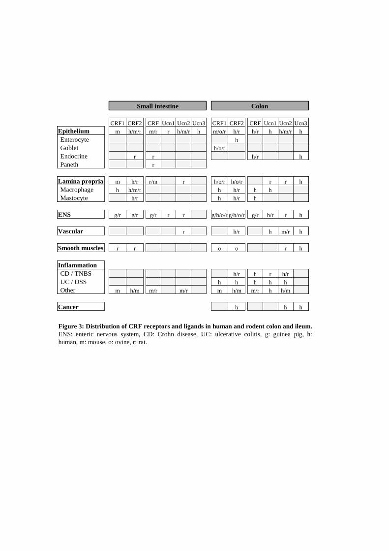
Figure 3: Distribution of CRF receptors and ligands in human and rodent colon and ileum. ENS: enteric nervous system, CD: Crohn disease, UC: ulcerative colitis, g: guinea pig, h: human, m: mouse, o: ovine, r: rat.
Small intestine Colon
CRF1 CRF2 CRF Ucn1 Ucn2 Ucn3 CRF1 CRF2 CRF Ucn1 Ucn2 Ucn3Epithelium m h/m/r m/r r h/m/r h m/o/r h/r h/r h h/m/r hEnterocyte hGoblet h/o/rEndocrine r r h/r hPaneth r
Lamina propria m h/r r/m r h/o/r h/o/r r r hMacrophage h h/m/r h h/r h hMastocyte h/r h h/r h
ENS g/r g/r g/r r r g/h/o/rg/h/o/r g/r h/r r h
Vascular r h/r h m/r h
Smooth muscles r r o o r h
InflammationCD / TNBS h/r h r h/rUC / DSS h h h h hOther m h/m m/r m/r m h/m m/r h h/m
Cancer h h h