Streptococcal toxins: role in pathogenesis and...
21
Microreview Streptococcal toxins: role in pathogenesis and disease Timothy C. Barnett, 1 Jason N. Cole, 1,2 Tania Rivera-Hernandez, 1 Anna Henningham, 1,2 James C. Paton, 3 Victor Nizet 2 and Mark J. Walker 1 * 1 Australian Infectious Diseases Research Centre and School of Chemistry and Molecular Biosciences, The University of Queensland, St Lucia, Queensland, Australia. 2 Department of Pediatrics and Skaggs School of Pharmacy and Pharmaceutical Sciences, University of California San Diego, La Jolla, California, USA. 3 Research Centre for Infectious Diseases, Department of Molecular and Cellular Biology, School of Biological Sciences, University of Adelaide, Adelaide, South Australia, Australia. Summary Group A Streptococcus (Streptococcus pyogenes), group B Streptococcus (Streptococcus agalactiae) and Streptococcus pneumoniae (pneumococcus) are host-adapted bacterial pathogens among the leading infectious causes of human morbidity and mortality. These microbes and related members of the genus Streptococcus produce an array of toxins that act against human cells or tissues, resulting in impaired immune responses and subversion of host physiological processes to benefit the invading microorganism. This toxin repertoire includes haemolysins, proteases, superantigens and other agents that ultimately enhance colonization and survival within the host and promote dissemination of the pathogen. Introduction The genus Streptococcus comprises several important species of human and animal pathogens, most specifically adapted to survive within a single host species. As host- adapted pathogens, streptococcal species have evolved distinctive repertoires of protein and non-protein toxins that play crucial roles in colonization, pathogenesis and dissemination. While there are examples of streptococcal toxins that are represented across species boundaries, many streptococcal toxins are species specific or even limited to certain clonal lineages within an individual species. A number of these toxins have found utility as vaccine antigens or as novel therapeutics. In this review, we divide our examination of streptococcal toxins into four groups: haemolysins, proteases, superantigens (SAgs) and miscellaneous toxins. The best-studied individual examples from each class from across the genus Streptococcus are used to illustrate the function of these toxin molecules and their contribution to the disease process. Haemolysins The complete lysis of red blood cells by streptococci, known as β-haemolysis, was first observed in 1895 and is embodied by the characteristic zone of clearing surround- ing bacterial colonies on the surface of blood agar medium (Ayers and Rupp, 1922; Molloy et al., 2011). This section summarizes our current understanding of streptococcal β-haemolysins/cytolysins (β-h/c) and their key role in pathogenesis (Table 1). Streptolysin S The potent membrane-active haemolysin streptolysin S (SLS) is secreted by 99% of all group A Streptococcus (GAS) isolates at stationary phase (Yoshino et al., 2010) and is related to the class I bacteriocin family of proteinaceous toxins and antimicrobial peptides (Nizet et al., 2000; Cotter et al., 2005). SLS belongs to the thiazole/oxazole-modified microcin class of natural prod- ucts, a family of diverse ribosomally produced peptides that are post-translationally modified to contain thiazole and (methyl)oxazole heterocycles from cysteine, threo- nine and serine residues (Mitchell et al., 2009; Melby et al., 2011). The activity of SLS is both temperature and concentration dependent and is principally responsible for the characteristic zone of β-haemolysis surrounding GAS colonies cultured on blood agar plates (Betschel et al., 1998; Nizet et al., 2000). SLS is encoded by the chromosomal SLS-associated gene (sag) locus, a con- served nine-gene operon comprised of contiguous genes sagA to sagI (Nizet et al., 2000). The mature SLS is a Received 29 June, 2015; revised 13 August, 2015; accepted 2 September, 2015. *For correspondence. E-mail mark.walker@uq. edu.au; Tel. (+61) 7 33461623; Fax (+61) 7 3365 4273. Cellular Microbiology (2015) doi:10.1111/cmi.12531 © 2015 John Wiley & Sons Ltd cellular microbiology
Transcript of Streptococcal toxins: role in pathogenesis and...
Microreview
Streptococcal toxins: role in pathogenesis and disease
Timothy C. Barnett,1 Jason N. Cole,1,2
Tania Rivera-Hernandez,1 Anna Henningham,1,2
James C. Paton,3 Victor Nizet2 and Mark J. Walker1*1Australian Infectious Diseases Research Centre andSchool of Chemistry and Molecular Biosciences, TheUniversity of Queensland, St Lucia, Queensland,Australia.2Department of Pediatrics and Skaggs School ofPharmacy and Pharmaceutical Sciences, University ofCalifornia San Diego, La Jolla, California, USA.3Research Centre for Infectious Diseases, Department ofMolecular and Cellular Biology, School of BiologicalSciences, University of Adelaide, Adelaide, SouthAustralia, Australia.
Summary
Group A Streptococcus (Streptococcus pyogenes),group B Streptococcus (Streptococcus agalactiae)and Streptococcus pneumoniae (pneumococcus)are host-adapted bacterial pathogens among theleading infectious causes of human morbidity andmortality. These microbes and related members ofthe genus Streptococcus produce an array oftoxins that act against human cells or tissues,resulting in impaired immune responses andsubversion of host physiological processes tobenefit the invading microorganism. This toxinrepertoire includes haemolysins, proteases,superantigens and other agents that ultimatelyenhance colonization and survival within the hostand promote dissemination of the pathogen.
Introduction
The genus Streptococcus comprises several importantspecies of human and animal pathogens, most specificallyadapted to survive within a single host species. As host-adapted pathogens, streptococcal species have evolveddistinctive repertoires of protein and non-protein toxins
that play crucial roles in colonization, pathogenesis anddissemination. While there are examples of streptococcaltoxins that are represented across species boundaries,many streptococcal toxins are species specific or evenlimited to certain clonal lineages within an individualspecies. A number of these toxins have found utility asvaccine antigens or as novel therapeutics. In this review,we divide our examination of streptococcal toxins into fourgroups: haemolysins, proteases, superantigens (SAgs)and miscellaneous toxins. The best-studied individualexamples from each class from across the genusStreptococcus are used to illustrate the function of thesetoxin molecules and their contribution to the diseaseprocess.
Haemolysins
The complete lysis of red blood cells by streptococci,known as β-haemolysis, was first observed in 1895 and isembodied by the characteristic zone of clearing surround-ing bacterial colonies on the surface of blood agar medium(Ayers and Rupp, 1922; Molloy et al., 2011). This sectionsummarizes our current understanding of streptococcalβ-haemolysins/cytolysins (β-h/c) and their key role inpathogenesis (Table 1).
Streptolysin S
The potent membrane-active haemolysin streptolysin S(SLS) is secreted by 99% of all group A Streptococcus(GAS) isolates at stationary phase (Yoshino et al., 2010)and is related to the class I bacteriocin family ofproteinaceous toxins and antimicrobial peptides (Nizetet al., 2000; Cotter et al., 2005). SLS belongs to thethiazole/oxazole-modified microcin class of natural prod-ucts, a family of diverse ribosomally produced peptidesthat are post-translationally modified to contain thiazoleand (methyl)oxazole heterocycles from cysteine, threo-nine and serine residues (Mitchell et al., 2009; Melbyet al., 2011). The activity of SLS is both temperature andconcentration dependent and is principally responsible forthe characteristic zone of β-haemolysis surrounding GAScolonies cultured on blood agar plates (Betschel et al.,1998; Nizet et al., 2000). SLS is encoded by thechromosomal SLS-associated gene (sag) locus, a con-served nine-gene operon comprised of contiguous genessagA to sagI (Nizet et al., 2000). The mature SLS is a
Received 29 June, 2015; revised 13 August, 2015; accepted 2September, 2015. *For correspondence. E-mail [email protected]; Tel. (+61) 7 33461623; Fax (+61) 7 3365 4273.
Cellular Microbiology (2015) doi:10.1111/cmi.12531
© 2015 John Wiley & Sons Ltd
cellular microbiology
~2.7 kDa oxygen-stable and broad-spectrum cytolytictoxin that forms hydrophilic pores in cholesterol-containingcytoplasmic membranes to induce irreversible osmoticlysis of host cells (Todd, 1938; Bernheimer, 1967;Ginsburg, 1999; Carr et al., 2001). The plasma membranedamage caused by pore-forming toxins induces signallingcascades in the host cell to promote membrane repair,metal ion homeostasis and a low-energy state wherebyprotein synthesis is arrested (Gonzalez et al., 2008, 2011).The sagA gene encodes pre-SLS, while the downstream
genes are required for post-translational modifications,heterocycle formation, processing and export of themature SLS exotoxin (Nizet et al., 2000; Nizet, 2002;Molloy et al., 2011). SLS enhances GAS pathogenicity by
lysing a broad spectrum of host cells including erythro-cytes (red blood cells), lymphocytes, neutrophils, plate-lets, sub-cellular organelles (e.g. lysosomes andmitochondria) and several other mammalian cell types(Ofek et al., 1970; Betschel et al., 1998), but not bacteriawith intact cell walls (Bernheimer, 1966). In a murinemodel of cutaneous infection, GAS strains lacking SLSactivity were less virulent than the wild-type (WT) isogenicparental strain (Betschel et al., 1998; Mitchell et al., 2009).Soft tissue damage in mice associated with bacterialproliferation, inflammation, vascular injury and formation ofnecrotic lesions also depends on the activity of SLS in GAS,group G Streptococcus (GGS) and Streptococcus iniae(Betschel et al., 1998; Limbago et al., 2000; Fuller et al.,
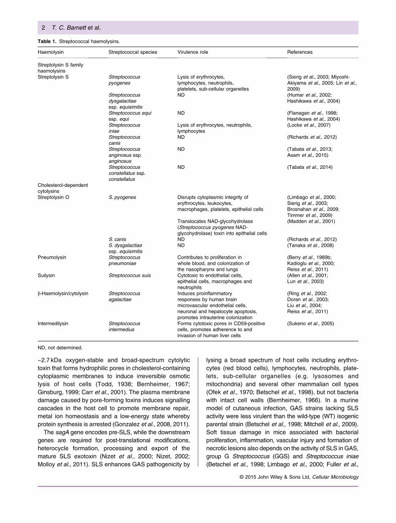
Table 1. Streptococcal haemolysins.
Haemolysin Streptococcal species Virulence role References
Streptolysin S familyhaemolysinsStreptolysin S Streptococcus
pyogenesLysis of erythrocytes,lymphocytes, neutrophils,platelets, sub-cellular organelles
(Sierig et al., 2003; Miyoshi-Akiyama et al., 2005; Lin et al.,2009)
Streptococcusdysgalactiaessp. equisimilis
ND (Humar et al., 2002;Hashikawa et al., 2004)
Streptococcus equissp. equi
ND (Flanagan et al., 1998;Hashikawa et al., 2004)
Streptococcusiniae
Lysis of erythrocytes, neutrophils,lymphocytes
(Locke et al., 2007)
Streptococcuscanis
ND (Richards et al., 2012)
Streptococcusanginosus ssp.anginosus
ND (Tabata et al., 2013;Asam et al., 2015)
Streptococcusconstellatus ssp.constellatus
ND (Tabata et al., 2014)
Cholesterol-dependentcytolysinsStreptolysin O S. pyogenes Disrupts cytoplasmic integrity of
erythrocytes, leukocytes,macrophages, platelets, epithelial cells
(Limbago et al., 2000;Sierig et al., 2003;Brosnahan et al., 2009;Timmer et al., 2009)
Translocates NAD-glycohydrolase(Streptococcus pyogenes NAD-glycohydrolase) toxin into epithelial cells
(Madden et al., 2001)
S. canis ND (Richards et al., 2012)S. dysgalactiaessp. equisimilis
ND (Tanaka et al., 2008)
Pneumolysin Streptococcuspneumoniae
Contributes to proliferation inwhole blood, and colonization ofthe nasopharynx and lungs
(Berry et al., 1989b;Kadioglu et al., 2000;Reiss et al., 2011)
Suilysin Streptococcus suis Cytotoxic to endothelial cells,epithelial cells, macrophages andneutrophils
(Allen et al., 2001;Lun et al., 2003)
β-Haemolysin/cytolysin Streptococcusagalactiae
Induces proinflammatoryresponses by human brainmicrovascular endothelial cells,neuronal and hepatocyte apoptosis,promotes intrauterine colonization
(Ring et al., 2002;Doran et al., 2003;Liu et al., 2004;Reiss et al., 2011)
Intermedilysin Streptococcusintermedius
Forms cytotoxic pores in CD59-positivecells, promotes adherence to andinvasion of human liver cells
(Sukeno et al., 2005)
ND, not determined.
2 T. C. Barnett et al.
© 2015 John Wiley & Sons Ltd, Cellular Microbiology
2002; Humar et al., 2002; Sierig et al., 2003). SLS mayinteract synergistically with other virulence factors (e.g. Mprotein and streptolysin O (SLO)) and host factors (e.g.neutrophil proteases and reactive oxygen species) to inducetissue necrosis and promote the development of necrotizingfasciitis in humans (Humar et al., 2002).
Recently, SLS and SLO have been shown to induceendoplasmic reticulum (ER) stress and an unfoldedprotein response (UPR) in host cells to reduce the surplusof unfolded proteins. The activation of UPR plays a keyrole in cellular defences against bacterial pore-formingtoxins and is an important downstream target of the p38mitogen-activated protein kinase pathway (Bischof et al.,2008), a central pathway of cellular immunity. In amechanism that is yet to be fully understood, UPR resultsin the transcriptional upregulation of asns, a geneencoding for asparagine synthetase, and the release ofasparagine into the extracellular environment. The extra-cellular asparagine sensed by GAS triggers a reduction inSLS and SLO transcription and stimulates bacterialgrowth (Baruch et al., 2014). Similarly, the pore-formingtoxin listeriolysin O (LLO) produced by Listeriamonocytogenes, a facultative intracellular bacterial path-ogen, induces ER stress and is required for UPRactivation (Pillich et al., 2012). LLO is activated withinthe acidic phagosome, allowing the bacterium to degradethe phagosome and escape to the cytosol (Dramsi andCossart, 2002).
Groups C and G streptococci streptolysin S homologues
Group C Streptococcus (GCS) and GGS comprise severalspecies of streptococci, with the significant humanpathogen Streptococcus dysgalactiae ssp. equisimilisresponsible for throat, skin and soft tissue infections andinvasive infections including endocarditis, bacteremia andtoxic shock. Nine-gene SLS-like loci are present in GCSand GGS. The SagA peptides of GCS and GGS share89% amino acid identity with SagA from GAS (Humaret al., 2002) and are responsible for the prototypical β-haemolytic phenotype on the surface of blood agar plates(Flanagan et al., 1998; Hashikawa et al., 2004). Heterol-ogous expression of GAS SagA in a non-haemolytic sagAmutant in GGS restored β-haemolytic activity on bloodagar (Humar et al., 2002).
Streptococcus iniae streptolysin S homologue
Streptococcus iniae is an emerging zoonotic pathogenresponsible for sporadic human infections through softtissue injuries suffered during the handling and prepara-tion of infected fish (Weinstein et al., 1997; Koh et al.,2009). The nine-gene S. iniae sag operon is 73%homologous to the sag operon from GAS and sharesthe same gene order (Fuller et al., 2002). Encoded by thesagA gene, the cytolysin of S. iniae shares 73% identity
with GAS SLS (Fuller et al., 2002; Locke et al., 2007) andlyses erythrocytes, neutrophils, lymphocytes and severaltissue culture cell lines. Heterologous expression of the S.iniae sagA gene in a non-haemolytic ΔsagA mutant ofserotype M49 GAS restored haemolytic activity (Fulleret al., 2002; Locke et al., 2007). S. iniae SLS promotesneither adherence and invasion of epithelial cells norresistance to opsonophagocytosis (Locke et al., 2007).However, a SagA-deficient mutant is highly attenuated forvirulence (Locke et al., 2007).
Intermedilysin
Streptococcus intermedius, Streptococcus constellatusand Streptococcus anginosus are members of theAnginosus group of streptococci (AGS) that colonize theoral cavity, upper respiratory, gastrointestinal and femalegenitourinary tracts (Whiley et al., 1992; Jacobs et al.,1995). AGS are opportunist human pathogens capable ofcausing liver and brain abscesses, dentoalveolar infec-tions and endocarditis (Jacobs et al., 1995). The secretedintermedilysin (ILY) of S. intermedius binds complementreceptor CD59 on human cells and forms cytotoxic pores,but only in the presence of sufficient levels of cholesterol(Nagamune et al., 1996; Farrand et al., 2008; Heuck et al.,2010; Johnson et al., 2013). ILY damages host tissuesand immune cells to promote bacterial survival anddissemination (Nagamune et al., 1996, 2000). ILY isconsidered to be a major virulence factor required foradherence to and invasion of human liver cells (Sukenoet al., 2005).
Streptolysin O
The oxygen-sensitive, 57 kDa thiol-activated SLO exotox-in is encoded by the highly conserved slo gene andsecreted by nearly all GAS isolates during exponentialand early stationary growth phases. SLO is a cholesterol-dependent cytolysin that disrupts the cytoplasmic mem-brane integrity of numerous eukaryotic cell types, includ-ing erythrocytes, leukocytes, macrophages, platelets,epithelial cells and various tissue culture cell lines(Limbago et al., 2000; Sierig et al., 2003). SLO contributesto GAS β-haemolysis under the surface of blood agarmedium and, in contrast to SLS, contributes negligibly toβ-haemolytic activity on the surface of blood agar (Molloyet al., 2011). The slo gene is co-transcribed with the ngagene encoding NAD-glycohydrolase (NADase), alsoknown as Streptococcus pyogenes NADase, which isactively translocated into the cytosol of human epithelialcells by SLO to deplete energy stores and promote hostcell injury (Madden et al., 2001; Bricker et al., 2005;Michos et al., 2006). Cathelicidin antimicrobial peptide LL-37 has been shown to upregulate the expression of sloand hyaluronan capsule, which promotes GAS resistanceto killing by human epithelial cells, neutrophils and
Streptococcal toxins role in pathogenesis and disease 3
© 2015 John Wiley & Sons Ltd, Cellular Microbiology
macrophages (Love et al., 2012). SLO blocks the clathrin-dependent pathway for GAS internalization throughdisruption of the keratinocyte cell surface (Logsdonet al., 2011) and induces keratinocyte apoptosis throughthe dysregulation of calcium signalling (Cywes Bentleyet al., 2005). SLO expression by intracellular GAS alsocontributes to the inhibition of dendritic cell maturation byinducing apoptosis (Cortes and Wessels, 2009). Uponmacrophage phagocytosis, SLO damages thephagolysosome membrane, preventing phagolysosomeacidification and resulting in the translocation of NADaseinto the macrophage cytosol (Bastiat-Sempe et al., 2014).SLO and NADase also inhibit the autophagic killing ofGAS in pharyngeal keratinocytes (O’Seaghdha andWessels, 2013). The toxic effects of NADase are furtherdiscussed below.In subcutaneous, intravenous and intraperitoneal mu-
rine models of invasive disease, slo-deficient GASmutants have decreased virulence compared with WTparental strains (Limbago et al., 2000; Ato et al., 2008;Timmer et al., 2009). A recent study showed that SLObinding to A549 epithelial cells does not require choles-terol, suggesting that cholesterol is not the membranereceptor for SLO (Mozola et al., 2014). In support of thishypothesis, SLO binding to a glycan on the surface ofhuman erythrocytes is essential for pore formation(Shewell et al., 2014), and the haemolytic activity ofSLO can be inhibited with a specific galactose-bindinglectin (Hasan et al., 2014). Similarly, an exposed F-typelectin domain at the N-terminus of lectinolysin (LLY), acholesterol-dependent cytolysin from Streptococcus mitis,promotes binding of LLY to fucose-rich sites on target cellmembranes (Farrand et al., 2008; Bouyain andGeisbrecht, 2012). SLO and an enzymatically inactivederivative are immunogenic and protective against GASchallenge in mouse vaccination models (Bensi et al.,2012; Chiarot et al., 2013).
Suilysin
Streptococcus suis is a pathogenof swine that is responsiblefor numerous diseases (meningitis, septicaemia and endo-carditis) and important economic losses to the porcineindustry worldwide (Fittipaldi et al., 2012). This speciesis also an emerging zoonotic agent of meningitis andstreptococcal toxic shock-like syndrome in humans. S.suis expresses the haemolysin suilysin (SLY), asecreted 54 kDa thiol-activated exotoxin that bindscholesterol and forms pores in eukaryotic cell mem-branes (Gottschalk et al., 1995; Palmer, 2001). It isclosely related to both GAS SLO and the pneumolysin(PLY) of Streptococcus pneumoniae and is cytotoxic toendothelial cells, epithelial cells, macrophages andneutrophils (Charland et al., 2000; Lalonde et al.,2000; Segura and Gottschalk, 2002; Chabot-Roy et al.,
2006). SLY allows S. suis to evade the innate immuneresponse by interfering with the complement cascadeand activating phagocytic cells to release proinflamma-tory cytokines (Lun et al., 2003; Segura et al., 2006).SLY-deficient mutants are attenuated for virulence in asystemic mouse infection model (Allen et al., 2001), butSLY is not required for full virulence in a piglet infectionmodel (Lun et al., 2003). Recently, SLY has beenshown to promote S. suis adherence to and invasion ofhuman HEp-2 epithelial cells (Seitz et al., 2013).
β-Haemolysin/cytolysin
Streptococcus agalactiae (group B Streptococcus, GBS)is the leading cause of meningitis, sepsis and pneumoniain human newborn infants and a significant agent ofinvasive infections among immunocompromised adults(e.g. diabetes and cancer patients) and pregnant womenworldwide (Farley, 2001). The β-h/c of GBS is a non-immunogenic oxygen-stable pore-forming cytolysin and amajor virulence factor expressed by most clinical isolates,with a predicted size of 78.3 kDa (Dal and Monteil, 1983;Nizet et al., 1996; Spellerberg, 2000). First described in1934 (Todd, 1934), cell surface-associated β-h/c isencoded by the cylE gene in the cyl locus, a unique12-gene operon involved in fatty acid biosynthesis(Pritzlaff et al., 2001) that is expressed by almost allstrains of GBS. CylE expression is invariably associatedwith the production of an orange to brick-red carotenoidpigment (Tapsall, 1987; Spellerberg et al., 2000) and isprimarily regulated by the two-component system covR/S(control of virulence) (Tapsall, 1987); some have recentlysuggested the pigment itself may convey the haemolyticactivity (Whidbey et al., 2013). CylE expression is requiredfor GBS survival in mouse and human blood ex vivo(Liu et al., 2004). Animal studies with WT and isogenicβ-h/c mutants demonstrate that haemolysin expressionhas proapoptotic, proinflammatory and cytotoxic effectsand is necessary for full GBS virulence in multiple in vivosystems, including mouse models of GBS arthritis (Pulitiet al., 2000), meningitis (Doran et al., 2003) andascending chorioamnionitis (Randis et al., 2014), a ratmodel of experimental GBS meningitis (Reiss et al.,2011), and rabbit models of GBS septicaemia (Ring et al.,2002) and pneumonia (Hensler et al., 2005).
Pneumolysin
Streptococcus pneumoniae (pneumococcus) is the caus-ative agent of pneumococcal pneumonia, meningitis,sepsis, otitis media and other less serious infections.PLY is a 53 kDa (471-amino-acid) (Walker et al., 1987)cholesterol-dependent pore-forming toxin with four func-tional domains (Mitchell and Dalziel, 2014). In a recentstudy, binding of PLY domain 4 to the sialyl LewisXglycolipid cellular receptor on the surface of human
4 T. C. Barnett et al.
© 2015 John Wiley & Sons Ltd, Cellular Microbiology
erythrocytes was shown to be an essential step beforemembrane insertion and pore formation (Shewell et al.,2014). PLY is a cytoplasmic thiol-activated toxin withcytolytic and complement-activating properties (Lucaset al., 2013). PLY is localized primarily to the cell wallcompartment in the absence of detectable cell lysis (Priceet al., 2012). However, unlike other cholesterol-bindingcytolysins, PLY lacks secretion signal sequences and isnot actively secreted into the extracellular milieu (Cassidyand O’Riordan, 2013). The cytolytic activity of PLY and itsrelease into the extracellular milieu are inhibited bybranched stem peptides in the peptidoglycan cell wall(Greene et al., 2015). PLY is released into the alveolarcompartment upon bacterial lysis induced by autolysis(Berry et al., 1989a, 1992) or by antibiotic treatment ofpneumococcal pneumonia patients (Anderson et al.,2007). However, in the absence of cell lysis, PLY isexported from the cytoplasm and attached to the cell wallby a yet-to-be-characterized mechanism (Price et al.,2012). PLY is directly toxic for a wide variety of host cellsand tissues and also elicits strong inflammatory responsesat the site of infection, triggering signalling via TLR4(Malley et al., 2003), as well as activation of the NLRP3inflammasome (McNeela et al., 2010). Circulating PLYalso induces myocardial injury in a mouse model ofinvasive pneumococcal disease and dose-dependentdamage to cardiomyocytes in vitro (Alhamdi et al.,2015). In pneumococcal meningitis, the majority of thedamage to the blood–brain barrier has been attributed toPLY (Zysk et al., 2001; Mitchell and Dalziel, 2014).Structural homology to the Fc region of immunoglobulinG (IgG) also allows PLY to activate the classicalcomplement pathway away from intact bacteria to depletehost serum complement levels and promote survival andspread (Paton et al., 1984; Mitchell et al., 1991; Rossjohnet al., 1998; Alcantara et al., 2001). Recently, PLY wasalso shown to contribute to the assembly of pneumococ-cal biofilms (Shak et al., 2013). PLY activates p38 in vitro(Ratner et al., 2006) and the NLRP3 inflammasome inmacrophages to stimulate the production of type Iinterferons following pneumococcal phagocytosis (Koppeet al., 2012). In murine infection models, PLY-deficientmutants have reduced proliferation in whole blood (Bentonet al., 1995), diminished capacity to colonize the naso-pharynx, induce less lung inflammation and neutrophilrecruitment and are rapidly cleared from the lung,compared with WT (Berry et al., 1989b; Kadioglu et al.,2000). Genetically inactivated PLY toxoids are immuno-genic and protective against lethal pneumococcal chal-lenge in mouse vaccination models (Paton et al., 1991;Alexander et al., 1994; Kirkham et al., 2006). Phase Iclinical trials demonstrated that PLY toxoid PlyD1 is safeand elicits functional neutralizing antibodies against thepneumococcus (Kamtchoua et al., 2013).
Proteases
Proteases (or peptidases) are enzymes that catalyse thehydrolysis of peptide bonds. The genus Streptococcuspossesses a wide array of proteases that have diversefunctions, including nutrient acquisition, protein maturationand quality control, and various host interactions. For thepurposes of this review, however, we will focus only onthose surface-exposed or secreted proteases that havedirect effects on pathogenesis through their activities onhost proteins and tissues.
Group A Streptococcus cysteine protease (SpeB)
The GAS cysteine protease SpeB is encoded in thegenomes of essentially all GAS strains (Bohach et al.,1988; Yu and Ferretti, 1991); homologous proteins, albeituncharacterized, are encoded in the genomes of theclosely related species Streptococcus didelphis, Strepto-coccus porcinus and Streptococcus pseudoporcinus.Expression of this protein is tightly regulated at thetranscriptional level, as well as post-transcriptionallythrough the maturation of the inactive zymogen to theactive mature enzyme (for a review, see Carroll andMusser, 2011). With diverse functions in pathogenesis,including direct action on host tissues as well as roles inthe maturation and surface display of other surface-exposed GAS proteins, SpeB is a veritable ‘Swiss armyknife’ of GAS biology.
SpeB is a broad-spectrum cysteine protease structurallyrelated to papain and has been shown to degradenumerous host proteins in vitro, including immunoglobu-lins, complement components, chemokines, cytokines,extracellular matrix proteins and numerous other hostproteins (for a review, see Nelson et al., 2011). However,despite these studies, it has recently been shown thatSpeB does not cleave immunoglobulins under physiolog-ically relevant conditions (Persson et al., 2013), and thus,the true in vivo substrate profile might be considerablysmaller. Multiple lines of evidence suggest that SpeB isimportant for GAS pathogenesis: passive immunization ofmice with anti-SpeB antibodies or a synthetic proteaseinhibitor protects against infection, and low-milligramamounts of SpeB are lethal when injected into mice(Bjorck et al., 1989; Kapur et al., 1994; Nelson et al.,2011). Furthermore, isogenic ΔspeB mutant GAS strainsare avirulent in mice following subcutaneous (Lukomskiet al., 1999; Cole et al., 2006; Terao et al., 2008) andintraperitoneal (Lukomski et al., 1997; Hollands et al.,2008) infection and exhibit decreased survival in humanwhole blood (Chiang-Ni et al., 2006) and serum (Honda-Ogawa et al., 2013).
Role in superficial infections. SpeB expression has astrong epidemiological association with isolates fromsuperficial disease (Ikebe et al., 2010; Cole et al., 2011).
Streptococcal toxins role in pathogenesis and disease 5
© 2015 John Wiley & Sons Ltd, Cellular Microbiology
While the exact role of this enzyme during superficial GASdisease was presumed to involve its degradation of hostimmune components, two recent reports have suggestedalternative or complementary functions. In the first report(Barnett et al., 2013), SpeB conferred the ability of GASstrains to replicate in the cytosol of infected epithelial cells.SpeB was shown to degrade the host proteins that directintracellular bacteria to the autophagy pathway, a hostsystem for degrading cytosolic components in lysosomesthat constitutes an important immune defence againstintracellular bacteria. Consequently, SpeB-expressingGAS strains are able to evade this pathway and replicatein the cytosol of infected cells. In a separate study, SpeBwas shown to promote the translocation of GAS acrossepithelial barriers by degrading occludin and E-cadherin(Sumitomo et al., 2013). Thus, SpeB may aid in theestablishment of an epithelial replicative niche anddissemination of GAS into deeper tissues.
Role in invasive disease. Epidemiological studies havedemonstrated that SpeB expression is inversely related todisease severity (Kansal et al., 2000; Ikebe et al., 2010).Loss of SpeB expression, through the accumulation ofmutations in the regulatory genes covR/S and to a lesserextent ropB, leads to the abolishment of speB expression.This genetic switch has multiple consequences that leadto a hypervirulent invasive state: loss of SpeB productionspares several GAS virulence factors (e.g. M protein,various SAgs and streptokinase) from proteolytic degra-dation, and covR/S mutations lead to the up-regulation ofmultiple virulence factors required for the invasive diseasephenotype (for a review, see Cole et al., 2011).While SpeB expression is strongly linked to GAS
isolates from superficial disease, immunohistochemicalanalysis has shown that human tissues from necrotizingfasciitis cases are strongly positive for SpeB, suggestingthat SpeB is elaborated during the establishment orprogression of this disease (Johansson et al., 2008).Similarly, SpeB is required for full virulence in invasiveinfection models in mice (Lukomski et al., 1999; Coleet al., 2006; Hollands et al., 2008; Olsen et al., 2010) andnonhuman primate animal models (Olsen et al., 2010).While these results seemingly contradict other studies thathave clearly demonstrated a genetic switch from a SpeB-positive phenotype to a SpeB-negative phenotype duringinvasive disease (Aziz et al., 2004; Cole et al., 2006;Sumby et al., 2006; Walker et al., 2007), it is possible thata mixed population of SpeB-positive and SpeB-negativebacteria contribute to the overall pathology of GASinvasive disease and penetration into deeper tissues(Cole et al., 2006).In addition to regulation through covR/S and ropB
mutations, it has also been recently demonstrated thatSpeB activity can be inhibited by the divalent cations of
zinc and copper (Chella Krishnan et al., 2014). While thephysiological consequence of this inhibition is currentlyunknown, it was postulated that reversible inhibition ofSpeB activity may reversibly preserve critical virulencefactors required during certain stages of the infectiousprocess.
Immune-modulating proteases
C5a peptidase. C5a peptidase (ScpA), is a serineendopeptidase that specifically cleaves and inactivatesthe C5a complement factor and has been implicated toplay a role in inhibiting the recruitment of phagocytes tothe infectious site (Ji et al., 1996; Collin and Olsen, 2003).While recombinant ScpA is very potent in inhibitingphagocyte chemotaxis in vitro, the effects of ΔscpAmutations in mice infections in vivo are much lessdramatic (Ji et al., 1996), possibly as a result of alternativechemoattractants, such as interleukin (IL)-8, and GASproteases that inhibit them, such as SpyCEP (below). C5apeptidase is a cell wall-anchored enzyme but can bereleased from the surface of GAS as a functionally activeenzyme by SpeB and can thus inactivate C5a at somedistance from the bacterium (Berge and Bjorck, 1995).C5a peptidase is expressed by strains of GAS (ScpA;Chen and Cleary, 1990), GBS (ScpB; Cleary et al., 1992)and Streptococcus equi ssp. zooepidemicus (ScpZ; Weiet al., 2013) and is encoded in the genomes of severalother streptococcal pathogens, including S. dysgalactiae,S. iniae, Streptococcus sanguinis, S.mitis and Strepto-coccus canis (GGS). In addition to its endopeptidaseactivity, the C5a peptidase proteins from GAS, GBS andS. equi ssp. zooepidemicus have also been suggested tofunction as an invasin (Cheng et al. , 2002b;Purushothaman et al., 2004; Wei et al., 2013). Vaccinationagainst ScpA is protective for GAS (Park and Cleary,2005), GBS (Cheng et al., 2002a; Santillan et al., 2008)and S. equi ssp. zooepidemicus (Wei et al., 2013).
SpyCEP. Group A Streptococcus SpyCEP is a subtilisin-like serine protease that can cleave human CXCchemokines. In its mature form, SpyCEP exists as adimer composed of two subunits (30 and 150 kDa)generated by intramolecular autocatalytic cleavage(Zingaretti et al., 2010). Substrates include granulocytechemotactic peptide-2 (CXCL6), growth-relatedoncogene-α, β, γ (CXCL1, 2, 3), neutrophil-activatingpeptide-78 (CXCL5), GRB2-related adapter protein 2and IL-8 (CXCL8), which correspond to the murine CXCchemokines MIP-2 and KC (Hidalgo-Grass et al., 2004;Sumby et al., 2008; Chiappini et al., 2012). As a result ofthis activity, SpyCEP impairs the recruitment of neutro-phils, monocytes and eosinophils to the site of infection(Zinkernagel et al., 2008; Chiappini et al., 2012). SpyCEPalso promotes resistance to neutrophil killing by reducing
6 T. C. Barnett et al.
© 2015 John Wiley & Sons Ltd, Cellular Microbiology
the production of neutrophil extracellular traps (Zinkerna-gel et al., 2008). SpyCEP-producing GAS strains are morevirulent in murine models of invasive disease, andSpyCEP production is up-regulated in human invasiveGAS isolates (Edwards et al., 2005; Hidalgo-Grass et al.,2006; Zinkernagel et al., 2008; Turner et al., 2009a).Furthermore, immunization with SpyCEP protects miceagainst GAS nasopharyngeal (Alam et al., 2013), intra-muscular and intranasal infection (Turner et al., 2009b)and intramuscular infection with S. equi (Turner et al.,2009b), demonstrating that a vaccine based on SpyCEPmay provide cross-protection against multiple streptococ-cal species. While SpyCEP is cell wall anchored, it can beshed into the supernatants of bacterial cultures duringstationary-phase growth (Chiappini et al., 2012), presum-ably as a result of protease processing. In addition tothese protease activities, SpyCEP also promotes theuptake of GAS into endothelial, but not epithelial, cells(Kaur et al., 2010). Homologous proteases have also beencharacterized in S. iniae (Zinkernagel et al., 2008) and S.equi (Turner et al., 2009b) and are present in the genomesof several other Streptococcus species (S. dysgalactiae,S. canis, S. didelphis, Streptococcus phocae, S. porcinus,S. pseudoporcinus and Streptococcus thermophilus).
IdeS. IdeS (also known as Mac) is a 35 kDa secretedcysteine protease produced by strains of GAS thatspecifically cleaves between the two glycine residues inpositions 236 and 237 in the lower hinge region of the IgGheavy chain, resulting in the separation of the Fc and Fabfragments (von Pawel-Rammingen et al., 2002; Vincentset al., 2004; von Pawel-Rammingen, 2012). As ahomologue of CD11b, IdeS has been proposed to alsoinhibit phagocytosis by inhibiting Fc receptor (CD16)recognition of IgG and/or complement deposition (Leiet al., 2001). This is hypothesized to prevent therecognition of antibody-opsonized bacteria by Fc recep-tors of immune cells and by the complement system (vonPawel-Rammingen et al., 2002). Protease activity isconsiderably higher against soluble IgG and IgG boundin an antigen-specific manner at the Fab region than toIgG bound non-specifically to GAS M protein at the Fcregion, suggesting that this enzyme has evolved to allowGAS to resist Ig-mediated phagocytosis and cytotoxicitywhile still allowing non-immune IgG interactions, which arebelieved to contribute to GAS pathogenesis (Su et al.,2011). However, IdeS was recently found to not beessential for phagocyte resistance or mouse virulence,and thus, the exact role of this protein in human diseaseremains to be identified (Okumura et al., 2013). Thehuman protease inhibitor cystatin C can act as a cofactorfor IdeS and greatly increase enzymatic activity (Vincentset al., 2008). All GAS strains examined possess one oftwo identified variants of IdeS (IdeS and Mac-2), with
considerable amino acid sequence divergence in themiddle third of the proteins but with indistinguishableenzymatic activity, with the exception of the Mac-2 proteinfrom M28 strains that displays only weak endopeptidaseactivity (Lei et al., 2001; von Pawel-Rammingen et al.,2002; Soderberg et al., 2008; von Pawel-Rammingen,2012). IdeS homologues that cleave IgG have also beenidentified in several other Streptococcus species, includ-ing S. equi ssp. equi and S. equi ssp. zooepidemicus(Lannergard and Guss, 2006; Hulting et al., 2009).Additionally, an IdeS homologue that cleaves porcineIgM has recently been found in S. suis (Seele et al., 2013).
Zinc metalloproteases
Streptococcus pneumoniae strains encode various com-binations of four zinc metalloproteases: IgA1 protease,ZmpB, ZmpC and ZmpD. An unusual characteristic of allof these proteins is the presence of an LPXTG cell wall-anchoring motif near their N-terminus (Bek-Thomsenet al., 2012), which in the case of IgA1 protease hasbeen shown to be important for proper localization andenzymatic function (Bender and Weiser, 2006).
IgA1 protease specifically cleaves the IgA1 hinge region,resulting in separation of the Fc and Fab fragments (Seniorand Woof, 2005a,b). This cleavage abrogates the protec-tive effects of IgA1 in mediating complement-dependentkilling of pneumococci by phagocytes (Fasching et al.,2007; Janoff et al., 2014) and promotes the adherence ofthis bacterium to respiratory epithelial cells (Weiser et al.,2003). It is likely that IgA1 protease has additional roles invirulence, as mutants have reduced virulence in mice yetmurine IgA1 is not a substrate for this enzyme (Chiavoliniet al., 2003; Janoff et al., 2014). Homologous IgA1proteases have been characterized in S. suis, which isimportant for virulence in pigs (Zhang et al., 2011), and S.sanguinis (Gilbert et al., 1988). IgA1 protease is encodedby all strains of S. pneumoniae, Streptococcuspseudopneumoniae, Streptococcus oralis and S.sanguinis and is variably present in S.mitis and Strepto-coccus infantis (Bek-Thomsen et al., 2012).
In contrast to IgA1 protease, the ZmpC protease hasmultiple targets and is an important virulence factor inexperimental pneumonia (Oggioni et al., 2003). ZmpC hasbeen shown to activate human matrix metalloprotease 9, ahost zinc protease involved in neutrophil migration andwound repair of the respiratory epithelium (Oggioni et al.,2003). ZmpC inhibits neutrophil flux by degrading the N-terminal region of P-selectin glycoprotein 1 (Surewaardet al., 2013). In addition, ZmpC induces specific epithelialcell shedding of both mucin 16, a vital defence barrier ofocular epithelial cells (Govindarajan et al., 2012; Menonand Govindarajan, 2013), and syndecan-1, a surfaceproteoglycan with diverse roles in cell–cell and cell–matrixbinding, cell signalling and cytoskeletal organization
Streptococcal toxins role in pathogenesis and disease 7
© 2015 John Wiley & Sons Ltd, Cellular Microbiology
(Chen et al., 2007). Genes encoding ZmpC are variablypresent in S. pneumoniae, S. pseudopneumoniae, S.mitis,S. oralis and Streptococcus gordonii and present in allstrains of S. sanguinis (Bek-Thomsen et al., 2012).In contrast to IgA1 protease and ZmpC, ZmpB and
ZmpD have not been well characterized, and theirsubstrates have not been identified. ZmpB induces tumournecrosis factor-α production in the respiratory tract, and azmpB mutant was attenuated in pneumonia andsepticaemia models of infection (Blue et al., 2003;Chiavolini et al., 2003). Genes encoding ZmpB are presentin all S. pneumoniae strains as well as several otherStreptococcus species, including S. pseudopneumoniae,S.mitis, S. oralis, S. sanguinis, S. gordonii, Streptococcusvestibularis and Streptococcus salivarius. Genesencoding ZmpD are sporadically distributed among S.pneumoniae, S. pseudopneumoniae and S.mitis (Bek-Thomsen et al., 2012).
SspA
Strains of S. suis encode a cell wall-anchored subtilisin-like serine protease termed SspA, which is important fordisease in pigs (Bonifait et al., 2010; Hu et al., 2010).SspA cleaves fibrinogen, preventing subsequent fibrinformation by thrombin. Purified SspA protease is toxicwhen added to brain microvascular endothelial cells(Bonifait et al., 2011b) and induces a proinflammatoryresponse in macrophages through a non-proteolyticmechanism (Bonifait and Grenier, 2011a). Homologousgenes are present in the genomes of several otherStreptococcus species, including S. thermophilus, S.
canis, S. agalactiae, S. dysgalactiae, S. gordonii, S.mitis,S. sanguinis and S. intermedius.
Superantigens
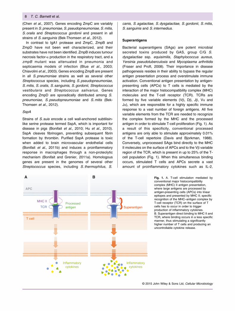
Bacterial superantigens (SAgs) are potent microbialsecreted toxins produced by GAS, group C/G S.dysgalactiae ssp. equisimilis, Staphylococcus aureus,Yersinia pseudotuberculosis and Mycoplasma arthritidis(Fraser and Proft, 2008). Their importance in diseasepathogenesis resides in their ability to bypass the regularantigen presentation process and overstimulate immuneactivation. Conventional antigen presentation by antigen-presenting cells (APCs) to T cells is mediated by theinteraction of the major histocompatibility complex (MHC)molecules and the T-cell receptor (TCR). TCRs areformed by five variable elements (Vβ, Dβ, Jβ, Vα andJα), which are responsible for a highly specific immuneresponse to a vast number of foreign antigens. All fivevariable elements from the TCR are needed to recognizethe complex formed by the MHC and the processedantigen in order to stimulate T-cell proliferation (Fig. 1). Asa result of this specificity, conventional processedantigens are only able to stimulate approximately 0.01%of the T-cell repertoire (Davis and Bjorkman, 1988).Conversely, unprocessed SAgs bind directly to the MHCII molecules on the surface of APCs and to the Vβ variableregion of the TCR, which is present in up to 25% of the T-cell population (Fig. 1). When this simultaneous bindingoccurs, stimulated T cells and APCs secrete a vastamount of proinflammatory cytokines such as IL-2,
Fig. 1. A. T-cell stimulation mediated byconventional major histocompatibilitycomplex (MHC) II antigen presentation,where large antigens are processed byantigen-presenting cells (APCs) into linearepitopes and presented by MHC II; specificrecognition of the MHC–antigen complex byT-cell receptor (TCR) on the surface of Tcells has to occur in order to triggerproduction of inflammatory cytokines.B. Superantigen direct binding to MHC II andTCR, where binding occurs in a less specificmanner, thus stimulating a significantlyhigher number of T cells and producing anuncontrollable cytokine release.
8 T. C. Barnett et al.
© 2015 John Wiley & Sons Ltd, Cellular Microbiology
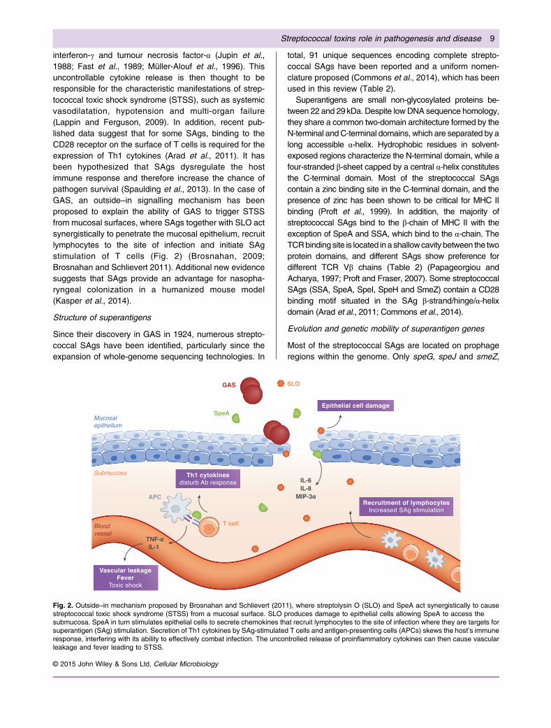
interferon-γ and tumour necrosis factor-α (Jupin et al.,1988; Fast et al., 1989; Müller-Alouf et al., 1996). Thisuncontrollable cytokine release is then thought to beresponsible for the characteristic manifestations of strep-tococcal toxic shock syndrome (STSS), such as systemicvasodilatation, hypotension and multi-organ failure(Lappin and Ferguson, 2009). In addition, recent pub-lished data suggest that for some SAgs, binding to theCD28 receptor on the surface of T cells is required for theexpression of Th1 cytokines (Arad et al., 2011). It hasbeen hypothesized that SAgs dysregulate the hostimmune response and therefore increase the chance ofpathogen survival (Spaulding et al., 2013). In the case ofGAS, an outside–in signalling mechanism has beenproposed to explain the ability of GAS to trigger STSSfrom mucosal surfaces, where SAgs together with SLO actsynergistically to penetrate the mucosal epithelium, recruitlymphocytes to the site of infection and initiate SAgstimulation of T cells (Fig. 2) (Brosnahan, 2009;Brosnahan and Schlievert 2011). Additional new evidencesuggests that SAgs provide an advantage for nasopha-ryngeal colonization in a humanized mouse model(Kasper et al., 2014).
Structure of superantigens
Since their discovery in GAS in 1924, numerous strepto-coccal SAgs have been identified, particularly since theexpansion of whole-genome sequencing technologies. In
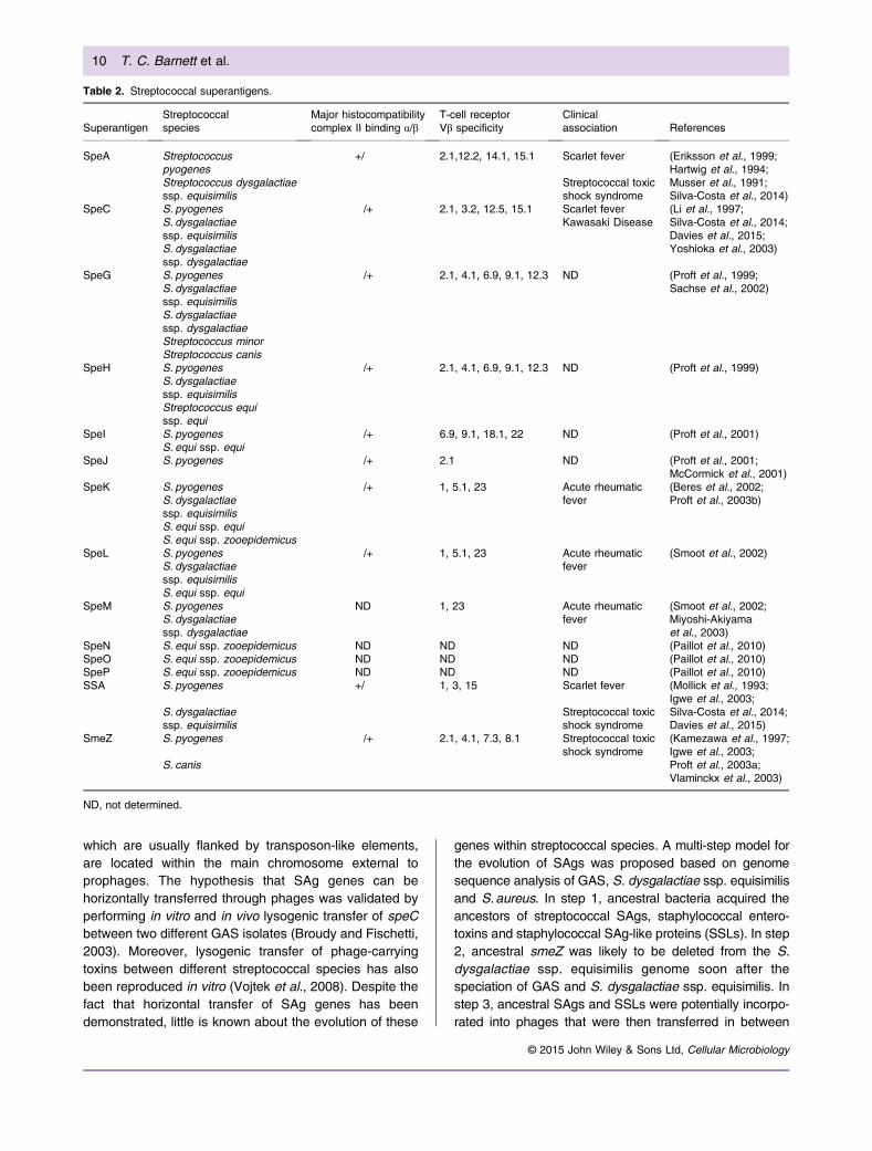
total, 91 unique sequences encoding complete strepto-coccal SAgs have been reported and a uniform nomen-clature proposed (Commons et al., 2014), which has beenused in this review (Table 2).
Superantigens are small non-glycosylated proteins be-tween 22 and 29kDa. Despite low DNA sequence homology,they share a common two-domain architecture formed by theN-terminal andC-terminal domains, which are separated by along accessible α-helix. Hydrophobic residues in solvent-exposed regions characterize the N-terminal domain, while afour-stranded β-sheet capped by a central α-helix constitutesthe C-terminal domain. Most of the streptococcal SAgscontain a zinc binding site in the C-terminal domain, and thepresence of zinc has been shown to be critical for MHC IIbinding (Proft et al., 1999). In addition, the majority ofstreptococcal SAgs bind to the β-chain of MHC II with theexception of SpeA and SSA, which bind to the α-chain. TheTCRbindingsite is located ina shallowcavity between the twoprotein domains, and different SAgs show preference fordifferent TCR Vβ chains (Table 2) (Papageorgiou andAcharya, 1997; Proft and Fraser, 2007). Some streptococcalSAgs (SSA, SpeA, SpeI, SpeH and SmeZ) contain a CD28binding motif situated in the SAg β-strand/hinge/α-helixdomain (Arad et al., 2011; Commons et al., 2014).
Evolution and genetic mobility of superantigen genes
Most of the streptococcal SAgs are located on prophageregions within the genome. Only speG, speJ and smeZ,
Fig. 2. Outside–in mechanism proposed by Brosnahan and Schlievert (2011), where streptolysin O (SLO) and SpeA act synergistically to causestreptococcal toxic shock syndrome (STSS) from a mucosal surface. SLO produces damage to epithelial cells allowing SpeA to access thesubmucosa. SpeA in turn stimulates epithelial cells to secrete chemokines that recruit lymphocytes to the site of infection where they are targets forsuperantigen (SAg) stimulation. Secretion of Th1 cytokines by SAg-stimulated T cells and antigen-presenting cells (APCs) skews the host’s immuneresponse, interfering with its ability to effectively combat infection. The uncontrolled release of proinflammatory cytokines can then cause vascularleakage and fever leading to STSS.
Streptococcal toxins role in pathogenesis and disease 9
© 2015 John Wiley & Sons Ltd, Cellular Microbiology
which are usually flanked by transposon-like elements,are located within the main chromosome external toprophages. The hypothesis that SAg genes can behorizontally transferred through phages was validated byperforming in vitro and in vivo lysogenic transfer of speCbetween two different GAS isolates (Broudy and Fischetti,2003). Moreover, lysogenic transfer of phage-carryingtoxins between different streptococcal species has alsobeen reproduced in vitro (Vojtek et al., 2008). Despite thefact that horizontal transfer of SAg genes has beendemonstrated, little is known about the evolution of these
genes within streptococcal species. A multi-step model forthe evolution of SAgs was proposed based on genomesequence analysis of GAS, S. dysgalactiae ssp. equisimilisand S.aureus. In step 1, ancestral bacteria acquired theancestors of streptococcal SAgs, staphylococcal entero-toxins and staphylococcal SAg-like proteins (SSLs). In step2, ancestral smeZ was likely to be deleted from the S.dysgalactiae ssp. equisimilis genome soon after thespeciation of GAS and S. dysgalactiae ssp. equisimilis. Instep 3, ancestral SAgs and SSLs were potentially incorpo-rated into phages that were then transferred in between
Table 2. Streptococcal superantigens.
SuperantigenStreptococcalspecies
Major histocompatibilitycomplex II binding α/β
T-cell receptorVβ specificity
Clinicalassociation References
SpeA Streptococcuspyogenes
+/! 2.1,12.2, 14.1, 15.1 Scarlet fever (Eriksson et al., 1999;Hartwig et al., 1994;
Streptococcus dysgalactiaessp. equisimilis
Streptococcal toxicshock syndrome
Musser et al., 1991;Silva-Costa et al., 2014)
SpeC S. pyogenes !/+ 2.1, 3.2, 12.5, 15.1 Scarlet fever (Li et al., 1997;S. dysgalactiaessp. equisimilis
Kawasaki Disease Silva-Costa et al., 2014;Davies et al., 2015;
S. dysgalactiaessp. dysgalactiae
Yoshioka et al., 2003)
SpeG S. pyogenes !/+ 2.1, 4.1, 6.9, 9.1, 12.3 ND (Proft et al., 1999;S. dysgalactiaessp. equisimilis
Sachse et al., 2002)
S. dysgalactiaessp. dysgalactiaeStreptococcus minorStreptococcus canis
SpeH S. pyogenes !/+ 2.1, 4.1, 6.9, 9.1, 12.3 ND (Proft et al., 1999)S. dysgalactiaessp. equisimilisStreptococcus equissp. equi
SpeI S. pyogenes !/+ 6.9, 9.1, 18.1, 22 ND (Proft et al., 2001)S. equi ssp. equi
SpeJ S. pyogenes !/+ 2.1 ND (Proft et al., 2001;McCormick et al., 2001)
SpeK S. pyogenes !/+ 1, 5.1, 23 Acute rheumaticfever
(Beres et al., 2002;S. dysgalactiaessp. equisimilis
Proft et al., 2003b)
S. equi ssp. equiS. equi ssp. zooepidemicus
SpeL S. pyogenes !/+ 1, 5.1, 23 Acute rheumaticfever
(Smoot et al., 2002)S. dysgalactiaessp. equisimilisS. equi ssp. equi
SpeM S. pyogenes ND 1, 23 Acute rheumaticfever
(Smoot et al., 2002;S. dysgalactiaessp. dysgalactiae
Miyoshi-Akiyamaet al., 2003)
SpeN S. equi ssp. zooepidemicus ND ND ND (Paillot et al., 2010)SpeO S. equi ssp. zooepidemicus ND ND ND (Paillot et al., 2010)SpeP S. equi ssp. zooepidemicus ND ND ND (Paillot et al., 2010)SSA S. pyogenes +/! 1, 3, 15 Scarlet fever (Mollick et al., 1993;
Igwe et al., 2003;S. dysgalactiaessp. equisimilis
Streptococcal toxicshock syndrome
Silva-Costa et al., 2014;Davies et al., 2015)
SmeZ S. pyogenes !/+ 2.1, 4.1, 7.3, 8.1 Streptococcal toxicshock syndrome
(Kamezawa et al., 1997;Igwe et al., 2003;
S. canis Proft et al., 2003a;Vlaminckx et al., 2003)
ND, not determined.
10 T. C. Barnett et al.
© 2015 John Wiley & Sons Ltd, Cellular Microbiology
bacterial strains. In the final step, staphylococcal SAgs(potential ancestors of streptococcal SAgs such as SSA andSpeA) were then horizontally transferred to Streptococcusbacteria (Okumura et al., 2012).
Streptococcal superantigens and disease correlation
Clinical manifestations of disease have been linked to thepresence of SAgs in streptococcal pathogens. Cases ofscarlet fever have been correlated with the carriage oracquisition of ssa, speA and speC in different studies(Silva-Costa et al., 2014; Davies et al., 2015). Additionally,speA and ssa, together with speK, and smeZ have beenassociated with invasive disease and STSS (Erikssonet al., 1999; Chatellier et al., 2000; Beres et al., 2002;Ikebe et al., 2002). A strong correlation has beenobserved between M18 isolates expressing speL andspeM and acute rheumatic fever (Smoot et al., 2002). M89isolates harbouring the speK gene have also beenassociated with acute rheumatic fever (Proft et al., 2003b).
A controversial correlation between speC and Kawasakidisease, a sporadic childhood inflammatory arthritis thatcan affect the coronary vessels (Shulman and Rowley,2015), has been proposed (Abe et al., 1992; Curtis et al.,1995); however, contradicting epidemiological studiesrender a direct correlation questionable (Yoshioka et al.,2003). Research on the causative agents of Kawasakidisease is still ongoing. In addition, speC has also beencorrelated to psoriasis in susceptible individuals (Leunget al., 1993; Lewis et al., 1993); however, the evidence isnot strong enough for a direct correlation, and the role ofSAgs in disease manifestations remains unclear (Traverset al., 1999; Thomssen et al., 2000).
Other Toxins
In addition to producing toxic SAgs, haemolysins andproteases, streptococcal species produce a number ofother toxins including proteins, enzymes and polysaccha-rides such as Christie Atkins Munch-Petersen (CAMP)factor, GBS toxin, adenosine diphosphate (ADP)-ribosyltransferase and NADase.
Christie Atkins Munch-Petersen factor
The CAMP reaction was initially described as the lysis oferythrocytes during a synergistic interaction of CAMPfactor produced by GBS with the β-toxin of S. aureus(Christie et al., 1944). Historically, this reaction has beenused for the clinical identification of GBS. The CAMPfactor is best characterized in GBS, where the toxin isencoded by the cfb gene (Podbielski et al., 1994). Otherstreptococcal species, including groups A, B, C, G, M, P,R and U, have also been reported to produce CAMPfactor; and in GAS, CAMP factor is encoded by the cfagene (Gase et al., 1999). The CAMP factor of GBS binds
glycophosphatidylinositol-anchored proteins (Lang et al.,2007) and subsequently functions as a pore-forming toxin,likely requiring self-oligomerization for activity (Lang andPalmer, 2003). Previously, GBS CAMP factor was calledprotein B, following reports that it could bind the Fc regionof IgG and IgM from several mammalian species (Jurgenset al., 1987). However, more recent studies could notdetect a non-immune binding association between CAMPfactor and human IgG, leading to the suggestion that thename protein B may be inappropriate (El-Huneidi et al.,2007). CAMP factor of GBS was determined to be non-pathogenic following administration to mice (Jurgenset al., 1987). Furthermore, CAMP factor was not essentialfor systemic virulence of GBS (Hensler et al., 2008),suggesting that CAMP factor (in the absence of S. aureusβ-toxin) does not play a direct role in GBS pathogenesis.Recent investigations have uncovered a novel CAMPfactor in GBS, designated CAMP factor II, that has beenproposed to have spread to other streptococci (Strepto-coccus uberis, S. dysgalactiae, S. dysgalactiae ssp.equisimilis and Streptococcus bovis) via integrative andconjugative elements (Chuzeville et al., 2012).
Group B Streptococcus toxin (CM101)
Group B Streptococcus toxin, also designated CM101, isa polysaccharide exotoxin that binds to embryonicreceptors expressed in the developing lung of theneonate, resulting in a strong inflammatory response(Wamil et al., 1997). GBS toxin also binds to tumourneovasculature in adults and for this reason has beenevaluated as an anti-cancer therapeutic. In a phase Iclinical trial administering GBS toxin as an anti-neovascularization agent in human cancer therapy (Har-ris, 1997), 5/15 patients exhibited tumour reduction orstabilization, and sera from all patients had elevatedsoluble E-selectin, indicative of tumour neovasculatureendothelial engagement in an inflammatory process(Wamil et al., 1997). Historically, GBS toxin was purifiedfrom culture media of GBS isolates from neonates whohad died because of GBS infection. GBS toxin has alsobeen identified in plasma, urine and cerebrospinal fluidfrom infants with GBS disease and could be used as anadditional tool to diagnose GBS infection in infants, whichcan sometimes prove difficult to diagnose (Sundell et al.,2000).
Adenosine diphosphate-ribosyltransferase (SpyA)
Adenosine diphosphate-ribosyltransferases covalentlytransfer ADP-ribose from NAD+ to eukaryotic proteins.SpyA is a surface-exposed membrane protein of GAS thathas been described as a C3-like ADP-ribosyltransferase(Korotkova et al., 2012). SpyA has been documented tomodify cytoskeletal proteins including vimentin, tropomy-osin and actin, and when expressed in HeLa cells
Streptococcal toxins role in pathogenesis and disease 11
© 2015 John Wiley & Sons Ltd, Cellular Microbiology
following transfection, SpyAactivity resulted in a loss of actinmicrofilaments (Coye and Collins, 2004) and inhibitedvimentin polymerization, resulting in the collapse of thevimentin cytoskeleton (Icenogle et al., 2012). In a mousesubcutaneous infection model, the ΔspyAmutant generatedsmaller lesions and had higher levels of mRNA encodingCXCL1 and CCL2 (both neutrophil and macrophagechemoattractants) and vimentin, compared with WT (Hoffet al., 2011). Vimentin plays many roles in the cell includingthe organization of cellular architecture (Ivaska et al., 2007),and the loss of vimentin functionality can impair woundhealing (Eckes et al., 2000). The findings of Hoff et al (2011)suggest that SpyA delays wound healing in the subcutane-ous infectionmodel. In amouse intravenous infectionmodel,the ΔspyA mutant caused higher mortality with impairedbacterial clearance, and in vivo, the mutant was resistant tokilling by macrophages (Lin et al., 2015). Lin and colleaguesdetermined that SpyA triggers pyroptosis, a caspase-1-dependent inflammasome in macrophages, resulting inmacrophage cell death and the release of proinflammatorycytokine IL-1β. This innate defence programme triggered inresponse to SpyA dramatically enhances clearance of GASand restricts bacterial growth, attenuating disease progres-sion.
NAD-glycohydrolase (Streptococcus pyogenes NADaseor NADase)
NAD-glycohydrolase (also known as S. pyogenes NADaseor Nga) is encoded by the nga gene, which is found in thesameoperon as slo. SLOandNADase act synergistically totrigger cytotoxicity. NADase is translocated through porescreated by SLO and delivered into the host cell cytoplasmiccompartment by a process termed cytolysin-mediatedtranslocation (Madden et al., 2001). NADase possessesboth ADP-ribosyl cyclase activity and cADPR hydrolaseactivity (Karasawa et al., 1995), and once inside the cell,NADase produces the potent second messenger cyclicADP-ribose. Cytotoxicity mediated by the enzymatic actionof NADase may be a consequence of the depletion of hostcell energy stores (e.g. intracellular NAD+) (Michos et al.,2006). NADase has been shown to act within humanpharyngeal keratinocytes, resulting in enhanced cellmembrane injury, inhibition of bacterial internalization andinduction of apoptosis (Bricker et al., 2002). SLO stimulatesxenophagy in these cells, and the co-expression of SLOand NADase results in prolonged intracellular survival ofGAS and prevents maturation of GAS-containingautophagosomes (O’Seaghdha and Wessels, 2013). Inaddition, SLO and NADase mediate GAS intracellularsurvival and cytotoxicity within macrophages, subsequent-ly enabl ing persistent infect ion by preventingphagolysosome acidification (Bastiat-Sempe et al., 2014).Recent studies have identified NADase-inactive variantsthat have emerged with polymorphism at multiple residues
(Chandrasekaran et al., 2013). This analysis revealed anNADase-independent cytotoxic activity that was retainedby the enzymatically inactive variants, suggesting anotherdomain of NADase can mediate cytotoxicity. NADase alsocontributes to virulence in two mouse skin tissue infectionmodels and a model of septicaemia, suggesting NADaseplays an important role in establishing infection in the host(Bricker et al., 2005; Tatsuno et al., 2010).
Conclusion
While several toxins are found across streptococcalspecies boundaries, each of these species expresses aspecific repertoire of toxic molecules that target specificaspects of host immunity and physiology. Indeed, manyclones within individual species express distinct toxinprofiles. These toxins play crucial roles within the host–pathogen interaction, allowing the pathogen to colonize,proliferate and disseminate. Several of these toxins havedemonstrated utility as candidate vaccine antigens, ininactive forms, and in several instances, the specificity oftoxin action has been experimentally utilized in thedevelopment of novel therapeutics, such as anti-canceragents. Deepening of our knowledge of the mode of actionof these toxin molecules will aid in our efforts to preventdisease caused in humans and animals by the variousstreptococcal pathogens.
Acknowledgements
The authors gratefully acknowledge the support of the NationalHealth and Medical Research Council of Australia and theNational Institutes of Health of the USA.
References
Abe, J., Kotzin, B.L., Jujo, K., Melish, M.E., Glode, M.P.,Kohsaka, T., and Leung, D.Y.M. (1992) Selectiveexpansion of T cells expressing T-cell receptor variableregions Vβ2 and Vβ8 in Kawasaki disease. Proc Natl AcadSci U S A 89: 4066–4070.
Alam, F.M., Bateman, C., Turner, C.E., Wiles, S., andSriskandan, S. (2013) Non-invasive monitoring of Strepto-coccus pyogenes vaccine efficacy using biophotonicimaging. PLoS One 8: e82123.
Alcantara, R.B., Preheim, L.C., and Gentry-Nielsen, M.J.(2001) Pneumolysin-induced complement depletion duringexperimental pneumococcal bacteremia. Infect Immun 69:3569–3575.
Alexander, J.E., Lock, R.A., Peeters, C.C.A.M., Poolman, J.T., Andrew, P.W., Mitchell, T.J., et al. (1994) Immunizationof mice with pneumolysin toxoid confers a significantdegree of protection against at least nine serotypes ofStreptococcus pneumoniae. Infect Immun 62: 5683–5688.
Alhamdi, Y., Neill, D.R., Abrams, S.T., Malak, H.A., Yahya,R., Barrett-Jolley, R., et al. (2015) Circulating pneumolysinis a potent inducer of cardiac injury during pneumococcalinfection. PLoS Pathog 11: e1004836.
12 T. C. Barnett et al.
© 2015 John Wiley & Sons Ltd, Cellular Microbiology
Allen, A.G., Bolitho, S., Lindsay, H., Khan, S., Bryant, C.,Norton, P., et al. (2001) Generation and characterization ofa defined mutant of Streptococcus suis lacking suilysin.Infect Immun 69: 2732–2735.
Anderson, R., Steel, H.C., Cockeran, R., von Gottberg, A., deGouveia, L., Klugman, K.P., et al. (2007) Comparison of theeffects of macrolides, amoxicillin, ceftriaxone, doxycycline,tobramycin and fluoroquinolones, on the production ofpneumolysin by Streptococcus pneumoniae in vitro. JAntimicrob Chemother 60: 1155–1158.
Arad, G., Levy, R., Nasie, I., Hillman, D., Rotfogel, Z., Barash,U., et al. (2011) Binding of superantigen toxins into theCD28 homodimer interface is essential for induction ofcytokine genes that mediate lethal shock. PLoS Biol 9:e1001149.
Asam, D., Mauerer, S., and Spellerberg, B. (2015) Strepto-lysin S of Streptococcus anginosus exhibits broad-rangehemolytic activity. Med Microbiol Immun 204: 227–237.
Ato, M., Ikebe, T., Kawabata, H., Takemori, T., andWatanabe, H. (2008) Incompetence of neutrophils toinvasive group A Streptococcus is attributed to inductionof plural virulence factors by dysfunction of a regulator.PLoS One 3: e3455.
Ayers, S.H., and Rupp, P. (1922) Differentiation of hemolyticstreptococci from human and bovine sources by thehydrolysis of sodium hippurate. J Infect Dis 30: 388–399.
Aziz, R.K., Pabst, M.J., Jeng, A., Kansal, R., Low, D.E., Nizet,V., and Kotb, M. (2004) Invasive M1T1 group A Strepto-coccus undergoes a phase-shift in vivo to preventproteolytic degradation of multiple virulence factors bySpeB. Mol Microbiol 51: 123–134.
Barnett, T.C., Liebl, D., Seymour, L.M., Gillen, C.M., Lim, J.Y.,Larock, C.N., et al. (2013) The globally disseminated M1T1clone of group A Streptococcus evades autophagy forintracellular replication. Cell Host Microbe 14: 675–682.
Baruch, M., Hertzog, B.B., Ravins, M., Anand, A., Cheng, C.Y., Biswas, D., et al. (2014) Induction of endoplasmicreticulum stress and unfolded protein response constitutesa pathogenic strategy of group A Streptococcus. Front CellInfect Microbiol 4: 105.
Bastiat-Sempe, B., Love, J.F., Lomayesva, N., and Wessels,M.R. (2014) Streptolysin O and NAD-glycohydrolaseprevent phagolysosome acidification and promotegroup a Streptococcus survival in macrophages. mBio 5:e01690–01614.
Bek-Thomsen,M.,Poulsen,K.,andKilian,M.(2012)Occurrenceand evolution of the paralogous zinc metalloproteases IgA1protease, ZmpB, ZmpC, and ZmpD in Streptococcuspneumoniae and related commensal species. mBio 3:e00303–e00312.
Bender, M.H., and Weiser, J.N. (2006) The atypical amino-terminal LPNTG-containing domain of the pneumococcalhuman IgA1-specific protease is required for proper enzymelocalization and function. Mol Microbiol 61: 526–543.
Bensi, G., Mora, M., Tuscano, G., Biagini, M., Chiarot, E.,Bombaci, M., et al. (2012) Multi high-throughput approachfor highly selective identification of vaccine candidates: thegroup A Streptococcus case. Mol Cell Proteomics 11:M111.015693.
Benton, K.A., Everson, M.P., and Briles, D.E. (1995) Apneumolysin-negative mutant of Streptococcus pneumoniae
causes chronic bacteremia rather than acute sepsis in mice.Infect Immun 63: 448–455.
Beres, S.B., Sylva, G.L., Barbian, K.D., Lei, B., Hoff, J.S.,Mammarella, N.D., et al. (2002) Genome sequence of aserotype M3 strain of group A Streptococcus: phage-encoded toxins, the high-virulence phenotype, and cloneemergence. Proc Natl Acad Sci U S A 99: 10078–10083.
Berge, A., and Bjorck, L. (1995) Streptococcal cysteineproteinase releases biologically active fragments of strep-tococcal surface proteins. J Biol Chem 270: 9862–9867.
Bernheimer, A.W. (1966) Disruption of wall-less bacteria bystreptococcal and staphylococcal toxins. J Bacteriol 91:1677–1680.
Bernheimer, A.W. (1967) Physical behavior of streptolysin S.J Bacteriol 93: 2024–2025.
Berry, A.M., Lock, R.A., Hansman, D., and Paton, J.C.(1989a) Contribution of autolysin to virulence of Strepto-coccus pneumoniae. Infect Immun 57: 2324–2330.
Berry, A.M., Yother, J., Briles, D.E., Hansman, D., and Paton,J.C. (1989b) Reduced virulence of a defined pneumolysin-negative mutant of Streptococcus pneumoniae. InfectImmun 57: 2037–2042.
Berry, A.M., Paton, J.C., and Hansman, D. (1992) Effect ofinsertional inactivation of the genes encoding pneumolysinand autolysin on the virulence of Streptococcuspneumoniae type 3. Microb Pathog 12: 87–93.
Betschel, S.D., Borgia, S.M., Barg, N.L., Low, D.E., and DeAzavedo, J.C. (1998) Reduced virulence of group Astreptococcal Tn916 mutants that do not produce strepto-lysin S. Infect Immun 66: 1671–1679.
Bischof, L.J., Kao, C.Y., Los, F.C., Gonzalez, M.R., Shen, Z.,Briggs, S.P., et al. (2008) Activation of the unfolded proteinresponse is required for defenses against bacterial pore-forming toxin in vivo. PLoS Pathog 4: e1000176.
Bjorck, L., Akesson, P., Bohus, M., Trojnar, J., Abrahamson,M., Olafsson, I., and Grubb, A. (1989) Bacterial growthblocked by a synthetic peptide based on the structure of ahuman proteinase inhibitor. Nature 337: 385–386.
Blue, C.E., Paterson, G.K., Kerr, A.R., Berge,M., Claverys, J.P.,and Mitchell, T.J. (2003) ZmpB, a novel virulence factor ofStreptococcus pneumoniae that induces tumor necrosisfactor alpha production in the respiratory tract. Infect Immun71: 4925–4935.
Bohach, G.A., Hauser, A.R., and Schlievert, P.M. (1988)Cloning of the gene, speB, for streptococcal pyrogenicexotoxin type B in Escherichia coli. Infect Immun 56:1665–1667.
Bonifait, L., and Grenier, D. (2011a) The SspA subtilisin-likeprotease of Streptococcus suis triggers a pro-inflammatoryresponse in macrophages through a non-proteolytic mech-anism. BMC Microbiol 11: 47.
Bonifait, L., de la Cruz Dominguez-Punaro, M., Vaillancourt,K., Bart, C., Slater, J., Frenette, M., et al. (2010) The cellenvelope subtilisin-like proteinase is a virulence determi-nant for Streptococcus suis. BMC Microbiol 10: 42.
Bonifait, L., Vaillancourt, K., Gottschalk, M., Frenette, M., andGrenier, D. (2011b) Purification and characterization of thesubtilisin-like protease of Streptococcus suis that contrib-utes to its virulence. Vet Microbiol 148: 333–340.
Bouyain, S., and Geisbrecht, B.V. (2012) Host glycanrecognition by a pore forming toxin. Structure 20: 197–198.
Streptococcal toxins role in pathogenesis and disease 13
© 2015 John Wiley & Sons Ltd, Cellular Microbiology
Bricker, A.L., Cywes, C., Ashbaugh, C.D., and Wessels, M.R.(2002) NAD+-glycohydrolase acts as an intracellular toxinto enhance the extracellular survival of group A strepto-cocci. Mol Microbiol 44: 257–269.
Bricker, A.L., Carey, V.J., and Wessels, M.R. (2005) Role ofNADase in virulence in experimental invasive group Astreptococcal infection. Infect Immun 73: 6562–6566.
Brosnahan, A.J., and Schlievert, P.M. (2011) Gram-positivebacterial superantigen outside–in signaling causes toxicshock syndrome. FEBS J 278: 4649–4667.
Brosnahan, A.J., Mantz, M.J., Squier, C.A., Peterson, M.L., andSchlievert, P.M. (2009) Cytolysins augment superantigenpenetration of stratified mucosa. J Immunol 182: 2364–2373.
Broudy, T.B., and Fischetti, V.A. (2003) In vivo lysogenicconversion of Tox(!) Streptococcus pyogenes to Tox(+)with lysogenic streptococci or free phage. Infect Immun 71:3782–3786.
Carr, A., Sledjeski, D.D., Podbielski, A., Boyle, M.D., andKreikemeyer, B. (2001) Similarities between complement-mediated and streptolysin S-mediated hemolysis. J BiolChem 276: 41790–41796.
Carroll, R.K., and Musser, J.M. (2011) From transcription toactivation: how group A streptococcus, the flesh-eatingpathogen, regulates SpeB cysteine protease production.Mol Microbiol 81: 588–601.
Cassidy, S.K., and O’Riordan, M.X. (2013) More than a pore:the cellular response to cholesterol-dependent cytolysins.Toxins 5: 618–636.
Chabot-Roy, G., Willson, P., Segura, M., Lacouture, S., andGottschalk, M. (2006) Phagocytosis and killing of Strepto-coccus suisby porcine neutrophils.MicrobPathog41: 21–32.
Chandrasekaran, S., Ghosh, J., Port, G.C., Koh, E.I., andCaparon, M.G. (2013) Analysis of polymorphic residuesreveals distinct enzymatic and cytotoxic activities of theStreptococcus pyogenes NAD+ glycohydrolase. J BiolChem 288: 20064–20075.
Charland, N., Nizet, V., Rubens, C.E., Kim, K.S., Lacouture,S., and Gottschalk, M. (2000) Streptococcus suis serotype2 interactions with human brain microvascular endothelialcells. Infect Immun 68: 637–643.
Chatellier, S., Ihendyane,N., Kansal,R.G., Khambaty, F., Basma,H., Norrby-Teglund, A., et al. (2000) Genetic relatedness andsuperantigen expression in group A Streptococcus serotypeM1 isolates from patients with severe and nonsevere invasivediseases. Infect Immun 68: 3523–3534.
Chella Krishnan, K., Mukundan, S., Landero Figueroa, J.A.,Caruso, J.A., and Kotb, M. (2014) Metal-mediated modu-lation of streptococcal cysteine protease activity and itsbiological implications. Infect Immun 82: 2992–3001.
Chen, C.C., and Cleary, P.P. (1990) Complete nucleotidesequence of the streptococcal C5a peptidase gene ofStreptococcus pyogenes. J Biol Chem 265: 3161–3167.
Chen, Y., Hayashida, A., Bennett, A.E., Hollingshead, S.K.,and Park, P.W. (2007) Streptococcus pneumoniae shedssyndecan-1 ectodomains through ZmpC, a metalloprotein-ase virulence factor. J Biol Chem 282: 159–167.
Cheng, Q., Debol, S., Lam, H., Eby, R., Edwards, L.,Matsuka, Y., et al. (2002a) Immunization with C5apeptidase or peptidase-type III polysaccharide conjugatevaccines enhances clearance of group B streptococci fromlungs of infected mice. Infect Immun 70: 6409–6415.
Cheng, Q., Stafslien, D., Purushothaman, S.S., and Cleary,P. (2002b) The group B streptococcal C5a peptidase isboth a specific protease and an invasin. Infect Immun 70:2408–2413.
Chiang-Ni, C., Wang, C.H., Tsai, P.J., Chuang, W.J., Lin, Y.S.,Lin, M.T., et al. (2006) Streptococcal pyrogenic exotoxin Bcauses mitochondria damage to polymorphonuclear cellspreventing phagocytosis of group A Streptococcus. MedMicrobiol Immun 195: 55–63.
Chiappini, N., Seubert, A., Telford, J.L., Grandi, G., Serruto,D., Margarit, I., and Janulczyk, R. (2012) Streptococcuspyogenes SpyCEP influences host–pathogen interactionsduring infection in a murine air pouch model. PLoS One 7:e40411.
Chiarot, E., Faralla, C., Chiappini, N., Tuscano, G., Falugi, F.,Gambellini, G., et al. (2013) Targeted amino acid substi-tutions impair streptolysin O toxicity and group A Strepto-coccus virulence. mBio 4: e00387–00312.
Chiavolini, D., Memmi, G., Maggi, T., Iannelli, F., Pozzi, G.,and Oggioni, M.R. (2003) The three extra-cellular zincmetalloproteinases of Streptococcus pneumoniae have adifferent impact on virulence in mice. BMC Microbiol 3: 14.
Christie, R., Atkins, N.E., and Munch-Petersen, E. (1944) Anote on a lytic phenomenon shown by group B streptococci.Aust J Exp Biol 22: 197–200.
Chuzeville, S., Puymege, A., Madec, J.Y., Haenni, M., andPayot, S. (2012) Characterization of a new CAMP factorcarried by an integrative and conjugative element inStreptococcus agalactiae and spreading in streptococci.PLoS One 7: e48918.
Cleary, P.P., Handley, J., Suvorov, A.N., Podbielski, A., andFerrieri, P. (1992) Similarity between the group B and Astreptococcal C5a peptidase genes. Infect Immun 60:4239–4244.
Cole, J.N., McArthur, J.D., McKay, F.C., Sanderson-Smith, M.L.,Cork, A.J., Ranson, M., et al. (2006) Trigger for group AstreptococcalM1T1 invasivedisease.FASEBJ20: 1745–1747.
Cole, J.N., Barnett, T.C., Nizet, V., and Walker, M.J. (2011)Molecular insight into invasive group A streptococcaldisease. Nature Rev Microbiol 9: 724–736.
Collin, M., and Olsen, A. (2003) Extracellular enzymes withimmunomodulating activities: variations on a theme inStreptococcus pyogenes. Infect Immun 71: 2983–2992.
Commons, R.J., Smeesters, P.R., Proft, T., Fraser, J.D.,Robins-Browne, R., and Curtis, N. (2014) Streptococcalsuperantigens: categorization and clinical associations.Trends Mol Med 20: 48–62.
Cortes, G., and Wessels, M.R. (2009) Inhibition of dendriticcell maturation by group A Streptococcus. J Infect Dis 200:1152–1161.
Cotter, P.D., Hill, C., and Ross, R.P. (2005) Bacteriocins:developing innate immunity for food. Nature Rev Microbiol3: 777–788.
Coye, L.H., and Collins, C.M. (2004) Identification of SpyA, anovel ADP-ribosyltransferase of Streptococcus pyogenes.Mol Microbiol 54: 89–98.
Curtis, N., Zheng, R., Lamb, J.R., and Levin, M. (1995)Evidence for a superantigen mediated process in Kawasakidisease. Arch Dis Child 72: 308–311.
Cywes Bentley, C., Hakansson, A., Christianson, J., andWessels, M.R. (2005) Extracellular group A Streptococcus
14 T. C. Barnett et al.
© 2015 John Wiley & Sons Ltd, Cellular Microbiology
induces keratinocyte apoptosis by dysregulating calciumsignalling. Cell Microbiol 7: 945–955.
Dal, M.-C., and Monteil, H. (1983) Hemolysin produced bygroup B Streptococcus agalactiae. FEMS Microbiol Lett 16:89–94.
Davies, M.R., Holden, M.T., Coupland, P., Chen, J.H.,Venturini, C., Barnett, T.C., et al. (2015) Emergence ofscarlet fever Streptococcus pyogenes emm12 clones inHong Kong is associated with toxin acquisition andmultidrug resistance. Nat Genet 47: 84–87.
Davis, M.M., and Bjorkman, P.J. (1988) T-cell antigen receptorgenes and T-cell recognition. Nature 334: 395–402.
Doran, K.S., Liu, G.Y., and Nizet, V. (2003) Group Bstreptococcal beta-hemolysin/cytolysin activates neutrophilsignaling pathways in brain endothelium and contributes todevelopment of meningitis. J Clin Invest 112: 736–744.
Dramsi, S., and Cossart, P. (2002) Listeriolysin O: a genuinecytolysin optimized for an intracellular parasite. J Cell Biol156: 943–946.
Eckes, B., Colucci-Guyon, E., Smola, H., Nodder, S., Babinet,C., Krieg, T., and Martin, P. (2000) Impaired wound healingin embryonic and adult mice lacking vimentin. J Cell Sci113(Pt 13): 2455–2462.
Edwards, R.J., Taylor, G.W., Ferguson, M., Murray, S.,Rendell, N., Wrigley, A., et al. (2005) Specific C-terminalcleavage and inactivation of interleukin-8 by invasivedisease isolates of Streptococcus pyogenes. J Infect Dis192: 783–790.
El-Huneidi, W., Mui, R., Zhang, T.H., and Palmer, M. (2007)Streptococcus agalactiae CAMP factor/protein B does notbind to human IgG. Med Microbiol Immun 196: 73–77.
Eriksson, B.K., Andersson, J., Holm, S.E., and Norgren, M.(1999) Invasive group A streptococcal infections: T1M1isolates expressing pyrogenic exotoxins A and B incombination with selective lack of toxin-neutralizing anti-bodies are associated with increased risk of streptococcaltoxic shock syndrome. J Infect Dis 180: 410–418.
Farley, M.M. (2001) Group B streptococcal disease innonpregnant adults. Clin Infect Dis 33: 556–561.
Farrand, S., Hotze, E., Friese, P., Hollingshead, S.K., Smith,D.F., Cummings, R.D., et al. (2008) Characterization of astreptococcal cholesterol-dependent cytolysin with a lewisy and b specific lectin domain. Biochemistry 47:7097–7107.
Fasching, C.E., Grossman, T., Corthesy, B., Plaut, A.G.,Weiser, J.N., and Janoff, E.N. (2007) Impact of themolecular form of immunoglobulin A on functional activityin defense against Streptococcus pneumoniae. InfectImmun 75: 1801–1810.
Fast, D.J., Schlievert, P.M., and Nelson, R.D. (1989) Toxicshock syndrome-associated staphylococcal and strepto-coccal pyrogenic toxins are potent inducers of tumornecrosis factor production. Infect Immun 57: 291–294.
Fittipaldi, N., Segura, M., Grenier, D., and Gottschalk, M.(2012) Virulence factors involved in the pathogenesis of theinfection caused by the swine pathogen and zoonotic agentStreptococcus suis. Future Microbiol 7: 259–279.
Flanagan, J., Collin, N., Timoney, J., Mitchell, T., Mumford, J.A., and Chanter, N. (1998) Characterization of thehaemolytic activity of Streptococcus equi. Microb Pathog24: 211–221.
Fraser, J.D., and Proft, T. (2008) The bacterial superantigenand superantigen-like proteins. Immunol Rev 225: 226–243.
Fuller, J.D., Camus, A.C., Duncan, C.L., Nizet, V., Bast, D.J.,Thune, R.L., et al. (2002) Identification of a streptolysin S-associated gene cluster and its role in the pathogenesis ofStreptococcus iniae disease. Infect Immun 70: 5730–5739.
Gase, K., Ferretti, J.J., Primeaux, C., and McShan, W.M.(1999) Identification, cloning, and expression of the CAMPfactor gene (cfa) of group A streptococci. Infect Immun 67:4725–4731.
Gilbert, J.V., Plaut, A.G., Fishman, Y., and Wright, A. (1988)Cloning of the gene encoding streptococcal immunoglob-ulin A protease and its expression in Escherichia coli. InfectImmun 56: 1961–1966.
Ginsburg, I. (1999) Is streptolysin S of group A streptococci avirulence factor? APMIS 107: 1051–1059.
Gonzalez, M.R., Bischofberger, M., Pernot, L., van der Goot,F.G., and Freche, B. (2008) Bacterial pore-forming toxins:the (w)hole story? Cell Mol Life Sci 65: 493–507.
Gonzalez, M.R., Bischofberger, M., Freche, B., Ho, S.,Parton, R.G., and van der Goot, F.G. (2011) Pore-formingtoxins induce multiple cellular responses promoting surviv-al. Cell Microbiol 13: 1026–1043.
Gottschalk, M.G., Lacouture, S., and Dubreuil, J.D. (1995)Characterization of Streptococcus suis capsular type 2haemolysin. Microbiology 141: 189–195.
Govindarajan, B., Menon, B.B., Spurr-Michaud, S., Rastogi,K., Gilmore, M.S., Argueso, P., and Gipson, I.K. (2012) Ametalloproteinase secreted by Streptococcus pneumoniaeremoves membrane mucin MUC16 from the epithelialglycocalyx barrier. PLoS One 7: e32418.
Greene, N.G., Narciso, A.R., Filipe, S.R., andCamilli, A. (2015)Peptidoglycan branched stem peptides contribute to Strep-tococcus pneumoniae virulence by inhibiting pneumolysinrelease. PLoS Pathog 11: e1004996.
Harris, A.L. (1997) Clinical trials of anti-vascular agent groupB Streptococcus toxin (CM101). Angiogenesis 1: 36–37.
Hartwig, U.F., Gerlach, D., and Fleischer, B. (1994) Majorhistocompatibility complex class II binding site for strepto-coccal pyrogenic (erythrogenic) toxin A. Med MicrobiolImmun 183: 257–264.
Hasan, I., Watanabe, M., Ishizaki, N., Sugita-Konishi, Y.,Kawakami, Y., Suzuki, J., et al. (2014) A galactose-bindinglectin isolated from Aplysia kurodai (sea hare) eggs inhibitsstreptolysin-inducedhemolysis.Molecules19: 13990–14003.
Hashikawa, S., Iinuma, Y., Furushita, M., Ohkura, T., Nada,T., Torii, K., et al. (2004) Characterization of group C and Gstreptococcal strains that cause streptococcal toxic shocksyndrome. J Clin Microbiol 42: 186–192.
Hensler, M.E., Liu, G.Y., Sobczak, S., Benirschke, K., Nizet,V., and Heldt, G.P. (2005) Virulence role of group BStreptococcus beta-hemolysin/cytolysin in a neonatalrabbit model of early-onset pulmonary infection. J InfectDis 191: 1287–1291.
Hensler, M.E., Quach, D., Hsieh, C.J., Doran, K.S., and Nizet,V. (2008) CAMP factor is not essential for systemicvirulence of group B Streptococcus. Microb Pathog 44:84–88.
Heuck, A.P., Moe, P.C., and Johnson, B.B. (2010) Thecholesterol-dependent cytolysin family of Gram-positivebacterial toxins. Subcell Biochem 51: 551–577.
Streptococcal toxins role in pathogenesis and disease 15
© 2015 John Wiley & Sons Ltd, Cellular Microbiology
Hidalgo-Grass, C., Dan-Goor, M., Maly, A., Eran, Y., Kwinn,L.A., Nizet, V., et al. (2004) Effect of a bacterial pheromonepeptide on host chemokine degradation in group Astreptococcal necrotising soft-tissue infections. Lancet363: 696–703.
Hidalgo-Grass,C.,Mishalian, I., Dan-Goor,M., Belotserkovsky, I.,Eran, Y., Nizet, V., et al. (2006) A streptococcal protease thatdegrades CXC chemokines and impairs bacterial clearancefrom infected tissues. EMBO J 25: 4628–4637.
Hoff, J.S., DeWald, M., Moseley, S.L., Collins, C.M., and Voyich,J.M. (2011) SpyA, a C3-like ADP-ribosyltransferase, contrib-utes to virulence in a mouse subcutaneous model ofStreptococcus pyogenes infection. Infect Immun 79:2404–2411.
Hollands, A., Aziz, R.K., Kansal, R., Kotb, M., Nizet, V., andWalker, M.J. (2008) A naturally occurring mutation in ropBsuppresses SpeB expression and reduces M1T1 group Astreptococcal systemic virulence. PLoS One 3: e4102.
Honda-Ogawa, M., Ogawa, T., Terao, Y., Sumitomo, T.,Nakata, M., Ikebe, K., et al. (2013) Cysteine proteinasefrom Streptococcus pyogenes enables evasion of innateimmunity via degradation of complement factors. J BiolChem 288: 15854–15864.
Hu, Q., Liu, P., Yu, Z., Zhao, G., Li, J., Teng, L., et al. (2010)Identification of a cell wall-associated subtilisin-like serineprotease involved in the pathogenesis of Streptococcussuis serotype 2. Microb Pathog 48: 103–109.
Hulting, G., Flock, M., Frykberg, L., Lannergard, J., Flock, J.I.,and Guss, B. (2009) Two novel IgG endopeptidases ofStreptococcus equi. FEMS Microbiol Lett 298: 44–50.
Humar, D., Datta, V., Bast, D.J., Beall, B., De Azavedo, J.C., andNizet, V. (2002) Streptolysin S and necrotising infectionsproduced by group G Streptococcus. Lancet 359: 124–129.
Icenogle, L.M., Hengel, S.M., Coye, L.H., Streifel, A., Collins,C.M., Goodlett, D.R., and Moseley, S.L. (2012) Molecularand biological characterization of streptococcal SpyA-mediated ADP-ribosylation of intermediate filament proteinvimentin. J Biol Chem 287: 21481–21491.
Igwe, E.I., Shewmaker, P.L., Facklam, R.R., Farley, M.M., vanBeneden,C., andBeall, B. (2003) Identificationof superantigengenes speM, ssa, and smeZ in invasive strains of beta-hemolytic groupCandGstreptococci recovered from humans.FEMS Microbiol Lett 229: 259–264.
Ikebe, T., Wada, A., Inagaki, Y., Sugama, K., Suzuki, R.,Tanaka, D., et al. (2002) Dissemination of the phage-associated novel superantigen gene speL in recentinvasive and noninvasive Streptococcus pyogenes M3/T3isolates in Japan. Infect Immun 70: 3227–3233.
Ikebe, T., Ato, M., Matsumura, T., Hasegawa, H., Sata, T.,Kobayashi, K., and Watanabe, H. (2010) Highly frequentmutations in negative regulators of multiple virulence genesin group A streptococcal toxic shock syndrome isolates.PLoS Pathog 6: e1000832.
Ivaska, J., Pallari, H.M., Nevo, J., and Eriksson, J.E. (2007)Novel functions of vimentin in cell adhesion, migration, andsignaling. Exp Cell Res 313: 2050–2062.
Jacobs, J.A., Pietersen, H.G., Stobberingh, E.E., andSoeters, P.B. (1995) Streptococcus anginosus, Strepto-coccus constellatus and Streptococcus intermedius. Clin-ical relevance, hemolytic and serologic characteristics. AmJ Clin Path 104: 547–553.
Janoff, E.N., Rubins, J.B., Fasching, C., Charboneau, D.,Rahkola, J.T., Plaut, A.G., and Weiser, J.N. (2014)Pneumococcal IgA1 protease subverts specific protectionby human IgA1. Mucosal Immunol 7: 249–256.
Ji, Y., McLandsborough, L., Kondagunta, A., and Cleary, P.P.(1996) C5a peptidase alters clearance and trafficking ofgroup A streptococci by infected mice. Infect Immun 64:503–510.
Johansson, L., Thulin, P., Sendi, P., Hertzen, E., Linder, A.,Akesson, P., et al. (2008) Cathelicidin LL-37 in severeStreptococcus pyogenes soft tissue infections in humans.Infect Immun 76: 3399–3404.
Johnson, S., Brooks, N.J., Smith, R.A., Lea, S.M., andBubeck, D. (2013) Structural basis for recognition of thepore-forming toxin intermedilysin by human complementreceptor CD59. Cell Rep 3: 1369–1377.
Jupin, C., Anderson, S., Damais, C., Alouf, J.E., and Parant,M. (1988) Toxic shock syndrome toxin 1 as an inducer ofhuman tumor necrosis factors and γ interferon. J Exp Med167: 752–761.
Jurgens, D., Sterzik, B., and Fehrenbach, F.J. (1987)Unspecific binding of group B streptococcal cocytolysin(CAMP factor) to immunoglobulins and its possible role inpathogenicity. J Exp Med 165: 720–732.
Kadioglu, A., Gingles, N.A., Grattan, K., Kerr, A., Mitchell, T.J., and Andrew, P.W. (2000) Host cellular immuneresponse to pneumococcal lung infection in mice. InfectImmun 68: 492–501.
Kamezawa, Y., Nakahara, T., Nakano, S., Abe, Y., Nozaki-Renard, J., and Isono, T. (1997) Streptococcal mitogenicexotoxin Z, a novel acidic superantigenic toxin produced bya T1 strain of Streptococcus pyogenes. Infect Immun 65:3828–3833.
Kamtchoua, T., Bologa, M., Hopfer, R., Neveu, D., Hu, B.,Sheng, X., et al. (2013) Safety and immunogenicity of thepneumococcal pneumolysin derivative PlyD1 in a single-antigen protein vaccine candidate in adults. Vaccine 31:327–333.
Kansal, R.G., McGeer, A., Low, D.E., Norrby-Teglund, A., andKotb, M. (2000) Inverse relation between disease severityand expression of the streptococcal cysteine protease,SpeB, among clonal M1T1 isolates recovered frominvasive group A streptococcal infection cases. InfectImmun 68: 6362–6369.
Kapur, V., Maffei, J.T., Greer, R.S., Li, L.L., Adams, G.J., andMusser, J.M. (1994) Vaccination with streptococcal extra-cellular cysteine protease (interleukin-1 beta convertase)protects mice against challenge with heterologous group Astreptococci. Microb Pathog 16: 443–450.
Karasawa, T., Takasawa, S., Yamakawa, K., Yonekura, H.,Okamoto, H., and Nakamura, S. (1995) NAD(+)-glycohydrolase from Streptococcus pyogenes shows cyclicADP-ribose forming activity. FEMS Microbiol Lett 130:201–204.
Kasper, K.J., Zeppa, J.J., Wakabayashi, A.T., Xu, S.X.,Mazzuca, D.M., Welch, I., et al. (2014) Bacterialsuperantigens promote acute nasopharyngeal infection byStreptococcus pyogenes in a human MHC class II-dependent manner. PLoS Pathog 10: e1004155.
Kaur, S.J., Nerlich, A., Bergmann, S., Rohde, M., Fulde, M.,Zahner, D., et al. (2010) The CXC chemokine-degrading
16 T. C. Barnett et al.
© 2015 John Wiley & Sons Ltd, Cellular Microbiology
protease SpyCEP of Streptococcus pyogenes promotes itsuptake into endothelial cells. J Biol Chem 285: 27798–27805.
Kirkham, L.A., Kerr, A.R., Douce, G.R., Paterson, G.K., Dilts,D.A., Liu, D.F., and Mitchell, T.J. (2006) Construction andimmunological characterization of a novel nontoxic protec-tive pneumolysin mutant for use in future pneumococcalvaccines. Infect Immun 74: 586–593.
Koh, T.H., Sng, L.H., Yuen, S.M., Thomas, C.K., Tan, P.L.,Tan, S.H., and Wong, N.S. (2009) Streptococcal cellulitisfollowing preparation of fresh raw seafood. ZoonosesPublic Health 56: 206–208.
Koppe, U., Hogner, K., Doehn, J.M., Muller, H.C., Witzenrath,M., Gutbier, B., et al. (2012) Streptococcus pneumoniaestimulates a STING- and IFN regulatory factor 3-dependent type I IFN production in macrophages, whichregulates RANTES production in macrophages, coculturedalveolar epithelial cells, and mouse lungs. J Immunol 188:811–817.
Korotkova, N., Hoff, J.S., Becker, D.M., Quinn, J.K., Icenogle,L.M., and Moseley, S.L. (2012) SpyA is a membrane-boundADP-ribosyltransferase of Streptococcus pyogenes whichmodifies a streptococcal peptide, SpyB. Mol Microbiol 83:936–952.
Lalonde, M., Segura, M., Lacouture, S., and Gottschalk, M.(2000) Interactions between Streptococcus suis serotype 2and different epithelial cell lines. Microbiology 146:1913–1921.
Lang, S., and Palmer, M. (2003) Characterization ofStreptococcus agalactiae CAMP factor as a pore-formingtoxin. J Biol Chem 278: 38167–38173.
Lang, S., Xue, J., Guo, Z., and Palmer, M. (2007)Streptococcus agalactiae CAMP factor binds to GPI-anchored proteins. Med Microbiol Immun 196: 1–10.
Lannergard, J., and Guss, B. (2006) IdeE, an IgG-endopeptidase of Streptococcus equi ssp. equi. FEMSMicrobiol Lett 262: 230–235.
Lappin, E., and Ferguson, A.J. (2009) Gram-positive toxicshock syndromes. Lancet Infect Dis 9: 281–290.
Lei, B., DeLeo, F.R., Hoe, N.P., Graham, M.R., Mackie, S.M.,Cole, R.L., et al. (2001) Evasion of human innate andacquired immunity by a bacterial homolog of CD11b thatinhibits opsonophagocytosis. Nat Med 7: 1298–1305.
Leung, D.Y.M., Walsh, P., Giorno, R., and Norris, D.A. (1993)A potential role for superantigens in the pathogenesis ofpsoriasis. J Invest Dermatol 100: 225–228.
Lewis, H.M., Baker, B.S., Bokth, S., Powles, A.V., Garioch, J.J., Valdimarsson, H., and Fry, L. (1993) Restricted T-cellreceptor Vβ gene usage in the skin of patients with guttateand chronic plaque psoriasis. Br J Dermatol 129: 514–520.
Li, P.L., Tiedemann, R.E., Moffat, S.L., and Fraser, J.D.(1997) The superantigen streptococcal pyrogenic exotoxinC (SPE-C) exhibits a novel mode of action. J Exp Med 186:375–383.
Limbago, B., Penumalli, V., Weinrick, B., and Scott, J.R.(2000) Role of streptolysin O in a mouse model of invasivegroup A streptococcal disease. Infect Immun 68:6384–6390.
Lin, A., Loughman, J.A., Zinselmeyer, B.H., Miller, M.J., andCaparon, M.G. (2009) Streptolysin S inhibits neutrophilrecruitment during the early stages of Streptococcuspyogenes infection. Infect Immun 77: 5190–5201.
Lin, A.E., Beasley, F.C., Keller, N., Hollands, A., Urbano, R.,Troemel, E.R., et al. (2015) A group A Streptococcus ADP-ribosyltransferase toxin stimulates a protective interleukin1beta-dependent macrophage immune response. mBio 6:e00133.
Liu, G.Y., Doran, K.S., Lawrence, T., Turkson, N., Puliti, M.,Tissi, L., and Nizet, V. (2004) Sword and shield: linkedgroup B streptococcal beta-hemolysin/cytolysin and carot-enoid pigment function to subvert host phagocyte defense.Proc Natl Acad Sci U S A 101: 14491–14496.
Locke, J.B., Colvin, K.M., Varki, N., Vicknair, M.R., Nizet, V.,and Buchanan, J.T. (2007) Streptococcus iniae beta-hemolysin streptolysin S is a virulence factor in fishinfection. Dis Aquat Organ 76: 17–26.
Logsdon, L.K., Hakansson, A.P., Cortes, G., and Wessels,M.R. (2011) Streptolysin O inhibits clathrin-dependentinternalization of group A Streptococcus. mBio 2:e00332–00310.
Love, J.F., Tran-Winkler, H.J., and Wessels, M.R. (2012)Vitamin D and the human antimicrobial peptide LL-37enhance group A Streptococcus resistance to killing byhuman cells. mBio 3: e00394-12.
Lucas, R., Czikora, I., Sridhar, S., Zemskov, E., Gorshkov, B.,Siddaramappa, U., et al. (2013) Mini-review: novel thera-peutic strategies to blunt actions of pneumolysin in thelungs. Toxins 5: 1244–1260.
Lukomski, S., Sreevatsan, S., Amberg, C., Reichardt, W.,Woischnik, M., Podbielski, A., and Musser, J.M. (1997)Inactivation of Streptococcus pyogenes extracellular cys-teine protease significantly decreases mouse lethality ofserotype M3 and M49 strains. J Clin Invest 99: 2574–2580.
Lukomski, S., Montgomery, C.A., Rurangirwa, J., Geske, R.S., Barrish, J.P., Adams, G.J., and Musser, J.M. (1999)Extracellular cysteine protease produced by Streptococcuspyogenes participates in the pathogenesis of invasive skininfection and dissemination in mice. Infect Immun 67:1779–1788.
Lun, S., Perez-Casal, J., Connor, W., and Willson, P.J. (2003)Role of suilysin in pathogenesis of Streptococcus suiscapsular serotype 2. Microb Pathog 34: 27–37.
Madden, J.C., Ruiz, N., and Caparon, M. (2001) Cytolysin-mediated translocation (CMT): a functional equivalent of type IIIsecretion in Gram-positive bacteria. Cell 104: 143–152.
Malley, R., Henneke, P., Morse, S.C., Cieslewicz, M.J.,Lipsitch, M., Thompson, C.M., et al. (2003) Recognition ofpneumolysin by Toll-like receptor 4 confers resistance topneumococcal infection. Proc Natl Acad Sci U S A 100:1966–1971.
McCormick, J.K., Pragman, A.A., Stolpa, J.C., Leung, D.Y.,and Schlievert, P.M. (2001) Functional characterization ofstreptococcal pyrogenic exotoxin J, a novel superantigen.Infect Immun 69: 1381–1388.
McNeela, E.A., Burke, A., Neill, D.R., Baxter, C., Fernandes, V.E.,Ferreira, D., et al. (2010) Pneumolysin activates the NLRP3inflammasome and promotes proinflammatory cytokines inde-pendently of TLR4. PLoS Pathog 6: e1001191.
Melby, J.O., Nard, N.J., and Mitchell, D.A. (2011) Thiazole/oxazole-modified microcins: complex natural products fromribosomal templates. Curr Opin Chem Biol 15: 369–378.
Menon, B.B., and Govindarajan, B. (2013) Identification of anatypical zinc metalloproteinase, ZmpC, from an epidemic
Streptococcal toxins role in pathogenesis and disease 17
© 2015 John Wiley & Sons Ltd, Cellular Microbiology
conjunctivitis-causing strain of Streptococcus pneumoniae.Microb Pathog 56: 40–46.
Michos, A., Gryllos, I., Hakansson, A., Srivastava, A.,Kokkotou, E., and Wessels, M.R. (2006) Enhancement ofstreptolysin O activity and intrinsic cytotoxic effects of thegroup A streptococcal toxin, NAD-glycohydrolase. J BiolChem 281: 8216–8223.
Mitchell, T.J., and Dalziel, C.E. (2014) The biology ofpneumolysin. Subcell Biochem 80: 145–160.
Mitchell, T.J., Andrew, P.W., Saunders, F.K., Smith, A.N., andBoulnois, G.J. (1991) Complement activation and antibodybinding by pneumolysin via a region of the toxin homologous toa human acute-phase protein. Mol Microbiol 5: 1883–1888.
Mitchell, D.A., Lee, S.W., Pence, M.A., Markley, A.L., Limm,J.D., Nizet, V., and Dixon, J.E. (2009) Structural andfunctional dissection of the heterocyclic peptide cytotoxinstreptolysin S. J Biol Chem 284: 13004–13012.
Miyoshi-Akiyama, T., Zhao, J., Kato, H., Kikuchi, K., Totsuka,K., Kataoka, Y., et al. (2003) Streptococcus dysgalactiae-derived mitogen (SDM), a novel bacterial superantigen:characterization of its biological activity and predictedtertiary structure. Mol Microbiol 47: 1589–1599.
Miyoshi-Akiyama, T., Takamatsu, D., Koyanagi, M., Zhao, J.,Imanishi, K., and Uchiyama, T. (2005) Cytocidal effect ofStreptococcus pyogenes on mouse neutrophils in vivo andthe critical role of streptolysin S. J Infect Dis 192: 107–116.
Mollick, J.A., Miller, G.G., Musser, J.M., Cook, R.G.,Grossman, D., and Rich, R.R. (1993) A novel superantigenisolated from pathogenic strains of Streptococcuspyogenes with aminoterminal homology to staphylococcalenterotoxins B and C. J Clin Invest 92: 710–719.
Molloy, E.M., Cotter, P.D., Hill, C., Mitchell, D.A., and Ross,R.P. (2011) Streptolysin S-like virulence factors: thecontinuing sagA. Nature Rev Microbiol 9: 670–681.
Mozola, C.C., Magassa, N., and Caparon, M.G. (2014) Anovel cholesterol-insensitive mode of membrane bindingpromotes cytolysin-mediated translocation by streptolysinO. Mol Microbiol 94: 675–687.
Müller-Alouf, H., Alouf, J.E., Gerlach, D., Ozegowski, J.H.,Fitting, C., and Cavaillon, J.M. (1996) Human pro- and anti-inflammatory cytokine patterns induced by Streptococcuspyogenes erythrogenic (pyrogenic) exotoxin A and Csuperantigens. Infect Immun 64: 1450–1453.
Musser, J.M., Hauser, A.R., Kim, M.H., Schlievert, P.M.,Nelson, K., and Selander, R.K. (1991) Streptococcuspyogenes causing toxic-shock-like syndrome and otherinvasive diseases: clonal diversity and pyrogenic exotoxinexpression. Proc Natl Acad Sci U S A 88: 2668–2672.
Nagamune, H., Ohnishi, C., Katsuura, A., Fushitani, K., Whiley,R.A., Tsuji, A., andMatsuda, Y. (1996) Intermedilysin, a novelcytotoxin specific for human cells secreted by Streptococcusintermedius UNS46 isolated from a human liver abscess.Infect Immun 64: 3093–3100.
Nagamune, H., Whiley, R.A., Goto, T., Inai, Y., Maeda, T.,Hardie, J.M., and Kourai, H. (2000) Distribution of theintermedilysin gene among the anginosus group strepto-cocci and correlation between intermedilysin productionand deep-seated infection with Streptococcus intermedius.J Clin Microbiol 38: 220–226.
Nelson, D.C., Garbe, J., and Collin, M. (2011) Cysteineproteinase SpeB from Streptococcus pyogenes – a potent
modifier of immunologically important host and bacterialproteins. Biol Chem 392: 1077–1088.
Nizet, V. (2002) Streptococcal beta-hemolysins: genetics androle in disease pathogenesis. Trends Microbiol 10:575–580.
Nizet, V., Gibson, R.L., Chi, E.Y., Framson, P.E., Hulse, M.,and Rubens, C.E. (1996) Group B streptococcal beta-hemolysin expression is associated with injury of lungepithelial cells. Infect Immun 64: 3818–3826.
Nizet, V., Beall, B., Bast, D.J., Datta, V., Kilburn, L., Low, D.E., and De Azavedo, J.C. (2000) Genetic locus forstreptolysin S production by group A Streptococcus. InfectImmun 68: 4245–4254.
Ofek, I., Bergner-Rabinowitz, S., and Ginsburg, I. (1970)Oxygen-stable hemolysins of group A streptococci. VII. Therelation of the leukotoxic factor to streptolysin S. J InfectDis 122: 517–522.
Oggioni, M.R., Memmi, G., Maggi, T., Chiavolini, D., Iannelli,F., and Pozzi, G. (2003) Pneumococcal zinc metallopro-teinase ZmpC cleaves human matrix metalloproteinase 9and is a virulence factor in experimental pneumonia. MolMicrobiol 49: 795–805.
Okumura, K., Shimomura, Y., Murayama, S.Y., Yagi, J.,Ubukata, K., Kirikae, T., and Miyoshi-Akiyama, T. (2012)Evolutionary paths of streptococcal and staphylococcalsuperantigens. BMC Genomics 13: 404.
Okumura, C.Y., Anderson, E.L., Dohrmann, S., Tran, D.N.,Olson, J., von Pawel-Rammingen, U., and Nizet, V. (2013)IgG protease Mac/IdeS is not essential for phagocyteresistance or mouse virulence of M1T1 group A Strepto-coccus. mBio 4: e00499–00413.
Olsen, R.J., Sitkiewicz, I., Ayeras, A.A., Gonulal, V.E., Cantu,C., Beres, S.B., et al. (2010) Decreased necrotizing fasciitiscapacity caused by a single nucleotide mutation that altersa multiple gene virulence axis. Proc Natl Acad Sci U S A107: 888–893.
O’Seaghdha, M., and Wessels, M.R. (2013) Streptolysin Oand its co-toxin NAD-glycohydrolase protect group AStreptococcus from xenophagic killing. PLoS Pathog 9:e1003394.
Paillot, R., Darby, A.C., Robinson, C., Wright, N.L., Steward,K.F., Anderson, E., et al. (2010) Identification of three novelsuperantigen-encoding genes in Streptococcus equisubsp. zooepidemicus, szeF, szeN, and szeP. InfectImmun 78: 4817–4827.
Palmer, M. (2001) The family of thiol-activated, cholesterol-binding cytolysins. Toxicon 39: 1681–1689.
Papageorgiou, A.C., and Acharya, K.R. (1997) Superantigensas immunomodulators: recent structural insights. Structure5: 991–996.
Park, H.S., and Cleary, P.P. (2005) Active and passiveintranasal immunizations with streptococcal surface proteinC5a peptidase prevent infection of murine nasal mucosa-associated lymphoid tissue, a functional homologue ofhuman tonsils. Infect Immun 73: 7878–7886.
Paton, J.C., Rowan-Kelly, B., and Ferrante, A. (1984)Activation of human complement by the pneumococcaltoxin pneumolysin. Infect Immun 43: 1085–1087.
Paton, J.C., Lock, R.A., Lee, C-J., Li, J.P., Berry, A.M.,Mitchell, T.J., et al. (1991) Purification and immunogenicityof genetically obtained pneumolysin toxoids and their
18 T. C. Barnett et al.
© 2015 John Wiley & Sons Ltd, Cellular Microbiology
conjugation to Streptococcus pneumoniae type 19F poly-saccharide. Infect Immun 59: 2297–2304.
von Pawel-Rammingen, U. (2012) Streptococcal IdeS and itsimpact on immune response and inflammation. J InnateImmun 4: 132–140.
von Pawel-Rammingen, U., Johansson, B.P., and Bjorck, L.(2002) IdeS, a novel streptococcal cysteine proteinase withunique specificity for immunoglobulin G. EMBO J 21:1607–1615.
Persson, H., Vindebro, R., and von Pawel-Rammingen, U.(2013) The streptococcal cysteine protease SpeB is not anatural immunoglobulin-cleaving enzyme. Infect Immun 81:2236–2241.
Pillich, H., Loose, M., Zimmer, K.P., and Chakraborty, T.(2012) Activation of the unfolded protein response byListeria monocytogenes. Cell Microbiol 14: 949–964.
Podbielski, A., Blankenstein, O., and Lutticken, R. (1994)Molecular characterization of the cfb gene encoding groupB streptococcal CAMP-factor. Med Microbiol Immun 183:239–256.
Price, K.E., Greene, N.G., and Camilli, A. (2012) Exportrequirements of pneumolysin in Streptococcus pneumoniae.J Bacteriol 194: 3651–3660.
Pritzlaff, C.A., Chang, J.C., Kuo, S.P., Tamura, G.S., Rubens,C.E., and Nizet, V. (2001) Genetic basis for the beta-haemolytic/cytolytic activity of group B Streptococcus. MolMicrobiol 39: 236–247.
Proft, T., and Fraser, J.D. (2007) Streptococcalsuperantigens. Chem Immunol Allergy 93: 1–23.
Proft, T., Moffatt, S.L., Berkahn, C.J., and Fraser, J.D. (1999)Identification and characterization of novel superantigensfrom Streptococcus pyogenes. J Exp Med 189: 89–102.
Proft, T., Arcus, V.L., Handley, V., Baker, E.N., and Fraser, J.D.(2001) Immunological and biochemical characterization ofstreptococcal pyrogenic exotoxins I and J (SPE-I and SPE-J)from Streptococcus pyogenes. J Immunol 166: 6711–6719.
Proft, T., Sriskandan, S., Yang, L., and Fraser, J.D. (2003a)Superantigens and streptococcal toxic shock syndrome.Emerg Infect Dis 9: 1211–1218.
Proft, T., Webb, P.D., Handley, V., and Fraser, J.D. (2003b)Two novel superantigens found in both group A and groupC Streptococcus. Infect Immun 71: 1361–1369.
Puliti, M., Nizet, V., von Hunolstein, C., Bistoni, F., Mosci, P.,Orefici, G., and Tissi, L. (2000) Severity of group Bstreptococcal arthritis is correlated with beta-hemolysinexpression. J Infect Dis 182: 824–832.
Purushothaman, S.S., Park, H.S., and Cleary, P.P. (2004)Promotion of fibronectin independent invasion by C5apeptidase into epithelial cells in group A Streptococcus.Indian J Med Res 119: S44–S47.
Randis, T.M., Gelber, S.E., Hooven, T.A., Abellar, R.G.,Akabas, L.H., Lewis, E.L., et al. (2014) Group B Strepto-coccus beta-hemolysin/cytolysin breaches maternal-fetalbarriers to cause preterm birth and intrauterine fetal demisein vivo. J Infect Dis 210: 265–273.
Ratner, A.J., Hippe, K.R., Aguilar, J.L., Bender, M.H., Nelson,A.L., and Weiser, J.N. (2006) Epithelial cells are sensitivedetectors of bacterial pore-forming toxins. J Biol Chem 281:12994–12998.
Reiss, A., Braun, J.S., Jager, K., Freyer, D., Laube, G.,Buhrer, C., et al. (2011) Bacterial pore-forming cytolysins
induce neuronal damage in a rat model of neonatalmeningitis. J Infect Dis 203: 393–400.
Richards, V.P., Zadoks, R.N., Pavinski Bitar, P.D., Lefebure,T., Lang, P., Werner, B., et al. (2012) Genomecharacterization and population genetic structure of thezoonotic pathogen, Streptococcus canis. BMC Microbiol12: 293.
Ring, A., Braun, J.S., Pohl, J., Nizet, V., Stremmel, W., andShenep, J.L. (2002) Group B streptococcal beta-hemolysininduces mortality and liver injury in experimental sepsis. JInfect Dis 185: 1745–1753.
Rossjohn, J., Gilbert, R.J., Crane, D., Morgan, P.J., Mitchell,T.J., Rowe, A.J., et al. (1998) The molecular mechanism ofpneumolysin, a virulence factor from Streptococcuspneumoniae. J Mol Biol 284: 449–461.
Sachse, S., Seidel, P., Gerlach, D., Gunther, E., Rodel, J.,Straube, E., and Schmidt, K.H. (2002) Superantigen-likegene(s) in human pathogenic Streptococcus dysgalactiae,subsp equisimilis: genomic localisation of the geneencoding streptococcal pyrogenic exotoxin G (speG(dys)). FEMS Immunol Med Microbiol 34: 159–167.
Santillan, D.A., Andracki, M.E., and Hunter, S.K. (2008)Protective immunization in mice against group B strepto-cocci using encapsulated C5a peptidase. Am J ObstetGynecol 198: 114.e1–114.e6.
Seele, J., Singpiel, A., Spoerry, C., von Pawel-Rammingen,U., Valentin-Weigand, P., and Baums, C.G. (2013)Identification of a novel host-specific IgM protease inStreptococcus suis. J Bacteriol 195: 930–940.
Segura, M., and Gottschalk, M. (2002) Streptococcus suisinteractions with the murine macrophage cell line J774:adhesion and cytotoxicity. Infect Immun 70: 4312–4322.
Segura, M., Vanier, G., Al-Numani, D., Lacouture, S., Olivier,M., and Gottschalk, M. (2006) Proinflammatory cytokineand chemokine modulation by Streptococcus suis in awhole-blood culture system. FEMS Immunol Med Microbiol47: 92–106.
Seitz, M., Baums, C.G., Neis, C., Benga, L., Fulde, M.,Rohde, M., et al. (2013) Subcytolytic effects of suilysin oninteraction of Streptococcus suis with epithelial cells. VetMicrobiol 167: 584–591.
Senior, B.W., and Woof, J.M. (2005a) Effect of mutations inthe human immunoglobulin A1 (IgA1) hinge on itssusceptibility to cleavage by diverse bacterial IgA1proteases. Infect Immun 73: 1515–1522.
Senior, B.W., and Woof, J.M. (2005b) The influences of hingelength and composition on the susceptibility of human IgAto cleavage by diverse bacterial IgA1 proteases. J Immunol174: 7792–7799.
Shak, J.R., Ludewick, H.P., Howery, K.E., Sakai, F., Yi, H.,Harvey, R.M., et al. (2013) Novel role for the Streptococcuspneumoniae toxin pneumolysin in the assembly of biofilms.mBio 4: e00655–00613.
Shewell, L.K., Harvey, R.M., Higgins, M.A., Day, C.J., Hartley-Tassell, L.E., Chen, A.Y., et al. (2014) The cholesterol-dependent cytolysins pneumolysin and streptolysin Orequire binding to red blood cell glycans for hemolyticactivity. Proc Natl Acad Sci U S A 111: E5312–E5320.
Shulman, S.T., and Rowley, A.H. (2015) Kawasaki disease:insights into pathogenesis and approaches to treatment.Nature Rev Rheumatol 11: 475–482.
Streptococcal toxins role in pathogenesis and disease 19
© 2015 John Wiley & Sons Ltd, Cellular Microbiology
Sierig, G., Cywes, C., Wessels, M.R., and Ashbaugh, C.D.(2003) Cytotoxic effects of streptolysin O and streptolysin Senhance the virulence of poorly encapsulated group Astreptococci. Infect Immun 71: 446–455.
Silva-Costa, C., Carriço, J.A., Ramirez, M., and Melo-Cristino,J. (2014) Scarlet fever is caused by a limited number ofStreptococcus pyogenes lineages and is associated withthe exotoxin genes ssa, speA and speC. Pediatr Infect DisJ 33: 306–310.
Smoot, L.M., McCormick, J.K., Smoot, J.C., Hoe, N.P.,Strickland, I., Cole, R.L., et al. (2002) Characterization oftwo novel pyrogenic toxin superantigens made by an acuterheumatic fever clone of Streptococcus pyogenes associ-ated with multiple disease outbreaks. Infect Immun 70:7095–7104.
Soderberg, J.J., Engstrom, P., and von Pawel-Rammingen,U. (2008) The intrinsic immunoglobulin g endopeptidaseactivity of streptococcal Mac-2 proteins implies a uniquerole for the enzymatically impaired Mac-2 protein of M28serotype strains. Infect Immun 76: 2183–2188.
Spaulding, A.R., Salgado-Pabón, W., Kohler, P.L., Horswill,A.R., Leung, D.Y.M., and Schlievert, P.M. (2013) Staphy-lococcal and streptococcal superantigen exotoxins. ClinMicrobiol Rev 26: 422–447.
Spellerberg, B. (2000) Pathogenesis of neonatal Streptococ-cus agalactiae infections. Microbes Infect 2: 1733–1742.
Spellerberg, B., Martin, S., Brandt, C., and Lutticken, R. (2000)The cyl genes of Streptococcus agalactiae are involved in theproduction of pigment. FEMS Microbiol Lett 188: 125–128.
Su, Y.F., Chuang, W.J., Wang, S.M., Chen, W.Y., Chiang-Ni,C., Lin, Y.S., et al. (2011) The deficient cleavage of Mprotein-bound IgG by IdeS: insight into the escape ofStreptococcus pyogenes from antibody-mediated immuni-ty. Mol Immunol 49: 134–142.
Sukeno, A., Nagamune, H., Whiley, R.A., Jafar, S.I., Aduse-Opoku, J., Ohkura, K., et al. (2005) Intermedilysin isessential for the invasion of hepatoma HepG2 cells byStreptococcus intermedius.Microbiol Immunol 49: 681–694.
Sumby, P., Whitney, A.R., Graviss, E.A., DeLeo, F.R., andMusser, J.M. (2006) Genome-wide analysis of group astreptococci reveals a mutation that modulates globalphenotype and disease specificity. PLoS Pathog 2 e5.
Sumby, P., Zhang, S., Whitney, A.R., Falugi, F., Grandi, G.,Graviss, E.A., et al. (2008) A chemokine-degradingextracellular protease made by group A Streptococcusalters pathogenesis by enhancing evasion of the innateimmune response. Infect Immun 76: 978–985.
Sumitomo, T., Nakata, M., Higashino, M., Terao, Y., andKawabata, S. (2013) Group A streptococcal cysteineprotease cleaves epithelial junctions and contributes tobacterial translocation. J Biol Chem 288: 13317–13324.
Sundell, H.W., Yan, H., Carter, C.E., Wamil, B.D., Wu, K.,Gaddipati, R., et al. (2000) Isolation and identification of thegroup B streptococcal toxin CM101 from infants withsepsis. J Pediatr 137: 338–344.
Surewaard, B.G., Trzcinski, K., Jacobino, S.R., Hansen, I.S.,Vughs, M.M., Sanders, E.A., et al. (2013) Pneumococcalimmune evasion: ZmpC inhibits neutrophil influx. CellMicrobiol 15: 1753–1765.
Tabata, A., Nakano, K., Ohkura, K., Tomoyasu, T., Kikuchi,K., Whiley, R.A., and Nagamune, H. (2013) Novel twin
streptolysin S-like peptides encoded in the sag operonhomologue of beta-hemolytic Streptococcus anginosus.J Bacteriol 195: 1090–1099.
Tabata, A., Sato, Y., Maya, K., Nakano, K., Kikuchi, K.,Whiley, R.A., et al. (2014) A streptolysin S homologue isessential for beta-haemolytic Streptococcus constellatussubsp. constellatus cytotoxicity. Microbiology 160:980–991.
Tanaka, D., Isobe, J., Watahiki, M., Nagai, Y., Katsukawa, C.,Kawahara, R., et al. (2008) Genetic features of clinicalisolates of Streptococcus dysgalactiae subsp. equisimilispossessing Lancefield’s group A antigen. J Clin Microbiol46: 1526–1529.
Tapsall, J.W. (1987) Relationship between pigment produc-tion and haemolysin formation by Lancefield group Bstreptococci. J Med Microbiol 24: 83–87.
Tatsuno, I., Isaka, M., Minami, M., and Hasegawa, T. (2010)NADase as a target molecule of in vivo suppression of thetoxicity in the invasive M-1 group A streptococcal isolates.BMC Microbiol 10: 144.
Terao, Y., Mori, Y., Yamaguchi, M., Shimizu, Y., Ooe, K.,Hamada, S., and Kawabata, S. (2008) Group A strepto-coccal cysteine protease degrades C3 (C3b) and contrib-utes to evasion of innate immunity. J Biol Chem 283:6253–6260.
Thomssen, H., Hoffmann, B., Schank, M., Elewaut, D.,Büschenfelde, K.H.M.Z., and Märker-Hermann, E. (2000)There is no disease-specific role for streptococci-responsive synovial T lymphocytes in the pathogenesis ofpsoriatic arthritis. Med Microbiol Immun 188: 203–207.
Timmer, A.M., Timmer, J.C., Pence, M.A., Hsu, L.C., Ghochani,M., Frey, T.G., et al. (2009) Streptolysin O promotes group AStreptococcus immune evasion by accelerated macrophageapoptosis. J Biol Chem 284: 862–871.
Todd, E.W. (1934) A comparative serological study ofstreptolysins derived from human and from animal infec-tions, with notes on pneumococcal haemolysin, tetanolysinand staphylococcus toxin. J Pathol Bacteriol 39: 299–321.
Todd, E.W. (1938) The differentiation of two distinct serolog-ical varieties of streptolysin, streptolysin O and streptolysinS. J Pathol Bacteriol 47: 423–445.
Travers, J.B., Hamid, Q.A., Norris, D.A., Kuhn, C., Giorno, R.C., Schlievert, P.M., et al. (1999) Epidermal HLA-DR andthe enhancement of cutaneous reactivity to superantigenictoxins in psoriasis. J Clin Invest 104: 1181–1189.
Turner, C.E., Kurupati, P., Jones, M.D., Edwards, R.J., andSriskandan, S. (2009a) Emerging role of the interleukin-8cleaving enzyme SpyCEP in clinical Streptococcuspyogenes infection. J Infect Dis 200: 555–563.
Turner, C.E., Kurupati, P., Wiles, S., Edwards, R.J., andSriskandan, S. (2009b) Impact of immunization againstSpyCEP during invasive disease with two streptococcalspecies: Streptococcus pyogenes and Streptococcus equi.Vaccine 27: 4923–4929.
Vincents, B., von Pawel-Rammingen, U., Bjorck, L., andAbrahamson, M. (2004) Enzymatic characterization of thestreptococcal endopeptidase, IdeS, reveals that it is acysteine protease with strict specificity for IgG cleavagedue to exosite binding. Biochemistry 43: 15540–15549.
Vincents, B., Vindebro, R., Abrahamson, M., and von Pawel-Rammingen, U. (2008) The human protease inhibitor
20 T. C. Barnett et al.
© 2015 John Wiley & Sons Ltd, Cellular Microbiology
cystatin C is an activating cofactor for the streptococcalcysteine protease IdeS. Chem Biol 15: 960–968.
Vlaminckx, B.J., Mascini, E.M., Schellekens, J., Schouls, L.M., Paauw, A., Fluit, A.C., et al. (2003) Site-specificmanifestations of invasive group a streptococcal disease:type distribution and corresponding patterns of virulencedeterminants. J Clin Microbiol 41: 4941–4949.
Vojtek, I., Pirzada, Z.A., Henriques-Normark, B., Mastny, M.,Janapatla, R.P., and Charpentier, E. (2008) Lysogenictransfer of group A Streptococcus superantigen geneamong streptococci. J Infect Dis 197: 225–234.
Walker, J.A., Allen, R.L., Falmagne, P., Johnson, M.K., andBoulnois, G.J. (1987) Molecular cloning, characterization,and complete nucleotide sequence of the gene forpneumolysin, the sulfhydryl-activated toxin of Streptococ-cus pneumoniae. Infect Immun 55: 1184–1189.
Walker, M.J., Hollands, A., Sanderson-Smith, M.L., Cole, J.N., Kirk, J.K., Henningham, A., et al. (2007) DNase Sda1provides selection pressure for a switch to invasive group Astreptococcal infection. Nat Med 13: 981–985.
Wamil, B.D., Thurman, G.B., Sundell, H.W., DeVore, R.F.,Wakefield, G., Johnson, D.H., et al. (1997) Soluble E-selectin in cancer patients as a marker of the therapeuticefficacy of CM101, a tumor-inhibiting anti-neovascularizationagent, evaluated in phase I clinical trail. J Cancer Res ClinOncol 123: 173–179.
Wei, Z., Fu, Q., Chen, Y., Li, M., Cong, P., Mo, D., and Liu, X.(2013) Streptococcus equi ssp. zooepidemicus C5apeptidase, a putative invasin, induces protective immuneresponse in mice. Res Vet Sci 95: 444–450.
Weinstein, M.R., Litt, M., Kertesz, D.A., Wyper, P., Rose, D.,Coulter, M., et al. (1997) Invasive infections due to a fishpathogen, Streptococcus iniae. S. iniae Study Group. NewEngl J Med 337: 589–594.
Weiser, J.N., Bae, D., Fasching, C., Scamurra, R.W., Ratner,A.J., and Janoff, E.N. (2003) Antibody-enhanced pneumo-coccal adherence requires IgA1 protease. Proc Natl AcadSci U S A 100: 4215–4220.
Whidbey, C., Harrell, M.I., Burnside, K., Ngo, L., Becraft, A.K.,Iyer, L.M., et al. (2013) A hemolytic pigment of Group BStreptococcus allows bacterial penetration of humanplacenta. J Exp Med 210: 1265–1281.
Whiley, R.A., Beighton, D., Winstanley, T.G., Fraser, H.Y., andHardie, J.M. (1992) Streptococcus intermedius,Streptococcusconstellatus, andStreptococcus anginosus (theStreptococcusmilleri group): association with different body sites and clinicalinfections. J Clin Microbiol 30: 243–244.
Yoshino, M., Murayama, S.Y., Sunaoshi, K., Wajima, T.,Takahashi, M., Masaki, J., et al. (2010) NonhemolyticStreptococcus pyogenes isolates that lack large regions ofthe sag operon mediating streptolysin S production. J ClinMicrobiol 48: 635–638.
Yoshioka, T., Matsutani, T., Toyosaki-Maeda, T., Suzuki, H.,Uemura, S., Suzuki, R., et al. (2003) Relation of strepto-coccal pyrogenic exotoxin C as a causative superantigenfor Kawasaki disease. Pediatr Res 53: 403–410.
Yu, C.E., and Ferretti, J.J. (1991) Frequency of theerythrogenic toxin B and C genes (speB and speC) amongclinical isolates of group A streptococci. Infect Immun 59:211–215.
Zhang, A., Mu, X., Chen, B., Han, L., Chen, H., and Jin, M.(2011) IgA1 protease contributes to the virulence ofStreptococcus suis. Vet Microbiol 148: 436–439.
Zingaretti, C., Falugi, F., Nardi-Dei, V., Pietrocola, G., Mariani, M.,Liberatori, S.,et al. (2010)Streptococcus pyogenesSpyCEP: achemokine-inactivating protease with unique structural andbiochemical features. FASEB J 24: 2839–2848.
Zinkernagel, A.S., Timmer, A.M., Pence, M.A., Locke, J.B.,Buchanan, J.T., Turner, C.E., et al. (2008) The IL-8 proteaseSpyCEP/ScpC of group A Streptococcus promotes resistanceto neutrophil killing. Cell Host Microbe 4: 170–178.
Zysk, G., Schneider-Wald, B.K., Hwang, J.H., Bejo, L., Kim,K.S., Mitchell, T.J., et al. (2001) Pneumolysin is the maininducer of cytotoxicity to brain microvascular endothelialcells caused by Streptococcus pneumoniae. Infect Immun69: 845–852.
Streptococcal toxins role in pathogenesis and disease 21
© 2015 John Wiley & Sons Ltd, Cellular Microbiology