Stenomalina rufigaster sp. n. from a pine bog in … · in southern Finland (Hymenoptera,...
6
Stenomalina rufigaster sp. n. from a pine bog in southern Finland (Hymenoptera, Chalcidoidea: Pteromalidae) Veli Vikberg Vikberg, V. 2014: Stenomalina rufigaster sp. n. from a pine bog in southern Fin- land (Hymenoptera, Chalcidoidea: Pteromalidae). — Entomol. Fennica 25: 180– 185. A new species, Stenomalina rufigaster sp. n., is described based on females and males found in Suurisuo, a bog in Janakkala, southern Finland. The female is eas- ily separated from all other species of the genus in having mostly yellowish red gaster. 13 females and 7 males have been captured mainly in July and August in 1979–1993 in the pine marshy area of the bog. The new species is compared with the closely similar S. micans (Olivier). The biology of the new species is un- known. V. Vikberg, Liinalammintie 11 as. 6, FI-14200 Turenki, Finland; e-mail: [email protected] Received 29 May 2012, accepted 4 November 2013 1. Introduction The species of the Stenomalina-group of Ptero- malidae (Hymenoptera: Chalcidoidea) were re- vised by Graham and Claridge (1965). The known species of the three genera of the group, Stenomalina Ghesquière, Chlorocytus Graham and Isocyrtus Walker, are entomophagous para- sitoids of stem-feeding hosts. The fauna of Finnish Stenomalina is poorly known. Hedqvist (2003) reported 13 species of the genus from many provinces in Sweden. Of these only 6 species have been recorded from Fin- land: S. epistena (Walker), S. favorinus (Walker) (Vikberg 1982), S. gracilis (Walker) = S. musca- rum auct. (Forsius 1925), S. illudens (Walker) (Koponen & Vikberg 1984) = S. crassicornis (Thomson) (Thuneberg 1959), S. liparae (Giraud) (Valkeila 1956), and S. micans (Olivier) = S. varians auct. (Thuneberg 1959). I have collected parasitoid wasps in the neigh- borhood of Turenki, Janakkala in South Häme, southern Finland since 1974. One of my favorite localities has been the bog Suurisuo which is lo- cated some 15 km towards Lammi. The peat and vegetation of this peat land has been studied e.g. by Ruuhijärvi (1979). In the area of the bog Suurisuo I have reared Stenomalina liparae from galls of Lipara lucens Meigen and L. pullitarsis Doskocil & Chvála (Diptera: Chloropidae) on Phragmites australis. In that bog, another strik- ing species of Stenomalina has been found re- peatedly in bog parts with pines (Pinus syl- vestris). This species is described as new to sci- ence in this article. 2. Material and methods The terminology of body parts follows those of Graham (1969) and Gibson (1997). The micros- copy technique is the same, and the measure- ments were made as explained in Vikberg (2011). © Entomologica Fennica. 30 December 2014
Transcript of Stenomalina rufigaster sp. n. from a pine bog in … · in southern Finland (Hymenoptera,...
Stenomalina rufigaster sp. n. from a pine bog
in southern Finland (Hymenoptera, Chalcidoidea: Pteromalidae)
Veli Vikberg
Vikberg, V. 2014: Stenomalina rufigaster sp. n. from a pine bog in southern Fin-land (Hymenoptera, Chalcidoidea: Pteromalidae). — Entomol. Fennica 25: 180–185.
A new species, Stenomalina rufigaster sp. n., is described based on females andmales found in Suurisuo, a bog in Janakkala, southern Finland. The female is eas-ily separated from all other species of the genus in having mostly yellowish redgaster. 13 females and 7 males have been captured mainly in July and August in1979–1993 in the pine marshy area of the bog. The new species is compared withthe closely similar S. micans (Olivier). The biology of the new species is un-known.
V. Vikberg, Liinalammintie 11 as. 6, FI-14200 Turenki, Finland; e-mail:
Received 29 May 2012, accepted 4 November 2013
1. Introduction
The species of the Stenomalina-group of Ptero-malidae (Hymenoptera: Chalcidoidea) were re-vised by Graham and Claridge (1965). Theknown species of the three genera of the group,Stenomalina Ghesquière, Chlorocytus Grahamand Isocyrtus Walker, are entomophagous para-sitoids of stem-feeding hosts.
The fauna of Finnish Stenomalina is poorlyknown. Hedqvist (2003) reported 13 species ofthe genus from many provinces in Sweden. Ofthese only 6 species have been recorded from Fin-land: S. epistena (Walker), S. favorinus (Walker)(Vikberg 1982), S. gracilis (Walker) = S. musca-
rum auct. (Forsius 1925), S. illudens (Walker)(Koponen & Vikberg 1984) = S. crassicornis
(Thomson) (Thuneberg 1959), S. liparae
(Giraud) (Valkeila 1956), and S. micans (Olivier)= S. varians auct. (Thuneberg 1959).
I have collected parasitoid wasps in the neigh-borhood of Turenki, Janakkala in South Häme,
southern Finland since 1974. One of my favoritelocalities has been the bog Suurisuo which is lo-cated some 15 km towards Lammi. The peat andvegetation of this peat land has been studied e.g.by Ruuhijärvi (1979). In the area of the bogSuurisuo I have reared Stenomalina liparae fromgalls of Lipara lucens Meigen and L. pullitarsis
Doskocil & Chvála (Diptera: Chloropidae) onPhragmites australis. In that bog, another strik-ing species of Stenomalina has been found re-peatedly in bog parts with pines (Pinus syl-
vestris). This species is described as new to sci-ence in this article.
2. Material and methods
The terminology of body parts follows those ofGraham (1969) and Gibson (1997). The micros-copy technique is the same, and the measure-ments were made as explained in Vikberg (2011).
© Entomologica Fennica. 30 December 2014

The following abbreviations are used:
– POL: Postocellar line, measured as the dis-tance between the inner margins of the lateralocelli.
– OOL: Ocello-ocular line, measured as the dis-tance between the outer margin of one lateralocellus and the inner margin of the compoundeye of the same side.
– OD: Ocellar diameter, measured as the maxi-mum width of one lateral ocellus.
3. Description ofStenomalina rufigaster sp. n.
Type material. Holotype female: 6.VIII.1993,Finland, South Häme: Janakkala, Suurisuo,EUREF grid 67634:83815, leg. V. Vikberg(sweep net), coll. Zoological Museum, Univer-sity of Helsinki, Finland. Paratypes: 1 $ 10.VII.1979, 1 # 20.VI.1980, 1 $ 24.VII.1980, 1 $1.VIII.1981, 1 $ 6.VIII.1981, 1 # 14.VII.1984, 2## 1 $ 13.VII.1991, 1 # 1 $ 4.VIII.1991, 2 $$20.VIII.1991, 1 # 2.IX.1991, 1 # 1.VII.1993, 2$$ 1.VIII.1993, 1 $ 4.VIII.1993, 1 $ 6.VIII.1993, swept from the same locality as the holo-
type, but labeled with uniform grid reference6766:3381, leg. V. Vikberg (sweep net), coll.Zoological Museum, University of Helsinki andcoll. V. Vikberg, Turenki, Finland.
Diagnosis. The female of Stenomalina
rufigaster sp. n. can be identified from other spe-cies of the genus by the following combination ofcharacters: 1) gaster mostly reddish yellow, 2)small size (body length 2.05–2.5 mm), 3) malarspace slightly less than half the length of an eye.The male of S. rufigaster sp. n. is characterizedby 1) head in exact dorsal view only 1.7 to 1.8 asbroad as its maximum length, 2) gaster with a dis-tinct, large reddish yellow spot, and 3) small size(body length 1.95–2.4 mm).
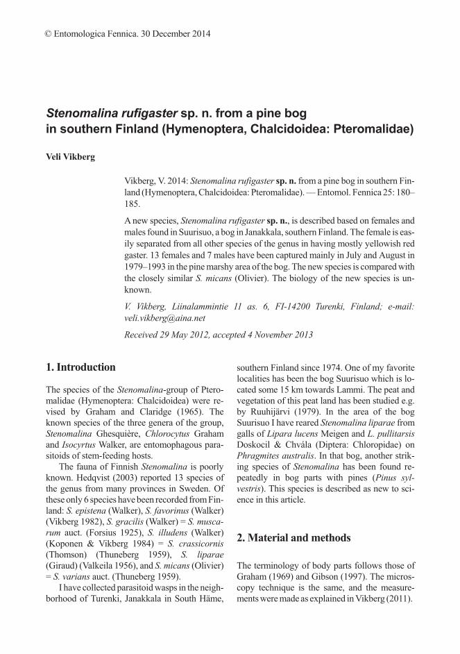
Description. Female holotype (Fig. 1).Colouration. Head dark blue. Eye pale,
slightly zinober. Ocelli amber red. Mandiblesreddish brown, palpi brown. Scape reddishbrown, apically slightly infuscate. Pedicel dark,with metallic tint. Flagellum dark, brownish toblackish. Mesosoma dark blue, with greenish me-tallic tint especially laterally and on propodeum,coxae concolorous with mesosoma. Legs other-wise yellowish red, fore femur greenish in-fuscate, hind tibia and tarsus brownish yellow,
ENTOMOL. FENNICA Vol. 25 • Stenomalina rufigaster sp. n. from Finland 181
Fig. 1. Stenomalina
rufigaster sp. n., holo-
type female, lateral
view. Body length 2.5
mm. Photograph by
Pekka Malinen.

with apical tarsomere brownish infuscate. Wingsslightly fumate. Gaster yellowish red, tergum 1with greenish tint, 3–4 caudal terga dark, withbronze tint.
Head. With strong reticulate sculpture, some-what dull. In dorsal view 1.83 times as broad aslong, temples converging rather strongly, about0.21 as long as the eyes. POL/OOL index 1.7;OOL/OD 1.8. Malar space 0.42 the height of aneye or 0.47 of the width of the mouth. Anteriormargin of antennal torylus slightly closer to themedian ocellus than to the anterior margin of theclypeus. Scape 0.79 times as long as height of aneye, reaching above top of vertex; combinedlength of pedicel and flagellum 1.16 times widthof head; flagellum slightly clavate; first funicularsegment as long as pedicel, 1.7 times as long asbroad, second and third segments distinctly lon-ger than broad, fourth segment slightly longerthan broad, fifth segment subquadrate and sixthsegment slightly transverse; clava with divisionsbetween its segments transverse; its third seg-ment, ventrally, produced 0.4 towards base ofclava and provided with a rather small patch ofmicropilosity which is constricted near apex.
Mesosoma. With strong reticulate sculpture,somewhat dull. Scutellum 1.23 times as long asbroad, moderately convex. Propodeum medially0.59 as long as scutellum, its median area withrather strong, nearly uniform, dense reticulation;
median carina irregularly wrinkled. Legs stout;hind femur 3.7 times as long as high in lateralview; hind tibia 6.7 times as long as apicallywide; spur of mid-tibia 0.8 times as long asbasitarsus. Fore wing 2.5 times as long as broad;basal fold with 3 hairs; marginal vein 2.0 times aslong as stigmal vein; postmarginal vein slightlyshorter than marginal vein.
Gaster. Broadly ovate, 1.57 times as long asbroad; ovipositor sheath in dorsal view con-cealed. Apex of hypopygium at level 0.56 of thelength of the gaster.
Measurements (length in mm, if not statedotherwise). Body 2.5. Head: length 0.40, width0.73, height 0.63. Scape 0.30. Eye: height 0.38,width 0.30. Distance between eyes on frons 0.45.Mesosoma: length 1.10, width 0.56, height 0.62.Fore wing 1.95. Hind leg: coxa 0.36, femur 0.57,tibia 0.70, tarsus 0.49. Gaster: length 1.08, width0.69, height 0.50.
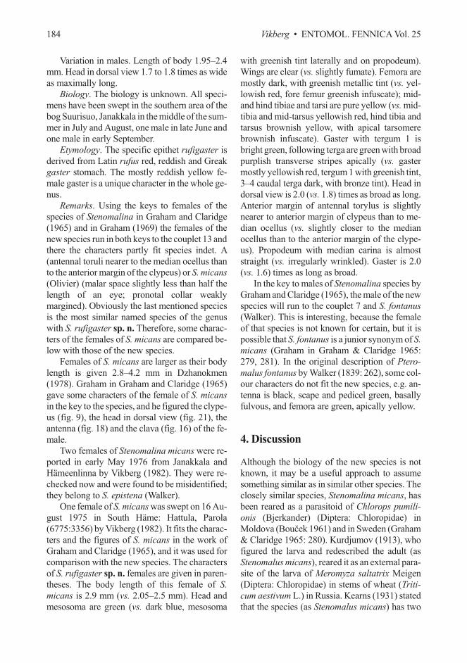
Variation in females. Length of body 2.05–2.5mm. Fore wing: marginal vein/stigmal vein 2.0–2.1; basal fold with 1–7 setae. A paratype female,collected on 10.VII.1979, shown in Fig. 2.
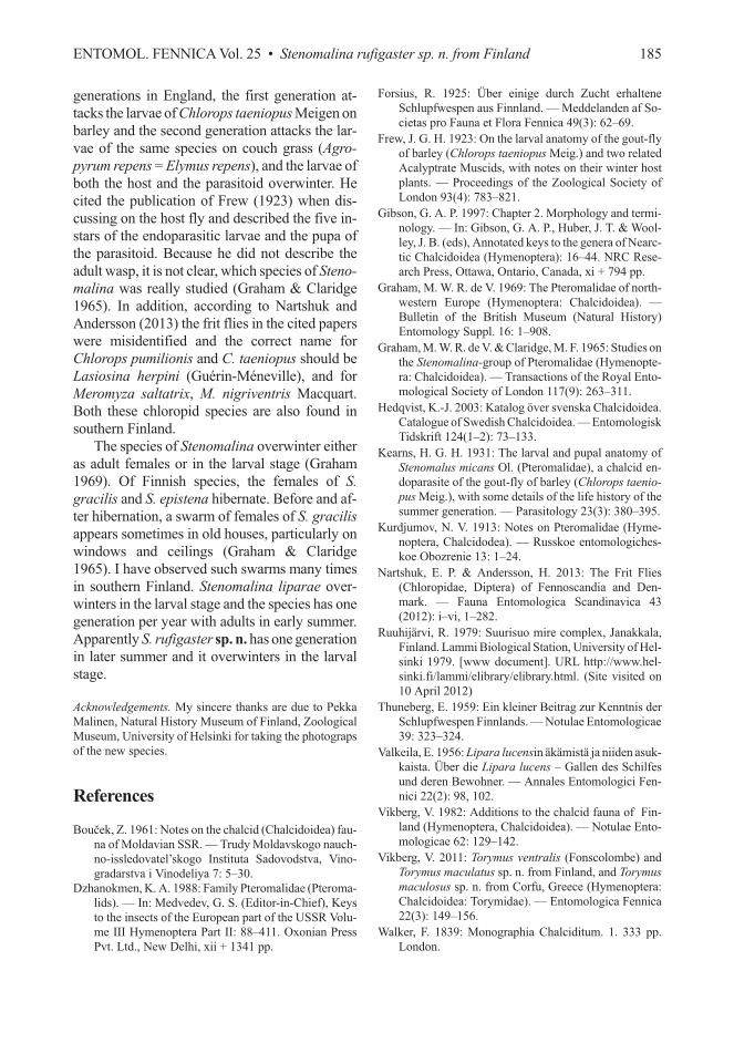
Male (paratype on 20.VI.1980) (Fig. 3).Colouration. Head dark blue. Scape brownish
yellow, apically above infuscate; pedicel brown,with greenish tint; flagellum dark brown. Meso-soma blue green, coxae blue green. Legs yellow-ish red, mid- and hind tibiae and tarsi more yel-
182 Vikberg • ENTOMOL. FENNICA Vol. 25
Fig. 2. Stenomalina
rufigaster sp. n., para-
type female, dorsal
view. Body length 2.3
mm. Photograph by
Pekka Malinen.

low; fore trochanter white. Wings slightlyinfumate; venation dark brownish. Gaster: terga1–2 reddish yellow, tergum 1 anteriorly broadlyinfuscate, with green tint; terga 3–6 dark bronze,tergum 7 bright green.
Head. In dorsal view 1.78 times as broad aslong. POL/OOL index 1.9; OOL/OD 1.5. Malarspace 0.44 the height of an eye or 0.48 of thewidth of the mouth. Anterior margin of antennaltorylus slightly closer to the median ocellus thanto the anterior margin of the clypeus. Scape 0.76times as long as the height of an eye, reachingslightly above the top of the vertex, anteroapicalmargin of scape with a shiny boss which extends0.63 of the length of the scape; combined lengthof pedicel and flagellum 1.76 times the width ofthe head; flagellum rather narrow, clava 1.25times as broad as width of pedicel in dorsal view;
first funicular segment 3.4 times as long as broad.Mesosoma. 2.1 times as long as wide. Scutel-
lum 1.29 times as long as broad, moderately con-vex. Propodeum medially 0.69 as long as scutel-lum, its median area with rather strong reticula-tion; median carina irregularly wrinkled. Forewing 2.4 times as long as broad; marginal vein1.86 times as long as stigmal vein. Hind femur 3.8times as long as high in lateral view. Gaster 2.3times as long as wide.
Measurements (length in mm, if not statedotherwise). Body 2.3. Head: length 0.36, width0.67, height 0.56. Scape 0.26. Eye: height 0.34,width 0.26. Distance between eyes on frons 0.42.Mesosoma: length 1.10, width 0.53, height 0.54.Fore wing 1.78. Hind leg: coxa 0.33, femur 0.53,tibia 0.66, tarsus 0.51. Gaster: length 0.98, width0.43, height 0.23.
ENTOMOL. FENNICA Vol. 25 • Stenomalina rufigaster sp. n. from Finland 183
Fig. 3. Stenomalina
rufigaster sp. n., para-
type male, dorsal view.
Body length 2.3 mm.
Photograph by Pekka
Malinen.
Variation in males. Length of body 1.95–2.4mm. Head in dorsal view 1.7 to 1.8 times as wideas maximally long.
Biology. The biology is unknown. All speci-mens have been swept in the southern area of thebog Suurisuo, Janakkala in the middle of the sum-mer in July and August, one male in late June andone male in early September.
Etymology. The specific epithet rufigaster isderived from Latin rufus red, reddish and Greakgaster stomach. The mostly reddish yellow fe-male gaster is a unique character in the whole ge-nus.
Remarks. Using the keys to females of thespecies of Stenomalina in Graham and Claridge(1965) and in Graham (1969) the females of thenew species run in both keys to the couplet 13 andthere the characters partly fit species indet. A(antennal toruli nearer to the median ocellus thanto the anterior margin of the clypeus) or S. micans
(Olivier) (malar space slightly less than half thelength of an eye; pronotal collar weaklymargined). Obviously the last mentioned speciesis the most similar named species of the genuswith S. rufigaster sp. n. Therefore, some charac-ters of the females of S. micans are compared be-low with those of the new species.
Females of S. micans are larger as their bodylength is given 2.8–4.2 mm in Dzhanokmen(1978). Graham in Graham and Claridge (1965)gave some characters of the female of S. micans
in the key to the species, and he figured the clype-us (fig. 9), the head in dorsal view (fig. 21), theantenna (fig. 18) and the clava (fig. 16) of the fe-male.
Two females of Stenomalina micans were re-ported in early May 1976 from Janakkala andHämeenlinna by Vikberg (1982). They were re-checked now and were found to be misidentified;they belong to S. epistena (Walker).
One female of S. micans was swept on 16 Au-gust 1975 in South Häme: Hattula, Parola(6775:3356) by Vikberg (1982). It fits the charac-ters and the figures of S. micans in the work ofGraham and Claridge (1965), and it was used forcomparison with the new species. The charactersof S. rufigaster sp. n. females are given in paren-theses. The body length of this female of S.
micans is 2.9 mm (vs. 2.05–2.5 mm). Head andmesosoma are green (vs. dark blue, mesosoma
with greenish tint laterally and on propodeum).Wings are clear (vs. slightly fumate). Femora aremostly dark, with greenish metallic tint (vs. yel-lowish red, fore femur greenish infuscate); mid-and hind tibiae and tarsi are pure yellow (vs. mid-tibia and mid-tarsus yellowish red, hind tibia andtarsus brownish yellow, with apical tarsomerebrownish infuscate). Gaster with tergum 1 isbright green, following terga are green with broadpurplish transverse stripes apically (vs. gastermostly yellowish red, tergum 1 with greenish tint,3–4 caudal terga dark, with bronze tint). Head indorsal view is 2.0 (vs. 1.8) times as broad as long.Anterior margin of antennal torylus is slightlynearer to anterior margin of clypeus than to me-dian ocellus (vs. slightly closer to the medianocellus than to the anterior margin of the clype-us). Propodeum with median carina is almoststraight (vs. irregularly wrinkled). Gaster is 2.0(vs. 1.6) times as long as broad.
In the key to males of Stenomalina species byGraham and Claridge (1965), the male of the newspecies will run to the couplet 7 and S. fontanus
(Walker). This is interesting, because the femaleof that species is not known for certain, but it ispossible that S. fontanus is a junior synonym of S.
micans (Graham in Graham & Claridge 1965:279, 281). In the original description of Ptero-
malus fontanus by Walker (1839: 262), some col-our characters do not fit the new species, e.g. an-tenna is black, scape and pedicel green, basallyfulvous, and femora are green, apically yellow.
4. Discussion
Although the biology of the new species is notknown, it may be a useful approach to assumesomething similar as in similar other species. Theclosely similar species, Stenomalina micans, hasbeen reared as a parasitoid of Chlorops pumili-
onis (Bjerkander) (Diptera: Chloropidae) inMoldova (Bou�ek 1961) and in Sweden (Graham& Claridge 1965: 280). Kurdjumov (1913), whofigured the larva and redescribed the adult (asStenomalus micans), reared it as an external para-site of the larva of Meromyza saltatrix Meigen(Diptera: Chloropidae) in stems of wheat (Triti-
cum aestivum L.) in Russia. Kearns (1931) statedthat the species (as Stenomalus micans) has two
184 Vikberg • ENTOMOL. FENNICA Vol. 25
generations in England, the first generation at-tacks the larvae of Chlorops taeniopus Meigen onbarley and the second generation attacks the lar-vae of the same species on couch grass (Agro-
pyrum repens = Elymus repens), and the larvae ofboth the host and the parasitoid overwinter. Hecited the publication of Frew (1923) when dis-cussing on the host fly and described the five in-stars of the endoparasitic larvae and the pupa ofthe parasitoid. Because he did not describe theadult wasp, it is not clear, which species of Steno-
malina was really studied (Graham & Claridge1965). In addition, according to Nartshuk andAndersson (2013) the frit flies in the cited paperswere misidentified and the correct name forChlorops pumilionis and C. taeniopus should beLasiosina herpini (Guérin-Méneville), and forMeromyza saltatrix, M. nigriventris Macquart.Both these chloropid species are also found insouthern Finland.
The species of Stenomalina overwinter eitheras adult females or in the larval stage (Graham1969). Of Finnish species, the females of S.
gracilis and S. epistena hibernate. Before and af-ter hibernation, a swarm of females of S. gracilis
appears sometimes in old houses, particularly onwindows and ceilings (Graham & Claridge1965). I have observed such swarms many timesin southern Finland. Stenomalina liparae over-winters in the larval stage and the species has onegeneration per year with adults in early summer.Apparently S. rufigaster sp. n. has one generationin later summer and it overwinters in the larvalstage.
Acknowledgements. My sincere thanks are due to PekkaMalinen, Natural History Museum of Finland, ZoologicalMuseum, University of Helsinki for taking the photograpsof the new species.
References
Bou�ek, Z. 1961: Notes on the chalcid (Chalcidoidea) fau-na of Moldavian SSR. — Trudy Moldavskogo nauch-no-issledovatel’skogo Instituta Sadovodstva, Vino-gradarstva i Vinodeliya 7: 5–30.
Dzhanokmen, K. A. 1988: Family Pteromalidae (Pteroma-lids). — In: Medvedev, G. S. (Editor-in-Chief), Keysto the insects of the European part of the USSR Volu-me III Hymenoptera Part II: 88–411. Oxonian PressPvt. Ltd., New Delhi, xii + 1341 pp.
Forsius, R. 1925: Über einige durch Zucht erhalteneSchlupfwespen aus Finnland. — Meddelanden af So-cietas pro Fauna et Flora Fennica 49(3): 62–69.
Frew, J. G. H. 1923: On the larval anatomy of the gout-flyof barley (Chlorops taeniopus Meig.) and two relatedAcalyptrate Muscids, with notes on their winter hostplants. — Proceedings of the Zoological Society ofLondon 93(4): 783–821.
Gibson, G. A. P. 1997: Chapter 2. Morphology and termi-nology. — In: Gibson, G. A. P., Huber, J. T. & Wool-ley, J. B. (eds), Annotated keys to the genera of Nearc-tic Chalcidoidea (Hymenoptera): 16–44. NRC Rese-arch Press, Ottawa, Ontario, Canada, xi + 794 pp.
Graham, M. W. R. de V. 1969: The Pteromalidae of north-western Europe (Hymenoptera: Chalcidoidea). —Bulletin of the British Museum (Natural History)Entomology Suppl. 16: 1–908.
Graham, M. W. R. de V. & Claridge, M. F. 1965: Studies onthe Stenomalina-group of Pteromalidae (Hymenopte-ra: Chalcidoidea). — Transactions of the Royal Ento-mological Society of London 117(9): 263–311.
Hedqvist, K.-J. 2003: Katalog över svenska Chalcidoidea.Catalogue of Swedish Chalcidoidea. — EntomologiskTidskrift 124(1–2): 73–133.
Kearns, H. G. H. 1931: The larval and pupal anatomy ofStenomalus micans Ol. (Pteromalidae), a chalcid en-doparasite of the gout-fly of barley (Chlorops taenio-
pus Meig.), with some details of the life history of thesummer generation. — Parasitology 23(3): 380–395.
Kurdjumov, N. V. 1913: Notes on Pteromalidae (Hyme-noptera, Chalcidodea). — Russkoe entomologiches-koe Obozrenie 13: 1–24.
Nartshuk, E. P. & Andersson, H. 2013: The Frit Flies(Chloropidae, Diptera) of Fennoscandia and Den-mark. — Fauna Entomologica Scandinavica 43(2012): i–vi, 1–282.
Ruuhijärvi, R. 1979: Suurisuo mire complex, Janakkala,Finland. Lammi Biological Station, University of Hel-sinki 1979. [www document]. URL http://www.hel-sinki.fi/lammi/elibrary/elibrary.html. (Site visited on10 April 2012)
Thuneberg, E. 1959: Ein kleiner Beitrag zur Kenntnis derSchlupfwespen Finnlands. — Notulae Entomologicae39: 323–324.
Valkeila, E. 1956: Lipara lucensin äkämistä ja niiden asuk-kaista. Über die Lipara lucens – Gallen des Schilfesund deren Bewohner. — Annales Entomologici Fen-nici 22(2): 98, 102.
Vikberg, V. 1982: Additions to the chalcid fauna of Fin-land (Hymenoptera, Chalcidoidea). — Notulae Ento-mologicae 62: 129–142.
Vikberg, V. 2011: Torymus ventralis (Fonscolombe) andTorymus maculatus sp. n. from Finland, and Torymus
maculosus sp. n. from Corfu, Greece (Hymenoptera:Chalcidoidea: Torymidae). — Entomologica Fennica22(3): 149–156.
Walker, F. 1839: Monographia Chalciditum. 1. 333 pp.London.
ENTOMOL. FENNICA Vol. 25 • Stenomalina rufigaster sp. n. from Finland 185