Stem cell therapy in rat hind limb ischemic injury - Jyx - Jyv¤skyl¤n
59
Stem cell therapy in rat hind limb ischemic injury Lilja Laatikainen Department of Biological and Environmental Science University of Jyväskylä 2007
Transcript of Stem cell therapy in rat hind limb ischemic injury - Jyx - Jyv¤skyl¤n

Stem cell therapy in rat hind limb ischemic injury
Lilja Laatikainen
Department of Biological and Environmental Science
University of Jyväskylä
2007
Preface
This study was carried out in the Medicity Research Laboratory of the University of
Turku in 2006-2007.
I wish to thank my supervisors PhD Mikko Laukkanen for the possibility to work as
a member of the Cellular Therapy Group on such an interesting topic and for excellent
instructing during this time, and MSc Juha Laurila for sharing the work load and giving a
hand whenever I needed, that is, great ‘colleagueness’.
Additionally, I want to acknowledge my mother for boosting my laborious weeks
with her delicious Sunday desserts, and my father and brother for their saintly patience and
technical assistance with all the computer-related difficulties.
Lilja Laatikainen
Turku, September 2007

Jyväskylän yliopisto Pro gradu –tutkielman tiivistelmä
Matemaattis-luonnontieteellinen tiedekunta
Tekijä: Lilja Laatikainen
Tutkielman nimi: Kantasoluterapia rotan takajalan iskeemisessä vauriossa
English title: Stem cell therapy in rat hind limb ischemic injury
Päivämäärä: 28.9.2007 Sivumäärä: 59
Laitos: Bio- ja ympäristötieteiden laitos
Oppiaine: Solubiologia
Tutkielman ohjaaja(t): FT Mikko Laukkanen ja FM Juha Laurila
Tiivistelmä: Mesenkymaalisia kantasolupopulaatioita (mesenchymal stem cells, MSC) on useimmissa
ihmiskehon kudostyypeissä, vaikkakin niiden prosentuaalinen osuus on hyvin pieni. Ne ovat multipotentteja
soluja, jotka toimivat luontaisesti kudosten uusiutumisessa ja vaurioiden korjautumisessa. Ihmisen sikiön
kantasoluista johdetut mesenkymaaliset kantasolut (human embryonic stem cell derived mesenchymal stem
cells, hESC MSC) on viimeaikoina kehitetty kantasolutyyppi, joka muistuttaa tavallisia MSC-soluja. Iskemia
syntyy verisuonten tukkeutuessa, jolloin kudoksen ravinnon- ja hapensaanti estyy. Tämä johtaa nekroosin,
apoptoosin ja tulehdusreaktion kautta tapahtuvaan kudostuhoon. Kudostuhon paranemiseen liittyy
uudissuonitus eli uusien suonien muodostuminen vanhoista haarautumalla, mikä palauttaa verenkierron
kudokseen. Uudissuonitusta säätelevät sekä kudoksen omat että tulehdussolut erittämällä useita
signaalimolekyylejä. Sonic hedgehog (Shh) on tärkeä yksilönkehityksen aikana toimiva morfogeeni, jonka on
todettu ottavan osaa verisuonten syntyyn myös aikuisessa kudoksessa.
Tämän tutkimuksen tarkoitus oli selvittää hESC MSC-solujen vaikutus rotan takajalan äkillisen
iskemian korjaantumiseen. HESC MSC-solut leimattiin lentiviruksilla, joissa oli joko lusiferaasi- tai GFP-
geeni. Luustolihasiskemia saatiin aikaan sulkemalla rottien oikea reisivaltimo kirurgisesti, ja 24 tuntia
myöhemmin lihakseen injektoitiin leimattuja kantasoluja Shh:n kanssa ja ilman. Lusiferaasi-kantasoluja
saaneet rotat kuvattiin 0, 6 ja 24 tuntia injektion jälkeen, ja GFP-kantasoluja saaneita rottia seurattiin kolme
päivää. Kudosnäytteet analysoitiin immunohistokemiallisesti värjäämällä verisuonispesifinen proteiini sekä
PCR-ajolla rotan genomisesta ja komplementaarisesta DNA:sta käyttäen ihmissekvenssin alukkeita. Värjätyt
hiussuonet laskettiin ja analysoitiin tilastollisesti.
HESC MSC-solujen kasvatus ja lyhyt Shh-altistus eivät muuttaneet niiden ulkonäköä, mutta
myöhemmässä vaiheessa pieniä muutoksia havaittiin in vitro. Solut myös leimaantuivat tehokkaasti
lentiviruksilla. Bioluminesenssikuvaus paljasti siirrettyjen solujen määrän laskevan nopeasti ensimmäisten 24
tunnin aikana. Joitakin GFP-positiivisia soluja kuitenkin nähtiin fluoresenssimikroskoopilla ja PCR-tulokset
vahvistivat ihmisen DNA:n ja lähetti-RNA:n olemassaolon rotan lihaksissa. Kontrolliin verrattuna
hiussuonten lukumäärä oli tilastollisesti merkittävästi suurempi molemmissa hESC MSC-soluja saaneissa
ryhmissä, mutta pelkkää Shh:a saaneessa ryhmässä ei, missä suonia oli vain hieman enemmän kuin
kontrolliryhmässä. Lisäksi ero kantasoluryhmien ja Shh-ryhmän välillä oli merkittävä.
Bioluminesenssikuvauksen perusteella suurin osa kantasoluista kuoli siirtopaikalle vuorokauden
kuluessa. Pieni osa soluista selviytyi seuranta-ajan loppuun, mutta varsinaista kudoksiin liittymistä ei
havaittu. Positiivinen vaikutus uudissuonten kehittymiseen oli silti selvä. Näiden tulosten perusteella hESC
MSC-solut näyttävät olevan tärkeitä uudissuonituksen alkuvaiheessa, jossa ne toimivat luultavasti
vahvistamalla kudoksessa jo olevaa hapenpuutteesta johtuvaa tulehdusreaktiota. Tässä työssä pienellä Shh-
morfogeeniannoksella ei ollut vaikutusta uudissuonituksen lisääntymiseen yksinään tai kantasolujen kanssa
annettuna. Pro gradu-työ muodostaa suuren osan laajemmasta tutkimuksesta, joka keskittyy kantasolu-
välitteiseen kudoksen uusiutumiseen iskeemisessä vauriossa. Tutkimusartikkeli, jossa kirjoittaja on toisena
kirjoittajana, on lähetetty tarkastettavaksi alan lehteen.
Avainsanat: hESC MSC, MSC, iskemia, angiogeneesi, Shh, soluterapia

University of Jyväskylä Abstract of Master´s Thesis
Faculty of Mathematics and Science
Author: Lilja Laatikainen
Title of thesis: Stem cell therapy in rat hind limb ischemic injury
Finnish title: Kantasoluterapia rotan takajalan iskeemisessä vauriossa Date: 28.9.2007 Pages: 59
Department: Department of Biological and Environmental Science
Chair: Cell Biology
Supervisor(s): PhD Mikko Laukkanen and MSc Juha Laurila
Abstract: Mesenchymal stem cells (MSC) are found in most tissue types throughout human body, though
their percentage is very small. They are multipotent cells which function in tissue turnover and repair. Human
embryonic stem cell derived mesenchymal stem cells (hESC MSC) have been developed recently and have
the properties of regular MSCs. Ischemia occurs after occlusion of blood vessels hindering nutrient and
oxygen supply to the respective tissue, and results in tissue damage including necrosis, apoptosis and
inflammation. Regeneration of ischemic tissue involves angiogenesis, the formation of new capillaries
through sprouting from existing vessels restoring the blood flow. It is regulated by a complex network of
signaling molecules secreted by several types of tissue and inflammatory cells. Morphogen sonic hedgehog
(Shh) is an important factor during embryogenesis and pattern formation. Additionally, it has been shown to
enhance angiogenesis in adults.
This study was carried out to determine the effect of hESC MSC and Shh on the recovery of rat hind
limb muscles from acute ischemia. Cells were labeled lentivirally with either luciferase or green fluorescent
protein (GFP), and injected with or without Shh into muscles in which ischemia had been induced 24 hours
earlier by ligating the proximal and distal ends of the femoral artery. Luciferase-hESC MSC transplanted
animals were imaged for bioluminescence at 0, 6 and 24 hours, whereas the GFP-hESC MSC transplanted
animals had three day follow-up period after transplantation. Tissue samples were analyzed with
immunohistochemical staining for capillary marker and with PCR for genomic DNA and complementary
DNA using human specific primers. Stained capillaries were calculated and analyzed statistically.
Expansion of hESC MSC and short-term incubation in Shh did not alter the stem cell morphology,
although changes were observed at late passages. They were also efficiently labeled with lentivirus vector.
Bioluminescence imaging showed a rapid decrease in the amount of transplanted cells during the first 24
hours. However, a few GFP positive cells were seen in the fluorescence microscope and PCR confirmed the
presence of human DNA and messenger-RNA in rat tissues. Number of capillaries increased significantly in
both hESC MSC groups as compared to control, but not in the group that received Shh alone, although there
was a minor increase. In addition, the difference between both stem cell groups and Shh group was
significant.
According to the bioluminescence imaging vast majority of the hESC MSC died at the
transplantation site during the first 24 hours in host tissues. Some cells survived through the follow-up period
but did not show any sign of engraftment. However, the angiogenic effect was clear. These results suggest
that the hESC MSC are important in initiation of angiogenesis, and that they function possibly by promoting
tissue inflammatory reaction first raised by hypoxia. In this study the small amount of morphogen Shh did
not have an effect on capillary formation either alone or when administered with the hESC MSC. The thesis
forms a major part of a study concentrating on stem cell mediated tissue regeneration in ischemic injury. A
research paper in which the author places as the second author has been submitted to a journal covering the
field.
Keywords: hESC MSC, MSC, ischemia, angiogenesis, Shh, cell therapy

Table of contents
PREFACE
ABSTRACTS
ABBREVIATIONS
INTRODUCTION .................................................................................................................................8
MESENCHYMAL STEM CELLS ...............................................................................................................8
Definition and properties on lab bench..........................................................................................8
MSCs in natural environment ......................................................................................................11
Applied research on MSCs...........................................................................................................13
ISCHEMIA...........................................................................................................................................17
Angiogenesis in response to ischemia..........................................................................................18
Research models ..........................................................................................................................22
Angiogenesis as clinical target ....................................................................................................24
AIM OF THE STUDY ........................................................................................................................28
MATERIALS AND METHODS........................................................................................................29
Plasmid production and purification ...........................................................................................29
Cell culture of HEK 293T ............................................................................................................30
Lentivirus production...................................................................................................................30
Lentivirus-GFP titer.....................................................................................................................30
Shh production and purification ..................................................................................................31
Cell culture of hESC MSC ...........................................................................................................31
Treatment of hESC MSC for transplantation...............................................................................32
Animal work .................................................................................................................................33
Bioluminescence imaging ............................................................................................................34
RT-PCR and PCR.........................................................................................................................34
Immunohistochemistry .................................................................................................................35
Statistics .......................................................................................................................................36
RESULTS.............................................................................................................................................37
EXPANSION OF HESC MSC ...............................................................................................................37
SHH PRODUCTION AND ITS EFFECT ON HESC MSC IN VITRO ..............................................................38
LV-GFP PRODUCTION AND TRANSDUCTION OF HESC MSC .............................................................39
Lentivirus-GFP titer.....................................................................................................................39
Transduction of hESC MSC turned out effective..........................................................................40
STEM CELL TRANSPLANTATION AND HESC MSC TRACKING.............................................................41

Amount of hESC MSC decreased rapidly at the transplantation site...........................................41
A few scattered GFP+ cells were seen in cryosections................................................................41
Human DNA and mRNA were found in the rat tissues three days post-transplantation..............41
EFFECT OF TRANSPLANTATION ON ANGIOGENESIS.............................................................................43
DISCUSSION.......................................................................................................................................45
REFERENCES ....................................................................................................................................49

Abbreviations
a. = arteria
αMEM = α-Minimum Eagle’s Medium
BLI = bioluminescence imaging
BM = bone marrow
bp, bps = base pair, base pairs
Dnase I = deoxyribonuclease I
EC = endothelial cell
ECM = extracellular matrix
EDTA = ethylenediaminetetraacetic acid
EF1-α = elongation factor 1α
FGF = fibroblast growth factor
Gag-pol = group antigen-polymerase
GFP = green fluorescent protein
hALU = human alu-element
HEK 293T = human embryonic kidney 293 cells with simian virus 40 large T antigen
hESC MSC = human embryonic stem cell derived mesenchymal stem cells
HRP = horseradish peroxidase
i.p. = intraperitoneal
IU = infective units
IVIS = in vivo imaging system
LBamp = lysogeny broth with ampicillin
LV = lentivirus
MOI = multiplicity of infection
MSC = mesenchymal stem cell
P = passage
PCR, RT-PCR = polymerase chain reaction, reverse transcriptase-polymerase chain
reaction
PDGF = platelet derived growth factor
Rnase = ribonuclease
RT = room temperature
s.c. = subcutaneous
SDS = sodium dodecyl sulphate
Shh = sonic hedgehog
SMC = smooth muscle cell
Tris = tris(hydroxymethyl)aminomethane
VEGF = vascular endothelial growth factor
Vsv-g = vesicular stomatitis virus envelope glycoprotein
vWF = von Willebrand factor

8
Introduction
Mesenchymal stem cells
Mesenchymal stem cells (MSC) were first described by Friedenstein and colleagues
in 1974 as bone marrow stromal cells that maintain the suitable microenvironment for
hematopoiesis to occur (Friedenstein et al., 1974; Friedenstein et al., 1976). Since then an
increasing amount of research around the world has been committed to discover the nature
and properties of these cells. The task has proven out more complicated than expected as
the cell population seems to be highly variable and flexible in vitro therefore rendering the
results from different groups controversial and difficult to compare. Additionally, due to
these properties it has been nearly impossible to study the MSCs in their undifferentiated
state and hence most of the current knowledge is based on research on cultured cells which
are likely to differ from their in vivo counterparts.
The nomenclature of mesenchymal stem cells reflects their diverse properties. Many
cell subpopulations of bone marrow and other tissues have names describing their in vitro
behavior leading to confusion among scientists on their true identity. The International
Society for Cellular Therapy (ISCT) has recently proposed (Horwitz et al., 2005) the name
‘mesenchymal stromal cell’ to be used instead of ‘mesenchymal stem cell’ in order to
maintain scientific accuracy, and thus the term stem cell should only be used of cells that
meet the accepted criteria. The acronym MSC can be used of both cell types as long as
scientists clearly state in their publications which one is in question. A recently developed
cell line used in this work, human embryonic stem cell derived mesenchymal stem cells
(hESC MSC), has not been characterized for stem cell properties but for consistency their
original name is used (Trivedi and Hematti, 2007).
Definition and properties on lab bench
Classical stem cell definition (Rosenthal, 2003) states that a stem cell is a multipotent
cell originating from a previous stem cell, and capable of self-renewing. In other words, a
single stem cell from homogenous population can produce daughter cells both similar to
the original cell and several differentiated types. A true stem cell remains in an
undifferentiated state in its niche, and as it proliferates through symmetric division the cell

9
population self-renews and expands. An asymmetric division accounts for the maintenance
of stem cell pool and multipotency as one daughter cell is committed to differentiate.
During embryogenesis the potency of stem cells decreases rapidly giving way to extensive
differentiation and proliferation. Strictly, a stem cell should also functionally and robustly
reconstitute tissue in vivo after transplantation but different stem cell populations seem to
have variable capability for this, e.g. hematopoietic stem cells readily initiate blood
formation in new bone marrow whereas MSCs engraft more occasionally. In 2006, the
International Society for Cellular Therapy announced somewhat less strict criteria to be
used when evaluating the stem cell properties of human MSCs (Dominici et al., 2006). In
short, they should be characterized by trilineage differentiation capacity to osteoblasts,
chondroblasts and adipocytes, plastic adherence, and a specific surface antigen pattern
(figure 1).
The ISCT criteria are met in the numerous studies supporting multipotent
differentiation abilities of MSCs. When cultured with proper components of extracellular
matrix, growth factors, and cytokines they can differentiate into cells of mesenchymal
tissue lineage such as osteoblasts, adipocytes, myocytes, endothelial cells and
chondrocytes (Pittenger et al., 1999; Reyes et al., 2001). Also various chemical substances
can induce similar differentiation (Grigoriadis et al., 1988). Furthermore, even the non-
mesenchymal differentiation of MSCs into hepatocytes and neural cells has been reported
(Lee et al., 2004; Woodbury et al., 2000). Their self-renewal is widely accepted as well
since carefully cultured cells continue to produce similar cells over passaging by
symmetric division. However, clonal assays have shown the populations to be
heterogeneous separate colonies showing variable potency (Colter et al., 2000; Colter et
al., 2001; Reyes et al., 2001) which indicates that a population consists of both primitive
stem cells and committed progenitor cells (Zohar et al., 1997). Thus, to date the actual
stem cell properties of adult MSCs are still under debate due to difficulties in gathering
solid evidence.
Cultured MSCs are generally described as plastic-adherent cells with spindle-shaped
morphology, although the latter changes according to the growth phase and confluence of
the culture so that the cells become larger and flatter at high densities (Digirolamo et al.,
1999; Sekiya et al., 2002). MSCs are capable of rapid proliferation in favorable in vitro
conditions resulting in even more than 1 million-fold expansion (Colter et al., 2000) but

10
this and the overall lifespan of the cells depend on their host organism, tissue of origin,
culture conditions such as plating density and medium composition, and sampling method
(Digirolamo et al., 1999; Pittenger et al., 1999; da Silva Meirelles et al., 2006). As the cells
are propagated the culture tends to become progressively homogenous and the
multipotency is gradually lost, presumably driven by the microenvironment and ageing
(Digirolamo et al., 1999; Majumdar et al., 1998; Muraglia et al., 2000). This may lead to
reduction of multipotentiality if a lineage restricted subpopulation is inadvertently favored
or the cells are induced to differentiate.
Cell surface markers of MSCs have been analyzed in a multitude of studies in order
to find unique markers for their more efficient recognition and isolation. According to
ISCT, cell populations that are positive for CD105, CD73, CD90, and negative for
hematopoietic and endothelial markers CD45, CD34, CD14 or CD11b, CD79 or CD19,
and HLA-DR along with the other requirements (figure 1) can be qualified as MSCs
(Dominici et al., 2006). In addition, many other non-specific markers and CD antigens
have been found but they vary between species and tissue of origin (Charbord et al., 2002).
Among them are e.g. SH2, SH3, Sca-1, CD9, CD29, CD44, CD71, CD105 (Martin et al.,
2002; Pittenger et al., 1999; Sun et al., 2003) and typical vascular smooth muscle cell
marker α-smooth muscle actin (α-SMA) (da Silva Meirelles et al., 2006) which also denote
the functions of MSCs in vivo as migratory, immunoregulatory and vascular support cells.
Most uninduced MSC lines are also positive for MHC I and CD58 at low level but not for
MHC II, CD40, CD54, CD80, or CD86 (Tse et al., 2003); a phenotype which is generally
considered nonimmunogenic and offers an advantage in allogeneic transplantation therapy.
So far the desired strictly MSC specific markers have not been identified.
The parameters mentioned in the context of expansion influence also MSC lineage
commitment resulting in broad variation among adult cell lines (da Silva Meirelles et al.,
2006). It is likely though, that MSCs from fetal tissue counterparts have higher potential
for clonal expansion and plasticity, i.e. the capacity to convert from one cell type to
another (see review by Moore and Quesenberry, 2003), than adult cells (Lee et al., 2006).
However, stem cell plasticity requires careful evaluation since at least five mechanisms can
cause false multipotential differentiation (see review by Pauwelyn and Verfaillie, 2006).
First, tissue under examination can contain multiple stem cell populations with specific
capacities. MSCs can also fuse with mature cells and thereby gain some of their
11
characteristics. When subjected to appropriate chemical conditions fully differentiated cells
can transdifferentiate directly into another matured type, or they can sometimes take a step
back, dedifferentiate, and then redifferentiate following the environmental cues. Finally, a
pluripotent stem cell population can coexist in a given tissue, and when tissue-specific
stem cells are purified they ‘contaminate’ the isolate.
MSCs in natural environment
Mesenchymal stem cells are derived from the mesenchyme of an embryo but the
early events that lead to their distribution into niches are poorly understood. A cell layer
bearing stromal characteristics under the hematopoietic layer in fetal dorsal aorta in the
aorto-gonadal-mesonephric (AGM) region has been suggested as the tissue of origin for
MSCs (Minasi et al., 2002). Evidence for MSC migration from here has not been presented
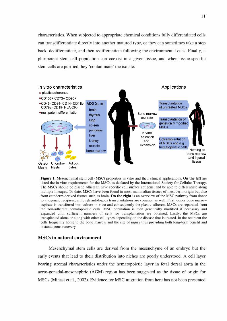
Figure 1. Mesenchymal stem cell (MSC) properties in vitro and their clinical applications. On the left are
listed the in vitro requirements for the MSCs as declared by the International Society for Cellular Therapy.
The MSCs should be plastic adherent, have specific cell surface antigens, and be able to differentiate along
multiple lineages. To date, MSCs have been found in most mammalian tissues of mesoderm origin but also
from ectoderm-derived tissues such as brain. On the right is an overview of the MSC pathway from donor
to allogeneic recipient, although autologous transplantations are common as well. First, donor bone marrow
aspirate is transferred into culture in vitro and consequently the plastic adherent MSCs are separated from
the non-adherent hematopoietic cells. MSC population is then genetically modified if necessary and
expanded until sufficient numbers of cells for transplantation are obtained. Lastly, the MSCs are
transplanted alone or along with other cell types depending on the disease that is treated. In the recipient the
cells frequently home to the bone marrow and the site of injury thus providing both long-term benefit and
instantaneous recovery.

12
but, interestingly, a large number of circulating stromal cells has been detected in fetuses
from seven to twelve weeks’ gestation after which they disappear (Campagnoli et al.,
2001). This cell population closely resembled conventional MSCs except that it had wider
and more long-lasting differentiation capacity. MSC-like fetal cells have been found also
in maternal tissues supporting the circulation hypothesis (see review by Bianchi, 2000).
Taken together the findings offer a plausible mechanism for MSCs to effectively spread
during the early development into their ultimate niches, the bone marrow stroma and other
tissues, from where they can operate throughout the life-span of an individual.
The actual niches accommodating the MSCs have long been subjects of keen
research. Many groups have isolated MSC populations from a wide range of tissues other
than bone marrow including adipose tissue (Zuk et al., 2001), skeletal muscle (Lee et al.,
2000), and dental pulp (Gronthos et al., 2002). Similar cells have also been found in lungs,
liver, pancreas, spleen, kidney, major blood vessels, and brain (figure 1) from both fetal
and adult tissues (da Silva Meirelles et al., 2006; in 't Anker et al., 2003). Blood has been
suggested as a source of circulating MSCs as well but as da Silva Meirelles et al. (2006)
pointed out a contamination from detached vessel wall MSCs is very likely unless specific
care is taken while obtaining a blood sample. However, even though research on MSC
distribution in mammalian organ system is leaping forward their exact in vivo location on
cellular level is still largely unknown.
Most tissues contain a population of MSCs but determining their niches has been
complicated as they remain nearly invisible. Traditionally the fraction of MSCs in a bone
marrow aspirate sample has been quantified using colony-forming unit fibroblast (CFU-F)
assay in which adherent cells from the sample are plated in low density and the number of
individual colonies assumed to derive from single cells is calculated (Digirolamo et al.,
1999; Owen and Friedenstein, 1988). CFU-F assays with human and feline BM MSCs
suggest they represent only 0.001% - 0.01% of all nucleated cells (Martin et al., 2002;
Pittenger et al., 1999). Similarly, the number of MSCs in other tissues is likely to be very
small. Their in vivo proliferation rate has also been difficult to determine. It is considered
that in general MSCs are quiescent in vivo, and that they enter the mitotic phase only in
specific conditions e.g. after exposure to activating signaling molecules or loss of contact
inhibition. This is supported by many observations of rapid in vitro growth after low
density plating (Colter et al., 2000; Sekiya et al., 2002).

13
During the lifetime of an individual its tissues need to be renewed, and larger scale
regeneration may be necessary after severe tissue damage. Tissue resident stem cells in e.g.
skin, gastrointestinal tract, and bone marrow have well-established roles in tissue turnover.
Such populations have also been described in skeletal muscle and liver (Lee et al., 2000;
Piscaglia et al., 2007), though BM-derived stem cells seem to participate actively in repair
of most tissues. The regenerative capacity of tissues decreases throughout the post-natal
life, presumably because the number of stem cells in BM and organs falls as the individual
ages (Bellows et al., 2003). Moreover, many tissues, such as central nervous system and
cardiac muscle, are known to lack this regenerative ability partially or completely. Thus,
after early childhood healing of damaged tissues occurs mainly through scar tissue
formation by fibroblast cells. The role of MSCs in natural turnover and repair of peripheral
tissues remains therefore unknown until more efficient and reliable methods for in vivo
tracking of MSCs are developed.
In general, very little is known about the functions of MSCs in their natural
environment. They seem to be resident in distinct tissues which would explain their
variable in vitro properties. Additionally, MSCs are thought to give rise to most of the
several bone marrow cell types such as osteoblasts, pericytes, and myofibroblasts
(Muguruma et al., 2006), thus being also responsible for hematopoietic stem cell
development (Calvi et al., 2003; Majumdar et al., 1998; Zhang et al., 2003) and early
maturation of T and B immune cells (Barda-Saad et al., 1999; Kurosaka et al., 1999). MSC
mobilization from bone marrow has not been documented under natural conditions, i.e.
without experimental intervention. Activated T cells may play a role as they secrete
various cytokines and chemokines that may modulate MSC behavior indirectly (Burger et
al., 1998) once they enter BM via circulation, but the actual factors inducing MSC
activation and migration to the site of injury have not been determined.
Applied research on MSCs
Over the years of research on MSCs, developing clinical treatments for various
pathologies has been one of the main goals. Stem cells offer a way to regenerate the
defective tissues instead of merely alleviating or suppressing the symptoms by medication,
irradiation or surgical operations. In this respect MSCs are considered especially valuable
as they possess many clinically relevant characteristics (see review by Giordano et al.,
14
2007). First, MSCs can be readily isolated from bone marrow aspirates which, despite the
seeming existence of these cells elsewhere in body, are their primary source. Second, their
relatively high proliferation rate enables expansion in culture in order to obtain relevant
numbers of cells for transplantations. Third, the ability to differentiate on such a wide scale
renders this one cell type very versatile. Finally, MSCs have a remarkable ability to
migrate to the sites of injury and suppress the host immune system. Researchers and
physicians are working ‘blindfolded’, however, since the exact mechanisms behind the
beneficial effects of MSCs are not known.
Most of the research on MSCs is still in preclinical phase, and only few MSC
applications have reached a clinical trial phase. Before the stem cell treatments are ready
for use in hospitals and clinics more basic knowledge about their biology and in vivo
behavior need to be gathered. It is of outmost importance to understand and to be able to
control the self-renewal and differentiation of stem cells for the safety of human patients.
For instance, due to slower growth rate and lack of telomerase activity in vitro MSCs have
considered less tumorigenic than ES cells but their immunosuppressive function can
actually promote tumor growth (see review by Prockop and Olson, 2007). Two studies
have reported MSC transformation either spontaneously (Rubio et al., 2005) or after
genetic manipulation (Tolar et al., 2007) albeit such findings are rare. Moreover,
calcification of myocardium presumably caused by transplanted BM cells or MSCs has
been observed (Yoon et al., 2004). Undoubtedly, in vivo models are invaluable as similar
in vitro settings are yet to be developed.
Engraftment of mesenchymal stem cells into new host is a popular topic in tissue
regeneration research. Many investigators have recently shown how isolated, tagged, and
culture expanded MSCs can home to bone marrow cavity after systemic infusion into
healthy recipients or straight to the site of experimental injury (figure 1) in allo- and
xenotransplantations to repair e.g. bone fracture (Devine et al., 2002), cerebral ischemia
(Wang et al., 2002), infarcted heart (Makkar et al., 2005), and wounded skin (Fathke et al.,
2004). Furthermore, after repopulating the BM the cells are able to migrate out to take part
in normal tissue turnover of the body (Devine et al., 2003; Opalenik and Davidson, 2005).
Arguments against MSC homing have been made, however, as they were found to lose
their homing ability following transfer to culture (Rombouts and Ploemacher, 2003). If
necessary, MSCs can also be targeted more accurately simply by implanting them to the
15
desired area. This has been the approach in numerous studies attempting to restore
mesodermal tissues such as bone (Kon et al., 2000), cartilage (Fuchs et al., 2003), cardiac
muscle (Strauer et al., 2002), as well as nervous tissue (Hofstetter et al., 2002) with
success. However, the percentage of engrafted cells is usually less than 3% (Devine et al.,
2003; Jackson et al., 2001) but much higher levels have been suggested (Direkze et al.,
2003). Debate on incorporation has arisen, though, after a couple of studies disputed the
engraftment of MSCs into newly formed blood vessels (Zentilin et al., 2006; Ziegelhoeffer
et al., 2004).
In addition to direct repair of damaged tissues, allogeneic MSCs have shown a
notable ability to evade and suppress host immune reactions. The finding is of particular
value in therapeutic transplantations in which tissue rejection and graft versus host disease
(GVHD) are severe threats. MSCs themselves do not seem to elicit immune responses as
indicated by in vitro studies with mixed lymphocyte cultures (Bartholomew et al., 2002),
and it is likely also in vivo since infused stem cells persist in recipient for weeks and even
over a year (Azizi et al., 1998; Devine et al., 2003; Liechty et al., 2000). In addition, they
can actively suppress immune cell proliferation and attenuate their cytolytic functions
(Augello et al., 2005; Sotiropoulou et al., 2006). This has been exploited in cell and organ
transplantations in which autologous or allogeneic MSCs delivered at the same time induce
tolerance in host and thus protect the implant (Bartholomew et al., 2002). Nevertheless, in
some occasions the MSCs have been rejected by the host to varying extent (Eliopoulos et
al., 2005; Nauta et al., 2006), creating a need for more in-depth research on direct and
indirect effects of MSCs on immune response.
Another interesting practice is to genetically modify mesenchymal stem cells and
only then transfuse them for a therapeutic purpose (figure 1). Retro- or adenoviral
transduction generally results in an efficient and long-term gene expression (Li et al.,
1995) but since the virus vectors pose safety threats non-viral methods have been
developed (Song et al., 2004). Favorable properties can be obtained depending on the
introduced gene, e.g. prolonged stem cell lifespan by activation of telomerase catalytic
subunit (Bodnar et al., 1998) or prosurvival protein Akt (Mangi et al., 2003). There has
also been successful attempts to introduce secretion of therapeutic factors for protein
deficiency disorders (Bartholomew et al., 2001), chemotherapy (Studeny et al., 2002), and
enhanced organ repair (Moutsatsos et al., 2001). These can aid in expanding the cells in

16
vitro, in improving their post-transplantation viability, and even in maintaining their
‘stemness’. Additional advantage in using MSCs arises from their ability to home to the
site of injury, also a modifiable trait, allowing delivery into inaccessible tissues by simple
infusion. Animal models have already shown interesting results in treatment of invasive
tumors (Nakamura et al., 2004), cardiac infarction (Sun et al., 2007) and bone repair
(Tsuchida et al., 2003).
The mechanisms by which mesenchymal stem cells attain the improvement of tissue
functions are constantly under research. Attempts to treat various damaged tissues by cell
transplantations have not shown robust evidence for wide scale tissue-specific
differentiation or engraftment of MSCs, and yet improvement has been observed.
Consequently, many studies have concentrated on alternative mechanisms. Differentiation
to supporting cells such as endothelial or smooth muscle cells in cardiac muscle (Jackson
et al., 2001; Tomita et al., 1999), myofibroblasts (Direkze et al., 2003), vascular supporting
cells (Ziegelhoeffer et al., 2004), or hepatic stellate cells (Baba et al., 2004) which in turn
contribute to healing have been observed. Moreover, paracrine secretion of factors which
induce e.g. angiogenesis, or proliferation and differentiation of endogenous stem cells has
gained attention recently. MSC conditioned growth medium has been shown to protect
tissue cultures ex vivo and improve cardiac repair after ischemic injury (Iso et al., 2007;
Kinnaird et al., 2004) but whether transplanted MSCs perform this in vivo has not been
demonstrated.
Mesenchymal stem cells are slowly entering clinics as the number of phase I/II
clinical trials is growing. Systematic preliminary trials on human patients began in the mid-
1990’s which showed that MSCs could be transplanted without unfavorable side-effects
(Lazarus et al., 1995). Since then their abilities have been put to test in attempts to treat a
wide variety of pathologies. Some trials have had promising outcomes in remedy of graft
versus host disease (Le Blanc et al., 2004), hematological diseases (Lazarus et al., 2005),
cardiac infarction (Strauer et al., 2002), osteogenesis imperfecta (Horwitz et al., 1999), and
in recovery after cancer chemotherapy (Koc et al., 2000). Other studies on, for instance,
neurological and inherited diseases have not directly shown positive results but
nevertheless have provided the clinicians and investigators with information on which to
built new techniques.

17
Ischemia
Blood circulation is a life supporting system responsible for the exchange of oxygen
and nutrients to carbon dioxide and metabolic waste products. Blood also carries the
chemical messages between individual tissues and evens differences in temperature,
acidity and such thus contributing to homeostasis, the dynamic equilibrium of the body.
Blood vessels consist of arteries which branch several times into smaller arterioles and
finally into capillaries, which in turn fuse to form larger venules and veins leading back to
heart. Two other types, collateral arteries and anastomoses, are also present in the vessel
system. The first describes two parallel arteries with same target tissue, and the latter is a
connective vessel joining two separate arteries. All these vessels operate in maintaining
and adjusting blood pressure and tissue perfusion according to the metabolic needs at a
given time. This is accomplished by contracting and dilating individual vessels and
precapillary sphincters which regulate the blood flow to capillary bed. Generally, blood
flows through larger vessels until exercise requires more efficient gas and fuel exchange
raising the need to utilize the extensive capillary network.
Tissue growth, such as vertebrate development, reproduction and repair but also
tumor growth, requires simultaneous development of vasculature because otherwise the
new cells would be too far away from oxygen and nutrient supply (Folkman et al., 1971).
Also, several pathological conditions result in narrowing the lumen of blood vessels or
formation of circulating blood clots, and instantaneous traumas may cause vessels to be
severed. When the blood supply to an organ or tissue is partially or completely hindered
the oxygen and nutrient content decreases rapidly and ischemia occurs. Often collateral
arteries and anastomoses can in part restore the blood flow to inflicted tissue (see review
by Heil and Schaper, 2004) thereby attenuating the damage caused by hypoxia. Even short-
term ischemia induces necrosis and apoptosis leading to inflammatory reaction.
Accumulating macrophages and other inflammatory cells (Barbera-Guillem et al., 2002)
together with e.g. fibroblasts and muscle cells (Gustafsson et al., 1999; Steinbrech et al.,
1999) secrete vascular endothelial growth factor (VEGF) which is necessary for the
initiation of healing process aiming at restoration of blood perfusion by constructing new
blood vessels through arteriogenesis and angiogenesis.
18
Angiogenesis in response to ischemia
All blood vessels share similar structures but their differences reflect their specific
functions. Arteries and smaller arterioles (figure 2) consist of an inner continuous layer of
endothelial cells (EC) attached to a collagenous basement membrane, forming the intima.
Smooth muscle cells (SMC) form the media which in turn is surrounded by fibroblasts and
connective tissue, the adventitia. Veins and venules (figure 2) have essentially the same
layer structure as arteries but are distinguished through thinner SMC layer and larger
diameter since similar durability is not required. Capillaries (figure 2) are the smallest
vessels with only basement membrane and a single layer of endothelial cells to minimize
resistance for gas and nutrient exchange. The small vessels - precapillary arterioles,
postcapillary venules, and capillaries - have an additional cell type, pericytes, embedded in
their basement membrane (see review by Armulik et al., 2005). These cells are found in
most tissues and do not appear as mere structural components but are involved in
facilitating and coordinating communication between the other vascular cells through gap
junctions. Pericytes may also have a more active role as sensors for varying extracellular
conditions and may initiate appropriate signaling cascades in the vessel wall. Together
pericytes and SMCs are often referred to as mural cells.
The adult vascular system remains quiescent until stimulated to form new vessels.
These stimuli derive from hypoxia (Forsythe et al., 1996), changes in blood flow (see
review by Heil and Schaper, 2004), and inflammation as the inflammatory cells and
involved tissues secrete angiogenic growth factors (see review by Shireman, 2007). In
ischemic injuries insufficient oxygen induces the expression of hypoxia-inducible factor 1
(HIF-1) which is a transcriptional activator of vascular endothelial growth factor (VEGF)-
A (Forsythe et al., 1996), and thus is a key regulator of angiogenesis. VEGF-A and VEGF-
D are considered the most important factors in early angiogenic response (Rissanen et al.,
2003), but other members of the VEGF family, such as placental growth factor (PlGF),
along with fibroblast growth factors (FGF), insulin-like growth factors (IGF),
angiopoietins (Ang), and platelet-derived growth factors (PDGF) are required for efficient
vessel remodeling, maturation, and stabilization (see review by Carmeliet, 2000). The
functional network of these factors is a complex one and many molecular and cellular
events remain still unknown.

19
Sonic hedgehog (Shh) is a morphogen and transcriptional activator from the
hedgehog family which functions in several key events of development and remains active
in post-natal life (van den Brink et al., 2001). It has very versatile activities as suggested by
a microarray analysis which revealed upregulation of e.g. cell cycle, adhesion and
apoptosis related molecules (Ingram et al., 2002). Interestingly, Shh has also been
implicated in ischemic injuries of skeletal muscles and myocardium (Kusano et al., 2005;
Pola et al., 2003), although the molecular mechanism triggering Shh expression is still
unknown. Both neoangiogenesis and collateral growth seem to depend to some extent on
Shh stimulus (Kusano et al., 2005) but the effect is indirect as the target cells of Shh
signaling are mainly fibroblasts while the ECs remain intact (Kusano et al., 2005; Pola et
al., 2001). The Shh induced cells initiate a robust expression of three VEGF isoforms,
namely VEGF121, VEGF 165 and VEGF 189, and the angiopoietins 1 and 2 resulting in strong
angiogenic response (Pola et al., 2001). However, a study by Kanda et al. (2003) has
shown that Shh can induce ECs to begin capillary morphogenesis. In either case the true
target cells and the intracellular signaling pathway remains elusive.
Figure 2. Blood vessel structure. Arterioles and venules have the same structural layers of the vessel wall,
namely, intima, media and adventitia. Arterioles have a thick smooth muscle (SMC) cell layer and narrow
lumen which maintain an adequate blood pressure. SMCs regulate blood pressure by contracting and
relaxing according to stimuli. The pressure in venules is lower, and therefore thinner walls and wide lumen
suffice. In these vessels fibroblasts produce collagenous connective tissue which supports the vessels.
Capillaries lack adventitia and media, and their walls are very thin in order to enable efficient gas and
nutrient exchange. In all vessel types endothelial cells and pericytes are closely connected through basement
membrane which allows rapid communication about the environmental changes.
20
Blood vessel formation has been categorized into three partly overlapping subtypes,
namely vasculogenesis, angiogenesis and arteriogenesis. Vasculogenesis refers to the de
novo formation of primitive vascular network, the vascular plexus, from angioblasts in
embryonic mesoderm (see review by Risau, 1997). After this period and during the
postnatal life new vessels are formed mainly through angiogenesis, a process in which new
capillaries sprout from existing ones (see review by Risau, 1997). It may also occur by
intussusception or bridging, often involved in remodeling of newly formed vessels, in
which a wall is formed in the lumen of a vessel by pericytes or endothelial cells, and the
vessel is split into two (see review by Risau, 1997). Arteriogenesis becomes important
during ischemia as it is responsible for the bulk blood flow to the tissues, though hypoxia
is not a prerequisite for its initiation (see review by Schaper and Scholz, 2003). It is
defined as the enlargement of anastomoses and small arterioles to form new collateral
arteries that improve the blood supply to the inflicted tissue by circumventing the site of
occlusion (see review by Schaper and Scholz, 2003).
Angiogenesis, whether physiological or pathological, is a multistep process involving
coordinated functions of numerous growth factors and cell types. It initiates with
upregulated HIF-1 expression from hypoxic cells of the tissue (Forsythe et al., 1996) and
nitric oxide induced vasodilatation (figure 3A). HIF-1 induced VEGF-family expression
enhances permeability of the vessel wall allowing leakage of plasma proteins into the
surrounding tissues to form a scaffold for migrating endothelial cells (see review by
Dvorak et al., 1995). VEGF also mobilizes bone marrow mononuclear cells which seem to
have a role in enhancing vessel sprouting (Grunewald et al., 2006; Zentilin et al., 2006).
Next, the intercellular junctions between pericytes, SMCs and ECs need to be loosened and
the basement membrane disintegrated in order to enable EC and pericyte migration (figure
3B). This is accomplished by destabilizing growth factors, such as angiopoietin-2
(Maisonpierre et al., 1997), and several proteinases from different families which
simultaneously release extracellular matrix (ECM)-bound growth factors such as FGF and
VEGF (Houck et al., 1992; Saksela and Rifkin, 1990) (figure 3B). The process must be
balanced to clear the way for migrating cells but to leave enough matrix to provide them
with necessary molecular cues and support since too extensive ECM degradation can
impair angiogenesis (Gutierrez et al., 2000).

21
The released growth factors stimulate the ECs, SMCs and pericytes to proliferate and
migrate after they have been detached from the growth-inhibiting environment of a
quiescent vessel wall (Hangai et al., 2002). The cells are guided by chemoattractants and
perivascular cells such as pericytes (Nehls et al., 1994) as well as by the different
components of the ECM scaffold such as collagen and elastin (Anderson et al., 2004;
Hangai et al., 2002) (figure 3C). In addition, molecules mediating cell-cell and cell-matrix
contacts, e.g. integrins (Davis and Camarillo, 1995), and other angiogenic factors, e.g.
nitric oxide (Genis et al., 2007), are essential in spreading of ECs. Several other growth
factors, hormones and cytokines are indicated in experimental angiogenesis but their roles
in vivo remain to be determined. As the cells migrate they form solid cords that penetrate
into surrounding tissue and fuse with the preexisting neighboring cords, possibly guided by
the mechanical force created by mobile ECs on the ECM (Davis and Camarillo, 1995).
Subsequently, lumen formation takes place as ECs in the middle of the cord undergo
apoptosis and vacuolated ECs secrete their vacuoles (Meyer et al., 1997) (figure 3C). More
ECs then settle into the vessel wall increasing the vessel diameter (Meyer et al., 1997).
The last phase of angiogenesis involves stabilization and maturation of the new
vessels which would otherwise soon regress without sufficient support from the
microenvironment. Recruitment of pericytes and SMCs is crucial for vessel maturation
(figure 3D), and here PDGF apparently plays an important role since its absence disrupts
the vessel development (Hellstrom et al., 2001; Lindahl et al., 1997). There is a short delay
before pericytes cover the EC lined tubules during which the capillary density is matched
to the oxygen supply (Benjamin et al., 1998). Connections between endothelial and mural
cells are stabilized by e.g. angiopoietin-1 and TGFβ-1 which inhibit their proliferation and
migration (Goumans et al., 2002; Suri et al., 1996). Moreover, the newly formed vessels
acquire special characteristics such as fenestrae in kidney and liver capillaries, or other
structures depending on their location in the body. After establishment of blood flow
higher fluid shear stress and blood pressure against the vessel wall promote further
remodeling of the cytoskeleton (Franke et al., 1984) but as soon as the forces are
normalized the new vessels become quiescent until angiogenic stimuli are introduced again
(figure 3D).

22
Research models
The process of angiogenesis is a complex interplay of numerous tightly regulated
molecular and cellular components. Moreover, it is dependent on the physical environment
of the tissue which offers a scaffold and appropriate signals for the migrating cells. Since
settings like this are difficult to establish in vitro most studies on angiogenesis must rely on
animal models. One objective in research of angiogenesis is to find suitable therapeutic
agents with which to promote or inhibit vessel growth according to the needs of
pathological state. Induced brief ischemia and subsequent vessel growth also serve in
Figure 3. Overview of angiogenesis. A Hypoxia is developed downstream from the site of occlusion and
induces HIF-1 expression (red dots) from hypoxic cells. HIF-1 further induces VEGF expression (green
dots) which initiates the angiogenic events. B VEGF recruited mononuclear cells (e.g. macrophages) and
nearby tissue cells produce several growth factors (green dots) and proteinases (yellow dots) which induce
basement membrane and connective tissue degradation. More growth factors and cryptic binding sites are
revealed from the connective tissue which aid in proliferation and migration of endothelial cells and
pericytes. C Endothelial cells form cords as they migrate into the hypoxic tissue guided by chemoattractants
and pericytes. These cords fuse together to form a network of primitive vessels. Later lumen is formed by
e.g. apoptotic removal of the innermost endothelial cells of the cords. D Vessel network matures as pericytes
and smooth muscle cells are recruited and other vessel structures are established. Blood flow induces shear
stress forces that further remodel the new vessels until they enter a quiescent state.
23
preconditioning an organ to be surgically operated (Murry et al., 1986) or transplanted
(Torras et al., 2002). However, results from a single model can not be used to draw
definitive conclusions because of the differences between e.g. species, target organs, tissue
types i.e. embryonic or adult, and agent delivery routes. Thus additional in vitro studies
and testing with models that more resemble human physiology are needed before
preclinical trials can be commenced. A recent review on in vivo angiogenesis models by
Norrby (2006) offers insight into assays currently in use.
Most angiogenesis assays developed can be used to study the angiogenic or anti-
angiogenic effects of a test substance but many of them have more variability. The first
assays to be introduced were the corneal micropocket assay in rodents and the chick
chorioallantoic membrane (CAM) assay in 1974 by Folkman and associates (Auerbach et
al., 1974; Gimbrone et al., 1974). They marked the beginning of systematic research but
have been refined since, and over the years new ones have been created such as sponge
implant and hollow fiber assays for studying tumor angiogenesis (Andrade et al., 1987;
Casciari et al., 1994), disc angiogenesis system (DAS) for wound healing (Fajardo et al.,
1988), and Matrigel plug assay for tissue regeneration (Passaniti et al., 1992). Imaging of
tumor angiogenesis can be done in live animals using green fluorescent protein expressing
tumor cells (Yang et al., 2001). Also non-mammalian systems utilizing zebrafish
(Serbedzija et al., 1999) or xenopus (Levine et al., 2003) have proven functional. These
assays along with those described in the review by Norrby (2006) overlap in terms of
applicability allowing the same experiment to be carried out in another model for
confirmation of the earlier results.
All animal models are not equal with respect to their similarity to human blood
vessel anatomy and responses to angiogenesis modulating factors. Among the myocardial
ischemia models domestic pig is the most popular due to the correct size and minimal
amount of coronary collateral circulation of the heart although dogs, baboons, and small
mammals have been used as well. Peripheral ischemia may occur in skeletal muscles and
internal organs due to e.g. a disease such as diabetes, severe infection, or surgery. Here,
rabbit hind limb model is the most frequently used and in different variations ischemia can
be induced e.g. by excising the femoral artery (Pu et al., 1994) or by its gradual
constriction (Baffour et al., 2000). Rats and mice are more economical but have naturally
smaller blood vessels which may complicate the operation and interpretation of the results.

24
A pig model of collateral artery growth was introduced by Buschmann et al. (2003) and it
offers a better temporal and size correlation to human. However, effective ischemia is
more difficult to induce because of the more extensive branching of the femoral artery.
As already stated, there are numerous factors that may have a profound effect on the
outcome of a study, and they should be taken into account when analyzing the collected
data. Animal species and strain, gender, and age can influence the rate and extent of
angiogenesis which are also subject to drug administration schedule, dosage and half-life
in the circulation (see review by Norrby, 2006). In addition to this animal experiments are
usually performed on healthy animals which are not as severely affected by ischemia as
diseased human patients (Sprengel et al., 1995). When only the pro- or anti-angiogenic
effect is tested the trauma caused by agent delivery should be minimized to avoid
angiogenesis induced by local inflammation or wound healing that reduce specificity and
sensitivity (see review by Norrby, 2006). Most assays requiring surgical operations are
prone to such unfavorable reaction, e.g. the corneal micropocket assay and the CAM
(Jakob et al., 1978; Wilting and Christ, 1992), but for instance the rodent mesentery
angiogenesis assay could be used to circumvent this as no surgery is involved (Norrby et
al., 1986). Lastly, performing an assay and interpreting the data usually requires a skilled
person to ensure scientific accuracy.
Angiogenesis as clinical target
Increasing amount of research is dedicated to angiogenesis-related phenomena each
year as new discoveries have shown its involvement in many more diseases than
previously thought. Excessive, insufficient or otherwise dysregulated angiogenesis in
human patients or experimental animals may appear in almost any organ of the body and
take part in e.g. obesity, endometriosis, impaired bone formation, and gastric ulcerations,
not to mention one of the most prominent causes of distress - cancer. A more complete list
is provided in a review by Carmeliet (2003). Angiogenesis is therefore an appealing target
for a multitude of therapies aiming at promotion or regression of vessel growth. The efforts
in research are justified by the 500 million people estimated to benefit from these
treatments in the coming decades (Niederhuber, 2006). Relatively few therapies in clinical
use exist so far, however, as the intricate processes of angiogenesis and collateral growth

25
produce frequently unexpected outcomes. Thus, research is still mainly focused on
clarifying these events in animal models.
A number of diseases derive from weakened angiogenesis or abnormally high vessel
regression. One such group is the cardiovascular diseases plaguing the western world
including atherosclerosis, hypertension and post-injury restenosis, which are characterized
by attenuated collateral growth (Van Belle et al., 1997), decreased capillary density (Noon
et al., 1997) and age-dependent impairment of endothelial cell growth (Gennaro et al.,
2003), respectively. Other organ systems can suffer from e.g. defective cytokine or growth
factor production, shown to be responsible for amyotrophic lateral sclerosis affecting the
motor neurons, neonatal respiratory distress and uterine bleeding (see review by Carmeliet,
2003). In addition, certain pathogens, such as Helicobacter pylori in the gastro-intestinal
tract, may up-regulate inhibitory cytokine expression (Jenkinson et al., 2002) and down-
regulate angiogenic growth factor receptors (Kim et al., 2004) thereby preventing
angiogenesis crucial for wound-healing. On the other hand, trauma-derived ischemia and
tissue damage, for example after stroke, could be attenuated by proangiogenic therapy
(Wang et al., 2004).
Both unrestrained and impaired angiogenesis can be detrimental to health of an
individual. It is effectively a consequence of angiogenic factor overexpression by any of
the cell components involved in the process. Often this is a result of genetic mutations
which have been found behind many diseases such as cavernous hemangioma or vascular
malformations (see review by Carmeliet, 2003), and cancer which hallmark is the
angiogenic balance shifted to promote vessel growth, termed the angiogenic switch
(Abdollahi et al., 2007). In most autoimmune and chronic inflammatory diseases vessel
growth is a continuous vicious circle as cytokine-recruited leukocytes produce angiogenic
factors which in turn lure more leukocytes thus supporting the inflammatory and
angiogenic response (Bottomley et al., 2000). For example, arthritis and allograft
vasculopathy are characterized by leukocyte infiltration (Uehara et al., 2006) but naturally
other factors such as hypoxia play a role (Berse et al., 1999). Overexpression of
proangiogenic mediators can also occur after viral infections as they express their own
angiogenic genes (Meyer et al., 1999) or induce an angiogenic cascade in the host leading
to tumor growth (Samols et al., 2007).

26
Considering the relatively clear role of cytokines, growth factors and other molecules
in the course of angiogenesis, therapy based on their inhibition, stimulation or enhanced
expression seems an effective approach. However, the perplexity of the system gives rise
to multiple difficulties that need to be overcome. Successful pro-angiogenic therapy
requires vessel maturation, and thus inadequate perfusion leading to lack of fluid shear
stress, or deficient mural cell recruitment could be detrimental (Lindahl et al., 1997). Anti-
angiogenic cancer therapy with only one or a few therapeutic substances is often
complicated by the numerous tumor cell-derived angiogenesis factors (Taylor et al., 2002).
Common problems are also targeting of the substances into the desired location, and
unpredictable effects of the therapeutic agent in various tissues and with endogenous
angiogenic factors. The former is more of a technical question yet under keen investigation
(Richardson et al., 2001) whereas the latter indicates that much still remains unknown
about the interconnections of angiogenic factors as well as the effect of age, gender and
individual metabolism of the test subject on angiogenesis.
Accumulating knowledge from both basic and applied angiogenesis-related research
continues to produce new approaches and to elaborate the old ones in treating the various
pathologies of the field. Probably the most popular means to modulate vessel growth is to
introduce specific stimulatory cytokines and growth factors, or their inhibitors. It has
become clear, though, that a combination of several factors (Richardson et al., 2001) or a
continuous low-dose medication, termed metronomic therapy, yield better results (Kamat
et al., 2007). Moreover, anti-inflammatory agents can suppress angiogenesis and T-cell
proliferation in diseases which involve cyclo-oxygenase-2-mediated inflammation such as
arthritis and tumors (Muthian et al., 2006). Essentially, any substance delivered in
therapeutic purpose influences the cells of the vascular and immune system either killing
or attenuating them, or stimulating them to carry out the desired task. If molecular therapy
and the body’s resources are not enough the angiogenic abilities of endothelial progenitor
cells (Rehman et al., 2003) or other stem cells can be exploited.
Mesenchymal stem cells have become a popular research subject on many fields in
the recent years, and their abilities in correcting various types of ischemic injuries have
already been tested in a myriad of studies. The faith in their applicability is based on
observations that speak for their central role in postnatal neovascularization as bone
marrow derived cells seem to contribute extensively to the development of endothelium

27
(Murayama et al., 2002). MSCs have been implicated in improving the functions of several
tissue types after induced ischemia, especially by participating in the blood vessel
formation. Much debate and suggestions have been brought up on the actual mechanism,
however. Some studies state the effect is paracrine referring to angiogenic cytokines
secreted by the stem cells result in vessel growth (Kinnaird et al., 2004), while others show
physiological engraftment as vascular supporting cells (Ziegelhoeffer et al., 2004) or even
as endothelial cells (Yan et al., 2007). Evidently, mesenchymal stem cell therapy with all
its promises is worth investigating but patience and consideration is needed as these cells
still have many tricks as well as treats.

28
Aim of the Study
Numerous studies have shown that mesenchymal stem cells participate at some level
to neoangiogenesis enhancing the recovery of the ischemic tissues involved. This study
was carried out to determine the suitability of recently developed hESC MSC cell line to
the treatment of acute ischemic injury in a rat hind limb model. The specific aims are as
follows:
1. To analyze the efficiency of hESC MSC transplantation and engraftment to
the site of ischemic injury.
2. To elucidate the faith of hESC MSC after transplantation.
3. To test the effect of morphogen Shh to the transplantation efficiency.
4. To determine the effect of hESC MSC and Shh on tissue recovery.

29
Materials and Methods
Plasmid production and purification
Lentiviral expression vectors pWPXLd (GFP with EF1-α promoter), psPAX2 (gag-
pol), and pMD2.G (vsv-g) were acquired from Addgene (Massachusetts, USA) and
transformed into either Stbl3 or DH5α E. coli. Each bacterial stock was expanded in
standard LBamp (20 µg/ml) medium for plasmid production and pelleted for purification.
Low copy number Shh plasmid was introduced into competent NEB 5α E. coli (New
England Biolabs) with heat shock following the manufacturer’s High Efficiency
Transformation Protocol with minor exceptions: after the heat shock the bacteria were
suspended in 300 µl SOC medium of which 15 µl and 285 µl were streaked on their
respective plates without diluting since by experience this does not affect the growth of
bacteria significantly. A single colony was then inoculated into a standard LBamp (20
µg/ml) culture. When sufficient culture density was reached chloramphenicol (25 µg/ml)
was added to increase plasmid copy number for an overnight period and after that the
bacteria were pelleted for purification.
All the plasmids were purified with High Purity Plasmid Maxiprep System
(Marligen Biosciences Inc.) according to the manufacturer’s instructions with some
exceptions. Firstly, to increase GFP+ lentivirus yield 12 ml each of cell suspension buffer,
cell lysis buffer and neutralization buffer were used. Cell lysates were then centrifuged
with 18 000 x g to remove excess cell debris, and an additional filtration with gauze was
included to prevent column clogging before loading the plasmid solutions into columns.
Plasmid preps were then precipitated with the less toxic 3M sodium acetate-ethanol
method after which the DNA pellets were gently rinsed with 70% ethanol and air dried.
Finally, plasmids were resuspended in sterile water instead of the suspension liquid
provided to prevent deoxyribonuclease (DNAse) contamination. The plasmid
concentrations and purities (A260/A280 ratio) were determined with Ultrospec 2100 pro
Uv/Visible Spectrophotometer.

30
Cell culture of HEK 293T
HEK 293T human renal epithelial cells were used for lentivirus (LV) and Shh protein
production. The cells were cultured on ∅10 cm cell culture plates in Dulbecco’s Modified
Eagle’s Medium (DMEM, Sigma) supplemented with 10% fetal bovine serum (Sigma),
1% penicillin-streptomycin (Sigma), and 1% L-glutamine (Sigma) in 5% CO2 at +37°C.
Plates were trypsinized (Sigma) and split every three to four days before reaching full
confluence. The day before transfection the cells were split on poly-D-lysine coated (1
µg/ml, Sigma) plates to obtain suitable cell density.
Lentivirus production
Lentiviruses containing GFP (LV-GFP) but incapable of replication were produced
by cotransfection of the plasmids pWPXLd, psPAX2, and pMD2.G. Only plasmids with
A260/A280 ratio over 1.80 were used. Transfections were carried out with a calcium-
phosphate-mediated transfection protocol described in the Molecular Cloning – A
Laboratory Manual, 3rd edition, Vol. 3, pages 16.19-16.20 (Sambrook&Russell, 2001).
DMEM was replaced with 6 ml of prewarmed αMEM 15 hrs post transfection, and three
LV-αMEM collections (6 ml per plate) were done with 24 h intervals starting 24 hrs after
changing the medium. Each batch was pooled and stored at -70°C for later use. First batch
of the collected media was tested for the presence of virus by incubating HEK 293T cells
for 48 hrs in the medium along with hexadimethrine bromide (1 µg/ml, Sigma), after
which fluorescence was visualized with a fluorescence microscope.
Lentivirus-GFP titer
In order to determine the LV-GFP titer, virus medium from the first batch was
serially diluted 1:1, 1:10, 1:100, 1:1000 and 1:10 000. On the previous day, HEK 293T
cells were seeded onto a six-well plate 1e105 cells per well, and at the time of transduction
the cells from one well were counted. The transduction was carried out as described in the
previous chapter. The cells were allowed to grow for 72 hrs, after which they were
trypsinized and fixed in 2% bovine serum albumin (BSA) /1% formaldehyde for FACS
analysis (Flow Cytometer LSR II, BD Biosciences).
Based on the results number of infective units per milliliter of medium (IU/ml) and
multiplicity of infection (MOI) were calculated. The titer in IU/ml is calculated from the
31
dilution which gives 10-20% GFP positive (GFP+) cells since it can then be assumed that a
single virus has infected the cell, though pseudotyping allows several of them to infect an
individual cell. The formulae used here were as follows:
1. percentage of GFP+ cells x number of cells at the time of transduction / 100 =
number of GFP+ cells in the dilution
2. number of GFP+ cells in the dilution x dilution factor = IU/ml
MOI indicates the number of virus particles per cell in the transduction medium. In
general, MOI higher than one with pseudotyped viruses should result in more effective
labeling.
3. (milliliters of virus medium x IU/ml) / number of cells on the plate = MOI
Shh production and purification
Transfection of HEK 293T cells with the Shh plasmid was done with PolyFect
(Qiagen) reagent following an appropriate protocol of the manufacturer. However, after
optimization of the constituents 6 µg of plasmid DNA and 300 µl serum free DMEM
together with 30 µl of PolyFect reagent were used. A260/A280 ratio of the plasmid was over
1.80. Medium was replaced with 8 ml αMEM 12-16 hrs post transfection to ensure
removal of excess DNA-PolyFect complexes from medium collections. Two Shh-αMEM
batches were collected as LV-αMEM mentioned above, syringe filtrated with 0.2 µm filter
units (Schleicher-Schuell) and stored at -70°C.
Part of the Shh-αMEM was taken separate to purify Shh from growth medium with 5
ml HiTrap Desalting column (Amersham Biosciences). Purification was performed
following the manufacturer’s instructions. Sample size was the maximum of 1.5 ml which
was eluted with 5 ml 25 mM NaCl in PBS in 500 µl fractions. Total protein concentration
in the fractions was determined with absorbance measurement and a standard curve, and
the presence of Shh in each fraction with a western blot.
Cell culture of hESC MSC
A vial of passage 4 human embryonic stem cell derived mesenchymal stem cells
(hESC MSC, received from Peiman Hematti, Wisconsin, USA) was thawed in +37°C
water bath and suspended in warm αMEM. Traces of dimethyl sulphoxide (DMSO) were

32
removed by centrifugation with 250 x g 3 min at RT after which the cells were gently
resuspended in αMEM and plated on a gelatinized (10 ml of 1 mg/ml solution, 4h) ∅10
cm cell culture plate (BD Falcon) in 10 ml αMEM. The cells were maintained by changing
8 ml of the medium every three days until near confluent. Plate was then washed twice
with PBS (pH 7.4) prior to trypsinization with 0.05% Trypsin-EDTA (Gibco), and
centrifuged and split in 1:5 on gelatinized plates as mentioned above. Originally two
centrifugations were recommended during splitting a plate but here the cells were
centrifuged only once to avoid causing excess stress.
HESC MSC cultures derived from the original vial were maintained for
characterization of the cells and for the transplantation purposes. Normal and GFP labeled
cells were passaged until their growth ceased. Part of the Shh medium was used for hESC
MSC incubation to find out its effect on the cells. The cells were incubated in Shh either
overnight or continuously. All cultures were examined daily with a microscope, and all
procedures and observations were recorded.
Treatment of hESC MSC for transplantation
For bioluminescence imaging the hESC MSCs were labeled at passage 6 with
luciferase containing lentivirus provided by Jari Heikkilä with titer of approx. 1x10⁷ IU/ml.
HESC MSCs were incubated in 5 ml fresh αMEM in which LV-luciferase with
multiplicity of infection (MOI) of approx. 20 was added with 40 µl hexadimethrine
bromide (1 mg/ml) for 11 hrs. Fresh αMEM was then added on the cells, and luciferase
expression was confirmed with IVIS 48 hrs after labeling. Before transplantation the cells
were split to passage 7 and when near confluent trypsinized, centrifuged with 170 x g for 4
min, resuspended in 150 µl PBS per plate, and transplanted into rats.
Unlabeled hESC MSCs were split to passage 7 a day before labeling them with the
GFP positive lentiviruses to obtain suitable cell density. Labeling was done by incubating
each plate in 7 ml LV-αMEM (MOI approx. 3) from the first collected batch overnight
with 56 µl of hexadimethrine bromide (1 mg/ml) to aid transduction of the virus. Batch
was chosen after testing the medium with HEK 293T cells. After the incubation the LV
medium was replaced with fresh αMEM, and the cells were allowed to recover and start
33
GFP expression for 48 hrs before transplantation. Untreated cells were then handled as
mentioned above with the exception of resuspending them in 300 µl PBS per plate.
Some of the GFP+ plates were incubated in fresh Shh-αMEM for 3 hrs shortly
before the transplantation. Cells were then trypsinized as mentioned above but resuspended
in 300 µl of purified Shh in 25 mM NaCl-PBS per plate, and injected into rats.
Animal work
All animal experiments were conducted under the license 1547/05 permitted by the
Experimental Animal Committee of Turku University, and followed the European Union
legislation. Male Fisher 344 rats (n=15, 107-159 g) were anesthetized with 150 µl i.p.
injections of fentanyl fluanisone (Hypnorm, fentanyl citrate 0.315 mg/ml and fluanisone 10
mg/ml, Vetapharma) and midazolame (Dormicum 1 mg/ml, Roche) diluted 1:1:4 in sterile
water, and when necessary additional injections of 30-50 µl were given during the
operation. Ischemia in the right hind limb was induced by surgically ligating the proximal
end of femoral artery (a. femoralis) and its branch a. circumflexa femoris lateralis right
below their bifurcation point, and the distal end above the branches a. saphena and a.
poplitea. Operations were performed by Juha Laurila. The wound was sutured and the
animals were injected with 50 µl naloxone (Narcanti 0.4 mg/ml, Bristol-Myers Squibb) i.p.
to antagonize anesthesia.
All animals received 1.5 mg (10 mg/kg) cyclosporine A (Fluka) in PBS s.c. at the
operation (day 1) and then once daily (days 2-4) total follow-up time being four days.
Group 1 rats (n=4) were transplanted with hESC MSC at day 2 each animal receiving
approx. 500 000 cells in 150 µl PBS at several locations i.m. in the ischemic limb. Group 2
and 3 rats (both n=4) were similarly injected with the Shh incubated hESC MSC or with
150 µl HiTrap purified Shh protein, respectively. Control rats (group 4, n=3) did not
receive stem cells or Shh protein. All injections were given after 15-20 s sedation with
O2/high CO2.
Immediately after killing (day 5) tissue samples were taken from both ischemic and
normal muscle. The histological samples were freshly frozen in 2-methylbutane cooled
with liquid nitrogen, and the tissue samples were frozen in cryotubes in liquid nitrogen.
Histological samples were sectioned promptly and the rest stored at -70°C.
34
Bioluminescence imaging
The same femoral artery ligation operation was performed on two male rats by Juha
Laurila, and the animals received similar cyclosporine A injections during the follow-up
period as mentioned above. Luciferase-labeled hESC MSC, approx. 1x10⁶ cells, were
transplanted in both rats 24 hrs after vessel ligation in 150 µl PBS on O2/high CO2
sedation. One rat was anesthetized as before for transplantation and injected i.p. with 200
µl D-luciferin (150 mg/kg, Synchem OHG) in PBS. After 16 minutes the wound was
opened and the rat was imaged with Xenogen IVIS 50 Imaging System for
bioluminescence. A second image was taken at six-hour time point 16 min after similar
injection of D-luciferin. For imaging the rat was killed and the skin removed from the
trunk to get a stronger signal. The other rat was imaged in the same manner at 24-hour time
point.
RT-PCR and PCR
Genomic DNA was isolated from muscle samples of two rats from both group 1 and
group 2 along with hESC MSC and normal rat tissue without stem cells. First, tissue was
lysed with with 5µl proteinase K (20 mg/ml, Finnzymes) in 5 ml lysis buffer (10 mM Tris,
1 mM EDTA, 300 mM sodium acetate, 1% SDS, autoclaved) overnight at +37°C shaking.
The initial amount of the proteinase K was not enough to disaggregate tissue and therefore
50 µl proteinase solution was added and the incubation continued at +55°C shaking until
tissue was completely dissolved. DNA was extracted by adding 5 ml phenol and 3 ml
chloroform, and then centrifuging with 2000 x g for 20 min. Top layer was transferred into
a new tube for further two extractions with 6 ml chloroform and similar centrifugations.
DNA was precipitated with 3M sodium acetate-ethanol overnight at -20°C and pelleted by
centrifuging 17 000 x g for 30 min at +4°C. Pellets were resuspended in sterile water
pooling the samples from both rats of the same group, and let stand for 2 hrs at RT and
overnight at +4°C to allow complete resuspension. Concentrations were determined with
Ultrospec 2100 pro Uv/Visible Spectrophotometer and the isolates stored at -20°C.
Total RNA of three rats from group 1 and from a control animal was isolated by first
homogenizing the tissue samples with 1 ml Tri Reagent (Sigma). RNA was extracted by
adding 200 µl chloroform to the homogenate and centrifuging with 17 000 x g for 15 min.
Top layer containing the RNA was then precipitated with 3M sodium acetate-ethanol 30
35
min at -20°C, centrifuged with 20 000 x g for 30 min at +4°C and stored at -70°C. For
reverse transcriptase (RT)-PCR messenger-RNA (mRNA) was purified with GenElute
mRNA Miniprep Kit (Sigma) following the manufacturer’s instructions. Before
complementary DNA (cDNA) synthesis purified mRNA was treated with ribonuclease
(RNase) inhibitor (Fermentas) and DNaseI (Fermentas) according to the manufacturer’s
instructions with 14 µl mRNA solution, 1.6 µl DNaseI buffer and 1 µl DNaseI.
RT-PCR was performed with Revertaid M-MuLV (Fermentas) reverse transcriptase
following the manufacturer’s instructions with the exception of mixing 1.6 µl random
hexamer primers (Promega), 20 µl M-MuLV buffer (Fermentas), 8 µl dNTPs (Finnzymes)
and 1 µl RNase inhibitor (Fermentas) before dividing it on the four DNaseI treated mRNA
samples. Additionally, the first short incubation at RT was not included. Reaction products
were stored at -20°C until amplification with PCR.
PCR for the prepared genomic DNA and cDNA with appropriate controls was done
with human ALU (hALU) primers (Oligomer) which recognize an approximately 100
basepair (bp) long sequence. Amplification reaction volume was ~25 µl in each tube
consisting of 10.5 µl water, 12.5 µl JumpStart REDTaq ReadyMix (Sigma), 1 µl both
forward and reverse hALU primers, and either ~7 ng genomic DNA template or 1 µl
cDNA template. In a 25-cycle reaction steps were as follows: 96°C 1 min, 60°C 1 min and
72°C 1 min with initial 4 min at 96°C and final 10 min at 72°C. The products were then
prepared for agarose gel electrophoresis. From each sample 10 µl was mixed with 6x
loading dye (Fermentas) and run on 2.5% gel with Generuler 100 bp DNA ladder
(Fermentas). Gel was stained 3 min with ethidium bromide (0.5 µg/ml), rinsed with tap
water and photographed (BioRad Gel Doc 2000).
Immunohistochemistry
Frozen muscle tissue was sectioned with a cryostat (Microm HM 500 M) into 10 µm
thin sections on glass slides and kept at -20°C. Right after sectioning five slides from each
group 1 and 2 animal were scanned with a fluorescence microscope (Olympus BX60) for
the presence of GFP positive transplanted cells. For von Willebrand factor (vWF) staining
of blood vessel endothelium endogenous peroxidase of the cryosections was blocked with
3% H2O2 diluted 1:10 in methanol for 30 min. Slides were rinsed briefly in PBS and
blocked 30 min with FcR Blocking Reagent (MACS, Miltenyi Biotec) diluted 1:50 in 2%
36
BSA/TBS. Rabbit polyclonal primary antibody (Abcam) for vWF was diluted 1:800 in
TBS and the slides stained for 1 hour at RT. Additional immunocontrol was incubated in
TBS. Slides were washed twice in TBS for 30 min prior to incubation with HRP-anti-
rabbit (Dakocytomation) secondary antibody for 30 min at RT. After two 10 min washes in
PBS staining was visualized with 3,3-diaminobentzidine (DAB) diluted 1:50 in DAB
substrate buffer (Dakocytomation). DAB reaction was allowed to proceed for 3 min. Slides
were then rinsed briefly in milli-Q water, counterstained with hematoxylin-eosin, and
mounted with Permount (Fisher Scientific) under coverslips.
Statistics
The vWF stained histological slides from each animal were blinded for two
investigators and capillaries were counted from twenty randomly chosen 0.066 mm2
rectangles from one section per animal using light microscope (Leica Aristoplan) with
500x (12.5x40) magnification. In each group, average number of capillaries per mm2 was
calculated, and the significances as compared to the control and Shh group were analyzed
with two-tailed Student’s t-test. Values of p<0.05 (*) and p<0.01 (**) were considered
significant.

37
Results
Expansion of hESC MSC
The hESC MSC at passage 4 (P4) obtained from Peiman Hematti (University of
Wisconsin, Madison WI, USA) were maintained in order to specify and supplement the
data in the recently published work of Trivedi and Hematti (2007).
In culture the cells remained in a typical mesenchymal spindle-shaped phenotype
with small filopodia and lamellipodia up until the passage 12, and were unaffected by one
freeze-thaw cycle (figure 4A). When the cells were nearly confluent they oriented into
wavy formations (figure 4A). However, at passage 13 the cells obtained a more neuronal
cell-like morphology (figure 4B), with an axon-like protrusion and a wide dendrite-like
lamella forming in the opposite ends. A small proportion of cells grew radial filopodia.
Additionally, the size of the cells increased and they lost orientation at confluence but
remained in good condition.
The growth rate of the hESC MSCs differed in the course of culturing based on the
visual observations and the frequency of splitting. The first ten passages up to P15 grew
slightly more rapidly than the last few, which is in concordance with the experiments of
Trivedi and Hematti (2007). The cells were also frozen and thawed once during this period,
Figure 4. HESC MSC morphology at early and late passages. A At early passage the hESC MSC had
recognizable spindle-like morphology. Round spots are detached cells. B Towards the end of their
lifespan most hESC MSC developed a neuron-like polarized morphology.
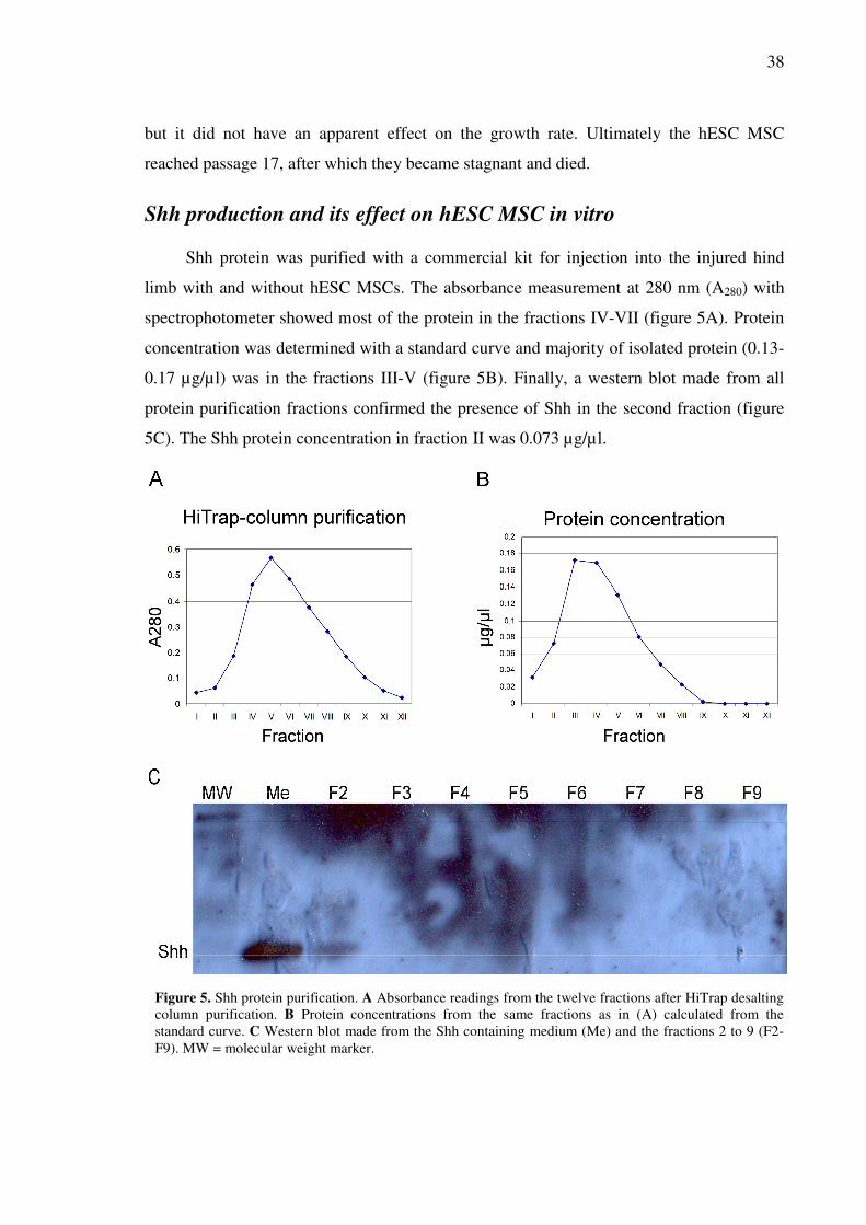
38
but it did not have an apparent effect on the growth rate. Ultimately the hESC MSC
reached passage 17, after which they became stagnant and died.
Shh production and its effect on hESC MSC in vitro
Shh protein was purified with a commercial kit for injection into the injured hind
limb with and without hESC MSCs. The absorbance measurement at 280 nm (A280) with
spectrophotometer showed most of the protein in the fractions IV-VII (figure 5A). Protein
concentration was determined with a standard curve and majority of isolated protein (0.13-
0.17 µg/µl) was in the fractions III-V (figure 5B). Finally, a western blot made from all
protein purification fractions confirmed the presence of Shh in the second fraction (figure
5C). The Shh protein concentration in fraction II was 0.073 µg/µl.
Figure 5. Shh protein purification. A Absorbance readings from the twelve fractions after HiTrap desalting
column purification. B Protein concentrations from the same fractions as in (A) calculated from the
standard curve. C Western blot made from the Shh containing medium (Me) and the fractions 2 to 9 (F2-
F9). MW = molecular weight marker.

39
In order to find out the effect of Shh on the hESC MSC they were incubated in
medium containing Shh. Either of the single overnight or long-term incubations did not
cause changes in the growth or appearance of the cells. Their lifespan was similar to that of
untreated cells with respect to growth rate and age.
LV-GFP production and transduction of hESC MSC
Lentivirus-GFP titer
By the first medium collection majority of the HEK 293T cells producing
lentiviruses with GFP (LV-GFP) were fluorescent indicating a successful transfection
(figure 6) but the presence of virus was further confirmed by running a transduction test
and by determining the titer with HEK 293T cells. Majority of the cells on the test
transduction plate were GFP positive two days after virus addition. LV-GFP titer was
calculated from the result of the fluorescence activated cell sorting analysis using the 1:10
dilution which gave 3.2% fluorescent cells:
1. 3.2% GFP+ cells * 275 000 cells / 100 = 8800 GFP+ cells in 100 µl medium
2. 8800 GFP+ cells in 100 µl medium * 10 = 0.88e105 IU/ml
Multiplicity of infection was determined for the 7 ml of transduction medium used for GFP
labeling of the hESC MSC when the number of cells on the plate was approx. 2e105:
3. (7 ml LV-medium * 0.88e105 IU/ml) / 2* 10
5 = 3.08 ~ 3 MOI
Figure 6. GFP+ lentivirus production with HEK 293T cells. A A fluorescent microscope image of
transfected HEK 293T cells. B Merged ultraviolet and visible light images. C Visible light image of the
cells in (A) and (B).

40
Transduction of hESC MSC turned out effective
HESC MSCs were labeled with luciferase-lentivirus to be used in bioluminescence
imaging (BLI) with Xenogen IVIS 50 Imaging System after transplantation. The cells were
labeled with multiplicity of infection (MOI) of approx. 20, and BLI 48 hrs after
transduction showed even luminescence on the plate, average value being 1.54*1010
p/sec/cm2/sr (figure 7A). The cells remained in good condition during the labeling
procedure and until the transplantation.
HESC MSCs were also labeled with GFP for visual detection from the rat muscle
tissue sections. MOI on the labeling was approx. 3, and incubation in LV-αMEM was
highly efficient. By the time of transplantation nearly 100% of the cells appeared
fluorescent in the microscope (figures 7B-C). However, due to the LV-αMEM being
somewhat depleted of nutrients by the virus producing HEK 293T cells and the viral
infection itself the condition of the stem cells weakened, although not critically. After
changing fresh αMEM medium for the next 48 hrs prior to transplantation the cells
recovered and displayed normal morphology.
The lentiviral transduction of the GFP and luciferase plasmid into the hESC MSC
resulted in a strongly positive fluorescent and luminescent phenotype, respectively, after a
single exposure to the virus. The cells retained this trait even after freezing and thawing
rendering the method a very efficient one. However, the first signs of alterations in the
morphology of GFP-labeled cells were observed already at P10 as the cells developed
neuron-like, and shortly after senescent traits. The life span of GFP+ hESC MSC was
Figure 7. Luciferase and GFP labeling of the hESC MSC. A Bioluminescence image of the luciferase+
hESC MSC on ∅10 cm plate prior to transplantation. B Fluorescence microscope image of the GFP+
hESC MSC on ∅10 cm plate prior to transplantation. C Visible light image of the cells in (B).

41
similar to that of the GFP negative cells in respect of passage number but the growth rate
was continuously faster.
Stem cell transplantation and hESC MSC tracking
Amount of hESC MSC decreased rapidly at the transplantation site
Two animals were imaged for bioluminescence at three time points after luciferase-
hESC MSC transplantation to elucidate their fate in rat tissues (figure 8A). Initial signal
from the transplanted cells was clear and centered at the site of injections (figure 8B). After
six hours the signal had decreased to 28% of the original while scattering of the cells was
not observed (figure 8C). By the 24 hour time point the signal intensity had weakened
further being only 1.5% of the original (figure 8D). The location of the signal had not
changed, and no new luminescent spots had appeared elsewhere indicating that the cells
had died in situ.
A few scattered GFP+ cells were seen in cryosections
Histological sections of the two transplantation groups were visually scanned for
GFP fluorescence with a fluorescence microscope immediately after sectioning.
Fluorescent spots presumed to be the hESC MSC were seen on the slides from both groups
scattered on the whole area of the sections which indicates that after three days the cells
were still present in rat tissues (figure 9A). Localization to the proximity of recognizable
structures such as blood vessels was not observed indicating that the cells did not
incorporate into the tissue, though present at the site of injury.
Human DNA and mRNA were found in the rat tissues three days post-
transplantation
PCR products of both genomic DNA and mRNA isolated from the rat muscle tissue
were run in the agarose gel to determine the presence of human nucleic acids. Positive
control, i.e. the genomic DNA of hESC MSCs, appeared as a bright band on both rows on
the gel, and the size of the fragment was the expected 100 bps according to the ladder.
Fragments of the same size were seen on the sample lanes containing the genomic or the
complementary DNA (cDNA). The band for genomic DNA was clear but less intense than
the positive control indicating the presence of hESC MSCs in the rat hind limb tissues
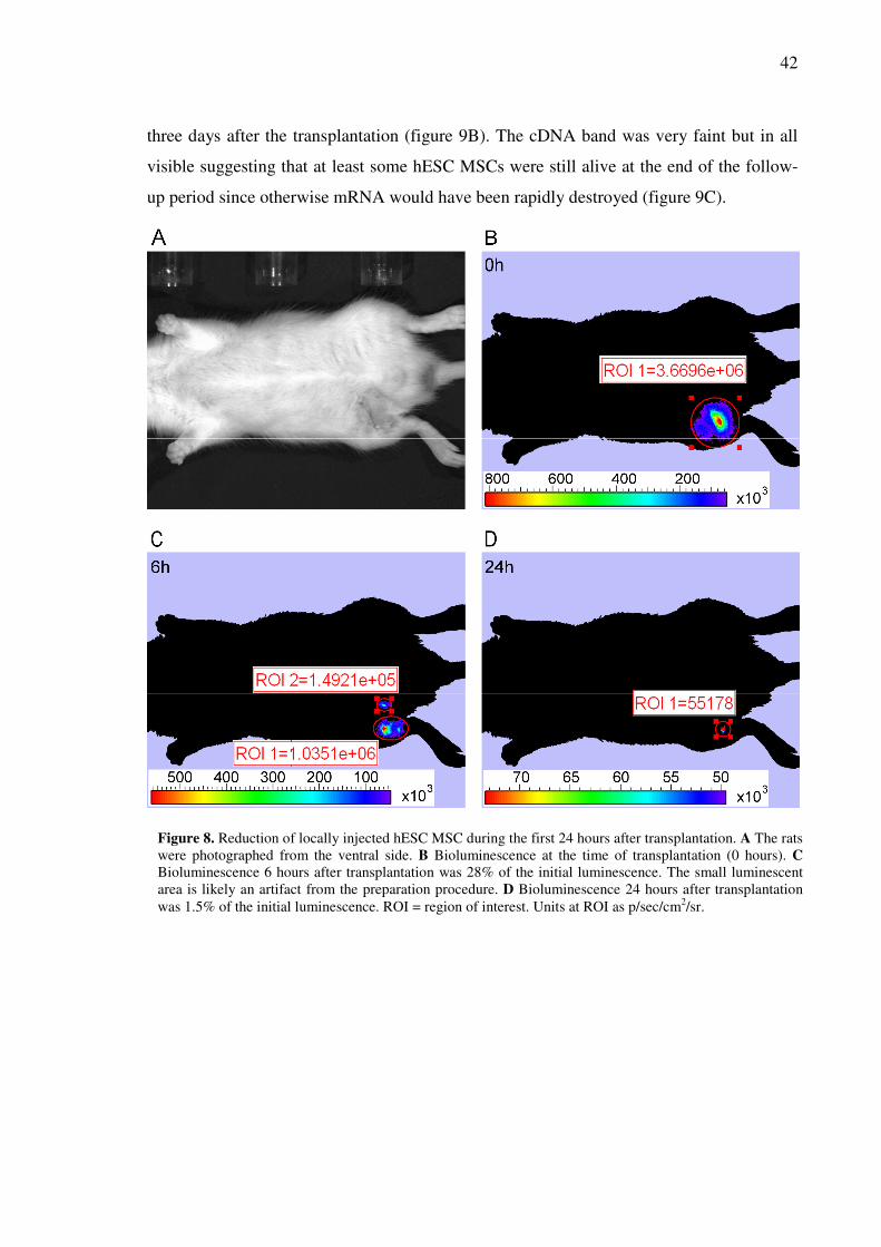
42
three days after the transplantation (figure 9B). The cDNA band was very faint but in all
visible suggesting that at least some hESC MSCs were still alive at the end of the follow-
up period since otherwise mRNA would have been rapidly destroyed (figure 9C).
Figure 8. Reduction of locally injected hESC MSC during the first 24 hours after transplantation. A The rats
were photographed from the ventral side. B Bioluminescence at the time of transplantation (0 hours). C
Bioluminescence 6 hours after transplantation was 28% of the initial luminescence. The small luminescent
area is likely an artifact from the preparation procedure. D Bioluminescence 24 hours after transplantation
was 1.5% of the initial luminescence. ROI = region of interest. Units at ROI as p/sec/cm2/sr.
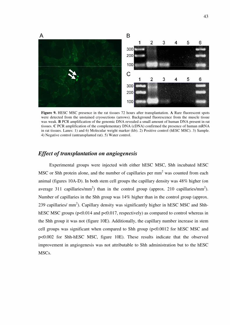
43
Effect of transplantation on angiogenesis
Experimental groups were injected with either hESC MSC, Shh incubated hESC
MSC or Shh protein alone, and the number of capillaries per mm2 was counted from each
animal (figures 10A-D). In both stem cell groups the capillary density was 48% higher (on
average 311 capillaries/mm2) than in the control group (approx. 210 capillaries/mm
2).
Number of capillaries in the Shh group was 14% higher than in the control group (approx.
239 capillaries/ mm2). Capillary density was significantly higher in hESC MSC and Shh-
hESC MSC groups (p<0.014 and p<0.017, respectively) as compared to control whereas in
the Shh group it was not (figure 10E). Additionally, the capillary number increase in stem
cell groups was significant when compared to Shh group (p<0.0012 for hESC MSC and
p<0.002 for Shh-hESC MSC, figure 10E). These results indicate that the observed
improvement in angiogenesis was not attributable to Shh administration but to the hESC
MSCs.
Figure 9. HESC MSC presence in the rat tissues 72 hours after transplantation. A Rare fluorescent spots
were detected from the unstained cryosections (arrows). Background fluorescence from the muscle tissue
was weak. B PCR amplification of the genomic DNA revealed a small amount of human DNA present in rat
tissues. C PCR amplification of the complementary DNA (cDNA) confirmed the presence of human mRNA
in rat tissues. Lanes: 1) and 6) Molecular weight marker (kb). 2) Positive control (hESC MSC). 3) Sample.
4) Negative control (untransplanted rat). 5) Water control.

44
Figure 10. Capillary formation in the rat
muscle. In A hESC MSC group and B Shh-
hESC MSC group the number of capillaries
was higher than in the group that received C
only Shh protein or D in control group.
Horseradish peroxidase stained capillaries are
visible as brown spots on thin sections. E
Statistical analysis showed similar significant
increase in both cell transplantation groups as
compared to control (*) and Shh group (**).
The difference between Shh group and control
group was not significant.

45
Discussion
Human embryonic stem cell derived mesenchymal stem cells (hESC MSC)
constitute a new cell line that has gained interest in the recent years in terms of their
derivation and possible clinical applications. This is justified by the benefits they have over
more traditional stem cell types, such as the practically endless source material, resistance
to malignant transformation (Barberi et al., 2007), and multilineage differentiation (Trivedi
and Hematti, 2007). These characteristics provide a good starting point for a range of
therapies. This study focused on the ability of hESC MSC to repair tissue damage after
induced ischemia and following inflammation in a rat hind limb model. The injury model
used here offers a rather simple and reliable way to generate acute ischemia, and thus it
was considered suitable in investigating the course of events after transplantation.
According to our observations the hESC MSC resemble normal bone marrow
derived mesenchymal stem cells (Sekiya et al., 2002). The cells were readily labeled with
lentiviral GFP and luciferase, and the label was stable through passaging and freeze-thaw
cycles. Additionally, the cell line was found to be finite with similar passage numbers as
described in the paper of Trivedi and Hematti (2007). Malignant transformation in stem
cells is always a concern when conducting research with clinical perspectives, but a
promising study suggested that this type of cells did not form tumors during six month
surveillance after transplantation into mice (Barberi et al., 2007). Here spontaneous
transformation was not detected in vitro but the follow-up time was too short to establish
this in vivo. However, alterations in morphology in vitro at late passages may indicate that
the hESC MSC loose their ‘stemness’, although it has been shown that differentiation
potential is preserved even in prolonged culturing (Barberi et al., 2007). In this study,
transplanted cells were at passage 7 whereas the hESC MSC have been found to be
multipotent still at passage 9 (Trivedi and Hematti, 2007).
Ischemic environment in skeletal muscle is characterized by massive cell death and
high amounts of free oxygen radicals, cytokines and inflammatory cells responding to
acute hypoxia. Consequently, apoptosis-inducing and cytolytic factors from necrotic cells,
and infiltrating monocyte-macrophages and granulocytes create harsh conditions for the
transplanted cells. Apoptosis or necrosis in situ has probably been the fate of most
transplanted hESC MSC since any signal from possible target organs of stem cell

46
migration was not detected. The presence of live hESC MSC was initially indicated by the
few scattered fluorescing dots in the histological sections and faint bioluminescence
obtained with the IVIS at 24 hour time point. Three day time point was not considered
necessary since bioluminescence had already decreased to 1.5% of the original 24 hours
after transplantation. Our RT-PCR and PCR results showed presence of human mRNA and
genomic DNA confirming that some hESC MSC were indeed alive after three days in the
host tissues. It is well known that free DNA and RNA are rapidly destroyed once the cell
has ruptured necessitating the presence of live cells in our experimental conditions.
Presence of a number of different signaling molecules in the ischemic site where the
hESC MSC were transplanted could possibly promote their differentiation. Multipotent
mesenchymal stem cells have a wide differentiation capacity from adipocytes and
myoblasts (Pittenger et al., 1999) even beyond mesenchymal borders to e.g. neuronal cells
(Woodbury et al., 2000). In our model, extensive tissue damage and inflammation would
likely promote differentiation of the stem cells towards fibroblast-like cells and result in
scar tissue formation (Yan et al., 2007). Other possibilities include myoblasts and
pericytes, which are naturally of mesenchymal origin and have been observed to arise from
transplanted autologous MSCs in ischemic muscle tissue (Tang et al., 2006). Our three day
follow-up time was far too short for the stem cells to fully differentiate since it usually
requires at least a few cell divisions. Additionally, the random distribution of the cells and
rapid decline in bioluminescence implies that the hESC MSC were not even engrafted into
host tissue which could be due to the unfavorable environment and in vitro culturing
(Rombouts and Ploemacher, 2003).
Neoangiogenesis is an absolute requirement for tissue healing, and it is regulated by
a complex signaling network of growth factors e.g. VEGF, FGF, and cytokines produced
by the participating cells such as endothelial and smooth muscle cells, monocyte-
macrophages, and fibroblasts. As a sign of active angiogenesis we observed significantly
increased capillary formation in both test groups that received hESC MSC. However, Shh
alone did not have any major effect. These results suggest a more important role for the
stem cells in tissue recovery, although Shh has been shown to take part in the angiogenetic
events (Kusano et al., 2005). Possible causes for our observations could be the low amount
of Shh in medium, its inefficient purification or inactivation of the purified protein during
47
the short storage in -70°C. These would also account for the seemingly ineffective in vitro
incubation of the hESC MSC in Shh medium.
Another substance that has been observed to affect angiogenesis is cyclosporine A.
Despite the side effects cyclosporine A has been used for long to suppress the host immune
system against a transplanted organ and its effect is based on the downregulation of the T
cell response. Administered alone it has been shown to inhibit VEGF-mediated
angiogenetic signaling pathway in vitro (Rafiee et al., 2004) but in our study the hESC
MSCs seemed to exert an antagonizing effect. Cyclosporine A has also been shown to be
important in enhancing MSC graft survival after transplantation into rat spinal cord
(Swanger et al., 2005); however, we did not see any sign of actual engraftment. Whether
the cyclosporine A affected the rapid clearance of the hESC MSCs from rat tissues in
unclear but, altogether, our results suggest an alternative mechanism for the MSC-induced
tissue recovery.
Results of mesenchymal stem cell transplantations vary immensely from study to
study suggesting either high engraftment efficiency (Direkze et al., 2003) or none at all
(Zentilin et al., 2006), or something between, although their therapeutic effect remains
undisputed. We did not observe engraftment but the contribution of hESC MSC to tissue
recovery was evident and therefore other mechanisms must underlie the positive effect. As
shown by several studies, the paracrine effect by the MSCs can improve cardiac muscle
recovery from ischemia (Iso et al., 2007; Kinnaird et al., 2004) but here the dead hESC
MSCs clearly outnumbered the live ones very rapidly rendering this mechanism unlikely or
at least highly ineffective. A more feasible explanation would be that the cell debris and
released cell contents activated resident macrophages and other tissue cells, thus
stimulating the expression of angiogenic factors as a result of inflammatory reaction first
initiated by hypoxia; however, we have not confirmed the activation of the immune
system. In the future, a thorough investigation of the signaling events and interactions
between the hESC MSC and host tissue are needed to estimate the potential of this cell
population in clinical settings.
In summary, we have demonstrated the ability of hESC MSCs to improve tissue
recovery through angiogenesis in ischemic muscle tissue of rat hind limb. Our data does
not support stem cell engraftment into host tissues after transplantation as shown by

48
bioluminescence imaging and rare GFP+ cells in the tissues, although a small number of
them had survived according to the results of PCR amplification using human specific
primers on rat tissue DNA and mRNA. The actual mechanism behind the significantly
enhanced angiogenesis and tissue healing still needs to be elucidated, and safety studies
regarding their tumorigenicity are required.

49
References
Abdollahi, A., C. Schwager, J. Kleeff, I. Esposito, S. Domhan, P. Peschke, K. Hauser, P. Hahnfeldt, L.
Hlatky, J. Debus, J.M. Peters, H. Friess, J. Folkman, and P.E. Huber. 2007. Transcriptional network
governing the angiogenic switch in human pancreatic cancer. Proc.Natl.Acad.Sci.U.S.A. 104:12890-12895.
Anderson, C.R., A.M. Ponce, and R.J. Price. 2004. Immunohistochemical identification of an extracellular
matrix scaffold that microguides capillary sprouting in vivo. J.Histochem.Cytochem. 52:1063-1072.
Andrade, S.P., T.P. Fan, and G.P. Lewis. 1987. Quantitative in-vivo studies on angiogenesis in a rat sponge
model. Br.J.Exp.Pathol. 68:755-766.
Armulik, A., A. Abramsson, and C. Betsholtz. 2005. Endothelial/pericyte interactions. Circ.Res. 97:512-523.
Auerbach, R., L. Kubai, D. Knighton, and J. Folkman. 1974. A simple procedure for the long-term
cultivation of chicken embryos. Dev.Biol. 41:391-394.
Augello, A., R. Tasso, S.M. Negrini, A. Amateis, F. Indiveri, R. Cancedda, and G. Pennesi. 2005. Bone
marrow mesenchymal progenitor cells inhibit lymphocyte proliferation by activation of the programmed
death 1 pathway. Eur.J.Immunol. 35:1482-1490.
Azizi, S.A., D. Stokes, B.J. Augelli, C. DiGirolamo, and D.J. Prockop. 1998. Engraftment and migration of
human bone marrow stromal cells implanted in the brains of albino rats--similarities to astrocyte grafts.
Proc.Natl.Acad.Sci.U.S.A. 95:3908-3913.
Baba, S., H. Fujii, T. Hirose, K. Yasuchika, H. Azuma, T. Hoppo, M. Naito, T. Machimoto, and I. Ikai. 2004.
Commitment of bone marrow cells to hepatic stellate cells in mouse. J.Hepatol. 40:255-260.
Baffour, R., J.L. Garb, J. Kaufman, J. Berman, S.W. Rhee, M.A. Norris, and P. Friedmann. 2000. Angiogenic
therapy for the chronically ischemic lower limb in a rabbit model. J.Surg.Res. 93:219-229.
Barbera-Guillem, E., J.K. Nyhus, C.C. Wolford, C.R. Friece, and J.W. Sampsel. 2002. Vascular endothelial
growth factor secretion by tumor-infiltrating macrophages essentially supports tumor angiogenesis, and IgG
immune complexes potentiate the process. Cancer Res. 62:7042-7049.
Barberi, T., M. Bradbury, Z. Dincer, G. Panagiotakos, N.D. Socci, and L. Studer. 2007. Derivation of
engraftable skeletal myoblasts from human embryonic stem cells. Nat.Med. 13:642-648.
Barda-Saad, M., L.A. Rozenszajn, H. Ashush, Y. Shav-Tal, A. Ben Nun, and D. Zipori. 1999. Adhesion
molecules involved in the interactions between early T cells and mesenchymal bone marrow stromal cells.
Exp.Hematol. 27:834-844.
Bartholomew, A., S. Patil, A. Mackay, M. Nelson, D. Buyaner, W. Hardy, J. Mosca, C. Sturgeon, M.
Siatskas, N. Mahmud, K. Ferrer, R. Deans, A. Moseley, R. Hoffman, and S.M. Devine. 2001. Baboon
mesenchymal stem cells can be genetically modified to secrete human erythropoietin in vivo. Hum.Gene
Ther. 12:1527-1541.
Bartholomew, A., C. Sturgeon, M. Siatskas, K. Ferrer, K. McIntosh, S. Patil, W. Hardy, S. Devine, D. Ucker,
R. Deans, A. Moseley, and R. Hoffman. 2002. Mesenchymal stem cells suppress lymphocyte proliferation in
vitro and prolong skin graft survival in vivo. Exp.Hematol. 30:42-48.
Bellows, C.G., W. Pei, Y. Jia, and J.N. Heersche. 2003. Proliferation, differentiation and self-renewal of
osteoprogenitors in vertebral cell populations from aged and young female rats. Mech.Ageing Dev. 124:747-
757.

50
Benjamin, L.E., I. Hemo, and E. Keshet. 1998. A plasticity window for blood vessel remodelling is defined
by pericyte coverage of the preformed endothelial network and is regulated by PDGF-B and VEGF.
Development. 125:1591-1598.
Berse, B., J.A. Hunt, R.J. Diegel, P. Morganelli, K. Yeo, F. Brown, and R.A. Fava. 1999. Hypoxia augments
cytokine (transforming growth factor-beta (TGF-beta) and IL-1)-induced vascular endothelial growth factor
secretion by human synovial fibroblasts. Clin.Exp.Immunol. 115:176-182.
Bianchi, D.W. 2000. Fetal cells in the mother: from genetic diagnosis to diseases associated with fetal cell
microchimerism. Eur.J.Obstet.Gynecol.Reprod.Biol. 92:103-108.
Bodnar, A.G., M. Ouellette, M. Frolkis, S.E. Holt, C.P. Chiu, G.B. Morin, C.B. Harley, J.W. Shay, S.
Lichtsteiner, and W.E. Wright. 1998. Extension of life-span by introduction of telomerase into normal human
cells. Science. 279:349-352.
Bottomley, M.J., N.J. Webb, C.J. Watson, L. Holt, M. Bukhari, J. Denton, A.J. Freemont, and P.E.
Brenchley. 2000. Placenta growth factor (PlGF) induces vascular endothelial growth factor (VEGF) secretion
from mononuclear cells and is co-expressed with VEGF in synovial fluid. Clin.Exp.Immunol. 119:182-188.
Burger, D., R. Rezzonico, J.M. Li, C. Modoux, R.A. Pierce, H.G. Welgus, and J.M. Dayer. 1998. Imbalance
between interstitial collagenase and tissue inhibitor of metalloproteinases 1 in synoviocytes and fibroblasts
upon direct contact with stimulated T lymphocytes: involvement of membrane-associated cytokines. Arthritis
Rheum. 41:1748-1759.
Buschmann, I.R., M. Voskuil, N. van Royen, I.E. Hoefer, K. Scheffler, S. Grundmann, J. Hennig, W.
Schaper, C. Bode, and J.J. Piek. 2003. Invasive and non-invasive evaluation of spontaneous arteriogenesis in
a novel porcine model for peripheral arterial obstructive disease. Atherosclerosis. 167:33-43.
Calvi, L.M., G.B. Adams, K.W. Weibrecht, J.M. Weber, D.P. Olson, M.C. Knight, R.P. Martin, E. Schipani,
P. Divieti, F.R. Bringhurst, L.A. Milner, H.M. Kronenberg, and D.T. Scadden. 2003. Osteoblastic cells
regulate the haematopoietic stem cell niche. Nature. 425:841-846.
Campagnoli, C., I.A. Roberts, S. Kumar, P.R. Bennett, I. Bellantuono, and N.M. Fisk. 2001. Identification of
mesenchymal stem/progenitor cells in human first-trimester fetal blood, liver, and bone marrow. Blood.
98:2396-2402.
Carmeliet, P. 2000. Mechanisms of angiogenesis and arteriogenesis. Nat.Med. 6:389-395.
Carmeliet, P. 2003. Angiogenesis in health and disease. Nat.Med. 9:653-660.
Casciari, J.J., M.G. Hollingshead, M.C. Alley, J.G. Mayo, L. Malspeis, S. Miyauchi, M.R. Grever, and J.N.
Weinstein. 1994. Growth and chemotherapeutic response of cells in a hollow-fiber in vitro solid tumor
model. J.Natl.Cancer Inst. 86:1846-1852.
Charbord, P., R. Oostendorp, W. Pang, O. Herault, F. Noel, T. Tsuji, E. Dzierzak, and B. Peault. 2002.
Comparative study of stromal cell lines derived from embryonic, fetal, and postnatal mouse blood-forming
tissues. Exp.Hematol. 30:1202-1210.
Colter, D.C., R. Class, C.M. DiGirolamo, and D.J. Prockop. 2000. Rapid expansion of recycling stem cells in
cultures of plastic-adherent cells from human bone marrow. Proc.Natl.Acad.Sci.U.S.A. 97:3213-3218.
Colter, D.C., I. Sekiya, and D.J. Prockop. 2001. Identification of a subpopulation of rapidly self-renewing
and multipotential adult stem cells in colonies of human marrow stromal cells. Proc.Natl.Acad.Sci.U.S.A.
98:7841-7845.
da Silva Meirelles, L., P.C. Chagastelles, and N.B. Nardi. 2006. Mesenchymal stem cells reside in virtually
all post-natal organs and tissues. J.Cell.Sci. 119:2204-2213.

51
Davis, G.E., and C.W. Camarillo. 1995. Regulation of endothelial cell morphogenesis by integrins,
mechanical forces, and matrix guidance pathways. Exp.Cell Res. 216:113-123.
Devine, M.J., C.M. Mierisch, E. Jang, P.C. Anderson, and G. Balian. 2002. Transplanted bone marrow cells
localize to fracture callus in a mouse model. J.Orthop.Res. 20:1232-1239.
Devine, S.M., C. Cobbs, M. Jennings, A. Bartholomew, and R. Hoffman. 2003. Mesenchymal stem cells
distribute to a wide range of tissues following systemic infusion into nonhuman primates. Blood. 101:2999-
3001.
Digirolamo, C.M., D. Stokes, D. Colter, D.G. Phinney, R. Class, and D.J. Prockop. 1999. Propagation and
senescence of human marrow stromal cells in culture: a simple colony-forming assay identifies samples with
the greatest potential to propagate and differentiate. Br.J.Haematol. 107:275-281.
Direkze, N.C., S.J. Forbes, M. Brittan, T. Hunt, R. Jeffery, S.L. Preston, R. Poulsom, K. Hodivala-Dilke,
M.R. Alison, and N.A. Wright. 2003. Multiple organ engraftment by bone-marrow-derived myofibroblasts
and fibroblasts in bone-marrow-transplanted mice. Stem Cells. 21:514-520.
Dominici, M., K. Le Blanc, I. Mueller, I. Slaper-Cortenbach, F. Marini, D. Krause, R. Deans, A. Keating, D.
Prockop, and E. Horwitz. 2006. Minimal criteria for defining multipotent mesenchymal stromal cells. The
International Society for Cellular Therapy position statement. Cytotherapy. 8:315-317.
Dvorak, H.F., L.F. Brown, M. Detmar, and A.M. Dvorak. 1995. Vascular permeability factor/vascular
endothelial growth factor, microvascular hyperpermeability, and angiogenesis. Am.J.Pathol. 146:1029-1039.
Eliopoulos, N., J. Stagg, L. Lejeune, S. Pommey, and J. Galipeau. 2005. Allogeneic marrow stromal cells are
immune rejected by MHC class I- and class II-mismatched recipient mice. Blood. 106:4057-4065.
Fajardo, L.F., J. Kowalski, H.H. Kwan, S.D. Prionas, and A.C. Allison. 1988. The disc angiogenesis system.
Lab.Invest. 58:718-724.
Fathke, C., L. Wilson, J. Hutter, V. Kapoor, A. Smith, A. Hocking, and F. Isik. 2004. Contribution of bone
marrow-derived cells to skin: collagen deposition and wound repair. Stem Cells. 22:812-822.
Folkman, J., E. Merler, C. Abernathy, and G. Williams. 1971. Isolation of a tumor factor responsible for
angiogenesis. J.Exp.Med. 133:275-288.
Forsythe, J.A., B.H. Jiang, N.V. Iyer, F. Agani, S.W. Leung, R.D. Koos, and G.L. Semenza. 1996. Activation
of vascular endothelial growth factor gene transcription by hypoxia-inducible factor 1. Mol.Cell.Biol.
16:4604-4613.
Franke, R.P., M. Grafe, H. Schnittler, D. Seiffge, C. Mittermayer, and D. Drenckhahn. 1984. Induction of
human vascular endothelial stress fibres by fluid shear stress. Nature. 307:648-649.
Friedenstein, A.J., R.K. Chailakhyan, N.V. Latsinik, A.F. Panasyuk, and I.V. Keiliss-Borok. 1974. Stromal
cells responsible for transferring the microenvironment of the hemopoietic tissues. Cloning in vitro and
retransplantation in vivo. Transplantation. 17:331-340.
Friedenstein, A.J., J.F. Gorskaja, and N.N. Kulagina. 1976. Fibroblast precursors in normal and irradiated
mouse hematopoietic organs. Exp.Hematol. 4:267-274.
Fuchs, J.R., D. Hannouche, S. Terada, J.P. Vacanti, and D.O. Fauza. 2003. Fetal tracheal augmentation with
cartilage engineered from bone marrow-derived mesenchymal progenitor cells. J.Pediatr.Surg. 38:984-987.
Genis, L., P. Gonzalo, A.S. Tutor, B.G. Galvez, A. Martinez-Ruiz, C. Zaragoza, S. Lamas, K. Tryggvason,
S.S. Apte, and A.G. Arroyo. 2007. Functional interplay between endothelial nitric oxide synthase and
membrane type 1-matrix metalloproteinase in migrating endothelial cells. Blood.

52
Gennaro, G., C. Menard, S.E. Michaud, and A. Rivard. 2003. Age-dependent impairment of
reendothelialization after arterial injury: role of vascular endothelial growth factor. Circulation. 107:230-233.
Gimbrone, M.A.,Jr, R.S. Cotran, S.B. Leapman, and J. Folkman. 1974. Tumor growth and
neovascularization: an experimental model using the rabbit cornea. J.Natl.Cancer Inst. 52:413-427.
Giordano, A., U. Galderisi, and I.R. Marino. 2007. From the laboratory bench to the patient's bedside: an
update on clinical trials with mesenchymal stem cells. J.Cell.Physiol. 211:27-35.
Goumans, M.J., G. Valdimarsdottir, S. Itoh, A. Rosendahl, P. Sideras, and P. ten Dijke. 2002. Balancing the
activation state of the endothelium via two distinct TGF-beta type I receptors. EMBO J. 21:1743-1753.
Grigoriadis, A.E., J.N. Heersche, and J.E. Aubin. 1988. Differentiation of muscle, fat, cartilage, and bone
from progenitor cells present in a bone-derived clonal cell population: effect of dexamethasone. J.Cell Biol.
106:2139-2151.
Gronthos, S., J. Brahim, W. Li, L.W. Fisher, N. Cherman, A. Boyde, P. DenBesten, P.G. Robey, and S. Shi.
2002. Stem cell properties of human dental pulp stem cells. J.Dent.Res. 81:531-535.
Grunewald, M., I. Avraham, Y. Dor, E. Bachar-Lustig, A. Itin, S. Jung, S. Chimenti, L. Landsman, R.
Abramovitch, and E. Keshet. 2006. VEGF-induced adult neovascularization: recruitment, retention, and role
of accessory cells. Cell. 124:175-189.
Gustafsson, T., A. Puntschart, L. Kaijser, E. Jansson, and C.J. Sundberg. 1999. Exercise-induced expression
of angiogenesis-related transcription and growth factors in human skeletal muscle. Am.J.Physiol. 276:H679-
85.
Gutierrez, L.S., A. Schulman, T. Brito-Robinson, F. Noria, V.A. Ploplis, and F.J. Castellino. 2000. Tumor
development is retarded in mice lacking the gene for urokinase-type plasminogen activator or its inhibitor,
plasminogen activator inhibitor-1. Cancer Res. 60:5839-5847.
Hangai, M., N. Kitaya, J. Xu, C.K. Chan, J.J. Kim, Z. Werb, S.J. Ryan, and P.C. Brooks. 2002. Matrix
metalloproteinase-9-dependent exposure of a cryptic migratory control site in collagen is required before
retinal angiogenesis. Am.J.Pathol. 161:1429-1437.
Heil, M., and W. Schaper. 2004. Influence of mechanical, cellular, and molecular factors on collateral artery
growth (Arteriogenesis). Circ.Res. 95:449-458.
Hellstrom, M., H. Gerhardt, M. Kalen, X. Li, U. Eriksson, H. Wolburg, and C. Betsholtz. 2001. Lack of
pericytes leads to endothelial hyperplasia and abnormal vascular morphogenesis. J.Cell Biol. 153:543-553.
Hofstetter, C.P., E.J. Schwarz, D. Hess, J. Widenfalk, A. El Manira, D.J. Prockop, and L. Olson. 2002.
Marrow stromal cells form guiding strands in the injured spinal cord and promote recovery.
Proc.Natl.Acad.Sci.U.S.A. 99:2199-2204.
Horwitz, E.M., K. Le Blanc, M. Dominici, I. Mueller, I. Slaper-Cortenbach, F.C. Marini, R.J. Deans, D.S.
Krause, A. Keating, and the International Society for Cellular Therapy. 2005. Clarification of the
nomenclature for MSC: The International Society for Cellular Therapy position statement. Cytotherapy.
7:393-395.
Horwitz, E.M., D.J. Prockop, L.A. Fitzpatrick, W.W. Koo, P.L. Gordon, M. Neel, M. Sussman, P. Orchard,
J.C. Marx, R.E. Pyeritz, and M.K. Brenner. 1999. Transplantability and therapeutic effects of bone marrow-
derived mesenchymal cells in children with osteogenesis imperfecta. Nat.Med. 5:309-313.
Houck, K.A., D.W. Leung, A.M. Rowland, J. Winer, and N. Ferrara. 1992. Dual regulation of vascular
endothelial growth factor bioavailability by genetic and proteolytic mechanisms. J.Biol.Chem. 267:26031-
26037.

53
in 't Anker, P.S., W.A. Noort, S.A. Scherjon, C. Kleijburg-van der Keur, A.B. Kruisselbrink, R.L. van
Bezooijen, W. Beekhuizen, R. Willemze, H.H. Kanhai, and W.E. Fibbe. 2003. Mesenchymal stem cells in
human second-trimester bone marrow, liver, lung, and spleen exhibit a similar immunophenotype but a
heterogeneous multilineage differentiation potential. Haematologica. 88:845-852.
Ingram, W.J., C.A. Wicking, S.M. Grimmond, A.R. Forrest, and B.J. Wainwright. 2002. Novel genes
regulated by Sonic Hedgehog in pluripotent mesenchymal cells. Oncogene. 21:8196-8205.
Iso, Y., J.L. Spees, C. Serrano, B. Bakondi, R. Pochampally, Y.H. Song, B.E. Sobel, P. Delafontaine, and
D.J. Prockop. 2007. Multipotent human stromal cells improve cardiac function after myocardial infarction in
mice without long-term engraftment. Biochem.Biophys.Res.Commun. 354:700-706.
Jackson, K.A., S.M. Majka, H. Wang, J. Pocius, C.J. Hartley, M.W. Majesky, M.L. Entman, L.H. Michael,
K.K. Hirschi, and M.A. Goodell. 2001. Regeneration of ischemic cardiac muscle and vascular endothelium
by adult stem cells. J.Clin.Invest. 107:1395-1402.
Jakob, W., K.D. Jentzsch, B. Mauersberger, and G. Heder. 1978. The chick embryo choriallantoic membrane
as a bioassay for angiogenesis factors: reactions induced by carrier materials. Exp.Pathol.(Jena). 15:241-249.
Jenkinson, L., K.D. Bardhan, J. Atherton, and N. Kalia. 2002. Helicobacter pylori prevents proliferative stage
of angiogenesis in vitro: role of cytokines. Dig.Dis.Sci. 47:1857-1862.
Kamat, A.A., T.J. Kim, C.N. Landen Jr, C. Lu, L.Y. Han, Y.G. Lin, W.M. Merritt, P.H. Thaker, D.M.
Gershenson, F.Z. Bischoff, J.V. Heymach, R.B. Jaffe, R.L. Coleman, and A.K. Sood. 2007. Metronomic
chemotherapy enhances the efficacy of antivascular therapy in ovarian cancer. Cancer Res. 67:281-288.
Kanda, S., Y. Mochizuki, T. Suematsu, Y. Miyata, K. Nomata, and H. Kanetake. 2003. Sonic hedgehog
induces capillary morphogenesis by endothelial cells through phosphoinositide 3-kinase. J.Biol.Chem.
278:8244-8249.
Kim, J.S., J.M. Kim, H.C. Jung, and I.S. Song. 2004. Helicobacter pylori down-regulates the receptors of
vascular endothelial growth factor and angiopoietin in vascular endothelial cells: implications in the
impairment of gastric ulcer healing. Dig.Dis.Sci. 49:778-786.
Kinnaird, T., E. Stabile, M.S. Burnett, C.W. Lee, S. Barr, S. Fuchs, and S.E. Epstein. 2004. Marrow-derived
stromal cells express genes encoding a broad spectrum of arteriogenic cytokines and promote in vitro and in
vivo arteriogenesis through paracrine mechanisms. Circ.Res. 94:678-685.
Koc, O.N., S.L. Gerson, B.W. Cooper, S.M. Dyhouse, S.E. Haynesworth, A.I. Caplan, and H.M. Lazarus.
2000. Rapid hematopoietic recovery after coinfusion of autologous-blood stem cells and culture-expanded
marrow mesenchymal stem cells in advanced breast cancer patients receiving high-dose chemotherapy.
J.Clin.Oncol. 18:307-316.
Kon, E., A. Muraglia, A. Corsi, P. Bianco, M. Marcacci, I. Martin, A. Boyde, I. Ruspantini, P. Chistolini, M.
Rocca, R. Giardino, R. Cancedda, and R. Quarto. 2000. Autologous bone marrow stromal cells loaded onto
porous hydroxyapatite ceramic accelerate bone repair in critical-size defects of sheep long bones.
J.Biomed.Mater.Res. 49:328-337.
Kurosaka, D., T.W. LeBien, and J.A. Pribyl. 1999. Comparative studies of different stromal cell
microenvironments in support of human B-cell development. Exp.Hematol. 27:1271-1281.
Kusano, K.F., R. Pola, T. Murayama, C. Curry, A. Kawamoto, A. Iwakura, S. Shintani, M. Ii, J. Asai, T.
Tkebuchava, T. Thorne, H. Takenaka, R. Aikawa, D. Goukassian, P. von Samson, H. Hamada, Y.S. Yoon,
M. Silver, E. Eaton, H. Ma, L. Heyd, M. Kearney, W. Munger, J.A. Porter, R. Kishore, and D.W. Losordo.
2005. Sonic hedgehog myocardial gene therapy: tissue repair through transient reconstitution of embryonic
signaling. Nat.Med. 11:1197-1204.

54
Lazarus, H.M., S.E. Haynesworth, S.L. Gerson, N.S. Rosenthal, and A.I. Caplan. 1995. Ex vivo expansion
and subsequent infusion of human bone marrow-derived stromal progenitor cells (mesenchymal progenitor
cells): implications for therapeutic use. Bone Marrow Transplant. 16:557-564.
Lazarus, H.M., O.N. Koc, S.M. Devine, P. Curtin, R.T. Maziarz, H.K. Holland, E.J. Shpall, P. McCarthy, K.
Atkinson, B.W. Cooper, S.L. Gerson, M.J. Laughlin, F.R. Loberiza Jr, A.B. Moseley, and A. Bacigalupo.
2005. Cotransplantation of HLA-identical sibling culture-expanded mesenchymal stem cells and
hematopoietic stem cells in hematologic malignancy patients. Biol.Blood Marrow Transplant. 11:389-398.
Le Blanc, K., I. Rasmusson, B. Sundberg, C. Gotherstrom, M. Hassan, M. Uzunel, and O. Ringden. 2004.
Treatment of severe acute graft-versus-host disease with third party haploidentical mesenchymal stem cells.
Lancet. 363:1439-1441.
Lee, C.C., F. Ye, and A.F. Tarantal. 2006. Comparison of growth and differentiation of fetal and adult rhesus
monkey mesenchymal stem cells. Stem Cells Dev. 15:209-220.
Lee, J.Y., Z. Qu-Petersen, B. Cao, S. Kimura, R. Jankowski, J. Cummins, A. Usas, C. Gates, P. Robbins, A.
Wernig, and J. Huard. 2000. Clonal isolation of muscle-derived cells capable of enhancing muscle
regeneration and bone healing. J.Cell Biol. 150:1085-1100.
Lee, K.D., T.K. Kuo, J. Whang-Peng, Y.F. Chung, C.T. Lin, S.H. Chou, J.R. Chen, Y.P. Chen, and O.K. Lee.
2004. In vitro hepatic differentiation of human mesenchymal stem cells. Hepatology. 40:1275-1284.
Levine, A.J., I. Munoz-Sanjuan, E. Bell, A.J. North, and A.H. Brivanlou. 2003. Fluorescent labeling of
endothelial cells allows in vivo, continuous characterization of the vascular development of Xenopus laevis.
Dev.Biol. 254:50-67.
Li, K.J., M.S. Dilber, M.R. Abedi, B. Bjorkstrand, C.I. Smith, H. Garoff, G. Gahrton, and K.G.
Xanthopoulos. 1995. Retroviral-mediated gene transfer into human bone marrow stromal cells: studies of
efficiency and in vivo survival in SCID mice. Eur.J.Haematol. 55:302-306.
Liechty, K.W., T.C. MacKenzie, A.F. Shaaban, A. Radu, A.M. Moseley, R. Deans, D.R. Marshak, and A.W.
Flake. 2000. Human mesenchymal stem cells engraft and demonstrate site-specific differentiation after in
utero transplantation in sheep. Nat.Med. 6:1282-1286.
Lindahl, P., B.R. Johansson, P. Leveen, and C. Betsholtz. 1997. Pericyte loss and microaneurysm formation
in PDGF-B-deficient mice. Science. 277:242-245.
Maisonpierre, P.C., C. Suri, P.F. Jones, S. Bartunkova, S.J. Wiegand, C. Radziejewski, D. Compton, J.
McClain, T.H. Aldrich, N. Papadopoulos, T.J. Daly, S. Davis, T.N. Sato, and G.D. Yancopoulos. 1997.
Angiopoietin-2, a natural antagonist for Tie2 that disrupts in vivo angiogenesis. Science. 277:55-60.
Majumdar, M.K., M.A. Thiede, J.D. Mosca, M. Moorman, and S.L. Gerson. 1998. Phenotypic and functional
comparison of cultures of marrow-derived mesenchymal stem cells (MSCs) and stromal cells. J.Cell.Physiol.
176:57-66.
Makkar, R.R., M.J. Price, M. Lill, M. Frantzen, K. Takizawa, T. Kleisli, J. Zheng, S. Kar, R. McClelan, T.
Miyamota, J. Bick-Forrester, M.C. Fishbein, P.K. Shah, J.S. Forrester, B. Sharifi, P.S. Chen, and M.
Qayyum. 2005. Intramyocardial injection of allogenic bone marrow-derived mesenchymal stem cells without
immunosuppression preserves cardiac function in a porcine model of myocardial infarction.
J.Cardiovasc.Pharmacol.Ther. 10:225-233.
Mangi, A.A., N. Noiseux, D. Kong, H. He, M. Rezvani, J.S. Ingwall, and V.J. Dzau. 2003. Mesenchymal
stem cells modified with Akt prevent remodeling and restore performance of infarcted hearts. Nat.Med.
9:1195-1201.

55
Martin, D.R., N.R. Cox, T.L. Hathcock, G.P. Niemeyer, and H.J. Baker. 2002. Isolation and characterization
of multipotential mesenchymal stem cells from feline bone marrow. Exp.Hematol. 30:879-886.
Meyer, G.T., L.J. Matthias, L. Noack, M.A. Vadas, and J.R. Gamble. 1997. Lumen formation during
angiogenesis in vitro involves phagocytic activity, formation and secretion of vacuoles, cell death, and
capillary tube remodelling by different populations of endothelial cells. Anat.Rec. 249:327-340.
Meyer, M., M. Clauss, A. Lepple-Wienhues, J. Waltenberger, H.G. Augustin, M. Ziche, C. Lanz, M. Buttner,
H.J. Rziha, and C. Dehio. 1999. A novel vascular endothelial growth factor encoded by Orf virus, VEGF-E,
mediates angiogenesis via signalling through VEGFR-2 (KDR) but not VEGFR-1 (Flt-1) receptor tyrosine
kinases. EMBO J. 18:363-374.
Minasi, M.G., M. Riminucci, L. De Angelis, U. Borello, B. Berarducci, A. Innocenzi, A. Caprioli, D.
Sirabella, M. Baiocchi, R. De Maria, R. Boratto, T. Jaffredo, V. Broccoli, P. Bianco, and G. Cossu. 2002.
The meso-angioblast: a multipotent, self-renewing cell that originates from the dorsal aorta and differentiates
into most mesodermal tissues. Development. 129:2773-2783.
Moore, B.E., and P.J. Quesenberry. 2003. The adult hemopoietic stem cell plasticity debate: idols vs new
paradigms. Leukemia. 17:1205-1210.
Moutsatsos, I.K., G. Turgeman, S. Zhou, B.G. Kurkalli, G. Pelled, L. Tzur, P. Kelley, N. Stumm, S. Mi, R.
Muller, Y. Zilberman, and D. Gazit. 2001. Exogenously regulated stem cell-mediated gene therapy for bone
regeneration. Mol.Ther. 3:449-461.
Muguruma, Y., T. Yahata, H. Miyatake, T. Sato, T. Uno, J. Itoh, S. Kato, M. Ito, T. Hotta, and K. Ando.
2006. Reconstitution of the functional human hematopoietic microenvironment derived from human
mesenchymal stem cells in the murine bone marrow compartment. Blood. 107:1878-1887.
Muraglia, A., R. Cancedda, and R. Quarto. 2000. Clonal mesenchymal progenitors from human bone marrow
differentiate in vitro according to a hierarchical model. J.Cell.Sci. 113 ( Pt 7):1161-1166.
Murayama, T., O.M. Tepper, M. Silver, H. Ma, D.W. Losordo, J.M. Isner, T. Asahara, and C. Kalka. 2002.
Determination of bone marrow-derived endothelial progenitor cell significance in angiogenic growth factor-
induced neovascularization in vivo. Exp.Hematol. 30:967-972.
Murry, C.E., R.B. Jennings, and K.A. Reimer. 1986. Preconditioning with ischemia: a delay of lethal cell
injury in ischemic myocardium. Circulation. 74:1124-1136.
Muthian, G., H.P. Raikwar, C. Johnson, J. Rajasingh, A. Kalgutkar, L.J. Marnett, and J.J. Bright. 2006.
COX-2 inhibitors modulate IL-12 signaling through JAK-STAT pathway leading to Th1 response in
experimental allergic encephalomyelitis. J.Clin.Immunol. 26:73-85.
Nakamura, K., Y. Ito, Y. Kawano, K. Kurozumi, M. Kobune, H. Tsuda, A. Bizen, O. Honmou, Y. Niitsu, and
H. Hamada. 2004. Antitumor effect of genetically engineered mesenchymal stem cells in a rat glioma model.
Gene Ther. 11:1155-1164.
Nauta, A.J., G. Westerhuis, A.B. Kruisselbrink, E.G. Lurvink, R. Willemze, and W.E. Fibbe. 2006. Donor-
derived mesenchymal stem cells are immunogenic in an allogeneic host and stimulate donor graft rejection in
a nonmyeloablative setting. Blood. 108:2114-2120.
Nehls, V., E. Schuchardt, and D. Drenckhahn. 1994. The effect of fibroblasts, vascular smooth muscle cells,
and pericytes on sprout formation of endothelial cells in a fibrin gel angiogenesis system. Microvasc.Res.
48:349-363.
Niederhuber, J.E. 2006. Angiogenesis Initiative Fueling Collaboration. NCI Cancer Bulletin. 9:1.

56
Noon, J.P., B.R. Walker, D.J. Webb, A.C. Shore, D.W. Holton, H.V. Edwards, and G.C. Watt. 1997.
Impaired microvascular dilatation and capillary rarefaction in young adults with a predisposition to high
blood pressure. J.Clin.Invest. 99:1873-1879.
Norrby, K. 2006. In vivo models of angiogenesis. J.Cell.Mol.Med. 10:588-612.
Norrby, K., A. Jakobsson, and J. Sorbo. 1986. Mast-cell-mediated angiogenesis: a novel experimental model
using the rat mesentery. Virchows Arch.B.Cell.Pathol.Incl Mol.Pathol. 52:195-206.
Opalenik, S.R., and J.M. Davidson. 2005. Fibroblast differentiation of bone marrow-derived cells during
wound repair. FASEB J. 19:1561-1563.
Owen, M., and A.J. Friedenstein. 1988. Stromal stem cells: marrow-derived osteogenic precursors. Ciba
found.Symp. 136:42-60.
Passaniti, A., R.M. Taylor, R. Pili, Y. Guo, P.V. Long, J.A. Haney, R.R. Pauly, D.S. Grant, and G.R. Martin.
1992. A simple, quantitative method for assessing angiogenesis and antiangiogenic agents using reconstituted
basement membrane, heparin, and fibroblast growth factor. Lab.Invest. 67:519-528.
Pauwelyn, K.A., and C.M. Verfaillie. 2006. 7. Transplantation of undifferentiated, bone marrow-derived
stem cells. Curr.Top.Dev.Biol. 74:201-251.
Piscaglia, A.C., T.D. Shupe, S.H. Oh, A. Gasbarrini, and B.E. Petersen. 2007. Granulocyte-colony
stimulating factor promotes liver repair and induces oval cell migration and proliferation in rats.
Gastroenterology. 133:619-631.
Pittenger, M.F., A.M. Mackay, S.C. Beck, R.K. Jaiswal, R. Douglas, J.D. Mosca, M.A. Moorman, D.W.
Simonetti, S. Craig, and D.R. Marshak. 1999. Multilineage potential of adult human mesenchymal stem cells.
Science. 284:143-147.
Pola, R., L.E. Ling, T.R. Aprahamian, E. Barban, M. Bosch-Marce, C. Curry, M. Corbley, M. Kearney, J.M.
Isner, and D.W. Losordo. 2003. Postnatal recapitulation of embryonic hedgehog pathway in response to
skeletal muscle ischemia. Circulation. 108:479-485.
Pola, R., L.E. Ling, M. Silver, M.J. Corbley, M. Kearney, R. Blake Pepinsky, R. Shapiro, F.R. Taylor, D.P.
Baker, T. Asahara, and J.M. Isner. 2001. The morphogen Sonic hedgehog is an indirect angiogenic agent
upregulating two families of angiogenic growth factors. Nat.Med. 7:706-711.
Prockop, D.J., and S.D. Olson. 2007. Clinical trials with adult stem/progenitor cells for tissue repair: let's not
overlook some essential precautions. Blood. 109:3147-3151.
Pu, L.Q., S. Jackson, K.J. Lachapelle, Z. Arekat, A.M. Graham, R. Lisbona, R. Brassard, S. Carpenter, and
J.F. Symes. 1994. A persistent hindlimb ischemia model in the rabbit. J.Invest.Surg. 7:49-60.
Rafiee, P., J. Heidemann, H. Ogawa, N.A. Johnson, P.J. Fisher, M.S. Li, M.F. Otterson, C.P. Johnson, and
D.G. Binion. 2004. Cyclosporin A differentially inhibits multiple steps in VEGF induced angiogenesis in
human microvascular endothelial cells through altered intracellular signaling. Cell.Commun.Signal. 2:3.
Rehman, J., J. Li, C.M. Orschell, and K.L. March. 2003. Peripheral blood "endothelial progenitor cells" are
derived from monocyte/macrophages and secrete angiogenic growth factors. Circulation. 107:1164-1169.
Reyes, M., T. Lund, T. Lenvik, D. Aguiar, L. Koodie, and C.M. Verfaillie. 2001. Purification and ex vivo
expansion of postnatal human marrow mesodermal progenitor cells. Blood. 98:2615-2625.
Richardson, T.P., M.C. Peters, A.B. Ennett, and D.J. Mooney. 2001. Polymeric system for dual growth factor
delivery. Nat.Biotechnol. 19:1029-1034.
57
Risau, W. 1997. Mechanisms of angiogenesis. Nature. 386:671-674.
Rissanen, T.T., J.E. Markkanen, M. Gruchala, T. Heikura, A. Puranen, M.I. Kettunen, I. Kholova, R.A.
Kauppinen, M.G. Achen, S.A. Stacker, K. Alitalo, and S. Yla-Herttuala. 2003. VEGF-D is the strongest
angiogenic and lymphangiogenic effector among VEGFs delivered into skeletal muscle via adenoviruses.
Circ.Res. 92:1098-1106.
Rombouts, W.J., and R.E. Ploemacher. 2003. Primary murine MSC show highly efficient homing to the bone
marrow but lose homing ability following culture. Leukemia. 17:160-170.
Rosenthal, N. 2003. Prometheus's vulture and the stem-cell promise. N.Engl.J.Med. 349:267-274.
Rubio, D., J. Garcia-Castro, M.C. Martin, R. de la Fuente, J.C. Cigudosa, A.C. Lloyd, and A. Bernad. 2005.
Spontaneous human adult stem cell transformation. Cancer Res. 65:3035-3039.
Saksela, O., and D.B. Rifkin. 1990. Release of basic fibroblast growth factor-heparan sulfate complexes from
endothelial cells by plasminogen activator-mediated proteolytic activity. J.Cell Biol. 110:767-775.
Samols, M.A., R.L. Skalsky, A.M. Maldonado, A. Riva, M.C. Lopez, H.V. Baker, and R. Renne. 2007.
Identification of cellular genes targeted by KSHV-encoded microRNAs. PLoS Pathog. 3:e65.
Schaper, W., and D. Scholz. 2003. Factors regulating arteriogenesis. Arterioscler.Thromb.Vasc.Biol.
23:1143-1151.
Sekiya, I., B.L. Larson, J.R. Smith, R. Pochampally, J.G. Cui, and D.J. Prockop. 2002. Expansion of human
adult stem cells from bone marrow stroma: conditions that maximize the yields of early progenitors and
evaluate their quality. Stem Cells. 20:530-541.
Serbedzija, G.N., E. Flynn, and C.E. Willett. 1999. Zebrafish angiogenesis: a new model for drug screening.
Angiogenesis. 3:353-359.
Shireman, P.K. 2007. The chemokine system in arteriogenesis and hind limb ischemia. J.Vasc.Surg.
45(Suppl A):48A-56A.
Song, L., L. Chau, Y. Sakamoto, J. Nakashima, M. Koide, and R.S. Tuan. 2004. Electric field-induced
molecular vibration for noninvasive, high-efficiency DNA transfection. Mol.Ther. 9:607-616.
Sotiropoulou, P.A., S.A. Perez, A.D. Gritzapis, C.N. Baxevanis, and M. Papamichail. 2006. Interactions
between human mesenchymal stem cells and natural killer cells. Stem Cells. 24:74-85.
Sprengel, U., B. Munkel, F. Robicsek, T.N. Masters, and J. Schaper. 1995. Ischemia affects cardiac proteins
in healthy animals less severely than in human patients. Ann.Thorac.Surg. 60:767-772.
Steinbrech, D.S., B.J. Mehrara, D. Chau, N.M. Rowe, G. Chin, T. Lee, P.B. Saadeh, G.K. Gittes, and M.T.
Longaker. 1999. Hypoxia upregulates VEGF production in keloid fibroblasts. Ann.Plast.Surg. 42:514-9;
discussion 519-20.
Strauer, B.E., M. Brehm, T. Zeus, M. Kostering, A. Hernandez, R.V. Sorg, G. Kogler, and P. Wernet. 2002.
Repair of infarcted myocardium by autologous intracoronary mononuclear bone marrow cell transplantation
in humans. Circulation. 106:1913-1918.
Studeny, M., F.C. Marini, R.E. Champlin, C. Zompetta, I.J. Fidler, and M. Andreeff. 2002. Bone marrow-
derived mesenchymal stem cells as vehicles for interferon-beta delivery into tumors. Cancer Res. 62:3603-
3608.
58
Sun, L., M. Cui, Z. Wang, X. Feng, J. Mao, P. Chen, M. Kangtao, F. Chen, and C. Zhou. 2007. Mesenchymal
stem cells modified with angiopoietin-1 improve remodeling in a rat model of acute myocardial infarction.
Biochem.Biophys.Res.Commun. 357:779-784.
Sun, S., Z. Guo, X. Xiao, B. Liu, X. Liu, P.H. Tang, and N. Mao. 2003. Isolation of mouse marrow
mesenchymal progenitors by a novel and reliable method. Stem Cells. 21:527-535.
Suri, C., P.F. Jones, S. Patan, S. Bartunkova, P.C. Maisonpierre, S. Davis, T.N. Sato, and G.D. Yancopoulos.
1996. Requisite role of angiopoietin-1, a ligand for the TIE2 receptor, during embryonic angiogenesis. Cell.
87:1171-1180.
Swanger, S.A., B. Neuhuber, B.T. Himes, A. Bakshi, and I. Fischer. 2005. Analysis of allogeneic and
syngeneic bone marrow stromal cell graft survival in the spinal cord. Cell Transplant. 14:775-786.
Tang, J., Q. Xie, G. Pan, J. Wang, and M. Wang. 2006. Mesenchymal stem cells participate in angiogenesis
and improve heart function in rat model of myocardial ischemia with reperfusion. Eur.J.Cardiothorac.Surg.
30:353-361.
Taylor, A.P., L. Osorio, R. Craig, J.A. Raleigh, Z. Ying, D.M. Goldenberg, and R.D. Blumenthal. 2002.
Tumor-specific regulation of angiogenic growth factors and their receptors during recovery from cytotoxic
therapy. Clin.Cancer Res. 8:1213-1222.
Tolar, J., A.J. Nauta, M.J. Osborn, A. Panoskaltsis Mortari, R.T. McElmurry, S. Bell, L. Xia, N. Zhou, M.
Riddle, T.M. Schroeder, J.J. Westendorf, R.S. McIvor, P.C. Hogendoorn, K. Szuhai, L. Oseth, B. Hirsch,
S.R. Yant, M.A. Kay, A. Peister, D.J. Prockop, W.E. Fibbe, and B.R. Blazar. 2007. Sarcoma derived from
cultured mesenchymal stem cells. Stem Cells. 25:371-379.
Tomita, S., R.K. Li, R.D. Weisel, D.A. Mickle, E.J. Kim, T. Sakai, and Z.Q. Jia. 1999. Autologous
transplantation of bone marrow cells improves damaged heart function. Circulation. 100:II247-56.
Torras, J., I. Herrero-Fresneda, N. Lloberas, M. Riera, J. Ma Cruzado, and J. Ma Grinyo. 2002. Promising
effects of ischemic preconditioning in renal transplantation. Kidney Int. 61:2218-2227.
Trivedi, P., and P. Hematti. 2007. Simultaneous generation of CD34+ primitive hematopoietic cells and
CD73+ mesenchymal stem cells from human embryonic stem cells cocultured with murine OP9 stromal
cells. Exp.Hematol. 35:146-154.
Tse, W.T., J.D. Pendleton, W.M. Beyer, M.C. Egalka, and E.C. Guinan. 2003. Suppression of allogeneic T-
cell proliferation by human marrow stromal cells: implications in transplantation. Transplantation. 75:389-
397.
Tsuchida, H., J. Hashimoto, E. Crawford, P. Manske, and J. Lou. 2003. Engineered allogeneic mesenchymal
stem cells repair femoral segmental defect in rats. J.Orthop.Res. 21:44-53.
Uehara, S., C.M. Chase, R.B. Colvin, J.C. Madsen, and P.S. Russell. 2006. T-cell depletion eliminates the
development of cardiac allograft vasculopathy in mice rendered tolerant by the induction of mixed
chimerism. Transplant.Proc. 38:3169-3171.
Van Belle, E., A. Rivard, D. Chen, M. Silver, S. Bunting, N. Ferrara, J.F. Symes, C. Bauters, and J.M. Isner.
1997. Hypercholesterolemia attenuates angiogenesis but does not preclude augmentation by angiogenic
cytokines. Circulation. 96:2667-2674.
van den Brink, G.R., J.C. Hardwick, G.N. Tytgat, M.A. Brink, F.J. Ten Kate, S.J. Van Deventer, and M.P.
Peppelenbosch. 2001. Sonic hedgehog regulates gastric gland morphogenesis in man and mouse.
Gastroenterology. 121:317-328.

59
Wang, L., Y. Li, J. Chen, S.C. Gautam, Z. Zhang, M. Lu, and M. Chopp. 2002. Ischemic cerebral tissue and
MCP-1 enhance rat bone marrow stromal cell migration in interface culture. Exp.Hematol. 30:831-836.
Wang, L., Z. Zhang, Y. Wang, R. Zhang, and M. Chopp. 2004. Treatment of stroke with erythropoietin
enhances neurogenesis and angiogenesis and improves neurological function in rats. Stroke. 35:1732-1737.
Wilting, J., and B. Christ. 1992. A morphological study of the rabbit corneal assay. Ann.Anat. 174:549-556.
Woodbury, D., E.J. Schwarz, D.J. Prockop, and I.B. Black. 2000. Adult rat and human bone marrow stromal
cells differentiate into neurons. J.Neurosci.Res. 61:364-370.
Yan, X., Y. Liu, Q. Han, M. Jia, L. Liao, M. Qi, and R.C. Zhao. 2007. Injured microenvironment directly
guides the differentiation of engrafted Flk-1(+) mesenchymal stem cell in lung. Exp.Hematol. 35:1466-1475.
Yang, M., E. Baranov, X.M. Li, J.W. Wang, P. Jiang, L. Li, A.R. Moossa, S. Penman, and R.M. Hoffman.
2001. Whole-body and intravital optical imaging of angiogenesis in orthotopically implanted tumors.
Proc.Natl.Acad.Sci.U.S.A. 98:2616-2621.
Yoon, Y.S., J.S. Park, T. Tkebuchava, C. Luedeman, and D.W. Losordo. 2004. Unexpected severe
calcification after transplantation of bone marrow cells in acute myocardial infarction. Circulation. 109:3154-
3157.
Zentilin, L., S. Tafuro, S. Zacchigna, N. Arsic, L. Pattarini, M. Sinigaglia, and M. Giacca. 2006. Bone
marrow mononuclear cells are recruited to the sites of VEGF-induced neovascularization but are not
incorporated into the newly formed vessels. Blood. 107:3546-3554.
Zhang, J., C. Niu, L. Ye, H. Huang, X. He, W.G. Tong, J. Ross, J. Haug, T. Johnson, J.Q. Feng, S. Harris,
L.M. Wiedemann, Y. Mishina, and L. Li. 2003. Identification of the haematopoietic stem cell niche and
control of the niche size. Nature. 425:836-841.
Ziegelhoeffer, T., B. Fernandez, S. Kostin, M. Heil, R. Voswinckel, A. Helisch, and W. Schaper. 2004. Bone
marrow-derived cells do not incorporate into the adult growing vasculature. Circ.Res. 94:230-238.
Zohar, R., J. Sodek, and C.A. McCulloch. 1997. Characterization of stromal progenitor cells enriched by
flow cytometry. Blood. 90:3471-3481.
Zuk, P.A., M. Zhu, H. Mizuno, J. Huang, J.W. Futrell, A.J. Katz, P. Benhaim, H.P. Lorenz, and M.H.
Hedrick. 2001. Multilineage cells from human adipose tissue: implications for cell-based therapies. Tissue
Eng. 7:211-228.
![Contentsusers.encs.concordia.ca/~grogono/procbibrep.pdf[2] Peter Grogono. The Evolution of Programming Languages. Jyv askyl a Summer School 1999, Univer-sity of Jyv askyl a, Finland,](https://static.fdocuments.in/doc/165x107/5ec62a8dc706d66909187f6b/grogonoprocbibreppdf-2-peter-grogono-the-evolution-of-programming-languages.jpg)