Review Article Meninges: from protective membrane to stem cell niche

ANRV255-CB21-25 ARI 8 September 2005 17:11
Stem Cell Niche:Structure and FunctionLinheng Li and Ting XieStowers Institute for Medical Research, Kansas City, Missouri 64110;email: [email protected], [email protected]
Annu. Rev. Cell Dev. Biol.2005. 21:605–31
First published online as aReview in Advance onJuly 1, 2005
The Annual Review ofCell and DevelopmentalBiology is online athttp://cellbio.annualreviews.org
doi: 10.1146/annurev.cellbio.21.012704.131525
Copyright c© 2005 byAnnual Reviews. All rightsreserved
1081-0706/05/1110-0605$20.00
Key Words
adult stem cell, self-renewal, differentiation, multipotentiality,signaling
AbstractAdult tissue-specific stem cells have the capacity to self-renewand generate functional differentiated cells that replenish lost cellsthroughout an organism’s lifetime. Studies on stem cells from di-verse systems have shown that stem cell function is controlled byextracellular cues from the niche and by intrinsic genetic programswithin the stem cell. Here, we review the remarkable progress re-cently made in research regarding the stem cell niche. We comparethe differences and commonalities of different stem cell niches inDrosophila ovary/testis and Caenorhabditis elegans distal tip, as well asin mammalian bone marrow, skin/hair follicle, intestine, brain, andtestis. On the basis of this comparison, we summarize the commonfeatures, structure, and functions of the stem cell niche and highlightimportant niche signals that are conserved from Drosophila to mam-mals. We hope this comparative summary defines the basic elementsof the stem cell niche, providing guiding principles for identificationof the niche in other systems and pointing to areas for future studies.
605
Ann
u. R
ev. C
ell D
ev. B
iol.
2005
.21:
605-
631.
Dow
nloa
ded
from
arj
ourn
als.
annu
alre
view
s.or
gby
Sta
nfor
d U
nive
rsity
Rob
ert C
row
n L
aw L
ib. o
n 03
/01/
09. F
or p
erso
nal u
se o
nly.

ANRV255-CB21-25 ARI 8 September 2005 17:11
Contents
INTRODUCTION. . . . . . . . . . . . . . . . . 606Stem Cell Behavior is
Regulated by Both ExtrinsicSignals and Intrinsic Programs . 606
The Hypothesis of and Evidencefor the Stem Cell Niche . . . . . . . 607
STEM CELL NICHES INDROSOPHILA OVARY ANDTESTIS . . . . . . . . . . . . . . . . . . . . . . . . . 608Germ Line Stem Cell and
Somatic Stem Cell Niches in theDrosophila Ovary . . . . . . . . . . . . . . 608
The Germ Line Stem Cell Nichein the Drosophila Testis . . . . . . . . . 610
THE GERM LINE STEM CELLNICHE IN C. ELEGANS . . . . . . . . 610
KNOWN STEM CELL NICHESIN MAMMALIAN SYSTEMS . . . 613The Hematopoietic Stem Cell
Niche . . . . . . . . . . . . . . . . . . . . . . . . . 613The Epithelial Stem Cell Niche
in Skin . . . . . . . . . . . . . . . . . . . . . . . . 615
The Intestinal Stem Cell Niche . . . 617The Neural Stem Cell Niche . . . . . 618The Germ Line Stem Cell Niche
in Mice . . . . . . . . . . . . . . . . . . . . . . . 619CONCLUSION AND
PROSPECTIVE . . . . . . . . . . . . . . . . . 622Common Features, Structures, and
Functions of the Stem CellNiche . . . . . . . . . . . . . . . . . . . . . . . . . 622
FUTURE DIRECTIONS. . . . . . . . . . . 622Cellular and Molecular
Components of the Stem CellNiche . . . . . . . . . . . . . . . . . . . . . . . . . 623
Asymmetric Versus SymmetricStem Cell Division . . . . . . . . . . . . 623
Stem Cell Maintenance andReversion from CommittedDaughter Cells . . . . . . . . . . . . . . . . 623
Normal Stem Cells and CancerStem Cells: Niche-Dependentor Niche-Independent . . . . . . . . . 623
CLOSING REMARKS . . . . . . . . . . . . . 624
INTRODUCTION
Stem Cell Behavior isRegulated by Both ExtrinsicSignals and Intrinsic Programs
Stem cells are a subset of cells that havethe unique ability to replenish themselvesthrough self-renewal and the potential to dif-ferentiate into different types of mature cells.These characteristics therefore play essentialroles in organogenesis during embryonic de-velopment and tissue regeneration. There aretwo main types of stem cells: embryonic andadult. The pluripotent embryonic stem cellis derived from the inner cell mass of blas-tocysts and has the ability to give rise to allthree embryonic germ layers—ectoderm, en-doderm, and mesoderm (Chambers & Smith2004, Thomson et al. 1998). As developmentproceeds, the need for organogenesis arises,
and the embryo proper forms germ line stemcells (GSCs) for reproduction and somaticstem cells (SSCs) for organogenesis. Althoughdiversified, GSCs and SSCs retain the fea-ture of self-renewal. They either are progres-sively restricted in development, giving riseto multiple lineages (including tissue-specificcells), or are unipotent, giving rise to singlelineage cells destined for certain tissues (Fuchset al. 2004, Rossant 2004, Weissman 2000).After birth, adult stem cells, including bothGSCs and SSCs, reside in a special microenvi-ronment termed the “niche,” which varies innature and location depending on the tissuetype. These adult stem cells are an essentialcomponent of tissue homeostasis; they sup-port ongoing tissue regeneration, replacingcells lost due to natural cell death (apoptosis)or injury. To sustain this function through-out the organism’s life span, a delicate balance
606 Li · Xie
Ann
u. R
ev. C
ell D
ev. B
iol.
2005
.21:
605-
631.
Dow
nloa
ded
from
arj
ourn
als.
annu
alre
view
s.or
gby
Sta
nfor
d U
nive
rsity
Rob
ert C
row
n L
aw L
ib. o
n 03
/01/
09. F
or p
erso
nal u
se o
nly.

ANRV255-CB21-25 ARI 8 September 2005 17:11
between self-renewal and differentiation mustbe maintained. The underlying mechanismsthat control this delicate balance are funda-mental to understanding stem cell regulation,the nature of cancer/tumor formation, andthe therapeutic use of stem cells in humandisease.
There are various intrinsic programs thatcontrol stem cell self-renewal and potency(Morrison et al. 1997). For example, HoxB4is sufficient to induce and expand hematopoi-etic stem cells (HSCs) when introduced intoembryonic stem cells (Kyba et al. 2002,Sauvageau et al. 1995). Bmi, a member ofthe polycomb family, is required for self-renewal of stem cells in the hematopoietic andneural systems (Lessard & Sauvageau 2003,Molofsky et al. 2003, Park et al. 2003). Toensure appropriate control of stem cell be-havior, these intrinsic genetic programs mustbe subject to environmental regulation. Thisis supported by many studies, some of whichare discussed later. Therefore, both environ-mental regulatory signals and intrinsic pro-grams are required to maintain stem cell prop-erties and to direct stem cell proliferation anddifferentiation.
The Hypothesis of and Evidencefor the Stem Cell Niche
In 1978, Schofield proposed the “niche”hypothesis to describe the physiologicallylimited microenvironment that supports stemcells (Schofield 1978). The niche hypothesishas been supported by a variety of cocultureexperiments in vitro and by bone marrowtransplantation, in which the niche is first“emptied” through irradiation or drug treat-ments (Brinster & Zimmermann 1994, Dexteret al. 1977, Moore et al. 1997, Rios & Williams1990, Roecklein & Torok-Storb 1995,Sitnicka et al. 1996). However, these studiesdid not resolve the issue of the exact stem celllocation and niche structure in vivo (Simmonset al. 2001, Verfaillie et al. 1999).
Although locating and further identifyingstem cell niches in mammals has been dif-
ficult owing to their extremely complicatedanatomic structures, studies regarding stemcells and their location/niche in other geneticmodel systems, including those of Drosophilaand Caenorhabditis elegans, have been fruit-ful. In Drosophila, GSCs were located in theanterior region of ovary germarium on thebasis of lineage tracing and laser ablation(Lin & Spradling 1993, Wieschaus & Szabad1979). In 2000, the germarial tip adjacent toGSCs was defined as the niche supportingGSCs in the Drosophila ovary (Xie & Spradling2000), whereas the hub, located at the tip ofDrosophila testis, served this function in testis(Kiger et al. 2001, Tulina & Matunis 2001). InC. elegans, a distal tip cell (DTC) located at thetip of the germ line organization region wasfound to function as the niche in supportingGSCs (Crittenden et al. 2002).
In mammals, the epithelial stem cell loca-tion was successfully identified in the bulgearea of hair follicles, and the intestinal stemcell location was identified near the crypt base.These were based on the adult stem cell’s abil-ity to retain the BrdU or 3H-thymidine labels(Cotsarelis et al. 1990, Potten et al. 2002). Re-cently, there has been significant progress re-garding stem cells and their surrounding mi-croenvironments in a variety of mammalianmodels. In 2003, two independent, simulta-neous studies using genetic mutant mousemodels led to the identification of osteoblas-tic cells, primarily those lining the trabecu-lar bone surface, as the key component of theHSC niche (Calvi et al. 2003, Zhang et al.2003). In the neural system, the stem cell nichewas found in endothelial cells located at thebase of the subventricular zone (SVZ) andsubgranular zone (SGZ) (Doetsch et al. 1999,Palmer et al. 1997, Shen et al. 2004).
Historically, “niche” is generally used todescribe the stem cell location. In our view,however, “niche” is composed of the cellu-lar components of the microenvironment sur-rounding stem cells as well as the signals em-anating from the support cells. In this review,we summarize the research defining thestem cell niche in Drosophila and mammals;
www.annualreviews.org • Stem Cell Niche 607
Ann
u. R
ev. C
ell D
ev. B
iol.
2005
.21:
605-
631.
Dow
nloa
ded
from
arj
ourn
als.
annu
alre
view
s.or
gby
Sta
nfor
d U
nive
rsity
Rob
ert C
row
n L
aw L
ib. o
n 03
/01/
09. F
or p
erso
nal u
se o
nly.

ANRV255-CB21-25 ARI 8 September 2005 17:11
compare the differences and commonalitiesof stem cell niches in these different systems;and use this information to define the basicfeatures, structures, and functions of the stemcell niche.
STEM CELL NICHES INDROSOPHILA OVARY ANDTESTIS
Germ Line Stem Cell andSomatic Stem Cell Niches in theDrosophila Ovary
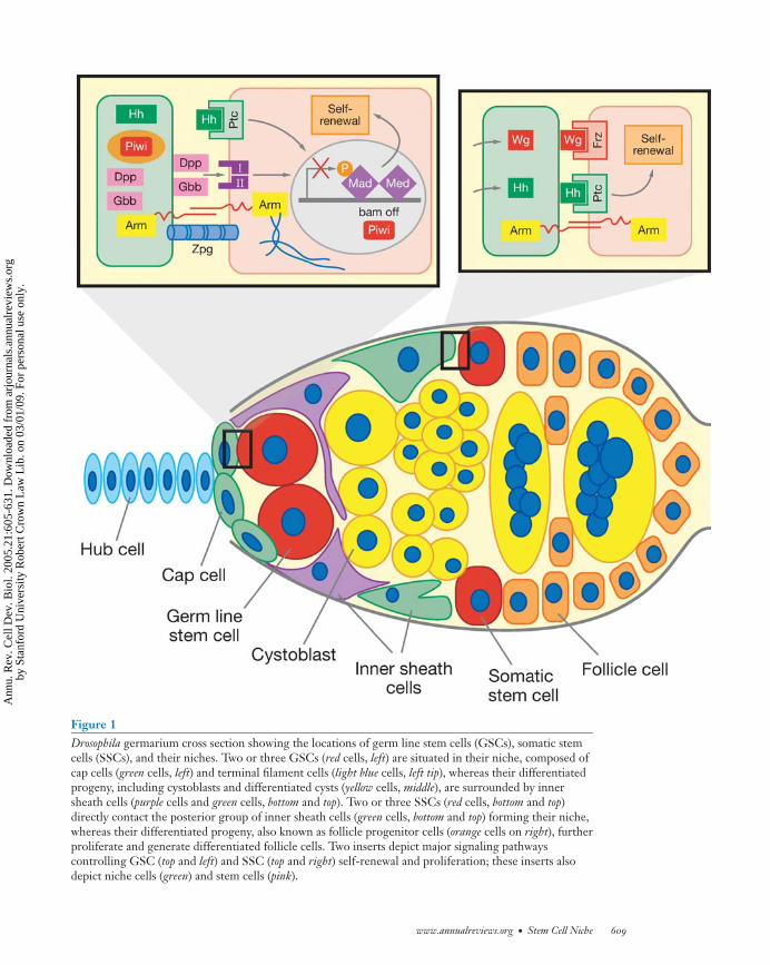
Two or three GSCs are located at the tip ofthe ovariole in the structure referred to asthe germarium. These GSCs are surroundedby three types of somatic cells: terminal fil-ament, cap cells, and inner germarial sheath(IGS) cells (Figure 1). The stem cells areeasily identified by their direct contact withcap cells and the presence of a spectrosome(Lin 2002, Xie & Spradling 2001). Normally,a GSC divides to generate two daughter cells:one daughter that stays in association with capcells and another daughter that moves awayfrom the cap cells to form a cystoblast, whicheventually becomes, through incomplete cy-tokinesis, an interconnected 16-cell cyst. Ge-netic and cell biological studies demonstratethat cap cells are the niche for GSCs (Xie &Spradling 2000). The anchorage of GSCs tocap cells through E-cadherin-mediated celladhesion is essential for maintaining GSCs(Song & Xie 2002). Also, the number of GSCscorrelates with the number of cap cells (Xie& Spradling 2000). Finally, cap cells expressgenes, such as dpp, gbb, hh, piwi, and Yb, thatare known to be important for maintainingGSCs (Cox et al. 2000, King et al. 2001,Song et al. 2004, Xie & Spradling 1998, 2000)(Figure 1).
BMP-, Hh-, and Piwi-mediated signalingpathways play an important role in the con-trol of ovarian GSC self-renewal (Figure 1).Two BMP-like genes, dpp and gbb, are ex-pressed in niche cells, and GSCs mutant fordpp, gbb, and their downstream components
are lost prematurely (Song et al. 2004, Xie& Spradling 1998, 2000). Dpp overexpres-sion completely prevents GSC differentiationand thereby causes GSC-like tumor forma-tion (Song et al. 2004, Xie & Spradling 1998).BMP signaling was recently shown to exertcontrol of GSC self-renewal by repressing ex-pression of bam (Chen & McKearin 2003,Song et al. 2004), which is necessary and suf-ficient for cystoblast differentiation (Ohlstein& McKearin 1997).
Piwi- and Yb-mediated signaling is alsorequired for controlling ovarian GSC self-renewal (Cox et al. 2000, King et al. 2001,Lin & Spradling 1997). Interestingly, Yb reg-ulates expression of piwi and hh in TF/capcells; these genes in turn control GSC self-renewal (King et al. 2001). Yb-mediated sig-naling is also involved in repressing bam ex-pression in GSCs (Chen & McKearin 2005,Szakmary et al. 2005). It would be interestingto know the relationship between BMP sig-naling and Piwi-mediated signaling in con-trolling GSC self-renewal. Zero populationgrowth (a Drosophila homolog of mammalianinnexin-4) is expressed in GSCs and is alsorequired for GSC maintenance, although theunderlying molecular mechanism for suchmaintenance is largely unknown (Gilboa et al.2003, Tazuke et al. 2002).
Two or three SSCs located in the middleof the germarium are responsible for generat-ing somatic follicle and stalk cells (Figure 1).The follicle cells encapsulate 16-cell cysts,whereas the stalk cells connect adjacent eggchambers. Although the ovarian SSCs lack aunique marker, they can be identified usinglineage tracing (Margolis & Spradling 1995,Song & Xie 2002, 2003, Zhang & Kalderon2001). SSCs have low levels of Fasciclin III(Fas 3) expression, whereas differentiated fol-licle cells have high levels of Fas 3 expression.Loss of adhesion between SSCs and IGS jeop-ardizes SSC self-renewal, suggesting that theproximal IGS cells are at least a part of theSSC niche, anchoring the SSCs (Song et al.2002). Although cap cells are not physicallyassociated with SSCs, they produce two
608 Li · Xie
Ann
u. R
ev. C
ell D
ev. B
iol.
2005
.21:
605-
631.
Dow
nloa
ded
from
arj
ourn
als.
annu
alre
view
s.or
gby
Sta
nfor
d U
nive
rsity
Rob
ert C
row
n L
aw L
ib. o
n 03
/01/
09. F
or p
erso
nal u
se o
nly.
ANRV255-CB21-25 ARI 8 September 2005 17:11
Figure 1Drosophila germarium cross section showing the locations of germ line stem cells (GSCs), somatic stemcells (SSCs), and their niches. Two or three GSCs (red cells, left) are situated in their niche, composed ofcap cells (green cells, left) and terminal filament cells (light blue cells, left tip), whereas their differentiatedprogeny, including cystoblasts and differentiated cysts (yellow cells, middle), are surrounded by innersheath cells (purple cells and green cells, bottom and top). Two or three SSCs (red cells, bottom and top)directly contact the posterior group of inner sheath cells (green cells, bottom and top) forming their niche,whereas their differentiated progeny, also known as follicle progenitor cells (orange cells on right), furtherproliferate and generate differentiated follicle cells. Two inserts depict major signaling pathwayscontrolling GSC (top and left) and SSC (top and right) self-renewal and proliferation; these inserts alsodepict niche cells (green) and stem cells (pink).
www.annualreviews.org • Stem Cell Niche 609
Ann
u. R
ev. C
ell D
ev. B
iol.
2005
.21:
605-
631.
Dow
nloa
ded
from
arj
ourn
als.
annu
alre
view
s.or
gby
Sta
nfor
d U
nive
rsity
Rob
ert C
row
n L
aw L
ib. o
n 03
/01/
09. F
or p
erso
nal u
se o
nly.

ANRV255-CB21-25 ARI 8 September 2005 17:11
diffusible growth factors, Hh and Wg, thatare required for controlling SSC maintenanceand proliferation (Forbes et al. 1996, Kinget al. 2001, Song & Xie 2003). This supportsthe hypothesis that these cap cells are alsoa part of the SSC niche (Forbes et al. 1996;King et al. 2001, Song & Xie 2003, Zhang &Kalderon 2001).
The Germ Line Stem Cell Niche inthe Drosophila Testis
In the apical tip of the Drosophila testis, twotypes of stem cells, GSCs and SSCs (the lat-ter are also known as cyst progenitor cells),are responsible for producing differentiatedgerm cells and somatic cyst cells, respectively(Fuller 1993, Kiger et al. 2001) (Figure 2).Seven to nine GSCs, each containing a spec-trosome, are attached to the hub (Hardyet al. 1979, Lindsley & Tokuyasu 1980, Ya-mashita et al. 2003). A male GSC dividesasymmetrically, giving rise to one stem cellthat remains in contact with the hub andone gonialblast that moves away from thehub and differentiates (Hardy et al. 1979,Lindsley & Tokuyasu 1980, Yamashita et al.2003). As a GSC divides to produce a go-nialblast, the neighboring SSCs also divideto generate two cyst cells, which envelopthe gonialblast. This process leads to pro-duction of 64 sperm (Gonczy & DiNardo1996, Hardy et al. 1979). The hub gener-ates signals, including Unpaired (Upd) andBMP, to control GSC self-renewal (Kawaseet al. 2004, Kiger et al. 2001, Shivdasani& Ingham 2003, Tulina & Matunis 2001)(Figure 2).
Upd from the hub activates the JAK-STATpathway in GSCs and promotes their self-renewal (Kiger et al. 2001, Tulina & Matsunis2001). Additionally, the activation of JAK-STAT signaling can reprogram mitotic germcysts into GSCs (Brawley & Matunis 2004).As in the ovary, BMP signaling is requiredfor controlling GSC self-renewal in the testis(Kawase et al. 2004, Schulz et al. 2004,
Shivdasani & Ingham 2003). Hub cells andsomatic cyst cells express gbb at high levelsand dpp at much lower levels; consequently,BMP downstream components are essentialfor controlling testicular GSC self-renewal(Kawase et al. 2004). Because dpp overexpres-sion fails to suppress completely spermatogo-nial cell differentiation, BMP signaling likelyplays a permissive role in controlling maleGSC self-renewal. BMP and JAK-STAT sig-naling pathways are required for controllingmale GSC self-renewal; thus, they must some-how interact with each other. The integra-tion between these two pathways in maleGSCs is an important area in need of futureexploration.
Gonialblast differentiation is tightly con-trolled by unknown signals from SSCs and so-matic cyst cells (Kiger et al. 2001, Tran et al.2000). In somatic cells mutant for Egfr and raf,GSC- and gonialblast-like single germ cellsare greatly increased in number and remainactive longer than do wild type cells.
One mechanism ensuring that only oneof the two stem cell daughters self-renews iscontrol of the spindle orientation of the stemcell so as to place one self-renewing daughterin the niche and the other daughter destinedto differentiate outside the niche (Figure 2).Cnn and APC1, centrosomal components inGSCs, control orientation of the spindle per-pendicular to the hub. Mutation in these com-ponents leads to an increase in GSC numberand subsequent crowding in the niche. APC2,which is concentrated at the junction be-tween GSCs and hub cells, also controls cor-rect GSC spindle orientation (Yamashita et al.2003).
THE GERM LINE STEM CELLNICHE IN C. ELEGANS
In the C. elegans hermaphrodite gonad, onlythe 225 germ cells closest to the distal tipcell (DTC) are mitotic; those further proximalare arrested in meiotic pachytene (Crittendenet al. 1994) (Figure 3). Specific stem cellswithin the mitotic region have not been
610 Li · Xie
Ann
u. R
ev. C
ell D
ev. B
iol.
2005
.21:
605-
631.
Dow
nloa
ded
from
arj
ourn
als.
annu
alre
view
s.or
gby
Sta
nfor
d U
nive
rsity
Rob
ert C
row
n L
aw L
ib. o
n 03
/01/
09. F
or p
erso
nal u
se o
nly.

ANRV255-CB21-25 ARI 8 September 2005 17:11
Figure 2Cross section of the apical tip of the Drosophila testis, showing the locations of germ line stem cells(GSCs), somatic stem cells (SSCs), and their niches. Hub cells (green) at the apical tip of the testis formniches for both GSCs (red) and SSCs (gray, left), which generate, respectively, spermatogonial cells(yellow) and somatic cyst cells (light gray) encapsulating differentiated spermatogonial cells. The insert ontop describes major signaling pathways involved in communication between GSCs and the niche cells forcontrolling self-renewal and proliferation.
www.annualreviews.org • Stem Cell Niche 611
Ann
u. R
ev. C
ell D
ev. B
iol.
2005
.21:
605-
631.
Dow
nloa
ded
from
arj
ourn
als.
annu
alre
view
s.or
gby
Sta
nfor
d U
nive
rsity
Rob
ert C
row
n L
aw L
ib. o
n 03
/01/
09. F
or p
erso
nal u
se o
nly.

ANRV255-CB21-25 ARI 8 September 2005 17:11
Figure 3Cross section of the C. elegans hermaphrodite gonad. The putative germ line stem cells (GSCs) (red ) aredirectly associated with their distal tip cell (DTC) niche cell (green), whereas their differentiated progeny(light yellow) move away from the DTC, progressing from the mitotic phase to the meiotic phase. TheGLP-1 (Notch-like) signaling pathway is involved in communication between the DTC and GSCs andrepresses functions of differentiation-promoting gene products, such as Gld-1, Gld-2, and Nos-3, whichregulate entry into meiosis (insert).
identified. The somatic DTC is required formaintaining these cells in mitosis (Kimble &White 1981). Although mitotic and meioticgerm cells in the tube share a central core ofcytoplasm, only those mitotic germ cells lo-cated at the most distal tip (i.e., GSCs) adja-cent to the DTC behave like stem cells, capa-
ble of self-renewing and generating differen-tiated gametes. The proximal mitotic neigh-bors behave more like transient amplifyingcell populations, described in other systems.As germ cells move further away from theDTC, they terminate their mitotic activitiesand commit meiosis. Only those germ cells
612 Li · Xie
Ann
u. R
ev. C
ell D
ev. B
iol.
2005
.21:
605-
631.
Dow
nloa
ded
from
arj
ourn
als.
annu
alre
view
s.or
gby
Sta
nfor
d U
nive
rsity
Rob
ert C
row
n L
aw L
ib. o
n 03
/01/
09. F
or p
erso
nal u
se o
nly.

ANRV255-CB21-25 ARI 8 September 2005 17:11
that physically interact with the DTC main-tain their GSC identity; thus, the signal fromthe DTC either must be short ranging or me-diated by a direct cell-cell interaction.
Signaling from the DTC to control GSCself-renewal is through a Notch-like cas-cade. The mitotic germ cells express theNotch-type receptor, GLP-1, which is acti-vated by the Delta-like signal from DTC,LAG-2 (Crittenden et al. 1994, Hendersonet al. 1994). Constitutive GLP-1 activitydownregulates the meiosis-promoting genesgld-1, gld-2, and nos-3 and thereby causes ex-pansion of germ cell numbers (Berry et al.1997). Because individual stem cells have notbeen identified in the mitotic region, it re-mains unclear whether they are maintainedthrough a population mechanism or an asym-metric division mechanism.
KNOWN STEM CELL NICHESIN MAMMALIAN SYSTEMS
The stem cell and the niche hypothesis, firstdeveloped in the hematopoietic system inmammals, has provided the conceptual back-ground for stem cell studies in Drosophilaand C. elegans (Schofield 1978, Weissman1994). Conversely, studies in Drosophila on themolecular pathways controlling the stem cellniche have provided important insight intoidentification of the stem cell niche in mam-malian systems (Lin 2002, Spradling et al.2001). In this section, we describe and com-pare the location and physical organization(if known) of adult stem cells in bone mar-row, skin/hair follicle, intestine, neuron, andtestis.
The Hematopoietic Stem Cell Niche
Bone marrow serves as the pioneer system forstudying stem cells; the concept and basic fea-tures of stem cells were defined from study-ing hematopoietic stem cells (HSCs) (Orkin2000, Till & McCulloch 1961, Weissmanet al. 2001). However, the way in which HSCsinteract with their local environment to pro-
mote stem cell maintenance has not beenclear. Most studies of HSCs have examinedtheir behavior in cell populations obtainedfrom their natural niche in the bone marrow.Thus far, however, only limited culture sys-tems exist that allow sustained maintenanceand expansion of HSCs in vitro, attesting tothe importance of as-yet poorly defined inter-actions in the bone marrow niche. Two inde-pendent studies recently 1) identified a sub-set of osteoblastic cells (N-cadherin+CD45−)to which HSCs physically attach in thebone marrow, 2) identified an N-cadherin/β-catenin adherens complex between HSCs andosteoblastic cells, 3) showed that Jagged1,generated from osteoblasts, influences HSCsby signaling through the Notch receptor,and 4) demonstrated that the number of N-cadherin+ osteoblastic lining cells controlsthe number of HSCs (Calvi et al. 2003, Zhanget al. 2003). Homing studies to trace the loca-tion of GFP-labeled HSCs after transplanta-tion also pointed to the endosteal surface as apossible stem cell niche (Nilsson et al. 2001).In vitro coculture of HSCs with osteoblastscan expand the HSC population (Taichman &Emerson 1998), and depletion of osteoblastsleads to loss of hematopoietic tissue (Visnjicet al. 2004). In addition, N-cadherin is a keytarget of Angiopoietin-1 (Ang-1)/Tie-2 sig-naling that maintains HSC quiescence (Araiet al. 2004) (Figure 4).
A primary function of the niche is toanchor stem cells. In addition to N-cadherin,other types of adhesion molecules, includingintegrin, play an important role in themicroenvironment/stem cell interaction(Simmons et al. 1997). Stromal cell-derivedfactor-1 (SDF-1) and its receptor CXCR4are involved in homing of HSCs (Lapidot &Kollet 2002) (Figure 4).
Although the analysis of the signals gen-erated by the niche has just begun, gene ex-pression profiling studies of HSCs have re-vealed which signals HSCs potentially receivefrom the niche. The components of evolu-tionally conserved and developmentally reg-ulated pathways are prominent in stem cells
www.annualreviews.org • Stem Cell Niche 613
Ann
u. R
ev. C
ell D
ev. B
iol.
2005
.21:
605-
631.
Dow
nloa
ded
from
arj
ourn
als.
annu
alre
view
s.or
gby
Sta
nfor
d U
nive
rsity
Rob
ert C
row
n L
aw L
ib. o
n 03
/01/
09. F
or p
erso
nal u
se o
nly.

ANRV255-CB21-25 ARI 8 September 2005 17:11
and are indeed involved in the regulation ofstem cell self-renewal or maintenance. Thesecomponents include the Shh, Wnt, Notch,and TGF-β/BMP pathways (Akashi et al.2003, Gomes et al. 2002, Ivanova et al. 2002,Park et al. 2002, Ramalho-Santos et al. 2002).
For example, the Wnt/β-catenin pathway isimportant for self-renewal of HSCs (Reyaet al. 2003). The Notch pathway is requiredfor maintaining HSCs in the undifferentiatedstate (Calvi et al. 2003, Duncan et al. 2005,Li et al. 1998, Varnum-Finney et al. 2000).
614 Li · Xie
Ann
u. R
ev. C
ell D
ev. B
iol.
2005
.21:
605-
631.
Dow
nloa
ded
from
arj
ourn
als.
annu
alre
view
s.or
gby
Sta
nfor
d U
nive
rsity
Rob
ert C
row
n L
aw L
ib. o
n 03
/01/
09. F
or p
erso
nal u
se o
nly.

ANRV255-CB21-25 ARI 8 September 2005 17:11
The BMP signal plays a role in control ofHSC number (Zhang et al. 2003). The Shhsignal mediated by the BMP pathway is ableto maintain stem cells in vitro (Bhatia et al.1999). (Figure 4).
The Epithelial Stem Cell Nichein Skin
Skin, with its appendix hair follicle structure,has well-organized architecture (Figure 5)and provides an excellent system for studyingthe molecular mechanisms that regulate stemcell self-renewal, proliferation, migration, andlineage commitment (Fuchs & Segre 2000).Each hair follicle is composed of a perma-nent portion, which includes sebaceous glandsand the underlying bulge area, and a dynamicrenewing portion, which undergoes cycles ofanagen phase (a period of active growth), cata-gen phase (apoptosis-driven retraction), andtelogen phase (a short period of rest) (Hardy1992). The bulge area functions as a niche,where epithelial stem cells (Niemann & Watt2002) are located and maintained (Cotsareliset al. 1990, Sun et al. 1991). Epithelial stemcells are multipotent, giving rise to daugh-ter cells that either migrate upward to serveas epidermal progenitors for generating epi-dermal cells during wound repair or migratedownward to convert to hair-matrix progeni-tors, which further give rise to the hair shaft(Niemann & Watt 2002, Oshima et al. 2001,Taylor et al. 2000).
During the early anagen phase, the der-mal papilla region may provide the dynamicsignals that activate stem cells; however, thecellular components of the niche in the bulgeare yet to be defined other than as stem cellsper se. The dermal sheath derived from mes-enchymal cells adjacent to the epithelial stemcells in the bulge area most likely providesthe niche function. The recent identificationof markers for epithelial stem cells, such asCD34, will be helpful in further identifyingthe adjacent niche cells and the related nichestructures, including adhesion molecules (i.e.,α6 integrin) (Blanpain et al. 2004).
Recent studies showed that label-retainingcells can regenerate the entire HF structurein transplantation experiments, thus demon-strating that these cells are bona fide epider-mal stem cells (Blanpain et al. 2004, Braunet al. 2003). Molecular analysis of epithe-lial stem cells has revealed the following fea-tures: 1) the expression of adhesion moleculesknown to be involved in stem cell-niche inter-action, 2) the presence of growth inhibitionfactors such as TGFβ/BMP molecules andcell cycle inhibitors, and 3) the components ofWnt signaling pathways, including receptorsand inhibitors such as Dkk, sFRP, and WIF.Taken together, these molecular features in-dicate that the epithelial stem cell niche isa growth- and differentiation-restricted en-vironment (Tumbar et al. 2004). This con-clusion is, in general, consistent with themany previous studies that have used genetic
←−−−−−−−−−−−−−−−−−−−−−−−−−−−−−−−−−−−−−−−−−−−−−−−−−−−−−−−−−−−−−−−−−−−−−Figure 4Illustration of the hematopoietic stem cell (HSC) niche. The HSC niche is located primarily on thesurface of trabecular bone, where a small subset of spindle-shaped N-cadherin-positive osteoblastic cells(indicated as SNO cells) are the key component of the HSC niche. N-cadherin and β-catenin form anadherens complex at the interface between stem cells and niche cells, assisting stem cells in attaching tothe niche. Multiple growth factors and cytokines are involved in stem-niche interaction. These includeSCF/Kit, Jagged/Notch, SDF-1/CXCR4, and Ang1/Tie2. BMP4 is expressed in osteoblastic cells, butthe type of receptor expressed in HSCs is unknown. The Wnt signal is important for stem cellself-renewal, but the Wnts present in the niche are unknown. The same is true for FGF and hedgehog.In vitro data suggest they affect HSC behavior; however, whether they are present as niche signals isunknown. Different types of stromal cells (illustrated as different colors and shapes) may regulate stemcell activation, proliferation, and differentiation by secreting different microenvironmental signals.Finally, maturated blood cells migrate and infiltrate into blood vessel.
www.annualreviews.org • Stem Cell Niche 615
Ann
u. R
ev. C
ell D
ev. B
iol.
2005
.21:
605-
631.
Dow
nloa
ded
from
arj
ourn
als.
annu
alre
view
s.or
gby
Sta
nfor
d U
nive
rsity
Rob
ert C
row
n L
aw L
ib. o
n 03
/01/
09. F
or p
erso
nal u
se o
nly.

ANRV255-CB21-25 ARI 8 September 2005 17:11
Figure 5Illustration of the epidermal stem cells. Stem cells are located in the bulge region of the hair folliclebeneath the sebaceous gland. Upon activation, stem cells undergo division; the daughter cells retained inthe bulge remain as stem cells while other daughter cells migrate down to become hair-matrixprogenitors responsible for hair regeneration. In neonatal mice or in damaged skin, stem cells can alsomigrate upward and convert to epidermal progenitors that replenish lost or damaged epidermis. Thebulge area is an environment that restricts cell growth and differentiation by expressing Wnt inhibitors,including DKK, Wif, and sFRP as well as BMPs. During the early anagen phase, Wnts from dermalpapilla (DP) and Noggin, which is derived from both DP and bulge (J. Zhang & L. Li, unpublished data),coordinate to overcome the restriction signals imposed by both BMPs and Wnt inhibitors; this leads tostem cell activation and subsequent hair regeneration. The FGF and Notch pathways are also involved inDP function for hair-matrix cell proliferation and lineage fate determination, but their influence onstem cells is not clear.
targeting and transgenic models to reveal thatsignaling molecules, including Wnts, Notch,and BMPs, have important roles in the reg-ulation of HF development and regeneration(Fuchs et al. 2001, Jones et al. 1995, Lavkeret al. 1993, Watt 2001).
Among these various signaling molecules,two family members are prominent, reflect-ing their important roles in controlling stemcell behavior. One is the Wnt signalingpathway which, through regulating β-cateninactivity, controls stem cell activation, fate
616 Li · Xie
Ann
u. R
ev. C
ell D
ev. B
iol.
2005
.21:
605-
631.
Dow
nloa
ded
from
arj
ourn
als.
annu
alre
view
s.or
gby
Sta
nfor
d U
nive
rsity
Rob
ert C
row
n L
aw L
ib. o
n 03
/01/
09. F
or p
erso
nal u
se o
nly.

ANRV255-CB21-25 ARI 8 September 2005 17:11
determination (by favoring HF over epider-mal cell lineages), and differentiation (Gatet al. 1998, Huelsken et al. 2001, Merrill et al.2001, Niemann et al. 2002). The second con-trolling pathway is the BMP signaling path-way (Hogan 1996). Although it is also re-quired for HF differentiation at a later stage,BMP signaling, as opposed to Wnt signaling,restricts the activation of stem cells and favorsepidermal cell fate (Botchkarev 2001, 2003,Kulessa et al. 2000). These observations alsosupport the theory that coordination betweenWnt and Noggin (through temporarily over-riding the BMP restriction on stem cells) isrequired to initiate each hair growth cycle(Jamora et al. 2003 and J. Zhang & L. Li, un-published data).
The Intestinal Stem Cell Niche
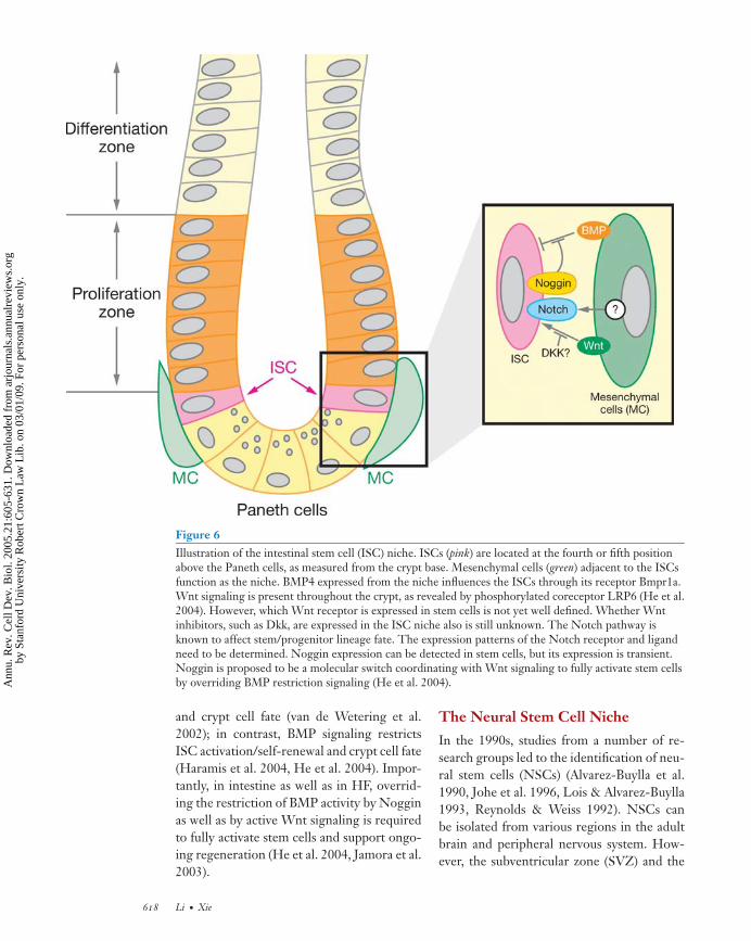
The intestinal architecture is composed ofa sequential array of zones (or compart-ments) along the villus-crypt axis (Figure 6).Intestinal regeneration begins with intesti-nal stem cells (ISCs), which give rise tofour different types of epithelial lineages:columnar enterocytes, mucin-producing gob-let cells, Paneth cells, and enteroendocrinecells (Bjerknes & Cheng 1999, Hermiston &Gordon 1995, Winton 2000). ISCs are gen-erally proposed to be located at the fourth orfifth position from the crypt bottom, abovethe Paneth cells (Booth & Potten 2000, Heet al. 2004, Sancho et al. 2004), as evidencedeither through a DNA-labeling retaining as-say (Booth & Potten 2000, He et al. 2004,Potten et al. 1997, 2002) or through re-generation dynamics using chimeric mouselines (Winton 2000, Bjerknes & Cheng 1999).A number of molecules—Telomerase, Tcf4,EphB3, P-PTEN, P-Akt, 14-3-3ζ , Noggin,and Musashi-1—are expressed in the pro-posed ISC position near the crypt base (Batlleet al. 2002, Booth & Potten 2000, He et al.2004, Korinek et al. 1998, Nishimura et al.2003). However, a combination of these mark-ers and the cell position is required to locateISCs more accurately.
During postnatal intestinal regeneration,mesenchymal cells subjacent to epithelial cellsplay a role in directing epithelial cell prolifer-ation, differentiation, and apoptosis. BMP4,expressing in the ISC-adjacent mesenchymalcells, is one of the putative niche signals (Heet al. 2004). However, the type of mesenchy-mal cells that expresses BMP4 adjacent to theISCs is yet to be identified. Endothelial cellscomposed of vascular vessels have also beenproposed to provide ISCs with survival signalssuch as FGF (Paris et al. 2001). Myofibrob-lasts that are distributed to the surroundingepithelial cells are proposed to be the candi-date “niche” supporting ISCs and influencingother epithelial cells (Mills & Gordon 2001).
We have just begun to understand whichniche signals regulate self-renewal and main-tain the balance between self-renewal anddifferentiation of ISCs. An increasing num-ber of molecules, including Wnt, BMP, FGF,Notch, and the underlying signal pathways,may play roles in this regard (Brittan &Wright 2002, Roberts et al. 1995, Sancho et al.2004). Gene expression profiling revealedthat Myc-related pathways and the PI3K/Aktpathway are predominantly present in thesestem/progenitors (Mills et al. 2002, Stappen-beck et al. 2003). Inappropriate activation ofthe Wnt/β-catenin, which targets on Myc,results in the development of tumors as aconsequence of an overproduction of stemcells (Clevers 2004). In addition, mutations inBMPRIA and its signaling mediator SMAD4have been found in Juvenile polyposis syn-drome (Howe et al. 1998a,b). Recent stud-ies using gene targeting demonstrated thatBMP signaling has a role in suppression ofWnt signaling and thereby maintains a bal-anced control of stem cell activation andself-renewal (Haramis et al. 2004, He et al.2004). Mechanistically, inhibition of Wntsignaling by the BMP signal involves boththe PTEN/PI-3k/Akt pathway and Smad-mediated transcriptional control (Haramiset al. 2004, He et al. 2004)
In summary, Wnt signaling plays a positiverole in promoting ISC activation/self-renewal
www.annualreviews.org • Stem Cell Niche 617
Ann
u. R
ev. C
ell D
ev. B
iol.
2005
.21:
605-
631.
Dow
nloa
ded
from
arj
ourn
als.
annu
alre
view
s.or
gby
Sta
nfor
d U
nive
rsity
Rob
ert C
row
n L
aw L
ib. o
n 03
/01/
09. F
or p
erso
nal u
se o
nly.
ANRV255-CB21-25 ARI 8 September 2005 17:11
Figure 6Illustration of the intestinal stem cell (ISC) niche. ISCs (pink) are located at the fourth or fifth positionabove the Paneth cells, as measured from the crypt base. Mesenchymal cells (green) adjacent to the ISCsfunction as the niche. BMP4 expressed from the niche influences the ISCs through its receptor Bmpr1a.Wnt signaling is present throughout the crypt, as revealed by phosphorylated coreceptor LRP6 (He et al.2004). However, which Wnt receptor is expressed in stem cells is not yet well defined. Whether Wntinhibitors, such as Dkk, are expressed in the ISC niche also is still unknown. The Notch pathway isknown to affect stem/progenitor lineage fate. The expression patterns of the Notch receptor and ligandneed to be determined. Noggin expression can be detected in stem cells, but its expression is transient.Noggin is proposed to be a molecular switch coordinating with Wnt signaling to fully activate stem cellsby overriding BMP restriction signaling (He et al. 2004).
and crypt cell fate (van de Wetering et al.2002); in contrast, BMP signaling restrictsISC activation/self-renewal and crypt cell fate(Haramis et al. 2004, He et al. 2004). Impor-tantly, in intestine as well as in HF, overrid-ing the restriction of BMP activity by Nogginas well as by active Wnt signaling is requiredto fully activate stem cells and support ongo-ing regeneration (He et al. 2004, Jamora et al.2003).
The Neural Stem Cell Niche
In the 1990s, studies from a number of re-search groups led to the identification of neu-ral stem cells (NSCs) (Alvarez-Buylla et al.1990, Johe et al. 1996, Lois & Alvarez-Buylla1993, Reynolds & Weiss 1992). NSCs canbe isolated from various regions in the adultbrain and peripheral nervous system. How-ever, the subventricular zone (SVZ) and the
618 Li · Xie
Ann
u. R
ev. C
ell D
ev. B
iol.
2005
.21:
605-
631.
Dow
nloa
ded
from
arj
ourn
als.
annu
alre
view
s.or
gby
Sta
nfor
d U
nive
rsity
Rob
ert C
row
n L
aw L
ib. o
n 03
/01/
09. F
or p
erso
nal u
se o
nly.

ANRV255-CB21-25 ARI 8 September 2005 17:11
subgranular zone (SGZ) of the hippocampusregion are the primary and well-characterizedgerminal regions in which NSCs resideand support neurogenesis in the adult brain(Doetsch 1999, 2003, Lois & Alvarez-Buylla1993, Palmer et al. 1997, Temple 2001).
There are four types of cells in the SVZ(Figure 7). A layer of ependymal cells lin-ing the lateral ventricle (LV) region separatesthe SVZ from the LV. SVZ astrocytes are lo-cated adjacent to the ependymal cells, with asingle cilium structure extending through theboundary of ependymal cells to contact the LVregion and to form a glial tunnel that embracesa group of neuroblasts. Immature cells derivedfrom SVZ astrocytes are precursors for neu-roblasts. A specialized basal lamina extendingfrom the blood vessels to the ependymal cellscontacts all cell types in the SVZ. The SVZ as-trocytes, which express astrocyte marker glialfibrillary acidic protein (GFAP), have stemcell features: They undergo self-renewal andgive rise to transient amplifying precursorC cells, which further give rise to neuroblasts.Neuroblasts differentiate into neurons thatmigrate toward the olfactory bulb and otherregions. In addition to producing neurons,SVZ astrocytes can also generate oligoden-drocytes (Doetsch 2003, Mirescu & Gould2003, Temple 2001). In the hippocampus, theSGZ is a germinal layer between the hilus andthe dentate gyrus, and is responsible for gen-erating dentate gyrus neurons (Palmer et al.1997). In the SGZ, neurogenesis occurs lo-cally in direct contact with blood vessels. SGZastrocytes also express GFAP and function asstem cells, undergoing self-renewal and gen-erating daughter cells that further producegranule neurons (Figure 7) (Doetsch 2003,Temple 2001).
In both the SVZ and SGZ structures, en-dothelial cells that form blood vessels and thespecialized basal lamina are an essential com-ponent of the NSC niche: These endothelialcells provide attachment for SVZ and SGZastrocytes and generate a variety of signalsthat control stem cell self-renewal and lin-eage commitment (Doetsch 2003, Shen et al.
2004). Signals generated from the niche in-clude BMPs and their antagonist Noggin,FGFs, IGF, VEGF, TGFα, and BDNF. TheBMP signal favors astrocyte lineage fate byinhibiting neuronal fate. In contrast, Nog-gin functions to inhibit BMP signaling andthereby favors neurogenesis (Temple 2001).An adherens junction composed of cadherinsand β-catenin also plays a role in mainte-nance of stem cells. Interestingly, overexpres-sion of β-catenin leads to expansion of theNSC population; this presumably reflects ac-tivation of Wnt signaling (Chenn & Walsh2002). This phenotype is very similar to over-expression of IGF in transgenic mice, in whichan increased brain size is also observed (Aberget al. 2003). Both EGF and bFGF are ableto expand NSCs in an in vitro culture sys-tem. In addition, signaling pathways, includ-ing Notch and PTEN/PI3K, are also involvedin NSC regulation (Doetsch 2003, Temple2001).
The Germ Line Stem Cell Nichein Mice
Stem cell transplantation capability, simpleanatomy, and genetics make the mouse testisan attractive model for studying GSCs andtheir niche. The GSCs in mice are single cellsthat are located in the periphery of seminif-erous tubules and that have the ability to self-renew and generate a large number of differ-entiated gametes (Brinster 2002) (Figure 8).GSCs in the mouse testis each divide asym-metrically to generate a GSC and a differenti-ated daughter, which forms an interconnectedApair spermatogonial cell. The Apair spermato-gonial cell then divides synchronously toform a chain of interconnected spermatogo-nial cells. Stem cells, spermatogonia, sper-matocytes, spermatids, and sperm cells canbe distinguished by their spatial relation todifferentiating sperm cells. GSCs are veryrare and can be isolated using fluorescence-activated cell sorting (FACS) as a populationof αv-integrin−/dim α6-integrin+ Thy-1lo/+ C-kit− cells (Kubota et al. 2003). Sertoli cells,
www.annualreviews.org • Stem Cell Niche 619
Ann
u. R
ev. C
ell D
ev. B
iol.
2005
.21:
605-
631.
Dow
nloa
ded
from
arj
ourn
als.
annu
alre
view
s.or
gby
Sta
nfor
d U
nive
rsity
Rob
ert C
row
n L
aw L
ib. o
n 03
/01/
09. F
or p
erso
nal u
se o
nly.

ANRV255-CB21-25 ARI 8 September 2005 17:11
Figure 7Illustration of the neural stem cell (NSC) niche. The subventricular zone (SVZ) and the subgranularzone (SGZ) are two well-characterized germinal regions in which NSCs (pink) are located. In the SVZ,astrocytes (B) lining the ependymal cells (E) function as NSCs; they give rise to transient amplifyingcells (C) (green), which further produce neuroblast cells (A) (blue). Endothelial cells in the blood vessel/laminar maintain contact with astrocytes, which regulate NSC self-renewal and proliferation bygenerating different types of signals. In the SGZ, astrocytes (B) directly attach to the blood vessel andreceive signals from the endothelial cells that direct NSCs to undergo self-renewal, proliferation (D),and differentiation (G). The figure is adapted and modified with permission from Doetsch 2003.
the somatic cells of the seminiferous tubulesthat physically interact with the stem cells,likely constitute functional niches for the stemcells by providing growth factors that promotestem cell self-renewal and/or proliferation.
Several studies support the idea that Sertolicells regulate the maintenance of the stem cellpool (although little is known about the un-derlying molecular mechanisms). First, stud-ies in which male GSCs and Sertoli cells
620 Li · Xie
Ann
u. R
ev. C
ell D
ev. B
iol.
2005
.21:
605-
631.
Dow
nloa
ded
from
arj
ourn
als.
annu
alre
view
s.or
gby
Sta
nfor
d U
nive
rsity
Rob
ert C
row
n L
aw L
ib. o
n 03
/01/
09. F
or p
erso
nal u
se o
nly.

ANRV255-CB21-25 ARI 8 September 2005 17:11
Figure 8Cross section of a small section of mouse testis. A germ line stem cell (GSC) (red) directly contacts aSertoli cell’s (purple) basement membrane (gray) secreted by myoid cells (pink), and specialized region(green), which together may form a putative GSC niche. Myoid cells (pink) may also participate in nichefunction, as they are close to GSCs. The differentiated spermatogonial cells (yellow) are germ-line cyststhat move through different domains formed by Sertoli cells toward the lumen, where mature sperm arereleased. The GDNF pathway, depicted in the insert (top), is a known major pathway for controllingGSC self-renewal in the mouse testis.
www.annualreviews.org • Stem Cell Niche 621
Ann
u. R
ev. C
ell D
ev. B
iol.
2005
.21:
605-
631.
Dow
nloa
ded
from
arj
ourn
als.
annu
alre
view
s.or
gby
Sta
nfor
d U
nive
rsity
Rob
ert C
row
n L
aw L
ib. o
n 03
/01/
09. F
or p
erso
nal u
se o
nly.
ANRV255-CB21-25 ARI 8 September 2005 17:11
are transplanted into infertile mice show thatSertoli cells indeed can support GSC main-tenance and spermatogenesis (Ogawa et al.2000, Shinohara et al. 2000, 2003). Second,GDNF, a member of the TGF-β superfam-ily produced by Sertoli cells, can controlGSC self-renewal and maintain GSCs in vitro(Kanatsu-Shinohara et al. 2004, Kubota et al.2004). Therefore, Sertoli cells contribute tothe function of the GSC niche. Future studyis needed to define the physical structure ofthe GSC niche and its associated signals inthe mouse testis.
CONCLUSION ANDPROSPECTIVE
Common Features, Structures, andFunctions of the Stem Cell Niche
After comparison of the stem cell nichesin the ovary and testis of Drosophila and inC. elegans, as well as in mammalian bone mar-row, hair follicle, intestine, brain, and testis,the common features, structures, and func-tions of the stem cell niche are summarizedas follows:
1. The stem cell niche is composed of agroup of cells in a special tissue loca-tion for the maintenance of stem cells.The niche’s overall structure is vari-able, and different cell types can pro-vide the niche environment. For exam-ple, N-cadherin-positive osteoblasticlining cells in the trabecular bone formthe niche for HSCs, whereas endothe-lial cells form the NSC cell niche.
2. The niche functions as a physical anchorfor stem cells. E-cadherin-mediatedcell adhesion is required for anchoringGSCs and SSCs in Drosophila, and N-cadherin may be important for anchor-ing HSC in the bone marrow niche.Other adhesion molecules, such as in-tegrins, may help anchor stem cells toextracellular matrixes.
3. The niche generates extrinsic factors
that control stem cell fate and num-ber. Many signal molecules have beenshown to be involved in regulationof stem cell behavior, including hh,Wnts, BMPs, FGFs, Notch, SCF, Ang-1, and LIF or Upd through the JAK-Statpathway. Among these, the BMP andWnt signal pathways have emerged ascommon pathways for controlling stemcell self-renewal and lineage fate fromDrosophila to mammals. Several path-ways can be utilized to control self-renewal of one stem cell type, whereasone growth factor can regulate severaldifferent stem cell types. The pres-ence of signaling components of mul-tiple conserved developmental regula-tory pathways in stem cells supports theideas that stem cells retain the ability torespond to these embryonic regulatorysignals and that orchestration of thesesignals is essential for proper regula-tion of stem cell self-renewal and lin-eage commitment.
4. In invertebrates and mammals, the stemcell niche exhibits an asymmetric struc-ture. Upon division, one daughter cellis maintained in the niche as a stem cell(self-renewal); the other daughter cellleaves the niche to proliferate and dif-ferentiate, eventually becoming a func-tionally mature cell.
FUTURE DIRECTIONS
Recent studies regarding the stem cell niche indifferent organisms, including various mam-malian organ systems, have resulted in signif-icant progress; fundamental principles aboutthe niche have been established. We hopethat the knowledge gained from these stud-ies discussed above will provide guidelinesfor defining the stem cell niche in othersystems. Using a combination of genetic,molecular, and cell biological approaches,several important signaling pathways fromthe various niches have been identified for
622 Li · Xie
Ann
u. R
ev. C
ell D
ev. B
iol.
2005
.21:
605-
631.
Dow
nloa
ded
from
arj
ourn
als.
annu
alre
view
s.or
gby
Sta
nfor
d U
nive
rsity
Rob
ert C
row
n L
aw L
ib. o
n 03
/01/
09. F
or p
erso
nal u
se o
nly.

ANRV255-CB21-25 ARI 8 September 2005 17:11
their ability to maintain and regulate self-renewal of stem cells. In general, multipleconserved developmental regulatory signalscoexist; therefore, orchestration of these sig-nals is essential for proper regulation of stemcell self-renewal and lineage commitment.Further studies of the cross-talk betweenthese signal pathways and the relationshipbetween these pathways and the intrinsic fac-tors required for self-renewal and mainte-nance of stem cells will provide further insightinto the molecular mechanisms governingstem cell self-renewal and differentiation.
Cellular and Molecular Componentsof the Stem Cell Niche
In uncovering other molecular componentsof the stem cell niche, genetic screening inDrosophila will continue to be an efficientmethod of identification of novel factors. Inmammals, systematic analysis of gene expres-sion in the niche cells (Hackney et al. 2002)will be as important and fruitful as analysisof gene expression in stem cells. For exam-ple, systematic analysis of the N-cadherin-positive osteoblastic lining cells, using genearray to compare to other types of marrowstromal cells, including N-cadherin-negativeosteoblastic cells, is required to uncover anyunique genes predominantly expressed in theHSC niche cells. Furthermore, comparisonsof niche- and stem cell–specific gene profilesin different systems will provide important in-sight into the critical niche signals and intrin-sic factors that potentially influence stem cellbehavior. Thus, conserved signal moleculesand intrinsic factors important for stem cellself-renewal and maintenance and specific fac-tors unique to each stem cell niche can beidentified.
Asymmetric Versus Symmetric StemCell Division
The stem cell niche exhibits structural asym-metry, and asymmetric division of stem cells isone of the proposed mechanisms controlling
the balance between self-renewal and differ-entiation. This has been well illustrated in theDrosophila system. Whether this mechanismis preserved in the mammalian system needsto be determined. The centrosome-associatedproteins APC1 and centrosomin are impor-tant in controlling spindle orientation dur-ing stem cell division in Drosophila (Yamashitaet al. 2003). It is important to investigatewhether control of spindle orientation is es-sential for asymmetric division of stem cells inother systems as well.
Stem Cell Maintenance andReversion from CommittedDaughter Cells
As described above, asymmetric stem cell di-vision leads to the retention of one daugh-ter cell in the niche (stem cell) and to theother daughter cell leaving the niche to be-come committed, an irreversible process inthe normal physiological condition. Whetherthe committed daughter cell can revert to astem cell if restored to the niche is an in-teresting and important question. Two recentstudies in Drosophila provide solid evidence in-dicating that this may be possible (Brawley& Matunis 2004, Kai & Spradling 2004). Itremains to be seen whether this is a generalfeature for different types of stem cells in in-vertebrates and mammals.
Normal Stem Cells and Cancer StemCells: Niche-Dependent orNiche-Independent
The concept of cancer stem cells has changedthe perspective on cancer, in which stem cellsand their underlying self-renewal is key. Inadults, the niche prevents tumorigenesis bycontrolling stem cells in the arrested state andmaintaining the balance between self-renewaland differentiation. In this context, any muta-tion that leads stem cells to escape from theniche control may result in tumorigenesis. It istherefore reasonable to hypothesize that oneof the differences between normal stem cells
www.annualreviews.org • Stem Cell Niche 623
Ann
u. R
ev. C
ell D
ev. B
iol.
2005
.21:
605-
631.
Dow
nloa
ded
from
arj
ourn
als.
annu
alre
view
s.or
gby
Sta
nfor
d U
nive
rsity
Rob
ert C
row
n L
aw L
ib. o
n 03
/01/
09. F
or p
erso
nal u
se o
nly.

ANRV255-CB21-25 ARI 8 September 2005 17:11
and cancer stem cells is that cancer stem cellsmay no longer be dependent on niche signal-ing. This hypothesis needs to be tested.
CLOSING REMARKS
Stem cell behavior is regulated by coordi-nation of environmental signals and intrin-sic programs. Environmental signals are pro-vided by the niche, which is composed ofspecialized cell populations located in uniquetopological relationships with the stem cellsin different adult tissues. In this review, wecompare the differences and commonalitiesof the niches in a variety of stem cell sys-tems across different species and provide evi-dence demonstrating the impact of the niche
on the homeostatic regulation of stem cells.Dissection of the niche’s cellular and molec-ular components has revealed the basic fea-tures and functions of the stem cell niche andwill provide important insights for identifica-tion of the stem cell niche in different sys-tems. We believe that the ability to reconsti-tute the stem cell niche in vitro will open anew avenue for maintenance and expansionof adult stem cells. Uncovering the importantsignals generated by the niche will shed lighton the mechanisms that regulate stem cell self-renewal and maintenance of stem cell multi-potentiality. Finally, understanding the inter-action between stem cells and their naturalpartners will substantially benefit therapeuticapproaches to human degenerative diseases.
ACKNOWLEDGMENTS
We thank L. Wiedemann for critical editing and D. di Natale for proofreading and manuscriptorganization. We are grateful for comments from P. Trainor. We apologize to those whosepapers are not cited here due to limited space. Our work is supported by the Stowers Institutefor Medical Research.
LITERATURE CITED
Aberg MA, Aberg ND, Palmer TD, Alborn AM, Carlsson-Skwirut C, et al. 2003. IGF-I has adirect proliferative effect in adult hippocampal progenitor cells. Mol. Cell Neurosci. 24:23–40
Akashi K, He X, Chen J, Iwasaki H, Niu C, et al. 2003. Transcriptional accessibility for genesof multiple tissues and hematopoietic lineages is hierarchically controlled during earlyhematopoiesis. Blood 101:383–89
Alvarez-Buylla A, Kirn JR, Nottebohm F. 1990. Birth of projection neurons in adult avianbrain may be related to perceptual or motor learning. Science 249:1444–46
Arai F, Hirao A, Ohmura M, Sato H, Matsuoka S, et al. 2004. Tie2/angiopoietin-1 signalingregulates hematopoietic stem cell quiescence in the bone marrow niche. Cell 118:149–61
Batlle E, Henderson JT, Beghtel H, van den Born MMW, Sancho E, et al. 2002. Beta-cateninand TCF mediate cell positioning in the intestinal epithelium by controlling the expressionof EphB/EphrinB. Cell 111:251–63
Berry LW, Westlund B, Schedl T. 1997. Germ-line tumor formation caused by activationof glp-1, a Caenorhabditis elegans member of the Notch family of receptors. Development124:925–36
Bhatia M, Bonnet D, Wu D, Murdoch B, Wrana J, et al. 1999. Bone morphogenetic proteinsregulate the developmental program of human hematopoietic stem cells. J. Exp. Med.189:1139–48
624 Li · Xie
Ann
u. R
ev. C
ell D
ev. B
iol.
2005
.21:
605-
631.
Dow
nloa
ded
from
arj
ourn
als.
annu
alre
view
s.or
gby
Sta
nfor
d U
nive
rsity
Rob
ert C
row
n L
aw L
ib. o
n 03
/01/
09. F
or p
erso
nal u
se o
nly.

ANRV255-CB21-25 ARI 8 September 2005 17:11
Bjerknes M, Cheng H. 1999. Clonal analysis of mouse intestinal epithelial progenitors. Gas-troenterology 116:7–14
Blanpain C, Lowry WE, Geoghegan A, Polak L, Fuchs E. 2004. Self-renewal, multipotency,and the existence of two cell populations within an epithelial stem cell niche. Cell 118:635–48
Booth C, Potten CS. 2000. Gut instincts: thoughts on intestinal epithelial stem cells. J. Clin.Invest. 105:1493–99
Botchkarev VA. 2003. Bone morphogenetic proteins and their antagonists in skin and hairfollicle biology. J. Invest. Dermatol. 120:36–47
Botchkarev VA, Botchkareva NV, Nakamura M, Huber O, Funa K, et al. 2001. Noggin isrequired for induction of the hair follicle growth phase in postnatal skin. FASEB J. 15:2205–14
Braun KM, Niemann C, Jensen UB, Sundberg JP, Silva-Vargas V, Watt FM. 2003. Manipu-lation of stem cell proliferation and lineage commitment: visualization of label-retainingcells in whole mounts of mouse epidermis. Development 130:5241–55
Brawley C, Matunis E. 2004. Regeneration of male germline stem cells by spermatogonialdedifferentiation in vivo. Science 304:1331–34
Brinster RL. 2002. Germline stem cell transplantation and transgenesis. Science 296:2174–76Brinster RL, Zimmermann JW. 1994. Spermatogenesis following male germ-cell transplanta-
tion. Proc. Natl. Acad. Sci. USA 91:11298–302Brittan M, Wright NA. 2002. Gastrointestinal stem cells. J. Pathol. 197:492–509Calvi LM, Adams GB, Weibrecht KW, Weber JM, Olson DP, et al. 2003. Osteoblastic cells
regulate the haematopoietic stem cell niche. Nature 425:841–46Chambers I, Smith A. 2004. Self-renewal of teratocarcinoma and embryonic stem cells. Onco-
gene 23:7150–60Chen D, McKearin D. 2003. Dpp signaling silences bam transcription directly to establish
asymmetric divisions of germline stem cells. Curr. Biol. 13:1786–91Chen D, McKearin D. 2005. Gene circuitry controlling a stem cell niche. Curr. Biol. 15:179–84Chenn A, Walsh CA. 2002. Regulation of cerebral cortical size by control of cell cycle exit in
neural precursors. Science 297:365–69Clevers H. 2004. At the crossroads of inflammation and cancer. Cell 118:671–74Cotsarelis G, Sun TT, Lavker RM. 1990. Label-retaining cells reside in the bulge area of
pilosebaceous unit: implications for follicular stem cells, hair cycle, and skin carcinogenesis.Cell 61:1329–37
Cox DN, Chao A, Baker J, Chang L, Qiao D, Lin HF. 1998. A novel class of evolutionar-ily conserved genes defined by piwi are essential for stem cell self-renewal. Genes Dev.12:3715–27
Cox DN, Chao A, Lin H. 2000. piwi encodes a nucleoplasmic factor whose activity modulatesthe number and division rate of germline stem cells. Development 127:503–14
Crittenden SL, Bernstein DS, Bachorik JL, Thompson BE, Gallegos M, et al. 2002. A con-served RNA-binding protein controls germline stem cells in Caenorhabditis elegans. Nature417:660–63
Crittenden SL, Troemel ER, Evans TC, Kimble J. 1994. GLP-1 is localized to the mitoticregion of the C. elegans germ line. Development 120:2901–11
Dexter TM, Moore MA, Sheridan AP. 1977. Maintenance of hemopoietic stem cells and pro-duction of differentiated progeny in allogeneic and semiallogeneic bone marrow chimerasin vitro. J. Exp. Med. 145:1612–16
Doetsch F. 2003. A niche for adult neural stem cells. Curr. Opin. Genet. Dev. 13:543–50
www.annualreviews.org • Stem Cell Niche 625
Ann
u. R
ev. C
ell D
ev. B
iol.
2005
.21:
605-
631.
Dow
nloa
ded
from
arj
ourn
als.
annu
alre
view
s.or
gby
Sta
nfor
d U
nive
rsity
Rob
ert C
row
n L
aw L
ib. o
n 03
/01/
09. F
or p
erso
nal u
se o
nly.

ANRV255-CB21-25 ARI 8 September 2005 17:11
Doetsch F, Caille I, Lim DA, Garcia-Verdugo JM, Alvarez-Buylla A. 1999. Subventricularzone astrocytes are neural stem cells in the adult mammalian brain. Cell 97:703–16
Duncan AW, Rattis FM, Dimascio LN, Congdon KL, Pazianos G, et al. 2005. Integration ofNotch and Wnt signaling in hematopoietic stem cell maintenance. Nat. Immunol. 6:314–22
Forbes AJ, Lin H, Ingham PW, Spradlin AC. 1996. hedgehog is required for the proliferationand specification of ovarian somatic cells prior to egg chamber formation in Drosophila.Development 122:1125–35
Fuchs E, Merrill BJ, Jamora C, DasGupta R. 2001. At the roots of a never-ending cycle. Dev.Cell 1:13–25
Fuchs E, Segre JA. 2000. Stem cells: a new lease on life. Cell 100:143–55Fuchs E, Tumbar T, Guasch G. 2004. Socializing with the neighbors: stem cells and their
niche. Cell 116:769–78Fuller MT. 1993. Spermatogenesis. In The Development of Drosophila, ed. M Bate, A Martinez-
Arias, pp. 71–147. Cold Spring Harbor, NY: Cold Spring Harbor Lab. PressGat U, DasGupta R, Degenstein L, Fuchs E. 1998. De novo hair follicle morphogenesis and
hair tumors in mice expressing a truncated beta-catenin in skin. Cell 95:605–14Gilboa L, Forbes A, Tazuke SI, Fuller MT, Lehmann R. 2003. Germ line stem cell differ-
entiation in Drosophila requires gap junctions and proceeds via an intermediate state.Development 130:6625–34
Gomes I, Sharma TT, Edassery S, Fulton N, Mar BG, Westbrook CA. 2002. Novel transcrip-tion factors in human CD34 antigen-positive hematopoietic cells. Blood 100:107–19
Gonczy P, DiNardo S. 1996. The germ line regulates somatic cyst cell proliferation and fateduring Drosophila spermatogenesis. Development 122:2437–47
Hackney JA, Charbord P, Brunk BP, Stoeckert CJ, Lemischka IR, Moore KA. 2002. A molec-ular profile of a hematopoietic stem cell niche. Proc. Natl. Acad. Sci. USA 99:13061–66
Haramis AP, Begthel H, van den Born M, van Es J, Jonkheer S, et al. 2004. De novo cryptformation and juvenile polyposis on BMP inhibition in mouse intestine. Science 303:1684–86
Hardy MH. 1992. The secret life of the hair follicle. Trends Genet. 8:55–61Hardy RW, Tokuyasu KT, Lindsley DL, Garavito M. 1979. The germinal proliferation center
in the testis of Drosophila melanogaster. J. Ultrastruct. Res. 69:180–90He XC, Zhang J, Tong WG, Tawfik O, Ross J, et al. 2004. BMP signaling inhibits intestinal
stem cell self-renewal through suppression of Wnt-beta-catenin signaling. Nat. Genet.36:1117–21
Helgason CD, Sauvageau G, Lawrence HJ, Largman C, Humphries RK. 1996. Overexpressionof HOXB4 enhances the hematopoietic potential of embryonic stem cells differentiatedin vitro. Blood 87:2740–49
Henderson ST, Gao D, Lambie EJ, Kimble J. 1994. lag-2 may encode a signaling ligand forthe GLP-1 and LIN-12 receptors of C. elegans. Development 120:2913–24
Hermiston ML, Gordon JI. 1995. Organization of the crypt-villus axis and evolution of itsstem cell hierarchy during intestinal development. Am. J. Physiol. Gastrointest. Liver Physiol.268:G813–22
Hogan BL. 1996. Bone morphogenetic proteins in development. Curr. Opin. Genet. Dev. 6:432–38
Howe JR, Ringold JC, Summers RW, Mitros FA, Nishimura DY, Stone EM. 1998a. A gene forfamilial juvenile polyposis maps to chromosome 18q21.1. Am. J. Hum. Genet. 62:1129–36
Howe JR, Roth S, Ringold JC, Summers RW, Jarvinen HJ, et al. 1998b. Mutations in theSMAD4/DPC4 gene in juvenile polyposis. Science 280:1086–88
626 Li · Xie
Ann
u. R
ev. C
ell D
ev. B
iol.
2005
.21:
605-
631.
Dow
nloa
ded
from
arj
ourn
als.
annu
alre
view
s.or
gby
Sta
nfor
d U
nive
rsity
Rob
ert C
row
n L
aw L
ib. o
n 03
/01/
09. F
or p
erso
nal u
se o
nly.

ANRV255-CB21-25 ARI 8 September 2005 17:11
Huelsken J, Vogel R, Erdmann B, Cotsarelis G, Birchmeier W. 2001. Beta-catenin controlshair follicle morphogenesis and stem cell differentiation in the skin. Cell 105:533–45
Ivanova NB, Dimos JT, Schaniel C, Hackney JA, Moore KA, Lemischka IR. 2002. A stem cellmolecular signature. Science 298:601–4
Jamora C, DasGupta R, Kocieniewski P, Fuchs E. 2003. Links between signal transduction,transcription and adhesion in epithelial bud development. Nature 422:317–22
Johe KK, Hazel TG, Muller T, Dugich-Djordjevic MM, McKay RD. 1996. Single factorsdirect the differentiation of stem cells from the fetal and adult central nervous system.Genes Dev. 10:3129–40
Jones PH, Harper S, Watt FM. 1995. Stem cell patterning and fate in human epidermis. Cell80:83–93
Kai T, Spradling A. 2004. Differentiating germ cells can revert into functional stem cells inDrosophila melanogaster ovaries. Nature 428:564–69
Kanatsu-Shinohara M, Inoue K, Lee J, Yoshimoto M, Ogonuki N, et al. 2004. Generation ofpluripotent stem cells from neonatal mouse testis. Cell 119:1001–12
Kawase E, Wong MD, Ding BC, Xie T. 2004. Gbb/Bmp signaling is essential for maintain-ing germline stem cells and for repressing bam transcription in the Drosophila testis.Development 131:1365–75
Kiger AA, Jones DL, Schulz C, Rogers MB, Fuller MT. 2001. Stem cell self-renewal specifiedby JAK-STAT activation in response to a support cell cue. Science 294:2542–45
Kimble JE, White JG. 1981. On the control of germ cell development in Caenorhabditis elegans.Dev. Biol. 81:208–19
King FJ, Lin H. 1999. Somatic signaling mediated by fs(1)Yb is essential for germline stemcell maintenance during Drosophila oogenesis. Development 126:1833–44
King FJ, Szakmary A, Cox DN, Lin H. 2001. Yb modulates the divisions of both germline andsomatic stem cells through piwi- and hh-mediated mechanisms in the Drosophila ovary.Mol. Cell 7:497–508
Korinek V, Barker N, Moerer P, van Donselaar E, Huls G, et al. 1998. Depletion of epithelialstem-cell compartments in the small intestine of mice lacking Tcf-4. Nat. Genet. 19:379–83
Kubota H, Avarbock MR, Brinster RL. 2003. Spermatogonial stem cells share some, but notall, phenotypic and functional characteristics with other stem cells. Proc. Natl. Acad. Sci.USA 100:6487–92
Kubota H, Avarbock MR, Brinster RL. 2004. Growth factors essential for self-renewal andexpansion of mouse spermatogonial stem cells. Proc. Natl. Acad. Sci. USA 101:16489–94
Kulessa H, Turk G, Hogan BL. 2000. Inhibition of Bmp signaling affects growth and differ-entiation in the anagen hair follicle. EMBO J. 19:6664–74
Kyba M, Perlingeiro RC, Daley GQ. 2002. HoxB4 confers definitive lymphoid-myeloid en-graftment potential on embryonic stem cell and yolk sac hematopoietic progenitors. Cell109:29–37
Lapidot T, Kollet O. 2002. The essential roles of the chemokine SDF-1 and its receptorCXCR4 in human stem cell homing and repopulation of transplanted immune-deficientNOD/SCID and NOD/SCID/B2m(null) mice. Leukemia 16:1992–2003
Lavker RM, Miller S, Wilson C, Cotsarelis G, Wei ZG, et al. 1993. Hair follicle stem cells: theirlocation, role in hair cycle, and involvement in skin tumor formation. J. Invest. Dermatol.101:16S–26S
Lessard J, Sauvageau G. 2003. Bmi-1 determines the proliferative capacity of normal andleukaemic stem cells. Nature 423:255–60
www.annualreviews.org • Stem Cell Niche 627
Ann
u. R
ev. C
ell D
ev. B
iol.
2005
.21:
605-
631.
Dow
nloa
ded
from
arj
ourn
als.
annu
alre
view
s.or
gby
Sta
nfor
d U
nive
rsity
Rob
ert C
row
n L
aw L
ib. o
n 03
/01/
09. F
or p
erso
nal u
se o
nly.
ANRV255-CB21-25 ARI 8 September 2005 17:11
Li L, Huang GM, Banta AB, Deng Y, Smith T, et al. Cloning, characterization, and thecomplete 57-kilobase DNA sequence of the human Notch4 gene. Genomics 51:45–58
Lin H. 2002. The stem-cell niche theory: lessons from flies. Nat. Rev. Genet. 3:931–40Lin H, Spradling AC. 1993. Germline stem cell division and egg chamber development in
transplanted Drosophila germaria. Dev. Biol. 159:140–52Lin H, Spradling AC. 1997. A novel group of pumilio mutations affects the asymmetric division
of germline stem cells in the Drosophila ovary. Development 124:2463–76Lin H, Yue L, Spradling AC. 1994. The Drosophila fusome, a germline-specific organelle, con-
tains membrane skeletal proteins and functions in cyst formation. Development 120:947–56Lindsley DT, Tokuyasu KT. 1980. Spermatogenesis. In Genetics and Biology of Drosophila,
ed. M Ashburner, TRF Wright, pp. 225–94. New York: Acad. PressLois C, Alvarez-Buylla A. 1993. Proliferating subventricular zone cells in the adult mammalian
forebrain can differentiate into neurons and glia. Proc. Natl. Acad. Sci. USA 90:2074–77Margolis J, Spradling A. 1995. Identification and behavior of epithelial stem cells in the
Drosophila ovary. Development 121:3797–807Merrill BJ, Gat U, DasGupta R, Fuchs E. 2001. Tcf3 and Lef1 regulate lineage differentiation
of multipotent stem cells in skin. Genes Dev. 15:1688–705Mills JC, Andersson N, Hong CV, Stappenbeck TS, Gordon JI. 2002. Molecular characteriza-
tion of mouse gastric epithelial progenitor cells. Proc. Natl. Acad. Sci. USA 99:14819–24Mills JC, Gordon JI. 2001. The intestinal stem cell niche: there grows the neighborhood. Proc.
Natl. Acad. Sci. USA 98:12334–36Mirescu C, Gould E. 2003. Stem cells in the adult brain. In Stem Cells: Adult and Fetal Stem
Cells, ed. R Lanza, pp. 219–24. Burlington, MA: Elsevier Acad.Molofsky AV, Pardal R, Iwashita T, Park IK, Clarke MF, Morrison SJ. 2003. Bmi-1 dependence
distinguishes neural stem cell self-renewal from progenitor proliferation. Nature 425:962–67
Moore KA, Ema H, Lemischka IR. 1997. In vitro maintenance of highly purified, transplantablehematopoietic stem cells. Blood 89:4337–47
Morrison SJ, Wright DE, Cheshier SH, Weissman IL. 1997. Hematopoietic stem cells: chal-lenges to expectations. Curr. Opin. Immunol. 9:216–21
Niemann C, Owens DM, Hulsken J, Birchmeier W, Watt FM. 2002. Expression of DeltaNLef1in mouse epidermis results in differentiation of hair follicles into squamous epidermal cystsand formation of skin tumours. Development 129:95–109
Niemann C, Watt FM. 2002. Designer skin: lineage commitment in postnatal epidermis. TrendsCell Biol. 12:185–92
Nilsson SK, Johnston HM, Coverdale JA. 2001. Spatial localization of transplanted hemopoi-etic stem cells: inferences for the localization of stem cell niches. Blood 97:2293–99
Nishimura S, Wakabayashi N, Toyoda K, Kashima K, Mitsufuji S. 2003. Expression of Musashi-1 in human normal colon crypt cells: a possible stem cell marker of human colon epithe-lium. Dig. Dis. Sci. 48:1523–29
Ogawa T, Dobrinski I, Avarbock MR, Brinster RL. 2000. Transplantation of male germ linestem cells restores fertility in infertile mice. Nat. Med. 6:29–34
Ohlstein B, McKearin D. 1997. Ectopic expression of the Drosophila Bam protein eliminatesoogenic germline stem cells. Development 124:3651–62
Orkin SH. 2000. Diversification of haematopoietic stem cells to specific lineages. Nat. Rev.Genet. 1:57–64
Oshima H, Rochat A, Kedzia C, Kobayashi K, Barrandon Y. 2001. Morphogenesis and renewalof hair follicles from adult multipotent stem cells. Cell 104:233–45
628 Li · Xie
Ann
u. R
ev. C
ell D
ev. B
iol.
2005
.21:
605-
631.
Dow
nloa
ded
from
arj
ourn
als.
annu
alre
view
s.or
gby
Sta
nfor
d U
nive
rsity
Rob
ert C
row
n L
aw L
ib. o
n 03
/01/
09. F
or p
erso
nal u
se o
nly.

ANRV255-CB21-25 ARI 8 September 2005 17:11
Palmer TD, Takahashi J, Gage FH. 1997. The adult rat hippocampus contains primordialneural stem cells. Mol. Cell Neurosci. 8:389–404
Paris F, Fuks Z, Kang A, Capodieci P, Juan G, et al. 2001. Endothelial apoptosis as the primarylesion initiating intestinal radiation damage in mice. Science 293:293–97
Park IK, He Y, Lin F, Laerum OD, Tian Q, et al. 2002. Differential gene expression profilingof adult murine hematopoietic stem cells. Blood 99:488–98
Park IK, Qian D, Kiel M, Becker MW, Pihalja M, et al. 2003. Bmi-1 is required for maintenanceof adult self-renewing haematopoietic stem cells. Nature 423:302–5
Potten CS, Booth C, Pritchard DM. 1997. The intestinal epithelial stem cell: the mucosalgovernor. Int. J. Exp. Pathol. 78:219–43
Potten CS, Owen G, Booth D. 2002. Intestinal stem cells protect their genome by selectivesegregation of template DNA strands. J. Cell Sci. 115:2381–88
Ramalho-Santos M, Yoon S, Matsuzaki Y, Mulligan RC, Melton DA. 2002. “Stemness”: tran-scriptional profiling of embryonic and adult stem cells. Science 298:597–600
Reya T, Duncan AW, Ailles L, Domen J, Scherer DC, et al. 2003. A role for Wnt signallingin self-renewal of haematopoietic stem cells. Nature 423:409–14
Reynolds BA, Weiss S. 1992. Generation of neurons and astrocytes from isolated cells of theadult mammalian central nervous system. Science 255:1707–10
Rios M, Williams DA. 1990. Systematic analysis of the ability of stromal cell lines derived fromdifferent murine adult tissues to support maintenance of hematopoietic stem cells in vitro.J. Cell Physiol. 145:434–43
Roberts DJ, Johnson RL, Burke AC, Nelson CE, Morgan BA, Tabin C. 1995. Sonic hedgehog isan endodermal signal inducing Bmp-4 and Hox genes during induction and regionalizationof the chick hindgut. Development 121:3163–74
Roecklein BA, Torok-Storb B. 1995. Functionally distinct human marrow stromal cell linesimmortalized by transduction with the human papiloma virus E6/E7 genes. Blood 85:997–1005
Rossant J. 2004. Embryonic stem cells in prospective. In Handbook of Stem Cells, ed. R Lanza,J Gearhart, BL Hogan, D Melton, R Pedersen, et al. London: Elsevier Acad.
Sancho E, Batlle E, Clevers H. 2004. Signaling pathways in intestinal development and cancer.Annu. Rev. Cell Dev. Biol. 20:695–723
Sauvageau G, Thorsteinsdottir U, Eaves CJ, Lawrence HJ, Largman C, et al. 1995. Overex-pression of HOXB4 in hematopoietic cells causes the selective expansion of more primitivepopulations in vitro and in vivo. Genes Dev. 9:1753–65
Schofield R. 1978. The relationship between the spleen colony-forming cell and the ham-atopopietic stem cell. A hypothesis. Blood Cells 4:7–25
Schulz C, Kiger AA, Tazuke SI, Yamashita YM, Pantalena-Filho LC, et al. 2004. A misex-pression screen reveals effects of bag-of-marbles and TGF beta class signaling on theDrosophila male germ-line stem cell lineage. Genetics 167:707–23
Shen Q, Goderie SK, Jin L, Karanth N, Sun Y, et al. 2004. Endothelial cells stimulate self-renewal and expand neurogenesis of neural stem cells. Science 304:1338–40
Shinohara T, Avarbock MR, Brinster RL. 2000. Functional analysis of spermatogonial stemcells in Steel and cryptorchid infertile mouse models. Dev. Biol. 220:401–11
Shinohara T, Orwig KE, Avarbock MR, Brinster RL. 2003. Restoration of spermatogenesis ininfertile mice by Sertoli cell transplantation. Biol. Reprod. 68:1064–71
Shivdasani AA, Ingham PW. 2003. Regulation of stem cell maintenance and transit amplifyingcell proliferation by tgf-beta signaling in Drosophila spermatogenesis. Curr. Biol. 13:2065–72
www.annualreviews.org • Stem Cell Niche 629
Ann
u. R
ev. C
ell D
ev. B
iol.
2005
.21:
605-
631.
Dow
nloa
ded
from
arj
ourn
als.
annu
alre
view
s.or
gby
Sta
nfor
d U
nive
rsity
Rob
ert C
row
n L
aw L
ib. o
n 03
/01/
09. F
or p
erso
nal u
se o
nly.

ANRV255-CB21-25 ARI 8 September 2005 17:11
Simmons PJ, Gronthos S, Zannettino AC. 2001. The development of stromal cells. InHematopoiesis: A Developmental Approach, ed. LI Zon, pp. 718–26. New York: Oxford Univ.Press
Simmons PJ, Levesque JP, Zannettino AC. 1997. Adhesion molecules in haemopoiesis. Bail-lieres Clin. Haematol. 10:485–505
Sitnicka E, Ruscetti FW, Priestley GV, Wolf NS, Bartelmez SH. 1996. Transforming growthfactor beta 1 directly and reversibly inhibits the initial cell divisions of long-term repop-ulating hematopoietic stem cells. Blood 88:82–88
Song X, Wong MD, Kawase E, Xi R, Ding BC, et al. 2004. Bmp signals from niche cells directlyrepress transcription of a differentiation-promoting gene, bag of marbles, in germline stemcells in the Drosophila ovary. Development 131:1353–64
Song X, Xie T. 2002. DE-cadherin-mediated cell adhesion is essential for maintaining somaticstem cells in the Drosophila ovary. Proc. Natl. Acad. Sci. USA 99:14813–18
Song X, Xie T. 2003. Wingless signaling regulates the maintenance of ovarian somatic stemcells in Drosophila. Development 130:3259–68
Song X, Zhu CH, Doan C, Xie T. 2002. Germline stem cells anchored by adherens junctionsin the Drosophila ovary niches. Science 296:1855–57
Spradling A, Drummond-Barbosa D, Kai T. 2001. Stem cells find their niche. Nature 414:98–104
Stappenbeck TS, Mills JC, Gordon JI. 2003. Molecular features of adult mouse small intestinalepithelial progenitors. Proc. Natl. Acad. Sci. USA 100:1004–9
Sun TT, Cotsarelis G, Lavker RM. 1991. Hair follicular stem cells: the bulge-activation hy-pothesis. J. Invest. Dermatol. 96:S77–78
Szakmary A, Cox DN, Wang Z, Lin H. 2005. Regulatory relationship among piwi, pumilio, andbag-of-marbles in Drosophila germline stem cell self-renewal and differentiation. Curr.Biol. 15:171–78
Taichman RS, Emerson SG. 1998. The role of osteoblasts in the hematopoietic microenviron-ment. Stem Cells 16:7–15
Taylor G, Lehrer MS, Jensen PJ, Sun TT, Lavker RM. 2000. Involvement of follicular stemcells in forming not only the follicle but also the epidermis. Cell 102:451–61
Tazuke SI, Schulz C, Gilboa L, Fogarty M, Mahowald AP, et al. 2002. A germline-specificgap junction protein required for survival of differentiating early germ cells. Development129:2529–39
Temple S. 2001. The development of neural stem cells. Nature 414:112–17Thomson JA, Itskovitz-Eldor J, Shapiro SS, Waknitz MA, Swiergiel JJ, et al. 1998. Embryonic
stem cell lines derived from human blastocysts. Science 282:1145–47Till JE, McCulloch EA. 1961. A direct measurement of the radiation sensitivity of normal
mouse bone marrow cells. Radiat. Res. 14:213Tran J, Brenner TJ, DiNardo S. 2000. Somatic control over the germline stem cell lineage
during Drosophila spermatogenesis. Nature 407:754–57Tulina N, Matunis E. 2001. Control of stem cell self-renewal in Drosophila spermatogenesis
by JAK-STAT signaling. Science 294:2546–49Tumbar T, Guasch G, Greco V, Blanpain C, Lowry WE, et al. 2004. Defining the epithelial
stem cell niche in skin. Science 303:359–63van de Wetering M, Sancho E, Verweij C, de Lau W, Oving I, et al. 2002. The beta-
catenin/TCF-4 complex imposes a crypt progenitor phenotype on colorectal cancer cells.Cell 111:241–50
630 Li · Xie
Ann
u. R
ev. C
ell D
ev. B
iol.
2005
.21:
605-
631.
Dow
nloa
ded
from
arj
ourn
als.
annu
alre
view
s.or
gby
Sta
nfor
d U
nive
rsity
Rob
ert C
row
n L
aw L
ib. o
n 03
/01/
09. F
or p
erso
nal u
se o
nly.
ANRV255-CB21-25 ARI 8 September 2005 17:11
Varnum-Finney B, Xu L, Brashem-Stein C, Nourigat C, Flowers D, et al. 2000. Pluripotent,cytokine-dependent, hematopoietic stem cells are immortalized by constitutive Notch1signaling. Nat. Med. 6:1278–81
Verfaillie CM, Gupta P, Prosper F, Hurley R, Lundell B, Bhatia R. 1999. The hematopoi-etic microenvironment: stromal extracellular matrix components as growth regulators forhuman hematopoietic progenitors. Hematology 4:321–33
Visnjic D, Kalajzic Z, Rowe DW, Katavic V, Lorenzo J, Aguila HL. 2004. Hematopoiesis isseverely altered in mice with an induced osteoblast deficiency. Blood 103:3258–64
Watt FM. 2001. Stem cell fate and patterning in mammalian epidermis. Curr. Opin. Genet. Dev.11:410–17
Weissman IL. 1994. Developmental switches in the immune system. Cell 76:207–18Weissman IL. 2000. Translating stem and progenitor cell biology to the clinic: barriers and
opportunities. Science 287:1442–46Weissman IL, Anderson DJ, Gage F. 2001. Stem and progenitor cells: origins, phenotypes,
lineage commitments, and transdifferentiations. Annu. Rev. Cell Dev. Biol. 17:387–403Wieschaus E, Szabad J. 1979. The development and function of the female germ line in
Drosophila melanogaster: a cell lineage study. Dev. Biol. 68:29–46Winton D. 2000. Stem cells in the epithelium of the small intestine and colon. In Stem Cell
Biology, ed. DR Marshak, RL Gardner, D Gottlieb, pp. 515–36. Cold Spring Harbor, NY:Cold Spring Harbor Lab. Press
Xie T, Spradling A. 2001. The Drosophila ovary: an in vivo stem cell system. In Stem CellBiology, ed. DR Marshak, RL Gardner, D Gottlieb, pp. 129–48. Cold Spring Harbor, NY:Cold Spring Harbor Lab. Press
Xie T, Spradling AC. 1998. decapentaplegic is essential for the maintenance and division ofgermline stem cells in the Drosophila ovary. Cell 94:251–60
Xie T, Spradling AC. 2000. A niche maintaining germ line stem cells in the Drosophila ovary.Science 290:328–30
Yamashita YM, Jones DL, Fuller MT. 2003. Orientation of asymmetric stem cell division bythe APC tumor suppressor and centrosome. Science 301:1547–50
Zhang J, Niu C, Ye L, Huang H, He X, et al. 2003. Identification of the haematopoietic stemcell niche and control of the niche size. Nature 425:836–41
Zhang Y, Kalderon D. 2001. Hedgehog acts as a somatic stem cell factor in the Drosophilaovary. Nature 410:599–604
www.annualreviews.org • Stem Cell Niche 631
Ann
u. R
ev. C
ell D
ev. B
iol.
2005
.21:
605-
631.
Dow
nloa
ded
from
arj
ourn
als.
annu
alre
view
s.or
gby
Sta
nfor
d U
nive
rsity
Rob
ert C
row
n L
aw L
ib. o
n 03
/01/
09. F
or p
erso
nal u
se o
nly.

Contents ARI 9 September 2005 15:36
Annual Review ofCell andDevelopmentalBiology
Volume 21, 2005
Contents
FrontispieceDavid D. Sabatini � � � � � � � � � � � � � � � � � � � � � � � � � � � � � � � � � � � � � � � � � � � � � � � � � � � � � � � � � � � � � � � � � � � � � � � � � � � � xiv
In Awe of Subcellular Complexity: 50 Years of Trespassing BoundariesWithin the CellDavid D. Sabatini � � � � � � � � � � � � � � � � � � � � � � � � � � � � � � � � � � � � � � � � � � � � � � � � � � � � � � � � � � � � � � � � � � � � � � � � � � � � � � 1
Mechanisms of Apoptosis Through Structural BiologyNieng Yan and Yigong Shi � � � � � � � � � � � � � � � � � � � � � � � � � � � � � � � � � � � � � � � � � � � � � � � � � � � � � � � � � � � � � � � � � � � �35
Regulation of Protein Activities by Phosphoinositide PhosphatesVerena Niggli � � � � � � � � � � � � � � � � � � � � � � � � � � � � � � � � � � � � � � � � � � � � � � � � � � � � � � � � � � � � � � � � � � � � � � � � � � � � � � � � � �57
Principles of Lysosomal Membrane Digestion: Stimulation ofSphingolipid Degradation by Sphingolipid Activator Proteins andAnionic Lysosomal LipidsThomas Kolter and Konrad Sandhoff � � � � � � � � � � � � � � � � � � � � � � � � � � � � � � � � � � � � � � � � � � � � � � � � � � � � � � � � �81
Cajal Bodies: A Long History of DiscoveryMario Cioce and Angus I. Lamond � � � � � � � � � � � � � � � � � � � � � � � � � � � � � � � � � � � � � � � � � � � � � � � � � � � � � � � � � 105
Assembly of Variant Histones into ChromatinSteven Henikoff and Kami Ahmad � � � � � � � � � � � � � � � � � � � � � � � � � � � � � � � � � � � � � � � � � � � � � � � � � � � � � � � � � 133
Planar Cell Polarization: An Emerging Model Points in theRight DirectionThomas J. Klein and Marek Mlodzik � � � � � � � � � � � � � � � � � � � � � � � � � � � � � � � � � � � � � � � � � � � � � � � � � � � � � � 155
Molecular Mechanisms of Steroid Hormone Signaling in PlantsGregory Vert, Jennifer L. Nemhauser, Niko Geldner, Fangxin Hong,and Joanne Chory � � � � � � � � � � � � � � � � � � � � � � � � � � � � � � � � � � � � � � � � � � � � � � � � � � � � � � � � � � � � � � � � � � � � � � � � � � � 177
Anisotropic Expansion of the Plant Cell WallTobias I. Baskin � � � � � � � � � � � � � � � � � � � � � � � � � � � � � � � � � � � � � � � � � � � � � � � � � � � � � � � � � � � � � � � � � � � � � � � � � � � � � � 203
RNA Transport and Local Control of TranslationStefan Kindler, Huidong Wang, Dietmar Richter, and Henri Tiedge � � � � � � � � � � � � � � � � � � � � 223
vi
Ann
u. R
ev. C
ell D
ev. B
iol.
2005
.21:
605-
631.
Dow
nloa
ded
from
arj
ourn
als.
annu
alre
view
s.or
gby
Sta
nfor
d U
nive
rsity
Rob
ert C
row
n L
aw L
ib. o
n 03
/01/
09. F
or p
erso
nal u
se o
nly.
Contents ARI 9 September 2005 15:36
Rho GTPases: Biochemistry and BiologyAron B. Jaffe and Alan Hall � � � � � � � � � � � � � � � � � � � � � � � � � � � � � � � � � � � � � � � � � � � � � � � � � � � � � � � � � � � � � � � � 247
Spatial Control of Cell Expansion by the Plant CytoskeletonLaurie G. Smith and David G. Oppenheimer � � � � � � � � � � � � � � � � � � � � � � � � � � � � � � � � � � � � � � � � � � � � � 271
RNA Silencing Systems and Their Relevance to Plant DevelopmentFrederick Meins, Jr., Azeddine Si-Ammour, and Todd Blevins � � � � � � � � � � � � � � � � � � � � � � � � � � � 297
Quorum Sensing: Cell-to-Cell Communication in BacteriaChristopher M. Waters and Bonnie L. Bassler � � � � � � � � � � � � � � � � � � � � � � � � � � � � � � � � � � � � � � � � � � � � � 319
Pushing the Envelope: Structure, Function, and Dynamics of theNuclear PeripheryMartin W. Hetzer, Tobias C. Walther, and Iain W. Mattaj � � � � � � � � � � � � � � � � � � � � � � � � � � � � � � 347
Integrin Structure, Allostery, and Bidirectional SignalingM.A. Arnaout, B. Mahalingam, and J.-P. Xiong � � � � � � � � � � � � � � � � � � � � � � � � � � � � � � � � � � � � � � � � � 381
Centrosomes in Cellular RegulationStephen Doxsey, Dannel McCollum, and William Theurkauf � � � � � � � � � � � � � � � � � � � � � � � � � � � 411
Endoplasmic Reticulum–Associated DegradationKarin Römisch � � � � � � � � � � � � � � � � � � � � � � � � � � � � � � � � � � � � � � � � � � � � � � � � � � � � � � � � � � � � � � � � � � � � � � � � � � � � � � � 435
The Lymphatic Vasculature: Recent Progress and ParadigmsGuillermo Oliver and Kari Alitalo � � � � � � � � � � � � � � � � � � � � � � � � � � � � � � � � � � � � � � � � � � � � � � � � � � � � � � � � � 457
Regulation of Root Apical Meristem DevelopmentKeni Jiang and Lewis J. Feldman � � � � � � � � � � � � � � � � � � � � � � � � � � � � � � � � � � � � � � � � � � � � � � � � � � � � � � � � � � 485
Phagocytosis: At the Crossroads of Innate and Adaptive ImmunityIsabelle Jutras and Michel Desjardins � � � � � � � � � � � � � � � � � � � � � � � � � � � � � � � � � � � � � � � � � � � � � � � � � � � � � � 511
Protein Translocation by the Sec61/SecY ChannelAndrew R. Osborne, Tom A. Rapoport, and Bert van den Berg � � � � � � � � � � � � � � � � � � � � � � � � � � � 529
Retinotectal Mapping: New Insights from Molecular GeneticsGreg Lemke and Michaël Reber � � � � � � � � � � � � � � � � � � � � � � � � � � � � � � � � � � � � � � � � � � � � � � � � � � � � � � � � � � � � 551
In Vivo Imaging of Lymphocyte TraffickingCornelia Halin, J. Rodrigo Mora, Cenk Sumen, and Ulrich H. von Andrian � � � � � � � � � � 581
Stem Cell Niche: Structure and FunctionLinheng Li and Ting Xie � � � � � � � � � � � � � � � � � � � � � � � � � � � � � � � � � � � � � � � � � � � � � � � � � � � � � � � � � � � � � � � � � � � 605
Docosahexaenoic Acid, Fatty Acid–Interacting Proteins, and NeuronalFunction: Breastmilk and Fish Are Good for YouJoseph R. Marszalek and Harvey F. Lodish � � � � � � � � � � � � � � � � � � � � � � � � � � � � � � � � � � � � � � � � � � � � � � � � 633
Specificity and Versatility in TGF-β Signaling Through SmadsXin-Hua Feng and Rik Derynck � � � � � � � � � � � � � � � � � � � � � � � � � � � � � � � � � � � � � � � � � � � � � � � � � � � � � � � � � � � 659
Contents vii
Ann
u. R
ev. C
ell D
ev. B
iol.
2005
.21:
605-
631.
Dow
nloa
ded
from
arj
ourn
als.
annu
alre
view
s.or
gby
Sta
nfor
d U
nive
rsity
Rob
ert C
row
n L
aw L
ib. o
n 03
/01/
09. F
or p
erso
nal u
se o
nly.

Contents ARI 9 September 2005 15:36
The Great Escape: When Cancer Cells Hijack the Genes forChemotaxis and MotilityJohn Condeelis, Robert H. Singer, and Jeffrey E. Segall � � � � � � � � � � � � � � � � � � � � � � � � � � � � � � � � � � 695
INDEXES
Subject Index � � � � � � � � � � � � � � � � � � � � � � � � � � � � � � � � � � � � � � � � � � � � � � � � � � � � � � � � � � � � � � � � � � � � � � � � � � � � � � � � � � � 719
Cumulative Index of Contributing Authors, Volumes 17–21 � � � � � � � � � � � � � � � � � � � � � � � � � � � 759
Cumulative Index of Chapter Titles, Volumes 17–21 � � � � � � � � � � � � � � � � � � � � � � � � � � � � � � � � � � � � 762
ERRATA
An online log of corrections to Annual Review of Cell and Developmental Biologychapters may be found at http://cellbio.annualreviews.org/errata.shtml
viii Contents
Ann
u. R
ev. C
ell D
ev. B
iol.
2005
.21:
605-
631.
Dow
nloa
ded
from
arj
ourn
als.
annu
alre
view
s.or
gby
Sta
nfor
d U
nive
rsity
Rob
ert C
row
n L
aw L
ib. o
n 03
/01/
09. F
or p
erso
nal u
se o
nly.