![Creating permissive microenvironments for stem cell ... · neuronal differentiation from NSPCs [33]. Elastic modulus Cell differentiation can be influenced, in part, by the mechanical](https://static.fdocuments.in/doc/165x107/5fcf1181a9c1051993304a8a/creating-permissive-microenvironments-for-stem-cell-neuronal-differentiation.jpg)
Stem Cell Network North Rhine-Westphalia rogram · Paul Frenette (New York) Blood vessels in...
107
_program & abstracts 9 th International Meeting Stem Cell Network North Rhine-Westphalia May 16–17, 2017 Final Program Poster Abstracts Company Profiles Contact
Transcript of Stem Cell Network North Rhine-Westphalia rogram · Paul Frenette (New York) Blood vessels in...

_pro
gram
& a
bstr
acts
9th International MeetingStem Cell Network North Rhine-Westphalia
May 16–17, 2017
Final ProgramPoster AbstractsCompany ProfilesContact

9th International MeetingStem Cell Network North Rhine-Westphalia
May 16–17, 2017
Final ProgramPoster AbstractsCompany ProfilesContact
54
Register Register
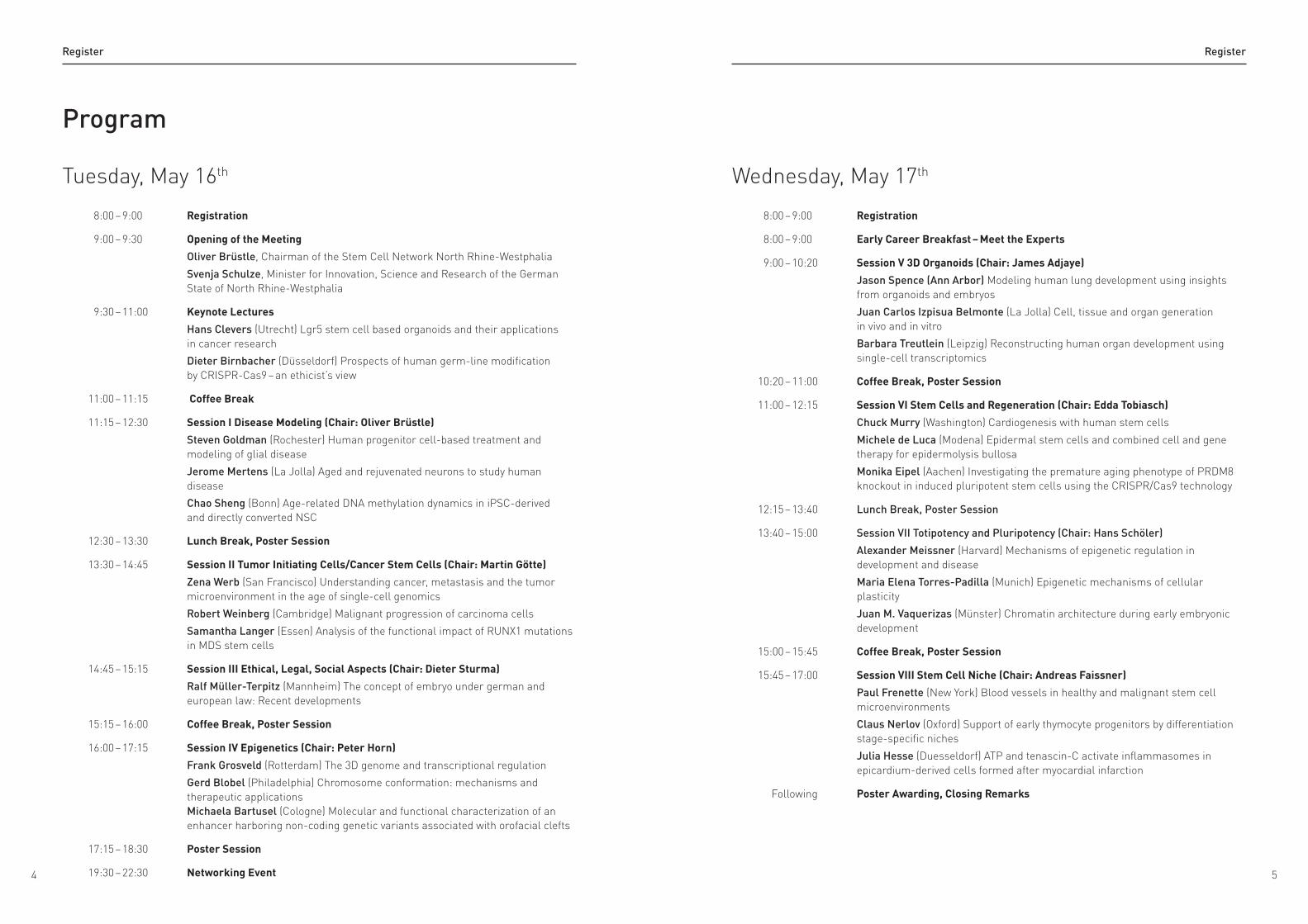
Program
Tuesday, May 16th
8:00 – 9:00 Registration
9:00 – 9:30 Opening of the Meeting
Oliver Brüstle, Chairman of the Stem Cell Network North Rhine-Westphalia
Svenja Schulze, Minister for Innovation, Science and Research of the German State of North Rhine-Westphalia
9:30 – 11:00 Keynote Lectures
Hans Clevers (Utrecht) Lgr5 stem cell based organoids and their applications in cancer research
Dieter Birnbacher (Düsseldorf) Prospects of human germ-line modification by CRISPR-Cas9 – an ethicist’s view
11:00 – 11:15 Coffee Break
11:15 – 12:30 Session I Disease Modeling (Chair: Oliver Brüstle)
Steven Goldman (Rochester) Human progenitor cell-based treatment and modeling of glial disease
Jerome Mertens (La Jolla) Aged and rejuvenated neurons to study human disease
Chao Sheng (Bonn) Age-related DNA methylation dynamics in iPSC-derived and directly converted NSC
12:30 – 13:30 Lunch Break, Poster Session
13:30 – 14:45 Session II Tumor Initiating Cells/Cancer Stem Cells (Chair: Martin Götte)
Zena Werb (San Francisco) Understanding cancer, metastasis and the tumor microenvironment in the age of single-cell genomics
Robert Weinberg (Cambridge) Malignant progression of carcinoma cells
Samantha Langer (Essen) Analysis of the functional impact of RUNX1 mutations in MDS stem cells
14:45 – 15:15 Session III Ethical, Legal, Social Aspects (Chair: Dieter Sturma)
Ralf Müller-Terpitz (Mannheim) The concept of embryo under german and european law: Recent developments
15:15 – 16:00 Coffee Break, Poster Session
16:00 – 17:15 Session IV Epigenetics (Chair: Peter Horn)
Frank Grosveld (Rotterdam) The 3D genome and transcriptional regulation
Gerd Blobel (Philadelphia) Chromosome conformation: mechanisms and therapeutic applications Michaela Bartusel (Cologne) Molecular and functional characterization of an enhancer harboring non-coding genetic variants associated with orofacial clefts
17:15 – 18:30 Poster Session
19:30 – 22:30 Networking Event
Wednesday, May 17th
8:00 – 9:00 Registration
8:00 – 9:00 Early Career Breakfast – Meet the Experts
9:00 – 10:20 Session V 3D Organoids (Chair: James Adjaye)
Jason Spence (Ann Arbor) Modeling human lung development using insights from organoids and embryos
Juan Carlos Izpisua Belmonte (La Jolla) Cell, tissue and organ generation in vivo and in vitro
Barbara Treutlein (Leipzig) Reconstructing human organ development using single-cell transcriptomics
10:20 – 11:00 Coffee Break, Poster Session
11:00 – 12:15 Session VI Stem Cells and Regeneration (Chair: Edda Tobiasch)
Chuck Murry (Washington) Cardiogenesis with human stem cells
Michele de Luca (Modena) Epidermal stem cells and combined cell and gene therapy for epidermolysis bullosa
Monika Eipel (Aachen) Investigating the premature aging phenotype of PRDM8 knockout in induced pluripotent stem cells using the CRISPR/Cas9 technology
12:15 – 13:40 Lunch Break, Poster Session
13:40 – 15:00 Session VII Totipotency and Pluripotency (Chair: Hans Schöler)
Alexander Meissner (Harvard) Mechanisms of epigenetic regulation in development and disease
Maria Elena Torres-Padilla (Munich) Epigenetic mechanisms of cellular plasticity
Juan M. Vaquerizas (Münster) Chromatin architecture during early embryonic development
15:00 – 15:45 Coffee Break, Poster Session
15:45 – 17:00 Session VIII Stem Cell Niche (Chair: Andreas Faissner)
Paul Frenette (New York) Blood vessels in healthy and malignant stem cell microenvironments
Claus Nerlov (Oxford) Support of early thymocyte progenitors by differentiation stage-specific niches
Julia Hesse (Duesseldorf) ATP and tenascin-C activate inflammasomes in epicardium-derived cells formed after myocardial infarction
Following Poster Awarding, Closing Remarks
76 76
Register Register
Register
Bioengineering & Biomaterials
Abstract 1
22A 3D model for bone tissue engineeringD Iandolo , C Pitsalidis , M Ferro , S Inal , A Hama , R Owens
Abstract 2
23Metrology-data driven non-invasive QC and scheduling of automated human iPSC generation and expansionA Elanzew , M Kulik , O Rippel , D Langendoerfer , S Jung , F Schenk , P Wanek , M Zenke , O Brüstle , S Haupt
Abstract 3
24Coiled-coil-based peptide hydrogels as synthetic extracellular matrix for stem cell differentiation K Hagen , N Ma , B Koksch
Abstract 4
25Novel activation techniques of ceramics for improved and accelerated tissue integration of medical implantsN Labude , F Böke , I Lauria , H Fischer , S Neuss
Abstract 5
26Expression of modified HGF for the tunable release from high strength ceramic implant materials S Reinhold , F Böke , N Labude , C Schmitz , I Lauria , W Jahnen-Dechent , H Fischer , S Neuss
Abstract 6
27Optimization of flow and hydrostatic pressure conditions for 3D differentiation of stem cellsC Kasper , D Egger , S Spitz , M Fischer , S Handschuh , M Glösmann , B Friemert , M Egerbacher , C Kasper
Abstract 7
28Scalable expansion of human mesenchymal stem cells in stirred-tank bioreactorsA Tack , V Dufey , A Tacheny , M Art , F De Longueville , M Sha
Direct Cell Fate Conversion
Abstract 8
29Complete reprogramming of embryonic stem sells to trophoblast stem cellsF Kaiser, C Kubaczka, H Schorle
Disease Modelling
Abstract 9
30Molecular and functional characterization of an enhancer harboring non-coding genetic variants associated with orofacial cleftsM Bartusel, M Nicolic, R Rehimi, A Rada-Iglesias
Abstract 10
31Human induced pluripotent stem cells with KIT D816V mutation for modeling leukemiaM Förster, M Szymanski de Toledo, S Sontag, N Chatain, K Gleixner, S Koschmieder, P Valent, T Brümmendorf, M Zenke
Abstract 11
32Loss of pain due to gain-of-function mutation: iPSC derived human nociceptors as a disease model of a Nav1.9 linked pain syndromeA Foerster, S Sontag, E Bressan, J Meents, C Roesseler, M Szymanski, M Hampl, HM Schüler, T Eggermann, I Kurth, M Zenke, A. Lampert
Abstract 12
33iPSC-derived neurons from patient with schizophrenia as a cellular model system for neuropsychiatric disordersL-M Grunwald, M Kriebel, M-C Eberle, A Fallgatter, S Buckenmaier, F Battke, Y Singh, H Volkmer
Abstract 13
34Modelling Batten Disease by genome editing in human iPS cellsG Gomez Giro, J Arias-Fuenzalida, H Zaheres, H Schöler, JC Schwamborn
Abstract 14
35The role of Adiponectin signalling in an iPSC-based model of nonalcoholic fatty liver disease N Graffmann, A Ncube, P Reuter, P Schulze-Matz, S Ring, M Bohndorf, W Wruck, C Czekelius, J Adjaye
Abstract 15
36A 3D lt-NES cell model of human Alzheimer’s disease recapitulates amyloid deposition and phospho-tau accumulationM Hebisch, B Weykopf, SH Choi, DY Kim, M Peitz, O Brüstle
Abstract 16
37Induced pluripotent stem cell-derived disease model of hereditary spastic paraplegia SPG5P Höflinger, S Hauser, Y Theurer, TW Rattay, I Björkhem, R Schüle, L Schöls
Abstract 17
38Mechanisms of microcephaly caused by defective centrosome biogenesis in human iPSC-derived brain C Jalade, R Gutsche, E Gabriel, K Natarajan, J Gopalakrishnan
Abstract 18
39Increased susceptibility of Machado Joseph Disease patient-specific neurons to stress J Jatho-Gröger, K Kleinsimlinghaus, D Poppe, J Ladewig, P Koch
Abstract 19
40Induced pluripotent stem cells for the investigation of schizophreniaM Jung, A Puls, J Schiller, A Klemenz, I Giegling, D Rujescu
Abstract 20
41Towards a stem cell model of retinoblastomaD Kanber, M Hiber, D Lohmann, D Lohmann, L Steenpass
98 98
Register Register
Abstract 33
54Induced pluripotent stem cell-derived hepatocyte-like cells for investigation of the protein quality control of the amyloidogenic protein transthyretinC Niemietz, L Fleischhauer, V Sauer, S Guttmann, S Reinartz Groba, P Ballmaier, A Zibert, HH-J Schmidt
Abstract 34
55Human iPSC-derived neural progenitors are an effective drug discovery model for neurological mtDNA diseaseA Prigione, C Lorenz, A Zink, G Inak, B Mlody, J Meier, D Meierhofer, Z Izvak, J Adjaye, M Schülke, E Wanker, A Lombes
Abstract 35
56Recent Zika virus isolates induce premature differentiation of neural progenitors in human brain A Ramani, E Gabriel, A Wason, M Kronke, J Gopalakrishnan
Abstract 36
57Multiparametric phenotypic assays for screening compounds in neurons derived from spastic paraplegia type 4 patientsK Rehbach, M Peitz, L Schöls, O Brüstle
Abstract 37
58Mechanisms of axonal transport in ALS-patient-derived iPSCs – an in vitro reconstitutionA Seifert, T Korten, L Scharrel, S Diez, X Lojewski, A Hermann
Abstract 38
59Generation of a 3D model to better mimic NAFLD in vitroIL-S Spitzhorn, M-A Kawala, N Graffmann, W Wruck, J Adjaye
Abstract 39
60Functional role of the CAD risk locus 9p21 in calcifying iPSC-derived SMCsA Trillhaase, U Haferkamp, B Schmidt, K Baldwin, V Lo Sardo, A Torkamani, J Erdmann, Z Aherrahrou
Abstract 40
61Generation of vascularized 3D tumor spheres using human iPSCsP Wörsdörfer, N Dalda, E Henke, S Ergün
Abstract 41
62Establishment of a human stem cell-based model of alcohol use disordersA Zink, G Inak, P Lisowski, J Priller, A Prigione
Ethical, Legal & Social Issues
Abstract 42
63The combination of stem sell technology and genome editing – legal and regulatory aspects for medicinal useT Faltus
Abstract 43
64The human pluripotent stem cell registray (hPSCreg)A Kurtz , J Dewender , R Müller , S Seltmann , B Aran Corbella , M Tognarelli , N Mah , A Veiga , G Stacey
Abstract 21
42Assessing the impact of mutant Huntingtin on neuronal mitochondrial functions using patient-derived iPSCsP Kannan, B Mlody, E Wanker, A Prigione
Abstract 22
43An ESC-based approach to investigate the role of autophagy in the reversion of steatotic conditions in non-alcoholic fatty liver diseaseM-A Kawala, A Ncube, N Graffmann, W Wruck, J Adjaye
Abstract 23
44Generation of patient-derived sensory neurons using iPSCs and smNPCs obtained from patients with Fabry diseaseT Klein, K Günther
Abstract 24
45Modeling myeloproliferative neoplasms with patient-derived and engineered iPS cellsC Küstermann, S Sontag, N Chatain, S Koschmieder, M Zenke, K Seré
Abstract 25
46A novel etiological mechanism for Branchio-Oculo-Facial Syndrome (BOFS) and other human neurocristopathiesM Laugsch, M Bartusel, A Karaolidou, H Alirzayeva, P Kolovos, F Grosveld, J Baptista, A Rada-Iglesias
Abstract 26
47Enabling phenotypic drug discovery for neurological mitochondrial DNA disorders with iPSC-derived neural progenitor cells from patientsC Lorenz, A Zink, G Inak, B Mlody, J Eichhorst, B Wiesner, EE Wanker, A Lombès, A Prigione
Abstract 27
48Nijmegen Breakage Syndrome patient-derived iPSCs as a tool for screening platform for anti-oxidants and underlying the mechanisms of microcephalyS Martins, W Wruck, B Mlody, K Sperling, J Adjaye
Abstract 28
49Establishing a disease model of Crigler-Najjar Syndrome with CRISPR/Cas9 and disease-specific induced pluripotent stem cellsJC Matte, T Ljubikj, M Bohndorf, P Schulze-Matz, J Adjaye
Abstract 29
50Modeling muscular dystrophies with patient – derived induced pluripotent stem cellsL Mavrommatis, K Ebert, V Badillo Lisakowski, M Vorgerd, B Brand-Saberi, H Zaehres
Abstract 30
51Rethinking the role of sodium channels in neuronal excitability and pain: new insights from iPSC-derived nociceptors of erythromelalgia patientsJ Meents, E Bressan, S Sontag, A Foerster, M Szymanski, P Hautvast, M Hampl, H Schüler, R Goetzke, T Kim Chi Le, W Wagner, E Jorum, B Namer, B Winner, M Zenke, A Lampert
Abstract 31
52Investigations on the stability of the CpG85 imprint in the human RB1 geneJ Menges, G Wiel, J Stanurova, D Kanber, D Lohmann, L Steenpass
Abstract 32
53Modelling Angelman Syndrome with neurons derived from patient-specific iPSCsA Neureiter, J Stanurova, B Horsthemke, PA Horn, L Steenpaß, H Klump
1110 1110
Register Register
Niche & Microenvironment
Abstract 54
75ATP and tenascin-C activate inflammasomes in epicardium-derived cells formed after myocardial infarctionJ Hesse , S Leberling , T Schmidt , Z Ding , J Schrader
Abstract 55
76Astroglial cells produce a stem cell niche-like extracellular matrix after cortical laser lesions in the mouseL Roll , UT Eysel , A Faissner
Abstract 56
77Evaluation of cellular and microenvironmental multidrug resistance and cancer stem-like cell subpopulation in metastatic and optically imageable iRFP-expressing 4T1 breast carcinoma xenograftsO Tezcan , LY Rizzo , F Kiessling , T Lammers
Abstract 57
78Automated generation of human neural organoidsJM Bruder , M Grabos , HR Schöler
Organogenesis & Regeneration
Abstract 58
79Expression patterns of stem cell factor in murine heartT Hu
Abstract 59
80An organoid-based model of cortical development identifies non-cell autonomous defects in ß-catenin signaling contributing to Miller-Dieker-SyndromeV Iefremova , G Manikakis , O Krefft , A Jabali , K Weynans , R Wilkens , F Marsoner , B Brändl , F-J Müller , P Koch , J Ladewig
Abstract 60
81HEARTI ToolBox: An online platform for the analysis in silico of cardiogenesisR Machado , JP Pinto , D Sabour , J Bragança , A Sachinidis , ME Futschik
Abstract 61
82In vitro approaches to induce re-assembly of adult human primary testicular cellsM Mincheva, R Sandhowe-Klaverkamp, J Wistuba, J-B Stukenborg, N Neuhaus, S Schlatt
Abstract 62
83Optimizing clearing protocols in neuronal organoids for high-throughput analysisM Otto , JM Bruder , M Grabos , HR Schöler
Abstract 63
84Towards nanoparticle-based delivery systems for human neural H Renner , M Grabos , JM Bruder , HR Schöler
Abstract 44
65Human artificial gametes: framing the normative issues of using artificial human gametes in research, cell-based therapy, and assisted reproductive technologiesS Sgodda , B Advena-Regnery , F Enghofer
Abstract 45
66Revisited ethical and legal concepts for precise genome engineering approaches of hereditary diseases (realign-HD)S Sgodda , S Schleidgen , S Kravcishin
Abstract 46
67Ethical dimensions of genom editing on stem cellH Zillmann
Induction & Maintenance of Pluripotency
Abstract 47
68Pluripotency factor UTF1 regulates testicular vasculature development and transgenerational epigenetic inheritance of pluripotencyP Droge , Q Bao , S Bhargy , K Pervushin
Abstract 48
69Transcriptomic and methylation changes in human neural stem cells subjected to reprogramming and subsequent redifferentiationC Haubenreich , M Lenz , L de Boni , J Walter , A Schuppert , P Koch , M Zenke , O Brüstle
Abstract 49
70Changing POU dimerization preferences converts Oct6 into a pluripotency inducerS Jerabek , CKL Ng , G Wu , MJ Arauzo-Bravo , K-P Kim , D Esch , V Malik , Y Chen , S Velychko , CM MacCarthy , X Yang , V Cojocaru , HR Schöler
Abstract 50
71Dissecting Oct4’s DNA binding in establishing and maintaining pluripotencyCM MacCarthy , F Merino , S Velychko , V Pogenberg , V Cojocaru , M Wilmanns , HR Schoeler
Abstract 51
72Generation of integration-free non-human primate iPSCs for basic research with clinical relevanceM Stauske , IR Polo , R Behr
Abstract 52
73SKM reprogramming of murine somatic cells to iPS cells S Velychko , G Wu , CM MacCarthy , K Adachi , HR Schöler
Abstract 53
74Detecting regulatory protein complexes that define pluripotencyT Will , V Helms

1312 1312
Register Register
Abstract 75
96Biased signalling mediated by toll-like receptor 4 influences cancer stem cell behaviourM-T Zeuner , GS Cottrell , D Widera
Stem Cell Differentiation
Abstract 76
97Is it possible to generate M2- in vitro-derived microglia from transgenic eGFP- mice?K Arnold , C Fabian , A Lourhmati , L Danielyan , A Stolzing
Abstract 77
98Differentiation potential of dental neural crest-derived progenitor cellsP Babczyk , D Schiper , P Ottensmeyer , W Götz , E Tobiasch
Abstract 78
99Impact of immortalization for establishment of hepatocyte-like cellsP Ballmaier , C Niemietz , S Guttmann , S Reinartz Groba , F Bernick , V Sauer , A Zibert , HH-J Schmidt
Abstract 79
100Oligodendroglial priming of adult neural stem cells: intrinsic or niche dependent?F Beyer , J Jadasz , L Dimou , NR Kannaiyan , M Rossne , S Griemsmann , N Klöcker , P Küry
Abstract 80
101LRP1 loss in radial glia and their progeny – a trigger for hyperexcitability?E Bres , D Safina , J Müller , P Bedner , C Steinhäuser , A Faissner
Abstract 81
102I’m not big boned, I’m just fat! - Oxidative stress induces adipogenesis in osteogenic differentating ASC and DFAT cultures hindering a sufficient osteogenesisA Bollmann , C Sons , C Suschek
Abstract 82
103The functional relevance of DNMT3A splice variants in hematopoietic differentiationT Boži , J Frobel , A Rai , S Heilmann-Heimbach , TW Goecke , E Jost , W Wagner
Abstract 83
104Human bone-marrow derived mesenchymal stromal cells exhibit large inter-donor differences in in vitro-osteogenic differentiationL Burmeister , Y Roger , B Quaas , T Flörkemeier , U Köhl , U Rinas , A Hoffmann
Abstract 84
105Generation of hematopoietic stem and progenitor cells from human pluripotent stem cells, in vitroM Cremanns , H Klump , B Giebel , PA Horn
Abstract 85
106Derivation of cochlear cells from pathological and isogenic human iPSCs for modeling hereditary hearing lossA Czajkowski , B Grobarczyk , L Borgs , K Hanon , P Lefebvre , L Nguyen , B Malgrange
Abstract 86
107Significance of microRNAs for the differentiation of human embryonic stem cells into definitive endoderm and mesodermU Diekmann , D Ishikawa , J Fiedler , A Just , T Thum , S Lenzen , O Naujok
Abstract 64
85KRAB ZNF gene biology: Do KRAB ZNF genes play an essential role in human stem cell driven differentiation and organogenesis? H-J Thiesen , P Lorenz , M Peitz , D Koczan , H Albony , M Al Chiblak , F Steinbeck , S Li , Y Li , O Brüstle
Abstract 65
86Molecular basis of controlled in vivo cellular reprograming that enables cardiac regenerationW Yao , G Ahuja , W Frank , D Bartsch , N Russ , C Dieterich , L Kurian
Somatic & Cancer Stem Cells
Abstract 66
87Hybrid clone cells derived from human breast epithelial cells and human breast cancer cells exhibit properties of cancer stem/ initiating cellsD Gauck , S Keil , B Niggemann , KS Zänker , T Dittmar
Abstract 67
88Inhibition of the notch signaling pathway as an experimental therapeutic approach in endometriosisM Götte , L Kettler , K Brüggemann , M Hubert , N Achmad , L Kiesel , A Schüring , S Schäfer , B Greve
Abstract 68
89The heparan sulfate proteoglycan Syndecan-1 regulates colon cancer stem cell function via a focal adhesion kinase – Wnt signaling axisM Götte , SK Katakam , R Reinbold , I Zucchi , GW Yip , B Greve
Abstract 69
90Upregulation of AhR and CYP1B1 is associated with the cancer stem cell phenotype of triple negative inflammatory breast cancer via WNT5a/b and ?-catenin signaling pathwayS Ibrahim , HT Mohamed , R Gadalla , EA El-Ghonaimy , M El-Shinawi , DH Sherr , MM Mohamed
Abstract 70
91Analysis of the functional impact of RUNX1 mutations in MDS stem cellsS Langer , M Kiljan , PA Horn , S Heinrichs
Abstract 71
92Self-renewal in primary colon cancer spheroidsJ Otte , W Wrick , L Dizdar , N Stoecklein , J Adjaye
Abstract 72
93CD44 and CD133 coexpression in poorly differentiated and early oral squamous cell carcinomaE Vencio , JSK Oliveira , MN Azevedo , TM Siqueira , AC Fraga Júnior
Abstract 73
94Expression of hypoxia-induced factor-1 alpha in early stage and in metastatic oral squamous cell carcinomaE Vencio , M Ribeiro , SR Teixeira , MN Azevedo , AC Fraga Júnior , APM Gontijo
Abstract 74
95MMP-9/ICAM-1 up-regulation by TNF-? increase cell fusion between MDA-MB435-pFDR.1 and M13SV1-Cre cells via NF-kB dependent pathway J Weiler , M Mohr , KS Zänker , T Dittmar
1514 1514
Register Register
Abstract 98
119Reprogramming of epicardial derived cells into functional cardiomyocytes by the cardiac microenvironment A Marzoq , Z Ding , O Sergeeva , D Friebe , J Hoell , X Ke , A Borkhardt , J Schrader
Abstract 99
120The role of Wnt5a during the BMP2/DLX3- induced osteogenic differentiation in dental follicle cellsL Mischkulnig , T Reichert , C Morsczeck
Abstract 100
121Dissecting the origin of dendritic cell and macrophage subsets in human hematopoiesisF Murke , A Görgens , PA Horn , B Giebel
Abstract 101
122FGF-2 but not FGF-7/-10 is a strong inhibitor of pancreatic lineage specification during differentiation of human embryonic stem cellsO Naujok, J Münchhoff, U Diekmann, J Kresse
Abstract 102
123The ubiquitin ligase LIN41 targets p53 to antagonize cell death and differentiation pathways during D Nguyen , FG Wulczyn
Abstract 103
124SCIS Toolbox: Design and implementation of online tools for in silico stem cell biologyJP Pinto , RK Kalathur , DV Oliveira , T Barata , RSR Machado , S Machado , I Pacheco-Leyva , JM Xavier , J Bragança , I Duarte , ME Futschik
Abstract 104
125Investigating the cardiac progenitor stage during cardiomyocyte induction of human pluripotent stem cellsR Quaranta , B Greber
Abstract 105
126Interpreting growth factor signals: How do ES cells encode and decode FGF/ERK signals? D Raina , C Schroeter
Abstract 106
127Involvement of transcription factor NF-kappa B during glutamatergic differentiation of adult human stem cellsL Ruiz-Perera , C Kaltschmidt , B Kaltschmidt
Abstract 107
128The potential of Bmp5/7 on the yield of midbrain dopaminergic neurons during in vitro differentiation of human induced pluripotent and neural stem cells A Salti , V Jovanovic , S Meyer , C Brodski , F Edenhofer
Abstract 108
129Identification of age- and species-dependent effects of mesenchymal stem cell-derived factors with pro-oligodendrogenic activitiesI Samper Agrelo , JJ Jadasz , L Tepe , L-S Spitzhorn , J Adjaye , P Küry
Abstract 109
130Direct differentiation of induced pluripotent stem cells into osteoblasts and endothelial cellsD Schipper , P Ottensmeyer , P Babczyk , H Hescheler , E Tobiasch
Abstract 87
108Investigating the premature aging phenotype of PRDM8 knockout in induced pluripotent stem cells using the CRISPR/Cas9 technologyM Eipel , C Weidner , C Rösseler , A Lampert , W Wagner
Abstract 88
109MEIS2 is a novel player involved in atrial cardiomyocyte specification of human pluripotent stem cellsJ Fell , R Quaranta , J Rao , B Greber
Abstract 89
110Hydrogels of human platelet lysate support differentiation of mesenchymal stromal cells and of iPSC-derived MSCsR Goetzke , J Franzen , A Ostrowska , M Vogt , G Klein , B Rath , M Zenke , W Wagner
Abstract 90
111Mechanisms and utility of cardiac subtype specification from hPSCs in development and disease B Greber , M Marzcenke , R Quaranta , M Pfeiffer , I Piccini , J Fell , G Seebohm
Abstract 91
112Single cell analyses of adult human neural crest-derived stem cells during neuronal differentiationA Höving , JF W Greiner , C Kaltschmidt , B Kaltschmidt
Abstract 92
113Transcriptional profiling of in vitro generated red blood cells from human adult HSCs and pluripotent stem cellsK Kessel , CM Bernecker , C Bartenhagen , A Bluemke , HR Schöler , H Zaehres , P Schlenke , I Dorn
Abstract 93
114In vitro generation of vascular wall-typical mesenchymal stem cells from murine induced pluripotent stem cellsD Klein , J Steens , M Zuk , N Teichweyde , J Hess , K Unger , A Görgens , H Klump
Abstract 94
115Bioluminescence imaging visualizes osteopontin-induced neurogenesis and neuroblasts migration in the mouse brain after strokeR Klein , A Pikhovych , A Bach , M Hoehn , S Couillard-Despres , GR Fink , M Schroeter , MA Rueger
Abstract 95
116Generation of functional neutrophils from human stem cells including induced pluripotent stem cellsN Ludwig , T Weinhage , C Kessel , D Föll , H Witkowski , K Kessel
Abstract 96
117The CellFinder on-line data resource and its applications for stem cell researchN Mah , K El Amrani , S Seltmann , H Stachelscheid , M Andrade-Navarro , A Kurtz
Abstract 97
118Neuronal differentiation of mouse pluripotent stem cells in vitro does recapitulate important aspects of the Foxg1 knockout phenotypeEM Mall , D Hermann , H Niemann
1716 1716
Register Register
Abstract 121
142Reprogramming enriches for somatic cell clones with small scale mutations in cancer-related genesM Kosanke , K Osetek , A Haase , L Wiehlmann , P Chouvarine , S Merkert , S Wunderlich , U Opel , S Menke , M Dorda , S Mielke , D Steinemann , A Schambach , U Martin
Abstract 122
143Epigenetic characterization of germ cells in human testis A Laurentino , J Gromoll , S Kliesch , S Schlatt , N Neuhaus
Abstract 123
144Cell cycle protein expression after the induction of cellular senescence in dental follicle stem cells C Morsczeck , A Reck , TE Reichert
Abstract 124
145Age-related DNA methylation dynamics in iPSC-derived and directly converted NSC C Sheng , J Jungverdorben , H Wiethoff , Q Lin , D Eckert , M Hebisch , J Kesavan , J Fischer , L Flitsch , A Till , B Weykopf , U Wüllner , W Wagner , M Peitz , O Brüstle
Stem Cells in Development
Abstract 125
146A germ cell score for diagnostic evaluation of prepubertal and pubertal testicular biopsies stored for fertility preservationL Heckmann , T Pock , J Wistuba , S Schlatt , S Kliesch , N Neuhaus
Abstract 126
147Ionizing irradiation impairs the cardiac differentiation of human embryonic stem cellsS Nitsch , I Schroeder , S Ritter
Abstract 127
148The extracellular matrix molecule tenascin-C and its regulation in neural stem cells and glial progenitorsU Theocharidis , E Schaberg , L Roll , C Stolt , M Wegner , A Faissner
Abstract 128
149Blockade of HCN/h current impedes proliferation and differentiation dynamics in neural stem cells in vitroSU Vay , AK Schlusche , I Jakovcevski , GR Fink , M Schroeter , D Isbrandt , MA Rueger
Abstract 129
150Do primordial germ cells migrate along nerve fibres? An interspecies comparison.E Wolff , M Suplicki , R Behr
Stem Cells in Regenerative Medicine
Abstract 130
151Dissecting the complexity of cell types present in urine, identifies renal progenitor cells with regenerative potentialJ Adjaye , M Bohndorf , H Kayar , F Asar , L-S Spitzhorn , W Wruck
Abstract 110
131Mechanical stimulation of human mesenchymal stem cells using a uniaxial cell stretcherT Schleypen , M Hoß , B Hoffmann , R Merkel , U Schnakenberg , S Neuss
Abstract 111
132Coordination of cell fate decisions through FGF signalingC Schröter , A Koseska
Abstract 112
133Exploration of Nrf2 pathway activation during neuronal differentiation of human neural stem cellsV Semkova , D Langendörfer , M Segschneider , C Bell , M Ingelman-Sundberg , A Till , O Brüstle , S Haupt
Abstract 113
134A scalable approach for the generation of human pluripotent stem cell-derived hepatic organoids with sensitive hepatotoxicity featuresM Sgodda , Z Dai , R Zweigerdt , AD Sharma , M Ott , T Cantz
Abstract 114
135Modeling IRF8 deficient human hematopoesis and dendritic cell development with engineered iPS cellsS Sontag , M Förster , J Qin , P Wanek , S Mitzka , HM Schüler , S Koschmieder , S Rose-John , K Sere , M Zenke
Abstract 115
136Rapid and efficient generation of human oligodendrocytes from induced pluripotent stem cells for in vitro disease modelling and drug discoveryL Starost , M Ehrlich , M Glatza , S Velychko , H Zaehres , J Antel , HR Schöler , T Kuhlmann
Abstract 116
137Effects of an antioxidative treatment with catalase on the osteogenic differentiation of human mesenchymal bone marrow stem cells D Strangmann , CV Suschek , D Strangmann
Abstract 117
138Biological characterisation of different human hepatocyte models by gene expression profilingT Waltermann , P Schulze-Matz , J Adjaye , H Ellinger-Ziegelbauer
Stem Cell Epigenetics
Abstract 118
139Epigenetic impairment of genomic integrity by a sphingolipid intermediate leads to senescence and aging of the heartG Ahuja , D Bartsch , W Yao , J Dodzian , S Frank , N Vargas , N Russ , J Messlin , J Hescheler , C Dieterich , M Petrascheck , A Aguirre , M Jain , D Valanzano , L Kurian
Abstract 119
140Changes in clonal dynamics during aging of the mouse subependymal zoneF Calzolari , L Bast , J Michel , M Strasser , EV Baumgart , F Theis , M Götz , C Marr , J Ninkovic
Abstract 120
141Human platelet lysate versus fetal Cclf serum: These supplements do not select for different mesenchymal stromal cellsE Fernandez-Rebollo , B Mentrup , R Ebert , J Franzen , G Abagnale , T Sieben , A Ostrowska , P Hoffmann , PF Roux , B Rath , M Goodhardt , JM Lemaitre , O Bischof , J Franz , W Wagner

1918 1918
Register Register
Abstract 142
163HOX A7 is downregulated during in vitro differentiation of mesenchymal stem cells towards osteoblastsP Ottensmeyer , D Schipper , P Babzcyk , M Witzler , F Elsayed , W Götz , M Schulze , E Tobiasch
Abstract 143
164Studies towards a pulmonary macrophage transplantation (TMP) strategy targeting alpha-1 antitrypsin deficiencyM Schwabbauer , E Janosz , M Hetzel , M Ackermann , C von Kaisenberg , S Janciauskiene , N Lachmann , T Moritz
Abstract 144
165Interspecies nuclear transfer: Way forward to generate horse specific nuclear transfer stem cellsN Selokar , TR Talluri , P Sharma , D Kumar , PS Yadav , BN Tripathi
Abstract 145
166The new approach for stimulation? Cell neogenesis in the insulin-dependent diabetes: Inhibition of inflammation and hematopoietic stem cells by reserpine and stimulation of ? cells progenitors by pegylated GLP-1E Skurikhin , P Olga , P Angelina , E Natalia , Y Anton , D Alexandr
Abstract 146
167Human ESC and iPSC-derived MSCs regenerate injured GUNN rat liver: a comparative studyL-S Spitzhorn , C Kordes , M Megges , I Sawitza , S Goetze , D Reichert , P Schulze-Matz , N Graffmann , W Wruck , D Herebian , E Mayatepek , R Oreffo , D Häussinger, J Adjaye
Abstract 147
168Human iPSC-derived iMSCs improve regeneration in a Goettingen mini-pig bone defect modelL-S Spitzhorn , J Grassmann , J Schneppendahl , S Tanner , M Bohndorf , W Wruck , V Grotheer , J Windolf , P Jungbluth , J Adjaye
Abstract 148
169Reported HSC expansion cocktails do not support human HSC/MPP expansionS Vitoriano da Conceicao Castro , A Görgens , PA Horn , B Giebel
Abstract 149
170Pathogen reduction through additive-free UV-C irradiation retains the optimal efficacy of human platelet lysaste for the expansion of human bone marrow mesenchymal stem cellsS Viau , L Chabrand , S Eap , J Lorant , K Rouger , F Goudaliez , C Sumian , B Delorme , F Goudaliez , B Delmore
Abstract 150
171A standardized and characterized clinical grade human platelet lysate for efficient expansion of human bon marrow mesenchymal stem cellsS Viau , S Eap , L Chabrand , J Lorant , K Rouger , P Bertholet , T Bouckenooghe , A Larange , F Goudaliez , B Delmore
Abstract 151
172Improved cryopreservation of human stem cells for regenerative medicineI Zahanich , U Ravens
Transcriptional Regulation & Non-Coding RNAs
Abstract 152
173Activation of Wnt/Tcf1 pathway induces cell cycle arrest in mouse embryonic stem cellsA de Jaime Soguero , F Aulicino , G Ertaylan , A Griego , A Cerrato , A Tallam , A del Sol , MP Cosma , F Lluis
Abstract 131
152Increasing biocompatibility of patient specific PEO-coated implants using endothelial pro-genitor cells and mesenchymal stem cells in bone defectsM Bienert , L Jauer , M Muether , C Ptock , B Lethaus , S Neuss
Abstract 132
153A mixed lymphocyte reaction as a functional assay for extracellular vesicles of different originsM Bremer , V Börger , PA Horn , B Giebel
Abstract 133
154Enhanced local immune cell response and angiogenesis are associated with robust cardiac regeneration induced by apoptotic USSCsZ Ding , K Tan , S Temme , C Alter , U Flögel , G Kögler , J Schrader
Abstract 134
155Imaging flow cytometry enables discrimination of distinct subpopulations of small extracellular vesiclesR Ferrer Tur , A Görgens , PA Horn , V Börger , B Giebel
Abstract 135
156Generation of early neuroepithelial progenitors from human fetal brain tissue for biomedical applicationsK Günther , P Wörsdörfer , MC Thier , F Edenhofer
Abstract 136
157iPSC-derived macrophages improve the disease phenotype of pulmonary alveolar proteinosis in vivoW Janosz , A Mucci , M Hetzel , M Ackermann , T Suzuki , C Happle , E Lopez-Rodriguez , JP Bankstahl , MP Kühnel , A Schambach , G Hansen , B Trapnell , N Lachmann , T Moritz
Abstract 137
158Precise concentration, viability, and phenotype analysis of adipose derived mesenchymal stem cells using a novel imaging cytometry methodB Kohring , H Yan , X Shao , F Kong , Y Zhang
Abstract 138
159Derivation and characterization of hepatocyte-like cells from iPSCs derived from urine progenitor cells of an african male bearing the CYP2D6 *4/*17 variant conferring intermediate drug metabolizing activityA Ncube , M Bohndorf , M-A Kawala , N Graffmann , W Wruck , J Adjaye
Abstract 139
160The role of pluripotency in chimera formation between mouse or monkey iPS cells and porcine embryosM Nowak-Imialek , S Wunderlich , D Herrmann , S Klein , U Baulain , A Lucas-Hahn , B Petersen , U Martin , H Niemann
Abstract 140
161Dissecting the complexity in amniotic fluid-derived cells obtained from cesarean sectionsS Rahman , L-S Spitzhorn , H-T Ho , M Bohndorf , W Wruck , L Schwindt , A Ncube , C Hagenbeck , P Balan , I Beyer , T Fehm , J Adjaye
Abstract 141
162Immunohistochemical studies of thymus and spleen of mice after skin graft transplantationM Ospanova , AS Alishevna , BT Sansyzbaevna , ST Nikolaevna , AM Bapovich
2120 2120
Register Register
Company Profiles
Acris Antibodies GmbH ................................................................................................................................................................................ 186Biolamina AB ..................................................................................................................................................................................................... 187BIOTREND Chemikalien GmbH .............................................................................................................................................................. 188BIOZOL Diagnostica GmbH ........................................................................................................................................................................ 189Brooks Automation Ltd. .............................................................................................................................................................................. 190Cygenia GmbH ................................................................................................................................................................................................... 191Eppendorf AG ..................................................................................................................................................................................................... 192I&L Biosystems GmbH ................................................................................................................................................................................. 193Labotect GmbH ................................................................................................................................................................................................. 194LGC Standards GmbH ................................................................................................................................................................................... 195LIFE and BRAIN GmbH ................................................................................................................................................................................ 196Lonza Cologne GmbH ................................................................................................................................................................................... 197Maco Pharma International GmbH ...................................................................................................................................................... 198Miltenyi Biotec GmbH ................................................................................................................................................................................... 199New England Biolabs GmbH .................................................................................................................................................................... 200neoFroxx GmbH ................................................................................................................................................................................................ 201PeproTech GmbH ............................................................................................................................................................................................. 202PL BioScience GmbH .................................................................................................................................................................................... 203PromoCell GmbH ............................................................................................................................................................................................ 204ReproCELL Europe Ltd ................................................................................................................................................................................ 205SERVA Electrophoresis GmbH ................................................................................................................................................................ 206STEMCELL Technologies Germany GmbH ...................................................................................................................................... 207Thermo Fisher Scientific ............................................................................................................................................................................ 208
Abstract 153
174Functional and topological characterization of poised enhancers in mouse embryonic stem cellsS de la Cruz Molina , P Respuela Alonso , C Tebartz , M Nikolic , P Kolovos , F Grosveld , A Rada Iglesias
Abstract 154
175Poised enhancers regulatory activity is topologically facilitated by polycombS de la Cruz Molina
Abstract 155
176Developmental programming by conserved lncRNA-TF pairs during the induction of the embryonic heartS Frank , J-E Messling , J Ku , G Ahuja , L Brant , N Russ , I Gesterira Costa Filho , A Papantonis , C Kanduri , L Kurian
Abstract 156
177miR-142-3p mediates stem cell regulation in breast cancer cellsB Greve , F Troschel , N Boehly , HT Eich , M Götte
Abstract 157
178Functions of murine long non-coding RNA in induced and physiological neurogenesisC Nakajima-Claverie , S Thakurela , VK Tiwari , B Berninger
Other
Abstract 158
179Banking of hiPSCs in a suspension bioreactorS Bur , A Schulz , L Gentile , J Neubauer , H Zimmermann
Abstract 159
180Transcriptome study with NGS and Affymetrix array: a comparative case study of cardiomyocyte specific differentiation process in murine stem cellL Gan , B Denecke
Abstract 160
181Cilia induction to prevent proliferation of glioma in vitroG Goranci-Buzhala , J Gopalakrishnan , A Mariappan
Abstract 161
182Identification and isolation of VASA positive putative oogonial stem cells from porcine and bovine ovariesI Röttinger , M Nowak-Imialek , D Herrmann , H Niemann
Abstract 162
183In vitro propagation of adult macaque spermatogonial stem cellsS Swati , N Terwort , N Neuhaus , S Schlatt
Abstract 163
184Modelling hereditary spastic paraplegia type 46 with induced pluripotent stem cells and CRISPR/Cas9U Ulmer , J Reichbauer , S Hauser , S Schuster , L Schöls , M Synofzik , R Schüle
22
Bioengineering & BiomaterialsAbstract 1
23
Abstract 2 Bioengineering & Biomaterials
A 3D model for bone tissue engineering
D Iandolo 1, C Pitsalidis 1, M Ferro 1, S Inal 2, A Hama 1, R Owens 1
1 CMP-EMSE, Gardanne, France 2 King Abdullah University of Science and Technology, Thuwal, Saudi Arabia
One of the latest trends in the fields of tissue engi-neering as well as oncological research is the devel-opment of in vitro systems mimicking specific target tissues. Indeed, there is an increasing demand for in vitro models recapitulating the tridimensional struc-ture and microenvironment experienced by cells in the target. These systems would integrate the 2D systems employed so far. Interestingly, in addition to chemical and mechanical cues, certain tissues are known to be regulated by endogenous bioelectrical cues. [1] One such tissue is the bone. Indeed, it has been demonstrated to exhibit piezoelectric proper-ties in vivo. [2] Electrical stimulation has been proven to sustain cell proliferation as well as to boost the expression of genes related to stem cells osteogenic differentiation and induce higher levels of enzymatic
activities related to bone matrix deposition. [3] We present the development of a device consisting of a 3D electroactive porous scaffold based on the elec-trochemically active polymer PEDOT: PSS, allowing both cells proliferation monitoring and electrical stimulation for osteoregeneration studies. Indeed, organic electronic materials offer a unique combina-tion of properties helping to transcend the current state of the art in transduction and stimulation of the electrical activity in cells. Among the proper-ties mentioned above, their ability to conduct ions provide a lower impedance ‘connection’ to cells. [4] The material developed and device are anticipated to be beneficial for the study of the responses of mul-tiple electroactive cell types in complex biomimicry environments.
Keywords: Bioelectronics, 3D model, regenerative medicine E-mail: [email protected]
Metrology-data driven non-invasive QC and scheduling of automated human iPSC generation and expansion
A Elanzew 1, M Kulik 2, O Rippel 1, D Langendoerfer 1, S Jung 2, F Schenk 2, P Wanek 3, M Zenke 3, O Brüstle 1, S Haupt 1
1 Life and Brain GmbH, University of Bonn, Bonn, Germany 2 Fraunhofer Institute for Production Technology, Aachen, Germany 3 Helmholtz-Institute for Biomedical Engineering, Aachen, Germany
The increasing demand for human induced pluripo-tent stem cells (hiPSCs) has created an urgent need for standardized and automated cell reprogramming. This demand is met by the StemCellFactory (www.stemcellfactory.de), a large system integration that provides automation for all required cell culture steps, ranging from adult human dermal fibroblast (HF) expansion via feeder-free, Sendai virus-based reprogramming to clonal selection and enzyme-free expansion of the obtained hiPSC clones and lines. The implementation of on-line measurement tech-nologies is key for the establishment of a fully auto-mated production process on the StemCellFactory. This is realized by evaluation of in-process generated data by a novel programmed control level software, which controls, triggers and allows user defined workflow assembly. Here we report the realization and biological validation of non-invasive measure-ment technologies for QC of hiPSC cultures and the scheduling of the fully automated splitting processes in 24- and 6-well plates. For on-line assessment of metabolic activity and detection of potential bacte-rial contamination we implemented a plate reader
and established instrument settings that enabled us to detect a shift of pH and/or turbidity. In addi-tion, an automated high-speed microscopy platform was implemented to schedule and perform fully automated cultivation of hiPSCs. To that end we developed a confluence detection assay that enables dynamic feedback by automatically computing con-fluence-based splitting ratios (SR) for subsequent cell passaging. For fully automated expansion of individual hiPSC clones in 24-well plates, well-based splitting protocols were developed. Hereby separate clones showing individual grow characteristics can be maintained in parallel within a single 24-well plate without compromising cell quality. Addition-ally, automated sub-cultivation for the scaled pro-duction of iPSC in 6-well plates was implemented. Automatically expanded hiPSCs can be efficiently harvested (95.6% ± 2.9%), maintain viability (94 ± 3%) and remain pluripotent for at least 10 passages. Our data show that dynamic feedback via genera-tion and analysis of in-process data can be used by the control level software to facilitate automation of highly dynamic cell culture processes.
Keywords: Stemcellautomation, reprogramming, metrology, robotic platform E-mail: [email protected]
2524
Abstract 3 Abstract 4 Bioengineering & BiomaterialsBioengineering & Biomaterials
Keywords: Ceramics, protein-immobilization, MSC, adhesion, osseointegration E-mail: [email protected]
Novel activation techniques of ceramics for improved and accelerated tissue integration of medical implants
N Labude 1, F Böke 1, I Lauria 1, H Fischer 1, S Neuss 1, 2
1 RWTH Aachen University, Aachen, Germany 2 Helmholtz-Institute for Biomedical Engineering, Aachen, Germany
High-performance oxide ceramics have successfully been used as medical implants for decades, espe-cially as articulating components of artificial joints, due to their high strength, high wear resistance and cytocompatibility. However, these ceramics are not suitable for direct bone contact due to an insufficient osseointegration. To enable a broader use in clinical practice, their bioinert surfaces need to be modified to promote cell recruitment and subsequent tissue integration. Therefore, we have developed a novel strategy to efficiently bioactivate oxide high-perfor-mance ceramic surfaces through tailored, stable sili-cate/silane coatings for peptide and protein coupling. High-performance oxide ceramics were coated with a 100 nm thick SiOx-layer via physical vapor deposition. Afterwards, a silane monolayer was applied to pre-sent either –NH2, -SH, or a mixture of both groups at the interface. The application process was verified via XPS, FTIR-ATR, EDX, and AFM. After silanisation, the –NH2 and –SH group density was calculated at > 90% on the mono-functionalized and > 80% on bi-func-tionalized surfaces. AFM and XPS analyses confirmed a silan-monolayer of approximately 0.7 nm thickness. Various peptides and proteins (RGD, RAD negative control, HGF and BMP-2) were covalently bound on mono- and bi-functionalized surfaces via specified crosslinkers. Coupling efficiency was proven by vari-ous methods: for RGD-peptides by I125-radioactive labeling, for HGF and BMP-2 by AuNP-labeling. To analyze the retained function of RGD after coupling, we modified a centrifugation-assay developed by Reyes et al1. Here, murine fibroblasts (L929) and human mesenchymal stem cells (MSC) were seeded on RGD-, RAD-coupled, and unmodified ceramics, centrifuged and the remaining cells on each surface
were counted. Potential loss-of-function of the HGF after coupling was examined by MSC and HuH7 (hu-man Hepatocyte-derived carcinoma cells) Boyden chamber assays. Osteogenic induction potential of functionalized and unmodified ceramics was evaluat-ed by RealTime-PCR.I125-labeling shows significant amounts of RGD when crosslinked on the surface in comparison to unspecific binding. A significant higher adhesion of L929 and MSC on RGD-loaded ceramics compared to RAD-loaded substrates was determined via centrifugation-assay, with approx. 65% (RGD) to 15% (RAD) adherent cells by centrifuge-forces from 60-70 nN per cell. This is in accordance with immunofluorescence studies showing higher amounts of phosphorylated FAK (PTK2) complexes, a key component of the integrin-downstream path-way, on RGD-loaded surfaces in comparison to RAD-controls. AuNP-labeling, followed by SEM analyses, confirmed the accessibility of BMP-2 and HGF after crosslinking. Migration studies of HGF after cou-pling showed significant higher amounts of cells migrating towards the modified surfaces. RT-PCR showed significant higher osteogenic differentiation of hMSC on BMP-2 coupled surfaces. In conclu-sion, individual chemical functionalization enables directed immobilization of cell adhesion, migration, and differentiation promoting peptides and proteins to significantly enhance osseointegration on high-performance oxide ceramics. Based on our thorough chemical, mechanical, and biological evaluation we show that the functionalized ceramic surfaces hold significant potential for an improved bone bonding behavior. In the next step the tailored surfaces will be tested for their osseointegration-potential in vivo.
Keywords: E-mail: [email protected]
Coiled-coil-based peptide hydrogels as synthetic extracellular matrix for stem cell differentiation
K Hagen 1, N Ma 2, B Koksch 3
1 Freie Universität Berlin, Berlin, Germany 2 Helmholtz-Zentrum Geesthacht, Teltow, Germany 3 Freie Universität Berlin, Berlin, Germany
Biomaterials have numerous applications in the fields of tissue engineering and regenerative medi-cine, which rely on the use of stem cells or pro-genitor cells. Various approaches and applications using peptides as substrates for directing stem cell behavior have been reported. Peptide-based bio-materials have many advantages, as for example i) biocompatibility and biodegradability, ii) modifiable properties based on the amino acid sequence, iii) ease of synthesis, iv) big repertoire of structural mo-tifs and higher-ordered assemblies. As an example, the coiled-coil structural motif consists of a core of periodical repeats of seven amino acids (heptad) (a-b-c-d-e-f-g)n, where especially the a and d po-sitions are occupied with hydrophobic amino acids. Coiled-coil motifs have benefits which make them ideal candidates for the design of self-assembling peptide based materials to influence stem cell fate, as for example i) straight-forward design based on
heptad repeats, ii) covalently modifiable, iii) ther-mal stability. Furthermore, coiled-coil peptides have been increasingly investigated as highly suitable scaffolds for cell culture applications due to their predictable self-assembly properties, which also allow multivalent ligand presentation. Moreover, temperature-responsive hydrogels have been shown to promote growth and differentiation of rat adrenal pheochromocytoma cells. To date, however, very few examples of 2D and 3D coiled-coil based scaffolds that influence stem cell behavior exist. Our group discovers the potential of coiled-coil peptides to directly influence stem cell fate. The theory of the stem cell niche describes that stem cells need the support of a defined microenvironment for prolifera-tion and differentiation, the so called extracellular matrix. We will test coiled-coil derived hydrogels that covalently display short recognition motifs to mimic the extracellular matrix of stem cells.
2726
Abstract 5 Abstract 6 Bioengineering & BiomaterialsBioengineering & Biomaterials
Keywords: Bioreactors, biomaterial, 3D dynamic cell culture E-mail: [email protected]
Optimization of flow and hydrostatic pressure conditions for 3D differentiation of stem cells
C Kasper 1, D Egger 1, S Spitz 1, M Fischer 1, S Handschuh 1, M Glösmann 1, B Friemert 1, M Egerbacher 1, C Kasper 1
1 University of Natural Resources and Life Science, Vienna, Austria
It is crucial but challenging to keep physiologic con-ditions during the cultivation of three-dimensional (3D) cell-scaffold constructs for the optimization of 3D cell culture processes. Therefore, we used a recently developed miniaturized perfusion bioreac-tor together with a specialized incubator system to optimize flow and hydrostatic pressure conditions for differentiation of human mesenchymal stem cells (MSC) on a 3D scaffold. The incubator system al-lows for cultivation of multiples samples under the same conditions and hence enables for screening of different conditions. First, a decellularized bone matrix was characterized and tested towards its suit-ability for 3D osteogenic differentiation under flow perfusion conditions. Subsequently, shear stress and hydrostatic pressure (HP) conditions were optimized for osteogenic differentiation of human MSC. X-ray computed microtomography and scanning electron microscopy (SEM) revealed a closed cell layer cover-
ing the entire matrix. Osteogenic differentiation which was assessed via alkaline phosphatase activity and SEM was found to be increased in all dynamic condi-tions. Furthermore, screening of different fluid shear stress conditions indicated 1.5 ml/min (equivalent to ~ 10 mPa shear stress) to be optimal for osteogenic differentiation. However, no distinct effect of HP com-pared to flow perfusion without HP on osteogenic differentiation was observed. Notably, throughout all experiments MSC cultivated under fluid shear stress or HP conditions displayed increased osteogenic differentiation which underlines the importance of physiologic conditions. In conclusion, shear stress and hydrostatic pressure conditions were screened and optimized for the osteogenic differentiation of MSC on a 3D scaffold. Furthermore, we hypothesize the bioreactor system used in this study may enhance the development and optimization of other 3D cell culture processes as well.
Keywords: HGF, biomaterial, cell migration, osseointegration, MSC E-mail: [email protected]
Expression of modified HGF for the tunable release from high strength ceramic implant materials
S Reinhold 1, F Böke 1, N Labude 1, C Schmitz 2, I Lauria 1, W Jahnen-Dechent 1, H Fischer 1, S Neuss 1, 2
1 RWTH Aachen University, Aachen, Germany 2 Helmholtz-Institute for Biomedical Engineering, Aachen, Germany
High strength oxide ceramics are versatile bioma-terials readily deployed in numerous orthopedic ap-plications. Although superior to the current gold standard titanium in various regards, their major drawback is a lack of osseointegration in vivo, re-sulting in fibrous encapsulation and ultimately loss of implant stability. To overcome this limitation both tissue integration and recruitment of stem cells towards the implant are required to be enhanced. For the latter, hepatocyte growth factor (HGF) is a key factor for mesenchymal stem cell (MSC) recruit-ment. The aim of this project is to modify wild-type HGF to contain a highly specific biomaterial coupling site combined with an enzymatic tPA (tissue plas-minogen activator) cleavage sequence for the tun-able release of HGF during wound healing situations in vivo. To this end, a cysteine tag was introduced at the N-terminus for immobilization on ceramic surfaces. This is followed by a spacer chain to al-low for a plain enzyme access. At first a proprietary HGF sequence from Spintec GmbH containing an N-terminal tPA-cleavage site and spacer molecule
was extended by a single cysteine via PCR using a mutagenic primer pair. The resulting template was cloned into a TOPO TA vector and used to transform E. coli Top10 cells. Sanger sequencing validated correct vector assembly. Trex FlpIn cells were then co-transfected with this vector and a helper plasmid for stable genomic integration. Clones were selected via antibiotic resistance towards hygromycin B. Suc-cessful genomic implementation was confirmed via PCR, Sanger sequencing and zeocin sensitivity. Following cell culture incubation, HGF secretion into the medium supernatant was significantly higher for the transfected cells, as shown by western blot analysis. Protein purification was achieved by affinity chromatography (IMAC). Preservation of function of the modified HGF was shown in Boyden chamber as-says. Secreted HGF after modification showed nearly the same affinity to induce motility in HuH7 cells in comparison to 75ng /mL recHGF (Sigma). This sug-gests that we generated a stable cell line which can express a functional modified HGF variant.
29
Direct Cell Fate Conversion
2928
Abstract 7 Abstract 8Bioengineering & Biomaterials
Keywords: Trophoblast stem cells, direct cell fate conversion, induced trophoblast stem cells, embryonic stem cells
E-mail: [email protected]
Complete reprogramming of embryonic stem sells to trophoblast stem cells
F Kaiser1, C Kubaczka2, H Schorle1
1 University of Bonn, Bonn, Germany2 Children’s Hospital Boston, Harvard Medical School, Cambridge, USA
The first cell fate decision takes place in the blasto-cyst, when totipotent cells of the zygote differentiate into pluripotent embryonic stem cells (ESC), multi-potent trophoblast stem cells (TSC) and extraem-bryonic endoderm stem cells (XEN). We showed that the overexpression of Gata3, Eomes, Tfap2c and Ets2 (GETE) induces TSC fate in somatic cells. Now, we are investigating whether the overexpression of the GETE factors and TSC transcription factor Cdx2 will also result in stable induction of TSC (iTSC) from ESC. ESC were transduced with the GETE factors (+/-Cdx2) which were named four factor (4F)- and five factor (5F)-ESC, respectively. Expression of the factors was induced for 72 hours either in fibroblast-conditioned serum TS medium or chemically defined TX medium. Cells were examined for expression of trophoblast specific markers, levels of pluripo-tency markers und DNA methylation patterns. After induction of GETE (+/-Cdx2), cells changed from a typical ESC morphology to a characteristic TSC morphology consisting of flat epithelial-like colonies. In TS Medium, only GETE +Cdx2 clones were stable in culture over several passages. In TX medium, however, iTSC can be generated using 4F- and 5F-ESC. Endogenous expression of TSC markers Elf5 and Cdx2 and levels of surface markers CD40 and PLET1 are higher in cells cultured in TX medium in comparison to cells in TS medium after three days of transgene induction. Further, we detected an in-
crease in Elf5 expression and CD40 and PLET1 levels past transgene induction suggesting a continuing stabilization of the TSC-fate. Protein levels of OCT4 remain constant up to two days after transgene in-duction for all clones. After four days, a loss of OCT4 protein in 5F clones and reduction of OCT4 levels in 4F clones was detected. Finally, we analyzed the promotor region of TSC-gatekeeper Elf5 that con-tains a region that is differentially methylated in ESC and TSC. 5F-iTSC in TS medium show unchanged methylation patterns after three days of transgene induction but modified CpGs after further passages indicative for a change in epigenetic regulation past transgene induction. The experiments demonstrate that TSC-fate can be induced in ESC by overexpres-sion of the GETE factors in the chemically defined TX medium only. In TS medium, also Cdx2 is required to induce stable TSC-fate. We hypothesize that in the TS medium, settings, serum and fibroblast de-rived factors support the pluripotency network pre-venting stable TS-fate induction. As Cdx2 has been demonstrated to act as a transcriptional repressor of the pluripotency marker Oct4, we suggest that the expression of Cdx2 leads to a breakdown of the pluripotency cascade which is essential to achieve TSC fate. Further analyses of gene expression, pro-tein levels, epigenetic mechanisms and functionality will show in how far the induced cells are bona-fide TSC.
Keywords: hMSC, microcarrier, stirred-tank bioreactor, large-scale cultivation, cell therapy E-mail: [email protected]
Scalable expansion of human mesenchymal stem cells in stirred-tank bioreactors
A Tack 1, V Dufey 2, A Tacheny 2, M Art 2, F De Longueville 2, M Sha 3
1 Eppendorf AG - Bioprocess Center, Jülich, Germany 2 Eppendorf Application Technologies 3 Eppendorf Inc.
The routine application of human pluripotent and human mesenchymal stem cells (hMSCs) in cell therapy and drug discovery will require the con-stant supply of high cell numbers in consistent, high quality. For stem cell cultivation two dimensional systems such as T-flask are widely used, however, they are limited in terms of control and scalability. Here we present results on expansion of human bone marrow-derived mesenchymal stem cells (hMSC-BM) and human adipose-derived mesen-chymal stem cells (AdMSC) in rigid-wall stirred-tank bioreactors. These facilitate the precise control of critical process parameters like pH and dissolved oxygen, and allow a more straightforward scale up to larger process dimensions. In proof of concept
studies we tested the suitability of the Eppendorf BioBLU® Single-Use Vessel portfolio for the cul-tivation of hMSCs. We successfully cultivated hM-SCs on microcarriers, using two rigid-wall, stirred-tank, single-use vessels with maximum working volumes of 250 mL and 3.75 L. The cells retained their stem cell properties and their differentiation potential. Stem cell cultivation in a 3.75 L work-ing volume facilitated the production of 1.6 x 10^9 cells per batch which is in the range estimated to be required per treatment dose in stem cell therapy. In summary, the presented examples demonstrate the suitability of stirred-tank, single-use bioreac-tors to produce clinically relevant numbers of stem cells.

30
Disease Modeling
3130
Abstract 9 Abstract 10 Disease Modeling
Molecular and functional characterization of an enhancer harboring non-coding genetic variants associated with orofacial clefts
M Bartusel1, M Nicolic1, R Rehimi1, A Rada-Iglesias1
1 University of Cologne, Cologne, Germany
Genome-wide association studies (GWAS) aim to associate common single nucleotide polymorphisms (SNPs) with quantitative traits and complex human disorders. However, most disease-associated SNPs occur in non-coding regions of the human genome and therefore cannot be directly connected to a gene. Consequently, the etiological relevance of most of these non-coding disease-associated SNPs remains unknown. Nevertheless, accumulating evidences suggest that these disease-associated SNPs might contribute to human disease susceptibility by dis-rupting enhancers, cis-regulatory elements that control the expression of target genes over long distances. As for many other complex disorders, the majority of SNPs associated with orofacial clefts, one of the most common congenital abnormalities in humans, are located within non-coding sequences. Orofacial clefts appear during embryonic develop-ment due to a defective fusion of facial structures that are derived from neural crest cells (NCCs), a transient embryonic cell type with wide differentia-tion potential that contributes to the formation of multiple tissues. Just like any other cell type, human NCCs possess a characteristic set of enhancers that
we recently characterized. We hypothesize that SNPs associated with orofacial clefts are frequently located within human NCC enhancers, which contribute to the etiology of the disorder. Here we investigate a candidate region on chromosome 2 (p24.3) where a conserved NCC enhancer overlaps an orofacial cleft associated haplotype. According to reporter assays the enhancer is active both in vitro in human NCCs (derived from induced pluripotent stem cells (hiPS)), and in vivo in NCCs and facial mesenchyme of chick-en embryos. Chromatin conformation capture in chicken facial mesenchyme as well as in human NCCs further revealed interaction between the en-hancer and the region surrounding the genes DDX1 and MYCN. Both genes were shown to be expressed in human NCCs and in chicken facial mesenchyme, though their role during embryonic development in general and the etiology of orofacial clefts specifi-cally is, to the best of our knowledge, unknown. Our aim is to functionally characterize both the candidate NCC enhancer and its two possible target genes, e.g. by deletion/knock-out using CRISPR/Cas9 in hiPS and in chicken embryos, to clarify their role in facial development.
Keywords: Complex disease, orofacial cleft, enhancer, regulation, neural crest cells E-mail: [email protected]
Human induced pluripotent stem cells with KIT D816V mutation for modeling leukemia
M Förster1, M Szymanski de Toledo1,2, S Sontag1,2, N Chatain2, K Gleixner3, S Koschmieder2, P Valent3, T Brümmendorf2, M Zenke1,2
1 Helmholtz-Institute for Biomedical Engineering, Aachen, Germany2 RWTH Aachen University, Aachen, Germany3 Ludwig Boltzmann Cluster Oncology, Medical University of Vienna, Vienna, Austria
Induced pluripotent stem cells (iPS cells) represent a particularly appealing tool for modeling of human diseases in vitro, such as myeloproliferative neo-plasms (MPN). MPN are chronic malignant diseases of the myeloid lineage and are associated with mu-tations in key signaling pathways. In mastocytosis patients a point mutation in the stem cell factor (SCF) receptor tyrosine kinase KIT (KIT D816V) leads to a constitutive active form of the receptor and con-fers resistance against the tyrosine kinase inhibitor Imatinib (Gleevec/Glivec). Thus, KIT D816V patients in the terminal phase of disease are left without ef-fective therapy, rendering this a fatal disease. Here we generated iPS cells from KIT D816V patients to investigate the mechanisms underlying their pathol-ogy and to develop models for screening drugs on a patient-specific background. We additionally intro-duced the KIT D816V mutation in human ES cells by CRISPR/Cas9n technology to generate isogenic pairs of ES cells with and without the KIT D816V muta-
tion. Patient-derived KIT D816V iPS cells and unmu-tated iPS cells were differentiated into hematopoi-etic progenitor cells in embryoid body (EB) assays. The progenitor cells obtained show characteristic expression of CD31, CD43, CD45 and KIT (CD117). Importantly, the KIT D816V mutation impacts on specific hematopoietic progenitor populations, which are being analyzed for KIT downstream signaling and screened for novel inhibitors. Further to this, KIT D816V progenitors and unmutated progenitors were differentiated into mast cells showing typical tryptase containing granules. The patient specific KIT D816V iPS cells and KIT D816V hematopoietic progenitors derived thereof, overcome the limitation of cell numbers from primary patient samples and are used for the development of novel therapeutic strategies. Patient-specific KIT D816V iPS cell clones can be used as a valuable in vitro model for further investigations of this pathology and might give new insights into the disease pathophysiology.
Keywords: KIT D816V, CRISPR/Cas9, iPS cells, disease modeling mastocytosis E-mail: [email protected]
3332
Abstract 11 Abstract 12 Disease ModelingDisease Modeling
Keywords: Neuropsychiatric disorders, schizophrenia, neurite outgrowth, synapse E-mail: [email protected]
iPSC-derived neurons from patient with schizophrenia as a cellular model system for neuropsychiatric disorders
L-M Grunwald1, M Kriebel1, M-C Eberle1, A Fallgatter1, S Buckenmaier1, F Battke2, Y Singh2, H Volkmer1
1 Eberhard Karls University of Tübingen, Reutlingen, Germany2 CeGaT GmbH, Tübingen, Germany
We used iPSC-derived neurons from patients with schizophrenia and autism to develop a human cel-lular model system for neuropsychiatric disorders. Both diseases signified by emotional and cogni-tive disturbances show aberrations in molecular pathways influencing synapse stability and synaptic connectivity. Fibroblasts of patients with schizophre-nia, autism as well as of healthy individuals were reprogrammed into iPSCs and characterized by im-munocytochemical stainings of stem cell markers and gene expression of stem cell specific genes. Subsequently, iPSCs were differentiated into neu-ral progenitor cells and finally into cortical neu-rons shown by expression of marker genes such as b-III-tubulin. Measurement of neurite outgrowth after neuronal differentiation revealed reduced neurite outgrowth of neurons from patients with schizophrenia or autism suggesting differences in the early neuronal development. Also a reduction of PSD95 cluster density of iPSC-derived neurons from patients with schizophrenia and autism may
point towards phenotypical differences with respect to synaptic differentiation. Calcium measurement revealed reduced amplitudes and increased inter-spike intervals in neurons derived from patients with schizophrenia or autism. Transcriptome analysis combined with principal component analysis ena-bled grouping of patient samples into respective disease groups. Closer analysis of individual tran-scriptomes showed overall heterogeneity of de-regulated genes. While one individual with schizo-phrenia showed highest scores for gene clusters associated with calcium signaling (e.g. CACNA2D4), another sample was associated with deregulated genes of the MHCII processing pathway (e.g. CIITA, HLA-DM, HLA-DQ, HLA-DR). In conclusion, iPSC-derived neurons from patients with schizophrenia and autism showed aberrations in morphological and functional characteristics. These observations described here may serve as valid model system for neurodegenerative disorders like schizophrenia and autism.
Keywords: Nociceptor, pain, sodium channel, inherited disease, electrophysiology E-mail: [email protected]
Loss of pain due to gain-of-function mutation: iPSC derived human nociceptors as a disease model of a Nav1.9 linked pain syndrome
A Foerster1, S Sontag1,2, E Bressan1, J Meents1, C Roesseler2, M Szymanski1,2, M Hampl1, HM Schüler1, T Eggermann1, I Kurth1, M Zenke1,2, A. Lampert1
1 RWTH Aachen University, Aachen, Germany2 Helmholtz-Institute for Biomedical Engineering, Aachen, Germany
Induced pluripotent stem cells (iPSCs) allow mod-eling of human diseases in vitro. About four years ago a de novo mutation in the gene coding for the sodium channel alpha subunit subtype Nav1.9, coded by the gene SCN11A, was linked to congenital insen-sitivity to pain (CIP). The patients are devoid of any pain perception, resulting in severe self-inflicted wounds. Experiments on dorsal root ganglia (DRGs) from knock-in mice and a rat neuroblastoma cell line showed that the loss of pain phenotype is linked to a gain-of-function mutation of Nav1.9. These results raised several questions. Although a hyperpolarizing shift in channel activation and slowed deactivation was seen in both models, a hyperpolarizing shift in the channel’s inactivation was solely observed in the mouse DRGs. Also, the results on sodium current density were conflicting and thus require further clarification. For our study we reprogrammed patient fibroblasts carrying the p.L811P mutation in SCN11A by induced expression of the Yamanaka factors. We
subsequently differentiated the thus gained iPSCs into nociceptors to investigate the role of Nav1.9 and its p.L811P variant in a native human context. To this end we perform a detailed analysis of nociceptor ex-citability by whole-cell patch clamp. Experiments fo-cus on resting membrane potential, action potential characteristics, spontaneous and evoked excitability. To allow for more detailed future experiments we are testing various differentiation protocols to enhance the expression of Nav1.9 in these human nocicep-tors. Nav1.9 is known to be an important contribu-tor to pain signaling and has not only been found to lead to congenital insensitivity to pain but is also playing a role in hereditary pain syndromes such as familial episodic pain, painful peripheral neuropathy and erythromelalgia. This study will hopefully help to elucidate the role of Nav1.9 in signal genera-tion and modulation in nociceptors and potentially help to develop new pain treatments with fewer side effects.

3534
Abstract 13 Abstract 14 Disease ModelingDisease Modeling
Keywords: NAFLD, adiponectin, fat induction, diabetes, metabolism E-mail: [email protected]
The role of Adiponectin signalling in an iPSC-based model of nonalcoholic fatty liver disease
N Graffmann1, A Ncube1, P Reuter1, P Schulze-Matz1, S Ring1, M Bohndorf1, W Wruck1, C Czekelius1, J Adjaye1
1 Heinrich Heine University, Duesseldorf, Germany
Metabolism in hepatocytes is highly susceptible to nutritional cues. In the presence of abundant calo-ries derived from fat and carbohydrates, hepatocytes store fatty acids as triacylglycerides in lipid droplets (LDs). This effect is increased by the action of insulin and results in the development of nonalcoholic fatty liver disease (NAFLD), the hepatic manifestation of the metabolic syndrome. While early stages of NAFLD are benign and reversible, many patients develop fibrosis (NASH), cirrhosis and even hepato-cellular carcinoma (HCC). The adipokine Adiponectin has been associated with many positive aspects on metabolism which result in increased insulin sen-sitivity as well as reduced gluconeogenesis and LDs in the liver. Adiponectin plasma levels are reduced in obesity, type 2 diabetes and insulin resistance in contrast to healthy individuals. We have established an in vitro model for NAFLD based on the differ-entiation of induced pluripotent stem cells (iPSCs) into hepatocyte like cells (HLCs). Treatment of HLCs with oleic acid (OA) or glucose and fructose induces the formation of LDs in parallel with an increase
of the LD-coating protein PLIN2. The influence of an individual’s genetic background on LD incorpo-ration and metabolism changes are assessed by using iPSCs derived from NAFLD patients and lean controls. Interestingly, we saw differences in the activation profile of PLIN2 as well as Peroxisome proliferator-activated receptor ? (PPAR?) between patient derived HLCs and healthy controls. Hepato-cytes express Adiponectin receptors 1 and 2 (AdipoR 1+2) which are involved in regulating glucose and lipid metabolism, inflammation and oxidative stress. We investigate these beneficial effects of Adiponectin in our model system using a small molecule ana-logue called AdipoRon. We observed time-depend-ent changes in the AdipoRon mediated activation of the AMP-activated protein kinase (AMPK) and Peroxisome proliferator-activated receptor ? (PPAR?) pathways which both increase fatty acid oxidation. Overall, our model system allows the dissection of molecular pathways which lead to NAFLD and it enables us to investigate possible pathways for intervention.
Keywords: Juvenile neuronal ceroid lipofuscinoses, CRISPR-Cas9, hiPSC, CLN3, genome editing E-mail: [email protected]
Modelling Batten Disease by genome editing in human iPS cells
G Gomez Giro1, J Arias-Fuenzalida1, H Zaheres2, H Schöler2, JC Schwamborn1
1 University of Luxembourg, Belvaux, Luxemburg2 Max Planck Institute for molecular Biomedicine, Münster, Germany
Neuronal Ceroid Lipofuscinoses (NCLs) are a group of lysosomal storage disorders affecting infants and young adults and considered the most common cause of neurodegeneration in children. Among the different subtypes of the disease, the juvenile form (JNCL) is the most frequent, appearing at about six years of age with rapid loss of sight as one of the first symptoms, accompanied by progressive motor and cognitive decline, epileptic seizures and pre-mature death between the second and third decade of life. JNCL is inherited in an autosomal recessive manner and is caused by mutations in the CLN3 gene, which encodes for a transmembrane protein mainly located to the late endosomal-lysosomal compartments but which function remains elusive. Lysosomal dysfunction in JNCL is associated with impaired autophagy, mitochondrial alterations and calcium homeostasis dysregulation. The possibility of reprogramming patient fibroblasts into human induced pluripotent stem cells (hiPSC) allowed the generation of human models for NCL. However, it remains unclear to which extent the genetic back-ground of the patient influences the disease pheno-types. Therefore, there is a growing need for isogenic
cell lines to study the effect of the CLN3 mutations in a patient-independent background. Using the FACS assisted CRISPR-Cas9 genome editing (FACE) we successfully introduced into the genome of wild-type hiPSC four different mutations that cause JNCL in the CLN3 gene (16p12.1). The 1.02kb deletion that abolishes exons 7 and 8, which is the most com-mon mutation found in the JNCL patients, was in-troduced. A nonsense mutation in exon 13 (Q352X) and two missense mutations in exon 15 (R405W and D416G) were also introduced. Thus, we gener-ated four different cell lines that will allow us to study the contribution of these mutations to the disease. As one of our first phenotyping approaches, we performed calcium imaging on the mutant lines to discover if they display calcium alterations com-pared to the wild-type situation, considering that it has been showed before that calcium pathways are implicated in Batten Disease. Since JNCL can affect different organs and systems, the generated hiPS cell lines constitute a valuable tool to study the disease in other tissues and open new possi-bilities for improved therapeutic strategies and drug testing.

3736
Abstract 15 Abstract 16 Disease ModelingDisease Modeling
Keywords: Disease modelling, hereditary spastic paraplegia, cortical neurons, hepatocyte-like cells
E-mail: [email protected]
Induced pluripotent stem cell-derived disease model of hereditary spastic paraplegia SPG5
P Höflinger1, S Hauser1, Y Theurer2, TW Rattay2, I Björkhem3, R Schüle2, L Schöls2
1 German Center for Neurodegenerative Diseases, Tübingen, Germany2 Hertie Institute for Clinical Brain Research, Tübingen, Germany3 Karolinska University Hospital, Karolinska Institutet, Stockholm, Sweden
Hereditary spastic paraplegia (HSP) refers to a group of rare monogenetic disorders, which are charac-terized by a progressive axonal degeneration of corticospinal motor neurons, leading to spasticity and weakness of the lower limb.Spastic paraplegia gene 5 (SPG5) is an autosomal recessive subtype of HSP caused by mutations in CYP7B1, encoding the cytochrome P-450 oxysterol 7-?-hydroxylase, essen-tial for the liver-specific alternative pathway in bile acid synthesis. In SPG5, a decreased enzyme activity leads to an accumulation of oxysterol substrates (e.g. 27-hydroxycholesterol) in plasma and cerebrospi-nal fluid (CSF) of patients. Research on molecular pathogenesis of HSP is limited by restricted access to primary neurons and hepatocytes from patients. In order to circumvent this obstacle we reprogrammed primary fibroblasts of five SPG5-patients and two age-matched controls using non-integrative episo-mal plasmids and characterized genetic integrity and
pluripotency of the generated induced pluripotent stem cells (iPSC). The disease-specific iPSCs provide an unlimited cell source for any somatic cell type. We established the differentiation into iPSC-derived neurons and hepatocyte-like cells, providing us with an in vitro human cell model.In cultures of motor neuron-like cells (NSC-34) and iPSC-derived neu-rons a neurotoxic effect of accumulating oxysterols could be demonstrated, supporting the hypothesis that substrates of CYP7B1 lead to progressive axonal degeneration. After validating the disease model, it will be further used to elucidate the disease mecha-nism in SPG5 thereby studying the changes in the lipidome and other cellular mechanisms caused by increased amounts of oxysterols. Ultimately, this cell model might be the basis for a compound screening platform. These studies will improve our insight in the pathogenesis of SPG5 and will help to develop new therapeutic approaches for HSP treatment.
Keywords: 3D culture, Alzheimer’s disease, A?, plaques, p-tau E-mail: [email protected]
A 3D lt-NES cell model of human Alzheimer’s disease recapitulates amyloid deposition and phospho-tau accumulation
M Hebisch1, B Weykopf2, SH Choi3, DY Kim3, M Peitz1,2, O Brüstle1,2
1 University of Bonn, Bonn, Germany2 Life and Brain GmbH, Bonn, Germany3 Massachusetts General Hospital, Harvard Medical School, Charlestown, USA
Alzheimer’s disease (AD) is the most prevalent neu-rodegenerative disease that will affect an increasing part of the population as life expectancies are on the rise. While age remains the largest risk factor, there is a plethora of mutations in several human genes causing early-onset disease variants (familial AD; fAD). Whether familial or idiopathic, AD is charac-terized by brain deposits of amyloid ? (A? plaques) and hyperphosphorylated tau (p-tau) in neurons. The “amyloid hypothesis” states that elevated levels of aggregation-prone amyloid ? (A?) species such as A?42 cause the downstream AD pathology.
Here we set out to implement a human cell-based model in a 3D setting in order to more closely model extracellular accumulation of A? and to protect early aggregates during culture maintenance. In this sys-tem cells are suspended in matrigel and deposited as 3D cultures of variable thickness (Kim YH, Nature Protoc. 2015). This setup has previously been used successfully with neuron-like cells derived from im-mortalized neural precursors (ReNcell VM; Choi SH et al., Nature 2014). In the present study, we used authentic human neurons derived from long-term self-renewing neuroepithelial stem (lt-NES) cells (Koch et al., PNAS 2009). Lt-NES cells are fully com-patible with 3D matrix culture and differentiate into neurons and astroglia similar to 2D conditions. Long-term cultures remain viable for at least 4 months and show ample expression of ?3-tubulin, neurofilament and S100?.
Induction of in vitro amyloid pathology depends criti-cally on high A? concentrations. To maximize A?42 production, we modified human induced pluripotent
stem cells (iPSCs) to conditionally overexpress amy-loid precursor protein (APP) carrying two fAD muta-tions, and a Presenilin-1 (PS-1) mutant lacking exon 9 (PS-1?E9). We combined the transgene cassette and the Tet3G transactivator system into a single vector (imAP), which we then inserted specifically into the human safe-harbor locus AAVS1. Finally, we generated lt-NES cells from validated homozygous imAP iPSCs.
Induced cultures exhibited constant transgene ex-pression for up to 4 months. Furthermore, induced cultures released large amounts of A?42 and A?40 into the supernatant. After 6 weeks, a photometric Thioflavin T (ThT) assay hinted at ?-sheet structures in induced imAP supernatants. After 8-10 weeks, we found large (up to 100 µm diameter) ThT-stained deposits with filamentous internal structure and characteristic DAPI autofluorescence in induced imAP 3D cultures. We observed compact, bright deposits, diffuse deposits, and a large number of punctae, all of which contained A? (antibody 6E10). Furthermore, after 16 weeks induced imAP cultures exhibited robust intracellular accumulation of hy-perphosphorylated tau (p-tau; antibody AT8) in the majority of cells. No overt difference in cell viability was detected in induced vs. non-induced cultures. From these data we conclude that 3D matrix cultures of lt-NES cells represent a suitable model to study intra- and extracellular AD pathology in an authentic human neuronal system. Due to the standardized and controllable transgene expression and ease of handling, this system may prove particularly use-ful in the context of industrial applications such as automated drug screening.

3938
Abstract 17 Abstract 18 Disease ModelingDisease Modeling
Keywords: SCA3, Ataxin3, neurodegeneration, iPSC E-mail: [email protected]
Increased susceptibility of Machado Joseph Disease patient-specific neurons to stress
J Jatho-Gröger1, K Kleinsimlinghaus1, D Poppe2, J Ladewig1, P Koch1
1 University of Bonn, Bonn, Germany2 School of Science, University of Western Australia, Perth, Australia
Machado Joseph disease (MJD)/ Spinocerebel-lar Ataxia Type 3 (SCA3) belongs to the group of polyglutamine (polyQ) expansion disorders and is the most prevalent autosomal dominantly inher-ited cerebellar ataxia worldwide. The cause of this progressive neurodegenerative disease is an ex-pansion of a CAG trinucleotide repeat in exon 10 of the MJD1 gene leading to an extended tract of repeated glutamines (polyQ) in the resulting Ataxin3 protein (ATXN3). Recent data suggest that either conformational changes of the expanded ATXN3 protein and/or a loss of free available ATXN3 due to recruitment of ATXN3 into aggregates leads to the dysregulation of multiple cellular pathways such as ubiquitination or transcriptional regulation. Here we used induced pluripotent stem cells (iPSCs) to study the influence of expanded and/or lost ATXN3 on gene expression in patient-specific neurons. To that end we generated isogenic iPSC-derived neu-
ral stem cell lines, expressing either the normal (ATXN3cont), the expanded (ATXN3exp) or no ATXN3 (ATXN3ko) using CRISPR/Cas9-mediated gene edit-ing. By transcriptional profiling of neurons generated from these lines we identified several genes, which were dysregulated in ATXN3exp and ATXN3ko neu-rons. Among those we identified a cluster of genes, which was significantly downregulated in ATXN3exp and ATXN3ko neurons compared to their isogenic controls. Functionally, these genes are important regulators of cell homeostasis and participate in an array of protective stress responses. Consequently, we found that ATXN3exp and ATXN3ko neurons are more susceptible to several types of cellular stress. Our findings support that ATXN3 has an important role in regulating the cellular stress response and that an increased susceptibility towards stress con-tributes to the pathogenesis of Machado Joseph Disease.
Keywords: Patient iPSCs, centrosomes, Cep135, microcephaly, human brain organoids E-mail: [email protected]
Mechanisms of microcephaly caused by defective centrosome biogenesis in human iPSC-derived brain
C Jalade1, R Gutsche1, E Gabriel1, K Natarajan1, J Gopalakrishnan1
1 University of Cologne, Cologne, Germany
Centrosomes are the major microtubule organizing centers of animal cells regulating the organization of bi-polar spindles for accurate cell division. Genetic mutations in the centrosomal genes like Cep135 cause primary microcephaly, a neurodevelopmental disorder in which neural stem cell homeostasis is perturbed resulting in severely reduced brain size in affected patients. However, it remains unclear how defective centrosomes could perturb neural stem cell functions and elicit defects during brain devel-opment. While conventional two-dimensional (2D) cultures can recapitulate many aspects of disease pathologies, they are unlikely to represent the full spectrum of individual cell functions occuring within the three-dimensional (3D) cytoarchitecture of hu-man organs. To address this challenge, we recently established induced pluripotent stem cells (iPSCs)-derived human brain organoids, which allows us to study and model neural progenitor cell (NPC) deple-
tion resulting in microcephaly. Building on this study, we propose experiments to obtain further insights into mechanisms by which defective centrosome biogenesis could perturb NPCs maintenance in a complex neural epithelial tissue of human brain or-ganoids. First, we will analyze defective centrosome structures and functions in patient-specific NPCs differentiated from iPSCs of a microcephaly patient carrying mutation in Cep135. We will then analyze if defective centrosome functions perturb NPCs pro-liferation and differentiation in 2D cultures. Finally, to study the importance of centrosome integrity in maintaining the neural stem cell pool, we will gen-erate patient-specific 3D human brain organoids to model microcephaly due to a mutation in Cep135. Together, our proposed experiments will uncover previously unknown mechanisms of centrosomes underlying NPCs maintenance in 3D human brain organoids.
4140
Abstract 19 Abstract 20 Disease ModelingDisease Modeling
Keywords: RB1, CRISPR/Cas9, neural retina, differentiation E-mail: [email protected]
Towards a stem cell model of retinoblastoma
D Kanber1, M Hiber1, D Lohmann1, D Lohmann1, L Steenpass1
1 University of Duisburg-Essen, Essen, Germany
Retinoblastoma is the most common eye tumor of early childhood. It is caused by inactivation of both alleles of the retinoblastoma gene, RB1. Our aim is to establish a human cell-based model for retino-blastoma. Using the CRISPR/Cas9 system we have modified the human embryonic stem cell line H1 to carry a mutation in RB1 exon 3 either on one or on both alleles. Deep sequencing was used to deter-mine the mutations on nucleotide-level on DNA. On RNA level, we detected expression of mutant RB1 transcripts reflecting the genotype in all clones and an additional mutant RB1 transcript with skipping of exon 3 in some clones. By western blot, the heterozy-gous clones still showed expression of RB1 protein (pRb), whereas absence of pRb was confirmed in the double-mutant clones. So far, we have characterized 3 heterozygous and 3 homozygous clones. Using these RB1-modified stem cell lines, we established in vitro differentiation in neural retina. Up to now we used a modified version of the 3D organoid pro-tocol published by Kuwahara in 2015. This protocol
uses the formation of aggregates with simultaneous induction into direction of the neuro-ectodermal lineage. Upon addition of growth factors, a retinal layer forms. Our results indicate that RB1mt/mt cells result in formation of smaller organoids with a thinner retinal layer, when compared to wildtype H1 cells. Comparative immunostainings of cryosec-tions at d19 show no difference in expression of the markers PAX6 and SOX2 between the wildtype and mutant hESCs. Further comparative immunostain-ings for markers specific for neural retina like e.g. RX and VSX2 at d19 and d61 are ongoing and will be presented. Meanwhile we have started to imple-ment the protocol published by Zhong in 2014. This protocol does not rely on the addition of external growth factors but on self-organization of cells into aggregates and later the eye field in a monolayer. After culturing the eye field structures in suspension, retinal cups will from. First results on comparative differentiation of wildtype and RB1mt/mt cells will be presented.
Keywords: Induced pluripotent stem cells, reprogramming, neural stem cells, neuron, differentiation
E-mail: [email protected]
Induced pluripotent stem cells for the investigation of schizophrenia
M Jung1, A Puls1, J Schiller1, A Klemenz1, I Giegling1, D Rujescu1
1 Martin Luther University Halle-Wittenberg, Halle (Saale), Germany
Objectives: Schizophrenia is a psychiatric disease affecting about 1.1% of the world population. Treat-ment strategies need to be improved, but the mo-lecular and cellular disease mechanisms are poorly understood. Copy number variations (CNVs) such as the heterozygous deletions have been associ-ated with schizophrenia, which highly recommends detailed analysis of the related cellular and molecu-lar signaling pathways. Reprogramming of somatic cells into induced pluripotent stem cells (iPS cells) provides an excellent approach to analyze disease mechanisms in patient-specific neural cells. Our major goal is characterization of known and the identification of new and unknown schizophrenia disease mechanisms.
Material and Methods: Patient-specific iPS cells from schizophrenia patients carrying heterozygote deletions were established from B-lymphoblas-toid cell lines (B-LCLs). Control iPS cell lines and schizophrenia-specific iPS cell lines were applied in a screening protocol for neural induction. Cell lines were differentiated into permanent neural stem cells (NSCs). Transcript and protein analy-sis confirmed the differentiation status of iPS cells and NSCs. NSCs were characterized in cellular 2D differentiation models. Electrophysiology was ana-lyzed in 2D protocols. NSCs were further differenti-ated as free-floating neurospheres to improve cell proliferation and 3D growth. To acquire functional 3D organoids from neurospheres, we used culture conditions suitable for the generation of cortical neurons. Transcript and protein analysis were ap-plied for the characterization cells obtained from 2D and 3D protocols.
Results: Gene expression analysis of specific stem cell markers including OCT4 and the activity of al-kaline phosphatases verified successful reprogram-ming of somatic cells. Screening of iPS clones re-vealed that the neural differentiation capacity was different. Clones with a high neural differentiation capacity were applied for 2D and 3D differentiation protocols. The analysis of NSC markers such as PAX6 indicated stable generation of permanent NSCs from patient-specific iPS cells. Patient-specific iPS cells were successfully differentiated into progenitor cells expressing a variety of neural lineage markers including neural makers such as TUBB3 as wells as glia cell markers such as GFAP. Protein expression analysis and patch-clamp recordings of differenti-ated neural cells showed the presence of different neuronal subtypes including inhibitory GABAergic neurons indicated by GABA and GABRA1 expres-sion. NSCs were also successfully applied for the generation of neurospheres, which were success-fully differentiated into 3D cerebral organoids. The expression pattern of cortical markers such as TBR1 revealed the induction of cortical layers mimicking the developing human cortex. Organoids showed 3D patterning of cell populations visualized by im-munostainings. The regional brain identity was con-firmed through the expression of telencephalic and hippocampal markers.
Conclusion: Together, we could demonstrate that iPS cells can be obtained from schizophrenia patients carrying schizophrenia-associated DNA variations. These iPS cells were successfully applied within neu-ral differentiation protocols modeling aspects of the neural development of the human brain. These cul-ture systems enable functional studies of healthy and diseased human cortical development for the analysis of psychiatric diseases including schizophrenia.
4342
Abstract 21 Abstract 22 Disease ModelingDisease Modeling
Keywords: NAFLD, steatosis, autophagy, disease modelling, hESCs E-mail: [email protected]
An ESC-based approach to investigate the role of autophagy in the reversion of steatotic conditions in non-alcoholic fatty liver disease
M-A Kawala1, A Ncube1, N Graffmann1, W Wruck1, J Adjaye1
1 Heinrich Heine University, Duesseldorf, Germany
The current prominence of unhealthy lifestyles in western countries leads to an increased appearance of non-alcoholic fatty liver disease (NAFLD), which is an umbrella term for several liver-associated dis-eases. NAFLD pathology comprises hepatic steato-sis, non-alcoholic steatohepatitis (NASH), fibrosis, cirrhosis and can finally lead to the development of hepatocellular carcinoma (HCC)1. Therefore, the un-derstanding of NAFLD development is an important task. To investigate the early pathogenesis of NAFLD we established a steatosis model by the induction of oleic acid (OA) or glucose-dependent lipid accumula-tion in embryonic stem cell (ESC) derived hepatocyte like cells (HLCs). Here, an ESC line (H1) was differen-tiated into HLCs and treated with 250mM glucose for 4 days or OA for 48h to initiate abnormal lipid droplet accumulation. To confirm the functionality of this model we examined steatosis related key genes like PLIN2 or PPARs2. The selected genes were previ-ously shown to be differentially expressed in patients with high steatosis compared to patients with low steatosis3,4. Based on these comparisons it was determined that most gene expression changes after addition of OA or glucose were comparable in our in vitro models and high steatosis patients. Recent
studies on fat metabolism revealed that autophagy plays an important role in the development of hepatic steatosis as it is involved in lipolysis, lipogenesis and the differentiation of adipocytes. Here, particu-larly the class I PI3K/Akt/mTOR signalling pathway plays a major role in the regulation of autophagy. The phosphorylation of PI3K causes the activation of mTOR, leading to the inhibition of autophagy and the accumulation of excess lipids5. We investigated the regulation of some autophagy related key genes, to determine the role of autophagy in steatosis de-velopment. Here, we found that LC3B, a well-known autophagy marker that is involved in the expansion of the autophagosome was down-regulated in our stea-tosis model compared to non-steatosis control. Ad-ditionally, more autophagy-related genes like ATG5, LC3A1, BECN1, ULK1, ATG4 and FOXO1 showed also a down-regulation in comparison to control. These findings lead us to the suggestion that steatosis con-ditioned by reduced autophagy can be modelled in our established in vitro model. By the supplementation of small molecules, which interfere with the PI3K/Akt/mTOR signalling pathway, like mTOR inhibitor rapamycin, we want to reverse steatosis conditions in NAFLD and improve disease progression.
Keywords: Induced Pluripotent stem cells (iPSCs), Huntington’s disease (HD), mitochondrial dynamics, NPCs, high throughput screening
E-mail: [email protected]
Assessing the impact of mutant Huntingtin on neuronal mitochondrial functions using patient-derived iPSCs
P Kannan1, B Mlody1, E Wanker1, A Prigione1
1 Max-Delbrück-Centrum for Molecular Medicine, Berlin, Germany
Induced Pluripotent stem cells (iPSCs) repro-grammed from patient somatic cells have been increasingly used to study various neurological dis-orders. Huntington’s disease (HD) is an autosomal dominant neurodegenerative disorder that causes motor dysfunction,behavioural abnormalities, cogni-tive impairment, and premature death. This disease is caused by expanded CAG repeats in the Huntingtin gene HTT. The presence of 40 CAG repeats or more give rise to the disease, with longer repeats associ-ated with more aggressive forms. Among the mecha-nism contributing to the HD pathogenesis, mitochon-drial dysfunction may play a significant role, as HD patients shows increased brain lactate concentration and decreased oxygen and glucose metabolism. In fact, mutated HTT protein can selectively bind to the mitochondrial outer membrane, thereby influencing mitochondrial dynamics and mitochondrial degrada-tion (Mitophagy). In this project, we aim to investigate
the neuronal mitochondrial impairment caused by mutated HTT, using patient iPSC-derived neurons. iPSCs were generated from four HD patients carry-ing different CAG lengths (ranging from 44 to 180) and four age and gender-matched healthy subjects. From the iPSCs, we generated neural progenitor cells (NPCs) using small molecules. NPCs were then transfected with lentiviruses expressing the neuronal-specific transcription factor NGN2. We now aim to use the obtained control and HD neurons to assess their mitochondrial functions, including mitochondrial bioenergetics, mitochondrial dynam-ics and mitophagy. These studies may help to shed light on the effects of expanded CAG repeats on neuronal mitochondrial functionality. Further, the identified cellular phenotypes may be used in the future for establishing high throughput screening platforms for the identification of disease modifying compounds.

4544
Abstract 23 Abstract 24 Disease ModelingDisease Modeling
Keywords: JAK2V617F, IRF8, iPS cells, myeloproliferative neoplasms E-mail: [email protected]
Modeling myeloproliferative neoplasms with patient-derived and engineered iPS cells
C Küstermann1, S Sontag1,2, N Chatain2, S Koschmieder2, M Zenke1,2, K Seré1,2
1 Helmholtz-Institute for Biomedical Engineering, Aachen, Germany2 RWTH Aachen University, Aachen, Germany
Myeloproliferative neoplasms (MPN) represent a group of clonal stem cell malignancies. MPN are characterized by an increase in distinct blood line-ages, hyperplasia of the bone marrow, splenomegaly and progression to acute myeloid leukemia (AML). Many MPN subtypes, including polycythemia vera (PV) and chronic myeloid leukemia (CML), are as-sociated with genetic abnormalities such as muta-tions and translocations. Janus kinases (JAK) are a family of tyrosine kinases that bind type I cytokine receptors, which lack intrinsic catalytic activity e.g. the erythropoietin receptor (EPO-R). The V617F mutation of JAK2 is found in 95% of patients with PV. This mutation turns JAK2 into a constitutively active kinase. Due to JAK2V617F expression cells become hypersensitive to cytokines like EPO, which results in expansion of the erythroid lineage in PV patients. We generated iPS cells from peripheral blood mononuclear cells (PBMC) obtained from a PV patient. Allele specific PCR confirmed establishment of both JAK2 and JAK2V617F iPS cell clones. JAK2 and JAK2V617F iPS cells were induced to differenti-ate into hematopoietic progenitor cells, which were harvested multiple times during differentiation. At each harvest JAK2V617F progenitors showed a sig-nificantly higher expansion in comparison to JAK2 progenitors. Yet, JAK2V617F expressing cells did not show a bias towards one specific hematopoietic lineage. Interestingly, upon EPO-induced differen-tiation towards red blood cell lineage JAK2V617F progenitors generated 10-fold more CD71+ CD235a+ red blood cells compared to JAK2 progenitors. Thus, JAK2V617F confers hypersensitivity to EPO in iPS
cell derived hematopoietic progenitors. This makes our patient-derived JAK2V617F iPS cells an impor-tant in vitro model system to study the molecular mechanism of disease development and clinically applied drugs. Interferon regulatory factor 8 (IRF8) is a transcription factor that coordinates development of myeloid lineages from hematopoietic stem and progenitor cells. Clinical data and genetic evidence indicate that there is a link between IRF8 and CML. Patients with IRF8 gene mutations show neutro-philia and increased immature hematopoietic cells, reminiscent of CML. Mice that lack IRF8 develop a CML-like myeloproliferative disease. To study how IRF8 deficiency contributes to CML we generated IRF8?/? iPS cells via CRISPR/Cas9 technology. We found that generation of hematopoietic progenitor cells was not hampered by the lack of IRF8. However, IRF8?/? progenitors formed more and larger colo-nies in a colony forming assay compared to IRF8+/+ progenitors. In particular, there was a prominent increase in frequency of granulocyte colonies (CFU-G). Furthermore, in liquid culture IRF8?/? progeni-tors generated almost exclusively eosinophils and neutrophils, whereas IRF8+/+ cultures harbored also other myeloid lineages like macrophages, mono-cytes, dendritic cells and mast cells. Finally, gene expression profiling further supported the bias of IRF8?/? progenitors towards granulocytes. Thus, hematopoietic differentiation from engineered IRF8?/? iPS cells recapitulates aspects of CML phe-notype. Thereby IRF8?/? iPS cells provide an excel-lent means to investigate how IRF8 acts as tumor suppressor.
Keywords: Fabry, iPSC, neurons, disease E-mail: [email protected]
Generation of patient-derived sensory neurons using iPSCs and smNPCs obtained from patients with Fabry disease
T Klein1, K Günther1
1 University of Würzburg, Würzburg, Germany
Background and Objective: Fabry disease (FD) is an X-linked lysosomal storage disorder that particularly affects heart, kidneys, and the nervous system. Due to deficiency of the alpha-galactosidase A (GLA), the glycolipid globotriaosylceramide 3 (Gb3) accumu-lates in cells, including human dermal fibroblasts (HDFs). The major neurological symptom in FD is acral burning pain, which manifests as episodic pain like attacks and crises, or as permanent pain. The pathophysiology of FD pain is unknown and research is hindered by the lack of suitable biomaterial. Our aim was to use a combination of established proto-cols to generate induced pluripotent stem cells (iP-SCs), followed by the derivation of small molecules neural precursor cells (smNPCs) and subsequently differentiation into nociceptors using patient-derived HDFs. Such an in vitro disease model would help to elucidate underlying mechanisms on pain patho-physiology in FD.
Methods: A six-mm skin punch biopsy was taken from the lateral lower leg of two female FD patients with a mutation of the GLA gene (patient 1: 25 years, exon 5, p.W236C missense mutation, patient 2: 50 years, exon 7, p.Gln357X nonsense mutation). HDFs were isolated and reprogrammed to iPSCs using a transgene-free synthetic mRNA approach. smNPCs were then derived from iPSCs, by means of dual-SMAD inhibition in suspension culture and use of chemically defined medium. smNPCs were further differentiated to nociceptors using a combination of three inhibitors, to promote sensory lineage commit-ment. Expression of pluripotency and NPC marker
proteins were analyzed using immunocytochemistry (ICC). iPSC clones were further characterized with fluorescence activated cell sorting (FACS) and dif-ferentiation into all three germ layers.
Results: We generated two iPSC lines from patient 1 and three lines from patient 2. All lines showed strong immunoreaction against the established pluripotency markers Oct-4, TRA-1-60 and SSEA4, highly suggesting pluripotency. Furthermore, we could show in all three lines from patient 2 that > 98% of the analyzed cells expressed TRA-1-60 and SSEA4 simultaneously, using FACS analysis. EB formation was done for one clone from patient 2 and ICC showed the presence of cells from all three germ layers. smNPCs were generated from one clone of each patient and showed expression of the NPC marker proteins SOX1, SOX2, Nestin, and PAX6. Pre-liminary differentiations showed neuronal outgrowth, with neurons being positive for the neuronal marker TUJ1 and the peripheral marker Peripherin, thus suggesting lineage commitment towards peripheral neurons which include sensory neurons.
Conclusions: We successfully induced pluripotency in HDFs of FD patients using synthetic mRNA. We further generated smNPCs from the iPSCs for neu-ronal differentiation, suggesting the presence of peripheral sensory neurons. Further analysis on neuronal subtypes needs to be done. Our aim is to generate functional neurons for multi-level charac-terization as the basis for pathophysiology research on FD associated pain.
4746
Abstract 25 Abstract 26 Disease ModelingDisease Modeling
Keywords: Neural progenitors, iPSCs, mitochondrial disorders, mtDNA mutations, drug discovery
E-mail: [email protected]
Enabling phenotypic drug discovery for neurological mitochondrial DNA disorders with iPSC-derived neural progenitor cells from patients
C Lorenz1, A Zink2, G Inak1, B Mlody1, J Eichhorst3, B Wiesner3, EE Wanker1, A Lombès4, A Prigione1
1 Max Delbrueck Center for Molecular Medicine, Berlin, Germany2 Charité University Hospital, Berlin, Germany3 Leibniz Institut for Molecular Pharmacology, Berlin, Germany4 INSERM U1016, Paris, France
Mitochondrial DNA (mtDNA) mutations cause rare genetic diseases predominantly affecting the nerv-ous system and for which no effective treatment exists. Animal models are scarce, due to the dif-ficulties in mtDNA engineering, and current cel-lular models lack the metabolic features of neural cells and the patient-specific mitochondrial-nucle-ar matched genome. The development of relevant model systems is therefore critical. We explored the possibility to employ iPSC-derived neural pro-genitor cells (NPCs) as a novel model system for mtDNA disorders. NPCs were efficiently generated from human iPSCs and hESCs using a direct small molecule-based approach. Global transcriptomics confirmed their neural identity with respect to pub-lished NPCs and human brain atlas repositories. We previously demonstrated that somatic mitochondria are dramatically reconfigured upon iPSC derivation, leading to a switch towards glycolytic metabolism. However, little was known about the mitochondrial state of neural-committed cells. Electron micro-scope studies, quantitative calcium imaging, and bioenergetic profiling demonstrated that NPCs ex-hibit mitochondrial maturation and a metabolic shift away from glycolysis. Moreover, deep sequencing of mtDNA established that the parental mtDNA profile
is maintained during reprogramming (both using ret-roviruses and episomal plasmids) and upon neural induction to NPCs. We next reprogrammed to iPSCs patient-derived fibroblasts carrying a homoplasmic m.9185T>C mutation within the MT-ATP6 gene. Pa-tient iPSC-derived NPCs relied on mitochondrial respiration and maintained the original parental mtDNA profile. Unlike fibroblasts, patient NPCs could not survive in glucose-free galactose media. Moreover, mutation-associated phenotypes could be identified in patient NPCs, including mitochon-drial hyperpolarization, defective calcium release, and plasma membrane depolarization. This sug-gests that the mutation may have more detrimental effects in NPCs than in fibroblasts, thereby pos-sibly interfering with correct neural functionality. Phenotype-based high-content screenings (HCS) with FDA-approved compounds were then carried out, leading to the identification of avanafil, which we found was able to partially rescue the calcium defect in patient NPCs and differentiated neurons. Patient-derived NPCs, exhibiting the correct genetic and metabolic features, might therefore represent a novel model system allowing the personalized discovery of innovative treatment strategies for de-bilitating mtDNA encephalopathies.
Keywords: Neural crest cells (NCC), human induced pluripotency stem cells (hiPSC), Branchio-Oculo-Facial Syndrome (BOFS), TFAP2A, topologically associating domain (TAD)
E-mail: [email protected]
A novel etiological mechanism for Branchio-Oculo-Facial Syndrome (BOFS) and other human neurocristopathies
M Laugsch1, M Bartusel1, A Karaolidou1, H Alirzayeva1, P Kolovos2, F Grosveld2, J Baptista3, A Rada-Iglesias1
1 University of Cologne, CMMC, Cologne, Germany2 University of Rotterdam, The Netherlands3 University of Exeter, Exeter, United Kingdom
BOFS is a rare autosomal dominant congenital dis-ease characterized by branchial skin, facial and ears defects. All BOFS cases reported to date are caused by heterozygous mutations in TFAP2A gene, a master regulator of neural crest cells (NCC) and craniofacial development that is also involved in the aetiology of orofacial clefting, a common congenital abnormality. Here we report a unique BOFS patient carrying a de novo 80 Mb heterozygous inversion in chromosome 6 in which the TFAP2A gene is intact. Combining our epigenomic data from human NCC with topologically associating domain (TAD) maps, we observed that TFAP2A is part of a large TAD containing many NCC active enhancers. As one of the inversion breakpoints in our BOFS patient is located between TFAP2A and its putative NCC enhancers, we hypothesize that the inversion alters the regulatory topology of the TFAP2A-containing TAD. Consequently, the inter-action between the TFAP2A promoter and its NCC enhancers might become compromised, resulting in abnormal TFAP2A expression during embryogenesis and ultimately in BOFS.To start evaluating this hy-pothesis, we performed Circular Chromosome Con-formation Capture sequencing (4C-seq) experiments that revealed strong physical interactions between the TFAP2A promoter and its enhancers in wild-type NCC.Moreover using CRISPRs/Cas9 technology,
we engineered human induced pluripotency stem cells (hiPSC) with creating numerous heterozygote genomic rearrangements within the TFAP2A locus, which should theoretically disrupt the communica-tion between TFAP2A and its NCC enhancers. In vitro differentiation of those hiPSC showed a significant delay in NCC development and decreased TFAP2A expression, strongly supporting the regulatory and functional importance of the TFAP2A-containing TAD. To investigate the functional and molecular consequences of the 80Mb inversion found in our BOFS patient, we generated patient-specific hiPSC lines. Currently we are evaluating them using in vitro differentiation models (i.e. NCC and NCC-derived mesenchyme), in vivo transplantation experiments and genomic approaches (i.e. RNA-seq, ChIP-seq and 4C-seq). We aim at revealing the major regultory networks controlled by TFAP2A during NCC develop-ment, which might provide important mechanistic insights into how loss of TFAP2A Function can lead to BOFS and other human neurocristopathies, such as OC. More generally, by investigating a potentially novel BOFS mechanism, we anticipate that our work can illustrate the pathological consequences of the structural disruption of gene regulatory landscapes, which represents an emerging yet largely unexplored cause of human disease.
4948
Abstract 27 Abstract 28 Disease ModelingDisease Modeling
Keywords: Crigler-Najjar Syndrome-iPSCs, UGT1A1, hepatocytes, bilirubin, CRISPR/Cas9 E-mail: [email protected]
Establishing a disease model of Crigler-Najjar Syndrome with CRISPR/Cas9 and disease-specific induced pluripotent stem cells
JC Matte1, T Ljubikj1, M Bohndorf1, P Schulze-Matz1, J Adjaye1
1 Heinrich Heine University, Duesseldorf, Germany
Crigler-Najjar Syndrome Type I (CNS-I) is a genetic disease of the liver characterized by a toxic increase of unconjugated bilirubin in the plasma - due to a complete aberration of UDP-glucuronosyltransferase 1A1 (UGT1A1) enzymatic activity. CNS-I is a rare dis-ease (< 1: 1 000 000), which has a poor prognosis and can rapidly lead to developmental delays in newborns and is fatal when left untreated – the only successful treatment is liver transplantation. Although over 130 mutations have been identified in the UGT1A1 locus, little progress has been made in terms of improving health care of patients. So far, CNS-I research is mostly conducted in the Gunn Rat model- a UGT1A1 mutant strain of the Witsar Rat colony (Cornelius et al., 1975). The Gunn Rat completely lacks UGT1A1 activity, hence is a relevant in vivo model of CNS-I. We aim to fill the gap in the research by building in vitro models of CNS-I, using two approaches, to bet-ter understand de-regulated genes and associated pathways. First, we focus on establishing an isogenic in vitro model of the disease by applying CRISPR/Cas9 technology in healthy induced pluripotent stem (iPS) cells. To achieve this we have used two gRNAs,
which target different regions of the first exon of the UGT1A1 gene. Transfection of the two gRNAs into iPS cells should lead to a fragment deletion, which will consequently result in a loss of function muta-tion. Further on, the cells are being analyzed on a genomic, transcriptome and protein level to show the down-regulation of the UGT1A1 enzymatic activ-ity. Our second approach involves deriving disease-specific iPS cells. GM09551 (Coriell Cell Depository) is a fibroblast cell line derived from a patient affected by CNS-I, as confirmed genetically and enzymati-cally. Here, the mutation in exon 2 of UGT1A1 was confirmed by PCR amplification and sequencing. Furthermore, iPSC were generated from these fi-broblasts by episomal-based reprogramming and characterized for the acquisition of full pluripotency. In conclusion, our project depicts a two way approach to build relevant in vitro models of CNS-I: generating disease-specific iPS cells and knocking out UGT1A1 in a healthy iPS cell line. This lays the foundation for further research into the etiology of CNS-I and also as a platform for toxicology and drug screening pertinent for alleviating this condition.
Keywords: Nijmegen Breakage Syndrome, pluripotent stem cells, genomic instability, antioxidants, microcephaly
E-mail: [email protected]
Nijmegen Breakage Syndrome patient-derived iPSCs as a tool for screening platform for anti-oxidants and underlying the mechanisms of microcephaly
S Martins1, W Wruck1, B Mlody2, K Sperling3, J Adjaye1
1 Heinrich Heine University, Duesseldorf, Germany2 Max-Delbrück-Centrum für Molekulare Medizin, Berlin, Germany3 Charité University Hospital Berlin, Berlin, Germany
Nijmegen Breakage Syndrome (NBS) is a rare au-tosomal recessive genetic disorder resulting in genomic instability, premature aging, microcephaly and other growth retardations, immune deficiency, and impaired puberty and fertility in females. The consequence of these manifestations is a severe decrease in average life span, caused by cancer or infection of the respiratory and urinary tract. It has already been reported that fibroblasts from NBS pa-tients can be reprogrammed into induced pluripotent stem cells (iPSCS) and thus premature senescence can be bypassed. We used NBS-iPSCS together with the NBS-fibroblasts as a disease model and drug screening platform and investigated the effect of antioxidants on intracellular levels of ROS and DNA damage. We observed that EDHB (activator of the HIF pathway) was able to reduce the level of DNA damage in the presence of high oxidative stress. Furthermore, we discovered that NBS fibroblasts,
but not NBS-iPSCs were more susceptible to the induction of DNA damage than their normal coun-terparts. Global transcriptome analysis comparing NBS to normal fibroblasts and NBS-iPSCs to hESCs unvealed deregulated cancer related pathways such as p53, cell cycle and glycolysis. In order to under-stand the underlying mechanisms leading to micro-cephaly in NBS patients, we performed comparative neural differentiation of NBS and healthy control iPSC lines. Key pathways related to neurogenesis, DNA damage response, cell cycle and apoptosis were differentially regulated. conclusion, our ob-servations are (i) NBS fibroblasts and iPSCs display deregulated cancer-related pathways such as p53, cell cycle and glycolysis (ii) the anti-oxidant-EDHB suppresses oxidative stress and DNA damage and (ii) NBS neuronal network display dysregulation of neurogenesis associated genes which might account for the development of microcephaly in NBS patients.

5150
Abstract 29 Abstract 30 Disease ModelingDisease Modeling
Keywords: iPSC, nociceptor, erythromelalgia, pain, sodium channel E-mail: [email protected]
Rethinking the role of sodium channels in neuronal excitability and pain: new insights from iPSC-derived nociceptors of erythromelalgia patients
J Meents1, E Bressan1, S Sontag1,2, A Foerster1, M Szymanski1,2, P Hautvast1, M Hampl1, H Schüler1, R Goetzke1,2, T Kim Chi Le1, W Wagner1,2, E Jorum3, B Namer4, B Winner4, M Zenke1,2, A Lampert1
1 RWTH Aachen University, Aachen, Germany2 Helmholtz-Institute for Biomedical Engineering, Aachen, Germany3 Oslo University Hospital-Rikshospitalet, Oslo, Norway4 Friedrich-Alexander-University Erlangen-Nuremberg, Erlangen, Germany
Induced pluripotent stem cells (iPSCs) allow mod-eling of human diseases in vitro. In recent years, the chronic inherited pain syndrome erythromelalgia (IEM) was convincingly linked to mutations in the voltage-gated sodium channel (Nav) subtype Nav1.7. We established a patient-specific, stem cell-derived human model system to investigate the effect of the IEM mutation Nav1.7/I848T: differentiation of patient-derived iPSCs into human nociceptors. To this end, we reprogrammed fibroblasts of a skin biopsy of two patients (mother and daughter) carrying the I848T mutation using the Yamanaka factors. Applying a small molecule differentiation protocol led to the generation of peripheral sensory neurons, which well characterize as nociceptor-like cells. Using whole-cell patch-clamp electrophysiology, we show that nociceptors from IEM patients display a decreased action potential threshold as well as higher action potential amplitude and stronger afterhyperpolariza-tion. These findings could well explain the increased pain experienced by the patients. To characterize the voltage-dependence of Nav activation, we used an adapted voltage-clamp pre-pulse protocol to avoid specific technical challenges that arise from the fast
gating of Navs and the long neurites of the cells. We were able to faithfully measure even small changes in voltage-dependence of activation of TTX-sensitive Navs. iPSC-derived human nociceptors of the IEM patients displayed a significantly hyperpolarized Nav activation compared to wildtype controls, thus con-firming that the IEM mutation induces this shift in a human, patient-specific system. Application of the Nav1.7 inhibiting spider toxin ProTx-II resulted in a loss of the mutation-induced shift of activation, con-firming that the shift is due to the mutation in Nav1.7. Hence, we are able to faithfully mimic and pharma-cologically influence the mutation-induced biophysi-cal changes of Nav1.7 in human stem cell-derived nociceptors, thereby providing a promising platform for innovative, potentially patient-specific drug de-sign. Furthermore, specifically blocking Nav1.7 al-lowed us to measure the voltage-dependence of other TTX-sensitive Nav isoforms. The results of these experiments change our current understand-ing of the role of the different Navs in the genera-tion of action potentials and suggest that in human nociceptors Nav1.7 plays a particular role, different to what has been believed so far.
Keywords: E-mail: [email protected]
Modeling muscular dystrophies with patient – derived induced pluripotent stem cells
L Mavrommatis1, K Ebert1, V Badillo Lisakowski1, M Vorgerd1,2, B Brand-Saberi1, H Zaehres1,3
1 Ruhr-University Bochum, Bochum, Germany2 Muskelzentrum Ruhrgebiet, Universitätsklinikum Bergmannsheil, Bochum, Germany3 Max Planck Institute for molecular Biomedicine, Münster, Germany
Muscular dystrophies (MD) comprise more than 30 genetically inherited distinct disorders affecting mil-lions of individuals worldwide. MD are characterized by progressive muscle wasting and weakness of variable distribution and severity. One predominant treatment for MDs throughout the years was con-sidered as modulation of adult skeletal muscle stem cell populations. Since these ‚satellite cells’ are still not expandable from human patients in sufficient amounts, skeletal muscle cells differentiated from induced pluripotent stem cells (iPSC) can be consid-ered as an emerging alternative. Furthermore, MD patient – derived iPSC can be utilized to model and study the pathogenesis of these diseases in vitro. We have generated human iPSC from two patients with Limb-girdle muscular dystrophy (LGMD2A)
(Güttsches, Brand-Saberi, Vorgerd et al., 2015) us-ing ectopic expression of the reprogramming factors Oct4, Sox2, Klf4 and cMyc. The latter patients have known mutations in the muscle-specific Calpain-3 gene (15q15.1-21.1). We have applied multi-step myogenic differentiation protocols to induce skel-etal precursors and muscle cells from these MD patient-derived iPSC. The iPSC derived cells dem-onstrate skeletal muscle morphology and differ-ent degrees of Pax3, Pax7 and Desmin expression, as demonstrated by immunohistochemistry, and gene expression profiling upon differentiation. We are further devising a strategy to edit the disease causing Calpain-3 mutations by CRISPR/Cas9 nu-cleases on the genomic level in the LGMD2A patient iPSCs.

5352
Abstract 31 Abstract 32 Disease ModelingDisease Modeling
Keywords: Angelman Syndrome, neural differentiation, epigenetics, disease modelling E-mail: [email protected]
Modelling Angelman Syndrome with neurons derived from patient-specific iPSCs
A Neureiter1, J Stanurova1, B Horsthemke1, PA Horn1, L Steenpaß1, H Klump1
1 University Hospital Essen, Essen, Germany
The Angelman Syndrome is a rare neurodevelop-mental disorder characterized by a developmental delay, ataxia, absence of speech, happy demean-our, as well as postnatal microcephaly, an abnormal EEG, seizures and sleep disturbances. The disease is caused by the absense of a functional Ubiquitin E3-ligase (UBE3A) in neurons of the central nervous system. The paternal copy of the UBE3A-gene is epi-genetically silenced during neuronal development. Therefore, neurons are dependent on the maternal allele and, as consequence, especially vulnerable to mutations in the maternal UBE3A locus. To date, the pathomechanistic basis how a missing UBE3A activity leads to this severe disease is currently un-known, in part because primary brain tissue from patients is not readily accessible for in vitro studies. As a relevant neuron-based test system is indispen-sable for understanding the molecular and cellular basis of the disease, we aimed to generate neurons from patient-specific induced pluripotent stem cells (iPSCs). For this purpose, we reprogrammed dermal fibroblasts isolated from an Angelman Syndrome patient carrying a defined three-base pair deletion in
the UBE3A gene. After extensive characterization of the iPSCs, we established a stepwise differentiation protocol towards neural progenitor cells and termi-nally differentiated neurons. During differentiation, the long non-coding RNA SNHG14 was expressed which mediated allele-specific silencing of pater-nal UBE3A expression, strongly indicating that the silencing of UBE3A which occurs in embryonic neu-rons, in vivo, is actually recapitulated in vitro. 1. In our hands, application of previously described long-term differentiation protocols often failed to generate neu-rons. Thus, we tested a recently published alterna-tive protocol which includes a combination of small molecules (CHIR99021: WNT agonist, DAPT: Notch antagonist, LDN193189: BMP inhibitor, SB431542: TGFß inhibitor, SU5402: FGF antagonist, PD0325901: MEK inhibitor, XAV939: WNT antagonist) targeting the BMP-, TGFß-, WNT-, NOTCH-, Erk and FGF-pathway. It describes the differentiation of functional cortical neurons in less than 16 days. 2. With this approach, we observed first Angelman-iPSC derived neurons on day 8 of differentiation resulting in a nearly pure neuronal culture after 16 days.
Keywords: RB1, CRISPR/Cas9, DNA methylation E-mail: [email protected]
Investigations on the stability of the CpG85 imprint in the human RB1 gene
J Menges1, G Wiel1, J Stanurova1, D Kanber1, D Lohmann1, L Steenpass1
1 University of Duisburg-Essen, Essen, Germany
Retinoblastoma is a tumor of the retina, occurring in very young children up to the age of five. Inactivation of both alleles of the RB1 gene, which is a tumor suppressor gene, is required for development of this tumor in the human. We have shown that human RB1 is an imprinted gene and as such is character-ized by DNA methylation of an CpG island (CpG85) in RB1 intron 2 on the maternal allele. As is typical for maternal imprint marks, it resides on a promoter, suppressing it in cis. On the paternal allele, CpG85 is not methylated and acts as a promoter for an alternative RB1 transcript, RB1-E2B. Experimental evidence suggests that transcription of RB1-E2B is the cause of skewing of regular RB1 expression in favor of the maternal allele that is observed in cells with methylation of the maternal CpG85. By defini-tion true gametic differentially methylated regions (gDMRs) are established in only one of the parental germ lines and they are supposed to be heritable in somatic cells with a stable state at least during early embryonic development. Therefore, gDMRs are expected to be present in all cells of an organism, resulting in an overall proportion of DNA methyla-tion of 50 percent. We could show that CpG85 is free of methylation in human sperm, which agrees with CpG85 being a maternal methylated gDMR. Pub-licly available methylome data from human oocyte genomes showed full methylation of CpG85. Initially we showed that the level of CpG85 methylation is
50 percent in blood, as expected. Next, we deter-mined the levels of DNA methylation at CpG85 in eight tissues of three individuals and observed a gain of methylation at CpG85 ranging from 60 to 65 percent in liver and skin and increasing to a level of 70 to 85 percent in the other tissues (heart, kidney, muscle, brain, lung and spleen). Interestingly, the degree of methylation was lower in fetal tissue than in adult tissue, as determined for brain and muscle. We also observed gain of methylation at CpG85 in two human embryonic stem cell lines and several induced pluripotent stem cell lines generated by reprogramming of dermal fibroblasts from healthy individuals or patients with Angelman syndrome. We also found complete methylation at CpG85 in 10 of 10 different retinoblastoma cell lines tested. These findings show that CpG85 is an unstable DMR and gains DNA methylation during differentiation, age-ing or in vitro culture. Based on the findings that methylation at gDMRs in oocyte DNA is established by transcriptional read-through from an upstream promoter, we hypothesize that gain of DNA methyla-tion at CpG85 is caused by run-through transcription from the upstream regular RB1 promoter. To test this hypothesis, we generated a genetic model car-rying modifications in the RB1 promoter and CpG85 using CRISPR/Cas9 technology. Data upon estab-lishment of the model and first results will also be presented.

5554
Abstract 33 Abstract 34 Disease ModelingDisease Modeling
Keywords: mitochondrial disease, drug discovery, NPCs, mitochondria, iPCs E-mail: [email protected]
Human iPSC-derived neural progenitors are an effective drug discovery model for neurological mtDNA disease
A Prigione1, C Lorenz1, A Zink1, G Inak1, B Mlody1, J Meier2, D Meierhofer3, Z Izvak1, J Adjaye4, M Schülke5, E Wanker1, A Lombes6
1 Max Delbrueck Center for Molecular Medicine, Berlin, Germany2 University of Braunschweig, Braunschweig, Germany3 Max Planck Institute for Molecular Genetics, Berlin, Germany4 Heinrich Heine University, Duesseldorf, Germany5 Charité University Hospital, Berlin, Germany6 INSERM U1016, Paris, France
Mitochondrial DNA (mtDNA) mutations frequently cause neurological diseases. Modeling of these defects is hindered by the challenges associated with engineering mtDNA. We show here that neu-ral progenitor cells (NPCs) derived from human induced pluripotent stem cells (iPSCs) retain the parental mtDNA profile and exhibit a neuronal-like metabolism depending on mitochondrial respira-tion. NPCs derived from patients carrying a delete-rious homoplasmic mutation in the mitochondrial gene MT-ATP6 (m. 9185T>C) exhibited defective ATP production and abnormally high mitochondrial membrane potential (MMP), plus altered calcium homeostasis, which represents a potential cause of neural impairment. Post-mitotic neurons differenti-
ated from patient NPCs showed the same cellular alterations, unlike fibroblasts or cybrids carrying the same MT-ATP6 mutation. High-content screen-ing of FDA-approved drugs carried out in patient NPCs using the MMP phenotype highlighted the PDE5 inhibitor avanafil, which we found capable of partially rescuing the calcium defect in patient NPCs and differentiated neurons. iPSC-derived NPCs from patients carrying other MT-ATP6 mutations (m. 8993T>C and m. 8993T>G) were used to confirm the disease phenotypes and the beneficial effect of avanafil. Overall, our results show that iPSC-derived NPCs provide an effective model system for drug screening of mitochondrial neurological disorders.
Keywords: Urine-derived cells, patient-specific iPSCs, HLC differentiation, transthyretin amyloidosis, protein quality control
E-mail: [email protected]
Induced pluripotent stem cell-derived hepatocyte-like cells for investigation of the protein quality control of the amyloidogenic protein transthyretin
C Niemietz1, L Fleischhauer1, V Sauer1, S Guttmann1, S Reinartz Groba1, P Ballmaier1, A Zibert1, HH-J Schmidt1
1 University of Münster, Münster, Germany
Background: Familial amyloid polyneuropathy (FAP) is caused by mutations in the transthyretin (TTR) gene, which is primarily (> 95%) expressed in the liver. The cytotoxicity of the amyloidogenic protein TTR is thought to have a high impact on neurode-generation of the peripheral nervous system, with the liver itself being not affected by the disease. Recently, several genes of the PQC system were implicated to be associated with the expression of variant TTR forms. AimsWhile most previous studies focused on the PQC system in the context of ectopic TTR expression in commercial cell lines, we took advantage of induced pluripotent stem cell-derived (iPSC) hepatocyte-like cells (HLCs) to study the gene expression of various PQC genes previously identified to have a role in FAP and other amyloidoses.
Methods: HLCs were differentiated from iPSCs which were generated from urine-derived cells of FAP patients and healthy individuals by using non-integrating episomal vectors. Reprogramming re-sulted in transient expression of the pluripotent transcription factors Sox2, Oct3/4, Klf4 and L-myc. The hepatic character of HLCs was assessed by functional analysis, gene expression profiling and immunostainings. qRT-PCR was used to analyse gene expression. Protein expression was determined by western blot and ELISA.
Results: HLCs derived from five FAP patients carry-ing three different mutations of TTR showed high ex-pression of hepatic markers, like albumin and trans-ferrin. TTR mRNA expression in HLCs was almost identical to primary human hepatocytes. 45 genes related to PQC were analysed in the HLCs, with 37 genes coding for chaperones that are predominantly located intracellularly and 8 located extracellularly. Out of these genes, seven showed significant (fold change > ±5) alteration of mRNA levels in at least two FAP patient-derived HLCs as compared to healthy controls. A high correlation (R >0.7) between the level of the PQC gene and TTR mRNA was observed in four (out of the seven) genes in FAP and healthy controls, all predominantly located extracellularly. Intriguingly, a knockdown of TTR resulted in the loss of this correlation for the FAP HLCs but not for healthy donor-derived HLCs, implying the existence of an extracellular chaperone network dedicated to the expression of variant TTR.
Conclusions: Our data show that HLCs derived from FAP patients are an excellent model to study patient-specific disease mechanisms in the genuine genetic background. Identification of PQC genes involved in chaperoning of variant TTR might illume the amy-loidogenic pathways and pave the way to new thera-peutic approaches for treatment of FAP.

5756
Abstract 35 Abstract 36 Disease ModelingDisease Modeling
Keywords: Induced pluripotent stem cells, cortical neurons, standardization, screening, automation
E-mail: [email protected]
Multiparametric phenotypic assays for screening compounds in neurons derived from spastic paraplegia type 4 patients
K Rehbach1, M Peitz1, L Schöls2, O Brüstle1,3
1 University of Bonn, Bonn, Germany2 Eberhard Karls University of Tübingen, Tübingen, Germany3 Life and Brain GmbH, Bonn, Germany
The use of iPSC models for drug discovery critically depends on the availability of robust differentia-tion paradigms and phenotypic assays that enable a fast read-out within hours or days after cell plating. We addressed these challenges in the context of spastic paraplegia type 4 (SPG4), the most frequent, autosomal dominant subtype of hereditary spastic paraplegia (HSP). We first established a standard-ized differentiation protocol yielding highly enriched cortical cultures comprising >80% glutamatergic neurons expressing the layer V/VI markers CTIP2 and TBR1. Cortical neuronal progenitors and neurons generated with this protocol can be cryopreserved as ready-to-use batches for downstream assays. In a second step, we developed three early phenotypic assays that could be transferred to a semi-auto-
mated 96-well drug testing setup for the readout of i) axonal swellings, a hallmark of HSP pathology that can already be reliably detected after 5 days in culture, ii) neurite outgrowth, which is reduced by 50% in SPG4 neurons already 24 hours after plat-ing, and iii) growth cone area, which is increased 2.5 times compared to controls 24 hours after plating. We next applied candidate small molecules to this setup and identified five compounds that positively modulated ?2 phenotypes, one candidate drug res-cued all three phenotypes in SPG4 neurons without affecting healthy control neurons. We expect this multiparametric and rapid phenotyping approach to accelerate the study of pathomechanisms underlying HSP as well as the identification and development of drugs for therapeutic intervention.
Keywords: E-mail: [email protected]
Recent Zika virus isolates induce premature differentiation of neural progenitors in human brain
A Ramani1, E Gabriel1, A Wason1, M Kronke1, J Gopalakrishnan1
1 University of Cologne, Cologne, Germany
The recent Zika virus (ZIKV) epidemic is associated with microcephaly in newborns. Although the con- nection between ZIKV and neurodevelopmental de- fects is widely recognized, the underlying mecha- nisms are poorly understood. Here we show that two recently isolated strains of ZIKV, an American strain from an infected fetal brain (FB-GWUH-2016) and a closely-related Asian strain (H/PF/2013), pro- duc-tively infect human iPSC-derived brain organo- ids. Both of thes strains readily target to and repli- cate in proliferating ventricular zone (VZ) apical progeni-
tors. The main phenotypic effect was prema- ture differentiation of neural progenitors associated with centrosome perturbation, even during early stages of infection, leading to progenitor depletion, disrup-tion of the VZ, impaired neurogenesis, and cortical thinning. The infection pattern and cellular outcome differ from those seen with the extensively passaged ZIKV strain MR766. The structural changes we see after infection with these more recently iso- lated viral strains closely resemble those seen in ZIKV-associated microcephaly.
5958
Abstract 37 Abstract 38 Disease ModelingDisease Modeling
Keywords: 3D, NAFLD, iPSC, hepatocyte-like cells, liver E-mail: [email protected]
Generation of a 3D model to better mimic NAFLD in vitro
IL-S Spitzhorn1, M-A Kawala1, N Graffmann1, W Wruck1, J Adjaye1
1 Heinrich Heine University, Duesseldorf, Germany
Introduction: Non-alcoholic fatty liver disease (NAFLD) has become one of the major risks for the development of hepatocellular carcinoma (HCC). Simple steatosis, which is the first stage of NAFLD, is characterized by abnormal lipid accumulation in hepatocytes. As the molecular processes which lead to steatosis and further progression to non-alcoholic steatohepatitis (NASH), fibrosis, cirrhosis and HCC, are currently not completely understood, many in vivo and in vitro models have been estab-lished. However, current existing in vitro models are wrought with limitations. Liver-biopsy derived primary hepatocytes have several limitations: They (i) are rare with a low number of healthy donors, (ii) have high inter-donor variability, (iii) show limited expansion in culture and (iv) rapid decline in function. Thus, the generation of hepatocyte like cells (HLCs) from induced pluripotent stem cells (iPSCs) can provide an alternative cell source. So far, these cells lack full maturity even though they express ALBU-MIN and cytochrome P450 family members. Mature HLCs are needed to maximize the relevance of the experimental outcome and applicability of these cells for toxicology and drug screening. Improved maturity and functionality of human iPSC-derived HLCs has been achieved employing three-dimensional (3D) ap-proaches incorporating MSCs and endothelial cells.
Methods: Our preliminary proof of principle experi-ments involved mixing of iPSC-derived mesenchymal
stem cells (iMSCs) with human umbilical vein en-dothelial cells (HUVECs) and HepG2 cells to generate 3D in vitro liver organoids. Furthermore, it is planned to generate MSCs, HLCs and endothelial cells from the same iPSC line (same genetic background). Ad-ditionally, spinner flasks were used to provide better medium flow and to improve liver bud growth. These liver buds were then challenged with high levels of glucose and oleic acid to mimic steatosis. For analysis of fat droplet formation sections of these organoids were generated.
Results: Within three weeks these mixed cells ag-gregated and formed vascularized liver buds when cultured on artificial extracellular matrices. These buds secrete urea, express ALBUMIN, VIMENTIN (MSC marker) and CD31 - an endothelial specific marker. Challenges with glucose and oleic acid re-sulted in elevated fat droplet formation as shown by BODIPY stainings.
Discussion and Conclusion: These iPSC-derived liver organoids have the added advantage of having pre-sent mesenchymal and endothelial cells from the same individual. Further studies are underway to better characterize these liver buds both molecular and biochemically for liver associated genes, path-ways and functions. This 3D iPSC-based approach is a better model for studying steatosis and comple-ments our current iPSC-based 2D models.
Keywords: Motor Neuron Disease, ALS, axonal tranport, FUS, microtubules E-mail: [email protected]
Mechanisms of axonal transport in ALS-patient-derived iPSCs – an in vitro reconstitution
A Seifert1, T Korten2,3, L Scharrel2,3, S Diez2,3, X Lojewski2, A Hermann1,2
1 German Center for Neurodegenerative Diseases, Dresden, Germany2 Technische Universität Dresden, Dresden, Germany3 Max-Planck-Institute of Molecular Cell Biology and Genetics, Dresden, Germany
The most common neurodegenerative disease of motor neurons is amyotrophic lateral sclerosis (ALS). It leads to the degeneration of upper and low-er motor neurons and results in complete loss of an individual’s motor function within 3-5 years. While about 90 % of cases are sporadic forms, the other 10 % are familial cases and are caused by specific genetic mutations. One of these mutations occurs in the fused-in-sarcoma (FUS) protein, which is involved in RNA metabolism by binding RNA. It is normally located in the nucleus, but forms patho-logical, cytoplasmic aggregates in FUS-mutants. Another pathological event in ALS is an impaired axonal structure or transport defects, with yet un-known cause. This study aimed at investigating the axonal transport in iPSC-derived motor neurons of ALS patients with mutations in FUS. This was done
by measuring the velocity of in vitro fluorescently labeled microtubules gliding over immobilized mo-tor proteins under the influence of FUS-ALS-patient cell lysates and wildtype control lysates. It was found that in vitro microtubule velocity is generally decreased when lysates are added to the gliding assay, but lysates of wildtype controls slow down microtubule velocity even more than FUS-ALS-patient lysates. Furthermore, the slowdown could be reversed by washing out the cell lysates. It is hence proposed that a compound, which is pre-sent in wildtype lysates but not in FUS-ALS-patient lysates, binds reversibly to microtubules or motor proteins and hinders their motility in vivo. Knowing the characteristics of this compound and its bind-ing mechanism potentially reveals crucial details of ALS pathology.
6160
Abstract 39 Abstract 40 Disease ModelingDisease Modeling
Keywords: Induced pluripotent stem cells, tumor, sphere culture, blood vessel, 3D E-mail: [email protected]
Generation of vascularized 3D tumor spheres using human iPSCs
P Wörsdörfer1, N Dalda1, E Henke1, S Ergün1
1 University of Würzburg, Würzburg, Germany
In vitro tumor models including tumor cells, stromal components and blood vessels are important tools for research and can serve as screening platforms for the development of new drugs. We used human induced pluripotent stem cells (iPSCs) to gener-ate mesodermal progenitors with splanchnopleuric identity. For that purpose, we induced mesodermal fate by Wnt-activation and provided further pattern-ing cues by applying additional cytokines e.g. BMP4. We assume that such cells should be able to give rise to vascular as well as hematopoetic cell types, both important components of the tumor stroma. The induced mesodermal progenitor cells were mixed
with GFP-labeled tumor cells (e.g. the breast cancer line MDA-MB 435s) and grown as three-dimensional sphere cultures with a diameter of approximately 500 µm. Within these spheres, capillary structures developed as demonstrated by immunofluorescence analyses using specific antibodies targeted against CD34, CD31 as well as VE-Cadherin. Moreover, smooth muscle actin positive stromal cell types as well as Collagen I deposition could be detected. By that, we present a new, all human in vitro tu-mor model system which could be used e.g. for preliminary screening of anticancer drugs in cell culture.
Keywords: Atheroscerosis, 9p21, iPS-derived calcifying VSMCs E-mail: [email protected]
Functional role of the CAD risk locus 9p21 in calcifying iPSC-derived SMCs
A Trillhaase1, U Haferkamp1, B Schmidt1, K Baldwin2, V Lo Sardo2, A Torkamani2, J Erdmann1, Z Aherrahrou1
1 University of Lübeck, Lübeck, Germany2 The Scripps Research Institute, La Jolla, USA
Using genome wide association studies (GWAS) our group contributed to the identification of 46 coro-nary artery disease (CAD) loci (PMID: 17634449, PMID: 21378990, PMID:23202125). The 9p21 locus was among these loci with a strong association to CAD and spans a region of 60 Kb. It is known that cardiovascular risk factors that contribute to the manifestation of CAD increase the risk of coronary or aortic calcification. The CAD risk locus 9p21 was reported very recently to be associated with coronary artery calcification (CAC) (PMID: 23561647). In this work, we aim to functionally analyse the effect of 9p21 locus on calcification using iPS-derived vascu-lar smooth muscle cells (VSMC). Induced-pluripotent stem cells (iPSCs) with a deletion of 60 Kb region within the 9p21 locus were successfully generated
and validated. A protocol for differentiating iPSCs into calcifying VSMCs was established in our labora-tory and studies on calcification in-vitro using iPSC-derived VSMCs from WT, KO and Heterozygous iPSC lines for 9p21 are on-going. The iPS-derived VSMCs express, as expected, SMC markers both at RNA and protein level. For the calcification assay calcifying media containing Pi or ß-glycerophosphate/L-ascor-bic acid phosphate were used to induce calcification in iPS-derived VSMCs. Calcification deposits were confirmed using calcification specific staining such as Alizarin-Red-S and Calcein, and quantified using a calcium assay kit. Results from the three 9p21 genotypes WT, Het and KO will be compared. The assay may be used in future as read out to screen for calcification inhibitors.

63
Ethical, Legal & Social Issues
6362
Abstract 41 Abstract 42Disease Modeling
Keywords: Stem cell technology, genome editing, legal, regulatory, ethics E-mail: [email protected]
The combination of stem sell technology and genome editing – legal and regulatory aspects for medicinal use
T Faltus 1
1 Martin-Luther-University Halle-Wittenberg, Halle an der Saale, Germany
Genome editing technologies especially in combina-tion with stem cell technology may in the future have therapeutic potential for presently various incurable diseases such as cancer, genetic disorders, or HIV/AIDS. Although, these technologies offer the poten-tial for the generation of transformative therapies for patients, their application for therapeutic modi-fication of human cells is still in its infancy. Conse-quently, development of ethical, legal and regulatory frameworks that ensure their safe and effective use is an increasingly important consideration. Regard-ing the application of these technologies two es-sential different ways can be observed: germ cell targeting or at least germ cell affecting methods and methods only targeting and affecting somatic cells. In this dichotomy, ethicists, lawyers, and regulators become most concerned when germ cells are the target, because any changes in germ cells can be potentially passed down to future generations, es-sentially introducing those changes into the human
population without having a respective consent. Re-garding the germ line affecting method members of the scientific community have already argued that a moratorium should be called on human germline editing. However, others have argued that it is un-ethical to withhold a technology that would eliminate devastating genetic diseases. Regarding the somatic use of the combination of stem cell technology and genome editing it seems that such ethical, legal, and regulatory aspects do only play a tangential role. Within somatic approaches it is more the ques-tion on how to regulate and implement these new technologies into medicinal therapy of standard care approaches. This research project therefore reviews issues that have the potential to impact the clini-cal implementation of the combination of stem cell technologies and genome editing technologies, and suggest paths forward for resolving them such that new therapies can be safely and rapidly translated to the clinic.
Keywords: AUD, KLB, CRISPR/Cas, hPSCs E-mail: [email protected]
Establishment of a human stem cell-based model of alcohol use disorders
A Zink1,2, G Inak2, P Lisowski2, J Priller1, A Prigione2
1 Charité University Hospital Berlin, Berlin, Germany2 Max Delbrueck Center for Molecular Medicine, Berlin, Germany
Alcohol use disorders (AUDs) can lead to serious health problems and represent an increasing health-care burden, as there are no efficient treatments available. It is known that alcohol can rapidly cross the blood-brain barrier and lead to increased do-pamine release in the ventral striatum in humans (Boileau et al., 2003). However, the molecular mecha-nisms underlying this effect remain to be fully clari-fied, given the difficulty to study human live neurons. Recent GWAS studies identified a polymorphism in the gene KLB (ß-Klotho) to be associated with in-creased predisposition to AUD (G. Schumann et al., 2016). KLB encodes for a transmembrane protein that functions as co-receptor for the hormone FGF21, which regulates sweet and alcohol preference (Ta-lukdar et al., 2016). KLB-knockout experiments in mice demonstrated that KLB expression in the brain significantly influence the FGF21 effect on alcohol
drinking (G. Schumann et al., 2016). In this project, we aim to develop a human model system of AUDs using human pluripotent stem cells (hPSCs). In particular we seek to dissect the role of the KLB-FGF21 signal-ing pathway in the context of alcohol exposure in hu-man dopaminergic (DA) neurons derived from hPSCs. As preliminary data, we have developed an efficient DA generation via neural progenitor cells (NPCs) and confirmed their functionality through electrophysiol-ogy analysis. We also verified the expression of KLB in DA cells as well as in undifferentiated hPSCs. We now aim to use the CRISPR/Cas9 technology to knock-out KLB in hPSCs and investigate the contribution of the KLB on the activity of DA neurons upon alcohol expo-sure. Our approach may allow to molecularly dissect the consequences of alcohol on human DA neurons and to shed light on the potential contributory role of the KLB-FGF21 signaling pathway.
6564
Abstract 43 Abstract 44 Ethical, Legal & Social IssuesEthical, Legal & Social Issues
The human pluripotent stem cell registray (hPSCreg)
A Kurtz 1, J Dewender 1, R Müller 1, S Seltmann 1, B Aran Corbella 2, M Tognarelli 3, N Mah 1, A Veiga 2, G Stacey 3
1 Charité University Hospital Berlin, Berlin, Germany
2 Stem Cell Bank, Center of Regenerative Medicine in Barcelona, Spain
3 National Institute for Biological Standards and Control, South Mimms, United Kingdom
To assess suitability of pluripotent stem cells (PSC) for clinical and research use, specific information is required about the donor, the manufacturing process and the cell line characterization. PSC lines are be-ing generated at multiple sites, which apply different means of donor characterization, cell manufactur-ing and characterization. To allow for comparison between different lines from different sources and to evaluate and select suitable PSC-lines, data stand-ards are required as well as a platform where the data are collected, validated and made compara-tively available to users for review. The EU-funded registry for human PSC lines (https://hpscreg.eu)
collects a wide range of PSC-related data in stand-ard formats, including ethical provenance, evidence of pluripotency, and genetic constitution. hPSCreg provides a unique identifier for each line based on an internationally accepted nomenclature to reduce the risk of misidentification and to allow traceability. Furthermore, hPSCreg validates the provided data and certifies lines for use in EU-funded research. Current developments of hPSCreg include the col-lection, validation and certification of PSC-lines for clinical use. As an extension of the hPSC project register, a clinical trial register focused on PSC will be established.
Keywords: Pluripotent stem cell, registry, ethics, clinical trial E-mail: [email protected]
Human artificial gametes: framing the normative issues of using artificial human gametes in research, cell-based therapy, and assisted reproductive technologies
S Sgodda 1, B Advena-Regnery 2, F Enghofer 3
1 REBIRTH-group Translational Hepatology and Stem Cell Biology, Hannover Medical School, Hannover, Germany
2 Philosophical and Theological University Vallendar, Vallendar, Germany
3 University of Passau, Passau, Germany
Unfulfilled genetic parenthood has been addressed as treatable condition by various means of assist-ed reproductive technology. Considering different causes of infertility several treatment options were discussed including the transplantation of own cryo-conserved gametes obtained prior to an ablative chemotherapy due to malignancy or the use of do-nated gametes from foreigners. Since the generation of artificial gametes has rapidly progressed within the past years, the application of this technique in reproductive medicine is getting into focus. In 2016 two groups reported the generation of artificial germ cells from ES- and iPS cells, which were proven to be functional by the birth of live offspring. The whole process of the gametogenesis was successfully es-tablished in vitro by reconstitution of the induction of primordial germ cell-like cells, the undergoing of meiosis and the in vitro maturation in mice. Our consortium hypothesizes that similar protocols will be applicable for the generation of human gametes in the near future and we started our project with framing the most imminent normative issues of artificial human gametes used for research, cell therapy and assisted reproductive technology. For the purposes of an adequate legal analysis, the legal status of artificial germ cells must be clarified. In this regard, it is decisive that mature artificial gametes do not qualify as ‘germ line cells’ within the meaning of Sec. 8(3), 5(1) Embryo Protection Act (EPA) and as ‘advanced therapy medicinal products’ (ATMPs) in accordance with Sec. 4(9) Medicinal Products Act
(MPA), Art. 2(1)(a) Regulation (EC) 1394/2007. Howev-er, mature artificial gametes are ‘germ cells’ within the meaning of Sec. 5(2) EPA. Therefore, according to Sec. 5(2) EPA, they must not be used for fertilisa-tion if they have been genetically modified before. As artificial germ cells are tissues according to Sec. 1a(4) Transplantation Act (TPA), the rules, inter alia, of the TPA and the TPA-Tissue Regulation have to be respected as well. The legal, in particular the constitutional, status of fertilised artificial gametes is an important aspect, too. In any case, their use for research purposes is prohibited under Sec. 2(1) EPA because they are embryos within the meaning of Sec. 8(1) EPA. Regarding the Basic Law, i.e. the German Constitution, several, frequently conflicting, norms must be taken into account to assess the use of arti-ficial gametes appropriately. The feasible availability of human artificial gametes concerns many fun-damental ethical questions regarding reproductive medicine. In human reproduction immortal germline cells transmit different properties from generation to generation in an uninterrupted process. Biologi-cal and genetic parents are those of whom the germ cells derive from. However, human artificial gametes are produced from somatic cells. The continuity of the germline is interrupted, despite the genetic re-lationship between parents and children. From the ethical perspective, it is not only the single individual whose rights are to be considered, but its role within the succession of generations as well.
Keywords: Gametogenesis, infertility, parenthood, artificial gametes, reproductive medicine E-mail: [email protected]

6766
Abstract 45 Abstract 46 Ethical, Legal & Social IssuesEthical, Legal & Social Issues
Keywords: Stem cell, genom editing, ethical dimensions, ethical objections E-mail: [email protected]
Ethical dimensions of genom editing on stem cell
H Zillmann 1
1 Martin-Luther-University Halle-Wittenberg, Halle (Saale), Germany
The philosophical site project of the scientific alli-ance project GenomELECTION has identified three dimensions on which objections against genom editing on stem cell are expressed. 1) Principled objections, 2) epistemic objections and 3) objections which refers to social live (this can mean political, economic and many other dimensions of social live). The three identified dimensions should be expressed on the poster and exemplified by basic objections and arguments. A systematization of the possible ethical objections is a prerequisite for a contention with these objections. Provided that an informed discourse about the possibilities and limitations of
genom editing on stem cell is the purpose then it is necessary to handle different objections in a differ-ent way. 1) Principled objections must be proofed on their presuppositions (Weltbilder). Can these objec-tions be modified by arguments or not? 2) One has to tell objections that base on questions which can be answered from questions which can be not. Ques-tions of the first type can be answered by further research. 3) Social objections concern to structures of power (monopoles) and asymmetries between different actors (scientific community and economic actors, scientific community and public actors). Here lawmakers are asked.
Keywords: CRISPR/Cas, gene therapy, risk-benefit assessment, iPS cells, pronuclear stage E-mail: [email protected]
Revisited ethical and legal concepts for precise genome engineering approaches of hereditary diseases (realign-HD)
S Sgodda 1, S Schleidgen 2, S Kravcishin 3
1 REBIRTH-group Translational Hepatology and Stem Cell Biology, Hannover Medical School, Hannover, Germany
2 Philosophical and Theological University Vallendar, Vallendar, Germany
3 University of Passau, Passau, Germany
Since the discovery of the comparatively efficient and easily applicable CRISPR/Cas system for genome-editing, new gene therapy approaches for heredi-tary diseases are evaluated in basic research and pre-clincial animal models. Fostered by the report on CRISPR/Cas9-mediated gene editing in human tripronuclear zygotes in 2015, there is an intensive ethical and legal debate about risks and safety issues of such genome engineering approaches. To develop a proposal for an ethically reflected framework pro-viding an appropriate basis for policy-making in the field of human gene therapy we will systematically analyze the scientific, ethical and legal questions arising from two scenarios: (A) gene therapy in hu-man iPS cells prior to differentiation into somatic cells for autologous cell therapy, and (B) gene ther-apy in human embryos in the pronuclear or zygote stage. For this purpose, biological attention needs to be drawn to current possibilities and applications of CRISPR/Cas-mediated genome editing includ-ing data-driven analysis of genetic, epigenetic and toxicity effects. From a legal perspective, first, it is essential to explore the regulatory status of iPSC-derived cell transplants and human embryos. Is a gene edited human embryo holder of human dignity (Art. 1 (1) GG)? Is it protected by the right to life from Art. 2 (2) GG? The right to biodata privacy (bio-informational self-determination) may collide with regulatory requirements which ensure safety, quality and efficiency of iPSC-derived cell transplants. We also need to take into consideration whether em-bryos are “subjective” holders of the right to biodata privacy or at least “objectively” protected by it.
Furthermore, against the background of the exist-ing prohibition of preimplantation diagnostics (Sec. 3a EPA) and artificial modification of gametes (Sec. 5(1), (2) EPA) on the one hand as well as the potential of CRISPR/Cas on the other, there is a necessity to overhaul the existing legal framework. From an ethi-cal perspective, its justifiability needs be examined with regard of at least three issues. (1) risk-benefit assessment: since no techniques exist for non-dis-ruptive genetic diagnosis of single-cell embryos, embryos would be genetically treated without knowl-edge of their genetic disposition, and the therapy results could only be controlled by preimplantation genetic diagnosis (PGD). This raises questions, e.g. about the concept of medical indication, selection criteria, and the fate of non-selected embryos. Ad-ditionally, since whole genome sequencing is not possible in a single cell taken for PGD, the question arises of whether limited PCR-based assays provide sufficiently safe information, (2) responsibility for future generations: as gene corrections in single-cell embryos may affect future generations, it has to be considered whether, and if so on which basis, it is justified or even necessary to involve or exclude future generations, and which consequences for giv-ing informed proxy consent would arise, (3) medical or lifestyle enhancement: since gene therapy could be taken for correcting additional genes in human embryos, the questions arise of how to delineate unfavorable genetic dispositions from mild or se-vere diseases and whether an intervention into the genome with enhancing intention could be ethically responsible.

68
Induction & Maintenance of Pluripotency
6968
Abstract 47 Abstract 48 Induction & Maintenance of Pluripotency
Transcriptomic and methylation changes in human neural stem cells subjected to reprogramming and subsequent redifferentiation
C Haubenreich 1, 2, M Lenz 3, L de Boni 1, J Walter 4, A Schuppert 3, P Koch 1, 2, M Zenke 3, O Brüstle 1, 2
1 University of Bonn, Bonn, Germany
2 Life and Brain GmbH, Bonn, Germany
3 RWTH Aachen University, Aachen, Germany
4 Saarland University, Saarbrücken, Germany
Cell reprogramming and the generation of induced pluripotent stem cells (iPSC) are associated with major epigenetic rearrangements. Here we ad-dress the question to what extent somatic stem cells passed through reprogramming and subsequent differentiation into their original fate maintain their native transcriptional and methylation signatures. To that end we reprogrammed human embryonic stem cell (ESC)-derived long-term self-renewing neuroepithelial stem cells (ESC lt-NES cells) into iPSC, which were subsequently re-differentiated into neuroepithelial stem cells (iPSC lt-NES cells). Global transcription profiling of this isogenic sys-
tem revealed remarkably similar transcriptomes of both lt-NES cell populations with the exception of 36 transcripts. Amongst these we detected a dis-proportionately large fraction of X-chromosomal genes, which regionally coincided with differential methylation. While our data point to a remarkable overall reinstallation of transcriptomic and meth-ylation signatures upon sojourn through pluri-potency, they also indicate that X chromosomal genes may escape this reinstallation process and thus corrupt downstream applications such as iPSC-based disease modeling and regenerative approaches.
Keywords: Human iPSC, reprogramming, isogenic, gene expression, DNA methylation E-mail: [email protected]
Keywords: Pluripotency, utf1, testicular vasculature, epigenetics, transgeneration E-mail: [email protected]
Pluripotency factor UTF1 regulates testicular vasculature development and transgenerational epigenetic inheritance of pluripotency
P Droge 1, Q Bao 1, S Bhargy 1, K Pervushin 1
1 Nanyang Technological University, Singapore, Singapore
Undifferentiated embryonic cell transcription factor 1 (UTF1) is highly expressed in pluripotent embryonic stem cells (ESCs) and germline cells, and serves as a reliable marker for full pluripotency in vitro and in vivo. The following molecular functions have been ascribed to UTF1: transcription factor, chromatin or-ganizer and epigenetic factor controlling H3K27me3 deposition at bivalent genes. This led to the hypoth-esis that UTF1 serves an important regulatory role during exit from ESC pluripotency, leading to effective cell differentiation. Here we report novel insights into the function(s) of UTF1 obtained through our Utf1-tomato pluripotency reporter mouse strain that car-ries Utf1 ko allele(s). We found that homozygous ko
mice are viable, and Utf1(-/-) males appear as fertile as Utf1(-/+) males. However, male ko mice exhibited substantial impairment in testicular vasculature, resulting in less blood supply and smaller testis. Furthermore, by comparing embryonic development potential of Utf1(-/-) offspring from either Utf1(-/-) or Utf1(-/+) males, we provide evidence that UTF1`s proposed role in maintaining full pluripotency must be linked to transgenerational epigenetic effects dur-ing spermatogenesis. These effects can be caused by either a compromised testicular vasculature in Utf1(-/-) males, a direct role of UTF1 in epigenetic regulation in spermatogonial stem cells, where UTF1 is expressed, or by a combination of both factors.
7170
Abstract 49 Abstract 50 Induction & Maintenance of PluripotencyInduction & Maintenance of Pluripotency
Keywords: Oct4, reprogramming, DNA binding, pluripotency E-mail: [email protected]
Dissecting Oct4’s DNA binding in establishing and maintaining pluripotency
CM MacCarthy 1, F Merino 1, S Velychko 1, V Pogenberg 2, V Cojocaru 1, M Wilmanns 2, HR Schoeler 1
1 Max Planck Insitute for molecular Biomedicine, Münster, Germany
2 EMBL - Hamburg
The transcription factor Oct4 employs a unique com-bination of specific and non-specific DNA interactions and cofactor cooperativity for binding its specific tar-gets in different chromatin contexts, distinguishing it from other POU family members, despite highly conserved DNA binding domains and binding mo-tifs. Such Oct4 properties are postulated to make the POU protein uniquely capable of restarting the pluripotency transcriptional program. Both the POU specific and homeodomain of Oct4 are important for DNA binding, however, they appear to function some-what independently. Data suggests that the Oct4 POU homeodomain employs predominantly non-specific DNA interactions. To dissect the contribution of Oct4’s bipartite domain to DNA recognition, we first exam-
ined the significance of select specific or non-specific amino acids in DNA binding among a subset of POU/HMG composite motifs in the presence or absence of Sox2. With a few exceptions, the in vitro binding of each motif was severely disrupted by an Oct4 trunca-tion or point mutation. Motif-dependent effects were observed upon the addition of Sox2. Interestingly, those mutants demonstrating reduced DNA binding in vitro with partially rescued binding upon Sox2 ad-dition were also capable of inducing pluripotency but not maintaining it, suggesting that Oct4 employs a unique mode of binding for reprogramming. We look to further understand how Oct4 engages chromatin for initiating and stabilizing the pluripotent transcrip-tional network during reprogramming.
Keywords: DNA binding, Oct4, POU factors, reprogramming to pluripotency E-mail: [email protected]
Changing POU dimerization preferences converts Oct6 into a pluripotency inducer
S Jerabek 1, CKL Ng 2, G Wu 1, MJ Arauzo-Bravo 3, 4, K-P Kim 1, D Esch 1, V Malik 5, 6, Y Chen 1, S Velychko 1, CM MacCarthy 1, X Yang 1, 5, 6, V Cojocaru 1, HR Schöler 1
1 Max Planck Insitute for molecular Biomedicine, Münster, Germany
2 Institute of Medical Biology, Singapore, Singapore
3 Biodonostia Health Research Institute, San Sebastián, Spain
4 Basque Foundation for Science, Bilbao, Spain5 South China Institute for Stem Cell Biology and Regenerative Medicine, Guangzhou, China
6 Guangzhou Institutes of Biomedicine and Health, Chinese Academy of Sciences, Guangzhou, China
The transcription factor Oct4 is a core component of molecular cocktails inducing pluripotent stem cells (iPSCs) whilst other members of the POU family can-not replace Oct4 with comparable efficiency. Rather, group III POU factors such as Oct6 induce neural lineages. Here, we sought to identify molecular fea-tures determining the differential DNA binding and reprogramming activity of Oct4 and Oct6. In enhanc-ers of pluripotency genes, Oct4 cooperates with Sox2 on heterodimeric SoxOct elements. By re-analyzing ChIP-Seq data and performing dimerization assays, we found that Oct6 homodimerizes on palindromic
OctOct more cooperatively and more stably than Oct4. Using structural and biochemical analysis we identified a single amino acid directing binding to the respective DNA elements. A change in this amino acid decreases the ability of Oct4 to generate iP-SCs whilst the reverse mutation in Oct6 does not augment its reprogramming activity. Yet, with two additional amino acid exchanges Oct6 acquires the ability to generate iPSCs and maintain pluripotency. Together, we demonstrate that cell type–specific POU factor function is determined by select residues that affect DNA-dependent dimerization.

7372
Abstract 51 Abstract 52 Induction & Maintenance of PluripotencyInduction & Maintenance of Pluripotency
Keywords: Reprogramming, Oct4, SKM, retroviral silencing, seesaw model E-mail: [email protected]
SKM reprogramming of murine somatic cells to iPS cells
S Velychko 1, G Wu 1, CM MacCarthy 1, K Adachi 1, HR Schöler 1
1 Max Planck Insitute for molecular Biomedicine, Münster, Germany
Induced pluripotent stem (iPS) cell technology holds great potential for regenerative medicine and drug discovery. Of the four factors capable of reprogram-ming somatic cells to pluripotency—Oct4, Sox2, Klf4, and cMyc—Oct4 is the only factor that cannot be replaced by members of its family. Contrary to previ-ous publications, here we show that Sox2, Klf4 and c-Myc (SKM) can reprogram somatic cells to pluri-potency in the absence of Oct4. Our data suggests that the simultaneous overexpression of Sox2 and c-Myc results in retroviral silencing and that this
silencing could be the reason for the discrepancy with previous studies that used retroviral elements to promote transgene expression. Time-course gene expression analysis revealed a normal progression of MET during SKM reprogramming, and Nanog, Sall4 and Zscan10 were the first core pluripotency factors to be upregulated by SKM expression. Our data challenges the ‘seesaw model’ of pluripotency, as we show that Sox2 and Klf4 do not need to be counteracted by meso-endodermal specifiers to generate iPSCs.
Keywords: Non-human primates, iPSCs, regenerative medicine E-mail: [email protected]
Generation of integration-free non-human primate iPSCs for basic research with clinical relevance
M Stauske 1, IR Polo 1, R Behr 1
1 German Primate Center GmbH, Göttingen, Germany
Background: Induced pluripotent stem cells (iP-SCs) are a unique tool to study human diseases and to explore the prospects of regenerative medicine, particularly cell replacement therapies. Non-human primates (NHP) as preclinical models offer advan-tages for specific purposes compared to other animal models because of their close phylogenetic relation to humans. This not only opens avenues for testing the efficacy of new drugs and vaccines but also offers the evaluation of potential new treatments for human diseases. In this context, NHP-iPSCs represent a valuable resource for the conduction of translational studies with high clinical relevance. We aimed to generate integration-free iPSCs from three NHP species, namely from the rhesus macaque (Macaca mulatta), the hamadryas baboon (Papio hamadryas), and the common marmoset (Callithrix jacchus) – which are frequently used in preclinical research. Methods and results: We reprogrammed different somatic cell types (skin fibroblasts, mesenchymal stem cells from bone marrow) into NHP-iPSCs us-ing two different approaches: the non-integrating episomal vector approach (Okita et al. 2011) and the excisable transposon-based six-factor-in-one piggyBac vector approach (Debowski et al. 2015). The generated NHP-iPSCs express endogenous pluripotency factors and can be cultivated under feeder-free conditions. In vitro differentiation experi-ments demonstrate the capability of these cells to
differentiate into derivatives of the three germ lay-ers. Vector-specific PCRs indicated the absence of the episomal reprogramming vectors in the iPSCs. Moreover, piggyBac-specific PCRs indicated excision and removal of the transposons from the genome. Furthermore, we were able to differentiate the rhe-sus monkey iPSCs into functional cardiomyocytes. However, the differentiation efficiency is currently rather low and not yet comparable with the efficien-cies of direct cardiac differentiation of human iPSCs.
Conclusions and outlook: In order bring iPSC-based therapies closer to clinical application, we consider it expedient to include NHP as model organisms in preclinical stem cell research. In this study, we estab-lished transgene-free iPSCs from three different NHP species. These cells showed the typical pluripotency gene expression pattern and were able to differentiate into the three germ layers. Importantly, the differen-tiation protocols established for human iPSCs are significantly less effective in NHP iPSCs indicating specific differences between human and NHP-iPSCs. In the future, we aim to improve the direct cardiac dif-ferentiation protocol for NHP-iPSCs and to compare the resulting NHP-iPSC-derived with human iPSC-derived cardiomyocytes on the molecular level as well as electrophysiologically. Our research might help to translate promising findings in different non-primate animal models to clinical application.

75
Niche & Microenvironment
7574
Abstract 53 Abstract 54Induction & Maintenance of Pluripotency
Keywords: ATP, EPDC, inflammasome, myocardial infarction, tenascin-C E-mail: [email protected]
ATP and tenascin-C activate inflammasomes in epicardium-derived cells formed after myocardial infarction
J Hesse 1, S Leberling 1, T Schmidt 1, Z Ding 1, J Schrader 1
1 Heinrich Heine University, Duesseldorf, Germany
Epicardium-derived mesenchymal cells (EPDC) play a pivotal role in heart development by differentiat-ing into coronary vascular precursors, fibroblasts and cardiomyocytes. In the adult heart, EPDC are reactivated by myocardial infarction (MI) and are considered as endogenous cell source involved in cardiac remodeling. Inflammasome activation is essential for cardiac remodeling after MI and has been reported for cardiomyocytes, fibroblasts, and smooth muscle cells. Since EPDC are a unique cell population formed after MI, we explored inflamma-some activation in cultured adult EPDC, isolated from rat hearts 5 days after ischemia/reperfusion. The NLRP3 inflammasome is the most clinically implicated inflammasome type. A typical activator of this inflammasome is extracellular ATP sign-aling via the receptor P2X7. Quantitative RT-PCR analysis revealed that EPDC expressed P2X7 and the three NLRP3 inflammasome components: the pattern recognition receptor NLRP3, the adaptor protein ASC, and the IL-1beta generating effec-tor enzyme caspase-1. Stimulation with the stable ATP analog BzATP induced the release of IL-1beta from EPDC as measured by Bio-Plex. The P2X7 in-hibitor A740003 reduced the BzATP-triggered IL-1beta secretion. Since pro-IL-1beta expression is
a prerequisite for inflammasome activation, we in addition investigated the role of the matricellular protein tenascin-C (TNC) as endogenous inducer of pro-IL-1beta expression via Toll-like receptor 4 (TLR4). TNC is absent from healthy adult hearts, however, its expression is dramatically activated after MI. Stimulation of EPDC with TNC resulted in a 4-fold increase of pro-IL-1beta expression as measured by quantitative RT-PCR and this effect was fully blocked by the TLR4 inhibitor CLI-095. Surprisingly, quantitative RT-PCR and immunofluo-rescence showed that EPDC abundantly express and secrete TNC themselves. Moreover, EPDC proved also to be able to release ATP from intracellular stores, thereby providing both ATP as well as TNC as endogenous inflammasome activators. Our data demonstrate that the NLRP3 inflammasome is pre-sent in EPDC and can be activated by extracellular ATP via P2X7 together with TNC via TLR4. EPDC release ATP and TNC, which can activate inflam-masomes in EPDC as well as in other myocardial cell types. Thus, the ATP/TNC-mediated inflamma-some activation in EPDC together with the ATP/TNC-secretion by EPDC is likely to importantly modulate central post-MI processes such as inflammation and remodeling.
Keywords: Pluripotency, stem cells, gene regulatory networks, protein-protein interactions, transcription factor complexes
E-mail: [email protected]
Detecting regulatory protein complexes that define pluripotency
T Will 1, V Helms 1
1 Saarland University, Saarbrücken, Germany
Eukaryotic gene expression is controlled by molecu-lar logic circuits that integrate regulatory signals of many different factors. In particular, complexation of transcription factors (TFs) and other regulatory proteins is a prevailing and evolutionary conserved mechanism of signal integration within critical regu-latory pathways. Knowledge on the exact assembly of such complexes can enable us to infer the target genes that are cooperatively controlled as well as the exerted regulatory mechanisms of all proteins involved, including potentially recruited coregula-tors. While the prediction of protein complexes from protein interactome data is a mature field, most of the existing methods are designed to detect self-contained functional modules. As such, they have difficulties to reveal dynamic combinatorial protein assemblies within highly interwoven subsets of phys-ically interacting proteins which is often the case for proteins concerned with the regulation of transcrip-tion and the chromatin state. Thus, unraveling the ensemble of biologically feasible protein complexes therein is of utmost importance for a meaningful prediction. We demonstrated for TF complexes in yeast that combining protein interaction data with domain–domain interaction data by our algorithm DACO yields superior predictions of the combina-
torial manifold of TF complexes. Furthermore, we were able to assign many of the predictions to target genes as well as to a potential regulatory effect in agreement with literature evidence. Currently we are upscaling and expanding the capabilities of our software tools. To generate sample-specific inter-actome data as the input for DACO, for example, we subsequently developed the tool PPIXpress that exploits expression data at the transcript-level and is able to construct contextualized protein and domain interaction networks with isoform-resolution that even account for the effects of alternative splicing. By inferring such specific interactomes for public data on human embryonic stem cells and other samples of the ENCODE project as well as terminally dif-ferentiated tissues from the Illumina BodyMap, we could predict the TF complexeomes found in those cell states. Our most recent methodological develop-ments finally allow to quantify the abundance of the complexes per sample and enabled us to pin down a set of differential TF complexes of significantly higher abundance in stem cells. Those particular complexes contain many known drivers of pluripo-tency and allowed us to construct a gene regulatory network of pluripotency that even considers coop-erativity between proteins.

7776
Abstract 55 Abstract 56 Niche & MicroenvironmentNiche & Microenvironment
Keywords: Multidrug resistance, metastasis, 3D spheroids, mammosphere E-mail: [email protected]
Evaluation of cellular and microenvironmental multidrug resistance and cancer stem-like cell subpopulation in metastatic and optically imageable iRFP-expressing 4T1 breast carcinoma xenografts
O Tezcan 1, LY Rizzo 1, F Kiessling 1, T Lammers 1
1 RWTH Aachen University, Aachen, Germany
Introduction: Multidrug resistance (MDR) is a major limiting event in chemotherapy. Development of MDR results from changes in several cellular mecha-nisms, such as altered gene expression profiles and signalling networks. These cellular MDR events were evaluated and compared with the sensitive parental counterparts by characterization of differences in expression profile. It is also reasonable that micro-environmental aspects in multidrug resistant tumors are differentially regulated. To examine this, we de-veloped 3D tumor spheroids mimicking in vivo tu-mor geometry and microenvironmental interactions. We also developed an optically imageable 4T1/iRFP metastatic tumor model to extend the evaluation of microenvironmental interactions from the in vitro to the in vivo situation. Additinoally, depending upon our recent observations, we have also characterized a dramatic increase in number of breast cancer stem-like structure, so called mammospheres in 4T1 resistant culture in comparison to the sensitive parental line.
Methods: Multidrug resistant 4T1 cells were devel-oped by stepwise selection using doxorubicin dose increments. To indicate molecular differences in between MDR cells and their more sensitive pa-rental cells, immunocytochemistry stainings (ICC) (e.g. P-gp, CD44, vimentin) were performed. 3D ho-mospheroids, consisting of only sensitive 4T1 cells and only multidrug resistant 4T1 cells as well as heterospheroids, based on co-culturing of 4T1 cells with NIH-3T3 fibroblasts (1:5) were also prepared. To provide proof-of-principle for follow-up in vivo studies, iRFP-expressing 4T1 cells were orthotopi-cally implanted into CD1 nude mice. Primary tumors and metastatic lesions were monitored using in vivo and ex vivo optical imaging techniques, including CT-
FMT, FRI and two-photon microscopy. The number of mammospheres was quantified by using bright field microscopy images.
Results: ICC stainings showed that multidrug re-sistant 4T1 cells expressed higher level of P-gp and CD44. A similar trend was observed in case of vi-mentin stainings. Collagen type I and alpha smooth muscle actin stainings showed that 3D heteroshper-oids (4T1 sensitive/NIH-3T3 and 4T1 resistant/NIH-3T3) were succesfully formed. iRFP-transfected 4T1 cells could be detected in vivo, by using computed tomography (CT) and hybrid computed tomogra-phy-fluorescence molecular tomography. Ex vivo analyses demonstrated metastatic colonization of multiple organs. The number of mammosphere was significantly higher in 4T1 resistant cell culture in comparison to the sensitive parental 4T1 cell culture.
Conclusion: Our findings show that P-gp level in-creased in resistant 4T1 breast cancer cells. Other stainings showed increases in cellular and microen-vironmental markers, related to cell adhesion (CD44) and epithelial-mesenchymal transition (vimentin) in resistant cells. We also provide the evidence that sensitive and multidrug resistant cells can form 3D homo- and heterospheroids (upon co-culturing with fibroblasts). Additionally, we show that 4T1/iRFP cells can be optically visualized in primary tumors and metastatic lesions in mice. Using these in vitro and in vivo model systems, we are currently evalu-ating the impact of MDR on microenvironmental features in solid tumors and metastases, as well as on treatment responses. We also showed that the tumor initiating breast cancer stem-like cell colony subpopulation (mammospheres) number was significantly higher in chemotherapy resistant cells.
Keywords: Extracellular matrix, brain injury, regeneration, astrocytes, neural stem/ progenitor cells
E-mail: [email protected]
Astroglial cells produce a stem cell niche-like extracellular matrix after cortical laser lesions in the mouse
L Roll 1, UT Eysel 1, A Faissner 1
1 Ruhr-University Bochum, Bochum, Germany
Regeneration of central nervous system (CNS) le-sions is very efficient in lower vertebrates, whereas it is severely limited in mammals. This indicates the existence of inhibitory factors limiting proper recov-ery, which is of importance for patients suffering from brain or spinal cord damage. The extracellular matrix (ECM) provides signals that regulate survival, proliferation, migration, differentiation, or neurite outgrowth. During development, these processes are of particular importance, but also after injury. Therefore, the postlesional ECM composition and the cell types that produce this matrix are potential candidates for the development of new treatment strategies. Infrared laser lesions of the mouse visual cortex were used as model for regeneration and plasticity in the brain, as it is easily accessible and extensive knowledge about plasticity on the cellular and molecular level in this region has been gained. Mice offer the advantage of knockout strains that are available. Immunohistochemical and RT-PCR analyses were performed to assess reactive gliosis and the expression of important ECM molecules. Re-active gliosis associated with an altered ECM com-
position was detected in the penumbra region. The stem cell-associated DSD-1 carbohydrate epitope was found near the lesion. Tenascin-C, a glycoprotein that regulates differentiation and axon growth during development, was up-regulated on mRNA and pro-tein level. After three and 14 days, astroglia express-ing the following markers showed a specific spatial distribution: Glial fibrillary acidic protein (GFAP) was broadly up-regulated, cells that in addition expressed vimentin were found in an intermediate pattern, and cells positive for GFAP and nestin were only present near the lesion. The postlesional ECM with factors like the DSD-1 epitope and tenascin-C resembles a stem cell niche-like environment. These factors and the expression of progenitor markers like nestin or vimentin indicate that astroglial subpopulations might regain multipotency after laser lesion, at least in vitro. This is in line with earlier reports about stem/progenitor-like cells appearing in the injured CNS. Therefore astrocytes represent, depending on their position relative to the lesion and hence in presence of a specific ECM composition, a potential source of new neurons.

78
Organogenesis & Regeneration
7978
Abstract 57 Abstract 58 Organogenesis & Regeneration
Keywords: E-mail: [email protected]
Expression patterns of stem cell factor in murine heart
T Hu 1
1 University of Bonn, Bonn, Germany
The c-kit-SCF signaling pathway has been exten-sively studied in several types of progenitor/stem cells for its essential role in proliferation, survival and migration. Current understanding of c-kit-SCF signaling in the heart remains controversial partially due to the difficulties of identifying the cardiac cells involved in c-kit signaling. However, c-kit positive cardiovascular progenitor cells, which can adopt vascular fates in newly formed vessels within the infarcted myocardium are potentially important for therapeutic purposes. To provide further insights into the biological role(s) of the SCF-c-kit pathway during heart development and disease we took advantage of a genetic mouse model to investigate the expression patterns of SCF in the heart. This genetic mouse model is a crossbreed between a Cre-expressing mouse line, SCF-CreERT2-IRES-RFP, and another loxP based reporter mouse line, Rosa26-mTmG, in which a single dose of tamoxifen administration leads to transient Cre activation, irreversible re-combination between LoxP sites and, eventually, permanent GFP expression in the SCF positive cells at the time of tamoxifen application. We administered tamoxifen at several time points of embryonic devel-opment to compare the fate maps of the SCF positive cell populations at different developmental stages. Our data showed that SCF expressing cardiac cell
populations include cardiomyocytes (CM), vascular endothelial cells (VECs), pro-epicardial organ and heart-valve interstitial cells. Interestingly, the SCF expressions in most of these cardiac cell types are highly dependent on the developmental stages and physiological conditions. We found that only newly formed CMs transiently express SCF, but not their progenitor cells. The transient expression pattern coincided with the formation of the first and second heart fields during midgestation. In addition, inflict-ing neonatal heart injury substantially increased the number of SCF positive CMs, which co-localized with different cell proliferation markers. Further-more, SCF is expressed in a small arterial-like VEC population in the heart throughout the entire lifes-pan of the mouse, whereas many sub-populations of VECs such as the subepicardial VEC progenitors, newly formed coronary vessel plexus in embryos and big vessel VECs in adult mice do not express SCF. Given the heterogeneities in both CMs and VECs, these observations could indicate a possible role of SCF during the process of differentiation, prolif-eration and (sub)lineage development in the heart. We are currently working on deciphering the mo-lecular identity of these SCF positive cardiac cells and their implications in heart development and disease.
Keywords: Organoids, iPS, automation, high-throughput, neural E-mail: [email protected]
Automated generation of human neural organoids
JM Bruder 1, M Grabos 1, HR Schöler 1
1 Max Planck Insitute for molecular Biomedicine, Münster, Germany
Three-dimensional organoids have given rise to new hopes of emulating cellular environments of tissues in vitro. This has a number of important implica-tions to better understand the molecular biology governing tissue morphogenesis, homeostasis, and catabolic processes such as disease and aging. Or-ganoids promise to bridge the gap between current two-dimensional in vitro and more complicated in vivo models. They maintain the complex cellular interplay between different cell types and their extra-cellular matrix in three dimensions, while providing a window into the processes during tissue generation and decline. So far, organoid generation protocols necessitate manual handling during key steps of their assembly. This severely limits the scalability of current protocols towards utilizing them in screen-ing applications. Also, manual handling introduces
heterogeneity into complex self-organizing systems that already suffer from significant stochastic out-comes. Here, we standardized human neural or-ganoid production to a degree that allowed us to implement the entire workflow from iPS to organoid in an automated liquid handler. The resulting or-ganoids show complex internal organization with a variety of cell types and are freely scalable in their number. This approach makes organoid technolo-gies suitable for high-throughput (HTS) applications and solves the bottle neck of tedious, manual steps during organoid generation. It is an essential step towards utilizing the power of HTS workflows in conjunction with emerging organoid technologies towards basic science in the form of small molecule and nucleic-acid-based approaches (CRISPR/CAS, RNAi) screens or drug testing.
8180
Abstract 59 Abstract 60 Organogenesis & RegenerationOrganogenesis & Regeneration
Keywords: Cardiogenesis, gene expression, systems biology, miRNAs E-mail: [email protected]
HEARTI ToolBox: An online platform for the analysis in silico of cardiogenesis
R Machado 1, JP Pinto 1, D Sabour 2, J Bragança 1, A Sachinidis 2, ME Futschik 1, 3
1 University of Algarve, Faro, Portugal
2 University of Cologne, Cologne, Germany
3 Plymouth University, Derriford, Plymouth, United Kingdom
Heart development or cardiogenesis is a highly complex process with many components that are finely tuned in a precise manner across time and space. Thus, regulation of gene expression plays an important role in this process. To capture this level of regulation, technologies such as microar-rays or next generation sequencing provide powerful tools, as they enable the simultaneous measure-ment of expression levels of thousands of genes and miRNAs. In the context of cardiogenesis, they have been applied to the study of in vitro stem cell differentiation to cardiomyocytes, reprograming of fibroblasts into cardiomyocytes and heart develop-ment in vivo by various groups. Therefore, at Sys-BioLab (http://www.sysbiolab.eu), we developed two tools that aim to look at heart development and try to understand its intricate mechanisms. These web tools for heart expression data analysis are freely available online: The Heart Expression Analysis of Relevant Transcripts and Interactions– HEARTI Tool-Box. The HEARTI toolbox is majorly composed by HeartEXpress and HeartmiR.HeartEXpress (http://heartexpress.sysbiolab.eu/) provides researchers a direct access to the collected gene expression data and enables independent investigations. HeartEX-press is a user-friendly tool which enables interac-tive exploration of expression patterns related to
heart development and morphogenesis. Users can query genes of interest to receive and visualize the corresponding expression data across different ex-perimental conditions. The data present in HeartEX-press was collected from public repositories: Gene Expression Omnibus and ArrayExpress, and covers nearly 130 experimental conditions from 23 differ-ent published studies. HeartmiR (http://heartmir.sysbiolab.eu/) is a webserver for the query of both miRNAs and mRNAs to identify relevant miRNA-mRNA interactions in the context of late heart de-velopment in mouse (Mus musculus). HeartmiR is based on integration of a large number of potential miRNA-mRNA interactions derived from experi-mental and computational approaches with a unique dataset comprising time-resolved dual expression profiles of miRNAs and mRNAs during murine heart development (from E10.5 to E19.5) and additionally of adult and old murine heart tissue.Overall, these tools provide not only a better understanding about gene expression during the different cardiogenesis contexts (reprogramming, differentiation and in vit-ro), but also provide insight in how miRNAs might participate in this complex event. Furthermore, these tools might have the potential to contribute in finding new major players and to understand their impact in heart related processes.
Keywords: Induced pluripotent stem cells, brain, neurodevelopmental disorders, lissencephaly, disease modeling
E-mail: [email protected]
An organoid-based model of cortical development identifies non-cell autonomous defects in ß-catenin signaling contributing to Miller-Dieker-Syndrome
V Iefremova 1, G Manikakis 1, O Krefft 1, A Jabali 1, K Weynans 1, R Wilkens 1, F Marsoner 1, B Brändl 2, F-J Müller 2, P Koch 1, J Ladewig 1
1 University of Bonn, Bonn, Germany
2 University Hospital Schleswig Holstein, Kiel, Germany
Miller-Dieker-Syndrome (MDS) represents a severe malformation of cortical development caused by a heterozygous deletion of chromosome 17p13.3 involving the genes LIS1 and YWHAE (coding for 14.3.3?). Here we used patient-specific forebrain-typeorganoids to identify pathological changes as-sociated with MDS. We demonstrate that patient-derived organoids show a significant reduction in size resulting from a switch of ventricular zone radial glia cells (vRGCs) from symmetric to asymmetric celldivision. This was associated with alterations in
the organization of vRGCs’ microtubule networks, a disruption of the architecture of the cortical niche and altered expression of cell adhesion molecules, leading to a non-cell autonomous disturbance of the Ncadherin/ß-catenin signaling axis. Reinstallation of active ß-catenin signaling rescues division modes and ameliorates growth defects. Our data highlight a new role of LIS1 and 14.3.3? in maintaining the cortical niche suggesting that organoid-based sys-temsserve as promising models to analyze complex cell-cell interactions in vitro.

8382
Abstract 61 Abstract 62 Organogenesis & RegenerationOrganogenesis & Regeneration
Keywords: Organoid, tissue clearing, imaging, staining E-mail: [email protected]
Optimizing clearing protocols in neuronal organoids for high-throughput analysis
M Otto 1, JM Bruder 1, M Grabos 1, HR Schöler 1
1 Max Planck Insitute for molecular Biomedicine, Münster, Germany
2 University of Münster, Münster, Germany
Human neural organoids are poised to become an experimental platform for developmental human brain research and drug testing. However, current organoid morphology prevents efficient imaging, as the numerous opaque tissue-like cell layers, extra cellular matrix, and size (1-4 mm) effectively at-tenuate light penetration. The aim of this study is to render organoids suitable for optical analysis in high-throughput applications. High-throughput ap-proaches require simple, cheap, and fast analysis methods, and currently available approaches such as tissue sectioning and staining are too tedious and inefficient to be used in a massive parallel scale or as part of automated screening campaigns. One simple possibility to enable light detection from deeper cell layers is tissue clearing. Clearing mini-mizes absorption and light refraction in biologicals samples. Briefly, samples are incubated in different solutions which remove light-absorbing molecules and/or adjust the refractive index of the tissue to the surrounding liquid. The advantage of this approach is the ability to automate the addition and removal of clearing solutions, thus rendering this strategy suitable for screening campaigns. Here we assess different clearing protocols such as CUBIC, ScaleSQ,
BABB, ClearT or X-ClarityTM in iPS-derived human neural organoids and quantify differences in light scattering and absorption. In addition, we measure the maximum detectable depth of DAPI stains us-ing a common laser confocal microscope. Overall, CUBIC and BABB yielded the highest increase in tissue transparency. CUBIC is a water-based method that allows follow-up analysis of samples via stand-ard immunostaining protocols. However, CUBIC is more time-consuming than BABB, which employs organic solvents and has only limited compatibility with antibody-based stains. BABB allows complete confocal imaging of all cells in a human neural or-ganoid, clearly rendering all internal structures. Since BABB compromises the integrity of epitopes, antibody staining following clearing requires further optimization. We are currently fine-tuning both the BABB clearing protocol to preserve more proteins and the staining protocol to improve the fluorescent signal in our samples. In summary, we were able to establish two simple methods, BABB and CUBIC, in order to boost signal detection in organoids in deep cell layers. Using these methods, we could increase the detection depth of fluorescently labelled cells from approximately 20 µm up to 500 µm.
Keywords: Tubulogenesis, in vitro culture testicular cells, testis morphogenesis E-mail: [email protected]
In vitro approaches to induce re-assembly of adult human primary testicular cellsM Mincheva 1, R Sandhowe-Klaverkamp 1, J Wistuba 1, J-B Stukenborg 2, N Neuhaus 1, S Schlatt 1
1 University of Münster, Münster, Germany
2 Karolinska Institutet, Stockholm, Sweden
Introduction: In vitro culture for tissue morphogen-esis was developed for different organs (kidney, lung, etc.) but not for testis. In vivo three-dimensional (3D) visualisation methods revealed specific early mecha-nisms in testis cord formation. Rodent conventional and 3D in vitro studies have described the capacity of dispersed testicular somatic cells to re-assemble to cords. Reorganization of adult human testicular cells cultured on decellularized testicular matrix was reported, but typical testis cord architecture was not achieved leaving the need of further refinement.
Materials and Methods: Testes obtained during sex reassignment surgery from 18 adult gender dys-phoria patients (mean age 39±11.8, SD) of varying spermatogenic status (spermatogonial arrest to complete spermatogenesis), were used for conven-tional (n=15) and for a 3D (n=3) culture approach. Cell suspensions were obtained by two-step enzymatic digestion. For the conventional approach, 5x10⁵ cells were plated onto glass inserts in 24-well plates. The supernatant was discarded after two days and culture of attached cells was continued. For the 3D approach, 1x10⁶ cells were seeded on Optimaix-3D collagen scaffolds placed in 24-well plates (Matri-cel, Herzogenrath, Germany) and incubated for 4h to allow for attachment before addition of medium, which was exchanged after two days. Cultures were maintained up to three (conventional) and two weeks (3D). For conventional approach, re-assembly into cord-like structures and morphological appearance were analysed, while the dynamics of cord formation were recorded by time lapse microscopy. Marker expression of Sertoli (SOX9) and peritubular cells (?-SMA) was evaluated by immunohistochemistry. For the 3D approach, architecture of cord-like structures and cellular morphological phenotype were evalu-ated by PAS staining.
Results: Phase-contrast microscopy analysis re-vealed that primary human testicular cells under-went a cascade of re-assembly into distinct mor-phological patterns, re-organizing in cord structures after two weeks. In the conventional approach, after attachment by day 2, somatic cells assembled into irregular- and spherically-shaped aggregates be-tween days 2 and 4. By day 6, they inter-connected via elongated spindle-shaped cells. The morphological cascade completed with the aggregates fusing to multi-layered cord-like structures between days 7 and 14. Time-lapse imaging revealed cord- formation dynamics: cell migration towards the aggregates, compaction of cord-like structures and their fu-sion via contractile myofibrillic cells. Immunohis-tochemistry showed that SOX9-positive Sertoli and ?-SMA-positive peritubular cells were present in cord-like structures. In the 3D approach, PAS analy-sis revealed that testicular cells colonized the sur-face of the scaffolds and reassembled in tubule-like structures by day 2. Subsequent migration of cells and cell reorganization deeper into the scaffolds was detected at day 7. Occasionally, individual germ cells were enclosed in Sertoli-like cell aggregates. Peritubular cells in both approaches underwent re-modelling in the in vitro formed cords –aligning in monolayer of flattened appearance, encircling the cord-like structures.
Conclusion: Both, conventional and 3D approaches revealed the capacity of primary adult testicular cell to re-capitulate steps in testis cord/tubule-like formation. They can serve as a model for fur-ther stabilization of in vitro culture to approximate physiological conditions. This will allow examining molecular and cellular mechanisms, testicular cell interactions and dynamics during human testicular tubulogenesis.

8584
Abstract 63 Abstract 64 Organogenesis & RegenerationOrganogenesis & Regeneration
Keywords: KRAB ZNF biology, differential gene regulation, cellular differentiation, transcriptional repression, organogenesis
E-mail: [email protected]
KRAB ZNF gene biology: Do KRAB ZNF genes play an essential role in human stem cell driven differentiation and organogenesis?
H-J Thiesen 1, P Lorenz 1, M Peitz 2, D Koczan 1, H Albony 1, M Al Chiblak 1, F Steinbeck 1, S Li 3, Y Li 3, O Brüstle 2, 4
1 University Medicine Rostock, Rostock, Germany
2 University of Bonn, Bonn, Germany
3 Shanghai Center for Bioinformation Technology, Shanghai, China
4 Life and Brain GmbH, Bonn, Germany
Since the late eighties of the last century C2H2 Krüppel-type zinc finger (ZFN) proteins have been suspected to be implicated in developmental control of gene expression in genomes from Drosophila to humans. Back in December 1988, the KRAB domain was identified for the first time as a heptad repeat of leucines in KOX1/ZNF 10 as reported in the An-nual Report 1988 of the Basel Institute of Immunol-ogy being reminiscent of leucine zipper structures as published in New Biol. 2, 363-374, 1990. Later, this domain described to encode an evolutionar-ily conserved domain was designated Krüppel as-sociated Box (KRAB) in PNAS 88, 3608-12, 1991. The KRAB domain represents one of the strongest repression domains in mammalian cells (PNAS 91, 4509-13, 1994) since 1995 being successfully utilized as a genetic tool for modulating transcriptional gene regulation by fusion to tetracycline repressor (MCB 15, 1907-14, 1995) or for downregulating gene ex-pression by dCas9-KRAB (Cell 154, 442–451, July 18, 2013). Approximately 400 human KRAB-ZNFs are assumed to have regulatory functions in many developmental processes. Little is known on their role in lineage specification. We set out to investigate KRAB ZNF gene expression signatures during neural differentiation starting with fibroblasts and their cor-responding human pluripotent stem cells (hPSCs). Hereto, we employed TaqMan arrays to quantitatively
analyze RNA expression of 367 different KRAB ZNF genes in human fibroblasts, induced pluripotent stem cells (hiPSCs), human embryonic stem cells (hESCs) and long-term self-renewing neuroepithe-lial stem cells (lt-NES cells). Data on KRAB ZNF ex-pression in hiPSCs are further compared to profiles obtained from parental human fibroblasts. Principal component analysis (PCA) reveal specific expression clusters for hPSCs, lt-NES cells and fibroblasts. In summary, differential expression analyses identify subsets of KRAB ZNF genes differentially regulated in lt-NES cells, hiPSCs and fibroblasts. Our KRAB ZNF data sets enable to group KRAB ZNF genes in functional subclasses to relate these subgroups to transcriptional transcriptome data determined on the same RNA samples by Affymetrix microarray analysis with the aim to select master KRAB ZNF genes for CRISPR-Cas9 knockout studies. Appar-ently, most of all KRAB domain containing proteins are expected to be binding to TRIM28, the native form of this protein has been described and visual-ized for the first time as SMP1 in TIS10 HeLa cells binding to TETR-KRAB (MCB 15, 1907-14, 1995). A fascinating riddle to be answered might be: How does a single protein such as TRIM28 manage to regulate its binding to more than 300 different KRAB containing proteins primarily implicated in driving what processes in which pathways within one cell?
Keywords: Organoids, iPS, 3D, nanoparticles, neural E-mail: [email protected]
Towards nanoparticle-based delivery systems for human neural
H Renner 1, M Grabos 1, JM Bruder 1, HR Schöler 1
1 Max Planck Insitute for molecular Biomedicine, Münster, Germany
Over the past few years, human stem cell-derived three-dimensional organoids have opened up new possibilities in the field of stem cell biology. 3D-Self-organization of stem cells in a microenvironment of their own construction mimics human tissues more closely in vitro than classic 2D cell cultures. More physiologically relevant in vitro models have implica-tions for areas ranging from basic biology and devel-opment to disease modeling and even drug screen-ing. Recently, different groups have demonstrated that human organoids can model developmental defects that could not be observed in 2D cultures or animal models. However, one issue that has yet to be overcome to facilitate more targeted and inter-ventional studies is the delivery of biologically active macromolecules into the center of the organoids. Their high cell density in conjunction with the pro-duction of extensive extracellular matrices severely limits diffusion-based approaches for delivering nu-cleic acids and proteins. Therefore, non-destructive technologies, which enable targeting of the cell mass within the organoids while preserving their highly complex structure, are urgently needed. Here, we present a pilot study applying two different types of
nanoparticles to human neural organoids to evalu-ate their feasibility as possible delivery and labelling vehicles. We tested protein-based Nanoparticles (pNPs, 230 nm diameter) as well as commercially available quantum dots (QDs, 6nm diameter) for their ability to penetrate to the center of our organoids. While the organoids tolerated the pNPs well, they did not penetrate through more than one or two cell layers on the outer surface of the organoids. The QDs were able to reach the center of the organoids but showed adverse, cytotoxic effects, especially at higher concentrations. In conclusion, we are able to deliver a proof of principle showing that nanoparti-cles are in fact able to fully penetrate human neural organoids and might be a suitable option for the delivery of biologically active macromolecules. How-ever, further research will be necessary to identify nanoparticles, which offer both, biological compat-ibility and sufficient penetration capability. One way to achieve this might be to chemically functionalize nanoparticles. Once optimized, the next step will be to couple macromolecules including DNA, RNA, and peptides to the nanoparticles and assess the efficiency of delivery to the center of organoids.

87
Somatic & Cancer Stem Cells
8786
Abstract 65 Abstract 66Organogenesis & Regeneration
Keywords: Cardiac regeneration, microRNAs, gene regulation, cell differentiation E-mail: [email protected]
Keywords: Cell fusion, mammosphere formation capacity, breast cancer, cancer stem/ initiating cells
E-mail: [email protected]
Hybrid clone cells derived from human breast epithelial cells and human breast cancer cells exhibit properties of cancer stem/ initiating cells
D Gauck 1, S Keil 1, B Niggemann 1, KS Zänker 1, T Dittmar 1
1 University of Witten/Herdecke, Witten, Germany
The biological phenomenon of cell fusion has been associated with cancer progression since normal cell × tumor cell fusion-derived hybrid cells could exhibit novel properties, like an increased drug re-sistance or an enhanced proliferating capacity, and even as a mechanism that could give rise to potential (metastatic) cancer stem or cancer initiating cells (CS/ICs). CS/ICs have been proposed as cancer cells exhibiting stem cell properties including the ability to (re)initiate tumor growth. In this study five M13HS hy-brid clone cells, which originated from spontaneous cell fusion events between M13SV1-EGFP-Neo hu-man breast epithelial cells and HS578T-Hyg human breast cancer cells, and their parental cells were analyzed for expression of stemness and EMT-re-lated marker proteins by Western Blot and confocal laser scanning microscopy. The amount of ALDH1 positive cells was determined by flow cytometry us-ing the AldeRed fluorescent dye. Conjointly, the cells colony forming capability as well as the cells ability to form mammospheres was investigated. The mi-gratory activity of the cells was analyzed using the 3D collagen matrix migration assay. The findings of this study illustrate that M13HS hybrid clone cells
revealed a co-expression of SOX9, SLUG, CK8 and CK14, which were differently expressed in parental cells. A variation in the ALDH1 positive putative stem cell population was observed among the five hybrids ranging from 1.44% (M13HS-7) to 13.68% (M13HS 2). In comparison to parental cells all five hybrid clone cells possessed an increased, but also unique colony formation and mammosphere formation capability. M13HS-4 hybrid clone cells exhibited the highest colony formation and second highest mammosphere formation capacity of all hybrids, whereby the mean diameter of mammospheres were comparable to parental cells. On the contrary, the largest mammos-pheres originated from M13HS-2 hybrid clone cells, whereas the cells mammosphere formation capacity was comparable to the parental breast cancer cells. All M13HS hybrid clones exhibited a mesenchymal phenotype and, which exception of one hybrid clone, responded to EGF with an increased migratory ac-tivity. Our in vitro cell fusion experiment could give rise to hybrid clone cells that possess certain CS/IC properties suggesting that cell fusion might be a mechanism how tumor cells exhibiting a CS/IC phenotype could originate.
Molecular basis of controlled in vivo cellular reprograming that enables cardiac regeneration
W Yao 1, G Ahuja 1, W Frank 1, D Bartsch 1, N Russ 1, C Dieterich 2, L Kurian 1
1 University of Cologne, Cologne, Germany
2 University of Heidelberg, Heidelberg, Germany
During embryogenesis the cellular fates remain re-stricted to keep the integrity and function of the tis-sue/ organ that it constitutes. Importantly, each cell type harbours a degree of plasticity which defines its ability for regenerative repair. Cardiac regeneration represents a natural and controlled reprogramming of cardiomyocytes to a pro-regenerative embryonic stage. Remarkably, lower vertebrates such as ze-brafish can regenerate the injured heart, in contrast to the mammalian counterparts which possess a limited regenerative capacity. Unfortunately, our knowledge on the mechanistic basis of cellular re-programming that enables cardiac regeneration is limited. To bridge this apparent gap-of-knowledge, firstly, we generated a high resolution, compre-hensive temporal transcriptomic map of zebrafish cardiac regeneration, mouse and human cardiac development. Secondly, we developed a robust com-putational algorithm, which integrates the known regeneration regulators, expression dynamics, cell-type specificity, evolutionary conservation, and micro-RNA (miR)/ transcription factors (TF)/ RNA binding proteins (RBP)- target predictions, to identify major co-regulatory nodes. Our analysis revealed two key
interdependent molecular nodes mediating de-dif-ferentiation and re-differentiation of cardiomyocytes. Central to each of these nodes were a set of novel and uncharacterized microRNAs, which were pre-dicted in silico to target the key RBPs, cardiac TFs and sarcomeric genes. Furthermore, by employing in vivo inhibitors and mimics for handful of these miRs, we found their potential role in cardiac development. For instance, mimics of the most promising candidate resulted in abnormal gastrulation and looping of the heart, even at lower concentrations. Interestingly, CRISPR/Cas9 mediated knockout of the predicted RBP target resulted in similar phenotypes, which fur-ther foster the hypothesis. Our preliminary evidences suggest that components of the novel pro-regener-ative networks are integral to programming cardiac fate as predicted. Encouraged by this, we propose to exogenous manipulation of these RNA-regulons induce regenerative reprogramming of mammalian cardiomyocytes in vitro and in vivo. Finally, character-ize the molecular mechanism of these RNA-regulons in cardiac regeneration. Together, we intend to gain key molecular insights and devise novel strategies to regenerate the mammalian heart.
8988
Abstract 67 Abstract 68 Somatic & Cancer Stem CellsSomatic & Cancer Stem Cells
Keywords: Heparan sulfate, tumor-initiating cells, cancer stem cells, integrins, WNT E-mail: [email protected]
The heparan sulfate proteoglycan Syndecan-1 regulates colon cancer stem cell function via a focal adhesion kinase – Wnt signaling axis
M Götte 1, SK Katakam 1, R Reinbold 2, I Zucchi 3, GW Yip 3, B Greve 1
1 University of Münster, Münster, Germany
2 ITB-CNR, Segrate-Milan, Milan, Italy
3 National University of Singapore, Singapore, Singapore
The cell surface heparan sulfate proteoglycan Syn-decan-1 acts as a coreceptor for growth factor-, morphogen-, and chemokine-mediated signaling, thereby regulating cancer cell proliferation, motility and invasiveness. In colon cancer, loss of Syndecan-1 is associated with increased invasiveness, metas-tasis and dedifferentiation. We demonstrate that siRNA-mediated Syndecan-1 downregulation in hu-man colon cancer cell lines enhances a cancer stem cell phenotype. Phenotypic marker analysis revealed an increase in the side population, enhanced ALDH1 activity, and higher expression of CD133, LGR5, EP-CAM, NANOG, SOX2, KLF2, and TCF4/TCF7L2 in Syndecan-1-depleted cells. Syndecan-1 knockdown enhanced sphere-formation capacity, cell viability, matrigel invasiveness and gene expression changes suggestive of epithelial-to-mesenchymal transi-tion. In vivo, growth of Syndecan-1-depleted HT-29 xenografts was significantly increased compared to siRNA controls. Mechanistic studies supported
by transcriptomic Affymetrix analysis revealed that decreased Syndecan-1 expression is associated with an increased activation of beta1-integrins, and focal adhesion kinase (FAK). Wnt signaling was increased upon Syndecan-1 siRNA depletion as revealed by TOP Flash assays. Pharmacological inhibitors of FAK and WNT-signalling blocked the enhanced stem cell phenotype. Sequential side population cell en-richment by flow cytometric sorting substantially enhanced the stem cell phenotype of Syndecan-1-depleted cells, which showed increased resistance to doxorubicin chemotherapy and irradiation in vitro. We conclude that reduced Syndecan-1 expression cooperatively enhances activation of integrins and FAK, which then generates signals for increased invasiveness and cancer stem cell properties. Our findings may provide a novel concept to target a stemness-associated signaling axis as a therapeutic strategy to reduce metastatic spread and cancer recurrence.
Keywords: Notch, Musashi, endometrium, endometriosis, induced differentiation E-mail: [email protected]
Inhibition of the notch signaling pathway as an experimental therapeutic approach in endometriosis
M Götte 1, L Kettler 1, K Brüggemann 1, M Hubert 1, N Achmad 1, L Kiesel 1, A Schüring 1, S Schäfer 1, B Greve 1
1 University of Münster, Münster, Germany
Background: Adult stem cells are involved in the re-generation of the endometrium - the inner lining iof the uterus- during the menstrual cycle. Recent data indicate that dysregulated stem cell function may also be implicated in the pathogenesis of endometriosis. We have previously demonstrated that expression of Msi1, a modulator of the stemness-associated notch signaling pathway, is upregulated in endometriosis and endometrial carcinoma. Interference with Msi1 function leads to a downregulation of notch-1 and to an induction of apoptosis in endometrial carcinoma cells. In the present study, we investigated the effects of notch pathway inhibition via gamma secretase inhibitors (GSI) on stemness-associated properties of the epithelial endometriotic cell line 12Z and on primary endometriotic stroma cells.
Methods: 12Z cells and primary endometriotic stroma cells were subjected to gamma secretase inhibitor treatment and analyzed for changes in gene expression by TaqMan low density arrrays and qPCR. The impact of notch pathway inhibition on stem cell properties was investigated by flow cytometric al-dehyde dehydrogenase activity assays, cell cycle analysis and annexin V apoptosis assay.
Results: qPCR analysis demonstrated expression of the notch pathway components notch 1-4, Msi1-2, numb, DLL1,3,4, Hes1, Hey1 and the presence of an ALDH+ cell pool, a surrogate marker of stem cell activity in this cell line. GSI treatment lead to a re-duction of ALDH+ cells, reduced cell viability in MTT assays, and increased apoptosis. In 12Z cells, the cell cycle shifted from the S- to the G2 phase after GSI-treatment. TaqMan Low density array analysis followed by qPCR confirmation revealed a signifi-cant downregulation of the pluripotency-associated transcription factor SOX2, previously shown to be associated with endometriosis, of the LIF receptor (confirmed by flow cytometry), IFITM1, a regulator of primordial germ cell function, and the stemness-associated factor PODXL. Expression of Msi1 and the notch antagonist numb was upregulated by GSI, while treatment of 12Z cells with recombinant notch-1 induced transcriptional downregulation of Msi1, numb, and the notch ligands DLL1 and DLL4.
Conclusions: Our data suggest that pharmacologi-cal interference with the notch signaling pathway may be a worthwhile approach in the treatment of endometriosis that warrants further investigation.
9190
Abstract 69 Abstract 70 Somatic & Cancer Stem CellsSomatic & Cancer Stem Cells
Keywords: RUNX1, Myelodysplastic Syndrome, mouse model E-mail: [email protected]
Analysis of the functional impact of RUNX1 mutations in MDS stem cells
S Langer 1, M Kiljan 1, PA Horn 1, S Heinrichs 1
1 University Hospital Essen, Essen, Germany
One of the most frequently mutated genes in my-elodysplastic syndromes (MDS) is RUNX1, which encodes a transcription factor involved in cell fate decisions in hematopoiesis. MDS-associated muta-tions of RUNX1 lead to proteins lacking transactiva-tion activity or DNA-binding mutants resulting in a loss of its tumor suppressor function. However, the exact functional consequences of mutating RUNX1 as well as the identity of RUNX1 target genes confer-ring its tumor suppressor function remain unclear. Here, we report that murine hematopoietic stem and progenitor cells (HSPCs) are immortalized in vitro by the expression of a dominant-negative, C-terminal truncated isoform of RUNX1 (dnRUNX1) that in-hibits wild-type Runx1 activity. Taking advantage of the doxycycline-inducible gene expression system Tet-ON that allows to reversibly induce the expres-sion of dnRUNX1, we demonstrated that survival of these cells perpetually depended on dnRunx1 activity. Immunophenotypical analysis revealed that
the majority of these cells (86 %) were Lin(-), 46 % of which were Lin(-) Sca-1(+) Kit(+) cells, confirming the presence of an immature subpopulation with self-renewal activity. Importantly, removal of doxycycline changed this phenotype and led to rapid differentia-tion and cell death. To define primary target genes of dnRUNX1, we identified 7262 dnRUNX1 genomic DNA binding sites in vitro in the immortalized mu-rine HSPCs using chromatin immunoprecipitation followed by sequencing (ChIP-seq). We also inves-tigated differential gene expression in dnRUNX1 ex-pressing HSPCs compared to those where dnRUNX1 expression was turned off. Combining the data set of mapped genomic binding sites with the gene ex-pression profiles, we aim to identify genes affected by mutant RUNX1. In conjunction with an in vivo competitive reconstitution approach, our model will contribute to defined molecular targets of the mu-tant RUNX1 advantage that plays a key role in MDS pathogenesis.
Keywords: inflammatory breast cancer, AhR, CYP1B1, WNT5a/b and ?-catenin E-mail: [email protected]
Upregulation of AhR and CYP1B1 is associated with the cancer stem cell phenotype of triple negative inflammatory breast cancer via WNT5a/b and ?-catenin signaling pathway
S Ibrahim 1, HT Mohamed 1, R Gadalla 1, EA El-Ghonaimy 1, M El-Shinawi 2, DH Sherr 3, MM Mohamed 1
1 Cairo University, Giza, Egypt
2 Ain Shams University, Cairo, Egypt
3 Boston University School of Public Health, Boston, USA
Introduction: Inflammatory breast cancer (IBC), a particularly aggressive form of breast cancer, is characterized by cancer stem cell (CSC) pheno-type. Due to a lack of targeted therapies, the iden-tification of molecular markers of IBC is of major importance. Aryl hydrocarbon receptor (AhR), the helix-loop-helix transcription factor, is involved in the regulation of cell differentiation, proliferation, and cancer progression. AhR/ cytochrome P4501B1 pathway plays a role in regulating the stemness of breast cancer, however, its precise role in IBC is still under-investigated.
Methods: We characterized expression level of AhR and cyp1b1 in clinical tissue specimen of IBC (n=28) and non-IBC (n=33) by qPCR, immunohistochem-istry and Western blotting. Assisted by immuno-histochemical staining and flow cytometry, we fur-ther established a correlation between expression of AhR and CYP1B1, and the CSC marker CD44 in IBC vs non-IBC. Moreover, as regulators of can-cer stemness properties we characterized the ex-
pression of the WNT signaling ligand WNT5a/b and ?-catenin in tissues of IBC vs non-IBC.
Results: Our data indicate upregulation of AhR and cyp1b1 in tissues of IBC vs non-IBC, with a higher ex-pression of AhR and cyp1b1 mRNA and protein levels in triple negative than hormonal positive molecular subtypes of both IBC and non-IBC. Moreover, the expression of AhR and CYP1B1 is significantly as-sociated with metastatic lymph nodes, tumor grade and lymphovascular invasion. Interestingly, a positive correlation between expression of AhR and CYP1B1, and CD44 was found in IBC. Mechanistically, the regulation of CSC exerted by AhR and CYP1B1 could be attributed to the higher expression of WNT5a/b and ?-catenin in tissues of IBC than non-IBC.
Conclusions: This study introduced AhR and CY-P1B1 as new targets that may regulate the stemness phenotype of triple negative IBC via WNT5a/b and ?-catenin. Therefore, targeting of AhR and CYP1B1 may represent therapeutic strategy for IBC.

9392
Abstract 71 Abstract 72 Somatic & Cancer Stem CellsSomatic & Cancer Stem Cells
Keywords: CD44, CD133, oral squamous cell carcinoma, metastasis E-mail: [email protected]
CD44 and CD133 coexpression in poorly differentiated and early oral squamous cell carcinoma
E Vencio 1, JSK Oliveira 1, MN Azevedo 1, TM Siqueira 1, AC Fraga Júnior 2
1 Federal University of Goias, Goiania, Brasil
2 Araujo Jorge Cancer Hospital, Goiania, Brasil
Cancer stem cells (CSC) seems to play a pivotal role in tumor development, progression, and therapy resistance. In oral cancer, there are still controver-sies. In the present study, expression of CD44 and CD133 markers were investigated among distinct oral squamous cell carcinoma (OSCC). A total of 70 paraffin-embedded blocks of OSCC, including 18 superficial OSCC, 45 conventional SCC, and 7 basaloid squamous cell carcinoma (BSCC), were analyzed with immunohistochemistry. Metastatic regional lymph nodes were also included. The results showed high expression of both CD44 and CD133 in superficial OSCC. In conventional OSCC, a significant enhance in the CD44 expression was detected mainly in the invasive front and in less differentiated tumors. Interesting, metastatic cells were CD133 positive and its expression correlated to its primary tumor,
regardless of topography (intratumoral, p=0.005 and invasive front, p=0.009). Indeed, CD44/CD133 coex-pression were significantly predominant in superfi-cial SCC (p=0.003) and in less differentiated tumors (p=0.007). Differential expression of CD44 and CD133 varies among OSCC subtypes and topography. OSCC seems to initiate in a CSC-rich population. In addi-tion, local invasion might involve CD44 overexpres-sion and lack of differentiation is related to CD44/CD133 coexpression in OSCC. Finally, CD133 may represent a potential novel marker for metastasis. The Vencio laboratory was supported by funding from CNPq-Conselho Nacional de Desenvolvimento Científico e Tecnológico, Brazil, N. 471879/2012-6 Universal 14/2012. JSKO received a Coordenação de Aperfeiçoamento Pessoal de Nível Superior (CAPES) scholarship from Brazil.
Keywords: Cancer stem cells, colon cancer, spheroid, organoid, self-renewal E-mail: [email protected]
Self-renewal in primary colon cancer spheroids
J Otte 1, W Wrick 1, L Dizdar 1, N Stoecklein 1, J Adjaye 1
1 Heinrich Heine University, Duesseldorf, Germany
In today’s view of tumor biology, cancer stem cells (CSC) are a subpopulation of tumor cells that pos-sess the ability to self-renew and serve as an ongo-ing source for differentiated tumor cells. With their stem cell-like properties, they play a major role in therapeutic resistance and the metastatic process. The CSC phenotype is an unstable feature not only in cell culture experiments but it is also fluid in primary cancer tissues meaning that it can be inhibited or induced by a permissive environment. We obtained biopsies from colorectal cancer patients for our pri-mary cancer culture. We used the unsorted bulk cell population and selected a stem cell-like cancer phe-notype by pluripotency-supporting culture conditions, independently of any cancer stem cell marker expres-sion. The (cancer) stem cell niche is dependent on juxtacrine and paracrine factors. Especially in the co-lon, the WNT-signaling pathway including its agonists like R-Spondin 3 (RSPO3) is essential for maintaining an undifferentiated phenotype in vivo as well as in vitro. We derived a RSPO3-secreting colon cancer cell line by transfection of a CMV-driven flag-tagged RSPO3 expression plasmid. To enable investigation of RSPO3-induced molecular effects related to self-renewal or other stem cell-related pathways, we ana-
lyzed the transcriptome of the RSPO3-secreting colon cancer cell line as well as primary tumor spheroids treated with RSPO3 conditioned media. We found a significant up-regulation of the WNT-pathway and its closest connected pathways such as oncogenic MAPK and TGF-? pathway as well as numerous cancer-associated cascades such as down-regulation of p53 signalling and an increased insulin response. The transcriptional network of pluripotent stem cells that facilitates self-renewal while also inhibiting dif-ferentiation is driven by FGF2 (Greber et al. 2007). We analyzed if this network is also active in our primary cancer culture. We indeed found a down-regulation of distinct FGF2 downstream targets such as TGFB1, GREM1, INHBA, CD44 and NANOG (pseudogene) when FGF2 was omitted in culture conditions or af-ter FGF-Receptor inhibition by SU-5402 treatment. In future experiments, we will further dissect stem cell-related self-renewal pathways at the protein and epigenetic level. Following the notion that these stem cell functions are not necessarily restricted to marker positive cells (e.g. CD133, CD44) but rather depend on extrinsic signals, we will also compare different subpopulations of tumor cells in terms of the inducibility of stemness.
9594
Abstract 73 Abstract 74 Somatic & Cancer Stem CellsSomatic & Cancer Stem Cells
Keywords: Cell fusion, TNF-?, NF-kB, MMP-9, ICAM-1 E-mail: [email protected]
MMP-9/ICAM-1 up-regulation by TNF-? increase cell fusion between MDA-MB435-pFDR.1 and M13SV1-Cre cells via NF-kB dependent pathway
J Weiler 1, M Mohr 1, KS Zänker 1, T Dittmar 1
1 University of Witten/Herdecke, Witten, Germany
Cell fusion is a fundamental biological process and involved in a wide range of physiological events. It is also known that cell fusion can trigger several patho-physiological events, such as tumor progression. In tumor tissue cells are able to fuse spontaneously with normal cells to generate hybrid cells with a more ag-gressive phenotype. However, the underlying mecha-nisms, which could promote such cell fusion events are unknown. Previous studies revealed that inflam-mation led to a tumor-promoting microenvironment, which could facilitate cell-fusion.To measure cell fusion events, a Cre-LoxP double fluorescence re-porter system was established to quantify fusion events between M13SV1-Cre_Puro breast epithelial cells and MDA-MB-435-pFDR.1 breast cancer cells. Here, it was observed that stimulation with the pro-inflammatory factor TNF-? enhanced the fusion be-tween M13SV1-Cre_Puro and MDA-MB-435-pFDR.1 cells. To further confirm this inflammatory effect by TNF-?, cells were treated with minocycline, a potent anti-inflammatory factor, and cell fusion experiments were performed. The results implicate that mino-cycline could abolish the pro-fusiogenic effect of TNF-?. A screening of cell fusion-relevant factors by cDNA microarray analysis, RT-PCR and Western blot revealed that a lot of different factors, including
the expression of the zymogen MMP-9 as well as of the adhesions molecule ICAM-1 were up-regulated by TNF-?. To investigate the fusiogenic role of MMP-9 and ICAM-1, specific inhibitors were added to the cells and cell fusion frequency was measured as described. The results show, that inhibition of both targets led to a constitutive reduction of cell fusion events. To study the intracellular pathway of TNF-?, kinase assays and chromatin immunoprecipitation were employed and showed that the up-regulation of MMP-9 and ICAM-1 was mediated through the activation of the NF-?B pathway. Inhibition of NF-?B decreased the phosphorylation of IKK? and I?B in M13SV1-Cre, followed by a decreasing NF-?B DNA-binding activity, which correlated with the cell fu-sion data. Finally, the protein-protein interaction of MMP-9 to ICAM-1 was investigated and showed that the inhibitory effect of MMP-9 was associated with the release of soluble ICAM-1. Taken together these data suggest that TNF-? is a positive trigger in the cell fusion process of MDA-MB-435-pFDR.1 with M13SV1-Cre_Puro and that this process is mediated by NF-?B, which induced the gene expression of fu-sion-relevant-factors, including MMP-9 and ICAM-1. This understanding could be useful for the developing and prevention of target-specific cancer therapies.
Keywords: Cancer stem cell, HIF-1 alpha, metastasis, oral squamous cell carcinoma E-mail: [email protected]
Expression of hypoxia-induced factor-1 alpha in early stage and in metastatic oral squamous cell carcinoma
E Vencio 1, M Ribeiro 1, SR Teixeira 1, MN Azevedo 1, AC Fraga Júnior 2, APM Gontijo 2
1 Federal University of Goias, Goiania, Brasil
2 Araujo Jorge Cancer Hospital, Goiania, Brasil
Low oxygen tension plays a crucial role in the self-renewal, metastatic potential, and therapy resistance of cancers. Hypoxia-induced factor-1 alpha (HIF-1 alpha) is expressed in niches of stem cell-like tumor cells, where it mediates invasion and metastasis. The clinical relevance of hypoxia in oral squamous cell carcinoma (OSCC) is controversial. Here, im-munohistochemistry analysis demonstrated that tumor location, subtype and topography affect HIF-1 alpha status. In contrast to tumors in other regions, tumors of the tongue and floor of the mouth showed HIF-1 alpha downregulation (p=0.025). In superfi-cial OSCCs, most tumor cells overexpress HIF-1 alpha, whereas HIF-1 alpha was restricted to the intratumoral region in invasive conventional SCCs. All basaloid squamous cell carcinomas (BSCCs) exhibited a downregulation of HIF-1 alpha. Inter-estingly, metastatic lymph nodes (91.7%, p=0.001)
and the intratumoral regions of the corresponding primary tumors (83.3%, p<, 0.001) were invaded by HIF-1 alpha-positive tumor cells. Overall survival was poor in patients with metastatic lymph nodes. In conclusion, HIF-1 alpha has distinct expression patterns in different OSCC subtypes, topographies and locations, suggesting that low oxygen tension promotes a growth pattern of superficial and invasive conventional SCC, but not BSCC. Indeed, the hy-poxic environment may facilitate regional metastasis and serve as a potential diagnostic and prognostic marker in primary tumors. The Vencio laboratory was supported by funding from CNPq-Conselho Na-cional de Desenvolvimento Científico e Tecnológico, Brazil, N. 471879/2012-6 Universal 14/2012. JSKO received a Coordenação de Aperfeiçoamento Pes-soal de Nível Superior (CAPES) scholarship from Brazil.

97
Stem Cell Differentiation
9796
Abstract 75 Abstract 76Somatic & Cancer Stem Cells
Keywords: In vitro-derived microglia, polarization, GFP E-mail: [email protected]
Is it possible to generate M2- in vitro-derived microglia from transgenic eGFP- mice?
K Arnold 1, C Fabian 1, A Lourhmati 2, L Danielyan 2, A Stolzing 3
1 University of Leipzig, Leipzig, Germany
2 Eberhard Karls University of Tübingen, Tübingen, Germany
3 Loughborough University, Loughborough, United Kingdom
Introduction: Over the last decade cell-based therapy gained in interest as promising tool for incurable diseases. Microglia might have interesting clinical properties for treatment of neurological diseases by providing neuroprotective and regenerative effects. As the resident and innate immune cells of the brain, they play a pivotal role in neuro-inflammation and exist in a heterogeneous population with a high mor-phological and functional plasticity. Their phenotypes are not permanently polarized into two categories. They exist along a continuum where they acquire dif-ferent profiles based on their local environment. The defined polarization status of the cells is activated by diverse stimuli, such as cytokines or chemokines. Simplified it is distinguished between 2 polarization states. Pro-inflammatory signals like LPS and IFN? lead to the classical, neurotoxic M1-polarization, whereas anti-inflammatory signals like IL-4 and IL-13 trigger the neuroprotective, alternative M2-state.
Aim: As the differential roles of microglia have tre-mendous clinical implications for treatment and understanding of neurological diseases our aim is to study polarization of in vitro-derived (IVD) micro-glia from C57Bl6- and transgenic eGFP-mice and whether GFP has an impact on the differentiation process.
Methods: To gain a pure, homogeneous culture, bone marrow (BM) -cells are sorted using CD45. CD45+ cells are cultured for 7 days with astrocyte-conditioned media. 24 h prior harvest the cells are stimulated with IL-4 towards an M2-polarization. Morphological and flow cytometry analyses as well as phagocytic activity assays are performed to de-termine the polarization states.
Results: First of all the number of isolated CD45+-BM cells is comparable between both mouse-strains but the yield of Bl6-IVD-microglia is higher compared to eGFP-IVD-microglia. Already during the cultiva-tion period morphological differences are visible. eGFP-IVD-microglia show stress fibers and have a larger cell soma compared to the Bl6-counterpart. Despite the same cultivation procedure, we observe different behaviors after IL-4 stimulation. Bl6-IVD-microglia express more CD206 (M2-phenotype mark-er, whereas the eGFP-cells express more MHCII (M1-phenotype marker).
Conclusion: Our study demonstrates that GFP-IVD microglia only show pro-inflammatory characteris-tics even though they were primed with IL-4 towards M2. We assume that eGFP affects the immune prop-erties and cell physiology of cells which might lead to an altered behavior at leat in vitro. Therefore the use of cells from eGFP-transgenic mice should be carefully considered.
Keywords: Cancer stem cells, TLR4, DAMPs, biased signaling E-mail: [email protected]
Biased signalling mediated by toll-like receptor 4 influences cancer stem cell behaviou
M-T Zeuner 1, GS Cottrell 1, D Widera 1
1 University of Reading, Reading, United Kingdom
A subpopulation of cells within malignant brain tu-mours (glioblastoma) are resistant to chemotherapy and radiation treatments and are responsible for recurrence of the tumour due to their stem cell-like properties (cancer stem cells). The tumour microen-vironment is characterized by aberrant proliferation, necrosis, and hypoxia and stimulates inflammation via release of danger/damage-associated molecu-lar patterns (DAMPs). These signals and specific pathogen-associated molecular patterns (PAMPs) are sensed by toll-like receptors (TLRs), which me-diate intial innate immune responses. However, the influence of TLR-mediated signalling on can-cer stem cell behaviour remains largely unknown. TLR4 recognizes lipopolysaccharides (LPS) from gram-negative bacteria and DAMPs to activate the transcription factors IRF3 and/or NF-kappaB. The pro-inflammatory transcription factor NF-kappaB stimulates cell motility and proliferation, whereas IRF3 has anti-inflammatory and anti-proliferative properties. Our work focuses on how TLR4-binding molecules influence cancer stem cell behaviour. In a glioblastoma cell line with stem cell-like properties
(U251), we first showed that the dynamics and kinet-ics of TLR4-induced NF-kappaB- and IRF3-activation is dependent on the nature of the ligand. Escheri-chia coli LPS promoted activation of NF-kappaB and upregulation of pro-inflammatory cytokines. In contrast, Salmonella minnesota LPS triggered activation and nuclear translocation of IRF3 as well as upregulation of the anti-inflammatory cytokine Interferon beta (IFNbeta). Hereinafter we demon-strated that NF-kappaB-biased TLR4-signalling me-diated by E.coli LPS promotes proliferation, whereas IRF3-activation due to exposure to S. minnesota LPS reduced migration of U251 cells. Long term (7d) stimulation with LPS further influences expres-sion levels of CD133, beta-III-tubulin, Nestin, and GFAP.Our results suggest that depending on the ligand, TLR4-mediated signalling is NF-kappaB or IRF3 biased, thereby influencing the migratory and proliferative behaviour of cancer stem cells. These findings further our understanding of how cancer stem cells respond to DAMPS and could provide insight for development of novel therapeutic options for treatment of glioblastoma.

9998
Abstract 77 Abstract 78 Stem Cell DifferentiationStem Cell Differentiation
Keywords: immortalization, urine-derived cells, hepatocyte-like cells E-mail: [email protected]
Differentiation potential of dental neural crest-derived progenitor cells
P Babczyk 1, D Schiper 1, P Ottensmeyer 1, W Götz 2, E Tobiasch 1
1 Bonn-Rhein-Sieg University of Applied Sciences, Rheinbach, Germany
2 University of Bonn, Bonn, Germany
Mesenchymal stem cells display a promising source for regenerative medicine approaches. Due to their ability of self-renewal and their differentiation poten-tial towards several lineages, they can be triggered to develop into cells of different tissues. In contrast, we have shown that adult stem cells derived from dental follicles of the head region are pre-committed towards the osteogenic lineage. That makes them an interesting stem cell source for jawbone recon-struction. However, the availability of these cells is restricted to the youth of the donors. Therefore, we investigated a cell type which might have a similar differentiation potential and can be found in adult teeth. In this study, the differentiation potential of dental neural crest-derived progenitor cells (dNC-PCs) from the apical pad, which can be found be-neath the papilla of maxillary third teeth, has been investigated. The differentiation potential towards the osteogenic lineage, adipogenic lineage and en-dothelial and smooth muscle cells was compared
to dental follicle cells (DFCs) and adipose tissue-derived mesenchymal stem cells (ATSC). Similar to DFCs, dNC-PCs are pre-committed towards the os-teogenic lineage and thus fail to differentiate towards the other mentioned cell types. DNC-PCs strongly differentiate towards osteoblasts within two weeks of differentiation which is faster than the other cell types and hints further pre-commitment towards the osteogenic lineage. Both dNC-PCs and DFCs are of a different embryonic origin compared to ATSC and can be named ecto-mesenchymal stem cells. However not all ecto-mesenchymal stem cells are pre-committed towards the osteogenic lineage. Den-tal pulp stem cells, ecto-mesenchymal stem cells of human exfoliated deciduous teeth, and stem cells from the apical papilla have a broader differentiation potential. Due to their strong osteogenic differen-tiation potential dNC-PCs might be an interesting source for jawbone reconstructive approaches in the future.
Keywords: Differentiation, dental neural crest-derived progenito cells, dental follicle cells, adipose tissue-derived stem cells
E-mail: [email protected]
Impact of immortalization for establishment of hepatocyte-like cells
P Ballmaier 1, C Niemietz 1, S Guttmann 1, S Reinartz Groba 1, F Bernick 1, V Sauer 1, A Zibert 1, HH-J Schmidt 1
1 University of Münster, Münster, Germany
Background: Hepatocyte-like cells (HLCs) generated from induced pluripotent stem cells (iPSCs) offer great opportunities to study diseases in a patient-specific manner. Urine-derived cells (UCs) are an easy attainable source of primary cells and can be used for the generation of iPSCs. Unfortunately, like primary hepatocytes, HLCs do not proliferate in vitro and consecutive differentiations are time consuming.
Aims: The use of genes reported to immortalize cells could significantly boost the current protocols of HLC generation. We wanted to investigate whether trans-fer of genes mediating immortalization modulates the process of reprogramming and differentiation.
Methods: Retroviral transduction using various gene combinations was performed at three different stag-es of reprogramming and differentiation (UCs and HLCs at an early and late stage of differentiation). Cells were transduced with either hTERT/p53, Cy-clinD1/CDK4(R24C), and HPV16E6E7 or combina-tions thereof. The influence of gene transfer was as-sessed by determination of cell proliferation, mRNA expression (q RT-PCR) and protein expression (e.g. flow cytometry, immunofluorescence).
Results: Untreated or GFP transduced UCs under-went senescence after 5-10 days as determined by senescence-associated beta-galactosidase as-say. In contrast, UCs could be cultured for several months (presently > 110 days) in the presence of HPVE6E7 (UCim, n=3). UCim which were also trans-duced with CyclinD1/CDK4(R24C) showed acceler-ated cell growth as compared to other combinations and single HPVE6E7 expression. Expression of renal, epithelial and fibroblast marker genes in UCs and UCim was in the same range. Using a transient, EBV-based expression system for reprogramming, UCim could be reprogrammed to iPS cells that expressed typical markers of pluripotency, like Oct3, SSEA4 and Tra-1-60. However, reprogramming of UCim resulted in slightly less iPSC-like colonies and was delayed (21 days versus 16 days, repectively). The expression of hepatic markers was assessed after hepatic dif-ferentiation of UCim and retroviral transduction at day 4 and day 14 of differentiation.
Outlook: Our experiments are highly valuable for the molecular understanding of the process of iPSC reprogramming and differentiation. Ultimately, im-mortalized patient-specific HLCs could represent ideal cell models to study disease and to screen for effective therapeutic compounds, e.g. for familial amyloidotic polyneuropathy or Wilson disease.
101100
Abstract 79 Abstract 80 Stem Cell DifferentiationStem Cell Differentiation
Keywords: LRP1, radial glia, differentiation, neural stem cells E-mail: [email protected]
LRP1 loss in radial glia and their progeny – a trigger for hyperexcitability?
E Bres 1, D Safina 1, J Müller 1, P Bedner 1, C Steinhäuser 1, A Faissner 1
1 Ruhr-University Bochum, Bochum, Germany
LRP1 (low density lipoprotein receptor-related pro-tein 1) is a multifunctional receptor belonging to the LDL receptor family and is expressed in various tis-sues including the brain. LRP1 interacts with more than 40 different ligands, is involved in ligand uptake, receptor mediated endocytosis, cellular signalling, lipoprotein transport and internalization, as well as in mediating cellular clearance of protease-inhibitor complexes. As the complete knock-out of LRP1 is lethal for the embryos, it is hard to study the role of LRP1 in embryonic brain development in vivo. However, various conditional knock-out models us-ing the cre-loxP system have been generated and shed more light onto the potential function of LRP1 in differentiated postmitotic neurons, adult fore-brain neurons and neuronal and astrocytic lineages during development. Recent publications from our
lab show that LRP1 is a novel carrier protein for LeX glycans, expressed by radial glia (neural stem precursor cells, NSPCs) in the developing and adult mouse CNS, and is important for neural stem cell differentiation: NSPCs with a conditional knock-out in vitro generate less oligodendrocytes and neurons, but more astrocytes. These results raised the ques-tion as to how LRP1 influences radial glia function-ing in the intact animal. We have therefore gener-ated an LRP1 conditional knock-out mouse model where LRP1 loss is restricted to the radial glia of the dorsal telencephalon and their neuronal and glial progeny. Here we present the impact of LRP1 deletion in vivo on neural stem cell properties and show that 1 month old knock-out animals develop a severe behavioural phenotype that may lead to an early death in a majority of cases.
Keywords: Heterogeneity, neural stem cells, oligodendroglial fate, CDKN1C, cell grafting E-mail: [email protected]
Oligodendroglial priming of adult neural stem cells: intrinsic or niche dependent?
F Beyer 1, J Jadasz 1, L Dimou 2, NR Kannaiyan 3, M Rossne 3, S Griemsmann 1, N Klöcker 1, P Küry 1
1 Heinrich Heine University, Duesseldorf, Germany
2 University Ulm, Ulm, Germany
3 Ludwig Maximilian University, Munich, Germany
Glial heterogeneity attracted increasing attention in the past years and led to the surprising finding of several cellular sub-populations among the four major glial cell types in the central nervous system (CNS), namely astrocytes, microglia, NG2 cells, and oligodendrocytes. In order to understand this hetero-geneity it is important to elucidate whether the dif-ferences of these sub-populations depend on region specific signals within the CNS or whether they are intrinsically encoded. Moreover, it will be of inter-est to reveal to what extent these mechanisms are conserved among different species and to describe fate determination and differentiation processes of neural stem cells (NSCs) giving rise to different glial cell types during both, development as well as in the adult. We found that the cyclin-dependent
kinase inhibitor protein CDKN1C (p57kip2) acts as negative regulator of Schwann cell- as well as of oligodendroglial precursor cell (OPC) differentia-tion without being associated with cell cycle control. In addition, it is also in charge of oligodendroglial fate determination of adult NSCs. Upon suppression of p57kip2 in adult NSCs of both, rat and mouse origin, these cells initiate an OPC-like expression profile concomitant with a downregulation of as-trocytic markers. Our current investigations aim to reveal whether the modulation of p57kip2 expres-sion in stem cells can lead to fully matured, my-elinating oligodendrocytes independent of extrinsic signalling and to what degree these oligodendro-glial cells contribute to glial heterogeneity in in vivo environments.

103102
Abstract 81 Abstract 82 Stem Cell DifferentiationStem Cell Differentiation
Keywords: HSPC, DNMT3A, DNA methylation, differentiation E-mail: [email protected]
The functional relevance of DNMT3A splice variants in hematopoietic differentiation
T Boži 1, J Frobel 1, A Rai 1, S Heilmann-Heimbach 2, TW Goecke 1, E Jost 1, W Wagner 1
1 RWTH Aachen University, Aachen, Germany
2 University of Bonn, Bonn, Germany
DNA methyltransferase 3A (DNMT3A) plays a pivotal role for de novo DNA methylation (DNAm) during development, but it is yet unclear how it governs the multitude of lineage-specific DNAm patterns. DNMT3A can be spliced into various different tran-scripts. In this study, we followed the hypothesis that specific splice variants of DNMT3A are of par-ticular relevance for hematopoietic differentiation. To this end, we have modulated expression of DN-MT3A transcripts in hematopoietic stem and pro-genitor cells (HSPCs) from cord blood: transcript 1+3 (Tr.1+3), transcript 2 (Tr.2), or transcript 4 (Tr.4) of DNMT3A were knocked down by short hairpin RNA or constitutively overexpressed by lentiviral infec-tion. Downregulation of either Tr.2 or Tr.4 reduced the proliferation rate of HSPCs significantly (n=3, p<, 0.05). HSPCs maintained CD34 expression for a higher number of cell divisions upon knockdown of Tr.2 (n=3, p<, 0.05). In colony forming unit (CFU) assays downregulation of Tr.4 resulted in a clear bias towards erythroid colonies (n=3, p<, 0.05). Overall, CFU frequency was reduced by knockdown of DN-
MT3A transcripts, whereas it was increased by over-expression. Subsequently, we analyzed the impact of specific DNMT3A variants on the DNAm patterns by Illumina 450k BeadChip technology: several CpG sites revealed DNAm changes upon knockdown of Tr.2 and Tr.1+3 (8,905 and 352 CpGs, respectively, n=3, adjusted p-value<, 0.05). Notably, these patterns were regulated in the opposite direction upon overex-pression of the same transcripts. Knockdown of Tr.4, which does not have the DNA-methyltransferase domain, did not evoke significant changes in DNAm. Furthermore, we observed significant changes in global gene expression upon knockdown of DNMT3A transcripts, which were reversed for specific genes upon overexpression (e.g. CD34). Our results dem-onstrate that specific DNMT3A transcripts impact on growth and differentiation of HSPCs. Knockdown and overexpression resulted in transcript-specific DNAm patterns. Thus, the alternative splicing of DNMT3A is of functional relevance for the site-specific epigenetic modifications in hematopoietic development.
Keywords: ASC, DFAT, osteogenesis, adipogenesis, ROS E-mail: [email protected]
I’m not big boned, I’m just fat! - Oxidative stress induces adipogenesis in osteogenic differentating ASC and DFAT cultures hindering a sufficient osteogenesis
A Bollmann 1, C Sons 1, C Suschek 1
1 Heinrich Heine University, Duesseldorf, Germany
The regeneration of bone after trauma is sometimes limited. When a defect reaches a size where it is not possible for the body to heal it completely, it is called a critical-size defect. To repair such a defect, one possibility is to transplant tissue engineered grafts produced out of stem cells. One choice is adipose-derived stromal cells (ASCs) isolated out of fatty tissue. Other cells with multi-lineage differentiation potential are dedifferentiated fat cells (DFATs), ob-tained by ceiling culture of mature adipocytes. They are a cell type of high uniformity with a fibroblast-like phenotype and express typical mesenchymal stem cell markers. In the in vitro osteogenic differentia-tion process of ASCs there is one peculiar problem occurring over and over: A fraction of some donors ASCs, about 1 out of 10 is not or just insufficiently able to differentiate in normal osteogenic induction medium - the so called “Non-responders”. A possible conclusion is that these donors are suffering from osteoporosis. One of the main causes of osteoporosis is oxidative stress. Reactive oxygen species (ROS) like H2O2 penetrate cells, damage them or even operate in signalling pathways, ultimately changing bone matrix structure. It seems that Non-responder and normal ASCs are altered by elevated ROS. To determine to which extent the DFATs of the same
donor are affected, was aim of this study. Can they be an alternative stem cell choice, for the produc-tion of bone-grafts, especially for Non-responder donors? ASCs and DFATs were isolated out of hu-man fatty tissue of several donors undergoing plas-tic surgeries. The cells osteogenic capacities were then compared with each other in vitro. The impact of H2O2 and katalse on the differentiation process was determined. In both cell types one thing stood out: The addition of H2O2 induced some cells to un-dergo adipogenesis even in the osteogenic induction condition simultaneously diminishing osteogenesis. Adipogenesis was prevented by addition of katalase, which remarkably induced the osteogenesis. DFATs in contrast to ASCs did undergo osteogenesis under much higher concentrations of H2O2 showing less adipocytes. It seems that DFATs are more potent to differentiate into bone under oxidative stress by H2O2 than ASCs. There is a chance that these cells will be the right choice for bone-grafts for Non-responder patients. In addition, adipogenesis seems to hinder sufficient in vitro osteogenesis, which is prevented by katalase-addition. It looks like the lack of katalase in the cells maybe is the overall cause of the occur-rence of Non-responders and potentially a cause of osteoporosis itself.

105104
Abstract 83 Abstract 84 Stem Cell DifferentiationStem Cell Differentiation
Keywords: Hematopoietic Stem Cells (HSCs), pluripotent stem cells (PSCs), hemogenic endothelium, hematopoietic progenitors, HOXB4
E-mail: [email protected]
Generation of hematopoietic stem and progenitor cells from human pluripotent stem cells, in vitro
M Cremanns 1, H Klump 1, B Giebel 1, PA Horn 1
1 University Hospital Essen, Essen, Germany
For somatic gene therapy of the hematopoietic system, Hematopoietic Stem Cells (HSCs) are key targets as they are the only cells capable of reconsti-tuting life-long, multilineage blood-formation after transplantation. Nevertheless, even after success-ful gene repair, sufficient, therapeutically reason-able numbers of gene-corrected HSCs need to be generated prior to their transplantation. However, significant in vitro expansion or even maintenance of HSCs has not been accomplished yet. In fact, even under the currently best available culture conditions, they rapidly differentiate or undergo apoptosis. In contrast, Pluripotent Stem Cells (PSCs), such as Embryonic Stem Cells (ESCs) or induced Pluripo-tent Stem Cells (iPSCs) can be efficiently expanded without loss of their stem cell properties. Impor-tantly, they have the ability to differentiate into all cell types of the body, including HSCs, thus making them an attractive source for generating HSCs, ex vivo. Despite all the progress made in the recent years, human PSC-derived HSCs capable of robust engraftment after transplantation into appropriate recipient animals have not been generated yet, even after ectopic expression of transcription factors in-volved in the development of earliest hematopoietic progenitors, such as GATA2, SCL, RUNX1/AML1 or HOXB4. One reason may be that a rate-limiting step of HSC formation in the embryo, the generation of the so-called hemogenic endothelium, is not effi-ciently promoted by the currently existing protocols.
Using human H1-ESCs, we tested a variety of differ-entiation protocols with the aim to identify the one most suitable for our purpose, namely the stepwise development towards the hemogenic endothelium, and subsequently, HSCs. Differentiation on mouse OP9 stroma cells or via Embryoid Body formation in medium containing different combinations of he-matopoietic cytokines did not lead to a reproducible and stable formation of hematopoietic progenitors. However, a commercially available two-stage dif-ferentiation protocol (STEMdiffTM Hematopoietic Kit, Stem Cell Technologies) mediated an efficient and stable development of hematopoietic CD43+CD34+ CD45+/- cells. During the first three days, the cells were provided with a medium described to enforce mesodermal development. During the following 9 days, morphological structures reminiscent of hemogenic endothelium colonies were observed with associated suspension cells which continued to proliferate. Harvested suspension cultures were functionally evaluated in hematopoietic CFC-as-says, in vitro, and mainly contained erythro-myeloid progenitors. Using this differentiation protocol as a starting point, our next goal is to increase the efficiency of CD144+CD31+CD61+ hemogenic en-dothelium formation by transient ectopic expression of selected transcription factors such as HOXB4. Selective expansion of this cell entity presumably will help to overcome one major rate-limiting step of in vitro HSC formation.
Keywords: Potency, osteogenesis, donor-specifity E-mail: [email protected]
Human bone-marrow derived mesenchymal stromal cells exhibit large inter-donor differences in in vitro-osteogenic differentiation
L Burmeister 1, Y Roger 1, B Quaas 2, T Flörkemeier 1, U Köhl 1, U Rinas 2, A Hoffmann 1
1 Hannover Medical School, Hannover, Germany
2 Leibniz University Hannover, Hannover, Germany
In our studies, we use primary bone marrow-derived mesenchymal stem cells isolated by density gradient centrifugation. To investigate the effects of differ-ent organic and inorganic chemicals and materials these cells are routinely induced in vitro to undergo osteogenic differentiation through the addition of differentiation media containing a phosphate and a vitamin C source as well as a signal inducer for osteogenic gene transcription. We were able to show that there are differences in gene expression levels and in the kinetics of differentiation depending on using an organic or inorganic phosphate source, ?-glycerophosphate or disodium phosphate, respec-tively, and either using dexamethasone or BMP-2 to induce osteogenic pathways. To assess and compare effects of different molecules in the process of dif-ferentiation one has to assure, that osteogenesis will take place effectively to be able to determine differ-ences under specific conditions. Nevertheless, we observed that the ability to differentiate shows high discrepancies between the donors used. Surprisingly
other studies show that there are no correlations between the ages of donors. Since there are plenty of studies looking at the differentiation potential and promising markers after osteogenic induction for 7 to 21 days in vitro, we work on developing a potency assay which will enable us to determine molecular characteristics with the most promising potential for successful osteogenic differentiation even before inducing the differentiation. It is already known, that the nature of “potency markers” dif-fers between the intended applications, like tissue development (regeneration), immunosuppressive potential or other. Therefore, it is difficult to develop a protocol for prediction or identification of the most potent donors in general. Furthermore we are trying to look at chondrogenesis and adipogenesis as well. To possibly find some marker genes common to all the different ways to differentiate MSCs and through that simplify the research characterizing MSCs de-rived from different donors in order to interpret the effects during stem cell differentiation.

107106
Abstract 85 Abstract 86 Stem Cell DifferentiationStem Cell Differentiation
Keywords: MicroRNA, purifed populations, endoderm, mesoderm E-mail: [email protected]
Significance of microRNAs for the differentiation of human embryonic stem cells into definitive endoderm and mesoderm
U Diekmann 1, D Ishikawa 1, J Fiedler 1, A Just 1, T Thum 1, S Lenzen 1, O Naujok 1
1 Hannover Medical School, Hannover, Germany
Background and aims: Pluripotent stem cells can be differentiated into all adult cell types and thus hold great promises for a potential cell replacement therapy of type 1 diabetes. MicroRNAs (miRNAs) are one class of non-coding RNAs and influence the ex-pression of their targets on the post-transcriptional level. In this study the miRNA expression profiles of purified definitive endoderm (DE), mesoderm and undifferentiated embryonic stem cells (ESCs) were analyzed. Additionally, selected miRNA were func-tionally investigated during differentiation to identify important regulators of these processes.
Materials and methods: Human ESC lines (HUES8, HES3) were differentiated into DE cells (CHIR-99021/ActivinA) and mesoderm (CHIR-99021/BMP4). Purifi-cation of the different populations was performed by FACS using the surface proteins CXCR4, EpCAM and NCAM. The expression of specific marker genes was analyzed by RT-qPCR. MiRNA expression profiles (754 miRNAs) were collected by qPCR-based array cards. Functional evaluation of selected miRNAs was performed upon transient transfection with mimics or inhibitors during differentiation. Validation of po-tential binding sites of selected miRNAs was carried out by luciferase-based reporter assays.
Results: The expression of DE-marker genes (FOXA2, SOX17) was significantly induced in FACS-sorted CX-CR4-positive cells. In contrast, mesoderm markers (VEGFR2, PDGFRa and CD34) were highly expressed in sorted EpCAM-negative/NCAM-positive cells, rep-resenting a mesodermal population. Analysis of the miRNA-arrays identified 19 DE-specific (e.g. miR-371, miR-489, miR-1263) and 28 mesoderm-specific miRNAs (e.g. miR-10a, miR-196b, miR-483) that
were differentially expressed in the respective popu-lation compared to the other two populations. Valida-tion of selected candidates revealed that the miR-371-373 cluster, miR-1263 and miR-489 were highly specifically enriched in DE cells. Upon transfection with miR-1263 mimic CXCR4-positive DE-cells arise earlier and in a significantly increased quantity. KLF4 was identified as potential target of miR-1263 upon in silico target prediction using two different algorithms and validated by the luciferase-based reporter assay. This indicates that the effect of miR-1263 is poten-tially mediated by interfering with the transcriptional network of pluripotent cells. Out of the mesodermal enriched miRNAs miR-199a, miR-214 and miR-483 were strongly induced in the mesoderm and thus used as candidates for functional tests. Analysis of these miRNAs revealed that transfection of miR-483 mimic during mesoderm differentiation yielded in higher amounts of mesodermal PDGFRa-positive cells, whereas inhibition of miR-483 reduced their quantity. PDGFRa-positive cells are considered as paraxial mesoderm, the progenitor population for cardiac, smooth muscle and mesenchymal lineages. Conclusion: This study identified miRNAs exhibiting particular functions for the early endodermal and mesodermal lineage. The miR-1263 was identified to facilitate endodermal differentiation potentially by interacting with the transcriptional network of pluripotent cells. In addition, miR-483 was identified as important regulator for the mesodermal PDGFRa-positive subpopulation, which is considered as pro-genitor population for cardiac, smooth muscle and mesenchymal lineages. Thus, the understanding of the interaction between non-coding RNAs and the transcriptome/proteome is important for a better understanding of underlying mechanisms.
Keywords: Alström syndrome, hiPSC, cochlea, deafness, CRISPR-CAS9 E-mail: [email protected]
Derivation of cochlear cells from pathological and isogenic human iPSCs for modeling hereditary hearing loss
A Czajkowski 1, B Grobarczyk 1, L Borgs 1, K Hanon 1, P Lefebvre 1, L Nguyen 1, B Malgrange 1
1 University of Liege, Esneux, Belgium
Alström Syndrome (AS) is a human autosomal re-cessive genetic disorder characterized by numer-ous clinical symptoms including deafness. AS is caused by mutations in the ALMS1 gene encoding for the ALMS1 protein located at the basal body and implicated in ciliogenesis, cell cycle and prolifera-tion (Jagger et al., 2011, Zulato et al., 2011 &, Shenje et al., 2014). Knowing that ALMS1 is express in the inner ear, we are interested in understanding the unknown mechanisms involving this protein in the genetic deafness in AS patients. To develop a model as close as possible to the human pathol-ogy, we use human induced pluripotent stem cells (hiPSCs) generated from healthy and AS patients. To exclude patient linked epigenetics and differentiation defects, we want to correct the genomic mutation in the AS hiPSCs to generate isogenic hiPSCs using the CRIPSR/Cas9 system (Mali et al., 2013). We are currently nucleofecting the AS hiPSCs and checking
for isogenic hiPSCs clones. Recently it was dem-onstrated that mouse and human embryonic stem cells can be directed to an otic fate (Oshima et al., 2010 &, Chen et al., 2012). Using a stepwise protocol, we demonstrate that healthy hiPSCs (wait-ing for isogenic hiPSCs) can generate a population of cells with gene and protein expression profiles consistent to the ones of otic progenitor cells (OSCs). When co-cultured with mouse feeder cells, human OSCs are then able to differentiate into hair cells (HCs). We successfully apply the differentiation pro-tocol to AS hiPSCs generated from AS patients. We are currently confirming genes expression pattern and testing HCs functionality. Experiments to study the proliferation and the ciliogenesis at different stages of the differentiation are ongoing. Thanks to the isogenic hiPSCs we will be able to confirm that the observed defects are well due to the ALMS1 mutation.

109108
Abstract 87 Abstract 88 Stem Cell DifferentiationStem Cell Differentiation
Keywords: Human pluripotent stem cells, cardiac Induction, atrial specification, MEIS2, ISL1 E-mail: [email protected]
MEIS2 is a novel player involved in atrial cardiomyocyte specification of human pluripotent stem cells
J Fell 1, R Quaranta 1, J Rao 1, B Greber 1
1 Max Planck Insitute for molecular Biomedicine, Münster, Germany
Cardiomyocytes derived from human pluripotent stem cells (hPSCs) serve as a valuable tool both for studying the process of cardiovascular devel-opment and for disease modelling purposes. We have recently developed a high efficiency procedure for cardiac induction of hPSCs into defined cardiac subtypes such as atrial and ventricular cells. We now utilize this platform for investigating the mecha-nisms which orchestrate differentiation of cardiac progenitor cells into different cardiomyocyte sub-populations. Retinoic acid (RA) causes atrial cardio-myocyte specification in vitro and in vivo by regulating downstream genes in a poorly understood manner. In ongoing work, we show that the transcription factor ISL1 acts as a key repressor of atrial specification. Indeed, according to our data, RA serves to diminish ISL1 function, thereby clearing this repressive atrial roadblock during cardiac induction. However, the question how RA regulates ISL1 remained unclear.
Here, we show that MEIS2 is induced by RA treat-ment during cardiac differentiation and investigate the hypothesis that MEIS2 mediates ISL1 inhibition downstream of RA signalling. Using a Doxycycline-inducible MEIS2 overexpression hPSC line, we could demonstrate that indeed, MEIS2 inhibits ISL1 ex-pression during cardiac induction, which establishes MEIS2 as a novel player in the atrial specification network. Furthermore, MEIS2 overexpression alone causes an up-regulation of key atrial genes as well as a down-regulation of ISL1 targets suggesting that potentially, MEIS2 may be sufficient for driving atrial differentiation in the absence of RA. Hence, these insights might suggest new and potentially facilitated ways of generating atrial cardiomyocytes bypassing the need for RA signalling manipulation, to serve in cardiac subtype-specific disease model-ling paradigms.
Keywords: Induced pluripotent stem cells, premature aging, neuronal developement, CRISPR Cas9
E-mail: [email protected]
Investigating the premature aging phenotype of PRDM8 knockout in induced pluripotent stem cells using the CRISPR/Cas9 technology
M Eipel 1, C Weidner 1, C Rösseler 1, A Lampert 1, W Wagner 1
1 RWTH Aachen University, Aachen, Germany
Dyskeratosis congenita (DKC) is a rare disease as-sociated with impaired telomere maintenance and a premature aging phenotype. In this study, we ana-lyzed peripheral blood DNA methylation (DNAm) profiles of DKC patients, using the 450K BeadChip platform from Illumina. We observed statistical sig-nificant differences in DNAm patterns compared to healthy controls, particularly in CpG sites related to the internal promoter of the histone methyltrans-ferase PR domain containing 8 (PRDM8) which was significantly hypermethylated. Gene expression analyses revealed a decrease of PRDM8 expression in DKC patients - thus uncovering the epigenetic regulatory effect of the promoter methylation. To
further investigate the physiological role of PRDM8 in DKC and to define whether the lack of gene ex-pression mimics the premature aging phenotype, we used CRISPR/Cas9 editing technology to delete PRDM8 in induced pluripotent stem cells (iPSCs). Since PRDM8 has an important role in neuronal development, we analyzed expression of neuronal markers in knockout cells derived from embryoid body assays and directed neuronal differentiation. The results clearly demonstrate a reduced differen-tiation potential of PRDM8 -/- cells into the neuronal lineage. Further CHIP-Seq and expression analyses will depict the impact of the gene knockout on neu-ronal development and histone modifications.
111110
Abstract 89 Abstract 90 Stem Cell DifferentiationStem Cell Differentiation
Keywords: Human pluripotent stem cells, cardiac induction, atrial specification, master regulator, cardiac disease modeling
E-mail: [email protected]
Mechanisms and utility of cardiac subtype specification from hPSCs in development and disease
B Greber 1, M Marzcenke 1, R Quaranta 1, M Pfeiffer 1, I Piccini 1, J Fell 1, G Seebohm 1
1 Max Planck Insitute for molecular Biomedicine, Münster, Germany
Human pluripotent stem cells (hPSCs) present an emerging system for studying the process of cardiac induction as well as for investigating cardiac dis-eases. We have previously established high-efficiency procedures driving cardiac specification in hPSCs and allowing their targeted genetic manipulation. Based on our recent functional analysis of the dif-ferentiation program, we have now been investigating key intermediate stages of the process. On the one hand, we have recently identified a master regulator of human cardiac induction in that its TET-ON-based dose-dependent activation is sufficient for driving cardiac differentiation at unprecedented efficiency and simplicity.Further downstream in the process, our functional analyses based on CRISPR-mediated gene disruption and stage-specific rescue using in-ducible overexpression suggests that the ISL1 tran-scription factor is a key functional player in the atrial specification network. Hence, building up on previous work by the Passier laboratory, we have extended the
gene regulatory atrial induction module by adding new players as well as by clarifying the relationship between retinoic acid signaling and ISL1 function. These data shed new light on human heart chamber specification thereby demonstrating the power of the hPSC system in this context. In parallel efforts, we have translated these findings to generating atrial hPSC-derived cardiomyocytes at high efficiency on our platform. Based on these advances, mutant hPSC lines were generated to potentially model familial atrial fibrillation (lone AF) at the cellular level. Our data gained from analyzing one of these putative AF models demonstrate the feasibility of modeling in-herited cardiac disorders in a subtype-specific man-ner. Furthermore, by challenging the system using drug administration, arrhythmia phenotypes could specifically be induced in the atrial mutant cells but not in wild-type or ventricular ones. These results excitingly suggest that the system may be translated to a phenotypic AF drug discovery platform.
Keywords: Mesenchymal stromal cells, induced pluripotent stem cells, iPSC-derived MSC, human platelet lysate, hPL-gel
E-mail: [email protected]
Hydrogels of human platelet lysate support differentiation of mesenchymal stromal cells and of iPSC-derived MSCs
R Goetzke 1, J Franzen 1, 2, A Ostrowska 1, 2, M Vogt 1, G Klein 3, B Rath 1, M Zenke 1, 2, W Wagner 1, 2
1 RWTH Aachen University, Aachen, Germany
2 Helmholtz-Institute for Biomedical Engineering, Aachen, Germany
3 Eberhard Karls University of Tübingen, Tübingen, Germany
Induced pluripotent stem cells (iPSCs) can be dif-ferentiated toward mesenchymal stromal cells (MSCs), but at least on epigenetic level this transi-tion remains incomplete with the current culture conditions. Hydrogels provide a more physiologic three-dimensional environment for in vitro cell cul-ture than conventional tissue culture plastic (TCP). In this study, we followed the hypothesis that growth and differentiation of primary MSCs and of iPSC-derived MSCs (iMSCs) can be enhanced on hydro-gels. To this end, we used a fibrin-based gel of hu-man platelet lysate (hPL), consisting of the same components as the over-layered culture medium. hPL-gel supported growth of primary MSCs and facilitated more structured deposition of extracel-lular matrix (ECM) components than TCP. Further-
more, iPSCs were effectively differentiated toward MSC-like cells if cultured on hPL-gel, whereas they did not grow if directly seeded into hPL-gel. Unex-pectedly, the differentiation process of iPSCs toward MSCs was hardly affected by the substrate: iMSCs that were either generated on TCP or hPL-gel did not reveal differences in morphology, immunophe-notype, differentiation potential, and gene expres-sion profiles. Moreover, DNA-methylation patterns were almost identical in iMSCs generated on TCP or hPL-gel. Taken together, hPL-gel provides a powerful matrix to support growth, ECM deposi-tion, and differentiation of MSCs and iMSCs. On the other hand, the soft hydrogels did not impact on cell fate decision during differentiation of iPSCs toward MSCs.

113112
Abstract 91 Abstract 92 Stem Cell DifferentiationStem Cell Differentiation
Keywords: Erythropoiesis, hematopoiesis, hiPSCs, gene expression E-mail: [email protected]
Transcriptional profiling of in vitro generated red blood cells from human adult HSCs and pluripotent stem cells
K Kessel 1, CM Bernecker 1, C Bartenhagen 1, A Bluemke 2, HR Schöler 2, H Zaehres 2, 3, P Schlenke 1, I Dorn 1
1 University of Münster, Münster, Germany
2 Max Planck Institute for molecular Biomedicine, Münster, Germany
3 Ruhr-University Bochum, Bochum, Germany
Although displaying an epigenetic memory, we re-ported a hematopoietic differentiation potential of human induced pluripotent stem cells, which is inde-pendent from the donor cell type of origin (Dorn, Klich et al., 2015). Nonetheless, erythroid differentiation from human iPSCs is less efficient than from adult-derived CD34+ stem/progenitor cells with regard to expansion rates and terminal maturation, especially enucleation and ?-like globin chain expression. Thus, the attempt to use iPSC-derived RBCs for transfu-sion purposes remains challenging. In order to in-vestigate potential differences on the transcriptome level between adult, cord blood and hESC- as well as hiPSC-derived RBCs, we collected samples on carefully chosen time points during in vitro erythroid differentiation and conducted a comparative gene expression study using microarray. Our results reveal
primitive to fetal gene expression patterns in hiPSC-derived RBCs. When comparing terminal maturation stages of iPSC-derived erythroid cells with those of adult origin, we found embryonic developmetal stage-specific genes upregulated in hiPSC-RBCs vs. PB-RBCs, such as HBZ, MYB and MIR144. Addition-ally, adult ?-globin and ?-globin were differentially expressed between adult- and iPSC-derived RBCs as determined by quantitative real-time-PCR. Fur-ther, we identified autophagy-related genes, such as BNIP3L and GABARAPL as low expressed in hiPSC-RBCs indicating essential processes of terminal erythroid differentiation as impaired. Low levels of TRIM58 might cause lower enucleation rates. Taken together our studies provide for a better understand-ing of iPSC-derived erythropoiesis and pave the way towards patient-specific RBCs, generated in vitro.
Keywords: Adult stem cells, single cell analyses, neuronal differentiation E-mail: [email protected]
Single cell analyses of adult human neural crest-derived stem cells during neuronal differentiation
A Höving 1, JF W Greiner 1, C Kaltschmidt 1, B Kaltschmidt 1
1 University of Bielefeld, Bielefeld, Germany
Understanding lineage choices of stem cells in context of development and differentiation is com-monly assessed using bulk samples masking cel-lular heterogeneity and dynamics. Addressing this challenge, profiling of individual cells serves a rap-idly developing state-of-the art technique to more precisely define stem cell populations as well as differentiated and intermediate cell types with great implications for understanding development and disease progression. Among the huge variety of adult human stem cells, neural crest derived inferior tur-binate stem cells (ITSCs) reveal a remarkably high differentiation potential into neuro-ectodermal an mesodermal cell-types. Nestin-positive ITSCs can be easily isolated from the inferior turbinate of the human nasal cavity and were reported to be capable of functionally recovering a PD rat model, showing their great regenerative potential in vivo. Here we describe for the first time the successful analysis of single ITSCs and nuclei in a differentiated and undifferentiated state using an adapted SMARTSeq2-protocoll followed by RT-PCR for transcriptional
profiling. GFP-transfected single stem cells were isolated from the cell population by flow cytometric fluorescence activated cell sorting (FACS). Transcrip-tional profiling revealed characteristic expression of Nestin, Snail and Twist in single ITSCs, validating their undifferentiated state. Comparing undifferenti-ated single ITSCs to those undergoing differentiation, we applied a defined neuronal induction medium comprising isobutyl-methyl-xanthine, dexametha-sone and indomethacin. After 28 days of directed differentiation, we observed the presence of mature ITSC-derived neurons indicated by NeuN expres-sion. Transcriptional profiling of successfully sorted NeuN-positive single nuclei revealed an increased expression of Neurofilament in single differentiated ITSCs accompanied by decreased expression of Nes-tin compared to undifferentiated single stem cells.The here established single cell profiling will allow us to further investigate cell-to-cell variability during differentiation processes of adult human neural crest derived stem cells for addressing developmental questions and designing accurate disease models.

115114
Abstract 93 Abstract 94 Stem Cell DifferentiationStem Cell Differentiation
Keywords: Bioluminescence imaging, osteopontin, neurogenesis, stroke, migration E-mail: [email protected]
Bioluminescence imaging visualizes osteopontin-induced neurogenesis and neuroblasts migration in the mouse brain after stroke
R Klein 1, A Pikhovych 1, A Bach 1, M Hoehn 2, S Couillard-Despres 3, GR Fink 1, M Schroeter 1, MA Rueger 1
1 University of Cologne, Cologne, Germany
2 Max Planck Institute for Metabolism Research, Cologne, Germany
3 Paracelsus Medical University, Salzburg, Austria
The phosphoglycoprotein osteopontin (OPN) is up-regulated in the brain following cerebral ischemia, where is exerts neuroprotective properties. We pre-viously demonstrated OPN to increase survival and proliferation of neural stem cells (NSC) in vitro and in vivo. In culture, OPN additionally promotes NSC migration as well as a neuronal differentiation fate. Based on these data, we hypothesized OPN to induce NSC migration as well as neurogenesis in vivo as well. We here aimed to establish and visualize these effects using non-invasive in vivo imaging. Trans-genic mice expressing luciferase (luc) under the doublecortin (DCX) promoter were used for brain-specific bioluminescence imaging (BLI) of DCX+ neuroblasts. Focal cerebral ischemia was induced via photothrombosis (PT) in n=27 mice, while n=16 mice served as healthy control (CNT). Mice were ran-domized to receive either OPN (n=14 PT, n=8 CNT), or saline (n=13 PT, n=8 CNT) via a single injection into the lateral ventricle of the brain. Magnetic resonance imaging (MRI) was performed to verify and localize infarcts. BLI data was repetitively obtained for a period of 28 days in each individual animal. Ex vivo,
immunohistochemistry for DCX+ neuroblasts served to validate imaging data at high resolution. In both healthy as well as stroke mice treated with OPN intracerebroventricularly, we observed enhanced mi-gration of DCX+ neuroblasts towards the site of OPN injection over a period of 28 days, as assessed by BLI (p<, 0.01 each). Moreover, the total flux of photons was increased in healthy mice 2 days after OPN in-jection, consistent with an expansion of neuroblasts numbers (p<, 0.01). Under ischemic conditions, OPN increased neuroblast numbers as surrogate for neurogenesis throughout the observation period of 28 days, as assessed by BLI (p<, 0.05). Data sug-gest positive effects of OPN on both the migration of neuroblasts as well as on neurogenesis in vivo. BLI visualizes and quantifies these effects non-invasively in the experimental animal in a longitudinal fashion, allowing to monitor the temporo-spatial dynamics of NSC mobilization under physiological conditions as well as in cerebral ischemia. The results con-firm OPN as a promising drug to promote regen-eration after stroke via its effects on neural stem cells.
Keywords: Vascular wall-derived mesenchymal stem cells, HOX gene, induced pluripotent stem cells, stem cell therapy
E-mail: [email protected]
In vitro generation of vascular wall-typical mesenchymal stem cells from murine induced pluripotent stem cells
D Klein 1, J Steens 1, M Zuk 1, N Teichweyde 1, J Hess 2, K Unger 2, A Görgens 2, H Klump 1
1 University Hospital Essen, Essen, Germany
2 Helmholtz Zentrum München GmbH, Oberschleißheim, Germany
Cell-based therapies using adult stem cells are a promising option for the prevention and treatment of a number of diseases including neurological and cardiovascular diseases. Among these, vascular wall-derived mesenchymal stem cells (VW-MSCs) are particularly well suited for the protection and curative treatment of vascular damage because of their tissue-specific action. Here we report for the first time the in vitro generation of VW-typical MSCs from induced pluripotent stem cells (iPSCs), based on a VW-MSC-specific gene code. Using a lentivi-
ral vector expressing the so-called Yamanaka fac-tors, we reprogrammed tail dermal fibroblasts from transgenic mice containing the GFP-gene integrated into the Nestin-locus (Nest-iPSCs) to facilitate line-age tracing after subsequent MSC differentiation. A lentiviral vector expressing a small set of recently identified human VW-MSC-specific HOX-genes then induced MSC differentiation. This direct program-ming approach successfully mediated the generation of VW-typical MSCs with classical MSC characteris-tics, both in vitro and in vivo.

117116
Abstract 95 Abstract 96 Stem Cell DifferentiationStem Cell Differentiation
Keywords: Transcriptome, database, web resource, ontology E-mail: [email protected]
The CellFinder on-line data resource and its applications for stem cell research
N Mah 1, K El Amrani 1, S Seltmann 1, H Stachelscheid 1, M Andrade-Navarro 2, A Kurtz 1
1 Charité University Hospital, Berlin, Germany
2 Johannes Gutenberg University, Mainz, Germany
CellFinder is a freely available on-line resource that aims to simplify access to diverse kinds of data associated with in vivo cells and in vitro cell lines. CellFinder has three main entry points for queries: the Semantic Body Browser, developmental tree or a simple text search. Use of the CELDA ontology provides a framework to organize relations between ontology terms (e.g. cell types, developmental trees) and data associated to the cell types, such as mo-lecular data or images. Molecular data is manu-ally curated with ontology terms, pre-processed for large-scale analysis, and stored in a PostgreSQL database. Current holdings include microarray ex-
pression data from tissue panels, pluripotent stem cells and engineered cell types. With the increas-ing popularity of single cell analysis, single-cell transcriptomic data will also be incorporated into the molecular database. We also develop analysis methods with the aim to provide these methods as user-friendly web-based tools in CellFinder, us-ing the expertly curated molecular database. Our suite of analysis tools currently includes a tool to generate differentially regulated genes from a stem cell dataset (CompareTool) and to generate tissue-specific markers from normal tissues (MarkerTool). CellFinder is available at: http://www.cellfinder.org.
Keywords: Granulocytes, hematopoiesis, hiPSC E-mail: [email protected]
Generation of functional neutrophils from human stem cells including induced pluripotent stem cells
N Ludwig 1, T Weinhage 1, C Kessel 1, D Föll 1, H Witkowski 1, K Kessel 1
1 University of Münster, Münster, Germany
The continuous generation of myeloid cells and neutrophils is of great interest to address basic research questions and to study cell function in health and disease. Primary human neutrophils present with very short ex vivo life span which sig-nificantly hampers in vitro studies. Thus, efficient and continuous generation of mature neutrophils from human pluripotent stem cells (hPSCs) using xeno-product free differentiation models is of par-ticular interest. However, the generation of large cell numbers of functional neutrophils from hPSCs seems to be compromised in absence of a stromal cell line. Here, we show efficient generation of ma-ture neutrophils from human induced pluripotent stem cells (hiPSCs) free from murine stromal cells. We established and optimized an embryoid body (EB)-based differentiation protocol for hiPSCs gen-erated from peripheral blood-derived (PB) CD34+ HSCs. Additionally, we differentiated PB-derived CD34+ HSCs into mature neutrophils and compared different culture conditions. As current data point at a more primitive character of pluripotent stem cell-derived hematopoiesis using EB differentia-tion, we compared these cells phenotypically with neutrophils derived from cord blood of neonates as well as from adult peripheral blood (buffy coats). We determined the neutrophil phenotype using flow
cytometry and cell morphology as well as functional assays such as phagocytosis and reactive oxygen species (ROS). Hereby, we are able to address de-velopmental stage-dependent functional features of hiPSC-derived neutrophils and identified side effects of in vitro differentiation. Here, we show effi-cient generation of mature neutrophils from human induced pluripotent stem cells (hiPSCs) free from murine stromal cells. To do so, we established and optimized an embryoid body (EB)-based differentia-tion protocol for hiPSCs generated from peripheral blood-derived (PB) CD34+ HSCs. Additionally, we differentiated PB-derived CD34+ HSCs into mature neutrophils and compared different culture condi-tions. Because strong evidence points at a more primitive character of pluripotent stem cell-derived hematopoiesis using EB differentiation, we differ-entially compared these cells phenotypically with neutrophils derived from cord blood of neonates as well as from adult peripheral blood (buffy coats). We determined the neutrophil phenotype using flow cytometry and cell morphology as well as functional assays such as phagocytosis and reactive oxygen species (ROS). Hereby, we are able to address de-velopmental stage-dependent functional features of hiPSC-derived neutrophils and identified side effects of in vitro differentiation.
119118
Abstract 97 Abstract 98 Stem Cell DifferentiationStem Cell Differentiation
Keywords: EPDCs, USSCs, stem cells E-mail: [email protected]
Reprogramming of epicardial derived cells into functional cardiomyocytes by the cardiac microenvironment
A Marzoq 1, Z Ding 1, O Sergeeva 1, D Friebe 1, J Hoell 1, X Ke 2, A Borkhardt 1, J Schrader 1
1 Heinrich Heine University, Duesseldorf, Germany
2 Hannover Medical School, Hannover, Germany
By recapitulating an embryonic program epicardium-derived cells (EPDC) become reactivated in the adult heart after myocardial infarction and may differenti-ate into coronary vascular precursors, fibroblasts, and cardiomyocytes. Signals which induce EPDC acti-vation are largely unknown, but are an important pre-requisite for exploitation of their regenerative poten-tial. We have previously observed that intramyocardial infection of unrestricted somatic stem cells (USSC) into the infarcted heart elicits a robust regenerative response with the formation of new cardiomyocytes. To find out which precursor cells population might be involved we investigated whether USSC–induced changes in the cardiac microenvironment can trig-ger the formation of functional cardiomyocytes from EPDC.Rat hearts were subjected to ischemia (60min) and reperfusion with and without injection of USSC into the infarct area. After 7 days, EPDC were iso-lated from the heart surface by an enzymatic pro-cedure and cultured. Remarkably, after 10 days of cultivation, EPDC only from USSC-treated hearts differentiated into clusters of TNT-positive spontane-ously beating cardiomyocyte-like cells. Patch-clamp recordings revealed Na-currents evoked by depolari-zation (+80mV from the holding potential normally -90mV). Spontaneous firing frequency of cells was suppressed by ACH and the HCN blocker ZD 7288. Conversely isoproterenol increased firing frequency. To further explore the molecular mechanism of EPDC differentiation into cardiomyocytes we carried out deep sequencing analysis on EPDC cultured for 3
and 10 days which were isolated from USSC un-treated (C1:day3, C2:day 10) and untreated (T1: day 3, T2: day 19) hearts (n=3 each) . We used miRNA and mRNA high-throughput sequencing to determine molecular signatures and regulatory mechanisms involved. Transcriptome sequencing (RNA-seq) of treated USSC (T2) revealed significant upregulation of several cardiac proteins such as cardiac troponin T2, alpha-cardiac actin, cardiac troponin C and myo-globin. We identified differentially expressed miR-NAs and correlated their expression with the cardiac associated genes. Ingenuity pathway analysis (IPA) was used to characterize the pathways connecting miRNA-mRNA pairs. Using filtered datasets, a total of 16 miRNA–gene interactions for 5 miRNA and 7 mRNA were predicted. Those miRNA-mRNA pairs including rno-miR-375, rno-miR-184, rno-miR-9a, miR-410 and mmu-miR-3473a may drive reprogram-ming of EDPC towards the cardiomyocytes lineage. We have also identified several canonical pathways that were significantly enriched in differentially ex-pressed miRNA-mRNA pairs such as the increase calcium signaling, regulation EMT pathway, factors promoting cardiogenesis and cardiomyocyte dif-ferentiation via BMP receptors. Overall, these data indicate that USSC-induced changes in the cardiac microenvironment can importantly modulate the fate of EPDC resulting in the formation of functional com-petent cardiomyocytes in culture. Thus, EPDC when properly instructed may be an important source of cardiomyocytes formed by the injured heart.
Keywords: Neuronal differentiation, Foxg1, CRISPR/Cas, corticogenesis in vitro E-mail: [email protected]
Neuronal differentiation of mouse pluripotent stem cells in vitro does recapitulate important aspects of the Foxg1 knockout phenotype
EM Mall 1, D Hermann 2, H Niemann 2
1 Hannover Medical School, Hannover, Germany
2 Friedrich Loeffler-Institute, Mariensee, Germany
Neuronal differentiation of pluripotent stem cells (PSCs) as 3D structures, depending on the intrinsic default of PSCs to develop into forebrain progeni-tors, is an elegant way to study early developmen-tal processes and complex interactions of different neuronal cell types that are not accessible in vivo. Foxg1 is a transcription factor critical for the devel-opment of the mammalian telencephalon. It controls proliferation of the dorsal telencephalon progenitor cells and specification of the ventral telencephalon. A homozygous knockout of Foxg1 in mice leads to severe microcephaly, attributed to nearly complete loss of telencephalic structures, such as the cer-ebral cortex. Other organ systems are developed normally, but animals do not initiate breathing and die shortly after birth. Here, we analyze whether the Foxg1 knockout phenotype can be recapitulated by an in vitro model of neuronal development. We used a serum free and morphogen reduced embryoid body like culture to study the effects of the Foxg1 knock-
out in different mPSCs. This protocol was described to recapitulate important steps of telencephalon development in vitro. Several mPSC lines with ho-mozygous mutations in the Foxg1 coding exon were produced using the CRISPR/Cas9 system leading to a truncated protein with loss of functional domains (Foxg1 knockout). We validated our protocol with a Foxg1 reporter line and were able to derive up to 90 % Foxg1 positive cells in the established culture. Quantitative reverse transcription (q) PCR revealed that different Foxg1 knockout mPSCs commonly express significantly lower levels of Foxg1, Emx1, and VGlut1 compared to wildtype controls, indicating reduced differentiation towards dorsal telencephalic progenitors. These results show that the loss of dorsal telencephalic progenitors can be recapitu-lated with a rather simple and rapid differentiation protocol. This study is an elegant proof of principle that this differentiation method can be used to study even extreme phenotypes, that are lethal in vivo.

121120
Abstract 99 Abstract 100 Stem Cell DifferentiationStem Cell Differentiation
Keywords: Hematopoiesis, hematopoietic stem cells, dendritic cell subtypes, macrophages, hematopoietic differentiation
E-mail: [email protected]
Dissecting the origin of dendritic cell and macrophage subsets in human hematopoiesis
F Murke 1, A Görgens 2, PA Horn 1, B Giebel 1
1 University Hospital Essen, Essen, Germany
2 Karolinska Institutet, Stockholm, Sweden
According to our recent findings multipotent progeni-tors (MPPs) do not create common lymphocyte (CLP) and common myeloid progenitors (CMP) as suggest-ed by the classical model of hematopoiesis. Instead, they create lymphoid-primed multipotent (LMPP) and erythro-myeloid progenitors (EMP). Thus, sub-sets of myeloid cells derive from both branches. In this context, we previously showed that neutrophils are derivatives of LMPPs and eosinophils and baso-phils of EMPs. Monocytes/Macrophages arise from progenitors of both branches. Without dissecting their concrete origin, dendritic cells (DCs) have been classicaly discriminated into lymphoid [plasmacytoid DC (pDC)] and myeloid DCs [monocyte-related DC (MoDC), myeloid DC1 (mDC1), myeloid DC2 (mDC2)] Now, the novel hematopoietic lineage relationships raise the question whether myeloid DCs derive from LMPPs and/or EMPs.Consequently, in our ongoing work, we aim to unravel the exact origin of both, the different human macrophage subsets as well as of the different DC subtypes. By comparing dif-ferent growth conditions, we have established and optimized in vitro differentiation assays allowing for
the generation and quantification of all current DC subtypes from umbilical cord blood-derived MPPs.
Upon characterizing obtained cells by multi-color flow cytometry (12 colors), we were able to increase the phenotypic resolution cur-rently used for in vitro generated DC-subtypes. Upon adding HLA-DR and CD11c as markers, we identify pDCs as HLA-DRdimCD14-CD1c-CD303+CD123+, mDC1s as HLA-DRdim/+CD14-CD1c+CD11c+CD141dim, and mDC2s as HLA-DRdim/+CD14-CD1c+CD11clowCD141+CLEC9a+ cells, now. According to the literature MoDCs are defined as HLA-DR+CD14-/+CD1c+CD11c+ cells. Although we obtain cells with this phenotype in our assays, the CD1c+ cell fraction is still a very hetero-geneous cell population which needs to be dissected any further. After defining the MoDC containing cell population in more detail, defined progenitors of the lymphomyeloid and the erythromyeloid branches will be analyzed for their exact DC potentials, finally al-lowing us to further unravel the model of the human hematopoietic tree.
Keywords: Dental follicle stem cells, WNT5A, osteogenic differentiation, BMP2 E-mail: [email protected]
The role of Wnt5a during the BMP2/DLX3- induced osteogenic differentiation in dental follicle cells
L Mischkulnig 1, T Reichert 1, C Morsczeck 1
1 University Hospital Regensburg, Regensburg, Germany
Human dental follicle stem cells (hDFCs) are the genuine precursor cells of the periodontium and have the potential to differentiate into craniofacial osteoblasts. It is known that a BMP2/DLX3 positive feedback mechanism plays a central role during the osteogenic differentiation of hDFCs. Thereby the growth factor BMP2 induces the transcription fac-tor DLX3, which in turn regulates the expression of BMP2 and several osteogenic differentiation markers such as Runx2. However, the molecular mechanism during the BMP2-induced osteogenic differentiation is not well understood. Current studies showed that
the noncanonical Wnt-signaling pathway is involved in the osteogenic differentiation of murine DFCs. In this study, we investigate the role of Wnt5a, a nonca-nonical Wnt ligand, during the BMP2/DLX3-induced osteogenic differentiation in hDFCs. Upon addition or knockdown of Wnt5a we analyzed the expression of osteogenic differentiation markers by using RT-PCR, western blot and ALP activity assay. We observed that Wnt5a regulates the expression of several osteogenic differentiation markers in hDFCs. Our data provide that Wnt5a is implicated in the BMP2-induced osteo-genic differentiation of hDFCs.
123122
Abstract 101 Abstract 102 Stem Cell DifferentiationStem Cell Differentiation
Keywords: Ubiquitination, neural differentiation, brain development E-mail: [email protected]
The ubiquitin ligase LIN41 targets p53 to antagonize cell death and differentiation pathways during
D Nguyen 1, FG Wulczyn 1
1 Charité University Hospital, Berlin, Germany
Rapidity and specificity are characteristic features of proteolysis mediated by the Ubiquitin-proteasome system (UPS). Therefore, the UPS is ideally suited for the remodelling of the embryonic stem cell proteome during the transition from pluripotent to differentiated states and its inverse, the generation of inducible pluripotent stem cells. The Trim-NHL family member LIN41 is among the first E3 ubiquitin ligases to be linked to stem cell pluripotency and reprogramming. Initially discovered in C. elegans as a downstream target of the let-7 miRNA, LIN41 is now recognized as a critical regulator of stem cell fates as well as the timing of neurogenesis. Despite being indispensable for embryonic development and neural tube closure in mice, the underlying mechanisms for LIN41 function in these processes are poorly understood. To better understand the specific contributions of the E3 ligase activity for
the stem cell functions of LIN41, we characterized global changes in ubiquitin or ubiquitin-like modi-fications using Lin41-inducible mouse embryonic stem cells. The tumor suppressor protein p53 was among the five most strongly affected proteins in cells undergoing neural differentiation in response to LIN41 induction. We show that LIN41 interacts with p53, controls its abundance by ubiquitination and antagonizes p53-dependent pro-apoptotic and pro-differentiation responses. In vivo, the lack of Lin41 is associated with upregulation of Grhl3 and widespread Caspase-3 activation, two downstream effectors of p53 with essential roles in neural tube closure. As Lin41-deficient mice display neural tube closure defects, we conclude that LIN41 is critical for the regulation of p53 functions in cell fate specification and survival during early brain development.
Keywords: Embryonic stem cells, pancreatic differentiation, fibroblast growth factors E-mail: [email protected]
FGF-2 but not FGF-7/-10 is a strong inhibitor of pancreatic lineage specification during differentiation of human embryonic stem cells
O Naujok1, J Münchhoff1, U Diekmann1, J Kresse1
1 Hannover Medical School, Hannover, Germany
The in vitro differentiation of pluripotent stem cells into pancreatic endoderm is nowadays robustly per-formed, although the efficiencies by which PDX1-positive cells arise vary significantly. Fibroblast and epidermal growth factors (FGF/EGF) are well known during mesenchyme-to-epithelium signaling in vivo and enhance epithelial growth while simultaneously suppressing premature differentiation during pan-creatic organogenesis. In this study, we analyzed the effect of growth factors on the expansion of PDX1-positive pancreatic precursor cells. The HES-3 hu-man embryonic stem cell line was subjected to a series of differentiation experiments. After definitive endoderm (DE) induction for 4 days the cells were further differentiated into PDX1-positive cells for an-other 4 days using a recently published protocol (2 µM IWR-1, 0.5 µM LDN-193189, 1 µM retinoic acid) and were compared cells cultured in media additionally supplemented with FGF-2, FGF-7, FGF-10 and EGF. The effects of small molecule inhibitors of the SHH-, JAK/STAT-, MEK/ERK- and PI3-Kinase were also as-sessed. The terminal analysis was done by RT-qPCR, IF, transcriptomics, FACS, and SHH-ELISA. Supple-mentation of the media with FGF-2 suppressed pan-creatic differentiation in a dose-dependent manner as shown by RT-qPCR and IF, while FGF-7, -10 and EGF did not. With respect to PDX1 gene expression an IC50 value of 9.2 ng/ml was calculated. Surpris-ingly FGF-2 suppressed PDX1 and HNF1B but signifi-cantly induced SHH and TBX1 expression. Secreted SHH protein, a strong inhibitor of pancreatic lineage selection during development, was detected in the differentiation medium, obviously secreted by the cells themselves. However, addition of a small mol-ecule SHH-signaling inhibitor did not rescue PDX1 expression thereby excluding a suppressive effect caused by SHH. Although the pathways downstream
of the FGF-receptors were individually suppressed by chemical inhibition (e.g. JAK/STAT-, MEK/ERK- and PI3K-signaling), a rescue of PDX1 expression was not detected. However, PI staining showed increased cell death of stem cells during differentiation in the pres-ence of FGF-2. Furthermore transcriptomic analysis of DE cells prior to pancreatic differentiation revealed that the cells express a variety of growth factors and inhibitors amongst them LEFTY1, LEFTY2, NODAL, BMP2/5, FGF8, and FGF17. Supplementation of the control medium with different concentrations of FGF-8 and FGF-17, which both share the same receptor subtypes with FGF-2 (FGFR3c, FGFR1c) but not the receptor subtype FGFR2b, which is predominantly used by FGF-7/-10, revealed that FGF-17 is also an in-hibitor of pancreatic differentiation. This study shows in contrast to previous reports that FGF-2, but not FGF-7/FGF-10, is a strong inhibitor of pancreatic lineage selection. SHH protein, induced by FGF-2 in a dose-dependent manner and then secreted to the media is not likely to be responsible for this effect. This is different to the situation in chicken, where SHH-inhibition is a prerequisite for the specification of the dorsal pancreatic plate. Finally, we found out that DE cells express growth factors inhibitory to pancreatic differentiation. Amongst them is FGF-17 which is believed to predominantly bind to FGFR3c, the same receptor subtype used by FGF-2. Thus, we speculate that the inhibitory effect of FGF-2 is mediated by this receptor subtype, potentially via the induction of apoptosis.

125124
Abstract 103 Abstract 104 Stem Cell DifferentiationStem Cell Differentiation
Keywords: Cardiomyocytes, ISL1, CRISPR/Cas9, retinoic acid, cardiac subpopulations E-mail: [email protected]
Investigating the cardiac progenitor stage during cardiomyocyte induction of human pluripotent stem cells
R Quaranta 1, B Greber 1
1 Max Planck Insitute for molecular Biomedicine, Münster, Germany
During embryogenesis two different lineages of cardiac progenitors originate from mesoderm and contribute to heart development: the First Heart Field (FHF), which gives rise mainly to left ventricle, and the Second Heart Field (SHF), which gives rise to atria, right ventricle, outflow and inflow tract. A complex network of signalling pathways and tran-scription factors directs the differentiation of cardiac progenitors into different cardiomyocyte subpopula-tions. The facts that most part of our knowledge concerning heart development comes from the study of animal models, and that the molecular mecha-nisms which govern cardiogenesis are far to be fully elucidated, make it necessary to obtain deeper in-sights in this field, using a human model. In this regard, human pluripotent stem cells (hPSCs) offer a great opportunity, due to their ability to differenti-ate in every possible cell types. Recently, we gen-
erated a universal cardiac differentiation protocol, through which we are able to obtain cardiomyocytes (CMs) from hPSCs with high efficiency under defined conditions. Here we used CRISPR/Cas9 technology targeting ISL1, a key gene of the cardiac progenitor network. We observed that, in absence of ISL1, cells differentiate preferably into CMs with atrial proper-ties. Furthermore, we identified a significant interac-tion among this gene and the pathway of retinoic acid (RA), known to induce atrial development in vitro and in vivo. The possibility to generate chamber-specific CMs by manipulating an early phase of specification gave us interesting insights on how the cardiac pro-genitor network regulates the fate of cardiac cells. Further investigation of the molecular mechanisms underlying these processes will help us to disclose in vitro how cardiac chamber formation is achieved during embryogenesis.
Keywords: E-mail: [email protected]
SCIS Toolbox: Design and implementation of online tools for in silico stem cell biology
JP Pinto 1, RK Kalathur 2, DV Oliveira 3, T Barata 1, RSR Machado 1, S Machado 1, I Pacheco-Leyva 1, JM Xavier 1, J Bragança 1, I Duarte 1, ME Futschik 1
1 University of Algarve, Faro, Portugal
2 Paracelsus Universität, Salzburg, Austria
3 Karolinska Institutet, Stockholm, Sweden
Stem cells’ ability for self-renewal and differentia-tion into mature cells holds enormous potential for clinical applications, but also for the fundamental understanding of cell biology, fuelling a considerable amount of both scientific and biomedical research. Additional interest in stem cells has been recently elicited by its potential involvement in neurode-generative diseases, diabetes and cancer. While this extensive research has led to many advances toward a better understanding of stem cells’ proper-ties, it has also generated large amounts of data, creating an outstanding challenge: “How to store, integrate, analyse and interpret the wealth of data from stem cell biology?”. In contrast with techno-logical advances on the experimental side, there is still a salient lack of dedicated computational
tools for stem cell data. Therefore, at the SysBioLab (http://www.sysbiolab.eu), we have started out to tackle this issue by developing a suite of connected and freely available web tools for stem cell data analysis: the Stem Cell in Silico Toolbox – SCIS Tool-box. Its first two major components, StemCellNet (http://stemcellnet.sysbiolab.eu) and StemCheck-er (http://stemchecker.sysbiolab.eu), have already been implemented.StemCellNet is a web-server that allows users to interactively query, visualize and interrogate molecular networks in the context of stem cell biology, while StemChecker enables researchers to rapidly check whether a given list of genes can be associated with stemness – a funda-mental property of stem cells, yet to be molecularly characterized.
127126
Abstract 105 Abstract 106 Stem Cell DifferentiationStem Cell Differentiation
Keywords: Inferior turbinate stem cells, glutamatergic differentiation, NF-kappa B subunits E-mail: [email protected]
Involvement of transcription factor NF-kappa B during glutamatergic differentiation of adult human stem cells
L Ruiz-Perera 1, C Kaltschmidt 1, B Kaltschmidt 1
1 University of Bielefeld, Bielefeld, Germany
Due to their broad differentiation potential and their persistence into adulthood, adult human neural crest-derived stem cells (NCSCs) are promising candidates for the use in regenerative medicine. Localized within the respiratory epithelium of hu-man nose, neural crest-derived inferior turbinate stem cells (ITSCs) are able to differentiate into a wide variety of mesodermal and neuro-ectodermal cell types. Regarding their capability to efficiently differentiate into functional mature glutamatergic neurons in vitro, ITSCs harbor immense potential as a model for the treatment of neurodegenerative dis-eases and other cell-based therapies. However, little is known about the molecular mechanisms regulat-ing fate decisions during differentiation, particularly into the neuronal fate. NF-kappaB is a ubiquitous transcription factor that is involved in very diverse cellular processes like cell survival, proliferation and immune response, with special functions within the nervous system such as neuroprotection/degenera-tion, neurite growth and synaptic plasticity. There-fore, we wanted to study the role of NF-kappaB in the specification of ITSCs during differentiation into glutamatergic neurons. In this work, we analyzed
the distribution of different NF-kappaB subunits by immunocytochemistry, in early stages of gluta-matergic differentiation. We show an increase in the presence of RelB at the first day of differentiation, similarly to what is observed for p52, which is fol-lowed by an increase in the presence of c-Rel after 2 days of differentiation. Also, IkappaB-alpha pursued a similar protein distribution than c-Rel, while p65 and p50, presented a lower protein level with little or no variation. These data suggests a differential composition of NF-kappaB subunits during early stages of neuronal differentiation, where RelB, p52 and c-Rel seem to have a predominant role in con-trast to p65 and p50. Furthermore, the increase of IkappaB-alpha might be involved in the regulation of these subunits, whose modulation could directly lead the specification of ITSCs towards the glutamatergic neuronal fate. Further studies of these less clas-sical NF-kappaB subunits will be of great interest to gain a better insight into their role during differ-entiation, for this reason, we designed a knockout using CRISPR/Cas9 system to further analyze the importance of c-Rel during this process in inferior turbinate stem cells.
Keywords: FGF, ERK dynamics, signaling E-mail: [email protected]
Interpreting growth factor signals: How do ES cells encode and decode FGF/ERK signals?
D Raina 1, C Schroeter 1
1 Max Planck Institute for Molecular Physiology, Dortmund, Germany
The same signaling ligands are reused multiple times during development to direct distinct fate decisions. In mouse embryonic stem cells (mESCs), FGF/ERK signaling plays critical and fate-specific roles in in-fluencing differentiation. For example: FGF signals promote exit from naïve pluripotency, and are also in-strumental in maintenance of the primed EpiSC state. It is unclear how these divergent roles are brought about by intracellular signal transduction. It is also not known how cells encode properties of signaling li-gands such as concentration. Previous work has sug-gested that dynamic temporal patterns of ERK activity are related to information such as signaling ligand concentration or identity, and can result in distinct cellular outcomes. In this study, I use an ERK activity
sensor along with long-term time-lapse imaging and tracking to analyze ERK activity dynamics in single ES cells maintained in different culture regimes. I discover unexpectedly rapid (<, 10 min) pulses of ERK phosphorylation related to the presence of an FGF ligand. Through a combination of quantitative analysis of single cell tracks, live-cell reporters for fate-specific markers, and inducible ERK activation systems, I will characterize how changes in the sign-aling input result in distinct patterns of ERK activation dynamics and gene expression. Using this approach, I will determine how FGF signals are encoded in these rapid temporal dynamics of ERK phosphorylation in ES cells, and how this code reflects the fate-specific cellular interpretation of FGF signals.
129128
Abstract 107 Abstract 108 Stem Cell DifferentiationStem Cell Differentiation
Keywords: NSC, oligodendrogenesis, MSC, MSC-CM, aged E-mail: [email protected]
Identification of age- and species-dependent effects of mesenchymal stem cell-derived factors with pro-oligodendrogenic activities
I Samper Agrelo 1, JJ Jadasz 1, L Tepe 1, L-S Spitzhorn 1, J Adjaye 1, P Küry 1
1 Heinrich Heine University, Duesseldorf, Germany
In demyelinating diseases, such as multiple scle-rosis, myelin repair activities based on recruitment, activation and differentiation of resident progeni-tor- and stem cells can be observed. However, the overall degree of successful remyelination is limited. Uncommitted, multipotent adult neural stem cells (aNSCs) of the central nervous system (CNS) have been described as an additional source for the re-generation of oligodendrocytes. In this regards, we and others have shown that stimulation of aNSCs with mesenchymal stem cell (MSC)-secreted fac-tors substantially enhances oligodendrogenesis by yet unknown mechanisms. Based on these previous findings, we investigated to what extent MSC-medi-ated effects are species- and /or age-dependent. To this end, we analyzed expression and regulation of oligodendroglial- and astroglial genes and -proteins after stimulation of rat aNSCs with MSC-conditioned medium (MSC-CM) from rat and human sources
of different ages along with iPSC (induced pluripo-tent stem cell)-generated MSCs. We observed that such treatment led to elevated levels of 2’, 3’-cyclic nucleotide 3’-phosphodiesterase (CNPase) and my-elin basic protein (MBP) expression, reduced glial fibrillary acidic protein (GFAP) expression and was accompanied by downregulation of prominent oligo-dendroglial inhibitory differentiation factors such as Id2 and Id4. Similar beneficial effects on oligoden-drogenesis were observed with 18-month-old (aged) rat- as well as with human fetal MSC-derived factors. Furthermore, we also show that fetal human iM-SCs (generated from fetal MSC derived iPSCs) have the same potential in instructing oligodendroglial differentiation and maturation of adult neural rat stem cells as fetal human MSCs. We thus currently conclude that MSC dependent promotion of stem cell dependent oligodendrogenesis is not restricted by age or limited to certain species.
Keywords: Pluripotent stem cells, BMP, dopaminergic differentiation, midbrain development E-mail: [email protected]
The potential of Bmp5/7 on the yield of midbrain dopaminergic neurons during in vitro differentiation of human induced pluripotent and neural stem cells
A Salti 1, V Jovanovic 2, S Meyer 3, C Brodski 2, F Edenhofer 1
1 University of Innsbruck, Innsbruck, Austria
2 Ben-Gurion University of the Negev, Be’er Sheva, Israel
3 University Wuerzburg, Wuerzburg, Germany
Pluripotent stem (PS) cells have been extensively studied in vitro owing to their potential in cell re-placement therapies and disease modeling. Trans-plantation of neural progenitor cells derived from human fetal midbrain tissue in open-label clinical trials, has yielded proof of concept for cell replace-ment therapy in Parkinson’s disease, however, the source of the cells still represents a major limita-tion. Subsequently, numerous preclinical studies have reported that human PS cells have the ability to differentiate into midbrain dopaminergic (mDA) neurons, and demonstrated the beneficial effects of these cells after transplantation in Parkinson’s disease animal models. In vitro approaches typically employ a floor-plate-based protocol, in an attempt to mimic the known developmental events occurring in vivo. The differentiation to ventral mDA neurons is achieved through the coordinated activation of SHH, FGF8 and WNT signaling pathways. Neuronal maturation is then accomplished by a cocktail of different factors, including BDNF, GDNF, TGFb3, db-cAMP and ascorbic acid. Our in vivo studies showed that Bmp5/7 deficient mice lack postmitotic mDA neurons, caused by reduced progenitor proliferation and ablated proneuronal gene expression combined with upregulated SHH expression. In addition, con-ditionally inactivating SMAD1, caused a significant reduction in cell cycle exit of mDA progenitors and PITX3 expression, leading to a reduced number of mDA neurons, predominantly of the substantia nigra. Based on these results, we aimed to investigate the role of BMP5/7 signaling on the targeted in vitro dif-
ferentiation of human PS cells. Thus far, data on the role of BMPs in the differentiation of mDA neurons in vitro have been inconclusive. Some results indicate that BMPs support the formation of DA neurons, while others indicate that inhibiting BMP signaling promotes the formation of DA neurons. The most efficient protocols for generating DA neurons from stem cells routinely feature SMAD inhibitors and compounds inactivating BMP activity during early stages to induce neural conversion, but also dur-ing later stages, together with other morphogens. However, the purity of DA neurons, that are obtained using current methods, lies approximately between 15-30% of the total neurons generated, suggesting that not all signals required for the differentiation of mDA neurons have been discovered. We applied BMP5/7 during defined stages of DA induction, spec-ification and maturation in vitro and observed a 2 to 3-fold enhancement in the generation of tyrosine hydroxylase (TH)-positive neurons. To validate dif-ferentiated putative DA neurons more comprehen-sively we performed immunostainings using A9-type specific combinations of cellular markers such as DAT, LMX1A, and GIRK2. Quantification revealed a 3 to 4-fold increase in the number of TH/LMX1A-positive population. TH-positive cells are also co-expressed with DAT and GIRK2 confirming their mDA identity. In conclusion, our study shows involvement of BMB5/7 signaling in DA neuronal specification enabling further insight into mid/hindbrain develop-ment and optimization of DA neuron generation for cell replacement therapies.
131130
Abstract 109 Abstract 110 Stem Cell DifferentiationStem Cell Differentiation
Keywords: MSC, mechanical stimulation, differentiation, real-time PCR E-mail: [email protected]
Mechanical stimulation of human mesenchymal stem cells using a uniaxial cell stretcher
T Schleypen 1, M Hoß 1, B Hoffmann 1, R Merkel 1, U Schnakenberg 1, S Neuss 1, 2
1 RWTH Aachen University, Aachen, Germany
2 Helmholtz-Institute for Biomedical Engineering, Aachen, Germany
Human mesenchymal stem cells (MSC) have the potential to undergo lineage-specific differentiation. The differentiation fate of MSC can be controlled by various factors like substrates (e.g. coatings/bio-materials), growth factors, as well as chemical and physical stimulation. It is well known that osteo-genic and myogenic differentiation of MSC can be induced in vitro by induction medium. In this project, we investigate the influence of physical stimula-tion in form of mechanical stimuli in combination with a biomaterial coating supporting osteogenic or myogenic fate. Mechanical stimuli are defined by various parameters (amplitude, frequency, duration of stretching per day). The best combination of these parameters for osteogenic and myogenic differentia-tion needs to be identified. In our study, we investi-gate cell reaction to mechanical stimulation using a uniaxial stretcher. Cell adhesion to polydimethylsi-loxsane (PDMS)-based stretcher system is ensured by coating with collagen A, fibrin or fibronectin, known to support osteogenic differentiation. Uniaxial stretcher is provided as a six chambers system by the associate partners (Institute of Complex Systems, Forschungszentrum Jülich GmbH) to perform par-allel experiments. Preparation of PDMS chambers, stretcher system operation and software operation can be performed autonomously by operator. Mark-
ers of osteogenic differentiation (core binding fac-tor ? 1 (CBF?1), runt-related transcription factor 2 (Runx2), alkaline phosphatase (ALP), osteopon-tin (OPN)) and myogenic differentiation (?-smooth muscle actin(?-SMA), Caldesmon, Calponin) were examined using real-time PCR. GAPDH was used as a housekeeping gene. Alizarin Red staining and EDX analysis were performed to visualize and quantify calcium and phosphate accumulations indicating osteogenic differentiation. Determination of coat-ing materials to support osteogenic differentiation is done by real-time PCR. MSC seeded on collagen A shows the highest expression and consequential collagen A was used for further experiments. EDX analysis and Alizarin Red staining shows a few cal-cium and phosphate accumulations for MSC under uniaxial stress with a frequency of 0.5 Hz, whereas 0.1 Hz does not show calcium and phosphate ac-cumulations. These results are confirmed by real-time PCR. Additionally, real-time PCR results shows upregulated expression of myogenic genes for a frequency of 0.1 Hz, whereas a frequency of 0.5 Hz does not seem to support myogenic differentiation. In conclusion, we could identify frequency of me-chanical stimulation as induction factor for osteo-genic and myogenic differentiation of mesenchymal stem cells.
Keywords: iPS, Differentiation, osteoblast, endothelial cells E-mail: [email protected]
Direct differentiation of induced pluripotent stem cells into osteoblasts and endothelial cells
D Schipper 1, P Ottensmeyer 1, P Babczyk 1, H Hescheler 2, E Tobiasch 1
1 Bonn-Rhein-Sieg University of Applied Sciences, Rheinbach, Germany
2 University of Cologne, Cologne, Germany
With the sharp rise in the aging population the need for effective regenerative medicine strategies is more important than ever. In the last decades, stem cell-based therapies have generated great hopes for the treatment of age-related diseases. With the discovery of induced pluripotent stem (iPS) cells, it is now possible to reprogram patient-specific somatic cells to a pluripotent state and in theory generate all cell types of adult tissues. Not astonishing, there is already a broad range of applications for these cells, including in vitro disease modeling, drug screening, or regenerative medicine approaches. Neverthe-less, a more comprehensive understanding of iPS cells and their differentiation processes is neces-sary when considering a safe clinical application in the future. In this study we therefore investigated the direct differentiation processes of an iPS cell clone towards osteoblasts and endothelial cells.
The IPS cells were successfully differentiated to-wards osteoblasts, adipocytes, smooth muscle and endothelial cells. The effectivity of the endothelial cell and osteogenic differentiation was verified by specific staining as well as by detecting the expres-sion of characteristic markers via semi-quantitative PCR. LDL-uptake proved the functionality of the differentiated endothelial cells. Interestingly, it was observed that the direct osteogenic differentiation of the iPS cells was approximately twice as fast as the osteogenic differentiation of mesenchymal stem cells. Bone replacement is a major obstacle in an aging population. Since iPS cells are available in abundance they are an interesting stem cell source. The finding that the osteogenic differentiation pro-cess of these cells is faster than in MSCs makes these cells even more attractive for bone tissue reconstruction.

133132
Abstract 111 Abstract 112 Stem Cell DifferentiationStem Cell Differentiation
Keywords: lt-NES, hPSC-derived neurons, differentiation, antioxidant response, oxidative stress E-mail: [email protected]
Exploration of Nrf2 pathway activation during neuronal differentiation of human neural stem cells
V Semkova 1, D Langendörfer 1, M Segschneider 1, C Bell 2, M Ingelman-Sundberg 2, A Till 3, O Brüstle 1, 3, S Haupt 1
1 Life and Brain GmbH, Bonn, Germany
2 Karolinska Institutet, Stockholm, Sweden
3 University of Bonn, Bonn, Germany
Organisms are exposed to reactive oxygen species (ROS) from internal metabolism and environmental toxicant exposure. Overproduction or incorporation of free radicals and imbalanced defence mechanisms against oxidative stress (OS) are linked to cell and organ toxicity. Exposure to ROS is counterbalanced by antioxidant defence systems such as the Nrf2/an-tioxidant response element (ARE) pathway, which is a prime regulator of endogenous antioxidant respons-es. On the other hand, ROS are also important regu-lators of intracellular signalling pathways involved in early neuronal development. The regulation of neuronal redox homeostasis via the Nrf2/ARE path-way in human pluripotent stem cell (hPSC)-derived neurons is not well understood and has major im-plications for the use of stem cell-based approaches in basic and translational research. We performed comprehensive time-course microarray studies in hPSC-derived long-term neuroepithelial stem cells (lt-NES, Koch et al. PNAS, 2009) that were differenti-ated for up to 12 weeks. Concurrent with neuronal and glial maturation we observed expression changes of components of the Nrf2 pathway. Dose-response analysis revealed an increased resistance of more mature cultures to the oxidative stress inducers Ro-
tenone and tert-butyl hydrogen peroxide as deter-mined by AlamarBlue assay. To assess whether this phenotype is associated with an altered Nrf2 signal-ling response, we performed qRT-PCR analyses of Nrf2 downstream targets after treatment with the positive Nrf2 inducer tert-butylhydroquinone (tBHQ). Interestingly, we observed that induction of the Nrf2 target genes NQO1 and HMOX1 upon treatment with tBHQ for 8h decreases with progressing maturation of lt-NES neurons. To assess the role of glia/neuron interactions in the neuronal susceptibility to oxidative stress, we established a shared medium co-culture model comprising lt-NES neurons and hiPSC-derived astrocytes. Following 48h of Rotenone treatment, a cell-type specific response of the Nrf2 pathway was observed with astrocytes displaying more robust downstream target gene expression changes than neurons. However, the co-cultured astrocytes exerted no protective effect on Rotenone-exposed neurons in this paradigm. Further elucidation of the cell type-specific and maturation-dependent dynamics of the Nrf2 pathway is expected to provide an important basis for interpreting and counteracting oxidative stress-mediated effects in disease modelling and cell therapy, respectively.
Keywords: Embryonic stem cells, differentiation, FGF, dynamical system, extraembryonic endoderm
E-mail: [email protected]
Coordination of cell fate decisions through FGF signaling
C Schröter 1, A Koseska 1
1 Max Planck Institute of Molecular Physiology, Dortmund, Germany
The emergence of two discrete cell types from an undifferentiated precursor population is a recur-rent theme in cell differentiation. During develop-ment, one of the first examples for such a process is the specification of cells with epiblast (Epi) and primitive endoderm (PrE) identity from inner cell mass cells of the mammalian preimplantation em-bryo. We ask how cell communication controls this fate decision and balances the proportions of cells adopting each of the two fates. At the single cell level, the Epi-vs-PrE fate decision is driven by a transcription-factor based toggle switch under the control of the fibroblast growth factor (FGF) signal. Using our recently developed ES cell model for this decision, we show that paracrine FGF4 signaling is required for the emergence of PrE-like cells, and that Fgf4 mRNA expression is negatively regulated
by transcription factors establishing PrE identity. This establishes an intercellular network topology of repressive interactions between individual cells, which has the potential to generate an inhomogene-ous steady state. The inhomogeneous steady state is a solution of the dynamical system defined by the molecular network, in which fate decisions emerge as a function of the behaviour of the cell popula-tion. We are currently testing the prediction of this model that proportions of cell fates should depend on coupling strength through FGF4 and be highly reproducible in small cell populations. Our idea that intercellular communication leads to a situation in which cell fate decisions are taken at the population instead of the single cell level is a new concept with broad implications for fate decisions beyond those of preimplantation development.

135134
Abstract 113 Abstract 114 Stem Cell DifferentiationStem Cell Differentiation
Keywords: Induced pluripotent stem cells, CRISPR/Cas9, genome editing, hematopoesis, dendritic cells, IRF8 transcription factor
E-mail: [email protected]
Modeling IRF8 deficient human hematopoesis and dendritic cell development with engineered iPS cells
S Sontag 1, M Förster 1, J Qin 1, P Wanek 1, S Mitzka 1, 2, HM Schüler 1, S Koschmieder 1, S Rose-John 3, K Sere 1, 2, M Zenke 1, 2
1 RWTH Aachen University, Aachen, Germany
2 Helmholtz-Institute for Biomedical Engineering, Aachen, Germany
3 Christian-Albrechts-University, Kiel, Germany
Modeling IRF8 Deficient Human Hematopoesis and Dendritic Cell Development with engineered iPS CellsHuman induced pluripotent stem cells (iPS cells) can differentiate into cells of all three ger-mlayers, including hematopoietic stem cells and their progeny. Additionally, iPS cells are efficiently modified by CRISPR/Cas technology and thus of-fer a particularly appealing approach for studying gene function during human development. Interferon regulatory factor 8 (IRF8), also known as interferon consensus sequence-binding protein (ICSBP), is a transcription factor, which acts as lineage determin-ing factor for myeloid cells, including dendritic cells (DC). Autosomal recessive or dominant IRF8 muta-tions occurring in patients cause severe monocytic and DC immunodeficiency. To study IRF8 in human immunodeficiencies we generated human IRF8–/– iPS cells and IRF8–/– embryonic stem cells (ES cells) using RNA guided CRISPR/Cas9n genome editing. Upon induction of hematopoietic differentiation, we
demonstrate that IRF8 is dispensable for iPS cell and ES cell differentiation into hemogenic endothelium and for endothelial-to-hematopoietic transition, and thus development of hematopoietic progenitors. We differentiated iPS cell and ES cell derived progeni-tors into CD141+ cross-presenting cDC1 and CD1c+ classical cDC2 and CD303+ plasmacytoid DC (pDC). We found that IRF8 deficiency compromised cDC1 and pDC development, while cDC2 development was largely unaffected. Additionally, in an unrestricted differentiation regimen, IRF8–/– iPS cells and ES cells exhibited a clear bias towards granulocytes at the expense of monocytes and DC. IRF8–/– DC showed reduced MHC class II expression and were impaired in cytokine responses, migration and an-tigen presentation. Taken together, we engineered a human IRF8 knockout model that allows studying molecular mechanisms of human immunodeficien-cies in vitro, including the pathophysiology of IRF8 deficient DC.
Keywords: Organoid, hepatic, scalable, suspension E-mail: [email protected]
A scalable approach for the generation of human pluripotent stem cell-derived hepatic organoids with sensitive hepatotoxicity features
M Sgodda 1, Z Dai 1, 2, R Zweigerdt 2, 3, AD Sharma 1, 2, M Ott 4, T Cantz 1, 2
1 REBIRTH Cluster of Excellence, Hannover Medical School, Hannover, Germany
2 Hannover Medical School, Hannover, Germany
3 Leibniz Research Laboratories for Biotechnology and Artificial Organs, Hannover, Germany
4 Twincore Centre for Experimental and Clinical Infection Research, Hannover, Germany
The quest for physiologically active human hepato-cyte-like cells for in vitro research, drug screening is high. The recent progress in the field of pluripotent stem cell (PSC) - derived hepatic cells within the last decade brings those cells closer to applica-tions in translational medicine. However, the use of the classical two-dimensional (2D) cell culture systems is limited, because relevant cell-cell in-teractions based on cell polarity, which is a major prerequisite for proper hepatic cell metabolisms, are not provided. Here we report of a scalable 3D suspension culture system in which hepatic differ-
entiated PSC can be cultured for up to three weeks with stable hepatic metabolism and gene expression from the range of a 1.5 ml up to a 15 mL system. Adjustments of culture conditions and, most impor-tant, the size of the organoids give rise of hepatic organoids exhibiting a homogenous cell popula-tion. Importantly, the generation of these hepatic organoids was highly reproducible and allowed, in contrast to monolayer hepatic PSC-derivatives, a sensitive assessment of acetaminophen-related tox-icity, the most common source for drug-induced liver failure.

137136
Abstract 115 Abstract 116 Stem Cell DifferentiationStem Cell Differentiation
Keywords: Human bone marrow-derived mesenchymal stem cells, osteogenic differentiation, reactive oxygen species, catalase, NADPH-Oxidase
E-mail: [email protected]
Effects of an antioxidative treatment with catalase on the osteogenic differentiation of human mesenchymal bone marrow stem cells
D Strangmann 1, CV Suschek 1, D Strangmann 1
1 Heinrich Heine University, Duesseldorf, Germany
As mesenchymal stem cells have become more widely used as a treatment for bone defects, multi-potent bone marrow-derived mesenchymal stem cells (BMSC) have gained greater importance in the field of regenerative medicine. And although the po-tential autologous re-transplantation of these cells is particularly advantageous due to a reduced risk of rejection, the reduced osteogenic differentiation potential of BMSCs observed in some individuals still represents a challenge. Osteogenic differentiation can be greatly affected by oxidative stress which is mediated by reactive oxygen species (ROS) – exert-ing inhibiting effects at elevated concentrations and promoting effects at physiological concentrations. An example of this phenomena is the ROS-generating NADPH-oxidase 4 (NOX4), which plays an essential role in affecting the osteogenic differentiation po-tential of bone marrow stem cells. In the present work, we have examined hBMSC cultures which have lost their osteogenic differentiation potential (non-responder cells, NR-hBMSCs), as well as re-sponder cells that showed a regular differentiation potential (R-hBMSCs). We assessed the impact of antioxidative approaches on their osteogenic dif-ferentiation potential with particular regard to the NOX4, catalase, superoxide dismutase (SOD) as well as the transcription factor FOXO1.
Methods: We received human adult BMSCs from our cooperation partners at the Hannover Medi-cal School, and proved those cells’ mesenchymal stem cell identity by gauging their plastic adher-ence, antigen-phentotype-characterization and
differentiation potential. Dexamethasone (500nM), ascorbate (50µM), and ?-glycerophosphate (10 mM) were added to induce osteogenic differentiation (OD) in these cells. The hBMSCs were then treated with catalase (125U and 250U) as an antioxidant. The osteogenic differentiation (OD) was evaluated by the detection and quantification of calcified matrix, alkaline phosphatase-expression and the expres-sion of osteoblast-specific proteins. The expression of redox-relevant proteins (catalase, SOD, NOX4, FOXO1) was quantified by Western blot analysis.
Results and Conclusion: Exogenous addition of cata-lase (125U and 250U) nearly completely restored the missing osteogenic differentiation potential of NR-BMSC cultures, as showed by a significantly enhanced formation of calcified matrix. This cata-lase-restored potential for osteogenic differentiation strongly correlated with the cellular decrease in NOX4, RUNX2 and catalase protein expression, as compared to untreated cultures. In contrast, catalase induced a strongly elevated expression of Mn-SOD as well as a strong increase in the Mn-SOD/NOX4 expression-ratio. Interestingly, the FOXO1/Mn-SOD-ratio was also highly increased, which underlined the role of FOXO1 as a pivotal regulator of osteoblast differentiation. In summary, our results give new important hints on the interaction of catalase, NOX4, Mn-SOD, and FOXO1 in a yet not fully understood osteogenesis-relevant redox-network which might represent a pivotal molecular target to bridge over impaired osteogenic potential of mesenchymal stem cells.
Keywords: Forward patterning, human iPSC, oligodendrocytes, myelination, disease modeling E-mail: [email protected]
Rapid and efficient generation of human oligodendrocytes from induced pluripotent stem cells for in vitro disease modelling and drug discovery
L Starost 1, M Ehrlich 1, M Glatza 1, S Velychko 2, H Zaehres 2, 3, J Antel 4, HR Schöler 2, T Kuhlmann 1
1 University of Münster, Münster, Germany
2 Max Planck Institute for molecular Biomedicine, Münster, Germany
3 Ruhr-University Bochum, Bochum, German
4 McGill University, Montreal, Canada
The generation of human oligodendrocytes (OL) from patient-specific induced pluripotent stem cells (iPSC) can be utilized for research on myelin disorders and the development of possible treat-ments. However, there is a lack of fast and efficient protocols. In this study, we established a rapid and efficient protocol that results in up to 70% of O4+ OL after 28 days of differentiation by inducing key transcription factors in iPSC-derived neural progeni-tor cells (NPC). We showed that these iPSC-derived OL (iOL) can be further differentiated into mature MBP+ cells within additional 7 days. Moreover, iOL are capable of myelin membrane formation as dem-onstrated by different in vitro myelination assays using nanofibers and human iPSC-derived neurons. Gene expression profiling furthermore revealed a highly similar profile of iOL compared to human
adult OL. Additionally, we treated iOL with different drug candidates to assess their potency to promote oligodendroglial differentiation. Here, we observed an enhanced differentiation in response to several of these candidates, indicating the applicability of iOL for the identification and testing of drugs. Finally, we used our protocol to generate patient-specific iOL from multiple sclerosis (MS) and healthy control patients. Analysis of the differentiation capability into O4 and MBP+ cells revealed no significant differenc-es between NPC from MS patients and their healthy counterparts. In summary, our new differentiation protocol provides rapid access to large numbers of iOL and thus may facilitate research on human myelin diseases and potentially the development of high-throughput screening platforms for drug discovery.

139
Stem Cell Epigenetics
139138
Abstract 117 Abstract 118Stem Cell Differentiation
Keywords: Histone deacetylases, cardiac aging, bioactive lipids, DNA damage E-mail: [email protected]
Epigenetic impairment of genomic integrity by a sphingolipid intermediate leads to senescence and aging of the heart
G Ahuja 1, D Bartsch 1, W Yao 1, J Dodzian 2, S Frank 1, N Vargas 1, N Russ 1, J Messlin 1, J Hescheler 1, C Dieterich 3, M Petrascheck 4, A Aguirre 5, M Jain 5, D Valanzano 2, L Kurian 1
1 University of Cologne, Cologne, Germany
2 Max Planck Institute for Biology of Ageing, Cologne, Germany
3 University Hospital Heidelberg, Heidelberg, Germany
4 The Scripps Research Institute, San Diego, USA5 University of California, La Jolla, USA
Decline in cardiac health is the primary cause of death in aged demography. Here we sought to de-termine the molecular basis of cardiac aging. Us-ing short lived vertebrate, Killifish, we discovered that early breakdown of sphingolipid biosynthesis and DNA damage response machinery as major contributors for cardiac aging. Importantly using untargeted metabolome analysis, we identified a metabolic intermediate that is enriched in the heart in an age dependent manner. Remarkably, connect-ing these two pathways, increasing the levels of this intermediate in human fetal- like cardiomyocytes leads to massive DNA damage eventually leading to cellular senescence marked by activation of p21, increased histone acetylation, senescence associ-ated beta-galactosidase activity and impaired nu-
clear architecture. Mechanistically, this intermediate directly inhibits HDAC1 activity. Inhibition of HDAC1 in cardiomyocytes leads to genome instability by impairing DNA damage response resulting in cel-lular senescence. Importantly, short term exposure of young zebrafish to increased levels of this me-tabolite leads to robust DNA damage and genome instability in cardiomyocytes in vivo. Moreover, this metabolite shows age dependent increase in hu-man heart samples coinciding with accumulation of senescent cardiomyocytes. Genome instability is a primary contributor to cellular senescence and organismal aging. Here we identified a novel meta-bolic mediator, whose age-dependent accumulation in the heart causes genome instability epigenetically by inhibiting histone deacetylases.
Keywords: Transcriptomics, induced pluripotent stem cells, differentiation, hepatocyte-like cells, human liver models
E-mail: [email protected]
Biological characterisation of different human hepatocyte models by gene expression profiling
T Waltermann 1, P Schulze-Matz 1, J Adjaye 1, H Ellinger-Ziegelbauer 2
1 Bayer AG, Wuppertal, Germany
2 Heinrich Heine University, Duesseldorf, Germany
Due to its important role in the metabolic elimina-tion of xenobiotics, the liver is a major target for drug-induced toxicity. The current gold standard for investigations of liver metabolism and detection of hepatotoxic effects in vitro are primary human hepat-ocytes (HHeps). Drawbacks associated with HHeps are limited availability, donor-to-donor variability and rapid de-differentiation in culture. Hepatocyte-like cells (HLCs) derived from human induced pluripo-tent stem cells (iPSCs) are suggested to overcome some of these limitations. Yet, HLCs generated with current protocols are more similar to fetal than to adult hepatocytes. This investigation compares gene expression profiles of (1) human iPSC-HLCs (3 dif-ferentiation protocols) with major human in vitro hepatocyte culture models as well as with human fetal and adult liver tissue and of (2) freshly isolated HHeps with HHeps cultured in a sandwich model or in a 3D co-culture model. Gene expression profiles of
human iPSC-HLCs clearly separated from those of primary human hepatocytes and revealed decreased expression of liver-specific genes. 65 genes could be identified whose expression was almost exclusively increased in iPSC-HLCs, compared to HHeps. 90% of these genes could roughly be assigned to develop-mental processes, supporting the impression of an on-going developmental process in iPSC-HLCs. Still the three differentiation protocols showed differenc-es with respect to their maturation status reflected by their correlation of the gene expression profile with HHeps, in particular 0.836 (protocol D1), 0.838 (protocol D2) and 0.849 (protocol D3). The increased correlation of protocol D3 with HHeps underlines that improvements are possible. With respect to 2D versus 3D culture of primary human hepatocytes, the latter was associated with increased long-term preservation of liver-specific gene expression com-pared to the sandwich model.
141140
Abstract 119 Abstract 120 Stem Cell EpigeneticsStem Cell Epigenetics
Changes in clonal dynamics during aging of the mouse subependymal zone
F Calzolari 1, L Bast 2, J Michel 2, M Strasser 2, EV Baumgart 2, F Theis 2, 3, M Götz 2, 4, C Marr 2, J Ninkovic 2, 4
1 Johannes Gutenberg-Universität, Mainz, Germany
2 Helmholtz Centre Munich GmbH, Oberschleißheim, Germany
3 Technical University Munich, Garching, Germany
4 Ludwigs-Maximilian University, Munich, Germany
The population of adult neural stem cells (NSCs) in the mammalian subependymal zone (SEZ) is re-sponsible for life-long generation of olfactory bulb neurons. While total NSC numbers and neurogenesis decline with age, how the behavior of actively prolifer-ating NSC changes in vivo during the animal lifespan is unknown. This prompted us to compare the clonal output of NSCs in young and aged mice to infer the population dynamics of lineage differentiation from NSCs. By means of in vivo clonal lineage-tracing, we have recently revealed that actively neurogenic NSCs in the SEZ of young mice (8-10wks) can rapidly gen-erate significant numbers of cells, but have limited long-term self-renewing ability and rapidly become exhausted (1). These observations suggested that the resulting reduction in NSC numbers could contrib-ute to the age-related decline in neurogenesis. We have now tested the neurogenic potential of NSCs remaining in the aged SEZ by clonally tracing the progeny of NSCs in the 12-14 months-old SEZ, when neurogenesis is markedly decreased. Unexpectedly we observed that, similarly to our observations in young animals, NSCs can repeatedly divide, produc-ing multiple waves of rapidly expanding progeny,
generating clones containing more than 100 cells in 3 weeks. Like in the young SEZ, however, most NSCs become exhausted a few weeks after activa-tion. These observations challenge the simple view that major differences in NSC behavior and lineage progression dynamics underlie aging-associated defects in adult neurogenesis. To clarify this issue, we studied the population dynamics of the system computationally, fitting over 4000 mathematical models of adult neurogenesis to population-level (2-4) and clonal data of young (1) and aged mice. In our models, we allow for different division strate-gies (symmetric vs. asymmetric, proliferation vs. differentiation) and differentiation rates of NSCs and their progeny. We find that the models need a considerable amount of asymmetric NSC divisions to explain the observed data, and that this changes during aging. Moreover, our approach indicates that more rapid activation/inactivation rates in the stem cell compartment shape the loss rate of NSCs dur-ing aging. We therefore propose that progressive but subtle changes in NSC dynamics can explain clonal and population-level observations performed in the adult SEZ.
Keywords: Neural stem cells, clonal analysis, aging, modelling E-mail: [email protected]
Human platelet lysate versus fetal Cclf serum: These supplements do not select for different mesenchymal stromal cells
E Fernandez-Rebollo 1, 2, B Mentrup 3, R Ebert 3, J Franzen 1, 2, G Abagnale 1, 2, T Sieben 1, 2, A Ostrowska 1, 2, P Hoffmann 4, PF Roux 5, B Rath 2, M Goodhardt 6, JM Lemaitre 7, O Bischof 5, J Franz 3, W Wagner 1, 2
1 Helmholtz-Institute for Biomedical Engineering, Aachen, Germany
2 RWTH Aachen University, Aachen, Germany
3 University of Würzburg, Würzburg, Germany
4 University of Bonn, Bonn, Germany5 INSERM U.993, Paris, France
6 INSERM UMRS1126, Paris, France
7 University of Montpellier, Montpellier France
Culture medium of mesenchymal stromal cells (MSCs) is usually supplemented with either human platelet lysate (HPL) or fetal calf serum (FCS). Many studies have demonstrated that proliferation and cellular morphology are affected by these supple-ments – it is therefore important to determine if they favor outgrowth of different subpopulations and thereby impact on the heterogeneous composition of MSCs. We have isolated and expanded human bone marrow derived MSCs in parallel with HPL or FCS and demonstrated that HPL significantly increas-
es proliferation and leads to dramatic differences in cellular morphology. Remarkably, global DNA methylation profiles did not reveal any significant differences. Even at the transcriptomic level, there were only moderate changes in pairwise comparison. Furthermore, the effects on proliferation, cytoskel-etal organization, and focal adhesions were revers-ible by interchanging to opposite culture conditions. These results indicate that cultivation of MSCs with HPL or FCS has no systematic bias for specific cell types.
Keywords: Epigenetics, methylation, transcriptomics, mesenchymal stromal cells, human platelet lysate, fetal calf serum
E-mail: [email protected]
143142
Abstract 121 Abstract 122 Stem Cell EpigeneticsStem Cell Epigenetics
Keywords: Spermatogonia, spermatogonial stem cells, epigenetics, DNA methylation, spermatogenesis
E-mail: [email protected]
Epigenetic characterization of germ cells in human testis
A Laurentino 1, J Gromoll 1, S Kliesch 1, S Schlatt 1, N Neuhaus 1
1 University of Münster, Münster, Germany
Spermatogonial stem cells (SSCs) are the founding adult male germline stem cells. These cells are a sub-population of adult spermatogonia and are essential for life-long sperm production by males. Mature sperm show an androgenetic pattern of DNA methylation, resulting from genome-wide repro-gramming, including fully established parental im-prints. In contrast, the DNA methylation pattern of human spermatogonia is mostly unknown. Defects in sperm DNA methylation have been associated with male infertility. The aim of this study was to employ deep bisulfite sequencing (DBS) to analyse the epi-genetic patterns of regulation (at DNA methylation level) of human spermatogonia (n=7) and compare it to testicular somatic cells (n=6) and sperm (n=5). Hu-man spermatogonia, including SSCs, were isolated from patients with qualitatively normal spermatogen-esis using a differential plating approach where the supernatant fraction (SN) is enriched in germ cells and the attached fraction (AT) contains mostly the testicular somatic component. Sperm samples were obtained from men with normal sperm parameters (according to the WHO) and purified by swim-up to exclude contamination by somatic cells. DNA meth-ylation of 4 imprinted genes (H19, MEST, KCNQ1OT1, MEG3), 4 spermatogenesis-related genes (VASA, FGFR3, RHOXF1, RHOXF2) and X-inactive specific
transcript gene (XIST) was analysed at single allele/sperm resolution using DBS (Roche GS Junior/454 platform). The SN samples showed completely es-tablished imprinting patterns, similar to those of mature sperm samples. In contrast, AT showed a somatic-like pattern of imprinting. Moreover, the 4 spermatogenesis-specific genes showed similarly low DNA methylation in SN and sperm, and high in AT. XIST is normally highly methylated in the blood of men, however here we show that it has very low methylation in sperm. This pattern can already be discerned in SN, showing that this transcript is acti-vated in the male germline but not AT, which showed high DNA methylation in this region similar to that found in blood. Our data indicate that imprinting re-establishment is completed in adult spermatogonia. Moreover, the results show that there is a discerning pattern of DNA methylation, already present in early stages of adult human spermatogenesis, which is maintained in sperm and can be used to distinguish germ cells from somatic cells. This panel, composed of the 9 genes analysed here, is useful for assessing the purity of human male germ cell cultures. Last but not least, the results presented here show that normal spermatogonia and sperm are epigenetically homogeneous regarding the DNA methylation pat-terns of imprinted genes.
Keywords: Induced pluripotent stem cells, small scale mutations, cancer-related genes, aging E-mail: [email protected]
Reprogramming enriches for somatic cell clones with small scale mutations in cancer-related genes
M Kosanke 1, 2, K Osetek 1, 2, 3, A Haase 1, 2, 3 4, L Wiehlmann 2, P Chouvarine 2, S Merkert 1, 2, 3, 4, S Wunderlich 1, 2, 3, 4, U Opel 1, 2, 3, 4, S Menke 1, 2, 3, 4, M Dorda 2, S Mielke 2, D Steinemann 2, A Schambach 2, U Martin 1, 2, 3, 4
1 Leibniz Research Laboratories for Biotechnology and Artificial Organs, Hannover, Germany
2 Hannover Medical School, Hannover, Germany
3 REBIRTH Cluster of Excellence, Hannover Medical School, Hannover, Germany
4 German Centre for Lung Research, Hannover, Germany
Recent studies demonstrated that the observed high mutational load in induced pluripotent stem cells (iPSCs) is largely derived from their parental cells, but it is not known whether reprogramming may enrich for specific mutations that pre-exist in the mosaic parental cell population. We have derived 30 human iPSC lines from endothelial cells of neonatal and aged individuals under comparable conditions. High accuracy exome and amplicon sequencing showed that all analyzed ‘true’ SNPs and INDELs pre-existed in their parental cells. Importantly, we provide first evidence that individual mutations present in small subpopulations of parental cells become highly enriched among iPSC clones dur-
ing reprogramming. Most of the genes affected by such mutations that became enriched are involved in the control of cell cycling, cell death or in stress responses. Disconcertingly, the majority of them is known to be cancer-related. Moreover, iPSCs from elderly patients show an increased number of such enriched mutations. These findings question the significance of reprogramming-induced de novo mu-tations in iPSCs and support the assumption that iPSCs from elderly patients are of lower biological quality. Reprogramming-associated selection for individual potentially pathogenic mutations that have been acquired during lifetime may impact the clinical value of patient-derived iPSCs.

145144
Abstract 123 Abstract 124 Stem Cell EpigeneticsStem Cell Epigenetics
Keywords: Direct cell fate conversion, induced neural stem cells, DNA methylation, aging, Machado-Joseph disease
E-mail: [email protected]
Age-related DNA methylation dynamics in iPSC-derived and directly converted NSC
C Sheng 1, J Jungverdorben 2, H Wiethoff 2, Q Lin 3, D Eckert 2, M Hebisch 2, J Kesavan 2, J Fischer 2, L Flitsch 2, A Till 2, B Weykopf 2, U Wüllner 2, W Wagner 3, M Peitz 2, O Brüstle 2, 4
1 German Centre for Neurodegenerative Diseases, Bonn, Germany
2 University of Bonn, Bonn, Germany
3 RWTH Aachen University, Aachen, Germany
4 Life and Brain GmbH, Bonn, Germany
Recent reports comparing induced neurons (iNs) with neurons differentiated from induced pluripotent stem cells (iPSCs) suggest that iNs, but not iPSC-de-rived neurons largely preserve age-associated traits. Hence, we became interested whether and to what extent directly converted stable induced neural stem cells (iNSCs) preserve epigenetic aging signatures. Employing restricted and integration-free expres-sion of the transcription factors SOX2 and c-MYC we successfully converted adult human peripheral blood cells into clonally expandable iNSCs, which remain highly responsive to regional patterning cues and give rise to functional neurons, astrocytes and oligoden-drocytes. We then studied the methylation status of CpG sites known to display differential methylation during aging, thereby enabling the determination of a ‘DNA methylation (DNAm) age’. Using the Horvath
model, DNAm ages of blood-derived iPSC and iPSC-derived neural progenitors were reset to a neonatal stage. In comparison, newly generated iNSCs ex-hibited DNAm ages situated between late childhood stage and the chronological age of the blood do-nors, suggesting a partial preservation of age-related DNAm signatures during direct cell fate conversion. Single cell-derived iNSC subclones showed variable DNAm ages, which might point to variable preserva-tion and/or erosion of age signatures during genera-tion and expansion of individual iNSCs. Interestingly, the DNAm age of iNSCs further decreased during extended passaging. Taken together, our findings ar-gue against an extensive preservation of age-related epigenetic alterations in iNSCs, which makes this cell population an interesting resource for both, in vitro disease modeling and regenerative approaches.
Keywords: Dental follicle cells, cellular senescence, cell cyclin protein E-mail: [email protected]
Cell cycle protein expression after the induction of cellular senescence in dental follicle stem cells
C Morsczeck 1, A Reck 1, TE Reichert 1
1 University Hospital Regensburg, Regensburg, Germany
Dental follicle stem cells (DFCs) have opened new promising treatment-alternatives for todays` dental health issues such as periodontal tissue regenera-tion. However, cellular senescence is a restricting factor resulting in limited lifespan and reduced cell differentiation potential. We showed previously that the onset of cellular senescence in DFCs inhibits the osteogenic differentiation. However, the mecha-nism for the induction of cellular senescence re-mains elusive. Two different pathways are involved in the induction of the cellular senescence, which are sustained either by the cell cycle protein P21 or by the cell cycle protein P16. We investigated the
expression of cell cycle proteins in DFCs after the induction of cellular senescence, which was proved by both an increased expression of ?-galactosidase and an increased population-doubling time after a prolonged cell culture. All tested cell cycle proteins were regulated after the induction of cellular senes-cence. Interestingly, only the cell cycle protein P16 was up regulated, while P21 was down regulated. Moreover, the down-regulation of cyclin dependent kinases CDK2 and CDK4 correlated to the reduced population doubling of senescent DFCs. In conclu-sion, our data suggest that a P16-dependent pathway drives the induction of cellular senescence in DFCs.

146
Stem Cells in Development
147146
Abstract 125 Abstract 126 Stem Cells in Development
Keywords: Cardiac differentiation, human embryonic stem cells, human embryogenesis, ionizing radiation
E-mail: [email protected]
Ionizing irradiation impairs the cardiac differentiation of human embryonic stem cells
S Nitsch 1, I Schroeder 1, S Ritter 1
1 GSI Helmholtzzentrum für Schwereionenforschung GmbH, Darmstadt, Germany
Embryonic stem cells are an important model to investigate the earliest steps of development and can be used for embryotoxicity studies. Due to increas-ing medical application of ionizing radiation (IR), it is crucial to determine how such treatments affect the human embryo since also pregnant women are treated who may not yet be aware of their pregnancy. To date, the impact of IR on the earliest stages of em-bryonic development is mostly investigated in animal models. Human embryonic stem cells (hESCs) are one of the most accurate in vitro models to inves-tigate this question. As the heart is the first organ that develops during human embryogenesis, we investigated the impact of IR on the directed cardiac differentiation of H9 hESCs in a monolayer system. In a previous study using mouse embryonic stem cells (mESCs) to form cardiomyocytes in embryoid bodies (EBs), we showed that IR leads to a delayed cardiac differentiation due to massive apoptosis1. However, when the surviving cells reached a certain threshold, EB formation and cardiac differentiation was comparable to the non-irradiated counterparts.
Based on these observations and the fact that also in hESCs pronounced apoptosis was observable, we adjusted the cell number of the irradiated hESCs to that of the controls excluding adverse effects caused by a too low initial cell number. Still, fewer beating clusters were observed after 1 Gy X-ray irradiation compared to controls. We analyzed the mRNA ex-pression of cardiac markers in differentiating cells using Real Time-PCR. The expression of cardiac pro-genitor markers, NKX2.5 and TBX5, and of TNNT2, a marker for immature cardiomyocytes, was reduced after irradiation compared to controls. The same observation was made concerning the more mature cardiac markers, like MYH7 and MYL2.Video-based functional analyses of the hESC-derived cardiac clusters also demonstrated an effect of IR on their beating rate. Cardiac clusters derived from irradi-ated hESCs did not reach the maximum beating rate of controls. These results hint at an impairment of the cardiac development following irradiation. This should be considered for risk assessments of medi-cal treatments that include IR.
Keywords: Spermatogonial stem cells, fertility preservation, cancer, Klinefelter syndrome E-mail: [email protected]
A germ cell score for diagnostic evaluation of prepubertal and pubertal testicular biopsies stored for fertility preservation
L Heckmann 1, T Pock 1, J Wistuba 1, S Schlatt 1, S Kliesch 1, N Neuhaus 1
1University of Münster, Münster, Germany
Survival rates of cancer patients have improved over the last decades. Therefore, quality of life of these patients after successful treatment has become an important concern. One side effect of treatment is temporary or permanent sub- or even infertility. Cry-opreservation of immature testicular tissues, con-taining spermatogonial stem cells (SSC), is therefore offered at the Centre of Reproductive Medicine and Andrology, Münster to prepubertal and pubertal boys at risk for germ cell loss. In these tissues, germ cells have not initiated spermatogenic differentiation, but experimental protocols for the derivation of sperm from the SSCs that are present in these tissues are currently under development. Critical factors for the success of these differentiation protocols are the absolute numbers of SSCs and the germ cell differentiation status. Our study aimed at evaluating absolute numbers of spermatogonia, including the SSC population and the differentiation status of the tissues to determine a patient-specific diagnostic germ cell score. Testicular tissues from 39 patients were assigned to 3 groups and evaluated. Group A: Patients suffering from diseases not directly affect-ing the testes, e.g. Hodgkin´s lymphoma (n=6, 6-14 years), group B: patients suffering from diseases directly affecting the testes, e.g. testicular tumors (n=14, 2-17 years) and group C: Klinefelter patients (n=19, 12-20 years). Immunohistochemical stainings (IHC) were performed for germ cell markers UTF1 and MAGEA4. Two stained cross sections of each patient were evaluated to determine the absolute numbers of spermatogonia per mm3 by morpho-metric analyses. Also, the differentiation status of the tissues were evaluated by UTF1 stainings which
stained mainly gonocytes and spermatogonia and MAGEA4 staining which labeled gonocytes, sper-matogonia and spermatocytes. Then, four catego-ries were applied: (a) gonocytes and spermatogonia, (b) solely spermatogonia, (c) spermatogonia and spermatocytes, (d) gonocytes, spermatogonia and spermatocytes. Each patient was assigned the let-ters (a), (b), (c) or (d), respectively depending on the presence of the different germ cell types. The cal-culated mean numbers of spermatogonia in group A were 100 343.0 (±21 664.8), in group B 35 726.8 (±34 819.3) and in group C 1 763.9 (±3 417.5) per mm3. Importantly, in group C, spermatogonia were only detected in 7 of 19 patients. To determine a diag-nostic germ cell score the 95th confidence interval of the absolute spermatogonia numbers of group A was calculated (75 437 SSCs per mm3). Patients with spermatogonial numbers above this value were allocated a (2), below this number a (1) and patients without any spermatogonia a (0). 9 patients aged between 2 and 13 years showed a differentiation status of (a) and 13 patients aged between 11 and 20 years were assigned a (c). Interestingly, in 4 pa-tients (10-17 years) all three germ cell types were detected and thus were assigned a (d). The analy-ses showed heterogeneous results depending on patient age and diagnosis, Therefore, we conclude that a germ cell score considering the absolute numbers of spermatogonia and the differentiation status seems to be advisable for each patient. These parameters most likely are decisive for the suc-cess of differentiation protocols that will be used in the future to derive sperm from these testicular tissues.

149148
Abstract 127 Abstract 128 Stem Cells in DevelopmentStem Cells in Development
Keywords: Potassium channel, ZD 7288, HCN/h current E-mail: [email protected]
Blockade of HCN/h current impedes proliferation and differentiation dynamics in neural stem cells in vitro
SU Vay 1, AK Schlusche 1, I Jakovcevski 1, GR Fink 1, M Schroeter 1, D Isbrandt 1, MA Rueger 1
1 University of Cologne, Cologne, Germany
Introduction: Neural stem cells (NSC) are self-re-newing, multipotent cells that play an essential role in the embryonic development of the brain. Struc-tural and/or functional abnormalities of the cerebral cortex may result from pathological NSC prolifera-tion, survival, differentiation and migration during embryonic development. The spontaneous activity of NSC is of great importance for regulating these developmental processes and is basically defined by their ion channel composition. Hyperpolariza-tion-activated cyclic nucleotide-gated non-selective cation (HCN) channels mediating the h current (Ih) shape the biophysical properties of NSC throughout brain development. Preliminary data suggest that the functional ablation of Ih in early prenatal brain development in mice result in a severe phenotype with pronounced microcephalus and reduced neo-natal viability. We here investigated the effects of a pharmacological blockade of Ih on NSC in vitro.
Methods: Primary fetal rat neural stem cells were cultured as homogenous monolayers and treated with different concentrations of ZD7288 to investi-gate the impact of Ih blockade in vitro. Fundamental properties of NSC were assessed following ZD7288 exposure, including survival, proliferative activity, migration, and differentiation potential.
Results: The functional ablation of Ih in vitro led to a decrease in NSC proliferation without inducing cell death. While Ih blockade did not change the overall fate of NSC differentiation into neurons, astrocytes, and oligodendrocytes, it fundamentally altered the temporal dynamics of their differentiation process compared to control. Migration of NSC was unaltered by functional ablation of Ih.
Conclusion: Data suggest Ih to be an essential intrin-sic regulator of the proliferation and differentiation of NSC. This supports the hypothesis that mutations in HCN channel genes may cause dysfunction of brain development.
Keywords: Neural stem cells, development, extracellular matrix, gliogenesis, stem cell niche E-mail: [email protected]
The extracellular matrix molecule tenascin-C and its regulation in neural stem cells and glial progenitors
U Theocharidis 1, E Schaberg 1, L Roll 1, C Stolt 1, M Wegner 1, A Faissner 1
1 Ruhr-University Bochum, Bochum, Germany
During embryonic development the cell types of the central nervous system arise in a timely and spatially highly regulated fashion from a common stem/pro-genitor cell type – the radial glia cells. They give rise to neurons in a first phase of neural development before glial cells (astrocytes and oligodendrocytes) are generated. The switch from neurogenesis to glio-genesis is accompanied by the occurrence of specific proteins, among them Tenascin-C (Tnc) which is an extracellular matrix glycoprotein. We detected Tnc at the onset of gliogenesis being expressed in the neural stem cell niches along the lateral ventricles in the developing forebrain. Cells isolated from this region and cultured in defined medium generated neurospheres which expressed Tnc. Its expression was mainly dependent on the growth factors which stimulate neural stem/progenitor cells to prolifera-tion. We found several transcription factors being able to regulate Tnc expression, among them Sox9, which is important for the onset of gliogenesis in the central nervous system. Sox9 can be detected in the same regional distribution as Tnc and binding of the transcription factor to regulatory regions for the Tnc gene could be shown. We analyzed Tnc in a conditional Sox9 mutant which lacked the transcrip-tion factors throughout the central nervous system.
To unravel the function of Tnc for the fate decisions of neural stem cells we cultivated these in a long term differentiation setup facing different matrices and proteins. Tnc turned out to be able to maintain the stem cell state of neural progenitors over a time period of several weeks. It can prevent neural stem cells from neuronal differentiation but favor glio-genesis. To investigate the importance of Tnc for the development of the astroglial lineage in detail, we performed histochemical analysis of brain slices from wildtype and Tnc knockout mice. An essential question in this context was whether Tnc induces the process of gliogenesis or if its expression increases during that process. By histochemical analyses and molecular biological studies, we determined the subpopulations of glial cells deriving from radial glia neural precursors. In different developmental stages we detected young astrocytes deriving from the stem cell niches in the brain showing distinct marker profiles. We compared Tnc knockout animals and wildtypes to see how the subpopulations of neu-ronal and glial cells differ between the genotypes. Thereby we laid our focus on the influence of Tnc on lineage decisions between neurogenic and gliogenic neural stem cells.

151
Stem Cells in Regenerative Medicine
151150
Abstract 129 Abstract 130Stem Cells in Development
Keywords: Urine renal progenitor cells, induced pluripotent stem cells, nephrogenesis, kidney tissue repair
E-mail: [email protected]
Dissecting the complexity of cell types present in urine, identifies renal progenitor cells with regenerative potential
J Adjaye 1, M Bohndorf 1, H Kayar 1, F Asar 1, L-S Spitzhorn 1, W Wruck 1
1 Heinrich Heine University, Duesseldorf, Germany
Urine is increasingly becoming the choice for isolat-ing cells for deriving induced pluripotent stem cells for the simple reason that urine is easily acces-sible, non-invasive, safe, and ethically unproblem-atic. Given the massive tubular network within the kidney, it is estimated that approximately 2000-7000 cells are flushed out in urine. Amongst these are renal, parietal, and renal tubular epithelial cells. Based on this, it is most likely that the majority of urine cell derived iPSCs are of a mixed cell origin. The aim of our study was to identify the numerous cell types shed into urine, identify progenitor/stem cells amenable for reprogramming into iPSCs. We obtained urine samples from 13 healthy individu-als of both genders with ages 19 to 62 years. Urine Renal Progenitor Cells (URPCs) were selected with a high proliferation-supporting medium. Similar to BM-MSCs, URPCs are Vimentin-positive, express CD73, CD105 and not CD14, CD20, CD34 and CD45. URPCs differentiate into adipose, osteoblasts and secrete trophic factors such as SDF-1, IL6, IL8, GDF-
15, SERPINE-1, Angiogenin, VEGF, and Thrombos-pondin-1. Like pluripotent stem cells, URPCs ex-press SSEA4, TRA-181, NANOG-NB, CD117, CD133, C-MYC. The transcriptomes of URPCs, BM-MSCs and pluripotent stem cells were compared. Path-way analysis identified active WNT, NOTCH, ERRB, FGF and TGF-Beta. Over-represented pathways and Gene Ontologies include kidney development, renal and urogenital system development. Furthermore, we uncovered a 140-gene signature specific to UR-PCs, amongst these are kidney and renal-associ-ated genes- PKHD1, ZBTB16, AQP4, SOX9, SIX2, SLCO4C1, PAX2 and PAX8. Finally, we could induce pluripotency in URPCs using episomal-based plas-mids without pathway perturbations and colonies appeared as early as 11 days post nucleofection. To summarize, we have optimized a selection protocol for Urine Renal Progenitor Cells with regenerative potential for kidney tissue repair, drug discovery, studying nephrogenesis and banking HLA homozy-gous iPS cells.
Keywords: Primordial germ cells, embryonic development, marmoset monkey, cell migration E-mail: [email protected]
Do primordial germ cells migrate along nerve fibres? An interspecies comparison.
E Wolff 1, M Suplicki 1, R Behr 1
1 German Primate Center GmbH, Göttingen, Germany
Primordial germ cells (PGCs) are the embryonic precursors of sperm and egg cells. In mammals, they arise early in embryonic development and have to translocate from their tissue of specification over a long distance to reach their destination, the genital ridges. The exact mechanism of translocation is still debated, however, it involves active cell migra-tion. A study on human embryos by Møllgard et al. published in 2010 demonstrated a very close spatial association between migrating PGCs and periph-eral nerves. Based on this, it was proposed that peripheral nerves may act as guiding structures for migrating human PGCs. The goal of our study was to test this hypothesis in other mammalian species to draw an inter-species comparison, in order to check whether the findings by Møllgard and colleagues for human embryos may represent a general strategy to guide PGCs in mammals. Therefore, we investigated
16 embryos of different developmental stages from the mouse and a non-human primate, the com-mon marmoset monkey (Callithrix jacchus), covering the phase from PGC emergence to their arrival in the gonadal ridge. We used immunohistochemical double-staining. Embryo sections were co stained for ß-III-tubulin to visualize neurons and OCT4 as marker for PGCs, and the distance between PGCs and the nearest detectable neuron was measured. We discovered that in all embryos analysed of both species, the majority of PGCs (> 94%) was found at a distance of 50 µm or more to the closest neuron and, more importantly, that the PGCs had reached the gonads before any ß-III-tubulin signal could be detected in the gonad’s vicinity. In conclusion, our data in a rodent and a non-human primate species does not support the theory of PGCs migrating along peripheral nerves to reach the gonad.
153152
Abstract 131 Abstract 132 Stem Cells in Regenerative MedicineStem Cells in Regenerative Medicine
Increasing biocompatibility of patient specific PEO-coated implants using endothelial pro-genitor cells and mesenchymal stem cells in bone defects
M Bienert 1, L Jauer 1, 2, M Muether 3, C Ptock 3, B Lethaus 1, S Neuss 1, 4
1 RWTH Aachen University, Aachen, Germany
2 Fraunhofer Institute for Laser Technology ILT, Aachen, Germany
3 Meotec GmbH & Co. KG, Aachen, Germany
4 Helmholtz-Institute for Biomedical Engineering, Aachen, Germany
Bone graft vascularization is a main challenge in tissue engineering to improve biocompatibility and clinical translation of bone constructs (Mercado-Pagán et al. 2015). Osseointegration and reconstruc-tion of function after implantation can be achieved by supporting bone environment rich in vascular networks. In here structural and vascular integra-tion is achieved by polyethylene oxide (PEO)-coated magnesium grafts cultured with autologous mes-enchymal stem cells (MSC) and endothelial pro-genitor cells from peripheral blood (EPC) to increase biocompatibility. Magnesium grafts for craniofacial defects are designed in a patient-specific way using DICOM data from the Fraunhofer ILT Aachen. Mate-rials are bated before PEO-coating by the company Meotech Aachen and coated and non-coated grafts are compared to each other. To enable EPC and MSC culture on the materials, materials need to be sterile before they are used in cell culture. Four different prevalent sterilization methods are tested. Since none of the sterilization methods changes the mor-phology of the grafts we decided to use UV-C light
exposure to sterilization the materials for cytotox-icity testing. Compared to the other methods UV-C provides a gentle method for sterilization (Schmitz 2011). Cytotoxcity studies for different magnesium grafts are performed like described in ISO 10993-5. Magnesium grafts with or without PEO-coating are incubated for 24h at 37°C in cell culture me-dia. Human vein endothelial cells (HUVEC) or hu-man MSC are seeded on cell culture plastic and are given one day to adhere. Media incubated with grafts is added to respective cells and incubated for additional 24h. Afterwards cells are stained with fluorescein diacetate (FDA) and propidium iodide (PI) and evaluated with fluorescence microscopy. Live/dead staining of the respective materials was performed Bating of the magnesium grafts signifi-cantly reduces HUVEC viability compared to negative control and PEO-coated material. This indicates that PEO-coating is essential for HUVEC viability. Neither bating nor bating and PEO-coating of the magnesium grafts showed a cytotoxic effect on MSC.
Keywords: EPCs, MSCs, HUVEC bone angiogenesis E-mail: [email protected]
A mixed lymphocyte reaction as a functional assay for extracellular vesicles of different origins
M Bremer 1, V Börger 1, PA Horn 1, B Giebel 1
1 University Hospital Essen, Essen, Germany
Extracellular vesicles (EVs), such as exosomes and microvesicles, are shed by all cell types and found in all body fluids. EVs transmit specific informa-tion from their cells of origin to specific target cells and are key factors in a novel form of intercellular communication. Depending on their origin, EVs can modulate immune responses and either act pro-in-flammatory (e.g. mature DC-EVs) or anti-inflamma-tory (e.g. mesenchymal stem cell- and many tumor cell-derived EVs). Aiming to analyze immune-mod-ulating properties of EVs from different sourcesin vitro, we established a novel form of a standard-ized mixed lymphocyte reaction (MLR) assay. Here, peripheral blood-derived mononuclear cells were pooled from 10-12 healthy donors warranting high cross-reactivity even being maintained after intro-ducing an optimized freezing and thawing cycle. After administering EVs to the MLR and an incubation time of 5 Days, the assay was analyzed. By analyzing the expression of defined lineage and activation markers via flow cytometry, the immunomodulatory effects of given EV samples on different immune cell types can now specifically be studied. The administration of EVs changed the expression level of activation and defined lineage marker on immune cells in the MLR
compared to the untreated control. The amount of CD14+ (monocytes) and CD56+ (natural killer cells, NK) cells was increased after incubation with MSC- and platelets- (PL) derived EVs, whereas CD4+ and CD8+ T cells showed a slight decrease. MSC-EVs lead to higher expression levels of CD19 (B cells) compared to PL-EVs and the untreated control. All activation markers (CD54, CD97, CD25 and CD71) were lower expressed on T cells as in the control without EVs. In addition, the expression of activation marker was increased for T cells or not different to the control, when treated with PL-EVs. Different pat-tern for the expression of CD97 and CD25 on NK, B cells and CD16+ cells was observed, where higher levels of these activation markers were seen.It could be observed that EVs of different origins have differ-ent effects on the immune cell types in the MLR. The lower activation of T Cells and the higher expression of CD97 and CD25 on NK and B cells could lead to the assumption that MSC-EVs are influencing the immune response to a more tolerant state. To give a clear statement, how MSC-EVs are modulating the immune response and do identify their function, we are currently investigating the lineage subsets in more detail using flow cytometry.
Keywords: Mesenchymal stem cell, extracellular Vesicles, immunomodulation, mixed lymphocyte reaction
E-mail: [email protected]
155154
Abstract 133 Abstract 134 Stem Cells in Regenerative MedicineStem Cells in Regenerative Medicine
Keywords: Mesenchymal stem cells, extracellular vesicles, imaging flow cytometry E-mail: [email protected]
Imaging flow cytometry enables discrimination of distinct subpopulations of small extracellular vesicles
R Ferrer Tur 1, A Görgens 2, PA Horn 1, V Börger 1, B Giebel 1
1 University Hospital Essen, Essen, Germany
2 Karolinska Institutet, Stockholm, Sweden
Mesenchymal stem/stromal cells (MSCs) are known to have regenerative and immunosuppressive prop-erties and their therapeutic potentials have been investigated in multiple studies and clinical trials. However, the results of such studies appear to be divergent and sometimes even controversial. Ini-tially, the MSC multilineage differentiation poten-tial and homing of the MSCs into damaged tissues were thought to be responsible for the observed therapeutic effects. However, recent investigations suggest that MSCs act in a paracrine rather than a cellular manner, and within the MSC secretion, the small extracellular vesicles (sEVs) have been identi-fied as key players for such therapeutic capacities. sEVs, which comprise exosomes (70-150 nm) and microvesicles (100-1000 nm), are usually identi-fied through the expression of markers like CD9, CD63 and CD81. Nevertheless, the actual knowledge of the different sEV subpopulations is very limited. This study intends to identify surrogate markers to predict the immunomodulation capacity of the
MSCs, and therefore also, of their sEVs. Through the comparison of several sEV fractions obtained from the conditioned media of different healthy-donor derived MSCs, relevant functional and molecular differences were observed. Based on the proteomic profiles of the sEV fractions several proteins being differentially expressed in the considered immuno-suppressive and non-immunosupressive MSC-sEVs were selected. Those surface markers were then analyzed at a single vesicle level in 6 different MSC-sEV fractions. For this purpose, a new antibody-based approach using imaging flow cytometry was successfully developed. This method allowed the identification of at least two sEV subpopulations in all samples: one population of sEVs expressing CD81 but not CD9, and another expressing CD9 but not CD81. This new technique rises as a powerful and high-resolution tool for the characterization of sEVs at a single vesicle level and it will soon enable basic research to further unravel the true heterogeneity of sEVs.
Keywords: USSC, myocardial infarct, immune response, apoptosis, regeneration E-mail: [email protected]
Enhanced local immune cell response and angiogenesis are associated with robust cardiac regeneration induced by apoptotic USSCs
Z Ding 1, K Tan 1, S Temme 1, C Alter 1, U Flögel 1, G Kögler 1, J Schrader 1
1 Heinrich Heine University, Duesseldorf, Germany
We have previously demonstrated that a unique cell population derived from human cord blood, namely unrestricted stromal stem cells (USSCs), evoked significant cardiac regeneration in the rat heart af-ter myocardial infarction (MI), even though most of USSCs undergo apoptosis after transplantation (Ding et al. Cell Transplant. 2013). Since cell death has been reported to open paths to regeneration by regulating the immune cell response, we explored the underlying mechanism by measuring the infil-trating immune cell subpopulations and cytokine/chemokine expression in the area of injection within the infarcted tissue with and without USSC treat-ment. Using a rat model of MI (60 min occlusion fol-lowed by reperfusion) we intramyocardially injected USSCs (1 x 10⁶) and sampled the infarcted area of the left ventricle 5 days after MI to isolate immune cells by methods described. Flow cytometry revealed that global immune cells (CD45+) in the USSC-treated hearts were significantly enriched (+ 60%, p<, 0.05, n=8) in comparison to PBS treated control hearts (n=8), with granulocytes increasing by a factor of 4.6, CD4 T cells by 3.4 and B cells by 3.2, while mono-cytes/macrophages remained unchanged. FACS data were confirmed by immunohistochemistry in heart sections, again showing a massive accumulation of CD45+CD3+ cells, and coincidently enhanced angio-
genesis (CD31+) solely in the USSC-injected area. In separate experiments on snap frozen samples from the infarcted area, a panel of 24 cytokines and chemokines were measured by quantitatively real-time TaqMan™ PCR. We found that USSCs boosted the expression of both pro-inflammatory cytokines including IFN-? (50 fold, p<, 0.01), IL-1b (10 fold, p<, 0.01), IL-6 (7 fold, p<, 0.01), IL-1a (3 fold, p<, 0.05), IL-22(3 fold, p<, 0.05), TNF-? (2 fold, p<, 0.05) and anti-inflammatory cytokines includ-ing IL-4 (4.5 fold, p<, 0.01), IL-10 (4.5 fold, p<, 0.01), TGF-? (3 fold, p<, 0.05). To mimic the in vivo situation ex-vivo, cultivated peritoneal macrophages and CD3+ T cells were incubated with either viable or apoptotic USSCs (100 nM strausporin or UV irradia-tion) for 24 hours. Only apoptotic USSCs vigorously induced the expression of both pro-inflammatory and anti-inflammatory cytokines, as well as trophic factors such as VEGF-? (40 fold, p<, 0.01) and FGF2 (20 fold, p<, 0.01). Our results demonstrate that the USSC-mediated regenerative response in the infarcted heart is associated with local angiogen-esis, strong immune infiltration and expression of various chemokines and cytokines. It is likely that these processes importantly shaped the cellular and molecular landscape favourable for the observed cardiac regenerative response.

157156
Abstract 135 Abstract 136 Stem Cells in Regenerative MedicineStem Cells in Regenerative Medicine
Keywords: Macrophages, iPSCs, pulmonary alveolar proteinosis E-mail: [email protected]
iPSC-derived macrophages improve the disease phenotype of pulmonary alveolar proteinosis in vivo
W Janosz 1, A Mucci 1, M Hetzel 1, M Ackermann 1, T Suzuki 2, C Happle 1, E Lopez-Rodriguez 1, JP Bankstahl 1, MP Kühnel 1, A Schambach 1, G Hansen 1, B Trapnell 2, N Lachmann 1, T Moritz 1
1 Hannover Medical School, Hannover, Germany
2 Cincinnati Children’s Hospital Medical Center, Ohio, USA
Hereditary pulmonary alveolar proteinosis (herPAP) is a rare, life-threatening lung disease typically di-agnosed in young children. The disease is caused by mutations in the granulocyte/macrophage colony stimulating factor (GM-CSF) receptor ? or ? chain (CSF2RA, CSF2RB) resulting in an inability of alveolar macrophages to clear the surfactant from the lung in response to GM-CSF signalling. Current treatment options are limited and symptomatic only, however, recent data suggest that pulmonary macrophage transplantation (PMT) employing healthy or gene corrected macrophages allows for marked and po-tentially life-long correction of the disease phenotype in a murine model of PAP. We present data that also murine induced pluripotent stem cell (iPSC)-derived macrophages can serve as a novel, safe and efficient therapeutic source for PMT in herPAP.In our studies Csf2rb-/- mice closely reflecting the human disease phenotype were used. Macrophages were derived from murine iPSCs employing previously established embryoid body-based differentiation protocols and
appropriate cytokine supplementation. Following single PMT of 4x10⁶ iPSC-derived macrophages, cells engrafted specifically into the alveolar spaces for up to six months as shown by tissue sections and PCR. Cells showed donor-specific CD45.1 ex-pression and typical macrophage morphology in vivo including a Siglec F+ phenotype in some of the mice. Concerning the safety of the procedure, no teratoma formation or tissue toxicity was detected in the organs of transplanted mice including the lung. Most importantly, following PMT a significant improvement of critical disease parameters such as protein, M-CSF, GM-CSF and surfactant protein-D (SP-D) concentration in the bronchoalveolar lavage fluid, PAS-positive material in lung sections, and lung opacity in computer tomography scans was observed. Thus, our data indicates iPSCs as a source of functional macrophages with profound therapeutic potential that upon pulmonary transplantation will integrate into the lung microenvironment to rescue a pulmonary disease phenotype such as herPAP.
Keywords: Neural progenitor cells, primary cells, stem cells, differentiation E-mail: [email protected]
Generation of early neuroepithelial progenitors from human fetal brain tissue for biomedical applications
K Günther 1, P Wörsdörfer 1, MC Thier 2, F Edenhofer 1, 3
1 University of Würzburg, Würzburg, Germany
2 DKFZ – HI-Stem, Heidelberg, Germany
3 University of Innsbruck, Innsbruck, Austria
In recent times, major advances in cellular re-programming for modeling neurological and neu-ropsychiatric diseases have been achieved. The generation of iPSCs and their differentiation into neural progenitor cells (NPCs) as well as the direct conversion of somatic cells into NPCs emerged into a promising strategy to obtain patient-specific precursors. However, it remains to be elucidated if those NPCs represent the physiological state. Primary human NPCs from fetal brain tissue might serve an alternative cell source for biomedical and comparative studies. Therefore, we assessed a chemically defined medium composition capable to modulate crucial signaling pathways such as SHH and FGF orchestrating early human devel-opment. Indeed, specific culture conditions con-taining compounds and growth factors enabled the formation of proliferating early neuroepithe-lial progenitor (eNEP) colonies of homogeneous morphology. Primary eNEPs were monoclonally expanded for more than 45 passages carrying a normal karyotype. Characterization by immuno-fluorescence, flow cytometry and quantitative RT-PCR revealed a distinct NPC profile including SOX1, PAX6, Nestin, SOX2 and CD133. NOTCH and HES5
upregulation combined with non-polarized mor-phology indicates an early neuroepithelial identity. Interestingly, eNEPs were detected to be of ventral midbrain/hindbrain regional identity. The validation of yielded cell types upon differentiation suggests a strong neurogenic potential, but also astrocytes and putative myelin structures indicating oligo-dendrocytes were identified. Electrophysiological recordings revealed functionally active neurons and immunofluorescence analysis indicated GABAergic, glutamatergic, dopaminergic and serotonergic sub-types. Additionally, putative physiological synapse formation was observed by immunostainings and ultrastructural examination. Notably, neurons posi-tive for the peripheral neuronal marker Peripherin could be found suggesting the potential of eNEPs to give rise to cells of neural tube and neural crest origin. Taken together, we here present a study demonstrating the derivation of novel fetal-derived eNEPs by defined media conditions. These cells might help to elucidate mechanisms of early hu-man neurodevelopment and serve as a comparative cell line. Moreover, eNEPs potentially represent a novel source for cell replacement therapies, drug screening and neural tissue engineering.
159158
Abstract 137 Abstract 138 Stem Cells in Regenerative MedicineStem Cells in Regenerative Medicine
Keywords: Hepatocyte-like cells, induced pluripotent stem cells, urine cells, CYP2D6 activity, drug metabolism
E-mail: [email protected]
Derivation and characterization of hepatocyte-like cells from iPSCs derived from urine progenitor cells of an african male bearing the CYP2D6 *4/*17 variant conferring intermediate drug metabolizing activity
A Ncube 1, M Bohndorf 1, M-A Kawala 1, N Graffmann 1, W Wruck 1, J Adjaye 1
1 Heinrich Heine University, Duesseldorf, Germany
Human induced pluripotent stem cells (iPSCs) re-semble embryonic stem cells (ESCs), due to their ability to generate any cell type. iPSCs can be gener-ated from a variety of somatic cell types via forced expression of pluripotency related transcription fac-tors. Urine cells are increasingly becoming a choice for the derivation of iPSCs as they are universally and easily accessible by non-invasive, simple and cost effective methods. It has already been reported that urine cells can be easily reprogrammed with greater efficiency than fibroblast cells, and therefore they can be used as a main source of cells for tis-sue engineering and regenerative medicine. Liver disease is a worldwide burden resulting in millions of deaths yearly, due to limited availability of therapy options and liver donors. Currently under study, is the generation of functional hepatocyte-like cells (HLCs) from iPSCs using a combination of small molecules and growth factors. CYP2D6 is a highly polymorphic gene, existing in more than 70 distinct allele variants. As it is involved in the metabolism of nearly all drugs, genetic variability affects the impact
of drugs, which might lead to deficiency or even toxicity. For individuals with high CYP2D6 activity usual drug doses are not sufficient, as medication is degraded quickly. In contrast, for individuals with low or missing CYP2D6 activity usual drug doses might be toxic. The four distinct CYP2D6 phenotypes are defined as poor metabolizer (PM), intermedi-ate metabolizer (IM), extensive metaboliser (EM) and ultra-rapid metaboliser (UM). This knowledge can be used for future drug development as well as the individualization of modern drug therapies. Using integration free episomal-based reprogram-ming without pathway inhibition, urine derived renal progenitor cells from an African male bearing the CYP2D6 *4/*17 variant were reprogrammed into iPSCs. Here we demonstrated that HLCs could be generated from urine derived iPSCs. The HLCs were characterised biochemically to show typical features of hepatocytes. We propose this iPS cell line and others with known CYP2D6 allele variants as the most optimal cellular tool for toxicology and drug screening platforms.
Keywords: Image cytometry, mesenchymal stem cells, CD-marker, fluorescence, therapy E-mail: [email protected]
Precise concentration, viability, and phenotype analysis of adipose derived mesenchymal stem cells using a novel imaging cytometry method
B Kohring 1, H Yan 1, X Shao 2, F Kong 2, Y Zhang 1
1 Shanghai RuiYu Biotech, Shanghai, China
2 Shanghai Jiao Tong University, Shanghai, China
Mesenchymal stem cells are a subset of pluripo-tent stem cells, which can be isolated from the mesoderm. With their self-replication renewal and multi-direction differentiation characteristics, they possess a high potential for various therapies in medicine. Mesenchymal stem cells have a unique immune phenotype and immune regulation abil-ity. Therefore, mesenchymal stem cells are already widely used in stem cell transplantations, tissue engineering and organ transplantation. And beyond these applications, they are used as an ideal tool in tissue engineering as seeder cells in a series of basic and clinical research experiments. Up to now, there
are no widely accepted methods and standards for the quality control of mesenchymal stem cells. The Countstar FL is capable of monitoring the concen-tration, viability and phenotype characteristics (and their changes) during the production and differentia-tion of these stem cells. The Countstar FL has also the advantage in obtaining additional morphological information, provided by the high-resolution bright-field and fluorescence based image acquisitions during the whole process of cell quality monitor-ing. The Countstar FL offers a fast, sophisticated and reliable method for the quality control of stem cells.

161160
Abstract 139 Abstract 140 Stem Cells in Regenerative MedicineStem Cells in Regenerative Medicine
Keywords: Amniotic fluidic derived cells (AFCs), mesenchymal Stem Cells (MSC), AF-derived mesenchymal stem cells (AFMSCs)
E-mail: [email protected]
Dissecting the complexity in amniotic fluid-derived cells obtained from cesarean sections
S Rahman 1, L-S Spitzhorn 1, H-T Ho 1, M Bohndorf 1, W Wruck 1, L Schwindt 1, A Ncube 1, C Hagenbeck 1, P Balan 1, I Beyer 1, T Fehm 1, J Adjaye 1
1 Heinrich Heine University, Duesseldorf, Germany
Introduction: Human amniotic fluidic derived cells (AFCs), first described by De Coppi et al. (2007), have immense therapeutic potential. Human AFCs are immune-privileged with low immunogenicity and anti-inflammatory properties with high prolifera-tive and differentiation potential thus amenable for cell replacement therapies. AFCs can be isolated from the amniotic fluid obtained during cesarean sections and at the time of amniocentesis. Amni-otic fluid contains heterogeneous populations of fetal-derived differentiated cells and undifferenti-ated progenitor cells. The different cell populations could originate from amniotic membrane and fetal skin derived epithelial cells and fetal urine derived kidney renal cells. AF-derived mesenchymal stem cells (AFMSCs) are able to self-renew and have a higher proliferation and differentiation potential than adult bone marrow-derived MSCs and cord blood-derived stem cells. Furthermore, AFCs are easily reprogrammed into induced pluripotent stem cells (iPSCs). Our earlier studies demonstrated that AFMSCs can be reprogrammed into iPSCs at high efficiencies with and without exogenous Yamanaka factors OCT4, SOX2, KLF4 and C-MYC.
Methods: We isolated AFCs from amniotic fluid ob-tained during cesarean sections and cultured the cells in distinct media formulations.
Results: The AFCs showed typical MSCs character-istics such as expression of CD73, CD90 and CD105 but not the haematopoietic markers CD14, CD20, CD34, and CD45. They had trilineage differentiation capacity to differentiate into adipocytes, chondro-cytes and osteoblasts. They also secrete cytokines such as C5/C5a, GM-CSF, GRO?, sICAM-1, IL-6, IL-8, MCP-1, MIF and Serpin E1. A sub-population of bulk cells expresses typical stem cell markers CD133, C-KIT and pluripotency-associated markers OCT4, NANOG, SOX2 and SSEA4. However, culturing in distinct media such as Chang C, ?MEM, MG-30 with/without PromoKera, and proliferation media sup-ported growth of cells with distinct morphologies, thus implying that the culture environment selects the cellular phenotype(s) of the amniotic fluid sam-ples. To assess this possibility, a further detailed investigation of these populations is needed. Fur-thermore, AFCs possess the ability to form sphere-like aggregates under non-adherent conditions in most of the conditions.
Discussion and Conclusion: Dissecting and charac-terization of the multiple cell types in amniotic fluid is crucial to enable robust establishment of distinct AFC and AFCMSC populations for research, toxicol-ogy, drug screening and cell replacement therapies in the future.
Keywords: Pig, interspecies chimera, iPSCs E-mail: [email protected]
The role of pluripotency in chimera formation between mouse or monkey iPS cells and porcine embryos
M Nowak-Imialek 1, S Wunderlich 3, D Herrmann 4, S Klein 4, U Baulain 4, A Lucas-Hahn 4, B Petersen 4, U Martin 3, H Niemann 4
1 REBIRTH Group Translational Hepatology and Stem Cell Biology, Hannover, Germany
2 Hannover Medical School, Hannover, Germany
3 Leibniz Research Laboratories for Biotechnology and Artificial Organs, Hannover Medical School, Hannover, Germany
4 Friedrich Loeffler-Institut, Mariensee, Germany
The growth of human organs that are more or less entirely derived from human induced pluripotent stem cells (iPSCs) in chimeric pigs, could be a new source for tissue therapy or organ replacement in case of terminal organ failure. It has been suggested that the pluripotent state of stem cells could be critical for successful chimera formation. Two major forms of pluripotency have been described: naïve and primed. Embryonic stem cells isolated from the inner cell mass of the blastocyst are referred to as naïve and contribute to chimera formation when injected into a blastocyst. Stem cells derived from postimplantation epiblast cells are usually referred to as primed because they cannot contribute to chi-mera formation upon transfer into preimplantation embryos. In the first experiment, we investigated primed cynomolgous iPSCs transgenic with the Ve-nus reporter (Venus cyiPSCs) with regard of their chimeric contribution potential in porcine develop-ing embryos. For embryo collection, six donor sows were inseminated, were 4 days later sacrificed, and 91 porcine embryos (42 4-cells - 8-16-cells and 49 morulae) were collected from the uterine horns and injected with clusters of Venus cyiPSCs. Injected embryos were transferred into two recipient wild-type sows and were recollected after seven days. Out of 88 chimeric embryos 34 were recovered and 32 could be analysed for potential chimera formation on day 11. We could not detect any Venus cyiPSCs localised in the embryonic disc. Fifty three percent
of the analysed embryos showed only single Ve-nus cyiPSCs (1-2 cells) in the trophectoderm. In the second experiment, we analysed whether na-ïve stem cells can contribute to porcine embryos with higher efficiency and used Venus transgenic mouse iPSCs (Venus miPSCs) in an interspecies chimera assay. Five synchronized donor wild-type sows were inseminated and 5 days later sacrificed and 119 porcine embryos (6 8-16-cells, 15 morulae and 98 blastocysts) were flushed from the uterus. These embryos were injected with clusters of Venus miPSCs and chimeric embryos were transferred into three wild-type recipient sows. From 117 transferred embryos 64 (55%) were recollected on day 11 and 40 embryos were analysed for the contribution of the Venus-expressing miPSCs. Single Venus miPSCs were discovered in the trophectoderm of all embryos recovered from sow #823 (an average of 96 miPSCs per embryo) and sow #827 (an average of 40 miPSCs per embryo). In two cases, we found Venus miPS cell clumps (15-20 cells) localized in trophectoderm. A few embryos (6 embryos from sow #823 and 3 em-bryos from sow #827) contained Venus miPS cell clumps in the porcine embryonic disc. Here, we show that naïve mouse iPSCs displayed a higher contri-bution to porcine embryos on day 11 than primed cynomolgous iPSCs. These results have led to the assumption that pluripotent state of cells used for the generation of interspecies chimera could be important for efficiency of interspecies formation.

163162
Abstract 141 Abstract 142 Stem Cells in Regenerative MedicineStem Cells in Regenerative Medicine
Keywords: Bone replacement, osteogenesis, HOX genes E-mail: [email protected]
HOX A7 is downregulated during in vitro differentiation of mesenchymal stem cells towards osteoblasts
P Ottensmeyer 1, D Schipper 1, P Babzcyk 1, M Witzler 1, F Elsayed 1, W Götz 2, M Schulze 1, E Tobiasch 1
1 Bonn-Rhein-Sieg University of Applied Sciences, Rheinbach, Germany
2 University of Bonn, Bonn, Germany
Bone regeneration therapies are of increasing in-terest due to challenges that accompany adrenalin sports, traffic accidents or tumor resections that of-ten result in critical sized bone defects. These defects require bone grafts to support healing. A promising new approach is the use of biomaterials combined with autologous stem cells differentiated towards osteoblasts. We showed recently, that dental follicle-derived stem cells are pre-committed towards hard tissues. They are also named ecto-mesenchymal stem cells due to their embryonic origin, which differs between the head and body region. This regulation is among others controlled by a family of transcrip-tion factors called HOX genes that define the cranio-caudal development in a highly conserved specific pattern. Since HOX A7 is not expressed in the already osteogenic committed stem cells of the head region, we addressed the question if HOX A7 plays a role in
osteogenic differentiation. First another ecto-mes-enchymal stem cell type was investigated to confirm the data of the dental follicle derived stem cells. Dental neural crest derived progenitor cells, that are also pre-committed towards hard tissues, also lack HOX A7 expression. Next mesenchymal stem cells (MSCs) isolated from human adipose tissue were differentiated towards the osteogenic lineage. The expression of HOX A7 was tested via an RNA-array, RT-PCR and Western blot and compared to the not differentiated cells. During the differentiation process HOX A7 downregulation was observed. Here we are highlighting the link of HOX A7 to human MSC differ-entiation towards osteoblasts in vitro. Depending on the donor, the quality of stem cells can differ greatly. Since therefore some stem cells differentiate poorly towards osteoblasts, osteogenesis might be improved by regulating HOX A7 expression.
Keywords: Transplantation, skin graft, bone marrow- derived mesenchymal stem cells, thymus and spleen, engraftment and rejection
E-mail: [email protected]
Immunohistochemical studies of thymus and spleen of mice after skin graft transplantation
M Ospanova 1, AS Alishevna 1, BT Sansyzbaevna 1, ST Nikolaevna 1, AM Bapovich 1
1 Scientific-Production Center of Transfusiology, Astana, Kazakhstan
Background: Transplantation of whole organs or even cells for treatment of end-stage organ failures, genetic, and metabolic diseases is one of the great-est achievements in immunology and surgery of this century. Occurring of rejection reactions against all grafts explains the statement that every individual has congenital individual antigenic differences in histocompatibility. Transplantation of a skin graft in experimental animals, especially in mice can be a practical model for the studies to study a number of functional problems in transplantation. The aim of this study is to investigate an impact of regenerative capabilities of bone marrow cells on the morpho-functional state of thymus and spleen posttransplant period.
Materials and Methods: In the study, two groups of mice were involved: a control group with the skin graft transplantation and main group with the skin graft transplantation combined with bone marrow-derived mesenchymal stem cells (BM-MSC). Both groups were examined through morphological meth-ods to obtain the effectiveness of multipotent BM-MSC 10, 20, 30 days after transplantation. Briefly, the thymus was fixed in Carnoy’s fixative, sections cut at 5 µ, and stained with Hematoxylin and Eosin. Morphometric analysis of the thymus was carried out with point net of Avtandilov. The analysis of the results performed using Axiovert 100 («Zeiss») mi-croscope. In addition, molecular markers of immu-
nological processes (TNF?, IFN?, and TGF?) in serum were analysed after transplantation.
Results and Discussion: The results demonstrated the depletion of regenerative zones of thymus and spleen took place over 10, 20, 30 days in the control group mice, which is coinciding with rejection reac-tion of the graft. Whereas there was an engraftment in the main group under the influence of BM-MSC and due to activation of reparative and regenera-tive processes of T and B zones of the thymus, and through stimulatory zones of the white pulp of the spleen. After three days, keratinocytes of the graft in the control group expressed a marked apoptosis, while in the main group expressed less apoptosis. The data revealed that the serum cytokine level of the mice with the skin graft transplantation combined with BM-MSC was slightly elevated in the dynamics of engraftment. However, the serum cytokine level of the control group had a downward trend.
Conclusion: The use of multipotent BM-MSC com-bined with transplantation of the skin graft leads the development of a number of regulatory processes such as stimulation and regulation of the thymus and spleen zones, regulation of apoptosis and prevention of graft rejection. It should be noted that the regu-latory processes during transplantation expressed more in the multipotent MSC than the hematopoietic stem cell fractions.
165164
Abstract 143 Abstract 144 Stem Cells in Regenerative MedicineStem Cells in Regenerative Medicine
Keywords: Horse, cloned, NT, buffalo, stem cell E-mail: [email protected]
Interspecies nuclear transfer: Way forward to generate horse specific nuclear transfer stem cells
N Selokar 1, TR Talluri 2, P Sharma 3, D Kumar 3, PS Yadav 3, BN Tripathi 2
1 Friedrich Löffler-Institut, Mariensee, Germany
2 ICAR-National Research Centre on Equines, Rajasthan, India
3 ICAR-Central Institute for Research on Buffaloes, Haryana, India
The recent advances in embryonic stem cell derived from nuclear transfer (NT) embryos in domestic ani-mals have enabled the production of animal specific embryonic stem cells in a number of species for po-tential cell-replacement therapy. Embryo and oocyte availability is a major limiting factor in species likes horse or wild endangered species to generate NT stem cells. A possible approach to generate new em-bryonic stem cells in such species could be the use of oocytes from domesticated animals to regenerate cloned embryos and finally to get NT stem cells. Con-sidering this in mind, we have attempted to produce NT embryos in horse by transferring donor genome from it and recipient slaughter oocyte from buffalo, which are easily available in India. Briefly, Fibroblasts from the skin tissues of horse were isolated and cultured until confluence and cryopreserved by the
slow freezing method and stored under Liq. N2 for future use. For the production of interspecies horse cloned embryos, buffalo oocytes were isolated from slaughter house ovaries and undergo recipient oo-cytes preparation such as maturation, cumulus/zona removal, and manual enucleation, and subsequently fused with horse somatic cells as per optimized lab protocol. Fused embryos were activated and cul-tured in K-RVCL media for 8 days in vitro. Total 53 interspecies horse cloned embryos were cultured till date and all these cloned embryos were developed to 16 to 32 cell stages. Preliminary results of our study indicated that buffalo oocytes supports an early embryonic development of horse genome and these embryos could be used for production of NT stem cells. Our future experiments are to attempts to generate NT stem cells from these embryos.
Keywords: Alpha-1 antitrypsin deficiency, lentiviral vector, pulmonary macrophage transplantation strategy
E-mail: [email protected]
Studies towards a pulmonary macrophage transplantation (TMP) strategy targeting alpha-1 antitrypsin deficiency
M Schwabbauer 1, E Janosz 1, M Hetzel 1, M Ackermann 1, C von Kaisenberg 1, S Janciauskiene 1, N Lachmann 1, T Moritz 1
1 Hannover Medical School, Hannover, Germany
Alpha-1antitrypsin (A1AT) deficiency constitutes a congenital disorder primarily affecting the liver and the lung. While a subset of patients experience liver failure already during childhood, the majority of pa-tients goes on the adult life, but later encounters severe lung disease such as emphysema as the result of uninhibited proteolytic activity due to the low A1AT levels. So far, life-long A1AT substitution therapy represents the only treatment option for the lung phenotype. While gene therapy approaches tar-geting the liver or muscle tissue to produce adequate amounts of A1AT and prevent the lung phenotype have failed, more recently promising results have been achieved following intratracheal application of an A1AT-expressing lentiviral vector targeting alveolar macrophages (AM, Wilson, JCI, 2010). Given that our group recently has established pulmonary macrophage transplantation (PMT) as a method of treatment in another severe lung disease (Happle, STM, 2014, Suzuki, Nature, 2014) we now aim to apply the PMT concept also to A1AT deficiency.To this point, the third generation SIN-lentiviral vector CBX3.EFS.?1AT.iGFP employing a shortened elonga-tion factor 1? promoter (EFS1?) and an Ubiquitous Chromatin Opening Element to overexpress human alpha-1 antitrypsin in combination with eGFP was designed. Following the transduction of human fi-broblasts (HEK, SC1), monkey kidney cells (COS7) as
well as human myeloid cell lines (U937 and K562) with the CBX3.EFS.?1AT.iGFP construct stable and long-term transgene expression was shown. This includes the demonstration of A1AT overexpression in U937, K562 and COS7 cell lysate as well as super-natant by A1AT-specific Western Blots. No adverse effect of A1AT expression with regard of prolifera-tion or cell survival was detected when propidium iodide staining or trypan blue exclusion microscopy were applied. Subsequently the CBX3.EFS.?1AT.iGFP vector was used to transduce human cord blood-derived CD34+ hematopoietic stem/progenitor cells, followed by M-CSF- and GM-CSF-based differentia-tion of these cells towards mature macrophages. Utilizing this protocol initial transduction efficiencies of 2-10% were achieved with the overall procedure resulting in an approximately tenfold expansion of transduced cells. Again, transduced CD34+ samples displayed no aberrations in growth and differentia-tion characteristics as tested by colony formation assays and in vitro differentiation towards CD14+, CD11b+, CD45+, CD163+ macrophages. In summary, we here describe a gene therapy vector allowing for the overexpression of human alpha-1 antitrypsin cell lines and primary macrophages. Long term we aim to transplant these A1AT-overexpressing macrophages into the lungs of A1AT patients using suitable mouse models as an intermediate step.
167166
Abstract 145 Abstract 146 Stem Cells in Regenerative MedicineStem Cells in Regenerative Medicine
Keywords: MSC, iMSC, GUNN Rat, Crigler-Najjar syndrome, liver E-mail: [email protected]
Human ESC and iPSC-derived MSCs regenerate injured GUNN rat liver: a comparative study
L-S Spitzhorn 1, C Kordes 1, M Megges 1, I Sawitza 1, S Goetze 1, D Reichert 1, P Schulze-Matz 1, N Graffmann 1, W Wruck 1, D Herebian 1, E Mayatepek 1, R Oreffo 1, 2, D Häussinger 1, J Adjaye1
1 Heinrich Heine University, Duesseldorf, Germany
2 University of Southampton, Southampton, United Kingdom
Introduction: The GUNN rat is an in vivo model for the Crigler-Najjar Syndrome 1. It is a rare genetic disorder characterized by elevated levels of bilirubin in the blood (hyperbilirubinemia) due to a mutation in the enzyme uridine 5’-diphospho-glucuronosyltrans-ferase (UGT1A1). The disease is associated with yel-lowing of the skin, mucous membranes and whites of the eyes (jaundice). Very time intensive photo-therapy is the standard current treatment option to prevent the neurotoxic effects of the bilirubin for the infant’s brain. Other treatment options like transplantation of primary hepatocytes or intact liver is restricted by the limited availability of matching donors. Alterna-tively, stem cell-based therapy might be a promis-ing avenue to pursue in the future. Mesenchymal stem cells (MSCs) are routinely used in liver-disease based transplantations. They have the potential to home at the sites of injury and have broad differen-tiation potential. It has been shown in vitro and in vivo that they can differentiate into functional liver cells. Furthermore, MSCs are known to modulate the immune system and secrete cytokines essential for paracrine signalling needed for regeneration. These features of MSCs endow them with high potential for direct or indirect participation in liver regeneration and treating liver-related diseases. To overcome limitations associated with the expansion of native MSCs from bone marrow and other organs from adults, iMSCs which are rejuvenated MSCs derived from induced pluripotent stem cells are now to be implemented in clinical trials of GvHD (https://www.bioinformant.com/cynata-gvhd-preclinical-study/). Chen et al. (2015) showed that hepatocyte-like cells derived from human iPSCs are capable of reducing bilirubin levels in immunosuppressed GUNN rats. In
our current study, we investigate if iMSCs are able to regenerate partial hepatectomized GUNN rat liver and improve the disease phenotype.
Methods: We derived rejuvenated MSCs (iMSCs) from (a) induced pluripotent stem cells (iPSCs) derived from fetal bone marrow MSCs as well as from the (b) human embryonic stem cell line H1. These iM-SCs and parental fetal MSCs were transplanted into the spleen of GUNN rats after partial hepatectomy without immunosuppression. After 2 months of liver regeneration, blood serum, liver and other organs were examined for the presence of transplanted iMSCs.
Results: The analysis revealed the presence of hu-man albumin in the rat serum as well as mRNA and protein expression in the liver. Furthermore, bilirubin levels were significantly reduced after 2 months. Hu-man cells expressing liver markers such as HNF4? and CK18 were identified in the liver sections. Moreo-ver, human cytokines (e.g. RANTES) were detected in the rat serum, indicating the presence of iMSCs. The iMSCS integrated into the rat liver, were not rejected, and differentiated into hepatocytes resulting in an improved disease phenotype.
Discussion and Conclusion: Native bone-marrow derived MSCs can be rejuvenated by first inducing pluripotency and then differentiating these into iM-SCs, hence by-passing cellular senescence. These iMSCs derived either from MSC-iPSCs or hESCs have the potential for future application in regenera-tive therapies of liver associated diseases such as Crigler-Najjar syndrome 1.
Keywords: Type 1 diabetes, inflammation, differentiation of ? cell progenitors, reserpine, pegylated GLP-1
E-mail: [email protected]
The new approach for stimulation? Cell neogenesis in the insulin-dependent diabetes: Inhibition of inflammation and hematopoietic stem cells by reserpine and stimulation of ? cells progenitors by pegylated GLP-1
E Skurikhin 1, P Olga 1, P Angelina 1, E Natalia 1, Y Anton 1, D Alexandr 1
1 Goldberg Research Institute of Pharmacology and Regenerative Medicine, Tomsk, Russia
The problem of type 1 diabetes is the destruction of insulin-producing and insulin-secreting cells. A inflammation considerably complicates neogenesis of insulin-producing cells in type 1 diabetes. Bone marrow hematopoietic stem cells (HSC) may is a potential source of inflammatory cells, can migrate to the pancreas and secrete inflammatory media-tors. Sympathetic nerve fibers are the element of niches for bone marrow HSCs. We proposed that drugs with sympathetic activity can act on HSCs and thus have an effect on inflammation. Previously we have been demonstrated the GLP-1 stimulated differentiation of ? cell progenitors in vitro. Mean-while, the stimulation of neogenesis by GLP-1 in vivo is difficult because the GLP-1 is destroyed by
proteases. In our study, we have evaluated the effect of reserpine on HSCs and inflammation and effect of the pegylated GLP-1 analogue (pegGLP-1) on progenitors of insulin-producing ? cells during the anti-inflammatory therapy. Reserpine treatment had an inhibitory effect on HSCs and inflammation in the pancreas in diabetes. The additional introduction of peg-GLP-1 has stimulated differentiation of progeni-tor cells into insulin-secreting ? cells in conditions of the anti-inflammatory therapy. We proposed a new approach to the pancreas regeneration in diabetes. At the heart of this approach is the reduction of the inflammation activity by reserpine with the following stimulation of ? cells precursors differentiation by pegGLP-1 administration.
169168
Abstract 147 Abstract 148 Stem Cells in Regenerative MedicineStem Cells in Regenerative Medicine
Keywords: E-mail: [email protected]
Reported HSC expansion cocktails do not support human HSC/MPP expansion
S Vitoriano da Conceicao Castro 1, A Görgens 1, 2, PA Horn 1, B Giebel 1
1 University Hospital Essen, Essen, Germany
2 Karolinska Institutet, Stockholm, Sweden
Ex vivo expansion of hematopoietic stem cells (HSCs) is a major goal in the field of stem cell research. Accordingly, researchers have set up a variety of different in vitro culture conditions comprising cock-tails of different cytokines and/or small molecules. Previously, several groups used insufficiently CD34 as HSC surrogate to interpret their results. Cur-rently, the ability to engraft and reconstitute hu-man hematopoietic cells in Non-Obese Diabetic/Severe Combined Immunodeficiency (NOD/SCID) mice, the repopulating cell (SRC) assay, is used as a gold standard to measure “HSC” expansion. Up to date, several conditions have been reported to allow SRC expansion. However, in addition to HSCs and multipotent progenitors (MPPs), lympho-my-eloid primed progenitors (LMPPs) were shown to also contain SRC activity. Thus, it is mandatory to analyze whether SRCs mainly derive from the HSC/MPP or the LMPP compartment. In our previous studies, we demonstrated that HSCs/MPPs can be
identified as CD133+CD45RA- CD34+ cells, while LMPPs reside within CD133+CD45RA+ CD34+ popu-lations. Thus, by using different “HSC” expanding conditions, most of which were shown to expand SRCs, we have analyzed the progenitor content of sort purified HSCs/MPPs.The cell surface expres-sion of CD133, CD34, CD45RA, CD90 and CD49f was monitored on arising progeny via flow cytometry at different time points. Remarkably, none of the conditions allowed expansion or even maintenance of CD133+CD45RA- CD34+ cells. In contrast, sev-eral conditions promoted CD133+CD45RA+CD34+ cell expansion. Upon using different in vitro read out systems, including colony forming cell (CFC), long-term culture initiating cell (LTC-IC) and natural killer initiating cell (NK-IC) assays, a loss of HSCs/MPPs but an expansion of LMPPs was confirmed at the functional level. In summary, our results chal-lenge the effectivity of current “HSC” expansion protocols.
Keywords: Bone healing, MSC, iMSC, Mini-Pig, osteogenesis E-mail: [email protected]
Human iPSC-derived iMSCs improve regeneration in a Goettingen mini-pig bone defect model
L-S Spitzhorn 1, J Grassmann 1, J Schneppendahl 1, S Tanner 1, M Bohndorf 1, W Wruck 1, V Grotheer 1, J Windolf 1, P Jungbluth 1, J Adjaye 1
1 Heinrich Heine University, Duesseldorf, Germany
Introduction: Treatment of bone defects is still a big challenge for orthopaedic surgeons and the positive outcome is dependent on many factors such as de-fect size and damage of the surrounding tissue. So far autologous bone grafting is the “gold-standard treatment” but is associated with increased opera-tion time, longer hospitalization and high donor site morbidity due to the harvesting procedures. Fur-thermore, the amount of autologous bone grafts is limited. Although suitable synthetic or natural bone substitute materials have been developed, they so far only serve as scaffolds for new bone material (osteo-conductive property) and do not show the ability to stimulate osteogenic progenitor cells to dif-ferentiate into osteoblasts (osteo-inductive property). Therefore additional treatments such as stem cell therapies are under intense investigation. Previous studies have shown the beneficial effect of autolo-gous transplants in mini pigs [Schneppendahl et al., 2016]. Mesenchymal stem cells (MSCs) already have shown great improvement in fracture non-union, and metabolic bone diseases such as osteogenesis imperfecta. MSCs are able to differentiate into adi-pocytes, osteoblasts and chondrocytes. Furthermore they are able to home into the site of injury and release immune-modulating cytokines thus making allogeneic transplantations possible. However, the expansion of native MSCs from bone marrow and other sources is wrought with several limitations. Due to this fact, MSCs (iMSCs) derived from induced pluripotent stem cells (iPSCs) are seen as sources of rejuvenated MSCs (Megges et al., 2017). They have been successfully used in a rat model of the Crigler-Najjar Syndrome 1 [Spitzhorn, Kordes et al., 2017] and are used in clinical trials for GvHD (https://www.bioinformant.com/cynata-gvhd-preclinical-study/).
Methods: Using a cannulated reamer, a cylindrical bone defect at the proximal tibia of 8 Goettingen mini-pigs was created. The defect was filled with carbonated, apatic calcium phosphate granules loaded with human iMSCs. The iMSCs were derived from induced pluripotent stem cells (iPSCs) origi-nating from human fetal foreskin fibroblasts (HFF). After regeneration time of six weeks the tibia was explanted for the histo-morphometric, the radiologic analysis and the CT scans.
Results: The iMSC derived from HFF-iPSCs showed fibroblast-like morphology and expressed the typi-cal MSC markers CD73, CD90 and CD105 but not the haematopoetic markers CD14, CD20, CD34 and CD45. Furthermore it has been shown that they se-creted cytokines which can modulate the immune system. The in vitro osteogenic differentiation poten-tial of the iMSCs was shown by Alizarin Red S stain-ing of calcium deposits and on the mRNA level by ex-pression of bone related genes such as RUNX2.Even though no immunosuppression was administered to the pig, post-operative events such as inflamma-tory reactions were not observed. Radiological and CT scan analysis revealed areas of mineralization. Histo-morphometric analysis based on toluidine blue staining revealed substantial new bone formation within the vicinity of the introduced defects.
Discussion and Conclusion: Derivation of iMSCs from induced pluripotent stem cells circumvents the shortfalls associated with the use of native MSCs such as limited expandability in vitro. The iMSC con-cept is a promising tool for future therapies.
171170
Abstract 149 Abstract 150 Stem Cells in Regenerative MedicineStem Cells in Regenerative Medicine
Keywords: ATMP, raw material, cell therapy, xenogeneic-free E-mail: [email protected]
A standardized and characterized clinical grade human platelet lysate for efficient expansion of human bon marrow mesenchymal stem cells
S Viau 1, S Eap 1, L Chabrand 1, J Lorant 2, K Rouger 2, P Bertholet 1, T Bouckenooghe 1, A Larange 1, F Goudaliez 1, B Delmore 1
1 Macopharma, Mouvaux, France
2 INRA, UMR 703 PAnTher, Nantes, France
Human platelet lysate (hPL) is rich in growth fac-tors (GF) and nutritive elements and represents a powerful xeno-free alternative to fetal bovine serum (FBS) notably for mesenchymal stem cell (hMSC) proliferation. However, there is a large variability in hPL sources and production protocols, resulting in discrepancies in product quality, low management of product safety and poor batch-to-batch stand-ardization. We describe here the development and the characterization of a standardized hPL prepared from transfusional grade screened normal human donor platelet concentrates (PCs), manufactured on an industrial scale (250 donors) and following a highly qualified process (clean room, trained opera-tors, validated aseptic filtration). PCs were frozen and thawed to lyse platelets. Cell debris were removed by centrifugation and the supernatant (hPL) was recov-ered. Clinical grade batches of aseptic filtered hPL were characterized. By contrast to hPL prepared from a limited number of donors, we observed a robust standardization between industrial batches of hPL in terms of GF contents (bFGF, EGF, VEGF, PDGF-AB, TGF-beta1 and IGF-1), biochemical analyses (total
proteins, albumin, vitamin B12 and triglycerides) and chemical parameters (osmolality and pH). We also documented the stability over time of hPL stored at -80°C and -20°C in terms of GF contents and chemi-cal parameters. Then we showed that clinical grade hPL enables an increase and batch-to-batch repro-ducible proliferation of bone marrow (BM)-hMSCs versus MSC-screened FBS (+/- bFGF). We compared the expression level of a large panel of membrane markers between hPL- and FBS-expanded hMSCs using RT-qPCR and flow cytometry analysis and observed that their variation between batches was higher in FBS conditions than in hPL conditions. We also documented an over expression of a number of membrane markers in hPL conditions (mRNA and protein levels). Finally, using quantitative methods, we observed a similar adipogenic and osteogenic differentiation potential and that immunosuppressive properties of BM-hMSCs (inhibition of T-cell prolifera-tion) cultivated in parallel in both conditions remained identical. In conclusion, we demonstrated the feasibil-ity to use a standardized, efficient and clinical grade hPL for research and cell therapy applications.
Keywords: Viral safety, raw material, ATMP, cell therapa, xenogeneic-free E-mail: [email protected]
Pathogen reduction through additive-free UV-C irradiation retains the optimal efficacy of human platelet lysaste for the expansion of human bone marrow mesenchymal stem cells
S Viau 1, L Chabrand 1, S Eap 1, J Lorant 2, K Rouger 2, F Goudaliez 1, C Sumian 1, B Delorme 1, F Goudaliez 1, B Delmore 1
1 Macopharma, Mouvaux, France
2 INRA, UMR 703 PAnTher, Nantes, France
We recently developed and characterized a stand-ardized and clinical grade human Platelet Lysate (hPL) that constitutes an advantageous substitute for fetal bovine serum (FBS) for human mesenchymal stem cell (hMSC) expansion required in cell therapy procedures, avoiding xenogenic risks (virological and immunological) and ethical issue. Because of the progressive use of pathogen reduced (PR) labile blood components, we evaluated the impact of the novel procedure THERAFLEX UV-Platelets for patho-gen reduction on hPL quality (growth factors content) and efficacy (as a medium supplement for hMSC expansion). This technology is based on short-wave ultraviolet light (UV-C) and has the main advantage not to need the addition of any photosensitizing ad-ditive compounds (that might secondary interfere with hMSCs). We applied THERAFLEX UV-Platelets procedure on fresh platelet concentrates (PCs) sus-pended in platelet additive solution and prepared hPL from these treated PCs. We compared the qual-ity and efficacy of PR-hPL with the corresponding
non-PR ones. We showed no impact on the content in 5 cytokines tested (EGF, bFGF, PDGF-AB, VEGF and IGF-1) and a significant decrease in TGF-beta1 (-21%, n=16, p<, 0.01). We performed large scale culture of hMSCs during 3 passages and showed that hPL or PR-hPL at 8% triggered comparable hMSC proliferation than FBS at 10% plus bFGF (n=3). Moreover, after proliferation of hMSCs in hPL or PR-hPL containing medium, their profile of membrane marker expression, their clonogenic potential and immunosuppressive properties (inhibition of T-cell proliferation) were maintained, in comparison with hMSCs cultured in FBS conditions. We quantitatively compared the potential to differentiate in adipogenic and osteogenic lineages of hMSCs cultured in paral-lel in the 3 conditions and showed that they remained also identical. In conclusion, we demonstrated the feasibility to use UV-C treatment to subsequently obtain pathogen reduced hPL, while preserving its optimal quality and efficacy for hMSC expansion for cell therapy applications.
173
Transcriptional Regulation & Non-Coding RNAs
173172
Abstract 151 Abstract 152Stem Cells in Regenerative Medicine
Keywords: Wnt pathway, cell cycle, embryonic stem cells, pluripotency, TCF factors E-mail: [email protected]
Activation of Wnt/Tcf1 pathway induces cell cycle arrest in mouse embryonic stem cells
A de Jaime Soguero 1, F Aulicino 2, G Ertaylan 3, A Griego 1, A Cerrato 4, A Tallam 5, A del Sol 6, MP Cosma 7, F Lluis 1
1 Katholieke Universiteit Leuven, Leuven, Belgium
2 Centre for Genomic Regulation, Barcelona, Spain
3 Maastricht University, Maastricht, The Netherlands
4 Istituto per l’Endocrinologia e l’Oncologia Sperimentale ‘Gaetano Salvatore’, Napoli, Italy5 TWINCORE, Zentrum fur Experimentelle und Klinische Infektionsforschung, Hannover, Germany
6 Luxembourg Centre for Systems Biomedicine, Luxembourg
7 Universitat Pompeu Fabra, Barcelona, Spain
Unraveling the mechanisms regulating the cell cy-cle, potency and differentiation of pluripotent stem cells is of high interest for their clinical application potential. Mouse embryonic stem cells (mESCs) are pluripotent and have the capacity to self-re-new. They show a fast cell cycle with very short G1 phase due to the lack of expression of cell cycle inhibitors. In mESCs the canonical Wnt/?-catenin signaling pathway is known to play a crucial role in pluripotency maintenance through Tcf3, a well-studied downstream transcription factor of the Tcf/Lef family. However, the regulatory role of the Wnt/?-catenin pathway in proliferation remains unknown.
We found that upon Wnt/?-catenin pathway acti-vation, Tcf1 binds to a new binding motif. Thereby Tcf1 regulates the transcription of genes involved in cell cycle arrest, but without affecting pluripo-tency. In presence of Wnt signaling, the G1 phase is prolonged resulting in a drastic reduction of the proliferation. Thus, we observe that distinct binding of Tcf1 and Tcf3 results in the regulation of distinct target genes and functions in mESCs. Altogether, we show that canonical Wnt/?-catenin signaling controls mESC pluripotency and cell cycle through non-overlapping functions of distinct Tcf/Lef family members.
Keywords: E-mail: [email protected]
Improved cryopreservation of human stem cells for regenerative medicine
I Zahanich 1, U Ravens 2
1 Kiev Institute, Kiev, Ukraine
2 Institute of Pharmacology and Toxicology, Medizinisch-Theoretisches Zentrum, Dresden, Germany
Hematopoietic and mesenchymal stem cells play an important role as a source of cells for regenerative medicine. Cell cryopreservation is used for freeze-storage of stem cells which are intended for further cell therapy. Whilst effective methods for cryopreser-vation and storage are available for various cell lines, questions still remain as to whether or not stem cell cryopreservation is clinically optimal. At present there is little information about cell volume regu-lation and the role of cell membrane channels in the process of cryopreservation. Therefore we are interested in the study of voltage-gated ion channels (VGICs) and hypertonicity-induced cation channels (HICCs) during recovery from cryopreservation. Cell suspensions containing human mesenchymal stem cells were frozen after addition of 5% DMSO and stored in cryovials at -80 degrees C or liquid nitro-gen. In culture following cryopreservation, human
mesenchymal stem cells appeared morphologically normal, and differentiated into osteocytes, adipo-cytes and chondrocytes similar to their freshly iso-lated counterparts. Since very little is known about the function of cell membrane channels in stem cells during freezing and after cryopreservation, we used the whole-cell configuration of the patch-clamp technique to study VGICs and HICCs in human mesenchymal stem cells after cryopreservation. In addition, it was shown that HICCs play an important role in cell volume regulation, and therefore maybe involved in the recovery from cryopreservation. To study the role of HICCs during cryopreservation, the peptide hormone vasopressin, an activator of HICCs, was applied before starting the cryo-protocol. By activation of HICCs, vasopressin increases viability of post-cryo cells and therefore improves efficiency of known cryo-protectants.

174
Transcriptional Regulation & Non-Coding RNAs
175174
Abstract 153 Abstract 154 Transcriptional Regulation & Non-Coding RNAs
Poised enhancers regulatory activity is topologically facilitated by polycomb
S de la Cruz Molina 1
1 University of Cologne, Cologne, Germany
Poised enhancers marked by H3K27me3 in pluripo-tent cells were previously proposed to facilitate the establishment of somatic expression programs upon embryonic stem cell (ESC) differentiation. However, the functional relevance and mechanism of action of poised enhancers remain unknown. Here, we use genetic deletions to demonstrate that poised en-hancers are necessary for the induction of major anterior neural regulators. Mechanistically, poised enhancers enable RNA Polymerase II recruitment to their cognate promoters upon differentiation. Inter-estingly, poised enhancers already establish physi-
cal interactions with their target genes in ESC in a Polycomb repressive complex 2 (PRC2) dependent manner. Loss of PRC2 led to neither the activation of poised enhancers nor the induction of their putative target genes in undifferentiated ESC. In contrast, loss of PRC2 severely and specifically compromised the induction of major anterior neural genes repre-senting poised enhancer targets. Overall, our work illuminates a novel function for polycomb proteins, which we propose facilitate neural induction by pro-viding major anterior neural loci with a permissive regulatory topology.
Keywords: poised enhancers, polycomb, neuroectoderm differentiation, exit from pluripotency, anterior neural genes induction
E-mail: [email protected]
Keywords: poised enhancers, polycomb, neuroectoderm differentiation, exit from pluripotency, anterior neural genes induction
E-mail: [email protected]
Functional and topological characterization of poised enhancers in mouse embryonic stem cells
S de la Cruz Molina 1, P Respuela Alonso 1, C Tebartz 1, M Nikolic 1, P Kolovos 2, F Grosveld 2, A Rada Iglesias 1
1 University of Cologne, Cologne, Germany
2 University of Rotterdam, Rotterdam, The Netherlands
Poised enhancers marked by H3K27me3 in pluripo-tent cells were previously proposed to facilitate the establishment of somatic expression programs upon embryonic stem cell (ESC) differentiation. However, the functional relevance and mechanism of action of poised enhancers remain unknown. Here, we use genetic deletions to demonstrate that poised en-hancers are necessary for the induction of major anterior neural regulators. Mechanistically, poised enhancers enable RNA Polymerase II recruitment to their cognate promoters upon differentiation. Inter-estingly, poised enhancers already establish physi-
cal interactions with their target genes in ESC in a Polycomb repressive complex 2 (PRC2) dependent manner. Loss of PRC2 led to neither the activation of poised enhancers nor the induction of their putative target genes in undifferentiated ESC. In contrast, loss of PRC2 severely and specifically compromised the induction of major anterior neural genes repre-senting poised enhancer targets. Overall, our work illuminates a novel function for polycomb proteins, which we propose facilitate neural induction by pro-viding major anterior neural loci with a permissive regulatory topology.

177176
Abstract 155 Abstract 156 Transcriptional Regulation & Non-Coding RNAsTranscriptional Regulation & Non-Coding RNAs
Keywords: Cancer stem cells, micro-RNA, KLF4, miR-142-3p E-mail: [email protected]
miR-142-3p mediates stem cell regulation in breast cancer cells
B Greve 1, F Troschel 1, N Boehly 1, HT Eich 1, M Götte 1
1 University of Münster, Münster, Germany
Purpose and Objectives: Cancer stem cells (CSC) are a subpopulation of tumorigenic, aggressive and resistant cells. Recent findings suggest that effec-tively targeting CSC drives outcomes in breast cancer patients. Interestingly, the microRNA miR-142-3p regulates the expression of the stem cell associated factor KLF4 in breast cancer cells. We thus wonder if functional properties of the stem cell phenotype are influenced by miR-142-3p and if further stem cell associated molecules are affected.
Materials and Methods: Using transient miRNA pre-cursor transfection we studied the influence of miR-142-3p overexpression on stem cell phenotype of two established breast cancer cell lines, MDA-MB-468 and MCF-7. Several stem cell associated markers and ALDH1 were quantified by flow cytometry. KLF4 and beta-catenin were additionally analyzed by West-
ern blotting. Finally, first and second generation mammosphere formation was investigated.
Results: All stem cell related features showed a decrease due to miR-142-3p overexpression (CD44+/CD24- and CD133+ population, ALDH1 high and sphere formation). Beside of KLF4 we could show a decrease of beta-catenin on protein level if we overexpress mir-142-3p.
Conclusion: MiR-142-3p negatively affects expres-sion of some important stem cell related markers in breast cancer cell lines with consequences for the ability to form spheres of first and second genera-tion. Due to the increasing options to specifically use precursor-miRNAs as therapeutics, the stemness associated miR-142-3p may be a promising candi-date for breast cancer treatment.
Keywords: lncRNA, cell-fate decision, embryogenesis, cardiac development E-mail: [email protected]
Developmental programming by conserved lncRNA-TF pairs during the induction of the embryonic heart
S Frank 1, J-E Messling 1, J Ku 2, G Ahuja 1, L Brant 1, N Russ 1, I Gesterira Costa Filho 2, A Papantonis 1, C Kanduri 3, L Kurian 1
1 University of Cologne, Cologne, Germany
2 RWTH Aachen University, Aachen, Germany
3 University of Gothenburg, Gothenburg, Sweden
Embryonic organogenesis requires precise tim-ing, patterning and coordinated activity of core transcriptional networks that drive developmen-tal progression. Understanding the mechanisms by which these transcriptional networks program developmental decisions is pivotal in devising ways to engineer specialized cell types or to re-engineer them for regenerative repair. Yet, our knowledge of the mechanisms that synchronize developmental progression of a tissue/organ is far from complete. Using a near synchronous human pluripotent stem cell based differentiation system recapitulating em-bryonic cardiac development, we discovered that a significant proportion of the key transcription fac-tors (TF) governing cardiac developmental cell-fate decisions are accompanied by lncRNA. This phe-nomenon is evolutionarily conserved in mammals. Importantly, they are chromatin associated, poly adenylated and robustly transcribed. Interestingly,
these lncRNAs associate with the promoter regions of key cardiac TFs and regulate their expression. These lncRNAs regulate the promoters of devel-opmental TFs by forming specific RNA: DNA triple helical structures. Comprehensive loss of function analysis revealed that these lncRNA transcripts are essential for the developmental transition at which they are expressed. In addition, we identified protein interacting partners of these lncRNAs, revealing further insights on the molecular mechanism by which this specific class of lncRNAs operate. Based on our data, we present a model where proximal lncRNAs enable the timely expression of develop-mental genes by facilitating the essential chromatin environment allowing for precision in embryonic cell-fate decisions. Together, we describe a regula-tory layer in embryogenesis where lncRNA shape the developmental transcriptional code that programs cell-fate decisions.

179
Other
179178
Abstract 157 Abstract 158Transcriptional Regulation & Non-Coding RNAs
Keywords: Human induced pluripotent stem cells, banking, alginate-microcarrier, suspension E-mail: [email protected]
Keywords: Cellular reprogramming, corticogenesis, lncRNA, RNA-seq, murine neurogenesis E-mail: [email protected]
Banking of hiPSCs in a suspension bioreactor
S Bur 1, A Schulz 1, L Gentile 1, J Neubauer 1, H Zimmermann 1
1 Fraunhofer Institut für Biomedizinische Technik, Saarbrücken, Germany
Human pluripotent stem cells (hiPSCs) are one of the most promising options for regenerative medi-cine, disease modeling and drug discovery. To pro-vide the needed mass of high-quality hiPSCs, tech-nologies and methodologies have to fundamentally evolve, not only to meet the clinical guidelines, but also to reduce costs. To overcome these current lim-itations, we developed a new technique for culturing human induced pluripotent stem cells (hiPSCs) in suspension on alginate-microcarrier. Most indus-trial scale-up systems are based on mechanical stirred suspension, but sensitive cells like hiPSCs were reported to spontaneously differentiate or to grow bad due to the associated high shear stress. To ensure homogeneous suspension and optimal sup-ply of nutrients and oxygen while avoiding high shear forces, we used the Hamilton BioLevitator with in-novative impeller-free vessels in combination with alginate-microcarrier that can be easily brought in suspension because of their density similar to wa-ter. Covalent coatings with Matrigel, vitronectin or laminin were established. Moreover, the properties of the alginate-microcarrier (e.g. stiffness, elastic-ity) are tunable, hence this substrate resembles the
physiological environment more than rigid plastic surfaces. The whole workflow from thawing until banking was performed in this system without im-plementation of traditional two-dimensional culture. Thereby the growth surface area in one vessel was up-scaled (to 200 cm2 in the BioLevitator compared to 600 cm2 in 2D cultivation) allowing the production of high cell mass in a small volume. Simultaneously the needed cultivation time from thaw until harvest was reduced to maximal 13 days, both approaches result in lower medium, matrix and consumable consumption. After only one passage between thaw and harvest, it was possible to bank 50 vials with at least one million cells per vial. Quality control of cryopreserved cells showed no differences in three different hiPS cell lines cultured in suspension compared to the standard two-dimensional culture in respect to viability, recovery, adhesion rate and expression of pluripotency and early differentiation markers. In conclusion, we present an innovative new system for banking of hiPSCs in suspension that overcomes the current limitations in accessing high-quality pluripotent stem cells.
Functions of murine long non-coding RNA in induced and physiological neurogenesis
C Nakajima-Claverie 1, S Thakurela 1, VK Tiwari 1, B Berninger 1
1 Johannes Gutenberg-Universität, Mainz, Germany
The mammalian genome is profusely transcribed resulting in the generation of a variety of non-coding RNAs. A subclass of these, long non-coding RNAs (lncRNAs), has been implicated in regulating mam-malian development including the central nervous system. One of the functions of lncRNAs exerts in-volvement in the recruitment of chromatin-modifying complexes to promoter or enhancer regions and thereby regulates coding-gene expression. In this study, we have focussed on long intervening ncR-NAs (lincRNAs) which are regulated during mouse cortical development in parallel to the expression of the nearest protein-coding gene. We identified
significantly upregulated 344 lincRNAs in mouse embryonic cortex (analysed separately for expres-sion in the ventricular zone, subventricular zone and cortical plate) compared to other tissues based on direct comparison of publically available RNA-sequencing data. We selected 6 lincRNAs to further annotate the RNAs like: orientations, full-length sequences, active promoter regions, transcribed exons and precise neuroanatomical localizations. Intriguingly, some are differentially expressed during astroglia-to-gabanergic neuron conversion by forced expression of Ascl1 and is suggested to facilitate the cellular reprogramming.
180
Other
181180
Abstract 159 Abstract 160 Other
Keywords: Glioblastoma multiforme, cilia, stem cells E-mail: [email protected]
Cilia induction to prevent proliferation of glioma in vitro
G Goranci-Buzhala 1, J Gopalakrishnan 1, A Mariappan 1
1 University of Cologne, Cologne, Germany
Glioblastoma multiform (GBM) is an aggressive brain malignancy characterized by high heterogeneity and invasiveness. Their cellular characteristics include rapid growth and efficient invasion of neoplastic cells into healthy brain tissue. GBM patients are treated with a combination of radiotherapy, chemotherapy and surgery. Until now, there is no treatment for pa-tients diagnosed with GBM, with an average survival time of 12-15 months from the initial diagnosis. It is interesting to note that GBM cells do not possess primary cilia which are associated with a multitude of tumor phenotypes, as observed in breast, pancreatic and prostate cancers4-6. Cilia are highly conserved organelles, present in a range of organisms from prokaryotes to eukaryotes. Typically, in a post-mitotic cell, cilium formation is mostly organized by centriole migration to the cell membrane where the mother centriole forms a basal body of the cilia. Cilium as-sembly and disassembly is synchronized with cell
cycle progression, where cilium formation coincides with cell cycle exit, and cilium disassembly allows cell cycle re-entry. It has recently been reported that cilium disassembly prior to mitotic cell division requires several proteins including OFD-1, Nde-1 and Nek-2. Here I would like to identify a mecha-nism that favors cilium disassembly and maintains the disassembled state. Several recent publications have indicated that Nek2, an S/G2 phase kinase is overexpressed in diverse forms of cancer. To study GBM cancer cell invasion and proliferation, I will use grown brain organoids. Using this 3D tissue like system, I will be able to study interactions between GBM cells and surrounding neurons and glial cells, which likely play a major role in tumor infiltration and growth. In summary, the experimental results will establish a basic understanding of GBM cells and serve as a foundation for future mechanistic stud-ies on how GBM cell proliferation can be controlled.
Keywords: Transcriptome, NGS, microarray E-mail: [email protected]
Transcriptome study with NGS and Affymetrix array: a comparative case study of cardiomyocyte specific differentiation process in murine stem cell
L Gan 1, B Denecke 1
1 RWTH Aachen University, Aachen, Germany
Profiling of the transcriptome plays an important role in stem cell research in many aspects such as differentiation as well as cell identity prediction. In the last decades, microarray technology has been wildly applied in transcriptome profiling studies. With the development of the next generation sequencing (NGS) technology, especially the price reduction in the last years, more and more profiling studies in-volve NGS instead of microarrays. In this study, we took samples at different time points (day0, day12, day19 and day26) from a cardiomyocyte specific dif-ferentiation study of murine embryonic stem cells and profiled the RNA samples with both NGS and microarray technologies. For microarray technology, Affymetrix GeneChip ® mouse exon ST arrays were applied for biological replica samples. For NGS tech-nology, libraries were prepared for both biological
duplicates and technical duplicates with TruSeq® stranded total RNA kit and subsequently sequenced on a NextSeq500 according to the manufacture ma-nuel. We compared the raw profiling results from both technologies as well as the differential analysis results to answer some questions around these two technologies in profiling studies: 1: coverage of NGS and microarray profiles in (a) coding genes and (b) non-coding genes like lncRNAs; 2: how compara-ble are the profiles from these two technologies: e.g., are the identified differentially expressed genes overlapped from both technologies? 3: will technical replicate benefit the study? Through our study, we gained insights into profiling strategies with NGS or Affymetrix microarray. This will sure benefit the similar studies in the stem cell research community in the future.

183182
Abstract 161 Abstract 162 OtherOther
Keywords: Spermatogonial stem cells, propagation, testicular, primate, culture E-mail: [email protected]
In vitro propagation of adult macaque spermatogonial stem cells
S Swati 1, N Terwort 1, N Neuhaus 1, S Schlatt 1
1 University of Münster, Münster, Germany
Introduction: Attempts to establish long-term cul-tures of primate spermatogonial stem cells (SSCs) have been unsuccessful to date. Isolation and culture of SSCs are however, the prerequisite to develop potential fertility preservation strategies for infer-tile patients. To date, feeder free culture conditions commonly used for pluripotent stem cell cultures have not been assessed for culturing primate sper-matogonia.
Aim: The aim of this study was to optimize culture conditions by comparing SSC propagation potential of three different complex media. Efficiency of un-coated and matrigel coated surfaces and culture conditions with and without testicular somatic cells were evaluated.
Methodology: Testicular tissue was obtained from four adult macaque monkeys (Macaca fascicula-ris). Tissue was digested using a 2-step enzymatic digestion. After overnight plating adherent (enriched for somatic cells) and supernatant fractions (germ cell enriched) were separated. Both fractions were further sub-cultured in three different media condi-tions including MEM? (Gibco) with 10% FCS and 1% Pen/Strep, Pluristem SCM130 (Millipore) and Stem-macs (Miltenyi Biotec) on matrigel (Corning) coated and uncoated surfaces. Cultures were maintained at 35°C and 5% CO? Cell samples from three frac-tions (supernatant, strictly adherent and supernatant of adherent culture condition) were collected for RNA expression analysis and fixed in 4% PFA for
protein analysis at day 7, 14 and 21. RNA expres-sion analysis for germ cell and somatic cell marker genes was performed using TaqMan® assays. Germ cell (MAGEA4) and somatic cell marker (SMA, SOX9) expression was evaluated by immunofluorescence analysis. Results: Live cell numbers in the super-natant fraction was low compared to live cells in the adherent fraction. RNA expression results show maintenance of undifferentiated germ cell popula-tions, specifically FGFR3 and UTF1 up to day 21 and MAGEA4 up to day 14 in supernatant and supernatant of adherent culture conditions. Whereas, a decrease in the expression of differentiated germ cell marker genes (BOULE and DDX4) was observed in all culture conditions over time. Immunofluorescence analy-sis confirms presence of undifferentiated germ cell clusters (MAGEA4+) up to day 14 in adherent culture condition.
Discussion: Uncoated surface and culture condition with MEM-? media with 10% FCS appears to be the most efficient to maintain primate spermatogonia in culture. Morphological observations and protein expression analysis demonstrate higher prolifera-tion efficiency in germ cells cultured with somatic cell populations. This indicates that the presence of somatic cell population in the cultures is a pre-requisite for germ cell maintenance and growth in in vitro conditions. This offers a new perspec-tive to understand germ cell dynamics and mecha-nisms involved in SSC propagation in in vitro culture conditions.
Keywords: oogonial stem cells, porcine, bovine, ovary E-mail: [email protected]
Identification and isolation of VASA positive putative oogonial stem cells from porcine and bovine ovaries
I Röttinger 1, M Nowak-Imialek 2, D Herrmann 2, H Niemann 1
1 Institute of Farm Animal Genetics, Neustadt, Germany
2 REBIRTH Cluster of Excellence, Hannover Medical School, Hannover, Germany
It is generally accepted that mammalian females are born with a limited pool of gametes and that this pool cannot be replenished. However, results of recent studies suggested the presence of a very rare population of mitotically active germ cells, named oogonial stem cells (OSCs), in adult mouse and hu-man ovaries (Johnson et al. 2004, White et al. 2012). Oogonial stem cells have the capacity for both, self-renewal and the production of functional oocytes (White et al. 2012). This cell population expressed a cell-surface variant of the DEAD box polypeptide 4 (DDX4, also commonly referred as VASA). In farm animals, little is known about the presence of OSCs. The goal of our study was to identify, isolate and maintain putative OSCs from adult porcine and bo-vine ovaries in the in vitro culture. Initially, we tested whether the surface-exposed epitope of VASA is ex-pressed in porcine and bovine dispersed ovaries and can be used for purification of viable OSCs. To obtain a single-cell suspension, ovarian cortexes were enzy-matically dissociated. Subsequently, flow cytometric analysis was successfully used to identify specific ovarian cell populations based on VASA expression. As positive control for the antibody, we used adult
mouse ovaries showing 1.59 ± 0.2% (mean ± s.e.m.) ovarian cells expressing VASA (n=3). We discovered a low proportion of VASA positive cells in both, porcine (2.83 ± 0.2%) (n=4) and bovine (3.17 ± 0.7%) (n=6) ovaries. We confirmed this observation by confocal microscopy and detected a very rare number of VASA positive cells in both, porcine (n=6) and bovine (n=5) dispersed ovary suspensions. Next, VASA expressing cells were purified from dispersed ovaries using an immunomagnetic beads-based assay (Magnetic-Activated Cell Sorting, MACS). Two cell populations, i.e. VASA+ and VASA- could be isolated. These cell populations were cultured in vitro on feeder cells with DMEM and MEM-Alpha, both supplemented with 1x concentrated N2-supplement, 10^3 units/ml LIF, 10ng/ml EGF, 1ng/ml bFGF and 40ng/ml GDNF. We observed populations of round, slowly proliferating cells in MEM-Alpha medium in porcine VASA+ cell fractions. These cells are subcultured and used for further analyses. To explore the characteristics of the in vitro cultured cells in detail, expression of different germ cell and pluripotency specific markers such as IFITM3, PRDM1, DAZL and OCT4, using qRT-PCR and immunostaining is currently underway.

185184
Abstract 163 Other
Keywords: E-mail: [email protected]
Modelling hereditary spastic paraplegia type 46 with induced pluripotent stem cells and CRISPR/Cas9
U Ulmer 1, J Reichbauer 1, 2, S Hauser 2, S Schuster 1, 2, L Schöls 1, 2, M Synofzik 1, 2, R Schüle 1, 2
1 Hertie Institute for Clinical Brain Research, Tübingen, Germany
2 German Center for Neurodegenerative Diseases, Tübingen, Germany
Hereditary spastic paraplegias (HSP) are a hetero-geneous group of inherited disorders with the patho-logical hallmark of progressive lower limb spasticity and weakness caused by degeneration of upper mo-tor neurons. HSP can be classified into either pure or complicated forms. Pure HSP manifests itself with progressive lower limb spasticity and weak-ness, whereas complicated forms are accompanied by additional neurological or non-neurological fea-tures. HSPs are genetically highly heterogeneous and can be inherited in an autosomal dominant, an autosomal recessive, as well as an X-linked manner. By today, more than 70 genomic loci are known to be associated with HSP. The most frequently af-fected pathways in HSP include axonal transport, lipid metabolism, ER morphogenesis, mitochon-dria maintenance and endosomal trafficking. One identified locus is the GBA2 gene on chromosome 9 causing SPG46. Mutations within this gene cause a complicated form of an autosomal recessive HSP. The GBA2 gene encodes the non-lysosomal ß-glu-cosidase. This enzyme catalyzes the breakdown of glucosylceramide into glucose and ceramide. GBA2 is localized at the ER and Golgi compartment. The protein is membrane associated but not integrated. The GBA2 mRNA is expressed ubiquitously, high levels are found in human liver, skeletal muscle,
kidney and brain. We have reprogrammed fibro-blasts derived from SPG46 patients into induced pluripotent stem cells (iPSC). Patient 1 carries a homozygous stop-mutation in Exon 4, caused by a point mutation. Patient 2 carries compound het-erozygous mutations in exon 7 and in exon 17. The mutation in Exon 7 is a missense mutation lead-ing to an amino acid exchange and the mutation in exon 17 introduces a premature stop codon. Van der Spoel and colleagues have demonstrated that all mutations impair GBA2 enzymatic activity. iPS cells were differentiated into cortical neurons using a modified protocol from Shi et al. By using Clustered Regularly Interspaced Short Palindromic Repeats (CRISPR) technology we have recently generated an isogenic control for SPG46 patient-derived iPSCs carrying the compound heterozygous mutation (pa-tient 2). To this end we have used the Cas9 protein approach to correct the mutated base position while preserving the genetic background. Currently we are generating the isogenic control for patient 1, bearing the homozygous stop mutation in exon 4. In cortical neurons differentiated from SPG46 iPSCs and respective isogenic controls we will assess con-sequences of pathogenic GBA2 mutations on lipid metabolism, mitochondrial and ER function and morphology.
Company Profiles
187186
Company ProfilesCompany Profiles
Biolamina AB
BioLamina offers premium laminin cell culture matrices for reliable expansion and differentiation of pluripotent cells and maintenance of special-ized cell types.
All our matrices are chemically defined and xeno-free and allow you to imitate the natural, cell-spe-cific niche for improved cell functionality.
Our technology is scientifically proven in high-impact journals.
Please visit us at www.biolamina.com to learn more about laminins and how they can be used to facilitate your research.
Contact: BioLaminaBiolamina AB Löfströms Alle 5a 17266 Stockholm SwedenTel +46 8 588 851 [email protected]
Acris Antibodies GmbH
Acris Antibodies-OriGene EU is your “1 stop solution provider”.
We support you as stem cell research scientists in your gene as well as protein directed approaches.
OriGene was founded as a research tool company focused on the creation of the largest com-mercial collection of full-length human cDNAs in a standard expression vector. For the past 20 years, we strived to become better and to prepare comprehensive, genome wide research tools and technology platforms to enable scientists to study complete biological pathways, thus enabling a bet-ter understanding of disease mechanisms includ-ing cancer and stem cell research.
Acris Antibodies was acquired by OriGene recently, and we are now the European Headquarter, serving the European customers from Germany, now able to offer about 1 million research products - everything you need for gene and protein based research: CRISPR, cDNA clones, shRNA, antibodies, tissues, lysates, proteins and many more.
Please contact us via [email protected] for technical inquiries and via [email protected] or +49-5221-34606-0 for any orders or general questions.
In case you need a personal contact for discussing your projects, please contact our Key Account Manager Freya Wedekind at [email protected] or call +49160-95873513.
Contact: OriGene EUAcris Antibodies GmbH Schillerstraße 5 32052 Herford [email protected]
www.acris-antibodies.com
189188
Company ProfilesCompany Profiles
BIOZOL Diagnostica GmbH
BIOZOL – Fit For Science
Established in 1989 as a private company, BIOZOL is located in Eching near Munich. Our mission is to serve the life science community with high-quality products. With more than 5.500.000 products of various producers BIOZOL is one of the most powerful vendors in the German market.
Our product range covers the areas of immunology, cell biology, molecular biology, pathology, cytogenet-ics and biochemistry, reagents and instruments for genomics and proteomics research. In these areas we offer primary and secondary antibodies, ELISA Kits, recombinant proteins, bio- and neurochemicals, small molecules, inhibitors, products for 3D cell culture, chromosome media, ready-to Use Gels (patented RunBlue Technologie), a new spectrophotometer, Gelfree 8100 Fractionation System for MS analysis (MW fractionation, patented) and more.
In the years to come, we will continue to expand our range of high-quality products to increase the flexibil-ity to meet our customers’ demands.
Our scientific team in Eching is looking forward to supporting your research!
Contact BIOZOL Diagnostica Vertrieb GmbH Obere Hauptstrasse 10b 85386 Eching Germany Tel. +49 (0) 89 3799 666-6 Fax + 49 (0) 89 3799 666-99 [email protected]
www.biozol.de
BIOTREND Chemikalien GmbH
After more than a decade of experience in the sale and marketing of research products for “Life Sci-ence Research”, Werner Hassler, Switzerland and Gunther Jaeger, Germany founded the Cologne based BIOTREND Chemikalien GmbH in May 1988.
BIOTREND was instigated in order to meet the growing demand from customers for an independ-ent and technologically orientated distribution facility offering high quality, yet cost-effective products for biochemical, immunochemical and pharmaceutical research.
With Celther Polska and Cline Scientific, we have strong partners offering innovative reagents and services for stem cell research:
Celther Polska offers human induced pluripotent stem cells (human iPSc), which can be easily differenti-ated to iNS cells, induced cardiomyocytes or hepatocytes as well as other specialised cell lines.
Cline Scientific is a world leading provider of in-vitro surface gradients and nano surfaces for cell biology research.
A surface gradient or nanosurface of biomolecules (e.g. growth factor, laminin, etc.) can be used to pre-cisely control cell development and designed to fit your exact needs.
Cline’s gradients and nano surfaces are today used in two main areas: stem cell differentiation and cell migration, for the development of stem cell therapies and within cancer research.
Contact: BIOTREND Chemikalien GmbHEupener Str. 15750933 CologneGermany
Fax +49 (0) 2173-940876
www.biotrend.com

191190
Company ProfilesCompany Profiles
Cygenia GmbH
Biomarkers for your Research!
Epigenetics opens new perspectives for compre-hensive cellular analysis. Cygenia has established methods for various epigenetic biomarkers that are continuously further developed. Initially, these assays have been designed at RWTH Aachen Uni-versity Medical School (e.g. Buccal Cell Signature, Epi-Pluri-Score, Epi-MSC-Score, Epigenetic-Ag-ing-Signature, Replicative-Senescence-Signature, Epimutation in DNMT3A; patents pending). Cygen-ia addresses particularly scientists and clinicians. You send genomic DNA of your samples - and the results are provided in a ready-to-publish format. Cygenia was founded in March 2014. It is the first company to provide this kind of service. Cygenia is also very open to all kinds of scientific coopera-tions on these assays or beyond.
Please visit us at www.cygenia.com
Contact: Cygenia GmbHFranz-Wallraff-Strasse 15852078 AachenGermanyTel +49 241 5310806-0 Fax +49 241 5310806-9
www.cygenia.com
Brooks Automation Ltd.
Comprehensive Sample Lifecycle Management Solutions
Brooks Life Science Systems, a division of Brooks Automation founded in 1978 is a leading worldwide provider of automa-tion and cryogenic solutions for multiple markets, includ-ing semiconductor manufacturing and life sciences.
Since 2011, Brooks has applied its automation and cryogenics expertise to meet the sample storage needs of customers in the life sciences industry.
Brooks provides comprehensive sample lifecycle management solutions including sample automation, cryogenics, a broad range of consumables including 2D barcoded tubes, compound and biological storage as well as flexible onsite or off site sample storage models.
The range is complemented by instruments such as decoding reader, decapper and recapper, seal-, piercing- and removal devices and many more. Brooks Life Science Systems is able to manage and store samples in temperatures down to -196°C.
Our products, services and technology solutions support hundreds of bioscience customers around the world including the top 20 biopharmaceutical companies.
Brooks is headquartered in Chelmsford, MA, with direct operations in North America, Europe and Asia.
Contact: Brooks Automation Ltd.Dr. Tanja HoffmannNorthbank, IrlamManchester M44 5 AYUnited Kingdom
[email protected] -49 (0) 178 9600 844+49 (0) 6084 9439 350

193192
Company ProfilesCompany Profiles
I&L Biosystems GmbH
The company was founded in 1991 and has been growing steadily ever since. The focus of I&L is on sales and distribution of high quality labora-tory equipment to customers and researchers in the microbiology, cell biology and biotechnology markets in Germany, Austria and Switzerland. I&L has also opened a Branch in the Netherlands, LA-Biosystems to provide service in the Benelux countries.
High quality of the products, competent advice and fast service are the basis for a long lasting relationship with our customers and partners in industry, research and development.
We present you at our booth 3 - different 3D Cell-Culture solutions, Multi-Well Microfluidics as well as Live Cell-Imaging Systems for your incubator:
LumascopesTM are small inverted microscopes that can be operated directly in a standard cell culture incubator. Long-term multi-channel recordings of cells (phase contrast, blue, green and red fluorescence) can thus be carried out without using an additional microscope environmental chamber. The Lumasco-peTM is available in different versions - from manual to completely motorized models. Installation and operation are very simple and can easily be carried out by users without microscopy ex-perience. In addition, the compact design helps to implement multidimensional fluorescence microscopy even in labs where only very limited space is available.
Contact: I&L Biosystems GmbHDr. David WalrafenProduct SpecialistKönigswinterer Str. 409a53639 KönigswinterTel +49 2223 9192-132 Fax +49 (0)2223 9192784 [email protected]
il-biosystems.eu
Eppendorf AG
About Eppendorf
Eppendorf is a leading life science company that develops and sells instruments, consumables, and services for liquid-, sample-, and cell handling in laboratories worldwide. Its product range includes pipettes and automated pipetting systems, dis-pensers, centrifuges, mixers, spectrometers, and DNA amplification equipment as well as ultra-low temperature freezers, fermentors, bioreactors, CO2 incubators, shakers, and cell manipulation systems. Consumables such as pipette tips, test tubes, microtiter plates, and single-use bioreactor vessels complement the range of highest-quality premium products.
Eppendorf products are most broadly used in academic and commercial research laboratories, e.g., in companies from the pharmaceutical and biotechnological as well as the chemical and food industries. They are also aimed at clinical and environmental analysis laboratories, forensics, and at industrial labora-tories performing process analysis, production, and quality assurance.
Eppendorf was founded in Hamburg, Germany in 1945 and has more than 3,000 employees worldwide. The company has subsidiaries in 25 countries and is represented in all other markets by distributors.
Eppendorf - We Know Bioprocessing
By exploiting the strong synergies in bioreactor technology and polymer manufacturing, Eppendorf has emerged as a global player and valuable resource to its customers in the bioprocess marketplace. With a comprehensive offering of single-use and traditional products for the growth of mammalian, microbial, insect, plant and algae cells, and working volumes of 60 mL – 2,400 L, the Eppendorf bioprocess portfolio can satisfy the demands of process development through production.
Contact: Eppendorf AGEuropean Bioprocess CenterRudolf-Schulten-Str. 552428 Jü[email protected]
www.eppendorf.com

195194
Company ProfilesCompany Profiles
LGC Standards GmbH
ATCC has provided stem cell solutions to the research commu-nity for more than a decade, with a grow-ing portfolio of cul-tures and reagents to choose from, including iPSC derived neu-ral progenitor cells, mouse embryonic stem cells, human mesenchymal stem cells (MSC), human iPS cells and media systems.
LGC is the exclusive European distributor for ATCC’s unique collections hence all ATCC products pur-chased through LGC are the original materials. LGC‘s partnership with ATCC facilitates the distribution of ATCC cultures and bioproducts to life science researchers throughout Europe and Africa through local stock holding in United-Kingdom of more than 5,000 individual culture items. Supported by our local office network, we are proud to deliver the highest levels of customer service and technical support.
Contact: LGC Standards GmbHMercatorstraße 5146485 WeselGermanyFax +49 (0) 281 9887 199
www.lgcstandards.com
Labotect GmbH
Labotect Labor-Technik-Göttingen GmbH – located in the centre of Germany – develops, produces and distributes a wide range of products for use in life-science laboratories and assisted reproduction. Outstanding quality and close contact to the cus-tomers are two important pillars of the company’s philosophy for more than 45 years.
Incubation technique is one of the key competence areas of Labotect. The benefit of the CO2-Incubators is perfect contamination control by most modern technical solutions. Very fast recovery times for all adjustable parameters are basic features in all Labotect-Incubators.
In addition to incubation technique the wide product range of the company includes devices and con-sumables for assisted reproduction, lab equipment and sterile products for gynaecological surgery.
Quality – Made in Germany!
Our product range:• CO2-Incubators 16 l, 60 l and 200 l (opt. with UV decontamination)• Benchtop incubator• Transport Incubators • Measuring device for CO2, temperature and O2
• Incubator Filter Box & Gas In-Line-Filter• Hot Plates• pH monitoring for Labotect incubators• … and much, much more
Contact: Labotect GmbHP.O. Box 20 02 12 37087 GoettingenGermanyTel. +49 (0) 551 50 50 125 Fax +49 (0) 551 50 50 111 [email protected]
www.facebook.com/labotectgmbh
www.labotect.com

197196
Company ProfilesCompany Profiles
Lonza Cologne GmbH
Lonza provides the pharma market with the tools that life-science researchers use to develop and test therapeutics, beginning with basic research stages on to the final product release. Lonza’s bioscience products and services range from cell culture and discovery technologies for research to quality control tests and software that ensures product quality.
Lonza Bioscience Solutions serves research cus-tomers worldwide in pharmaceutical, biopharma-ceutical, biotechnology and personal care compa-nies. The company delivers physiologically relevant cell biology solutions and complete solutions for rapid microbiology.
Contact: Lonza Cologne GmbHNattermannallee 150829 KölnGermanyTel +49 (0) 221 99199 0 Fax +49 (0) 221 99199 [email protected]
www.lonza.com
LIFE and BRAIN GmbH
Closing the gap between university research and business – this is the rational behind LIFE & BRAIN. Located in the immediate vicinity of the University of Bonn medical center, LIFE & BRAIN acts as revolving door. Innovative research results are recognized early and developed further into marketable biomedical products and services. LIFE & BRAIN Cellomics provides market ready neural stem cell-based cellular products for screening applications, customized automated solutions for stem cell-based applications and stem cell-related genomic services. We address the unmet need of process standardization and automation for the robust production of human iPS cell-based disease models with industrial scale and standards. Our proprietary technology enables the production of human pluripotent stem cell-derived neural cell types for cell-based disease modelling, identification of new drug targets and screening applications. We offer complementary microarray based genomic services via our LIFE & BRAIN Genomics platform including genome wide SNP genotyping, molecular karyotyping, pluripo-tency check (pluritest), methylation- and expres-sion profiling of differentiated stem cell-derived cell types.
Contact: LIFE and BRAIN GmbHUniversity of BonnSigmund-Freud-Strasse 2553105 BonnGermanyTel. +49-228-6885-470Fax +49-228-6885-471

199198
Company ProfilesCompany Profiles
Miltenyi Biotec GmbH
Miltenyi Biotec is a global provider of products and services that advance biomedi-cal research and cellular therapy. Our innovative tools sup-port research at every level, from basic re-search to translational research to clinical application. This integrated portfolio enables scientists and clinicians to obtain, analyze, and utilize the cell.
Our technologies cover techniques of sample preparation, cell isolation, cell sorting, flow cytometry, cell culture, molecular analysis, and preclinical imaging. Our more than 25 years of expertise spans research areas including immunology, stem cell biology, neuroscience, and cancer, and clinical research areas like hematology, graft engineering, and apheresis. In our commitment to the scientific community, we also of-fer comprehensive scientific support, consultation, and expert training.
Today, Miltenyi Biotec has more than 1,800 employees in 25 countries – all dedicated to helping research-ers and clinicians around the world make a greater impact on science and health.
Contact: Miltenyi Biotec GmbHFriedrich-Ebert-Str. 6851429 Bergisch GladbachGermanyTel +49 (0) 2204 8306 0 [email protected]
www.miltenyibiote.com
Maco Pharma International GmbH
Your partner in cell and tissue therapy, regenerative medicine and transplantation worldwide
With 40 years of expertise in Transfusion medicine and Infusion therapy, Maco Pharma Group has become a global player, manufacturing millions of medical devices and pharmaceutical products every year.
Building on the group experience, and drawing on a close network of supporting customers and partners, Maco Pharma Biotherapy has designed and developed a range of products dedicated to cellular therapy and regenerative medicine.
Ranging from the collection of cells and tissues to their treatment, expansion and preservation, Maco Pharma Biotherapy devices and solutions are now used by hospitals, research labs, cell therapy cent-ers, biotech companies and blood agencies across the globe, from Sydney to Vancouver.
Contact: Maco Pharma International GmbHRobert-Bosch-Str. 1163225 LangenGermanyTel + 49 (0) 6103 9008 0Fax + 49 (0) 6103 9008 90
www.macopharma.de
201200
Company ProfilesCompany Profiles
neoFroxx GmbH
neoFroxx GmbH Germany
neoFroxx GmbH develops, manufactures and markets products for research and industrial purposes. Even being a young company the people working at neoFroxx are having a long experience in the market of chemicals and biochemicals.
In addition neoFroxx distributes products of the company HiMedia Laboratories and Biological Industries (BI).
BI develops, manufactures and supplies life science products to universities, governmental research and healthcare institutions and to the biopharmaceutical industry.
With an in-house R&D department and 30 years of experience, as well as through scientific collaborations with academic researchers, they have introduced a series of serum-free media and many other products for animal cell culture and molecular biology.
In recent years, they have developed NutriStem® – a serum-free, xeno-free gold standard stem cell cul-ture medium for optimal growth and expansion of induced pluripotent stem cells and mesenchymal stem cells, advancing the fields of stem cell-based therapies.
BI distributes its products in more than 40 countries worldwide, and exclusively represents Israel in many world-leading international companies. They are committed to a Culture of excellence through high quality, cost-effective and innovative products and services, tailored to customer’s specific needs.
Contact: neoFroxx GmbHMarie-Curie-Str. 364683 EinhausenGermanyTel +49 (0) 6251 989 24-0 Fax +49 (0) 06251 989 24-10 [email protected]
www.neofroxx.com
New England Biolabs GmbH
Founded by research scientists in 1999, Cell Signaling Technol-ogy (CST) is a private, family-owned com-pany with over 400 employees worldwide. Our research focus is in the field of ap-plied systems biol-ogy, particularly as it relates to cancer. As fellow scientists, we believe that you have the right to expect more reliable results from your antibodies
and research reagents. We don’t believe in compromising science for profits. It’s why we produce all of our antibodies in-house, and they are painstakingly validated in multiple applications. And the same CST sci-entists who produce our antibodies also provide technical support for our customers, helping them design experiments, troubleshoot, and obtain trustworthy data. We do this because that’s what we’d want if we were in the lab. Because, actually, we are.
In Germany and Austria, CST products are exclusively distributed by New England Biolabs GmbH.
Cell Signaling Technology In Deutschland und Österreich exclusively by
Contact: New England Biolabs GmbHBrüningstrasse 50, Geb. B85265926 Frankfurt am MainTel +49 (0)69 305 23140 Fax: +49 (0)69 305 23149 [email protected]
www.cellsignal.de
www.cellsignal.com

203202
Company ProfilesCompany Profiles
PL BioScience GmbH
PL BioScience is a young, ambitious and dynamic German life science startup. Started in 2015, PL BioScience offers a portfolio of novel products and innovative technologies for cell expansion in research and clinical applications. The company aims to accelerate the translation of basic re-search into cell therapy, regenerative medicine and therapeutic products.
PL BioScience’s main products are various types of human platelet lysate which is a better alterna-tive to fetal calf serum as cell culture supplement. Especially adult stem cells and various other types of primary cells grow optimally in platelet lysate. PL BioScience provides the lysate in research grade and GMP grade, together with the essential natural heparin or synthetic heparin.
Moreover, PL BioScience offers a fibrinogen-depleted version of the lysate which does not require any more supplementary heparin. Next to the lysate, PL BioScience has developed a patented cell culture matrix. This PLMATRIXenables the cells of interest to grow in three dimensions with much better results in terms of population doubling, proliferation rate etc. Due to reliable partnerships, PL BioScience has constant access to human platelet units ready for research activities. Interested in cell culture 2.0? PL Bio Science is happy to be your competent partner of choice.
Contact: PL BioScience GmbHDr. Hatim HemedaDennewartstraße 25–2752068 AachenGermanyTel +49 (0)241 95 719 100 Fax +49 (0)241 95 719 109
www.pl-bioscience.com
PeproTech GmbH
Supporting life science research since 1988, Pep-roTech is the trusted source for the development and manufacturing of high quality cytokine prod-ucts for the life-science and cell therapy markets. Over the past 29 years the company has grown into a global enterprise with state-of-the-art manufac-turing facilities in the US, and offices around the world.
With over 2,000 products PeproTech has developed and refined innovative protocols to ensure quality, reliability and consistency.
Our mission is to provide the highest quality prod-ucts and premium support that address the needs and demands of today’s scientists and research-ers.
We pride ourselves on being a trusted partner within the scientific community.• Research Use Only (RUO) proteins and antibodies• GMP-Compliant products for Cell, Gene and Tissue Therapy• Animal Free Cytokine Range• ELISA kits• Media Kits / Supplements
Contact: PeproTech GmbHOberaltenallee 822081 Hamburg GermanyTel +49 (0) 40 73435 7770Fax +49 (0) 40 73435 7779 [email protected] [email protected]
www.peprotech.com
205204
Company ProfilesCompany Profiles
ReproCELL Europe Ltd
The ReproCELL group is dedicated to providing researchers with single source bench-to-bedside solutions for the advancement of stem cell and human tissue research in drug discovery and regenerative medicine.
The ReproCELL brands include:
ReproCELL• Global provider of differentiated cell types;
iPSC-derived hepatocytes, cardiomyocytes, and neurons
• Extensive portfolio of embryonic and induced pluripotent stem cell culture reagents
Bioserve• Extensive biorepository of human tissue sam-
ples • Network of clinical sites for prospective sam-
ple collection • Molecular services
Stemgent• RNA Reprogramming systems and services • Reagents for pluripotent cell culture and dif-
ferentiation• Extensive portfolio of small molecules
Alvetex• 3D cell culture technology creating an in vivo
like cell environment• Protocols for stem cell, oncology, and other
tissue research applications
Biopta• Experts in human tissue contract research
services for drug development• Predictive safety, efficacy and ADME assays in
human and animal tissues
Contact: ReproCELL Europe LtdWeipers CentreGarscube EstateBearsden RoadGlasgow, G61 1QHUnited Kingdom
www.reprocell.com
PromoCell GmbH
PromoCell is a premier manufacturer of cell culture products. We are known in particular for our broad range of human primary cells, stem cells and blood cells, optimized cell culture media, and comprehensive line of cell biology research products.
We believe that our cell culture products are the most suitable ones for biomedical research be-cause they provide scientists with physiologically accurate models and therefore enable them to obtain better results in their research.
Stem and Blood Cell CulturePromoCell offers a collection of adult stem cells, as well as differentiated blood cells from normal human bone marrow, umbilical cord tissue, placenta, adipose tissue, peripheral blood, and cord blood. High-qual-ity media that simulate the in vivo environment as well as optimized differentiation media systems are also available for these cell types.
Human Primary Cell CulturePromoCell primary cells are available from a wide variety of tissues and organs. All of our cells are avail-able either cryopreserved or proliferating. Optimized media for robust growth performance are available for each cell type.
Cell Biology Research ToolsIn our PromoKine brand, we offer a wide range of proven products for cell biology research, such as kits and reagents for cell analysis (e.g. viability/proliferation, cytotoxicity, apoptosis, cell stress/aging and metabolism), cell transfection, fluorescent staining and labeling, mycoplasma detection, elimination and prevention, and numerous antibodies, ELISAs, cytokines and growth factors.
Contact: PromoCell GmbHSickingenstraße 63/6569126 [email protected]

207206
Company ProfilesCompany Profiles
STEMCELL Technologies Germany GmbH
We specialize in the development of spe-cialty cell culture me-dia and cell isolation systems. Our products are used to drive the fields of stem cell, immunology, cancer, regenerative medicine and cellular therapy research forward. We develop our product lines to support entire experimental work-flows, from cell isola-tion through charac-terization, expansion, differentiation, main-
tenance and storage. Our products are designed and developed by highly trained, world class scientists who often collaborate with academic partners to develop products specific to a given research field. We are Scientists Helping ScientistsTM
In addition to expanding our original line of haematology products, we have grown our product portfolio to include a comprehensive suite of immune cell isolation tools as well as products that support research using pluripotent stem cells and mesenchymal stem cells. We have also developed suites of products optimized for neural, mammary, prostate, pulmonary, pancreatic, and intestinal cell biology research. Additionally, STEMCELL’s Contract Assay Services department supports researchers by developing tailor-made in vitro assays customized for their research needs. In addition we offer primary cells, antibodies and cytokines and instruments to provide scientists with all of the necessary tools to carry out their research.
To learn more about how STEMCELL Technologies facilitates life science research, please visit www.stem-cell.com.
Contact: STEMCELL Technologies Germany GmbHStolberger Str. 20050933 KölnGermanyTel: +49 (0) 221 888 799 0 Fax: +49 (0) 221 888 799 19
www.stemcell.com
SERVA Electrophoresis GmbH
SERVA Electrophoresis is a Ger-man life science company founded in 1953. Its unique product port-folio includes electrophoresis and laboratory devices, accessories, fine biochemicals and enzymes for research and clinical applications.
SERVA Collagenase NB qualities are the first choice for researchers aiming for isolation of high-yield vi-able cells including stem cells. The application of pharmaceutical manufacturing standards guarantees stringent quality control, reliable lot-to-lot consistency and excellent performance. SERVA was the first supplier of GMP Grade Collagenase qualities designed for cell isolation which are today an established tool world wide.
SERVA’s superior Collagenase NB 6 GMP Grade is particularly suitable for isolation and passaging of stem cells destined for clinical applications. TSE safety is has been certified by EDQM and low endotoxin level assures high yield of viable cells.
Contact: SERVA Electrophoresis GmbHCarl-Benz-Str. 769115 HeidelbergGermanyTel +49 (0) 6221 13840 0 Fax +49 (0) 6221 13840 [email protected] [email protected]
www.serva.de

209208
Company Profiles
Thermo Fisher Scientific
Thermo Fisher Scientific supplies innovative solutions for the world’s stem cell research. With applications that span basic research and commercial scale-up to disease mod-eling and downstream clinical research – we provide a broad range of products and services including high quality media, non-integrating reprogramming technologies, reagents and instruments for characterization and analysis, and cutting edge plastics.
Contact: Thermo Fisher ScientificLife Technologies GmbHFrankfurter Strasse 129B64293 DarmstadtTel: +49 172 655 7340 [email protected]
www.thermofisher.com

210
Contact & Disclaimer
Ort der Abendveranstaltung/Location of the Networking Event Historisches Rathaus Münster, Prinzipalmarkt 10, 48143 Münster
_ContactStem Cell Network North Rhine-Westphaliac/o Ministry for Innovation, Science and ResearchVölklingerstraße 4940221 DüsseldorfGermany
Phone +49 211 896 4042Fax +49 211 896 4050
www.stemcells.nrw.de
_Disclaimer
Der Abdruck der wissenschaftlichen Abstracts erfolgt wie beim Kompetenznetzwerk Stammzellforschung NRW eingereicht. Für inhaltliche und orthografische Fehler sind die Autoren selbst verantwortlich.
The scientific abstracts are printed as handed in to the office of the Stem Cell Network NRW. The authors are responsible for any continual and orthographic errors.

Congress VenueMesse und Congress Centrum Halle Münsterland Albersloher Weg 32 48155 Münster Germany
Contact usStem Cell Network NRW c/o Ministry for Innovation, Science and Research of the State of North Rhine-Westphalia Völklinger Straße 49 40221 Düsseldorf Germany
Phone: + 49(0)211.896-4043 Fax: + 49(0)211.896-4050 Email: [email protected]
www.stemcells.nrw.de