Status Epilepticus in Children: A Study of 54 Cases · Status Epilepticus (SE) is one of the most...
5
Remedy Publications LLC. Annals of Pediatric Research 2018 | Volume 2 | Issue 2 | Article 1015 1 Status Epilepticus in Children: A Study of 54 Cases OPEN ACCESS *Correspondence: Fu-Yong Jiao, Department of Pediatrics, Jiaotong Univeristy, Children’s Hospital of Shaanxi Provincial People’s Hospital, Xian, 710068, China, Tel: (86) 029 -85368194; 85521331 ext 2361; E-mail: [email protected] Received Date: 06 Oct 2018 Accepted Date: 15 Nov 2018 Published Date: 19 Nov 2018 Citation: Jiao F-Y, Ma L, Ouvrier R, Ho P. Status Epilepticus in Children: A Study of 54 Cases. Ann Pediatr Res. 2018; 2(2): 1015. Copyright © 2018 Fu-Yong Jiao. This is an open access article distributed under the Creative Commons Attribution License, which permits unrestricted use, distribution, and reproduction in any medium, provided the original work is properly cited. Research Article Published: 19 Nov, 2018 Introduction Status Epilepticus (SE) is one of the most common emergencies in paediatric neurology and is associated with a high mortality and morbidity, with an incidence of 20 per 100,000 children per year [1,2]. SE is more frequent in children than in adults. SE occurs in a variety of settings especially in children with infections and patients with previously established epilepsy, cerebral malformations, hypoxia, hypoglycemia and head trauma, but in many cases SE can be the first unprovoked manifestation of a seizure disorder. Mortality rates associated with SE in children of between 3 and 20 percent have been reported, with higher rates if a significant metabolic disturbance is present or there is a delay in termination [3,4]. Although many studies have been devoted to the clinical, EEG, neuro pathological features of SE, little information is available concerning etiological and prognostic features of SE. is paper presents the etiological and clinical characteristics, effect of treatment and prognosis in a large group of children with status epilepticus. Special attention was paid to their intensive care course and the presence of anticonvulsant toxicity. Materials and Methods e study was approved by the ethics committee of the Royal Alexandra Hospital for Children, West mead, Sydney, Australia. Using the International Classification of Disease (ICD-9-CM) codes for seizures (780.3), SE (345.3) and convulsions (345.9) for ascertainment. 62 patients presenting with SE hospitalized from 01 January, 1996 to 31 December, 1997 (with 88 admissions) were identified, only 56 patients (with 61 admissions) had records available during the time of research. SE has been defined as any continuous seizure lasting longer than 30 minutes or a series of seizures without return of consciousness for at least 30 minutes. Seizures were classified as generalized (absence, myoclonic, clonic, tonic, tonic-clonic, atonic) or partial (simple, complex, secondarily generalized) according to the classification of International League against Epilepsy [5]. e etiology of SE was definite using the modification of the classification of Hasue et al., [4,6] as follows: Idiopathic: No acute CNS or metabolic dysfunction. Febrile: Status epilepticus provoked only by fever (>38.4 ° C). Abstr act Objectives: To review the clinical character, the management and outcome of status epilepticus in childhood. Methodology: We conducted a retrospective review of 54 cases treated between 1996 and 1997 at the Royal Alexandra Hospital for Children, Sydney, Australia. Results: Of the status epilepticus, 61% were in male and 39% in female, with age range of 3 months to 15 years (average 5.3 years). Etiology of status epilepticus is age related, with acute causes common in the 1 to 3 years and 4 to 7 years, both totals together were 44 (81.5%) cases. e etiology of status epilepticus included febrile (35.18%), acute symptomatic (27.58%) and idiopathic (16.6%), total was 44 (81.4%) cases. Median length of PICU stay and days of mechanical ventilation were 3.02 ± 1.6 days and 1.24 ± 0.5 days respectively. Mortality was 5.3%. Most patients had used diazepam and phenytoin. e total number of the patients with good outcome or effect was 41 (75.89%). Conclusion: One of the most common neurologic emergencies in children-status epilepticus remains a major problem in morbidity and mortality. e commonest cause of status epilepticus is still idiopathic. Intravenously administered phenytoin and diazepam remains the first-line therapy for status epilepticus. Most of the patients will respond to the treatment. Keywords: Status epilepticus; Children, Management Fu-Yong Jiao 1 *, Lei Ma 1 , Robert Ouvrier 2 and Patrick Ho 2 1 Department of Pediatrics, Children’s Hospital of Shaanxi Provincial People’s Hospital, China 2 Department of Pediatrics, Royal Alexandra Hospital for Children, Australia
Transcript of Status Epilepticus in Children: A Study of 54 Cases · Status Epilepticus (SE) is one of the most...

Remedy Publications LLC.
Annals of Pediatric Research
2018 | Volume 2 | Issue 2 | Article 10151
Status Epilepticus in Children: A Study of 54 Cases
OPEN ACCESS
*Correspondence:Fu-Yong Jiao, Department of Pediatrics, Jiaotong Univeristy, Children’s Hospital
of Shaanxi Provincial People’s Hospital, Xian, 710068, China, Tel: (86) 029
-85368194; 85521331 ext 2361;E-mail: [email protected]
Received Date: 06 Oct 2018Accepted Date: 15 Nov 2018Published Date: 19 Nov 2018
Citation: Jiao F-Y, Ma L, Ouvrier R, Ho P. Status
Epilepticus in Children: A Study of 54 Cases. Ann Pediatr Res. 2018; 2(2):
1015.
Copyright © 2018 Fu-Yong Jiao. This is an open access article distributed
under the Creative Commons Attribution License, which permits unrestricted
use, distribution, and reproduction in any medium, provided the original work
is properly cited.
Research ArticlePublished: 19 Nov, 2018
IntroductionStatus Epilepticus (SE) is one of the most common emergencies in paediatric neurology and
is associated with a high mortality and morbidity, with an incidence of 20 per 100,000 children per year [1,2]. SE is more frequent in children than in adults. SE occurs in a variety of settings especially in children with infections and patients with previously established epilepsy, cerebral malformations, hypoxia, hypoglycemia and head trauma, but in many cases SE can be the first unprovoked manifestation of a seizure disorder. Mortality rates associated with SE in children of between 3 and 20 percent have been reported, with higher rates if a significant metabolic disturbance is present or there is a delay in termination [3,4]. Although many studies have been devoted to the clinical, EEG, neuro pathological features of SE, little information is available concerning etiological and prognostic features of SE. This paper presents the etiological and clinical characteristics, effect of treatment and prognosis in a large group of children with status epilepticus. Special attention was paid to their intensive care course and the presence of anticonvulsant toxicity.
Materials and MethodsThe study was approved by the ethics committee of the Royal Alexandra Hospital for Children,
West mead, Sydney, Australia. Using the International Classification of Disease (ICD-9-CM) codes for seizures (780.3), SE (345.3) and convulsions (345.9) for ascertainment. 62 patients presenting with SE hospitalized from 01 January, 1996 to 31 December, 1997 (with 88 admissions) were identified, only 56 patients (with 61 admissions) had records available during the time of research. SE has been defined as any continuous seizure lasting longer than 30 minutes or a series of seizures without return of consciousness for at least 30 minutes. Seizures were classified as generalized (absence, myoclonic, clonic, tonic, tonic-clonic, atonic) or partial (simple, complex, secondarily generalized) according to the classification of International League against Epilepsy [5]. The etiology of SE was definite using the modification of the classification of Hasue et al., [4,6] as follows:
Idiopathic: No acute CNS or metabolic dysfunction.
Febrile: Status epilepticus provoked only by fever (>38.4°C).
AbstractObjectives: To review the clinical character, the management and outcome of status epilepticus in childhood.
Methodology: We conducted a retrospective review of 54 cases treated between 1996 and 1997 at the Royal Alexandra Hospital for Children, Sydney, Australia.
Results: Of the status epilepticus, 61% were in male and 39% in female, with age range of 3 months to 15 years (average 5.3 years). Etiology of status epilepticus is age related, with acute causes common in the 1 to 3 years and 4 to 7 years, both totals together were 44 (81.5%) cases. The etiology of status epilepticus included febrile (35.18%), acute symptomatic (27.58%) and idiopathic (16.6%), total was 44 (81.4%) cases. Median length of PICU stay and days of mechanical ventilation were 3.02 ± 1.6 days and 1.24 ± 0.5 days respectively. Mortality was 5.3%. Most patients had used diazepam and phenytoin. The total number of the patients with good outcome or effect was 41 (75.89%).
Conclusion: One of the most common neurologic emergencies in children-status epilepticus remains a major problem in morbidity and mortality. The commonest cause of status epilepticus is still idiopathic. Intravenously administered phenytoin and diazepam remains the first-line therapy for status epilepticus. Most of the patients will respond to the treatment.
Keywords: Status epilepticus; Children, Management
Fu-Yong Jiao1*, Lei Ma1, Robert Ouvrier2 and Patrick Ho2
1Department of Pediatrics, Children’s Hospital of Shaanxi Provincial People’s Hospital, China
2Department of Pediatrics, Royal Alexandra Hospital for Children, Australia

Fu-Yong Jiao, et al., Annals of Pediatric Research
Remedy Publications LLC. 2018 | Volume 2 | Issue 2 | Article 10152
Acute Symptomatic: SE occurring during an acute illness with known CNS (eg: meningitis).
Remote Symptomatic: SE without acute causes occurring in patients with a prior history of CNS insult known to be associated with increased risk of convulsion.
Progressive Neurological: SE occurring during progressive neuropathy (eg: neuro cutaneous diseases).
Utilizing the advice definitions, 54 patients were selected, 47 patients had generalized convulsive status (grand mal), 2 patients had partial SE, 3 patients had non-convulsive SE and 2 patients had tonic status. All patients’ diagnosis had been confirmed by Computed Tomography (CT), MRI, EEG, cerebrospinal fluid finding and (or) culture except 2 patients who suffered neurofibromatosis and neuro cutaneous disease (Sturge-Weber syndrome) were documented by biopsy. The 2 patients who died underwent autopsy. All patients received standard initial evaluation and therapy including physical examination, CPK, SGOT, CBC, phenytoin and phenobarbital serum levels, other anticonvulsant levels as indicated.
Standards of the hospital’s clinical laboratory were used as the criteria for therapeutic and sub therapeutic range. The therapeutic range for phenytoin was 40 μmol/L to 89 μmol/L, phenobarbital 60 μmol/L to 120 μmol/L and carbamazepine 15 μmol/L to 40 μmol/L.
SE is a medical emergency requiring prompt therapy. In the hospital the treatment protocol we often use for SE has consisted of three steps as most protocols recommend:
(a) Benzodiazepine (BZD) and phenytoin (PHT): Benzodiazepine (BZD) and Phenytoin (PHT) to terminate and prevent seizure recurrence. Main BZD used was Diazepam (DZP) and Clonazepam (CZP). Diazepam’s initial dose was 0.25 mg/kg to 0.4 mg/kg (max 10 mg), rate of <2 mg/min, often need repeated within 10 minutes to 15 minutes (Max 40 mg over 24 hours). Some patients received rectal administration at an initial dose of 0.5 mg/kg (max 20 mg). The way is the most useful in prehospital settings. Phenytoin IV (Intravenous) loading dose of 15 mg/kg to 20 mg/kg, rate <0.5 mg/kg to 1.0 mg/kg·min (<4 years old) or 25 mg/min (>4 years old). The combination of DZP and PHT stopped seizures in almost all the patients.
If this fails,
(b) Phenobarbitone (PB): Phenobarbitone (PB) as the third drug will be needed, loading dose of 20 mg/kg (max 100 mg), rate <100 mg/min.
(c) General anaesthesia (GA): General Anaesthesia (GA) will be needed once SE continues >60 minutes or the first and second line drugs fail. Short barbiturate anaesthesia: this is induced by a short-act in barbiturate thiopentone sodium, used by at 30 mg/kg over 1
hour, then 5 mg/hr as maintenance infusion, increased to 10 to 20 mg/kg/h. The patients were admitted to the PICU intubated and adapted to a respirator, mechanical ventilated, continuous EEG and cardio respiratory monitoring. An arterial line, vascular access and intravenous access were placed in all patients. Anaesthesia was usually maintained at this rate for approximately 24 hours and if electrical status returns as the general anaesthesia slowly withdraws, EEG should be re-converted to burst suppression or isoelectric tracing with GA during the following 24 hours. After oral trachea cannula and administering oxygen, cardio respiratory and BP monitoring was carried out, then were hypoxia, acidosis, hypotension, cerebral edema and causes and triggers of SE.
ResultsAge and sex
The 54 cases with SE were identified, of which were 33 males and 21 females with a ratio of 1.57:1.24 (44.4%) cases were under 3 years old and 20 (37.1%) cases between 4 to 7 years old. Both totals together were 44 (81.5%). So SE in children is more common among the 1 to 3 and 4 to 7 years. The average age was 5.3 years, with an age range of 3 months to 15 years old (Table 1).
Etiology of status epilepticusEtiology of SE was idiopathic in 9 cases, febrile in 19 cases and
acute symptomatic in 16 cases. The most common etiologies of SE were febrile, idiopathic and acute symptomatic. Total was 44 (81.5%) cases (Table 2).
Type of seizureThe seizure type grand mal was the commonest type involved in
47 (87.3%) cases, remain were partial SE 2 cases, tonic status 2 cases, non convulsant SE 3 cases, previously diagnosed and treated epilepsy was present in 12 (22.22%) cases.
Serum anticonvulsant level (use of phenytoin)Within the 61 admissions, intravenous phenytoin was employed
in 29 (47.5%) cases (maximum serum levels were known, ranging from 65 to 270 μmol/L, therapeutic range of 40 to 80 μmol/L). Phenytoin was helpful in preventing PICU admission in 5 admissions, 3 of which belonged to the febrile category, 1 idiopathic and 1 progressive. One child was admitted to ICU without mechanical ventilation
Number of Cases
Age Male Female Total
1-3M 2 1 3
4M-1Y 5 5 10
1Y-3Y 5 6 11
4Y-7Y 12 8 20
8Y-15Y 7 3 10
Table 1: Age and Sex Distribution of Status Epilepticus.
The Etiology of Status Epilepticus Number (%)
Idiopathic 9
Febrile 19
Acute Symptomatic 16
CNS infection 9
Brain Trauma 3
Sepsis 2
Hydrocephalus (shunt obstruction) 2
Remote Symptomatic
Congenital CNS malformation 3
Prior CNS insult 3
Progressive Neurological
Neurocutanaeous 2
Metabolic 2
Table 2: The Etiology of Status Epilepticus.
Fu-Yong Jiao, et al., Annals of Pediatric Research
Remedy Publications LLC. 2018 | Volume 2 | Issue 2 | Article 10153
after phenytoin. In one case the child was experiencing respiratory depression while on phenobarbitone. Phenytoin substituted for phenobarbitone because of its good seizure control and no respiratory complication. In 4 admissions, there were progressive ataxia, nystagmus or intention tremors associated with phenytoin use. No cardiac complication was reported. These were the children with acute bacterial meningitis and ALL. The deaths were not considered as a consequence of phenytoin use. None of the children suffering from the side effects of phenytoin had been on prior treatment with phenytoin. Three levels were available: 85, 113 and 270 μmol/l.
Use of phenobarbitoneIntravenous phenobarbitone was used in 20 (33%) admissions
suffering SE, 5 with etiology of febrile, 3 acute CNS, 7 idiopathic, 2 chronic CNS and 3 progressive. 10 maximum serum levels were obtainable, ranging from75 μmol/l to 195 μmol/l (therapeutic 40 to 130 μmol/l). In 2 cases, PICU admission was not required for the management of SE. One child had febrile SE, the other had lissencephaly before using phenobarbitone. In all the other cases, phenobarbitone use was associated with mechanical ventilation. In 1 case, irritability developed and was attributed to phenobarbitone. The child who passed away with ALL also had phenobarbitone injection. Phenobarbitone levels were not known in either case.
Median length of PICU stay and days of mechanical ventilation
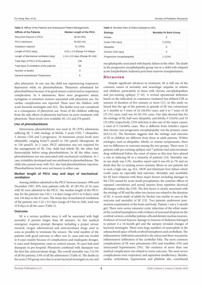
Among children admitted to the PICU between January 1996 and December 1997, 45% were patients with SE. 45 (83.3%) of 54 cases with SE were admitted to the PICU. The median length of the PICU stay for the patients was 3.02 ± 1.6 days (range of 0.5 to 9 days), total was 136 days in the 45 cases. The mean day of mechanical ventilation of the patients was 1.24 ± 0.5 days (range of 9 hm to 10d), total was 55.8 days in all the cases (Table 3).
OutcomeSE is a serious problem since it will be associated with high
mortality if persists longer than 60 minutes. So this medical emergency requires prompt therapies including a proper airway ensured, oxygen administered and anticonvulsant drugs used as soon as possible to terminate the seizure. The total number of the patients with good outcome or effect was 41 cases and was invalid in 8 cases mainly because of complications and inadequate dosages. 6 cases used thiopentone came to control seizure, 36 cases had used diazepam in pre-hospital. Phenytoin combined with diazepam was the first line anticonvulsant drugs. The overall mortality was 3 (5.3% of all the patients, 4.9% of all the admissions) (Table 4). The deaths in the acute CNS group were due to acute bacterial meningitis in one and
encephalopathy associated with hepatic failure in the other. The death in the progressive encephalopathy group was in a child with relapsed acute lymphoblastic leukemia post bone marrow transplantation.
DiscussionDespite significant advances in treatment, SE is still one of the
common causes of mortality and neurologic sequelae in infants and children, particularly in those with chronic encephalopathies and pre-existing epilepsy [7-10]. A revised operational definition based on the indication to commence treatment has defined CSE as seizures of duration of five minutes or more [11]. In this study we found that the age of the patients at episode of SE was commonest in 3 months to 3 years of 24 (44.4%) cases, and 4 to 7 years of 20 (37.1%) cases, total was 44 (81.5%) cases. Our data showed that for the etiology of SE, half were idiopathic and febrile, 9 (16.6%) and 19 (35.18%) respectively. CNS infection is also one of the major causes, a total of 9 (16.66%) cases. This is different from Scholte’s report that chronic non-progressive encephalopathy was the primary cause [8,9,12,13]. The literature suggests that the etiology and outcome of SE in children are different from those in adult patients. Within etiologies, such as idiopathic, febrile, or remote symptomatic, there was no difference in outcome among the two groups. There were 12 patients with pre-existing epilepsy and 7 patients had anticonvulsant drug withdrawal before the onset of status so this may have played a role in inducing SE in a minority of patients [14]. Mortality rate in our study was 5.3%. Another report said it was 6% to 7% and we think that the co-existing severe systemic illness in our patients led to such a high rate, eg: ALL, MOF and fulminant liver failure which could cause an especially bad outcome. Mortality and morbidity for SE have relations with three major factors including damages to the CNS caused by acute insult precipitation, the systemic effects of repeated convulsions and neural injuries from repetitive electrical discharges within the CNS. The first factor is clearly associated with the etiology of SE and the other two factors are related to the duration of SE. A recent study of adults by Stecker was similar to ours at the outcome and mortality of SE [15]. Two patients underwent post-mortem examination of the brain and body. Patient 1 was a 3-month girl. There were severe extensive cystic infarction of the white mater of the cerebral hemispheres with evidence of neuronal dropout on the cerebral cortices, cerebellar pukinje cells and dentate nucleus neurons. Evidence of recent hypoxic damage to neurons of thalamus belonged to patient 2-a 16-month girl and the autopsy revealed changes of bacterial meningitis. There were large numbers of neutrophils in the subarachnoid space of both cerebral hemispheres and cerebellum. The inflammatory infiltration extended to the sulcus and there was patchy inflammatory infiltration of the cerebellar folia. The most common complications of SE were pneumonia (4%) and tonsillitis (5%) and intracranial hypertension (5%). The existence of more than one medical complication was related to worse outcome. The most severe complications were respiratory and aspiration insufficiency. Besides, cardiac arrhythmia, hypotension and phlebitis also contributed
Afflicts of the Patients Median Length of the PICU
Recurrent Seizure in PICU 28 (51.8%)
PICU Admission 45 (83.1%)
Intubation required 41 (75%)
Length of PICU (day) 3.02 ± 1.6 (Range 0.5-9days)
Length of Mechanical ventilation (day) 1.24 ± 0.5 days (Range 9h-10d)
Total days of PICU of the patients 136
Total days of ventilation of the patients 58
Number of deaths 3
General anaesthesia-Thiopentone 6
Table 3: Afflicts of the Patients and Subsequent Patient Managements.
Etiology Mortality l% Each Group
Febrile 0
Acute CNS injury 2 (22%)
Idiopathic 0
Chronic CNS injury 0
Progressive encephalopathy 1 (11%)
Table 4: Mortality Rate of Status Epilepticus (SE).

Fu-Yong Jiao, et al., Annals of Pediatric Research
Remedy Publications LLC. 2018 | Volume 2 | Issue 2 | Article 10154
to a poor result. A study in India shows appropriate prehospital management and treatment targeting resolution of cardiovascular dysfunction during resuscitation could reduce mortality in children with SE [10,16,17]. So importance of prompt treatment of medical complications during general convulsions-SE has been stressed. The commonest cause of SE was idiopathic, and then was febrile. The acute CNS, the chronic CNS and the progressive encephalopathy groups were of equal distribution. Most of the presentations of SE required PICU admission. Most of the PICU admissions required mechanical ventilation probably reflecting good clinical judgment of the Emergency and PICU physicians in selecting admitted cases. This study confirmed the findings of Maytal et al. [4] regarding the mortality. Status epilepticus itself is associated with low mortality. It is the concurrent pathological processes (acute CNS injury or progressive encephalopathy) that mostly determine the cause of death [7,9,10,17]. There was great concern for the emergency or PICU physicians regarding the oversubscribing of phenzodiazepines or barbiturates in the acute resuscitative period, hence the necessitation of PICU admission. The use of phenytoin has been encouraged. Most emergency centers will recommend the use of intravenous phenytoin when the seizure persists after 30 minutes. According to this study, around 50% of the cases employed phenytoin for seizure control. In 21% (6/29) of children receiving intravenous phenytoin, the need for intubation was avoided and in terms of budget, 5 PICU beds were saved. However, it could not be established whether the SE stopped spontaneously because of the direct effect of phenytoin. 13.8% (4/29) of children receiving intravenous phenytoin suffered from acute cerebellar toxicity. Except for 1 child with level of 270 μmol/l, there was no correlation with the levels, nor the prior treatment with phenytoin. There were no other side effects seen in this study although we did not follow any of these children up prospectively. With this relatively small and crude study, it seems the use of phenytoin should be encouraged to prevent artificial ventilation and its possible adverse effects. Most of the phenobarbitone use was in the febrile or the idiopathic group. It prevented only 2 mechanical ventilations. This might reflect the sedative nature of the drug itself. Only 1 behavioral side effect was recorded. We have demonstrated that intravenously administered diazepam and phenytoin remain the first-line therapy for SE [18]. Phenytoin combined with diazepam is effective and safe. Besides, there has been a lot of research on whether Intravenous (IV) levetiracetam or IV phenytoin is the better second line treatment for the emergency management of CSE in children, suggesting that IV levetiracetam is safer and more effective [13,19-21]. More than a half of patients will response to the initial treatment. The SE treatment also includes high-dose phenobarbital or inhalation of anaesthetic agents, proper airway must be ensured, oxygen administered, blood pressure supported and intracranial pressure controlled and soon. One of the most recent major advances in management of seizure disorders is availability of methods for rapid determination of anticonvulsant blood level. Dosage schedule of anticonvulsant can now be adjusted to maintain serum drug level within the therapeutic ranges, this provides a means of checking patient compliance and provides insights of drug metabolism of these compounds in normal individual, particularly in young infants who may have variable absorption when seizures are uncontrolled on therapy, drug toxicity is suspected, or changes in drug treatment are planned. Diazepam is a good agent to arrest SE, but should not be used in combination with phenobarbital because of the risk of hypotension and respiratory depression, particularly in infants [13,17,21].
AcknowledgementThe authors would like to acknowledge the support of the New
Children’s Hospital for the study in the tow hospital.
References1. Chin RF, Neville BG, Peckham C, Bedford H, Wade A, Scott RC; NLSTEPSS
Collaborative Group. Incidence, cause, and short-term outcome of convulsive status epilepticus in childhood: prospective population-based study. Lancet. 2006;368(9531):222-9.
2. Novorol CL, Chin RF, Scott RC. Outcome of convulsive status epilepticus: a review. Arch Dis Child. 2007;92(11):948-51.
3. Frisby JR. Status Epilepsy in Intensive Care Manual on TE (Editor). Butterworths Sydney. 1990;266-9.
4. Maytal J, Shinnar S, Moshé SL, Alvarez LA. Low morbidity and mortality of status epilepticus in children. Pediatrics. 1989;83(3):323-31.
5. FE Dreifuss, J Bancaud, O Henriksen. Commission on Classification and Terminology of the International League Against Epilepsy, Proposal for Revised Clinical and Electroencephalographic Classification of Epileptic Seizures. Epilepsia. 1981;22(4):489-501.
6. Hauser WA, Anderson VE, Loewenson RB, McRoberts SM. Seizure recurrence after a first unprovoked seizure. N Engl J Med. 1982;307(9):522-8.
7. Phillips SA, Shanahan RJ. Etiology and mortality of status epilepticus in children. A recent update. Arch Neurol. 1989;46(1):74-6.
8. Jiao F, Guo X, Lin J, Cui W, Li H. A Randomized Trial of Ligustrazini Hydrochlorioi in the Treatment of Viral Encephalitis in Children. J Nepal Pediatric Society. 2010;30:2.
9. Jiao F, Zhang X, Bai T, Lin J, Cui W, Liu B. Clinical evaluation of the function of hypothalamo-pituitary-thyroid axis in children with central nervous system infections. Ital J Pediatr. 2011;37:11.
10. Jiao FY, Cao HC, Liu ZY, Wu S, Wong HB. The use of blood glucose/cerebrospinal fluid glucose ratio in the diagnosis of CNS infection in infants and children. J Singapore Pediatr Soc. 1992;34(3-4):191-8.
11. Lowenstein DH, Bleck T, Macdonald RL. It's time to revise the definition of status epilepticus. Epilepsia. 1999;40(1):120-2.
12. Scholtes FB, Renier WO, Meinardi H. Generalized convulsive status epilepticus: causes, therapy, and outcome in 346 patients. Epilepsia. 1994;35(5):1104-12.
13. Jiao F, Gao DY, Takuma Y, Wu S, Liu ZY, Zhang XK, et al. Randomized, controlled trial of high-dose intravenous pyridoxine in the treatment of recurrent seizures in children. Pediatr Neurol. 1997;17(1):54-7.
14. Maytal J, Novak G, Ascher C, Bienkowski R. Status epilepticus in children with epilepsy: the role of antiepileptic drug levels in prevention. Pediatrics. 1996;98(6):1119-21.
15. Stecker MM, Kramer TH, Raps EC, O'Meeghan R, Dulaney E, Skaar DJ. Treatment of refractory status epilepticus with propofol: clinical and pharmacokinetic findings. Epilepsia. 1998;39(1):18-26.
16. Santhanam I, Yoganathan S, Sivakumar VA, Ramakrishnamurugan R, Sathish S, Thandavarayan M. Predictors of Outcome in Children with Status Epilepticus during Resuscitation in Pediatric Emergency Department: A Retrospective Observational Study. Ann Indian Acad Neurol. 2017;20(2):142-8.
17. Jiao FY, Guo RL, Ma FR. Differential diagnosis of CNS infections by Alkaline phosphatase in CSF . N Z Med J. 1986;99(796):124.
18. Cascino GD. Generalized convulsive status epilepticus. Mayo Clinic Proc.1996;71(8):787-92.
19. Dalziel SR, Furyk J, Bonisch M, Oakley E, Borland M, Neutze J, et al. A

Fu-Yong Jiao, et al., Annals of Pediatric Research
Remedy Publications LLC. 2018 | Volume 2 | Issue 2 | Article 10155
multicentre randomised controlled trial of levetiracetam versus phenytoin for convulsive status epilepticus in children (protocol): Convulsive Status Epilepticus Paediatric Trial (ConSEPT) - a PREDICT study. BMC Pediatr. 2017;17(1):152.
20. Lyttle MD, Gamble C, Messahel S, Hickey H, Iyer A, Woolfall K, et al. Emergency treatment with levetiracetam or phenytoin in status epilepticus in children-the EcLiPSE study: study protocol for a randomised controlled trial. Trials. 2017;18(1):283.
21. Jiao F, Wang J, Zhang X, Mu Z. The Situation of Suppression Seizures with Diazepam by Rectal Administration in China. J Pediatr. 2016;1(1):1-4.