
STARS Syllabus
39
1 Geoffrey K. Lighthall PhD, MD Geoffrey K. Lighthall PhD, MD Assistant Professor, Anesthesia and Critical Care Assistant Professor, Anesthesia and Critical Care Stanford University School of Medicine Stanford University School of Medicine Palo Alto Veterans Affairs Health Care System Palo Alto Veterans Affairs Health Care System STARS STARS Simulation Simulation Training for Training for Acute Acute Resuscitation Resuscitation Skills Skills
Transcript of STARS Syllabus
1
Geoffrey K. Lighthall PhD, MDGeoffrey K. Lighthall PhD, MDAssistant Professor, Anesthesia and Critical CareAssistant Professor, Anesthesia and Critical CareStanford University School of MedicineStanford University School of MedicinePalo Alto Veterans Affairs Health Care SystemPalo Alto Veterans Affairs Health Care System
STARSSTARSSimulationSimulationTraining forTraining forAcuteAcuteResuscitationResuscitationSkillsSkills

STAR Course Syllabus Contents
Welcome to Critical Care 3
Immediate Assessment and Stabilization 4
Respiratory Failure 6
Hypoxemia 8
Production and Elimination of Carbon Dioxide 10
Therapy for Respiratory Problems 12
Physiology and Disruption of Oxygen Transport 14
Circulatory Shock: 16
Therapy for Hemodynamic Instability 20
Acute Blood Loss 23
Acute pain 25
Cognitive Aids and Flow ChartsHypoxemia 9
Carbon Dioxide 10-11
Shock / Hypotension Diagnostic Tree 19
Shock / Hypotension Treatment Outline 22
2

Welcome to Critical Care
The ICU serves every clinic, specialty, ward or procedure room in the hospital, and at every hour of the day. Care of a critically ill patient demands flexibility and the ability to perform under stress, but also a calm, methodical approach to dynamic situations and the ability to identify priorities. Often, the need to have expert hands at the bedside leaves the medical student out of the loop in terms of hands on experience and in the ability to ask questions as they arise. The purpose of this course is to break down some of this activity into its component parts and to provide some experiential learning in an environment that creates no risk to patients. The goals we have set for this experience are to:
Alert you to some of the types of patients that have unexpected admissions to the intensive care unit
Appropriately equip you with cognitive tools and conditioned responses to events that arise in medical emergencies.
Make use of medical simulation to:- “bring to life” basic knowledge of physiology and pharmacology by relating such to real medical conditions and lifelike “patients”- allow you first-hand experience in managing patient emergencies- reinforce standard methods of patient evaluation- provide you with expert videotape review of the management of
patient emergencies
The hands-on experience with patient simulation is the most unique and exciting aspect of the course. In addition to the thrill and responsibility of caring for a critically ill patient, you should also leave with the following: How to recognize a sick patient--an appreciation and ability to detect early
warning signs of deterioration The techniques and priorities in stabilizing patients with ABC problems;
what you can do for a patient in the pre-arrest phase A sense of urgency (and fear) in evaluating a patient with an abnormal
vital status The pathophysiology of problems with the airway, breathing, and
circulation
Components of the course include:
An on-screen simulator and worksheet that need to be completed before the simulation sessions.
This text and a pre-test A four-hour session in the Human Patient Simulation lab where you will class
manage a variety or critical incidents. Review and debriefing of simulated critical incidents A post course questionnaire and post test.
3
Immediate Assessment and Stabilization
The overall emphasis of this course and its training exercises is to understand the physiology of deteriorating patients and think of problems, therapy, and end points in these terms. While subtle, this represents a departure from how most branches of medicine operate: “make a diagnosis then treat the problem.” The lack of a clear diagnosis seems to cause a bit of mental paralysis during the first 10-15 minutes of a critical patient encounter and this is exactly when initiating therapy is likely to have the greatest impact. A physiologic approach to patient care allows one to work with broad categories of problems and find diagnoses within this framework. More significantly, one gains the ability to start treatment for that class of problems at an earlier stage.
This chapter will serve provide a general overview of patient resuscitation as a lead in to more detailed chapters on therapy for airway, breathing and circulatory disorders.
An example You are called to evaluate a patient who is hypotensive. You arrive and find a patient with intact mental function, but with a BP of 85/49. The patient is warm to the touch and pulses are full. The patient has a respiratory rate of 24 with clear breath sounds; the chest seems to move with each heartbeat. From this brief exam, you can be fairly sure that the patient has no problem with cardiac function or with breathing. The extremities are warm, so you rule out an increase in vascular tone that one may expect if is the patient's hypotension was due to bleeding. Instead, you attribute the hypotension to vasodilation. Now you’re not quite sure whether the problem is sepsis due to cellulitis (the admitting condition), or due to an anaphylactic reaction associated with a recent antibiotic. Either way, you know that in a hypotensive patient with low SVR, administration of fluids and vasopressors is the mainstay of therapy. Better IV access is obtained, 50 mg of diphenhydramine and 2L of a crystalloid solution are quickly administered. The blood pressure improves with several 100mcg boluses of phenylephrine, during which time more fluids are given. Later, when the patient is more stable, you look up anaphylaxis and are reminded that a tryptase level would confirm this diagnosis, and a serum sample is sent. In this case, establishing a definitive diagnosis (worsening sepsis) lagged a few hours behind supportive therapy, but at least knowing the nature of the physiologic problem prevented time wasted on diagnostic studies, and facilitated early stabilization and probably a better clinical result.
Caring for an unstable patient can be unnerving without some ability to prioritize problems. In the initial stages of any resuscitation, the focus is on cardiovascular and respiratory function and the absolute necessity of oxygen assimilation and delivery to the tissues in order to sustain life. The initial interventions are therefore: address the ABCs, collect and assess vital signs, assess the components of oxygen delivery.
4

While defining an unstable patient's problem in physiologic terms, it is
important to also consider diagnoses that require external personnel or interventions as early as possible ( ie cardiac cath lab, surgical operation, special procedure); if in doubt, call for help at a stage when it can be useful.
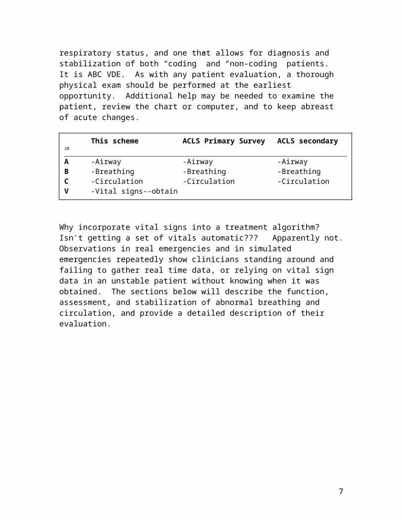
A more deliberate algorithm describing these priorities is summarized below, as well as a comparison of the ABCD survey taught in advanced cardiac life support (ACLS) courses. With cardiac arrest, survival correlates with conditions (heart rhythms) amenable to rapid correction by cardioversion, and accordingly, a high priority is assigned to identifying "shockable rhythms." Likewise, understanding the problem underlying the instability (ventricular tachycardia, pulseless electrical activity) will help you prevent a second code. In the absence of an emergency requiring defibrillation, the ACLS ABCD survey doesn’t provide any insight or guidance into the assessment and management of unstable patients, rather it suggests you establish a diagnosis for the instability. A preferable method is one that encompasses the overall issues of tissue oxygen delivery as it relates to cardiovascular and respiratory status, and one that allows for diagnosis and stabilization of both “coding” and “non-coding” patients. It is ABC VDE. As with any patient evaluation, a thorough physical exam should be performed at the earliest opportunity. Additional help may be needed to examine the patient, review the chart or computer, and to keep abreast of acute changes.
Why incorporate vital signs into a treatment algorithm? Isn't getting a set of vitals automatic??? Apparently not. Observations in real emergencies and in simulated emergencies repeatedly show clinicians standing around and failing to gather real time data, or relying on vital sign data in an unstable patient without knowing when it was obtained. The sections below will describe the function, assessment, and stabilization of abnormal breathing and circulation, and provide a detailed description of their evaluation.
5
Without a handle on these three basic points, it is unbelievably easy to get distracted with secondary phenomena and to waste crucial time on insignificant problems.
This scheme ACLS Primary Survey ACLS secondary 20
A -Airway -Airway -AirwayB -Breathing -Breathing -BreathingC -Circulation -Circulation -CirculationV -Vital signs--obtainD -DO2—assess with ABG -Defibrillation -DiagnosisE -Endocrine (check glucose)
Respiratory Failure: Physiologic and Anatomic Considerations
Respiratory failure is a general term referring to impairment of the body’s ability to assimilate oxygen and transport it to the pulmonary capillary blood, eliminate carbon dioxide from the blood, or both. Organ-based derangements underlying such problems are diverse and include metabolic disturbances, dysfunction of neural circuitry from the brainstem down to the neuromuscular junction, abnormalities in the chest wall, alveoli, pulmonary parenchyma, upper and lower airways, and blood flow to the lung.
In respiratory failure, diagnosis and intervention occur simultaneously, with support being the higher priority.
Inspection and auscultation of the patient may provide enough information to initiate therapy, however, knowing whether the problem had a fast or slow onset will provide diagnostic information (i.e. was there a medicine recently given, was the patient eating, ambulating, etc). Respiratory failure usual involves an interplay between a disease process and host factors, all of which need to be considered as part of a management strategy.
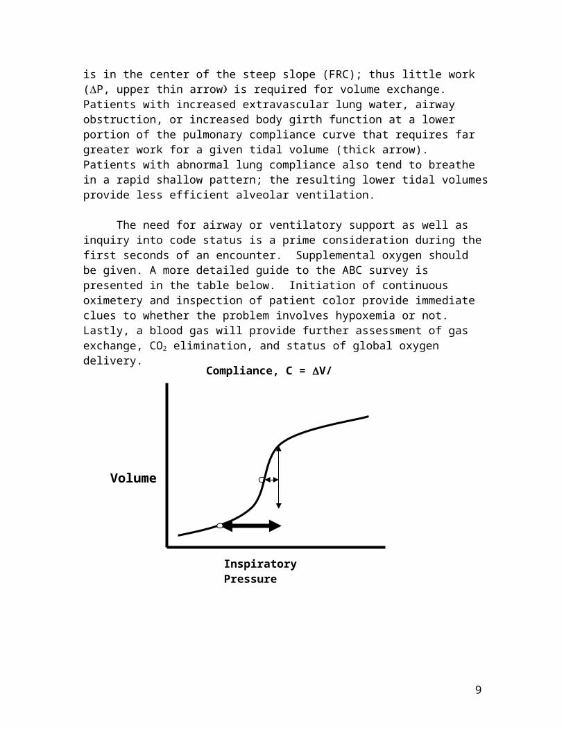
Most patients can tolerate an increased demand on the respiratory system for a few hours, some less. When a high workload is sustained, many patients experience respiratory muscle fatigue, and regardless of the inciting event, develop hypercapnic respiratory failure. When evaluating the patient, consider subjective complaints of fatigue and distress, and form your own impression of the work requirements of respiration. A more formal way to think of the patient’s work of breathing is in terms of the pulmonary compliance curve on the following page. The normal set point is in the center of the steep slope (FRC); thus little work (P, upper thin arrow is required for volume exchange. Patients with increased extravascular lung water, airway obstruction, or increased body girth function at a lower portion of the pulmonary compliance curve that requires far greater work for a given tidal volume (thick arrow). Patients with abnormal lung compliance also tend to breathe in a rapid shallow pattern; the resulting lower tidal volumes provide less efficient alveolar ventilation.
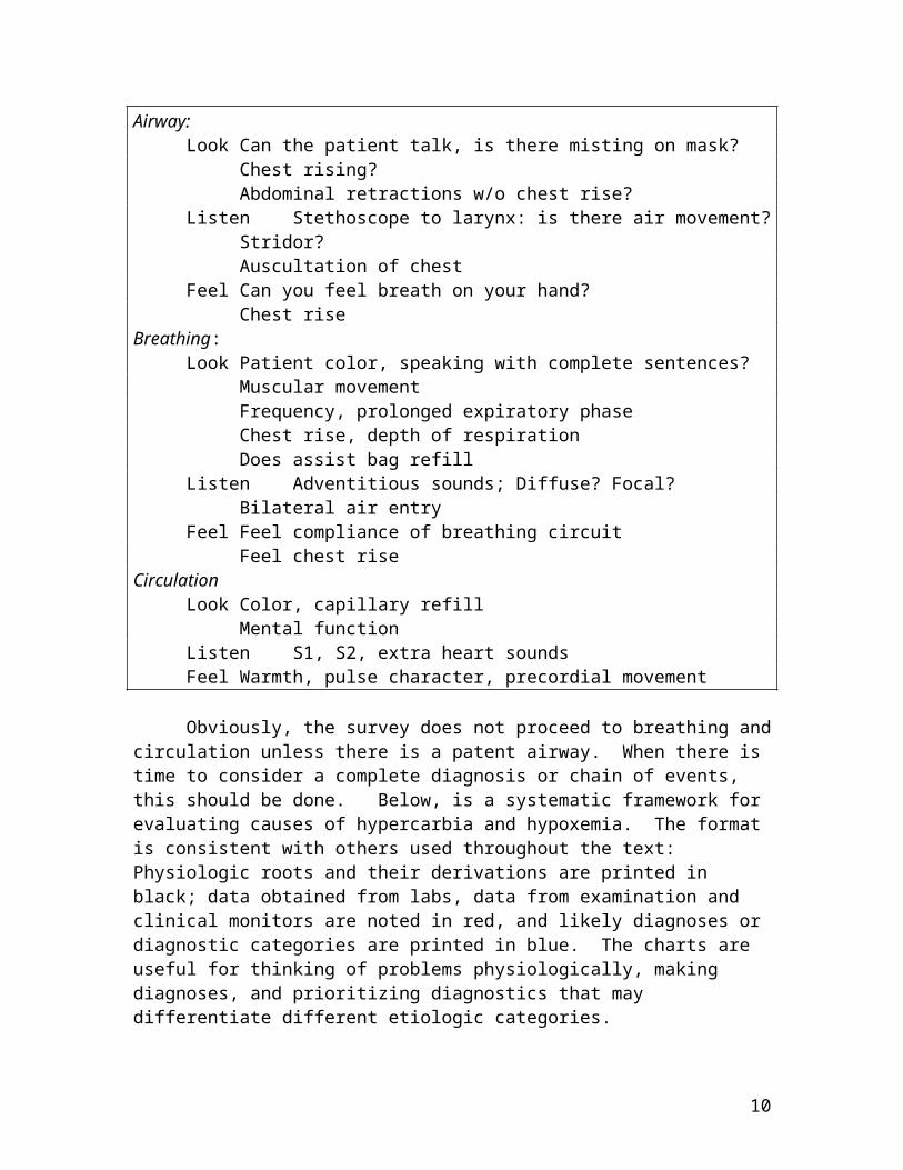
The need for airway or ventilatory support as well as inquiry into code status is a prime consideration during the first seconds of an encounter. Supplemental oxygen should be given. A more detailed guide to the ABC survey is presented in the table below. Initiation of continuous oximetery and inspection of patient color provide immediate clues to whether the problem involves hypoxemia or not. Lastly, a blood gas will provide further assessment of gas exchange, CO2 elimination, and status of global oxygen delivery.
6
Airway:Look Can the patient talk, is there misting on mask?
Chest rising?Abdominal retractions w/o chest rise?
Listen Stethoscope to larynx: is there air movement?Stridor?Auscultation of chest
Feel Can you feel breath on your hand?Chest rise
Breathing:Look Patient color, speaking with complete sentences?
Muscular movementFrequency, prolonged expiratory phaseChest rise, depth of respirationDoes assist bag refill
Listen Adventitious sounds; Diffuse? Focal?Bilateral air entry
Feel Feel compliance of breathing circuitFeel chest rise
CirculationLook Color, capillary refill
Mental functionListen S1, S2, extra heart soundsFeel Warmth, pulse character, precordial movement
7
Inspiratory Pressure
Volume
Compliance, C = V/ P

Obviously, the survey does not proceed to breathing and circulation unless there is a patent airway. When there is time to consider a complete diagnosis or chain of events, this should be done. Below, is a systematic framework for evaluating causes of hypercarbia and hypoxemia. The format is consistent with others used throughout the text: Physiologic roots and their derivations are printed in black; data obtained from labs, data from examination and clinical monitors are noted in red, and likely diagnoses or diagnostic categories are printed in blue. The charts are useful for thinking of problems physiologically, making diagnoses, and prioritizing diagnostics that may differentiate different etiologic categories.
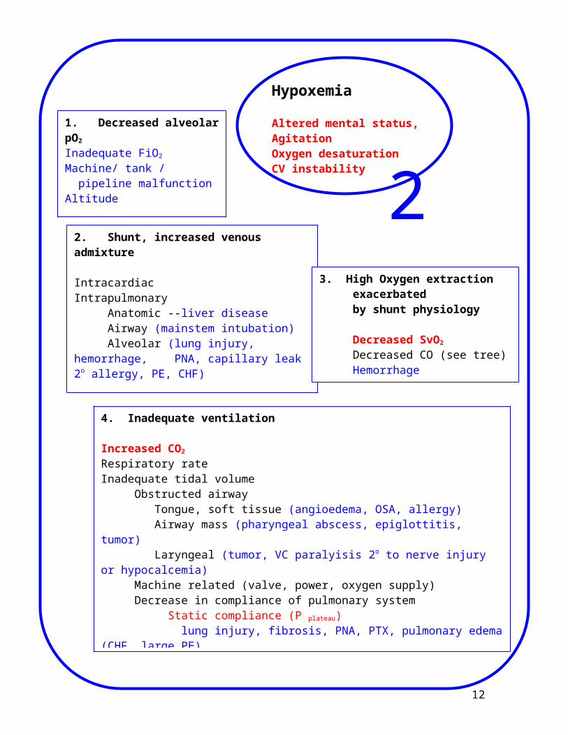
Hypoxemia
Hypoxemia can manifest itself in a subtle manner, with secondary signs such as change in mentation, delirium, and organ dysfucntion. In such cases the challenge is to correct the hypoxia and figure out why the patient has this problem. Alternately, hypoxia can be intertwined with a primary complaint as chest pain in major pulmonary embolus, or cardiogenic shock. In either case, your job is to provide rapid stabilization, and address the underlying process as soon as possible. Etiologic categories are listed on the following chart. As with other charts in this text, broad physiologic categories are noted in black, clinical, laboratory and monitor findings are noted in red, and possible diagnoses are written in blue type.
8
9
1. Decreased alveolar pO2
Inadequate FiO2
Machine/ tank / pipeline malfunctionAltitude
4. Inadequate ventilation
Increased CO2 Respiratory rate Inadequate tidal volume
Obstructed airway Tongue, soft tissue (angioedema, OSA, allergy) Airway mass (pharyngeal abscess, epiglottitis, tumor) Laryngeal (tumor, VC paralyisis 2o to nerve injury or hypocalcemia)Machine related (valve, power, oxygen supply)Decrease in compliance of pulmonary system
Static compliance (P plateau) lung injury, fibrosis, PNA, PTX, pulmonary edema (CHF, large PE)Dynamic compliance (P peak) Bronchospasm, mucus plug in airway or in ET, foreign body hyperinflation
Hypoxemia
Altered mental status, AgitationOxygen desaturationCV instability
2. Shunt, increased venous admixture
IntracardiacIntrapulmonary
Anatomic --liver diseaseAirway (mainstem intubation)Alveolar (lung injury, hemorrhage, PNA, capillary leak 2o allergy, PE, CHF)
23. High Oxygen extraction
exacerbated by shunt physiology
Decreased SvO2
Decreased CO (see tree)HemorrhageHigh VO2 from sepsis, fever, etc
Production and Elimination of Carbon Dioxide
Carbon dioxide content is one of the primary determinants of acid / base status. Obtaining an ABG is essential to understanding CO2 levels, as well as assessing their contribution to body pH. Abnormally high or low paCO2 values, and deviations from baseline values can often provide a valuable window into the patient’s ventilatory status. Total ventilation (denoted by convention as VE) consists of ventilation of alveoil (VA) as well as non-perfused parts of the lung (dead space ventilation, VD), such that VE = VD + VA. Many critically ill patients have exhaled CO2 sampled and displayed on the bedside monitor; the value displayed (usually referred to as the end tidal CO2). is a useful adjunct to arterial CO2 monitoring. The ET CO2 is always going to be less than alveolar CO2 by a “gradient” that is 5-7 mm Hg in normal subjects, but increases in proportion to increases in VD/ VE. Patients with emphysema, for example, may have a gradient of 15. Patients with a shallow respiratory pattern will also have a higher gradient.
HypocapneaHypocarbia acutely causes a decrease in cerebral blood flow, with
attendant changes in mental status or consciousness. Alkalemia if present, can cause coronary vasospasm, bronchoconstriction and a left shift in the hemoglobin dissociation curve. Causes of hypocapnea that you will commonly see are pain, anxiety, and as compensation for metabolic acidosis.
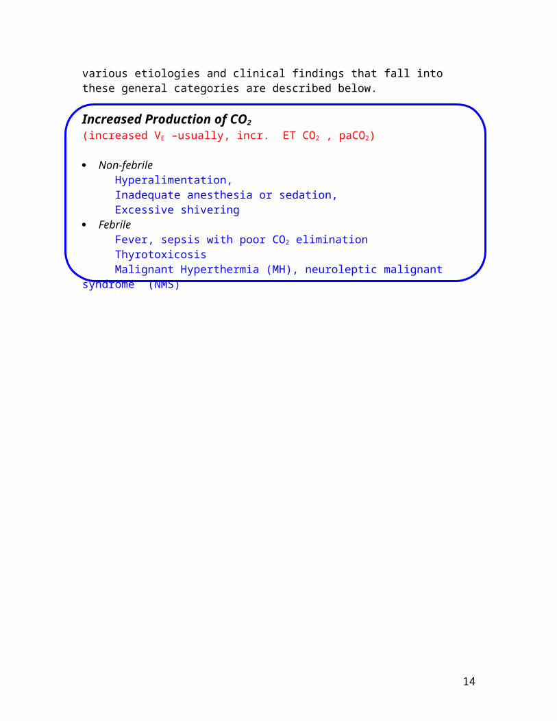
HypercapneaHypercarbia often manifests as a depressed mental status, tachypnea,
hypertension, tachcardia, and ectopy. Increases in CO2 are attributable to either decreased elimination of CO2, increased production, or both. The various etiologies and clinical findings that fall into these general categories are described below.
Increased Production of CO2 (increased VE –usually, incr. ET CO2 , paCO2)
Non-febrileHyperalimentation,Inadequate anesthesia or sedation, Excessive shivering
FebrileFever, sepsis with poor CO2 eliminationThyrotoxicosisMalignant Hyperthermia (MH), neuroleptic malignant syndrome (NMS)
10

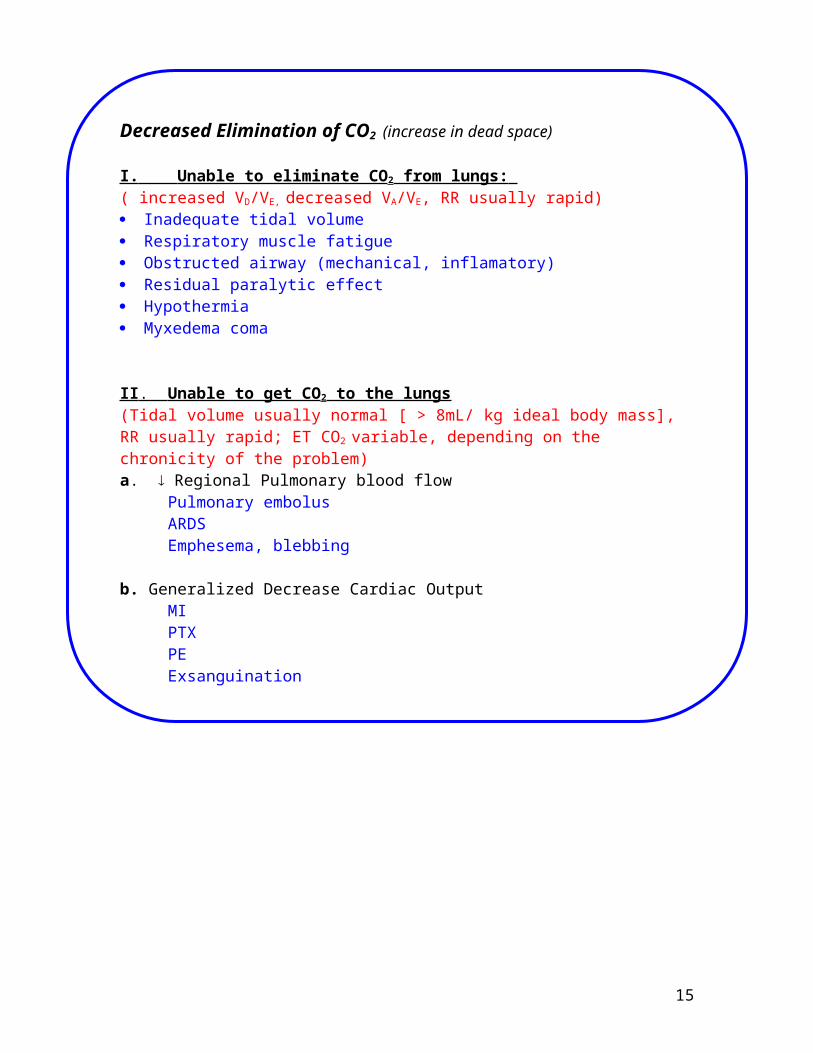
Decreased Elimination of CO2 (increase in dead space)
I. Unable to eliminate CO 2 from lungs: ( increased VD/VE, decreased VA/VE, RR usually rapid) Inadequate tidal volume Respiratory muscle fatigue Obstructed airway (mechanical, inflamatory) Residual paralytic effect Hypothermia Myxedema coma
II . Unable to get CO 2 to the lungs(Tidal volume usually normal [ > 8mL/ kg ideal body mass], RR usually rapid; ET CO2 variable, depending on the chronicity of the problem)a. Regional Pulmonary blood flow
Pulmonary embolusARDSEmphesema, blebbing
b. Generalized Decrease Cardiac Output MIPTXPEExsanguination
11
Therapy for Respiratory Problems
Therapy for Airway problems
I. Foreign body Scoop and sweep, forceps removal Heimlich maneuver
II. Soft tissue or tongue obstruction, over sedation Chin lift, jaw thrust Supplemental high flow oxygen (100% non-rebreather bag @ 15 L/ min) Nasal and / or oral airway Reverse paralytics and sedatives
Benzos--Flumazenil, 0.2 mg IV push, repeat up to 1.0 mgNon-succinylcholine paralytics--5mg neostigmine + 1.0 mg glycopyrolateOpiates--40-80 mcg naloxone, repeat as needed(note: a typical vial contains 400 mcg)
Bag-assist ventilation via Ambu Bag or Jackson-Reese circuit (need to call anesthesiologist for help)
III. Stridor, (extrathoracic airway obstruction edema,tumor, infection, VC paralysis) Supplemental high flow oxygen (100% non-rebreather bag @ 15 L/ min) Immediate call to anesthesiologist; s/he will help you determine need for
surgical airway Need to avoid stimulation and stress; this increases turbulence of airflow Bag-assist ventilation via a Jackson-Reese circuit or BiPAP machine
(need to call anesthesiologist for help, AMBU bag may not work without addition of a PEEP valve)
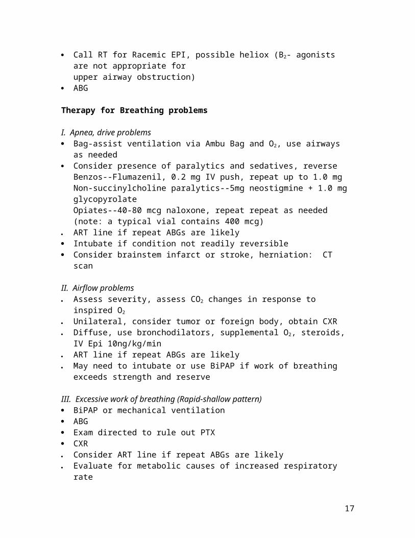
Call for expert help with airway management: anesthesiology and ENT Call RT for Racemic EPI, possible heliox (B2- agonists are not appropriate for
upper airway obstruction) ABG
Therapy for Breathing problems
I. Apnea, drive problems Bag-assist ventilation via Ambu Bag and O2, use airways as needed Consider presence of paralytics and sedatives, reverse
Benzos--Flumazenil, 0.2 mg IV push, repeat up to 1.0 mgNon-succinylcholine paralytics--5mg neostigmine + 1.0 mg glycopyrolateOpiates--40-80 mcg naloxone, repeat repeat as needed(note: a typical vial contains 400 mcg)
ART line if repeat ABGs are likely Intubate if condition not readily reversible
12

Consider brainstem infarct or stroke, herniation: CT scan
II. Airflow problems Assess severity, assess CO2 changes in response to inspired O2
Unilateral, consider tumor or foreign body, obtain CXR Diffuse, use bronchodilators, supplemental O2, steroids, IV Epi 10ng/kg/min ART line if repeat ABGs are likely May need to intubate or use BiPAP if work of breathing exceeds strength and
reserve
III. Excessive work of breathing (Rapid-shallow pattern) BiPAP or mechanical ventilation ABG Exam directed to rule out PTX CXR Consider ART line if repeat ABGs are likely Evaluate for metabolic causes of increased respiratory rate
• Remember: patients in extremis have a high catecholamine tone (assume an invisible Epi drip at 50-75 ng/kg/min). Any type of sedative, anxiolytic, or induction agent will decrease the catechol drive and cause a rapid circulatory collapse. Placement of an ART line, and assembly of vasoactive drugs and fluids should be done whenever possible prior to intubation.
13
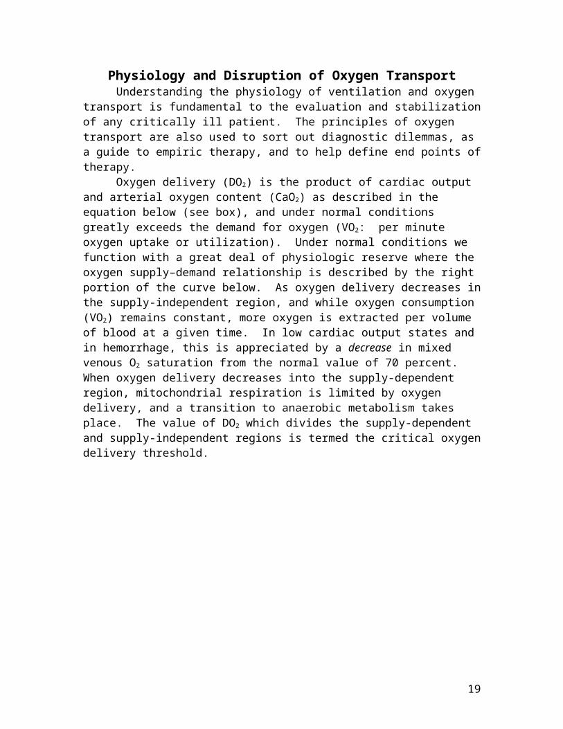
Physiology and Disruption of Oxygen Transport Understanding the physiology of ventilation and oxygen transport is
fundamental to the evaluation and stabilization of any critically ill patient. The principles of oxygen transport are also used to sort out diagnostic dilemmas, as a guide to empiric therapy, and to help define end points of therapy.
Oxygen delivery (DO2) is the product of cardiac output and arterial oxygen content (CaO2) as described in the equation below (see box), and under normal conditions greatly exceeds the demand for oxygen (VO2: per minute oxygen uptake or utilization). Under normal conditions we function with a great deal of physiologic reserve where the oxygen supply–demand relationship is described by the right portion of the curve below. As oxygen delivery decreases in the supply-independent region, and while oxygen consumption (VO2) remains constant, more oxygen is extracted per volume of blood at a given time. In low cardiac output states and in hemorrhage, this is appreciated by a decrease in mixed venous O2 saturation from the normal value of 70 percent. When oxygen delivery decreases into the supply-dependent region, mitochondrial respiration is limited by oxygen delivery, and a transition to anaerobic metabolism takes place. The value of DO2 which divides the supply-dependent and supply-independent regions is termed the critical oxygen delivery threshold.
14
Blood pressure is maintained within a certain range for each individual by homeostatic mechanisms that include antonomic reflexes, capillary fluid shifts, and modulation of neurohormones. Blood pressure is important to the body because the various solid organs have adapted their ability to maintain constant blood flow to a certain range of blood pressures (autoregulation, see figure below). For healthy, normotensive people the brain and kidney autoregulate their blood flow at mean pressures between 50-150 mm Hg (black curve in the figure below). However, many of our patients have curves shifted to the right--meaning that kidneys, brain and brainstem, etc. may need higher pressures in order to maintain uniform tissue oxygen delivery (the red curve). The overall autoregulatory range of your patient needs to be deliberately considered, and can be inferred in many cases, by a combination of bedside examination and inspection of historical blood pressure data and its correlation with organ function. Poorly controlled hypertensives have fairly dramatic right shifts to their autoregulatiory curves.
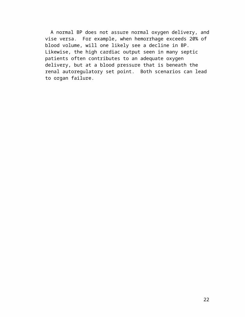
A normal BP does not assure normal oxygen delivery, and vise versa. For example, when hemorrhage exceeds 20% of blood volume, will one likely see a decline in BP. Likewise, the high cardiac output seen in many septic patients often contributes to an adequate oxygen delivery, but at a blood pressure that is beneath the renal autoregulatory set point. Both scenarios can lead to organ failure.
15
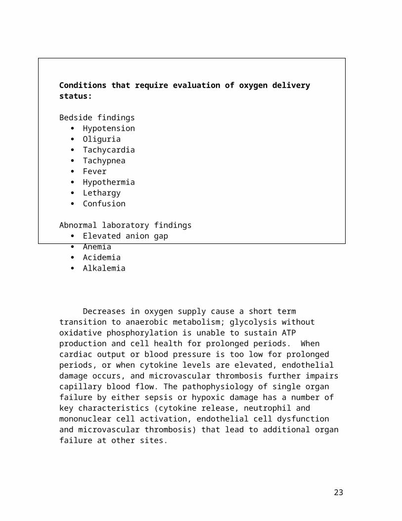
Shock results from a mismatch between oxygen supply and demand, the latter leads to changes in cellular function that may be either short-lived or permanent.

Conditions that require evaluation of oxygen delivery status:
Bedside findings Hypotension Oliguria Tachycardia Tachypnea Fever Hypothermia Lethargy Confusion
Abnormal laboratory findings Elevated anion gap Anemia Acidemia Alkalemia
Decreases in oxygen supply cause a short term transition to anaerobic metabolism; glycolysis without oxidative phosphorylation is unable to sustain ATP production and cell health for prolonged periods. When cardiac output or blood pressure is too low for prolonged periods, or when cytokine levels are elevated, endothelial damage occurs, and microvascular thrombosis further impairs capillary blood flow. The pathophysiology of single organ failure by either sepsis or hypoxic damage has a number of key characteristics (cytokine release, neutrophil and mononuclear cell activation, endothelial cell dysfunction and microvascular thrombosis) that lead to additional organ failure at other sites.
There are definitions for the various forms of shock that are useful for research protocols and epidemiologic studies. Attaining diagnostic criteria for shock is not a prerequisite for aggressive administration of fluids, inotropes, vasopressors, or admission to an intensive care unit. Overall, organ failure is further accelerated by prior injury or vulnerability—for example, renal failure or atherosclerotic disease of any major vascular bed. Known solid organ vulnerability should push one toward early aggressive therapy.
16
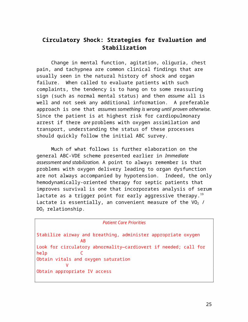
Circulatory Shock: Strategies for Evaluation and Stabilization
Change in mental function, agitation, oliguria, chest pain, and tachypnea are common clinical findings that are usually seen in the natural history of shock and organ failure. When called to evaluate patients with such complaints, the tendency is to hang on to some reassuring sign (such as normal mental status) and then assume all is well and not seek any additional information. A preferable approach is one that assumes something is wrong until proven otherwise. Since the patient is at highest risk for cardiopulmonary arrest if there are problems with oxygen assimilation and transport, understanding the status of these processes should quickly follow the initial ABC survey.
Much of what follows is further elaboration on the general ABC-VDE scheme presented earlier in Immediate assessment and stabilization. A point to always remember is that problems with oxygen delivery leading to organ dysfunction are not always accompanied by hypotension. Indeed, the only hemodynamically-oriented therapy for septic patients that improves survival is one that incorporates analysis of serum lactate as a trigger point for early aggressive therapy.19 Lactate is essentially, an convenient measure of the VO2 / DO2 relationship.
Patient Care Priorities
Stabilize airway and breathing, administer appropriate oxygen ABLook for circulatory abnormality—cardiovert if needed; call for help CObtain vitals and oxygen saturation VObtain appropriate IV accessAddress tissue oxygen delivery with ABG DConsider other studies: stat glucose if pt. is unresponsive, ECG, electrolytes EUse physical exam to evaluate CO, SVR, blood loss, pulmonary function
Treatment for stroke, myocardial infarction, hemorrhagic trauma and major pulmonary embolus are surrounded by the concept of a “golden hour” or similar time frame within which treatment should be instituted. Treatment of hypotension, organ dysfunction, sepsis, and most other medical conditions lack clear time guidelines. Even with this being the case, it’s hard to think of any physiologic abnormality for which it is acceptable to leave the bedside before being corrected. Decisions regarding whether a patient belongs in an ICU or step down unit, the rapidity of resuscitation, how to budget your time, and whether to call in help depend not only on the current vital status, but also on the extent of organ dysfunction. A general inventory of end-organ function can be gained by considering the question on the chart below.
17

Evaluation of Tissue Function and Oxygen Delivery
Question How Addressed Is oxygenation and ventilation normal? O2 sat, PaO2, PaCO2
Is the body in an anaerobic mode? pH, base deficit, lactate, anion gap Is the problem with bound oxygen? Hemoglobin, co-oximetery Is cardiac output adequate SvO2 or surrogate, capillary refill,
pH in presence of normal [Hb], cold patientMost blood gas analyzers can answer all of these questions with a single sampleOther supporting data can come from exam and laboratory findings that address “organs at risk:”
CNS or brainstem injury? Orientation, memory, respiratory pattern Metabolic abnormalities? Electrolyte analysis, anion gap, lactate Renal function? Creatinine and urine output Liver function? Tansaminases, bilirubin, PT, PTT
Treating hypotension or a low perfusion state is usually accomplished with a combination of fluids, blood, inotropes, or direct vasopressors. The most appropriate choice in a given situation hinges on whether the problem is one of preload, anemia, contractility, or lack of vascular tone. After quickly establishing the presence of a blood pressure or organ blood flow problem, it becomes essential to understand its root cause. The figure on the following page breaks down decreases in BP and DO2 into their constituent parts. As with similar figures in this text, physiologic roots are printed in black, findings from monitors, labs and exam are in red, and common diagnoses for each category are printed in blue.
While some of the physiologic data is presented as values obtainable by way of a pulmonary artery (Swan-Ganz) catheter, having such a catheter is not considered a necessity in caring for the critically ill. What is important is to ask the right physiologic questions and to assemble them in a coherent picture.
Life Without a Pulmonary Artery Catheter: Examining Physiology by Physical Exam
CO Cold extremities, distant pulses, acidosis, SvO2. Try to figure out whether a decrease in CO is due to hypovolemia or due to pump failure.
Pump Failure Distended neck veins, S3, cold extremities Preload (CVP) Flat or absent neck veins, tachycarida. Preload (CVP) jugular vein distention, enlarged veins elsewhereSVR BP and mental state may be NORMAL.
Findings: Cold extremities, distant pulsesSVR Hypotension is likely. Patient may be warm with full pulses if CO
is normal or elevated.Other valuable studies:
Spot Echo exam of the heart: addresses tamponade, CHF, ischemia, hypovolemia O2 saturation from CVP line or PICC line: provides indirect but meaningful estimates of the
adequacy of DO2, cardiac function.
18
19
Shock / Hypotension Diagnostic Tree
Goals : Stabilize BP with empiric fluids and vasopressorsIdentify etiology and treat more specifically
Exception: Arterial ruptures: AAA, dissection, intracranial bleed
Shock / Hypotension Diagnostic Tree
Arterial Oxygen Content-- Hemorrhage, Anemia Severe hypoxemia
SVR Sepsis Spinal shock Anaphylaxis Heatstroke Iatrogenic
Heart Rate Stroke Volume
Cardiac Output
Oxygen delivery (DO2)Lactic acidosispH, anion gap, BEPeripheral cyanosis end organ function Blood Pressure
or
or
or
Decreased effective stroke volume--classes of abnormalities
1. Contractility: MI, Cardio shock, ( CVP, PaOP, SVR)
2. Preload / filling PTX, tamponade, ( CVP, PaOP, SVR) Hypovolemia, hemorrhage ( CVP, PaOP, SVR)Tachycardia, dysrhythmia (loss of “atrial kick”)
3. Obstructive: Pulm Embolus, ( CVP, SVR) High PVR with RV failure, ( CVP, PAP, SVR)High SVR, or AS with LV failure ( CVP, PaOP, SVR)

Therapy for Hemodynamic Instability
Unstable patients require simultaneous diagnosis and treatment of underlying physiologic derangements.
In evaluating an unstable patient, it becomes crucial to develop some sense of how soon you need to normalize a patient’s status (5 minutes, 10 minutes, one hour?). This type of decision can be made quickly, based on known medical problems, what is know about the acute condition and its natural history, and the patient’s current status (acidotic, ST changes, chest pain, oliguria, hypotensive, etc.). For example, you may be able to show a bit more patience in waiting for fluid infusion to normalize the blood pressure in a young patient with an infection, than in an elderly patient who also has renal insufficiency, cerebral vascular disease, or coronary disease. If in doubt, treat early.
Resuscitation and supportive care of many critically ill patients ends up using a careful combination of fluids, inotropes, and vasopressors, based on considerations of the patient’s cardiac status, normal blood pressure, and current status of oxidative metabolism and solid organ function. The typical manner for maximizing cardiac performance, oxygen delivery and blood pressure is in order: titrate in fluids, evaluate cardiac output and use inotropes as indicated, and finally use alpha agonists to maintain blood pressure in an acceptable range. Because you’re not going to arrive at this “solution” immediately, you are certainly justified in treating an abnormally low blood pressure by making crude estimates of filling pressures, cardiac performance, and SVR and giving fluids or vasoactive drugs according to your hunches. This will keep your patient alive in the short term, and buy you some time to get it right later.
In attempting to maximize oxygen delivery, often one forgets the need to minimize oxygen consumption. The table below gives some examples of how certain stresses can increase oxygen demand.
Condition % Increase Over Resting VO2
Fever 8% (for each 1C increase)Work of breathing 40%Severe Infection 60%Shivering 50% - 100%Burns up to 100%Endotracheal tube suctioning 27%Sepsis 50% -100%Head injury, patient sedated 89%Head injury, patient unsedated 138%
Thus, seemingly insignificant acts such as keeping the patient warm, treating fever, and planning diagnostic procedures can be as efficacious as increasing cardiac output with fluids or inotropes. A patient with a severe metabolic acidosis
20

or lung injury can have about 30% of his oxygen consumption dedicated to the work of breathing. In other conditions where pulmonary compliance is poor, the amount of work required to maintain a reasonable minute ventilation can be likewise astronomical.
On the following pages, a more general scheme for early resuscitation of hypotension and shock is presented.
21
When blood flow to vital organs is compromised, institution of mechanical ventilation will “free up” diaphragmatic blood requirements, and increase the oxygen available to the rest of the body. Mechanical ventilation should be seriously considered on these merits alone, independent of lung injury.
Shock / Hypotension Treatment Outline
The goal of most resuscitative strategies is rapid normalization of blood pressure and oxygen delivery. The plan for complete resuscitation and normalization of blood pressure is more negotiable in cases hemorrhagic shock, cerebral hemorrhage, and arterial bleeding. Rapid identification of such processes and consultation with an appropriate service is a high priority, and discussions regarding resuscitation end points should take place quickly. Likewise, suspicion for cardiogenic shock should lead to immediate consultation with cardiology regarding early revascularization. Following the ABC-VDE evaluation, use the guide below as a checklist and a guide to pharmacotherapy. Different centers may have different methods and protocols for resuscitation. Whichever you end up using, the most important thing is that you make a constant cycle between: a plan for action the action an assessment of the action and its impact and a reassessment of the situation which leads to the next plan of action, and so on. In management circles this is known as a PDSA cycle for “plan, do, study, act.”
Priority #1. Normalize Blood Pressure: Crystalloid fluid: 1-2L over 10-15 minutes. 2 X 18g, or 16g or percutaneous
introducer. May need pressure bag. Triple lumens are generally inadequate. Vasopressor or inotrope to maintain oxygen delivery and BP. Want to normalize BP
in 2-5 minutes after IV access is established. A new stable peripheral IV or PICC line is OK to use for initiial use of vasopressors.
• Phenylephrine 100mcg IV q 1-5 minutes (may HR)• Ephedrine 5-10mg q 3-5 minutes (will HR)• Epinephrine 10-30mgc IV q 2-5 minutes (will HR)• Dopamine 3-10 mcg/kg/min. May need bolus of phenylephrine or ephedrine until target dopamine levels are achieved
Place Art line. Consider femoral (16g single lumen) if MAP < 55 Place central line for CVP, vasoactive drips, venous 02 saturation
Priority #2. Define nature and severity of injury: Labs and Diagnostics: Exam:ABG skin color and warmthCBC, PT/PTT*, DIC*, pulse characterLactate alertness, memoryElectrolytes including glucose breath soundsMixed venous O2 heart soundsType and screen* painCXR* subjective complaintsEKG,* Echo* evidence of bleedingCultures* *as indicated by clinical situation
Priority #3. Treat underlying cause as soon as it is known or suspected: Antibiotics, Gluccocorticoids, Blood and fluid replacement
22
Acute Blood Loss
Patients may experience sudden and massive blood loss for a variety of reasons-the most common being traumatic injury and/or surgical blood loss. However medical patients can also experience sudden and massive blood loss (i.e. GI bleed, hemoptysis, leaking AAA) and thus require an expeditious resuscitation to reduce morbidity and mortality. Traumatic blood loss and resuscitation is an especially dynamic process involving factor and fibrionogen consumption, dilutional coagulopathy, and further interaction between the coagulation system, temperature, and anoxic tissues. A full discussion on the management of hemorrhagic shock is beyond the scope of this text; instead, the concentration will be on non-traumatic blood loss.
When faced with massive and sudden blood loss in a patient, one should immediately call for help to mobilize all available resources. Help includes fellow residents, a stat surgical consult, anesthesia, crisis/ICU nurses, and the ICU fellow/team. Initial patient assessment should include ABCs assuring an adequate airway and placing the patient on 100% oxygen. Circulation should be assessed with the goal of aggressive treatment of tachycardia and hypotension with IV fluids.
Stat laboratory tests for CBC, ABG, coags, lactate, and type/cross should be drawn immediately by bedside personnel and taken directly to the lab for urgent processing. Care team members should establish adequate IV access,14-16g peripheral IVs or a “Cordis” introducer for rapid fluid administration. A triple lumen cath is inadequate. If present and it is difficult to find additional access consider changing to an introducer over a wire. Nurses should be alerted that you want blood pumps connected to blood warmers so that these will be ready at the time you place the IV lines.
If blood is needed immediately, before the blood bank has time to set up type and crossed units, send a runner to the blood bank and request a “trauma bucket” which should contain 6 units of O negative blood. Ideally type and crossed blood should be given. In the event that is not ready, then type specific uncross-matched blood is preferable followed by O negative. As the initial resuscitation proceeds, plans for definitive treatment of the bleeding should be made (i.e. OR/cath lab, etc) as well as transfer to the ICU for further care and management. Depending on the rate and amount of the blood loss you may have to replace the circulating blood volume several times until the bleeding is controlled. As one approaches 1.5 blood volumes transfused (adult range of 7-8 liters), a dilutional thrombocytopenia and coagulopathy may develop. Expect to give fresh frozen plasma (FFP) and platelets after two blood volumes have been transfused or earlier if there is laboratory evidence to suggest a more aggressive transfusion need (platelet count less than 80 or PT/PTT greater than 1.5x normal). FFP, if given in adequate volume, should replace clotting factors and fibrinogen. However, if the fibrinogen continues to be less than 1.0 g/L then
23

cryoprecipitate should be considered. Throughout the resuscitation heart rate, blood pressure, SaO2, mental status, and urine output should be monitored continuously (consider Art line/CVP).
Rapid Transfusion
Rapid transfusion of large volumes of stored red blood cells can result in several potential dangerous complications.
When the rate of transfusion exceeds 1 unit every 5 minutes there is a risk of hypocalcaemia from citrate toxicity. Signs of hypocalcaemia include hypotension, flat T waves, prolonged QT interval and a widened QRS complex. This should be treated with 500-1000 mg of IV CaCl2 via a central line or calcium gluconate via a peripheral IV.
The reduced concentration of 2,3-DPG in the hemoglobin of stored PRBC results in a left shift in the Hb-oxygen dissociation curve and less availability of oxygen at the tissue level.
Hyperkalemia can result from increased potassium from lysed red cells in the stored blood. In blood stored greater than 21 days K can be as high as 35 mEq/L.
Micro aggregate formation occurs in the stored units and can cause pulmonary insufficiency and ARDS when patients receive massive transfusions (>10 units/24hrs).
PRBC are stored at temperatures between 1-6 degree C. Failure to use a blood warming device can result in significant hypothermia. This can result in increased risk for the development of DIC, and less activity of existing clotting factors. Furthermore, hypothermia is associated with a higher mortality rate in massively transfused patients.
Acute pain
24
Throughout the resuscitation, heart rate, blood pressure, SaO2, mental status, and urine output should be monitored continuously (consider Art-line / CVP / foley).
Failure to warm the blood prior to transfusion can result in significant hypothermia.
Replacement of red blood cells should take priority over coagulation factors when resuscitating the acutely hemorrhaging patient.
There is no diagnostic or therapeutic yield in leaving a patient in pain. Major abdominal pathology, dyspnea and bone pain can still undergo appropriate evaluation without the added distress of nociception. Some points on the practical physiology of pain can be summarized:
The perception of pain is highly subjective and cannot be easily quantified across a heterogeneous population.
Pain is a significant stimulant of the sympathetic nervous system, and can exacerbate coronary and vascular pathology.
Pain exacerbates psychiatric pathology. Controlling pain later is generally more difficult than treating at the earliest opportunity. Poorly controlled pain can lead to other limb-pain syndromes or chronic pain. Agents such as NSAIDS and acetaminophen are terrible PRN drugs, but great as round-the-
clock agents in a comprehensive pain control scheme. Opiate tolerance is not opiate addiction, and may be acceptable in the short term.
Like other abnormalities in the vital status, extreme pain needs to be controlled acutely at the bedside.
Here is an example of the acute management of incisional pain: Fentanyl in 25-50 mcg boluses are given to the opiate naïve person with a small
incision; 50-100 mcg with a younger patient, 100-200 if opiate tolerant. Look at prior dosing histories to get some idea of tolerance and effective doses (cath
lab reports, anesthesia records and ER sheets). Continue boluses every 2-4 minutes until the pain score becomes 2-3. Give a few doses of morphine 2-5 mg and monitor for respiratory depression. A nurse can continue with morphine 2-5 mg q 15 min, until a PCA is ready.Drug Equianalgesic Cautions / notes_______ Dose Morphine 10 mg Venodilation via histamine mechanism, n/v, pruritisFentanyl 100mcg No venodilationDilaudid 2 mg Less pruritis and n/v than morphineMeperedine 75 mg Aviod for pain, use 25-50 mg for shivering & rigorsVicodin 8 tablets 4g Acetaminophen at this dose--don't exceedMethadone 10 mg No role for acute control
Some like morphine for acute coronary syndromes because its venodilatory property also lowers myocardial wall stress.
Reversing opiates The ER uses 0.4 mg of Naloxone to reverse possible opiate effects in
comatose patients. For the respiring patient, this is too high a starting dose. Overdosing naloxone will make your patient absolutely wild, and produce a dangerous hypertensive crisis.
1/10 of a vial (40 mcg) in q 1-2 minute escalating doses will reverse respiratory depression without antagonizing the analgesic properties.
25