Standardized endpoint definitions for transcatheter … consensus... · Composite endpoints for...
18
..................................................................................................................................................................................... ..................................................................................................................................................................................... ..................................................................................................................................................................................... CLINICAL RESEARCH Valvular medicine Standardized endpoint definitions for transcatheter aortic valve implantation clinical trials: a consensus report from the Valve Academic Research Consortium † Martin B. Leon * , Nicolo Piazza, Eugenia Nikolsky, Eugene H. Blackstone, Donald E. Cutlip, Arie Pieter Kappetein, Mitchell W. Krucoff, Michael Mack, Roxana Mehran, Craig Miller, Marie-ange ` le Morel, John Petersen, Jeffrey J. Popma, Johanna J.M. Takkenberg, Alec Vahanian, Gerrit-Anne van Es, Pascal Vranckx, John G. Webb, Stephan Windecker, and Patrick W. Serruys Columbia University Medical Center, Center for Interventional Vascular Therapy, 173 Fort Washington Avenue, Heart Center, 2nd floor, New York, NY 10032, USA. Received 8 July 2010; revised 30 September 2010; accepted 6 October 2010 Objectives To propose standardized consensus definitions for important clinical endpoints in transcatheter aortic valve implan- tation (TAVI), investigations in an effort to improve the quality of clinical research and to enable meaningful compari- sons between clinical trials. To make these consensus definitions accessible to all stakeholders in TAVI clinical research through a peer reviewed publication, on behalf of the public health. Background Transcatheter aortic valve implantation may provide a worthwhile less invasive treatment in many patients with severe aortic stenosis and since its introduction to the medical community in 2002, there has been an explosive growth in pro- cedures. The integration of TAVI into daily clinical practice should be guided by academic activities, which requires a har- monized and structured process for data collection, interpretation, and reporting during well-conducted clinical trials. Methods and results The Valve Academic Research Consortium established an independent collaboration between Academic Research organizations and specialty societies (cardiology and cardiac surgery) in the USA and Europe. Two meetings, in San Francisco, California (September 2009) and in Amsterdam, the Netherlands (December 2009), including key physician experts, and representatives from the US Food and Drug Administration (FDA) and device manufacturers, were focused on creating consistent endpoint definitions and consensus recommendations for implementation in TAVI clinical research programs. Important considerations in developing endpoint definitions included (i) respect for the historical legacy of surgical valve guidelines; (ii) identification of pathophysiological mechanisms associated with clinical events; (iii) emphasis on clinical relevance. Consensus criteria were developed for the following end- points: mortality, myocardial infarction, stroke, bleeding, acute kidney injury, vascular complications, and prosthetic valve performance. Composite endpoints for TAVI safety and effectiveness were also recommended. Conclusion Although consensus criteria will invariably include certain arbitrary features, an organized multidisciplinary process to develop specific definitions for TAVI clinical research should provide consistency across studies that can facilitate the † The Valve Academic Research Consortium (VARC) consists of representatives from several independent Academic Research Organizations, several Surgery and Cardiology Societies, members of the U.S. Food and Drug Administration, and several independent experts (Appendices 1 and 2). * Corresponding author. Tel: +1 212 342 3617, Fax: +1 212 342 3660, Email: [email protected] Published on behalf of the European Society of Cardiology. The article has been co-published in the Journal of the American College of Cardiology. All rights reserved. & The Author 2010. For permissions please email: [email protected]. The online version of this article has been published under an open access model. Users are entitled to use, reproduce, disseminate, or display the open access version of this article for non-commercial purposes provided that the original authorship is properly and fully attributed; the Journal, Learned Society and Oxford University Press are attributed as the original place of publication with correct citation details given; if an article is subsequently reproduced or disseminated not in its entirety but only in part or as a derivative work this must be clearly indicated. For commercial re-use, please contact [email protected] European Heart Journal (2011) 32, 205–217 doi:10.1093/eurheartj/ehq406 by guest on January 27, 2016 http://eurheartj.oxfordjournals.org/ Downloaded from
Transcript of Standardized endpoint definitions for transcatheter … consensus... · Composite endpoints for...

. . . . . . . . . . . . . . . . . . . . . . . . . . . . . . . . . . . . . . . . . . . . . . . . . . . . . . . . . . . . . . . . . . . . . . . . . . . . . . . . . . . . . . . . . . . . . . . . . . . . . . . . . . . . . . . . . . . . . . . . . . . . . . . . . . . . . . . . . . . . . . . . . . . . . . . . . . . . . . . . . . . . . . . . . . . . . . . . . . . . .
. . . . . . . . . . . . . . . . . . . . . . . . . . . . . . . . . . . . . . . . . . . . . . . . . . . . . . . . . . . . . . . . . . . . . . . . . . . . . . . . . . . . . . . . . . . . . . . . . . . . . . . . . . . . . . . . . . . . . . . . . . . . . . . . . . . . . . . . . . . . . . . . . . . . . . . . . . . . . . . . . . . . . . . . . . . . . . . . . . . . .
. . . . . . . . . . . . . . . . . . . . . . . . . . . . . . . . . . . . . . . . . . . . . . . . . . . . . . . . . . . . . . . . . . . . . . . . . . . . . . . . . . . . . . . . . . . . . . . . . . . . . . . . . . . . . . . . . . . . . . . . . . . . . . . . . . . . . . . . . . . . . . . . . . . . . . . . . . . . . . . . . . . . . . . . . . . . . . . . . . . . .
CLINICAL RESEARCHValvular medicine
Standardized endpoint definitions fortranscatheter aortic valve implantation clinicaltrials: a consensus report from the ValveAcademic Research Consortium†
Martin B. Leon*, Nicolo Piazza, Eugenia Nikolsky, Eugene H. Blackstone,Donald E. Cutlip, Arie Pieter Kappetein, Mitchell W. Krucoff, Michael Mack,Roxana Mehran, Craig Miller, Marie-angele Morel, John Petersen, Jeffrey J. Popma,Johanna J.M. Takkenberg, Alec Vahanian, Gerrit-Anne van Es, Pascal Vranckx,John G. Webb, Stephan Windecker, and Patrick W. Serruys
Columbia University Medical Center, Center for Interventional Vascular Therapy, 173 Fort Washington Avenue, Heart Center, 2nd floor, New York, NY 10032, USA.
Received 8 July 2010; revised 30 September 2010; accepted 6 October 2010
Objectives To propose standardized consensus definitions for important clinical endpoints in transcatheter aortic valve implan-tation (TAVI), investigations in an effort to improve the quality of clinical research and to enable meaningful compari-sons between clinical trials. To make these consensus definitions accessible to all stakeholders in TAVI clinicalresearch through a peer reviewed publication, on behalf of the public health.
Background Transcatheter aortic valve implantation may provide a worthwhile less invasive treatment in many patients with severeaortic stenosis and since its introduction to the medical community in 2002, there has been an explosive growth in pro-cedures. The integration of TAVI into daily clinical practice should be guided by academic activities, which requires a har-monized and structured process for data collection, interpretation, and reporting during well-conducted clinical trials.
Methodsand results
The Valve Academic Research Consortium established an independent collaboration between Academic Researchorganizations and specialty societies (cardiology and cardiac surgery) in the USA and Europe. Two meetings, inSan Francisco, California (September 2009) and in Amsterdam, the Netherlands (December 2009), including keyphysician experts, and representatives from the US Food and Drug Administration (FDA) and device manufacturers,were focused on creating consistent endpoint definitions and consensus recommendations for implementation inTAVI clinical research programs. Important considerations in developing endpoint definitions included (i) respectfor the historical legacy of surgical valve guidelines; (ii) identification of pathophysiological mechanisms associatedwith clinical events; (iii) emphasis on clinical relevance. Consensus criteria were developed for the following end-points: mortality, myocardial infarction, stroke, bleeding, acute kidney injury, vascular complications, and prostheticvalve performance. Composite endpoints for TAVI safety and effectiveness were also recommended.
Conclusion Although consensus criteria will invariably include certain arbitrary features, an organized multidisciplinary process todevelop specific definitions for TAVI clinical research should provide consistency across studies that can facilitate the
†The Valve Academic Research Consortium (VARC) consists of representatives from several independent Academic Research Organizations, several Surgery and CardiologySocieties, members of the U.S. Food and Drug Administration, and several independent experts (Appendices 1 and 2).
* Corresponding author. Tel: +1 212 342 3617, Fax: +1 212 342 3660, Email: [email protected]
Published on behalf of the European Society of Cardiology. The article has been co-published in the Journal of the American College of Cardiology. All rights reserved. & TheAuthor 2010. For permissions please email: [email protected] online version of this article has been published under an open access model. Users are entitled to use, reproduce, disseminate, or display the open access version of this articlefor non-commercial purposes provided that the original authorship is properly and fully attributed; the Journal, Learned Society and Oxford University Press are attributed as theoriginal place of publication with correct citation details given; if an article is subsequently reproduced or disseminated not in its entirety but only in part or as a derivative work thismust be clearly indicated. For commercial re-use, please contact [email protected]
European Heart Journal (2011) 32, 205–217doi:10.1093/eurheartj/ehq406
by guest on January 27, 2016http://eurheartj.oxfordjournals.org/
Dow
nloaded from

evaluation of this new important catheter-based therapy. The broadly based consensus endpoint definitionsdescribed in this document may be useful for regulatory and clinical trial purposes.
- - - - - - - - - - - - - - - - - - - - - - - - - - - - - - - - - - - - - - - - - - - - - - - - - - - - - - - - - - - - - - - - - - - - - - - - - - - - - - - - - - - - - - - - - - - - - - - - - - - - - - - - - - - - - - - - - - - - - - - - - - - - - - - - - - - - - - - - - - - - - - - - - - - - - - - - - - -Keywords Transcatheter aortic valve implantation
IntroductionSince the introduction of transcatheter aortic valve implantation(TAVI) in 20021, there has been increasing interest in the field ofcatheter-based treatment of high-surgical-risk patients with symp-tomatic aortic stenosis (AS).2 – 7 Introduction of this new technol-ogy should ideally follow the standard bench-to-bedsideevidence-based medicine pattern, starting with pre-clinical testingand advancing to clinical investigations. Unfortunately, the explo-sive growth of TAVI (Figure 1) has created a ‘clinical data conun-drum’: investigators were not prepared to optimally organize andinterpret clinical data for this radically different treatment, render-ing thoughtful assessment of clinical trial outcomes difficult andinter-study results comparisons problematic.8– 11
Surgical valve clinical research guidelines have been developedusing a more traditional ‘multi-society approach’, have beenrevised approximately every 10 years, incorporate not merely clini-cal endpoints but also issues such as structural valve deteriorationand non-structural valve dysfunction, and often divide clinicalevents into those that are valve and non-valve related.12 Interven-tional cardiology has a tradition of agreed upon clinical endpointdefinitions and clinical trial methodologies13,14 and recently hasincorporated a consensus process to standardize key endpointdefinitions by convening an Academic Research Consortium(ARC) among Academic Research Organizations (AROs) fromthe USA and Europe joined by representatives from the USFDAand device manufacturers.14 The ARC process demonstrated the
power of a well-managed international goal-directed academicconsortium collaborating effectively with the FDA and industryto establish consensus clinical endpoint definitions and toimprove the conduct of clinical research.
In the spirit of the ARC-mission statement,14 the ‘Valve Aca-demic Research Consortium’ (VARC) was organized as anamalgam of the ARO and multi-society guideline models withstrong participation from independent experts, the FDA, andmedical device manufacturers (Appendices 1 and 2). Twoin-person meetings on 19 September 2009 in San Francisco, CAand on 5–6 December 2009 in Amsterdam, the Netherlands,involving VARC study group members and invited guests (includingthe FDA and industry representatives) provided much of the sub-stantive discussion from which this consensus manuscript wasderived.
The goals of VARC are to combine the expertise of surgeons,interventionalists, medical cardiologists, clinical trialists, andother specialists (representing relevant disciplines including echo-cardiography, vascular medicine, and neurology) to arrive at aconsensus for (i) selecting appropriate clinical endpoints reflectingdevice, procedure and patient-related effectiveness and safety,and (ii) standardizing definitions for single and composite clinicalendpoints.
Importantly, this first consensus manuscript was not intended asa ‘guidelines statement’ or a ‘guidance document’, but rathershould be viewed as a roadmap to facilitate the standardizationof future TAVI and other aortic valve clinical research
Figure 1 Current generation transcatheter aortic valve therapies. (A) balloon-expandable, stainless steel support structure, bovine pericardialvalve; (B) self-expanding, nitinol support structure, porcine pericardial valve.
M.B. Leon et al.206
by guest on January 27, 2016http://eurheartj.oxfordjournals.org/
Dow
nloaded from

Principles for selecting anddefining clinical endpoints fortranscatheter aortic valveimplantation investigations:general considerations
Criteria for endpoint definitionsThe definitions of major clinical endpoints must follow a multi-stepthought process.
† Each major endpoint should address issues that establish eitherthe safety and/or the effectiveness of the proposed new therapy.
– Safety is characterized by the avoidance of device-related orprocedural complications.
– Effectiveness is a more complex descriptor, as it encompassesboth the avoidance of negative disease-related outcomes andobjective measures of clinical functional benefit.
† The endpoints should relate short- and long-term pathophysiolo-gical mechanisms to meaningful clinical events.
† Endpoint definitions must be consistent with the body of pub-lished literature, but still reflect unique or evolving aspects ofthe new therapy.
† The emphasis should be on definitions that accurately representessential patient-oriented clinical outcomes.
† The endpoints must be well defined (preferably through blindedadjudication processes) such that they can be to be subjected tostatistical analysis.
It is helpful to reference a standardized definition format regarding(i) the specific treatment, (ii) the place of occurrence, (iii) the timeof occurrence, and (iv) the specific type of endpoint.
Device, procedure, and patient-orientedoutcomesEndpoint definitions for TAVI will in most cases be characterized inrelation to the specific implant device, the implant procedure, andthe resultant patient-oriented outcomes, which can occur at anytime after the procedure. During the early phases of therapy devel-opment, particular attention must be directed to the safety andperformance of the device. Therefore, VARC tries to strike a com-promise by also elucidating device and procedure-related events,which are essential to the understanding of a new class of catheter-based therapies.
Since TAVI is fundamentally the placement of a prosthetic aorticvalve and will be compared with surgical aortic valve replacement(AVR), tradition should be respected and crucial endpoints such asall-cause mortality and device durability must be assessed longitud-inally for the life of the implant.12 However, primary clinical end-points used in pivotal clinical trials for regulatory approval ofTAVI devices should incorporate a shorter time domain of 1 to2 years after the index procedure. These recommended shortertime horizons should not discourage the standard long-termfollow-up procedures for prosthetic heart valves.
Proposed safety and efficacyendpoints
MortalityAll-cause mortality in surgical clinical trials has become the ‘goldstandard’ in previously published consensus and guideline docu-ments.12 The advantage of reporting all-cause mortality is that itis both objective (without bias) and pragmatic from the standpointof ascertainment and adjudication. However, the use of all-causemortality in high-risk TAVI patients may be misleading, resultingin disproportionate reporting of mortal events unrelated toeither the treatment device or the procedure. Therefore, VARCproposes to use all-cause mortality as a primary clinical endpoint,but also recommends further subdivision of mortality, specificallydenoting cardiovascular mortality as an important secondary end-point (Table 1). Of note, ‘unknown’ deaths should be consideredas cardiovascular in origin and to improve the ascertainment ofdeath, the social security death index or national death registriesshould be utilized in cases of patients lost to follow-up.
Consistent with surgical guidelines and surgical clinical trialpractices,12 mortality should be formally assessed and reportedat 30 days after the index procedure (or longer if the patientwas not discharged from the treatment hospital or a secondaryconvalescent facility). Since there may be either unknown orunder-reporting of early device failure modes, a more appropriateduration for all-cause mortality as a primary endpoint in TAVIclinical trials is 1 year after the index procedure. After 1 year,mortality should be recorded at yearly intervals for a minimumof 5 years, or ideally, for the duration of the prosthetic valveimplant, in the form of well-defined post-approval surveillanceregistries.
Myocardial infarctionIn 2007, the joint ESC/ACC/AHA/WHF task force for the redefini-tion of Myocardial infarction (MI) established diagnostic criteriaand updated guidelines for a universal MI definition to be used inclinical trials.13 This universal MI definition is highly sensitive,relying heavily on the measurement of cardiac biomarkers (prefer-ably troponin). Conversely, surgical valve guidelines have adopted a
. . . . . . . . . . . . . . . . . . . . . . . . . . . . . . . . . . . . . . . . . . . . . . . . . . . . . . . . . . . . . . . . . . . . . . . . . . . . . . . .
Table 1 Mortality
Cardiovascular mortality
Any one of the following criteria:
Any death due to proximate cardiac cause (e.g. MI, cardiactamponade, worsening heart failure)
Unwitnessed death and death of unknown cause
All procedure-related deaths, including those related to acomplication of the procedure or treatment for a complicationof the procedure
Death caused by non-coronary vascular conditions such ascrebrovascular disease, pulmonary embolism, ruptured aorticaneurysm, dissecting aneurysm, or other vascular disease
VARC consensus endpoints after TAVI for high risk AS 207
by guest on January 27, 2016http://eurheartj.oxfordjournals.org/
Dow
nloaded from

‘minimalist’ approach to MI definitions, usually ignoring biomarkerdiagnoses and excluding both intra-operative and post-operativeMIs, unless the MI was caused by a coronary embolus.12
Valve Academic Research Consortium proposes a more ‘cen-trist’ approach to MI definitions after TAVI, recognizing thatmany patients have coexistent aortic valve and coronary arterydisease,15 which requires an MI definition that does notexclude peri-procedural or late MIs that may impact patient out-comes. Valve Academic Research Consortium proposes to defineperi-procedural MI as an acute ischaemic event that is associatedwith documented and clinically significant myocardial necrosis(Table 2). This definition does not include ischaemic eventsafter TAVI or surgery defined solely by biomarker elevationswithout a clinically evident ischaemic insult. Since troponinmeasurements are an extremely sensitive biomarker of myocar-dial necrosis, VARC recommends that CPK-MB should be theperi-procedural biomarker of choice in TAVI clinical trials. Bio-marker samples ideally should be obtained at baseline, twiceafter the procedure (separated by at least 6 h), and if still elev-ated, daily thereafter until values are declining. Since TAVI mayinvolve open surgical procedures (e.g. transapical access), the bio-marker diagnosis of peri-procedural MI requires a .20% increase
in the second post-procedure sample and a threshold elevationof ten times the upper normal range. An electrocardiogramshould be collected at baseline and at least once after the pro-cedure prior to discharge to document the presence orabsence of new Q-waves. The peri-procedural interval is inclusiveof all events that begin within 72 h of the index procedure. Acuteischaemic events occurring after 72 h are considered spontaneousMIs and are defined in accordance with the universal MI guide-lines,13 as further modified by ARC14 (Table 2). Finally, a con-firmed coronary embolus, occurring at any time, should bereported as an independent event, if biomarker changes andassociated findings fulfil definition criteria.
StrokeRecently, two reports have indicated a high frequency of new per-fusion abnormalities (presumably embolic) detected by diffusion-weighted magnetic resonance imaging (MRI) studies soon afterTAVI,16,17 although the clinical significance of these early perfusiondefects remains unclear. Strokes during and after TAVI may occurdue to embolic events from multiple sources, procedure-relatedaortic dissections, ischaemia from hypotension, or haemorrhagiccomplications associated with adjunctive pharmacotherapy. Insightson stroke definitions are in a state of evolution and VARC examinedviewpoints derived from several sources, including recent multi-society consensus documents12,18,19 and multi-center randomizedtrials, in which stroke was an important endpoint.20–29 Valve Aca-demic Research Consortium considered five important issues in arriv-ing at clinically relevant stroke definitions, as follows: (i) a clinicaldiagnosis of stroke which ruled out metabolic or toxic encephalopa-thy, pharmacological influences, and non-central neurological symp-toms, (ii) the role of neuroimaging studies for confirmation of thediagnosis, (iii) the distinction of stroke vs. transient ischaemic attack(TIA) (including timing), (iv) categorization of stroke into major andminor events based on the degree of disability as defined by conven-tional neurological assessment tools, and (v) sub-classification ofstrokes into haemorrhagic, ischaemic, and undetermined categories.
Table 3 outlines the diagnostic criteria and specific definitions forTIA and stroke as proposed by VARC. There is growing accep-tance that neuroimaging is an important biomarker for the diagno-sis of neuronal injury and stroke18,19 and diffusion-weighted MRI isgenerally considered the procedure of choice in the context ofacute neurological syndromes.30 If a stroke is reported without evi-dence of confirmation of the diagnosis by the methods outlined inTable 3, the event may still be considered a stroke on the basis ofthe clinical presentation alone, but formal adjudication by qualifiedneurologists who are members of, or consultants to, a clinicalevents committee is mandatory. Patients with a global encephalo-pathy will not be reported as a stroke without unequivocal neuroi-maging findings. Diagnosis of stroke in patients with a previouslydocumented neurological deficit is more problematic and requiresclinical assessment by a neurologist accompanied by appropriatenew CT or MRI findings. In patients with a previous stroke and per-sistent neurological deficits, baseline pre-treatment neurologicalconsultation and neuroimaging studies are recommended.
The earliest time of new neurological symptoms is defined asthe time of onset of the stroke or TIA. When a patient awakensor begins responding (if previously unconscious) after the index
. . . . . . . . . . . . . . . . . . . . . . . . . . . . . . . . . . . . . . . . . . . . . . . . . . . . . . . . . . . . . . . . . . . . . . . . . . . . . . . .
Table 2 Myocardial infarction
Peri-procedural MI (≤72 h After the index procedure)
New ischaemic symptoms (e.g. chest pain or shortness of breath),or new ischaemic signs (e.g. ventricular arrhythmias, new orworsening heart failure, new ST-segment changes,haemodynamic instability, or imaging evidence of new loss ofviable myocardium or new wall motion abnormality),
AND
Elevated cardiac biomarkers (preferably CK-MB) within 72 h afterthe index procedure, consisting of two or more post-proceduresamples that are .6–8 h apart with a 20% increase in the secondsample and a peak value exceeding 10x the 99th percentile upperreference limit (URL), or a peak value exceeding 5x the 99thpercentile URL with new pathological Q waves in at least twocontiguous leads.
Spontaneous MI (.72 h after the index procedure)
Any one of the following criteria:
Detection of rise and/or fall of cardiac biomarkers (preferablytroponin) with at least one value above the 99th percentile URL,together with evidence of myocardial ischaemia with at least oneof the following:
ECG changes indicative of new ischaemia [new ST-T changes ornew left bundle branch block (LBBB)]
New pathological Q waves in at least two contiguous leads
Imaging evidence of new loss of viable myocardium or new wallmotion abnormality
Sudden, unexpected cardiac death, involving cardiac arrest, oftenwith symptoms suggestive of myocardial ischaemia, andaccompanied by presumably new ST elevation, or new LBBB,and/or evidence of fresh thrombus by coronary angiography and/or at autopsy, but death occurring before blood samples couldbe obtained, or at a time before the appearance of cardiacbiomarkers in the blood.
Pathological findings of an acute myocardial infarction.
M.B. Leon et al.208
by guest on January 27, 2016http://eurheartj.oxfordjournals.org/
Dow
nloaded from

procedure with obvious new signs of a neurological deficit, thestroke or TIA is considered to have occurred during the index pro-cedure. The diagnosis of a transient ischemic attack is defined ascomplete resolution of new neurological symptoms usuallywithin 1–2 h but always within 24 h and also requires a normalneuroimaging study.18,19 A stroke fulfiling the diagnostic criteriain Table 3 is classified as a major stroke based upon ongoing signifi-cant clinical disability, defined as a Modified Rankin Score ≥2.Although the initial Modified Rankin Score should be recordedafter 7 days or at the time of hospital discharge, the attributionof clinically significant disability requires a Modified Rankin Score≥2 at both 30 and 90 days follow-up (allowing sufficient timefor stroke disability to stabilize). The Modified Rankin Score deter-minations should be performed by qualified individuals who haveundergone a certification process.31 –34 A minor stroke must alsofulfil stroke diagnostic criteria, with either resolution of newneurological symptoms within 24 h or persistence of symptoms.24 h and a Modified Rankin Score ,2 at both 30 and 90 daysfollow-up. If there is discordance between the 30 and 90-dayModified Rankin Scores, the final determination of major vs.
minor strokes should be adjudicated by the neurology membersof the clinical events committee. For the purposes of clinical trialendpoints, VARC advocates that only major strokes should be con-sidered as an important safety endpoint, however, all neurologicalevents should be reported as adverse events.
Stroke will be further stratified into ischaemic, haemorrhagic, orundetermined origin utilizing newly proposed definitions by anFDA consensus panel.35 Ischaemic stroke is as an acute sympto-matic episode of focal cerebral, spinal, or retinal dysfunctioncaused by an infarction of central nervous system tissue. Haemor-rhagic stroke is an acute symptomatic episode of focal or globalcerebral or spinal dysfunction caused by a non-traumatic intrapar-enchymal, intraventricular, or subarachnoid haemorrhage. Anundetermined stroke is a stroke with insufficient information toallow categorization as either of ischaemic or haemorrhagic origin.
Bleeding complicationsBleeding is a critical safety endpoint in evaluating contemporarypharmacological agents and interventional devices.36– 44 Valve Aca-demic Research Consortium carefully reviewed several literaturesources including (i) landmark clinical trials assessing the effectsof anti-thrombotic medications in stable and acute coronary syn-dromes,45– 59 (ii) a report by the Control of Anticoagulation Sub-committee of the International Society on Thrombosis andHaemostasis,60 and (iii) surgery guidelines after cardiac valveprocedures.12
The definition of clinically meaningful bleeding was guided by thefollowing principles: (i) the definition must be based on objectivecriteria, including an obvious source of bleeding or number oftransfusions; (ii) serious or meaningful bleeding must result indeath, be life-threatening, be proven to be associated with
. . . . . . . . . . . . . . . . . . . . . . . . . . . . . . . . . . . . . . . . . . . . . . . . . . . . . . . . . . . . . . . . . . . . . . . . . . . . . . . .
. . . . . . . . . . . . . . . . . . . . . . . . . . . . . . . . . . . . . . . . . . . . . . . . . . . . . . . . . . . . . . . . . . . . . . . . . . . . . . . .
Table 4 Bleeding
Life-threatening or disabling bleeding
Fatal bleeding OR
Bleeding in a critical area or organ, such as intracranial, intraspinal,intraocular, or pericardial necessitating pericardiocentesis, orintramuscular with compartment syndrome OR
Bleeding causing hypovolemic shock or severe hypotensionrequiring vasopressors or surgery OR
Overt source of bleeding with drop in haemoglobin of ≥5 g/dL orwhole blood or packed red blood cells (RBCs) transfusion ≥4unitsa
Major bleeding
Overt bleeding either associated with a drop in the haemoglobinlevel of at least 3.0 g/dL or requiring transfusion of two or threeunits of whole blood/RBC AND
Does not meet criteria of life-threatening or disabling bleeding
Minor Bleeding
Any bleeding worthy of clinical mention (e.g. access sitehaematoma) that does not qualify as life-threatening, disabling ormajor
aGiven one unit of packed RBC typically will raise blood haemoglobinconcentration by 1 g/dL, an estimated decrease in haemoglobin will be calculated.
. . . . . . . . . . . . . . . . . . . . . . . . . . . . . . . . . . . . . . . . . . . . . . . . . . . . . . . . . . . . . . . . . . . . . . . . . . . . . . . .
Table 3 Stroke
Stroke diagnostic criteria
Rapid onset of a focal or global neurological deficit with at least oneof the following: change in level of consciousness, hemiplegia,hemiparesis, numbness or sensory loss affecting one side of thebody, dysphasia or aphasia, haemianopia, amaurosis fugax, orother neurological signs or symptoms consistent with stroke
Duration of a focal or global neurological deficit ≥24 h; OR , 24 h,if therapeutic intervention(s) were performed (e.g. thrombolytictherapy or intracranial angioplasty); OR available neuroimagingdocuments a new haemorrhage or infarct; OR the neurologicaldeficit results in death
No other readily identifiable non-stroke cause for the clinicalpresentation (e.g. brain tumour, trauma, infection,hypoglycaemia, peripheral lesion, pharmacological influences)a
Confirmation of the diagnosis by at least one of the following:
Neurology or neurosurgical specialist
Neuroimaging procedure (MR or CT scan or cerebralangiography)
Lumbar puncture (i.e. spinal fluid analysis diagnostic ofintracranial haemorrhage)
Stroke definitions
Transient ischaemic attack:
New focal neurological deficit with rapid symptom resolution(usually 1–2 h), always within 24 h
Neuroimaging without tissue injury
Stroke: (diagnosis as above, preferably with positive neuroimagingstudy)
Minor—Modified Rankin score ,2 at 30 and 90 daysb
Major—Modified Rankin score ≥2 at 30 and 90 days
aPatients with non-focal global encephalopathy will not be reported as a strokewithout unequivocal evidence based upon neuroimaging studies.bModified Rankin Score assessments should be made by qualified individualsaccording to a certification process. If there is discordance between the 30 and 90day Modified Rankin Scores, a final determination of major vs. minor stroke will beadjudicated by the neurology members of the clinical events committee.
VARC consensus endpoints after TAVI for high risk AS 209
by guest on January 27, 2016http://eurheartj.oxfordjournals.org/
Dow
nloaded from

increased long-term mortality, cause chronic sequellae, orconsume major health-care resources. The VARC definition ofbleeding complications (Table 4) is divided into life-threateningor disabling bleeding, major bleeding, and minor bleeding; anythingbut minor bleeding constitutes a serious or meaningful bleedingevent.
Given the ample body of literature suggesting that administrationof whole blood or red blood cell (RBC) transfusions in patientswith cardiovascular pathology may be potentially harmful,41 –44
VARC considers that any whole blood or RBC transfusion needsto be reported in the case report forms, including the numberof transfused units, regardless of the presence or absence ofovert bleeding. Transfusions also need to be further stratifiedinto those associated with overt bleeding and those in theabsence of overt bleeding. Bleeding complications and transfusionsshould also be characterized relative to the time of occurrenceincluding during the procedure, within the index hospitalization,or post-discharge.
Acute kidney injuryThe natural history of acute kidney injury (AKI) in a variety of clini-cal settings61–70 is now well understood, including the recognitionthat even small decreases in kidney function can have a dramaticimpact on the risk for subsequent mortality.69,70 In recentreports, AKI has been observed in 12–28% of patients undergoingTAVI and was associated with a four times higher post-proceduralmortality.71,72
In defining the stages of AKI, VARC proposes adopting serumcreatinine criteria from the ‘modified’ RIFLE (Risk, Injury, Failure,Loss, and End-stage kidney disease) classification (Table 5).73 TheRIFLE classification74– 76 has been validated in the setting of inten-sive care units and cardiac surgery77–81 and provides practical defi-nitions for early stages of renal dysfunction when kidney injury canstill be prevented, as well as stages when the kidney has alreadybeen damaged and renal failure is established. Modifications ofthe original RIFLE classification include two important changes:(i) smaller changes in serum creatinine (0.3 mg/dL) are includedin stage 1 (‘Risk’)82; (ii) the ‘Loss’ and ‘End-stage kidney disease’
categories have been removed due to a lack of uniform indicationsand timing of renal replacement therapy (RRT) and variability inRRT resources in different countries. An outer bound of 72 hfrom the index procedure for diagnosing AKI was selected basedon evidence that adverse outcomes were observed when theelevation occurred within 24 to 48 h of the procedure83 and toensure that the process was both acute and related to the pro-cedure itself rather than as a consequence of post-proceduremulti-organ system failure. Risk, Injury, Failure, Loss, and End-stagekidney disease classifications also stress the predictive value ofurine output criteria in defining AKI, but VARC has not includedthis measure in the definition of AKI since urine outputs maynot be measured accurately or routinely in all cases.
Valve Academic Research Consortium proposes to utilize themodified RIFLE classification to (i) capture even the earlieststages of AKI (stage 1) on case report forms, (ii) define AKI aseither stage 2 or 3, and (iii) report any case of RRT (haemodialysis,peritoneal dialysis, or haemofiltration) occurring during the indexhospitalization or within 30 days after the index procedure.Given the well-recognized damaging impact of contrast media onrenal function, VARC also recommends to report the volumeand type of contrast medium used during the index procedure.
Vascular complicationsRecent TAVI literature indicates that major vascular complicationsusing various non-standardized definitions (e.g. with or withoutincluding the need for blood transfusions) occur at a frequencyof 4–34% and are associated with a two- or three-fold higher30-day mortality.84– 87 In defining vascular complications, VARCreferenced the reporting standards of the Society of VascularSurgery for defining and reporting vascular complications followingendovascular aortic graft repair procedures.88
Valve Academic Research Consortium proposes to report bothmajor and minor vascular complications, but to only considermajor vascular complications as an important clinical endpoint.Of note, the ‘access site’ is defined as any location (arterial orvenous) traversed by a guide-wire, a catheter or a sheath [includingthe left ventricular (LV) apex and the aorta] and ‘access related’ isdefined as any adverse clinical consequence possibly associatedwith any of the access sites used during the procedure. TheVARC definitions for major and minor vascular complications aredescribed in Table 6.
Many vascular situations require special notice. Femoral vascularaccess and closure in many centers is routinely achieved using sur-gical cut-down procedures, and therefore, pre-planned surgicalaccess and/or closure should be considered as part of the pro-cedure and not as a complication. Similarly, uncomplicated non-femoral (e.g. retroperitoneal, iliac, subclavian, or aortic) surgicalaccess for sheath entry (planned or unplanned) is not considereda vascular complication, unless untoward clinical consequences aredocumented (e.g. bleeding complications). However, interven-tional or surgical repair for failed percutaneous closure of thearteriotomy site during the index procedure without other clinicalsequellae (Table 6) is considered a minor vascular complication.Considering the recent proliferation of vascular access approachesand the recognition that specific access sites and techniques maybe associated with either increased or decreased complications,
. . . . . . . . . . . . . . . . . . . . . . . . . . . . . . . . . . . . . . . . . . . . . . . . . . . . . . . . . . . . . . . . . . . . . . . . . . . . . . . .
Table 5 Acute kidney injury (modified RIFLEclassification)
Change in serum creatinine (up to 72 h) compared withbaseline
Stage 1 Increase in serum creatinine to 150–200% (1.5–2.0 × increase compared with baseline) or increase of ≥0.3 mg/dL(≥26.4 mmol/L)
Stage 2 Increase in serum creatinine to 200–300% (2.0–3.0 × increase compared with baseline) or increase between .0.3mg/dL (.26.4 mmol/L) and ,4.0 mg/dL (,354 mmol/L)
Stage 3a Increase in serum creatinine to ≥300% (.3 × increasecompared with baseline) or serum creatinine of ≥4.0 mg/dL (≥354mmol/L) with an acute increase of at least 0.5 mg/dL (44 mmol/L)
aPatients receiving renal replacement therapy are considered to meet Stage 3criteria irrespective of other criteria.
M.B. Leon et al.210
by guest on January 27, 2016http://eurheartj.oxfordjournals.org/
Dow
nloaded from

VARC strongly recommends that detailed information is recordedon the access site and technique for each procedure.
A special circumstance relates to complications associated withthe left-ventricular apex site during transapical TAVI procedures.Although such complications are less frequent than transarterialvascular complications, a recent report indicates that the clinicalconsequences (including death) can be more serious.89 Major com-plications associated with transapical TAVI procedures includebleeding, pseudoaneurysm formation (with or without rupture),and haemodynamic instability requiring urgent transarterial cardio-pulmonary bypass support. Valve Academic Research Consortiumproposes that all such complications associated with transapicalTAVI be reported in case report forms and that clinical conse-quences resulting from such complications be registered underthe appropriate clinical endpoints (e.g. bleeding, stroke, mortality).
Prosthetic valve performanceThe clinical presentation of patients with prosthetic valve dysfunc-tion is usually consistent with symptoms and signs of either valvularregurgitation or stenosis. Valve Academic Research Consortiumproposes only two criteria to evaluate impaired prosthetic valveperformance: (i) prosthetic valve haemodynamics assessed byechocardiography and (ii) associated clinical findings indicatingimpaired cardiovascular or valvular function (e.g. new or worseningcongestive heart failure). Transthoracic echocardiography (TTE) is
usually adequate to evaluate prosthetic aortic valve function,90
although transoesophageal echocardiography (TEE) may be veryuseful in the setting of technically challenging or complex cases.Serial echocardiography evaluations after surgical AVR and TAVIshould be performed at baseline, soon after the index procedure(ideally within 24–48 h, but always before discharge), at 1month (especially for TAVI), 12 months, and yearly thereafter.91,92
This follow-up schedule is more intensive than recommended inthe AHA/ACC and ESC guidelines for follow-up after surgicalAVR,91,92 but more frequent documentation of valve functionand position is considered desirable for TAVI clinical trials.
Although the VARC definitions for impaired prosthetic valveperformance discount mechanistic characterizations, valve failuremode(s) should be recorded whenever possible in case reportforms (Table 7). In addition to echocardiography, multi-slice com-puted tomography may also provide useful insights into theresponsible patho-biological mechanisms of devicemalfunction.93,94
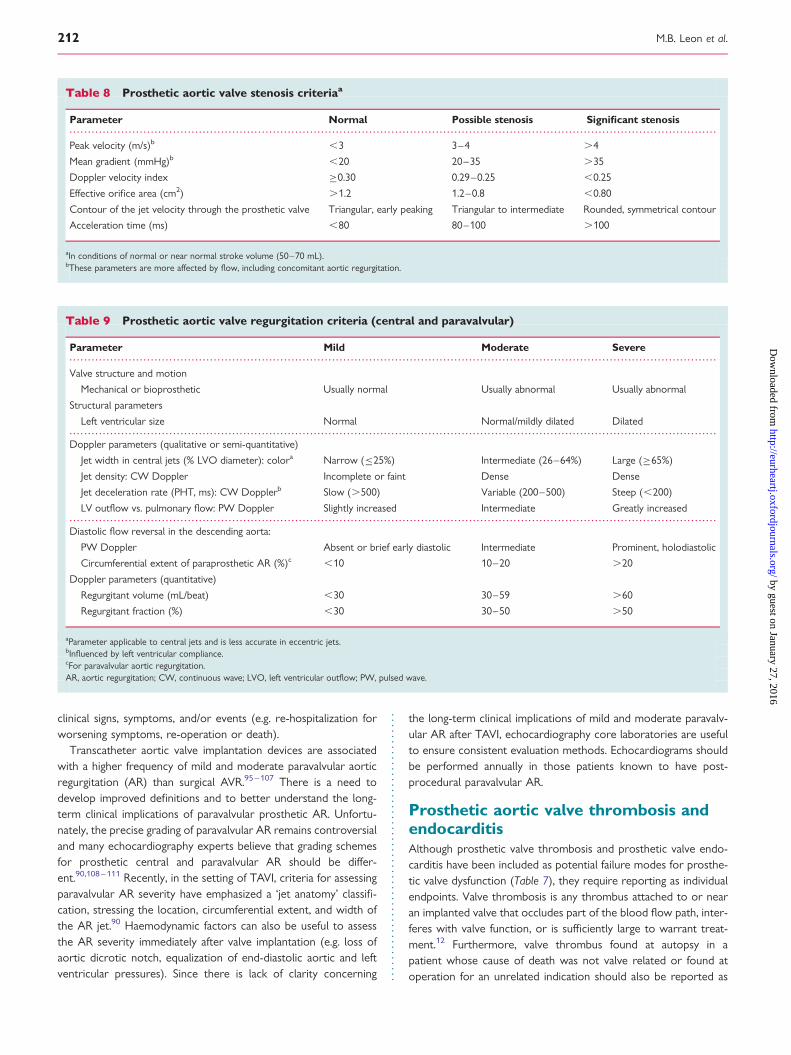
Prosthetic aortic stenosis andregurgitationUtilizing the recent prosthetic valve echocardiography guidelines,90
the severity of prosthetic aortic valve stenosis is graded as (i)normal, (ii) possible, or (iii) significant (Table 8) and prostheticaortic valve regurgitation (central or paravalvular) as (i) mild, (ii)moderate, or (iii) severe (Table 9). The clinical significance of pros-thetic valve dysfunction is further supported by the presence of
. . . . . . . . . . . . . . . . . . . . . . . . . . . . . . . . . . . . . . . . . . . . . . . . . . . . . . . . . . . . . . . . . . . . . . . . . . . . . . . .
Table 7 Potential failure modes of prosthetic valvedysfunction
Aortic stenosis
Stent creep
Pannus
Calcification
Support structure deformation (out-of-round configuration),under-expansion, fracture, or trauma (cardio-pulmonaryresuscitation, blunt chest trauma)
Mal-sizing (prosthesis-patient mismatch)
Endocarditis
Prosthetic valve thrombosis
Native leaflet prolapse impeding prosthetic leaflet motion
Aortic regurgitation
Pannus
Calcification
Support structure deformation (out-of-round configuration), recoil,under-expansion, fracture, insufficient radial strength, or trauma(cardio-pulmonary resuscitation, blunt chest trauma)
Endocarditis
Prosthetic valve thrombosis
Malposition (too high, too low)
Acute mal-coaptation
Leaflet wear, tear/perforation, prolapse, or retraction
Suture breakage or disruption
Native leaflet prolapse impeding prosthetic leaflet motion
. . . . . . . . . . . . . . . . . . . . . . . . . . . . . . . . . . . . . . . . . . . . . . . . . . . . . . . . . . . . . . . . . . . . . . . . . . . . . . . .
Table 6 Vascular access site and access-relatedcomplications
Major vascular complications
Any thoracic aortic dissection
Access site or access-related vascular injury (dissection, stenosis,perforation, rupture, arterio-venous fistula, pseudoaneurysm,haematoma, irreversible nerve injury, or compartmentsyndrome) leading to either death, need for significant bloodtransfusions (≥4 units), unplanned percutaneous or surgicalintervention, or irreversible end-organ damage (e.g. hypogastricartery occlusion causing visceral ischaemia or spinal artery injurycausing neurological impairment)
Distal embolization (non-cerebral) from a vascular source requiringsurgery or resulting in amputation or irreversible end-organdamage
Minor vascular complications
Access site or access-related vascular injury (dissection, stenosis,perforation, rupture, arterio-venous fistula or pseudoaneuysmsrequiring compression or thrombin injection therapy, orhaematomas requiring transfusion of ≥2 but ,4 units) notrequiring unplanned percutaneous or surgical intervention andnot resulting in irreversible end-organ damage
Distal embolization treated with embolectomy and/orthrombectomy and not resulting in amputation or irreversibleend-organ damage
Failure of percutaneous access site closure resulting ininterventional (e.g. stent-graft) or surgical correction and notassociated with death, need for significant blood transfusions(≥4 units), or irreversible end-organ damage
VARC consensus endpoints after TAVI for high risk AS 211
by guest on January 27, 2016http://eurheartj.oxfordjournals.org/
Dow
nloaded from
clinical signs, symptoms, and/or events (e.g. re-hospitalization forworsening symptoms, re-operation or death).
Transcatheter aortic valve implantation devices are associatedwith a higher frequency of mild and moderate paravalvular aorticregurgitation (AR) than surgical AVR.95– 107 There is a need todevelop improved definitions and to better understand the long-term clinical implications of paravalvular prosthetic AR. Unfortu-nately, the precise grading of paravalvular AR remains controversialand many echocardiography experts believe that grading schemesfor prosthetic central and paravalvular AR should be differ-ent.90,108 – 111 Recently, in the setting of TAVI, criteria for assessingparavalvular AR severity have emphasized a ‘jet anatomy’ classifi-cation, stressing the location, circumferential extent, and width ofthe AR jet.90 Haemodynamic factors can also be useful to assessthe AR severity immediately after valve implantation (e.g. loss ofaortic dicrotic notch, equalization of end-diastolic aortic and leftventricular pressures). Since there is lack of clarity concerning
the long-term clinical implications of mild and moderate paravalv-ular AR after TAVI, echocardiography core laboratories are usefulto ensure consistent evaluation methods. Echocardiograms shouldbe performed annually in those patients known to have post-procedural paravalvular AR.
Prosthetic aortic valve thrombosis andendocarditisAlthough prosthetic valve thrombosis and prosthetic valve endo-carditis have been included as potential failure modes for prosthe-tic valve dysfunction (Table 7), they require reporting as individualendpoints. Valve thrombosis is any thrombus attached to or nearan implanted valve that occludes part of the blood flow path, inter-feres with valve function, or is sufficiently large to warrant treat-ment.12 Furthermore, valve thrombus found at autopsy in apatient whose cause of death was not valve related or found atoperation for an unrelated indication should also be reported as
. . . . . . . . . . . . . . . . . . . . . . . . . . . . . . . . . . . . . . . . . . . . . . . . . . . . . . . . . . . . . . . . . . . . . . . . . . . . . . . . . . . . . . . . . . . . . . . . . . . . . . . . . . . . . . . . . . . . . . . . . . . . . . . . . . . . . . . . . . . . . . . . . . . . . . . . . . . . . . . . . . . . . . . . . . . . . . .
. . . . . . . . . . . . . . . . . . . . . . . . . . . . . . . . . . . . . . . . . . . . . . . . . . . . . . . . . . . . . . . . . . . . . . . . . . . . . . . . . . . . . . . . . . . . . . . . . . . . . . . . . . . . . . . . . . . . . . . . . . . . . . . . . . . . . . . . . . . . . . . . . . . . . . . . . . . . . . . . . . . . . . . . . . . . . . .
. . . . . . . . . . . . . . . . . . . . . . . . . . . . . . . . . . . . . . . . . . . . . . . . . . . . . . . . . . . . . . . . . . . . . . . . . . . . . . . . . . . . . . . . . . . . . . . . . . . . . . . . . . . . . . . . . . . . . . . . . . . . . . . . . . . . . . . . . . . . . . . . . . . . . . . . . . . . . . . . . . . . . . . . . . . . . . .
Table 9 Prosthetic aortic valve regurgitation criteria (central and paravalvular)
Parameter Mild Moderate Severe
Valve structure and motion
Mechanical or bioprosthetic Usually normal Usually abnormal Usually abnormal
Structural parameters
Left ventricular size Normal Normal/mildly dilated Dilated
Doppler parameters (qualitative or semi-quantitative)
Jet width in central jets (% LVO diameter): colora Narrow (≤25%) Intermediate (26–64%) Large (≥65%)
Jet density: CW Doppler Incomplete or faint Dense Dense
Jet deceleration rate (PHT, ms): CW Dopplerb Slow (.500) Variable (200–500) Steep (,200)
LV outflow vs. pulmonary flow: PW Doppler Slightly increased Intermediate Greatly increased
Diastolic flow reversal in the descending aorta:
PW Doppler Absent or brief early diastolic Intermediate Prominent, holodiastolic
Circumferential extent of paraprosthetic AR (%)c ,10 10–20 .20
Doppler parameters (quantitative)
Regurgitant volume (mL/beat) ,30 30–59 .60
Regurgitant fraction (%) ,30 30–50 .50
aParameter applicable to central jets and is less accurate in eccentric jets.bInfluenced by left ventricular compliance.cFor paravalvular aortic regurgitation.AR, aortic regurgitation; CW, continuous wave; LVO, left ventricular outflow; PW, pulsed wave.
. . . . . . . . . . . . . . . . . . . . . . . . . . . . . . . . . . . . . . . . . . . . . . . . . . . . . . . . . . . . . . . . . . . . . . . . . . . . . . . . . . . . . . . . . . . . . . . . . . . . . . . . . . . . . . . . . . . . . . . . . . . . . . . . . . . . . . . . . . . . . . . . . . . . . . . . . . . . . . . . . . . . . . . . . . . . . . .
Table 8 Prosthetic aortic valve stenosis criteriaa
Parameter Normal Possible stenosis Significant stenosis
Peak velocity (m/s)b ,3 3–4 .4
Mean gradient (mmHg)b ,20 20–35 .35
Doppler velocity index ≥0.30 0.29–0.25 ,0.25
Effective orifice area (cm2) .1.2 1.2–0.8 ,0.80
Contour of the jet velocity through the prosthetic valve Triangular, early peaking Triangular to intermediate Rounded, symmetrical contour
Acceleration time (ms) ,80 80–100 .100
aIn conditions of normal or near normal stroke volume (50–70 mL).bThese parameters are more affected by flow, including concomitant aortic regurgitation.
M.B. Leon et al.212
by guest on January 27, 2016http://eurheartj.oxfordjournals.org/
Dow
nloaded from

valve thrombosis. The diagnosis of prosthetic valve thrombosis isbest discerned during an echocardiographical examination orduring surgical exploration. There have already been casereports and anecdotes112 of transcatheter prosthetic valve throm-bosis with and without important clinical consequences.
The diagnosis of prosthetic valve endocarditis is based on one ofthe following criteria12:
† reoperation with evidence of abscess, paravalvular leak, pus, orvegetation confirmed as secondary to infection by histologicalor bacteriological studies;
† autopsy findings of abscess, pus, or vegetation involving arepaired or replaced valve;
† in the absence of reoperation or autopsy, fulfiling the Duke Cri-teria for endocarditis.113
Isolated case reports of transcatheter aortic valve endocarditis havealready been published.114,115 Owing to the variability in transcathetervalve designs and positioning within the aortic root, meticulousreporting of the pattern of endocarditis is mandatory.116
Prosthetic valve ‘associated’complicationsDepending on the design characteristics and final implant position,prosthetic aortic valves may come in close contact with the anteriormitral valve leaflet, the intervalvular fibrosa, the aortic annulus, theventricular septum, the aortic sinuses and root, the coronaryarteries, and the cardiac conduction system. Collectively, these ana-tomic structures, which are contiguous with the prosthetic aorticvalve, are referred to as the aortic valvar complex (Figure 2). Assuch, prosthetic aortic valve procedures, and in particular TAVI,may have untoward effects on any of these structures which mayresult in important clinical consequences. Therefore, VARC
proposes to group these complications as a separate endpoint cat-egory. However, it must be noted, that some of these adverseevents may not be directly related to The Valve prosthesis itself,but may occur before or after valve implantation (e.g. conductiondisturbances after pre-implant balloon aortic valvuloplasty).
Conduction disturbances and cardiacarrhythmiasThe close anatomical relationship between the aortic valvarcomplex and the branching atrioventricular bundle explains thepossible development of conduction abnormalities following pros-thetic aortic valve procedures.117,118 Following surgical AVR, new-onset bundle branch block has been reported in 16–32% ofpatients and the need for permanent pacemakers in 3–8% ofpatients.119 –123 In early experiences with TAVI, new-onsetbundle branch block has occurred in up to 45% of patients andthe need for permanent pacemakers has varied from as low as4% to as high as 33%.95– 98,124– 128 Differences among devicesand heterogeneity in physician and country-based healthcarethresholds may explain the significant inter-hospital variability innew permanent pacemaker requirements after TAVI.
Although the implications of persistent left bundle branch block(LBBB) after TAVI are currently unknown, the presence of newbundle branch block after surgical AVR has been associatedwith increased risk of subsequent arrhythmic events duringfollow-up (specifically, syncope, AV dissociation, and suddendeath).119,120 Owing to this association between conductionsystem abnormalities and adverse patient outcomes following sur-gical AVR and several anecdotal reports after TAVI of either earlypost-discharge severe bradyarrhythmic events or sudden cardiacdeath,15 VARC recommends to carefully document the occur-rence of new conduction system abnormalities (left bundlebranch block and third degree atrioventricular block), as well asthe requirements and indications for new permanent pacemakerswithin 30 days after the procedure. The timing (days) and location(intra-procedural, in-hospital, or post-discharge) of the eventshould also be recorded. To accurately capture such events,daily ECGs and continuous telemetry ECG monitoring shouldbe considered while the patients are in-hospital, and should berequired in patients with any evidence of new conductionabnormalities or arrhythmias.
Although conduction abnormalities associated with TAVI havebeen a recent concern, it bears noting that new onset atrial fibrilla-tion and ventricular arrhythmias have also been observed afterboth TAVI and surgical AVR procedures.128 In particular, newonset atrial fibrillation occurs in as many as 20–30% of patientsafter conventional surgical AVR129,130 and any valid comparisonof transcatheter vs. surgical aortic valve treatment strategiesshould include a careful analysis of post-therapy supra-ventricularand ventricular arrhythmias.
Coronary obstructionMechanical coronary artery obstruction following TAVI or surgicalAVR is rare and occurs in ,1% of patients.96,97 The obstructiontypically occurs during the index procedure. Importantly, clinicalsigns and symptoms may be subtle and not appreciated until
Figure 2 The aortic valvar complex, including the aortic valve,annulus, sinuses, aorta, coronary arteries, membranous septum,and the mitral valve.
VARC consensus endpoints after TAVI for high risk AS 213
by guest on January 27, 2016http://eurheartj.oxfordjournals.org/
Dow
nloaded from

after the procedure. Possible mechanisms for mechanical coronaryobstruction include (i) impingement of the coronary ostia by TheValve support structure in the setting of suboptimal valve position-ing and/or ‘small aortic root’ anatomy; (ii) embolization fromcalcium, thrombus, air, or endocarditis; (iii) displacement ofnative aortic valve leaflets towards the coronary ostia duringTAVI,131,132 and (iv) suture-related kinking or obstruction orcannulation-related obstruction of the coronary ostia associatedwith surgical AVR.
The diagnosis of TAVI-associated coronary obstruction can bedetermined by imaging studies (coronary angiography, intravascularultrasound, multi-slice CT angiography, or echocardiography), sur-gical exploration, or autopsy findings. Cardiac biomarker elevationsand ECG changes indicating new ischaemia provide corroborativeevidence.
Other prosthesis-related adverse eventsThe short- and long-term consequences of contact, trauma, orimpingement on the anterior mitral valve leaflet by the ventricularend of a transcatheter aortic valve are currently unknown. Never-theless, any new mitral valve dysfunction (e.g. worsening mitralregurgitation or stenosis) or disruption (e.g. chordal rupture,leaflet perforation, anterior mitral valve leaflet aneurysm) relatedto contact with the transcatheter valve implant or mitral valveendocarditis114– 116 should be carefully documented. Other infre-quent complications following TAVI include new ventricularseptal defects and aortic root rupture/perforation/dissection,occurring either during the pre-implant balloon aortic valvulo-plasty, or during the transcatheter valve implant.133,134
Clinical benefit endpointsIn addition to the avoidance of mortality, specific endpoints toestablish the clinical benefit after TAVI are important. Objectivebenefit parameters derived from the heart failure literature canbe adapted in valve-related clinical trials.135 Several choices areavailable, including exercise performance,136 assessment ofNew York Heart Association (NYHA) functional status,137 andvarious quality of life138 and frailty questionnaires.139 Each ofthese symptom evaluation tools has strengths and weaknesses inthe TAVI patient population, which is disproportionately rep-resented by elderly, frail, individuals with multiple co-morbidities.For instance, exercise test performance is an appealing endpoint,but as aortic valve therapy studies are unblinded, they may bebiased and they can be difficult to perform in high-risk TAVIpatients.
Valve Academic Research Consortium has also considered a cat-egorical endpoint of clinical benefit which captures failure ofcurrent AS therapy; hospitalization for symptoms of cardiac orvalve-related decompensation, at least 30 days after the index pro-cedure (surgical AVR or TAVI). This endpoint mandates carefuladjudication by a clinical events committee and is defined as hos-pitalization for symptoms of valve or cardiac deterioration (e.g.new or worsening heart failure, angina, or syncope) requiringeither a valve procedure (surgery or interventional treatment) orintensification of medical management (new or increased use ofinotropes, vasopressors, diuretics, and/or vasodilators).
Quality-of-life and healthcare economic instruments can beuseful to assess disability and impairment due to congestiveheart failure (e.g. Kansas City Cardiomyopathy Questionnaire)140
and for mapping health status compared with population-levelutility weights (e.g. EuroQOL questionnaire).141 –143 However,quality-of-life questionnaires are also prone to bias and must beuniformly administered. The time points for assessment of theaforementioned clinical benefit endpoints should be at 30 days,at 6 months, and at 1 year after initiating therapy. Valve AcademicResearch Consortium recommends that if any measure of clinicalbenefit is utilized in clinical trials, there must be careful oversightand adjudication by experienced clinical events committees.
The assessment of ‘frailty’ in patients with advanced valvularheart disease has become increasingly important and is usuallynot included in surgical-risk algorithms. Frailty is loosely definedas a biological syndrome of decreased reserve and resistance tostressors, resulting from cumulative declines across multiple phys-iological systems, and causing vulnerability to adverse outcomes.139
Various frailty indices have been developed and have been corre-lated with worsening clinical outcomes in geriatric patients inintensive care units and after surgery.144 –146 In general, the evalu-ation of frailty demands a composite analysis of several categoricaland continuous variables including mobility, strength, endurance,activities of daily living, cognitive impairment, and nutritionalstatus (as discerned by body mass index and biomarkers such asserum albumin).147,148 Although there is no standard frailty indexwhich has been applied and validated in high-risk AS patients, mul-tiple preliminary efforts are ongoing and VARC proposes toinclude measures of frailty in future clinical trials as a componentof clinical benefit endpoints.
Therapy-specific endpointsGiven the complex nature of TAVI procedures and the rapid evol-ution of devices and procedural techniques, VARC proposes torecord in case report forms (but not as formal endpoints) anopen category of therapy-specific endpoints which may be relevantto clinical outcomes or device performance. Examples of suchevents include the following: (i) the unplanned use of cardio-pulmonary bypass to manage haemodynamic compromise or toreverse procedural complications; (ii) conversion from a ‘failed’percutaneous transcatheter procedure to an ‘open’ surgical AVRor to a surgical-access TAVI149,150; (iii) ventricular perforation(for any reason) with and without cardiac tamponade151; (iv) pros-thetic valve migration or dislocation from the native aortic valvelanding zone152,153; (v) frequency, reasons, and results of post-TAVIballoon dilation; (vi) frequency, reasons, and results after place-ment of a second valve over the original valve, so-called TAVI‘valve-in-valve’154,155; (vii) integrity of the support structure, includ-ing strut fractures, compression or other evidence of geometry dis-tortion (requires careful serial imaging modalities includingcine-fluoroscopic analyses and echocardiography)90,156 –158; (viii)instances of device recapture (with or without repositioning), orretrieval (removal from the body) which occur during the indexprocedure; (ix) re-intervention (either percutaneous or surgical)for any reason after the index procedure.159 As appropriate, thetiming of these events (during the index procedure, in-hospital,
M.B. Leon et al.214
by guest on January 27, 2016http://eurheartj.oxfordjournals.org/
Dow
nloaded from

or post-discharge) should be carefully recorded. This category isintended to be a dynamic platform and should be added to thecase report forms.
Clinically relevant compositeendpointsAlthough VARC discourages the overuse of composite endpoints,to achieve overall impressions of safety and effectiveness mayrequire the incorporation of more than single endpoints. Thesestrategic assessments of TAVI as an alternative therapy shouldideally include device, procedure, and patient-oriented factors.Valve Academic Research Consortium proposes three compositeendpoints (Table 10): device success (intra-procedure), a combinedsafety endpoint (at 30 days), and a combined efficacy endpoint (at1 year or longer).
Device success is a ‘technical’ composite endpoint meant tocharacterize the acute device and procedural factors whichunderlie vascular access, delivery, and performance of the TAVIsystem. Echocardiography should be routinely utilized as the stan-dard for measuring prosthetic valve stenosis and regurgitationimmediately after TAVI, and should always be performed in aresting state, either within 24–48 h after the index procedure orbefore hospital discharge.
The 30-day combined safety endpoint is a hierarchical compositeof the most relevant patient-oriented safety endpoints previouslydefined by VARC (Table 10). In addition, a repeat procedure in
the first 30 days (either surgery or intervention) to treatvalve-related dysfunction is also incorporated in this endpoint.Examples of urgent repeat procedures would include balloonaortic valvuloplasty or repeat TAVI (valve-in-valve) to treateither paravalvular or central severe AR after the TAVI. Thefocus on 30-day events after the index procedure is meant toisolate safety concerns largely pertaining to early device perform-ance and the procedure. Nonetheless, overall patient safety alsorequires a careful examination of pertinent individual safety end-points over the life history of the device.
The time-sensitive assessment of TAVI effectiveness requires amore delayed combined efficacy endpoint incorporating major clini-cal and valve performance factors. Valve Academic Research Con-sortium proposes a 1-year (or longer) time interval for thecombined efficacy endpoint integrating three important endpoints:(i) all-cause mortality after 30 days, meant to reflect therapy effec-tiveness by measuring prevention of AS-related mortality overtime; (ii) failure of the current therapy for AS, requiring hospitaliz-ation for symptoms of valve-related or cardiac decompensation(adjudicated episodes of heart failure, angina, or syncope requiringan aortic valve procedure or intensification of medical manage-ment); (iii) evidence of prosthetic valve dysfunction, definedusing strict echocardiography criteria, possibly in conjunctionwith other signs of functional deterioration.
DiscussionThe VARC was convened in response to an urgent call for standar-dized clinical research processes involving the emerging field oftranscatheter valve therapies, and more specifically, TAVI inhigh-surgical-risk patients with AS. The inter-disciplinary natureof TAVI, combining aspects of both surgical and interventionaltherapies, presented special challenges and required an enlightenedand collaborative approach to the development of clinical researchrecommendations and endpoint definitions.160,161 The VARCinitiative is an attempt to achieve a necessary consensus amongthe various subspecialties and stakeholders, such that this innova-tive treatment strategy may be evaluated objectively and accordingto a set of practical endpoint definitions.
This consensus manuscript is not intended to be interpreted asa ‘guidelines’ or ‘guidance’ document and although thoroughlyreviewed by individuals from seven cardiology and cardiac surgerysocieties, the content has not been subjected to a formal societyguidelines review process. These standardized endpoints are mea-sureable, apply to both predicate surgical and new transcathetertherapies, can be adjudicated by clinical events committees, andcan be used to compare findings from different clinical trials. Byintent, this consensus manuscript was not device-specific and thedefinitions can be applied to next generation and iterative TAVIdevices already under early stages of clinical investigation.162–165
Given the rapid growth in transcatheter valve therapies, and thepotential exposure of this technology to lower risk patient popu-lations, it is certain that this preliminary attempt to arrive at con-sensus endpoint definitions for TAVI will need refinement in thefuture. In principle, the consensus process calls for the higheststandards of clinical research, including (i) inter-disciplinaryexperts gathering to arrive at standardized endpoint definitions,
. . . . . . . . . . . . . . . . . . . . . . . . . . . . . . . . . . . . . . . . . . . . . . . . . . . . . . . . . . . . . . . . . . . . . . . . . . . . . . . .
. . . . . . . . . . . . . . . . . . . . . . . . . . . . . . . . . . . . . . . . . . . . . . . . . . . . . . . . . . . . . . . . . . . . . . . . . . . . . . . .
Table 10 Composite endpoints
Device success
Successful vascular access, delivery and deployment of the deviceand successful retrieval of the delivery system
Correct position of the device in the proper anatomical location
Intended performance of the prosthetic heart valve (aortic valvearea .1.2 cm2 and mean aortic valve gradient ,20 mmHg orpeak velocity ,3 m/s, without moderate or severe prostheticvalve AR)
Only one valve implanted in the proper anatomical location
Combined safety endpoint (at 30 days)
All-cause mortality
Major stroke
Life-threatening (or disabling) bleeding
Acute kidney injury—Stage 3 (including renal replacement therapy)
Peri-procedural MI
Major vascular complication
Repeat procedure for valve-related dysfunction (surgical orinterventional therapy)
Combined efficacy endpoint (at 1 year or longer)
All-cause mortality (after 30 days)
Failure of current therapy for AS, requiring hospitalization forsymptoms of valve-related or cardiac decompensation
Prosthetic heart valve dysfunction (aortic valve area ,1.2 cm2 andmean aortic valve gradient ≥20 mmHg or peak velocity ≥3 m/s,OR moderate or severe prosthetic valve AR)
VARC consensus endpoints after TAVI for high risk AS 215
by guest on January 27, 2016http://eurheartj.oxfordjournals.org/
Dow
nloaded from

(ii) harmonized and well-structured data collection, interpretation,and reporting for specific TAVI-related clinical events, and (iii) theconsistent use of central core laboratories and independent,blinded endpoint adjudication.
Many of the endpoints discussed in this manuscript are suffi-ciently general that they can be applied to other AS populationsand to other valvular heart disease clinical research scenarios,both surgical and interventional. This is particularly germane toTAVI clinical research, as new studies involving lower risk ASpatients are already being considered. Importantly, recentreports and randomized trials using new catheter-based mitralvalve therapies to treat mitral regurgitation166 – 168 also sufferfrom non-standardized endpoint definitions and might wellbenefit from a comparable VARC consensus effort.
This consensus manuscript, which represents the ‘first step’ in amuch longer road to help improve clinical research in valvularheart disease, has several limitations. The endpoint definitionswere intended to be reasonably broad, but nonetheless in someinstances are also intentionally narrow to address the specific con-siderations of TAVI in high-surgical-risk patients with severe AS.Therefore, application of all of these endpoint definitions toother patient populations may be problematic. The importantarea of pre-clinical device testing, both assessments of valve andsupport structure properties and in vivo animal studies, is beyondthe scope of this manuscript. Other aspects of clinical trialdesign and clinical trial methodologies are also essential to opti-mize clinical research, but similarly, a comprehensive treatmentof these subjects could not be included in this manuscript.Finally, many global endpoints, such as stroke and bleeding andsome specific endpoints, such as paravalvular regurgitation, arethemselves in a state of evolution, subject to modifications byother consensus committees in the near future.
The VARC process embodied in this manuscript was an ambi-tious multi-disciplinary attempt to bring order through consensus,thereby providing standardization of clinical research in the bur-geoning area of transcatheter aortic valve therapy. Hopefully, thistemplate can also serve as a model to improve clinical researchmethodologies in the evaluation of new therapies for other cardio-vascular diseases.
FundingGrants were provided to the ARC Board including representatives ofThe Cardiovascular Research Foundation, Cardialysis, Duke ClinicalResearch Institute and Harvard Clinical Research Institute to coverthe costs of travel, meeting rooms, and lodging for academic attendeesat the San Francisco and Amsterdam meetings by Edwards Lifesciencesand Medtronic Corporation. All funds not utilized for the aforemen-tioned travel-related purposes have been returned to the sponsors.Funding to pay the Open Access publication charges for this articlewas provided by Cardialysis BV on behalf of the Valve AcademicResearch Consortium.
Conflict of interest: VARC Participants will provide Conflict ofInterest Disclosures individually prior to publication. The VARC meet-ings involved members of the Interventional Cardiology DevicesBranch, of the Office of Device Evaluation, Center for Devices andRadiological Health, USFDA. The opinions or assertions herein arethe private views of the authors and are not to be construed as reflect-ing the views of the FDA.
Appendix 1
Valve Academic ResearchConsortium Contributing Groups(1) Academic Research Organizations
Cardialysis (Rotterdam, the Netherlands)Cardiovascular Research Foundation (New York, NY,
USA)Duke Clinical Research Institute (Durham, NC, USA)Harvard Clinical Research Institute (Boston, MA,
USA)(2) Societies
American Association for Thoracic SurgeryAmerican College of CardiologyAmerican Heart AssociationEuropean Association for CardioThoracic SurgeryEuropean Society of CardiologySociety of Cardiac Angiography and InterventionSociety of Thoracic Surgeons.
(3) US Food and Drug Administration(4) Industry Representatives
Appendix 2
Valve Academic ResearchConsortium participants(1) Clinical Research Organizations
(1) Cardialysis/Erasmus MC—Rotterdam, the Netherlands
Morel M.A.Piazza, N.Serruys, P.W.Van Es, G.A.Van Mieghem, N.Vranckx, P.
(2) Cardiovascular Research Foundation—New York City,NY, USA
Caixeta, A.Dalton, K.Haratani, N.Kirtani, A.Kodali, S.Lansky, A.Leon, M.B.Mehran, R.Nikolsky, E.Williams, M.
(3) Duke Clinical Research Institute—Durham, NC, USA
Krucoff, M.W.Petersen, J.
(4) Harvard Cardiovascular Research Institute—Boston, MA,USA
Cutlip, D.E.Pinto, D.
M.B. Leon et al.216
by guest on January 27, 2016http://eurheartj.oxfordjournals.org/
Dow
nloaded from
(2) Physician Society Representatives and Experts
Surgeons
Adams, D.—Mt. Sinai Medical Center, New York City,USAKappetein, A.P.—Erasmus MC, Rotterdam, theNetherlandsMack, M.—Medical City Hospital, Dallas, TX, USAMiller, C.—Stanford University, CA, USAMohr, F.—Herzzentrum Universitat Leipzig, Leipzig,GermanyNataf, P.—CHU Bichat, Paris, FranceSmith, C.—Columbia University Medical Center,New York City, NY, USAVerrier, E.—University of Washington, Seattle, WA, USA
Cardiologists
Bailey, S.—University of Texas, San Antonio, TX, USABonan, R.—Montreal Heart Institute, Montreal, CanadaBonhoeffer, P.—Great Ormond Street Hospital for Chil-dren, London, UKBonow, R.—Northwestern University, Chicago, IL, USADouglas, P.—Duke University, Durham, NC, USAGillam, L.—Columbia University Medical Center,New York City, NY, USAIung, B.—CHU Bichat, Paris, FrancePopma, J.J.—Beth Israel—Deaconess Medical Center,Boston, MA, USAThomas, M.—Guys and St Thomas Hospital, London, UKTuzcu, M.—Cleveland Clinic Foundation, Cleveland, OH,USAVahanian, A.—CHU Bichat, Paris, FranceVirmani, R.—CV Pathology Institute, Gaithersburg, MD,USAWebb, J.G.—St Paul’s Hospital, Vancouver, BC, CanadaWindecker, S.—University Hospital Bern, Bern,SwitzerlandBiostatistics/Clinical Epidemiology
Blackstone, E.—Cleveland Clinic Foundation, Cleveland,OH, USAPocock, S.—London School of Hygiene and TropicalMedicine, London, UKTakkenberg, J.J.M.—Erasmus MC, Rotterdam, theNetherlandsUS Food and Drug Administration
Hillebrenner, M.Swain, J.Zuckerman, B.Medicines and Healthcare products Regulatory Agency
(MHRA)
Ludgate, S.Industry Representatives
Akin, J.—Edwards Lifesciences, Orange, CA, USAArmitage, T.—Medtronic, Minneapolis, MN, USAMartin, K.—Sadra Medical, Los Gatos, CA, USASheahan, B.—Direct Flow Medical, Santa Rosa, CA, USASimonton, C.—Abbott Vascular, Santa Clara, CA, USA
Wilson, R.—Heart Leaflet Technologies, Maple Grove,MN, USA
Appendix 3
Minimum data collection andendpoint requirements after TAVI† Mortality (all cause and cardiovascular)† Myocardial Infarction (peri-procedural and spontaneous)† Stroke (major and minor)† Bleeding (life threatening or disabling and major)† Acute kidney injury (modified RIFLE stage 2 and 3, including
RRT)† Vascular complications (major)† Prosthetic valve performance (requires serial echocardiography
assessments)
(a) Prosthetic valve stenosis (possible and significant) andregurgitation (moderate or severe with special referenceto paravalvular regurgitation)
(b) Prosthetic valve thrombosis(c) Prosthetic valve endocarditis
† Prosthetic valve-associated complications
(a) Conduction disturbances and cardiac arrhythmias (includingnew LBBB, new permanent pacemaker implantation, andnew supra-ventricular or ventricular arrhythmias) and
(b) Coronary obstruction† Clinical benefit endpoints
(a) Symptom status (global assessments using NHYA classifi-cation and some measure of quality of life)
(b) Repeat hospitalization (.30 days after the index pro-cedure) for valve-related or cardiac decompensation)
† Therapy specific endpoints (ventricular perforation at any timeresulting in cardiac tamponade, prosthetic valve embolization,and acute or delayed valve-in-valve treatment)
† Composite endpoints
(a) Device success(b) Combined safety endpoint (at 30 days)(c) Combined efficacy endpoint (at 1 year or longer)
References1. Cribier A, Eltchaninoff H, Bash A, Borenstein N, Tron C, Bauer F, Derumeaux G,
Anselme F, Laborde F, Leon MB. Percutaneous transcatheter implantation of anaortic valve prosthesis for calcific aortic stenosis: first human case description.Circulation 2002;106:3006–3008.
2. Cribier A, Eltchaninoff H, Tron C, Bauer F, Agatiello C, Sebagh L, Bash A,Nusimovici D, Litzler PY, Bessou JP, Leon MB. Early experience with percuta-neous transcatheter implantation of heart valve prosthesis for the treatmentof end-stage inoperable patients with calcific aortic stenosis. J Am Coll Cardiol2004;43:698–703.
3. Iung B, Baron G, Butchart EG, Delahaye F, Gohlke-Barwolf C, Levang OW,Tornos P, Vanoverschelde JL, Vermeer F, Boersma E, Ravaud P, Vahanian A. Aprospective survey of patients with valvular heart disease in Europe: The EuroHeart Survey on Valvular Heart Disease. Eur Heart J 2003;24:1231–1243.
4. Stuge O, Liddicoat J. Emerging opportunities for cardiac surgeons within struc-tural heart disease. J Thorac Cardiovasc Surg 2006;132:1258–1261.
5. Varadarajan P, Kapoor N, Bansal RC, Pai RG. Clinical profile and natural historyof 453 nonsurgically managed patients with severe aortic stenosis. Ann ThoracSurg 2006;82:2111–2115.
6. Bramstedt KA. Aortic valve replacement in the elderly: frequently indicated yetfrequently denied. Gerontology 2003;49:46–49.
VARC consensus endpoints after TAVI for high risk AS 217
by guest on January 27, 2016http://eurheartj.oxfordjournals.org/
Dow
nloaded from

7. Iung B, Cachier A, Baron G, Messika-Zeitoun D, Delahaye F, Tornos P,Gohlke-Barwolf C, Boersma E, Ravaud P, Vahanian A. Decision-making inelderly patients with severe aortic stenosis: why are so many denied surgery?Eur Heart J 2005;26:2714–2720.
8. Piazza N, Yoshinobu O, de Jaegere P, Serruys PW. Guidelines for reportingcardiac mortality and morbidity after cardiac valve interventions—need for areappraisal? Ann Thorac Surg 2009; 87:357–358.
9. Akins CW, Blackstone EH, Miller C, Kouchoukos NT, Turina MI. Guidelines forreporting cardiac mortality and morbidity after cardiac valve interventions—need for a reappraisal? (Response) Ann Thorac Surg 2009;87:359–360.
10. Thomas M, Wendler O. Transcatheter aortic valve implantation (TAVI): how tointerpret the data and what data is required? EuroIntervention 2008;5:25–27.
11. Piazza N, Cutlip DE, Yoshinobu O, Kappetein AP, de Jaegere P, Serruys PW.Clinical endpoints in transcatheter aortic valve implantation: a call to ARC forstandardised definitons. EuroIntervention 2008;5:28–29.
12. Akins CW, Miller DC, Turina MI et al. Guidelines for reporting mortality andmorbidity after cardiac valve interventions. J Thorac Cardiovasc Surg 2008;135:732–738.
13. Thygesen K, Alpert JS, White HD, Jaffe AS, Apple FS, Galvani M, Katus HA,Newby LK, Ravkilde J, Chaitman B, Clemmensen PM, Dellborg M, Hod H,Porela P, Underwood R, Bax JJ, Beller GA, Bonow R, Van der Wall EE,Bassand JP, Wijns W, Ferguson TB, Steg PG, Uretsky BF, Williams DO,Armstrong PW, Antman EM, Fox KA, Hamm CW, Ohman EM, Simoons ML,Poole-Wilson PA, Gurfinkel EP, Lopez-Sendon JL, Pais P, Mendis S, Zhu JR,Wallentin LC, Fernandez-Aviles F, Fox KM, Parkhomenko AN, Priori SG,Tendera M, Voipio-Pulkki LM, Vahanian A, Camm AJ, De Caterina R, Dean V,Dickstein K, Filippatos G, Funck-Brentano C, Hellemans I, Kristensen SD,McGregor K, Sechtem U, Silber S, Widimsky P, Zamorano JL, Morais J,Brener S, Harrington R, Morrow D, Lim M, Martinez-Rios MA, Steinhubl S,Levine GN, Gibler WB, Goff D, Tubaro M, Dudek D, Al-Attar N. Universal defi-nition of myocardial infarction. J Am Coll Cardiol 2007;50:2173–2195.
14. Cutlip DE, Windecker S, Mehran R, Boam A, Cohen DJ, van Es GA, Steg PG,Morel MA, Mauri L, Vranckx P, McFadden E, Lansky A, Hamon M,Krucoff MW, Serruys PW. Clinical end points in coronary stent trials: a casefor standardized definitions. Circulation 2007;115:2344–2351.
15. Antonini-Canterin F, Leiballi E, Capanna M, Burelli C, Cassin M, Macor F,Grandis U, Nicolosi GL. Association between carotid and coronary arterydisease in patients with aortic valve stenosis: an angiographic study. Angiology2009;60:596–600.
16. Kahlert P, Knipp SC, Schlamann M, Theilmann M, Al-Rashid F, Weber M,Johansson U, Wendt D, Jakob HG, Forsting M, Sack S, Erbel R, Eggebrecht H.Silent and apparent cerebral ischemia after percutaneous transfemoral aorticvalve implantation: a diffusion-weighted magnetic resonance imaging study. Circu-lation 2010;121:870–878.
17. Ghanem A, Muller A, Nahle CP, Kocurek J, Werner N, Hammerstingl C,Schild HH, Schwab JO, Mellert F, Fimmers R, Nickenig G, Thomas D. Risk andfate of cerebral embolism after transfemoral aortic valve implantation: a pro-spective pilot study with diffusion-weighted magnetic resonance imaging. J AmColl Cardiol 2010;55:1427–1432.
18. Easton JD, Saver JL, Albers GW, Alberts MJ, Chaturvedi S, Feldmann E,Hatsukami TS, Higashida RT, Johnston SC, Kidwell CS, Lutsep HL, Miller E,Sacco RL. Definition and evaluation of transient ischemic attack: a scientificstatement for healthcare professionals from the American Heart Association/American Stroke Association Stroke Council; Council on CardiovascularSurgery and Anesthesia; Council on Cardiovascular Radiology and Intervention;Council on Cardiovascular Nursing; and the Interdisciplinary Council on Periph-eral Vascular Disease. The American Academy of Neurology affirms the value ofthis statement as an educational tool for neurologists. Stroke 2009;40:2276–2293.
19. Saver JL. Proposal for a universal definition of cerebral infarction. Stroke 2008;39:3110–3115.
20. Ong AT, Serruys PW, Mohr FW, Morice MC, Kappetein AP, Holmes DR Jr,Mack MJ, van den Brand M, Morel MA, van Es GA, Kleijne J, Koglin J,Russell ME. The SYNergy between percutaneous coronary intervention withTAXus and cardiac surgery (SYNTAX) study: design, rationale, and run-inphase. Am Heart J 2006;151:1194–1204.
21. Farkouh ME, Dangas G, Leon MB, Smith C, Nesto R, Buse JB, Cohen DJ,Mahoney E, Sleeper L, King S 3rd, Domanski M, McKinlay S, Fuster V. Designof the Future REvascularization Evaluation in patients with Diabetes mellitus:Optimal management of Multivessel disease (FREEDOM) trial. Am Heart J2008;155:215–223.
22. Gurm HS, Yadav JS, Fayad P, Katzen BT, Mishkel GJ, Bajwa TK, Ansel G,Strickman NE, Wang H, Cohen SA, Massaro JM, Cutlip DE. Long-term resultsof carotid stenting versus endarterectomy in high-risk patients. N Engl J Med2008;358:1572–1579.
23. Ringleb PA, Allenberg J, Bruckmann H, Eckstein HH, Fraedrich G, Hartmann M,Hennerici M, Jansen O, Klein G, Kunze A, Marx P, Niederkorn K, Schmiedt W,Solymosi L, Stingele R, Zeumer H, Hacke W. 30 day results from the SPACE trialof stent-protected angioplasty versus carotid endarterectomy in symptomaticpatients: a randomised non-inferiority trial. Lancet 2006;368:1239–1247.
24. Hobson RW II, Howard VJ, Roubin GS, Brott TG, Ferguson RD, Popma JJ,Graham DL, Howard G. Carotid artery stenting is associated with increasedcomplications in octogenarians: 30-day stroke and death rates in the CRESTlead-in phase. J Vasc Surg 2004;40:1106–1111.
25. Maggioni AP, Franzosi MG, Santoro E, White H, Van de Werf F, Tognoni G. Therisk of stroke in patients with acute myocardial infarction after thrombolytic andantithrombotic treatment. Gruppo Italiano per lo Studio della Sopravvivenzanell’Infarto Miocardico II (GISSI-2), and The International Study Group. N EnglJ Med 1992;327:1–6.
26. ISIS-3 (Third International Study of Infarct Survival) Collaborative Group. ISIS-3:a randomised comparison of streptokinase vs tissue plasminogen activator vsanistreplase and of aspirin plus heparin vs aspirin alone among 41,299 cases ofsuspected acute myocardial infarction. Lancet 1992;339:753–770.
27. Gore JM, Granger CB, Simoons ML, Sloan MA, Weaver WD, White HD,Barbash GI, Van de Werf F, Aylward PE, Topol EJ. Stroke after thrombolysis.Mortality and functional outcomes in the GUSTO-I trial. Global use of strategiesto open occluded coronary arteries. Circulation 1995;92:2811–2818.
28. Halperin JL. Ximelagatran compared with warfarin for prevention of throm-boembolism in patients with nonvalvular atrial fibrillation: Rationale, objectives,and design of a pair of clinical studies and baseline patient characteristics(SPORTIF III and V). Am Heart J 2003;146:431–438.
29. Ezekowitz MD, Connolly S, Parekh A, Reilly PA, Varrone J, Wang S, Oldgren J,Themeles E, Wallentin L, Yusuf S. Rationale and design of RE-LY: randomizedevaluation of long-term anticoagulant therapy, warfarin, compared with dabiga-tran. Am Heart J 2009;157:805–810, 810 e1–2.
30. Totaro P, Toni D, Durastanti L, Bozzao L, Gualdi GF, Raz E, Kouleridou A,Pantano P. Diffusion-weighted MRI in patients with non-diagnostic CT in thepost-acute phase of cerebral ischemia. Eur Neurol 2010;63:94–100.
31. Brott T, Adams HP Jr, Olinger CP, Adams HP Jr, Tomsick T, Barsan WG, Biller J,Eberle R, Hertzberg V, Walker M. Measurements of acute cerebral infarction: aclinical examination scale. Stroke 1989;20:864–870.
32. Bonita R, Beaglehole R. Modification of Rankin Scale: recovery of motor functionafter stroke. Stroke 1988;19:1497–1500.
33. Goldstein LB, Samsa GP. Reliability of the National Institutes of Health StrokeScale. Extension to non-neurologists in the context of a clinical trial. Stroke1997;28:307–310.
34. Lyden PD, Lau GT. A critical appraisal of stroke evaluation and rating scales.Stroke 1991;22:1345–1352.
35. Standardized Definitions for Cardiovascular Outcomes Trials: FDA Draft Rec-ommendations (personal communication; 24 March 2010).
36. Manoukian SV, Voeltz MD, Eikelboom J. Bleeding complications in acute coron-ary syndromes and percutaneous coronary intervention: predictors, prognosticsignificance, and paradigms for reducing risk. Clin Cardiol 2007;30:II24–II34.
37. Feit F, Voeltz MD, Attubato MJ, Lincoff AM, Chew DP, Bittl JA, Topol EJ,Manoukian SV. Predictors and impact of major hemorrhage on mortality follow-ing percutaneous coronary intervention from the REPLACE-2 trial. Am J Cardiol2007;100:1364–1369.
38. Ndrepepa G, Berger PB, Mehilli J, Seyfarth M, Neumann FJ, Schomig A,Kastrati A. Periprocedural bleeding and 1-year outcome after percutaneous cor-onary interventions: appropriateness of including bleeding as a component of aquadruple end point. J Am Coll Cardiol 2008;51:690–697.
39. Budaj A, Eikelboom JW, Mehta SR, Afzal R, Chrolavicius S, Bassand JP, Fox KA,Wallentin L, Peters RJ, Granger CB, Joyner CD, Yusuf S; OASIS 5 Investigators.Improving clinical outcomes by reducing bleeding in patients withnon-ST-elevation acute coronary syndromes. Eur Heart J 2009;30:655–661.
40. Mehran R, Pocock SJ, Stone GW, Clayton TC, Dangas GD, Feit F, Manoukian SV,Nikolsky E, Lansky AJ, Kirtane A, White HD, Colombo A, Ware JH, Moses JW,Ohman EM. Associations of major bleeding and myocardial infarction with theincidence and timing of mortality in patients presenting with non-ST-elevationacute coronary syndromes: a risk model from the ACUITY trial. Eur Heart J2009;30:1457–1466.
41. Rao SV, Jollis JG, Harrington RA, Granger CB, Newby LK, Armstrong PW,Moliterno DJ, Lindblad L, Pieper K, Topol EJ, Stamler JS, Califf RM. Relationshipof blood transfusion and clinical outcomes in patients with acute coronary syn-dromes. JAMA 2004;292:1555–1562.
42. Yang X, Alexander KP, Chen AY, Roe MT, Brindis RG, Rao SV, Gibler WB,Ohman EM, Peterson ED. The implications of blood transfusions for patientswith non-ST-segment elevation acute coronary syndromes: results from theCRUSADE National Quality Improvement Initiative. J Am Coll Cardiol 2005;46:1490–1495.
M.B. Leon et al.217a
by guest on January 27, 2016http://eurheartj.oxfordjournals.org/
Dow
nloaded from
43. Nikolsky E, Mehran R, Sadeghi HM, Grines CL, Cox DA, Garcia E, Tcheng JE,Griffin JJ, Guagliumi G, Stuckey T, Turco M, Fahy M, Lansky AJ, Stone GW. Prog-nostic impact of blood transfusion after primary angioplasty for acute myocardialinfarction: analysis from the CADILLAC (Controlled Abciximab and DeviceInvestigation to Lower Late Angioplasty Complications) trial. JACC CardiovascInterv 2009;2:624–632.
44. Shishehbor MH, Madhwal S, Rajagopal V, Hsu A, Kelly P, Gurm HS, Kapadia SR,Lauer MS, Topol EJ. Impact of blood transfusion on short- and long-term mor-tality in patients with ST-segment elevation myocardial infarction. JACC Cardio-vasc Interv 2009;2:46–53.
45. The GUSTO investigators. An international randomized trial comparing fourthrombolytic strategies for acute myocardial infarction. N Engl J Med 1993;329:673–682.
46. Assessment of the Safety and Efficacy of a New Thrombolytic Regimen(ASSENT)-3 Investigators. Efficacy and safety of tenecteplase in combinationwith enoxaparin, abciximab, or unfractionated heparin: the ASSENT-3 random-ised trial in acute myocardial infarction. Lancet 2001;358:605–613.
47. White H. Thrombin-specific anticoagulation with bivalirudin versus heparin inpatients receiving fibrinolytic therapy for acute myocardial infarction: theHERO-2 randomised trial. Lancet 2001;358:1855–1863.
48. Mehran R, Brodie B, Cox DA, Grines CL, Rutherford B, Bhatt DL, Dangas G,Feit F, Ohman EM, Parise H, Fahy M, Lansky AJ, Stone GW. The HarmonizingOutcomes with RevasculariZatiON and Stents in Acute Myocardial Infarction(HORIZONS-AMI) trial: study design and rationale. Am Heart J 2008;156:44–56.
49. Antman EM, McCabe CH, Gurfinkel EP, Turpie AG, Bernink PJ, Salein D, BayesDe Luna A, Fox K, Lablanche JM, Radley D, Premmereur J, Braunwald E. Enox-aparin prevents death and cardiac ischemic events in unstable angina/non-Q-wave myocardial infarction. Results of the thrombolysis in myocardialinfarction (TIMI) 11B trial. Circulation 1999;100:1593–1601.
50. Cohen M, Demers C, Gurfinkel EP, Turpie AG, Fromell GJ, Goodman S,Langer A, Califf RM, Fox KA, Premmereur J, Bigonzi F. A comparison oflow-molecular-weight heparin with unfractionated heparin for unstable coron-ary artery disease. Efficacy and Safety of Subcutaneous Enoxaparin inNon-Q-Wave Coronary Events Study Group. N Engl J Med 1997;337:447–452.
51. Goodman SG, Fitchett D, Armstrong PW, Tan M, Langer A. Randomized evalu-ation of the safety and efficacy of enoxaparin versus unfractionated heparin inhigh-risk patients with non-ST-segment elevation acute coronary syndromesreceiving the glycoprotein IIb/IIIa inhibitor eptifibatide. Circulation 2003;107:238–244.
52. Blazing MA, de Lemos JA, White HD, Fox KA, Verheugt FW, Ardissino D,DiBattiste PM, Palmisano J, Bilheimer DW, Snapinn SM, Ramsey KE,Gardner LH, Hasselblad V, Pfeffer MA, Lewis EF, Braunwald E, Califf RM.Safety and efficacy of enoxaparin vs unfractionated heparin in patients withnon-ST-segment elevation acute coronary syndromes who receive tirofibanand aspirin: a randomized controlled trial. JAMA 2004;292:55–64.
53. SYNERGY Executive Committee. The SYNERGY trial: study design and ration-ale. Am Heart J 2002;143:952–960.
54. Yusuf S, Mehta SR, Chrolavicius S, Afzal R, Pogue J, Granger CB, Budaj A,Peters RJ, Bassand JP, Wallentin L, Joyner C, Fox KA. Comparison of fondapar-inux and enoxaparin in acute coronary syndromes. N Engl J Med 2006;354:1464–1476.
55. Lincoff AM, Kleiman NS, Kereiakes DJ, Feit F, Bittl JA, Jackman JD, Sarembock IJ,Cohen DJ, Spriggs D, Ebrahimi R, Keren G, Carr J, Cohen EA, Betriu A,Desmet W, Rutsch W, Wilcox RG, de Feyter PJ, Vahanian A, Topol EJ. Long-term efficacy of bivalirudin and provisional glycoprotein IIb/IIIa blockade vsheparin and planned glycoprotein IIb/IIIa blockade during percutaneous coron-ary revascularization: REPLACE-2 randomized trial. JAMA 2004;292:696–703.
56. Stone GW, Bertrand M, Colombo A, Dangas G, Farkouh ME, Feit F, Lansky AJ,Lincoff AM, Mehran R, Moses JW, Ohman M, White HD. Acute Catheterizationand Urgent Intervention Triage strategY (ACUITY) trial: study design and ration-ale. Am Heart J 2004;148:764–775.
57. Subherwal S, Bach RG, Chen AY, Gage BF, Rao SV, Newby LK, Wang TY,Gibler WB, Ohman EM, Roe MT, Pollack CV Jr, Peterson ED, Alexander KP.Baseline risk of major bleeding in non-ST-segment-elevation myocardial infarc-tion: the CRUSADE (Can Rapid risk stratification of Unstable angina patientsSuppress ADverse outcomes with Early implementation of the ACC/AHAGuidelines) bleeding score. Circulation 2009;119:1873–1882.
58. Mehta SR, Bassand JP, Chrolavicius S, Diaz R, Fox KA, Granger CB, Jolly S,Rupprecht HJ, Widimsky P, Yusuf S. Design and rationale of CURRENT-OASIS7: a randomized, 2 × 2 factorial trial evaluating optimal dosing strategies for clo-pidogrel and aspirin in patients with ST and non-ST-elevation acute coronarysyndromes managed with an early invasive strategy. Am Heart J 2008;156:1080–1088 e1.
59. James S, Akerblom A, Cannon CP, Emanuelsson H, Husted S, Katus H, Skene A,Steg PG, Storey RF, Harrington R, Becker R, Wallentin L. Comparison of
ticagrelor, the first reversible oral P2Y(12) receptor antagonist, with clopidogrelin patients with acute coronary syndromes: rationale, design, and baselinecharacteristics of the PLATelet inhibition and patient Outcomes (PLATO)trial. Am Heart J 2009;157:599–605.
60. Schulman S, Kearon C. Definition of major bleeding in clinical investigations ofantihemostatic medicinal products in non-surgical patients. J Thromb Haemost2005;3:692–694.
61. Levy EM, Viscoli CM, Horwitz RI. The effect of acute renal failure on mortality. Acohort analysis. JAMA 1996;275:1489–1494.
62. McCullough PA, Wolyn R, Rocher LL, Levin RN, O’Neill WW. Acute renalfailure after coronary intervention: incidence, risk factors, and relationship tomortality. Am J Med 1997;103:368–375.
63. Mangano CM, Diamondstone LS, Ramsay JG, Aggarwal A, Herskowitz A,Mangano DT. Renal dysfunction after myocardial revascularization: risk factors,adverse outcomes, and hospital resource utilization. The Multicenter Study ofPerioperative Ischemia Research Group. Ann Intern Med 1998;128:194–203.
64. Gruberg L, Mintz GS, Mehran R, Dangas G, Lansky AJ, Kent KM, Pichard AD,Satler LF, Leon MB. The prognostic implications of further renal functiondeterioration within 48 h of interventional coronary procedures in patientswith pre-existent chronic renal insufficiency. J Am Coll Cardiol 2000;36:1542–1548.
65. Best PJ, Lennon R, Ting HH, Bell MR, Rihal CS, Holmes DR, Berger PB. Theimpact of renal insufficiency on clinical outcomes in patients undergoing percu-taneous coronary interventions. J Am Coll Cardiol 2002;39:1113–1119.
66. Nash K, Hafeez A, Hou S. Hospital-acquired renal insufficiency. Am J Kidney Dis2002;39:930–936.
67. Rihal CS, Textor SC, Grill DE, Berger PB, Ting HH, Best PJ, Singh M, Bell MR,Barsness GW, Mathew V, Garratt KN, Holmes DR Jr. Incidence and prognosticimportance of acute renal failure after percutaneous coronary intervention. Cir-culation 2002;105:2259–2264.
68. Antunes PE, Prieto D, Ferrao de Oliveira J, Antunes MJ. Renal dysfunction aftermyocardial revascularization. Eur J Cardiothorac Surg 2004;25:597–604.
69. Lassnigg A, Schmidlin D, Mouhieddine M, Bachmann LM, Druml W, Bauer P,Hiesmayr M. Minimal changes of serum creatinine predict prognosis in patientsafter cardiothoracic surgery: a prospective cohort study. J Am Soc Nephrol 2004;15:1597–1605.
70. Chertow GM, Burdick E, Honour M, Bonventre JV, Bates DW. Acute kidneyinjury, mortality, length of stay, and costs in hospitalized patients. J Am SocNephrol 2005;16:3365–3370.
71. Aregger F, Wenaweser P, Hellige GJ, Kadner A, Carrel T, Windecker S, Frey FJ.Risk of acute kidney injury in patients with severe aortic valve stenosis under-going transcatheter valve replacement. Nephrol Dial Transplant 2009;24:2175–2179.
72. Bagur R, Webb JG, Nietlispach F, Dumont E, De Larochelliere R, Doyle D,Masson JB, Gutierrez MJ, Clavel MA, Bertrand OF, Pibarot P, Rodes-Cabau J.Acute kidney injury following transcatheter aortic valve implantation: predictivefactors, prognostic value, and comparison with surgical aortic valve replacement.Eur Heart J 2010;31:865–874.
73. Lameire N, Van Biesen W, Vanholder R. The changing epidemiology of acuterenal failure. Nat Clin Pract Nephrol 2006;2:364–377.
74. Bellomo R, Ronco C, Kellum JA, Mehta RL, Palevsky P. Acute renal failure defi-nition, outcome measures, animal models, fluid therapy and information technol-ogy needs: the Second International Consensus Conference of the AcuteDialysis Quality Initiative (ADQI) Group. Crit Care 2004;8:R204–R212.
75. Bellomo R, Kellum JA, Ronco C. Defining acute renal failure: physiological prin-ciples. Intensive Care Med 2004;30:33–37.
76. Kellum JA, Levin N, Bouman C, Lameire N. Developing a consensus classificationsystem for acute renal failure. Curr Opin Crit Care 2002;8:509–514.
77. Abosaif NY, Tolba YA, Heap M, Russell J, El Nahas AM. The outcome of acuterenal failure in the intensive care unit according to RIFLE: model application, sen-sitivity, and predictability. Am J Kidney Dis 2005;46:1038–1048.
78. Bell M, Liljestam E, Granath F, Fryckstedt J, Ekbom A, Martling CR. Optimalfollow-up time after continuous renal replacement therapy in actual renalfailure patients stratified with the RIFLE criteria. Nephrol Dial Transplant 2005;20:354–360.
79. Hoste EA, Clermont G, Kersten A, Venkataraman R, Angus DC, De Bacquer D,Kellum JA. RIFLE criteria for acute kidney injury are associated with hospitalmortality in critically ill patients: a cohort analysis. Crit Care 2006;10:R73.
80. Kuitunen A, Vento A, Suojaranta-Ylinen R, Pettila V. Acute renal failure aftercardiac surgery: evaluation of the RIFLE classification. Ann Thorac Surg 2006;81:542–546.
81. Uchino S, Bellomo R, Goldsmith D, Bates S, Ronco C. An assessment of theRIFLE criteria for acute renal failure in hospitalized patients. Crit Care Med2006;34:1913–1917.
VARC consensus endpoints after TAVI for high risk AS 217b
by guest on January 27, 2016http://eurheartj.oxfordjournals.org/
Dow
nloaded from
82. Mehta RL, Kellum JA, Shah SV, Molitoris BA, Ronco C, Warnock DG, Levin A.Acute kidney injury network: report of an initiative to improve outcomes inacute kidney injury. Crit Care 2007;11:R31.
83. Guitterez NV, Diaz A, Timmis GC, O’Neill WW, Stevens MA, Sandberg KR,McCullough PA. Determinants of serum creatinine trajectory in acute contrastnephropathy. J Interv Cardiol 2002;15:349–354.
84. Van Mieghem NM, Nuis RJ, Piazza N, Apostolos T, Ligthart J, Schultz C, deJaegere PP, Serruys PW. Vascular complications with transcatheter aortic valveimplantation using the 18 Fr Medtronic CoreValve System(R): the Rotterdamexperience. EuroIntervention 2010;5:673–679.
85. Ducrocq G, Francis F, Serfaty JM, Himbert D, Maury JM, Pasi N, Marouene S,Provenchere S, Iung B, Castier Y, Leseche G, Vahanian A. Vascular complicationsof transfemoral aortic valve implantation with the Edwards SAPIEN prosthesis:incidence and impact on outcome. EuroIntervention 2010;5:666–672.
86. Tchetche D, Dumonteil N, Sauguet A, Descoutures F, Luz A, Garcia O, Soula P,Gabiache Y, Fournial G, Marcheix B, Carrie D, Fajadet J. Thirty-day outcome andvascular complications after transarterial aortic valve implantation using bothEdwards Sapien and Medtronic CoreValve(R) bioprostheses in a mixed popu-lation. EuroIntervention 2010;5:659–665.
87. Kahlert P, Al-Rashid F, Weber M, Wendt D, Heine T, Kottenberg E,Thielmann M, Kuhl H, Peters J, Jakob HG, Sack S, Erbel R, Eggebrecht H. Vascularaccess site complications after percutaneous transfemoral aortic valve implan-tation. Herz 2009;34:398–408.
88. Chaikof EL, Blankensteijn JD, Harris PL, White GH, Zarins CK, Bernhard VM,Matsumura JS, May J, Veith FJ, Fillinger MF, Rutherford RB, Kent KC. Reportingstandards for endovascular aortic aneurysm repair. J Vasc Surg 2002;35:1048–1060.
89. Bleiziffer S, Ruge H, Mazzitelli D, Hutter A, Opitz A, Bauernschmitt R, Lange R.Survival after transapical and transfemoral aortic valve implantation: talkingabout two different patient populations. J Thorac Cardiovasc Surg 2009;138:1073–1080.
90. Zoghbi WA, Chambers JB, Dumesnil JG, Foster E, Gottdiener JS, Grayburn PA,Khandheria BK, Levine RA, Marx GR, Miller FA Jr., Nakatani S, Quinones MA,Rakowski H, Rodriguez LL, Swaminathan M, Waggoner AD, Weissman NJ,Zabalgoitia M. Recommendations for evaluation of prosthetic valves with echo-cardiography and doppler ultrasound: a report From the American Society ofEchocardiography’s Guidelines and Standards Committee and the Task Forceon Prosthetic Valves, developed in conjunction with the American College ofCardiology Cardiovascular Imaging Committee, Cardiac Imaging Committee ofthe American Heart Association, the European Association of Echocardiogra-phy, a registered branch of the European Society of Cardiology, the JapaneseSociety of Echocardiography and the Canadian Society of Echocardiography,endorsed by the American College of Cardiology Foundation, AmericanHeart Association, European Association of Echocardiography, a registeredbranch of the European Society of Cardiology, the Japanese Society of Echocar-diography, and Canadian Society of Echocardiography. J Am Soc Echocardiogr2009;22:975–1014; quiz 1082–4.
91. Vahanian A, Baumgartner H, Bax J, Butchart E, Dion R, Filippatos G,Flachskampf F, Hall R, Iung B, Kasprzak J, Nataf P, Tornos P, Torracca L,Wenink A. Guidelines on the management of valvular heart disease: The TaskForce on the Management of Valvular Heart Disease of the European Societyof Cardiology. Eur Heart J 2007;28:230–268.
92. Bonow RO, Carabello BA, Chatterjee K, de Leon AC Jr, Faxon DP, Freed MD,Gaasch WH, Lytle BW, Nishimura RA, O’Gara PT, O’Rourke RA, Otto CM,Shah PM, Shanewise JS. Focused update incorporated into the ACC/AHA2006 guidelines for the management of patients with valvular heart disease: areport of the American College of Cardiology/American Heart AssociationTask Force on Practice Guidelines (Writing Committee to Revise the 1998Guidelines for the Management of Patients With Valvular Heart Disease):endorsed by the Society of Cardiovascular Anesthesiologists, Society for Cardi-ovascular Angiography and Interventions, and Society of Thoracic Surgeons. Cir-culation 2008;118:e523–e661.
93. Schultz CJ, Weustink A, Piazza N, Otten A, Mollet N, Krestin G, van Geuns RJ,de Feyter P, Serruys PW, de Jaegere P. Geometry and degree of apposition ofthe CoreValve ReValving system with multislice computed tomography afterimplantation in patients with aortic stenosis. J Am Coll Cardiol 2009;54:911–918.
94. Schultz C, Piazza N, van Geuns RJ, de Feyter P, Serruys P, de Jaegere P. Tips andTricks for Imaging Percutaneously Implanted Aortic Valve Prostheses. New York:Informa Healthcare, 2010. p275–287.
95. Cribier A, Eltchaninoff H, Tron C, Bauer F, Agatiello C, Nercolini D, Tapiero S,Litzler PY, Bessou JP, Babaliaros V. Treatment of calcific aortic stenosis with thepercutaneous heart valve: mid-term follow-up from the initial feasibility studies:the French experience. J Am Coll Cardiol 2006;47:1214–1223.
96. Webb JG, Altwegg L, Boone RH, Cheung A, Ye J, Lichtenstein S, Lee M,Masson JB, Thompson C, Moss R, Carere R, Munt B, Nietlispach F,
Humphries K. Transcatheter aortic valve implantation: impact on clinical andvalve-related outcomes. Circulation 2009;119:3009–3016.
97. Eberhard Grube LB, Ralf M, Barthel S, Bernfried Z, Dinesh N, Harald B,Thomas F, Stein I, Ulrich G. Progress and current status of percutaneousaortic valve replacement: results of three device generations of the corevalverevalving system. Circ Cardiovasc Intervent 2008;1:167–175.
98. Piazza N, Grube E, Gerckens U, den Heijer P, Linke A, Luha O, Ramondo A,Ussia G, Wenaweser P, Windecker S, Laborde JC, de Jaegere P, Serruys PW.Procedural and 30-day outcomes following transcatheter aortic valve implan-tation using the third generation (18 Fr) corevalve revalving system: resultsfrom the multicentre, expanded evaluation registry 1-year following CE markapproval. EuroIntervention 2008;4:242–249.
99. Walther T, Simon P, Dewey T, Wimmer-Greinecker G, Falk V, Kasimir MT,Doss M, Borger MA, Schuler G, Glogar D, Fehske W, Wolner E, Mohr FW,Mack M. Transapical minimally invasive aortic valve implantation: multicenterexperience. Circulation 2007;116:1240–1245.
100. Svensson LG, Dewey T, Kapadia S, Roselli EE, Stewart A, Williams M,Anderson WN, Brown D, Leon M, Lytle B, Moses J, Mack M, Tuzcu M,Smith C. United States feasibility study of transcatheter insertion of a stentedaortic valve by the left ventricular apex. Ann Thorac Surg 2008;86:46–54.
101. Himbert D, Descoutures F, Al-Attar N, Iung B, Ducrocq G, Detaint D,Brochet E, Messika-Zeitoun D, Francis F, Ibrahim H, Nataf P, Vahanian A.Results of transfemoral or transapical aortic valve implantation following auniform assessment in high-risk patients with aortic stenosis. J Am Coll Cardiol2009;54:303–311.
102. De Jaegere P, Piazza N, Galema T, Otten A, Soliman O, Van Dalen B,Geleijnse M, Kappetein A, Garcia Garcia H, Van Es G, Serruys P. Early echocar-diographic evaluation following percutaneous implantation with the self-expanding CoreValve ReValving System aortic valve bioprosthesis. Eurointerven-tion 2008;4:351–357.
103. Detaint D, Lepage L, Himbert D, Brochet E, Messika-Zeitoun D, Iung B,Vahanian A. Determinants of significant paravalvular regurgitation after trans-catheter aortic valve: implantation impact of device and annulus discongruence.JACC Cardiovasc Interv 2009;2:821–827.
104. Clavel MA, Webb JG, Pibarot P, Altwegg L, Dumont E, Thompson C, DeLarochelliere R, Doyle D, Masson JB, Bergeron S, Bertrand OF,Rodes-Cabau J. Comparison of the hemodynamic performance of percutaneousand surgical bioprostheses for the treatment of severe aortic stenosis. J Am CollCardiol 2009;53:1883–1891.
105. Davila-Roman VG, Waggoner AD, Kennard ED, Holubkov R, Jamieson WR,Englberger L, Carrel TP, Schaff HV. Prevalence and severity of paravalvularregurgitation in the Artificial Valve Endocarditis Reduction Trial (AVERT) echo-cardiography study. J Am Coll Cardiol 2004;44:1467–1472.
106. Ionescu A, Fraser AG, Butchart EG. Prevalence and clinical significance of inci-dental paraprosthetic valvar regurgitation: a prospective study using transoeso-phageal echocardiography. Heart 2003;89:1316–1321.
107. O’Rourke DJ, Palac RT, Malenka DJ, Marrin CA, Arbuckle BE, Plehn JF. Outcomeof mild periprosthetic regurgitation detected by intraoperative transesophagealechocardiography. J Am Coll Cardiol 2001;38:163–166.
108. Rallidis LS, Moyssakis IE, Ikonomidis I, Nihoyannopoulos P. Natural history ofearly aortic paraprosthetic regurgitation: a five-year follow-up. Am Heart J1999;138:351–357.
109. Kupferwasser II, Mohr-Kahaly S, Erbel R, Nolting A, Dohmen G, Oelert H,Meyer J. Improved assessment of pathological regurgitation in patients with pros-thetic heart valves by multiplane transesophageal echocardiography. Echocardio-graphy 1997;14:363–374.
110. Mohr-Kahaly S, Kupferwasser I, Erbel R, Oelert H, Meyer J. Regurgitant flow inapparently normal valve prostheses: improved detection and semiquantitativeanalysis by transesophageal two-dimensional color-coded Doppler echocardio-graphy. J Am Soc Echocardiogr 1990;3:187–195.
111. Mohr-Kahaly S, Kupferwasser I, Erbel R, Wittlich N, Iversen S, Oelert H, Meyer J.Value and limitations of transesophageal echocardiography in the evaluation ofaortic prostheses. J Am Soc Echocardiogr 1993;6:12–20.
112. Trepels T, Martens S, Doss M, Fichtlscherer S, Schachinger V. Images in cardio-vascular medicine. Thrombotic restenosis after minimally invasive implantationof aortic valve stent. Circulation 2009;120:e23–e24.
113. Durack DT, Lukes AS, Bright DK. New criteria for diagnosis of infective endo-carditis: utilization of specific echocardiographic findings. Duke EndocarditisService. Am J Med 1994;96:200–209.
114. Comoglio C, Boffini M, El Qarra S, Sansone F, D’Amico M, Marra S, Rinaldi M.Aortic valve replacement and mitral valve repair as treatment of complicationsafter percutaneous core valve implantation. J Thorac Cardiovasc Surg 2009;138:1025–1027.
115. Wong DR, Boone RH, Thompson CR, Allard MF, Altwegg L, Carere RG,Cheung A, Ye J, Lichtenstein SV, Ling H, Webb JG. Mitral valve injury late
M.B. Leon et al.217c
by guest on January 27, 2016http://eurheartj.oxfordjournals.org/
Dow
nloaded from

after transcatheter aortic valve implantation. J Thorac Cardiovasc Surg 2009;137:1547–1549.
116. Piazza N, Marra S, Webb J, D’Amico M, Rinaldi M, Boffini M, Comoglio C,Scacciatella P, Kappetein A, de Jaegere P, Serruys P. Two cases of Aneurysmof the Anterior Mitral Valve Leaflet associated with Transcatheter AorticValve Endocarditis (TAVE): a mere coincidence? J Cardiothorac Surg 2010;140:e36–8. Published online ahead of print 18 February 2010.
117. Piazza N, de Jaegere P, Schultz C, Becker AE, Serruys PW, Anderson RH.Anatomy of the aortic valvar complex and its implications for transcatheterimplantation of the aortic valve. Circ Cardiovasc Intervent 2008;1:74–81.
118. Moreno R, Dobarro D, Lopez de Sa E, Prieto M, Morales C, Calvo Orbe L,Moreno-Gomez I, Filgueiras D, Sanchez-Recalde A, Galeote G,Jimenez-Valero S, Lopez-Sendon JL. Cause of complete atrioventricular blockafter percutaneous aortic valve implantation: insights from a necropsy study. Cir-culation 2009;120:e29–e30.
119. El-Khally ZTB, Staniloae C, Theroux P, Dubuc M, Roy D, Guerra P, Macle L,Talajic M. Prognostic significance of newly acquired bundle branch block afteraortic valve replacement. Am J Cardiol 2004;94:1008–1011.
120. Thomas JL, Dickstein RA, Parker FB Jr., Potts JL, Poirier RA, Fruehan CT,Eich RH. Prognostic significance of the development of left bundle conductiondefects following aortic valve replacement. J Thorac Cardiovasc Surg 1982;84:382–386.
121. Limongelli G, Ducceschi V, D’Andrea A, Renzulli A, Sarubbi B, De Feo M,Cerasuolo F, Calabro R, Cotrufo M. Risk factors for pacemaker implantation fol-lowing aortic valve replacement: a single centre experience. Heart 2003;89:901–904.
122. Gordon RS, Ivanov J, Cohen G, Ralph-Edwards AL. Permanent cardiac pacingafter a cardiac operation: predicting the use of permanent pacemakers. AnnThorac Surg 1998;66:1698–1704.
123. Dawkins S, Hobson AR, Kalra PR, Tang AT, Monro JL, Dawkins KD. Permanentpacemaker implantation after isolated aortic valve replacement: incidence, indi-cations, and predictors. Ann Thorac Surg 2008;85:108–112.
124. Sinhal A, Altwegg L, Pasupati S, Allard M, Martin P, Cheung A, Ye J, Kerr C,Lichenstein SV, Webb JG. Atrioventricular block after transcatheter balloonexpandable aortic valve implantation. JACC Cardiovasc Interv 2008;1:305–309.
125. Jilaihawi H, Chin D, Vasa-Nicotera M, Jeilan M, Spyt T, Ng GA, Bence J,Logtens E, Kovac J. Predictors for permanent pacemaker requirement aftertranscatheter aortic valve implantation with the CoreValve bioprosthesis. AmHeart J 2009;157:860–866.
126. Calvi V, Puzzangara E, Pruiti GP, Conti S, Di Grazia A, Ussia GP, Capodanno D,Tamburino C. Early conduction disorders following percutaneous aortic valvereplacement. Pacing Clin Electrophysiol 2009;32 Suppl 1:S126–S130.
127. Gutierrez M, Rodes-Cabau J, Bagur R, Doyle D, DeLarochelliere R, Bergeron S,Lemieux J, Villeneuve J, Cote M, Bertrand OF, Poirier P, Clavel MA, Pibarot P,Dumont E. Electrocardiographic changes and clinical outcomes after transapicalaortic valve implantation. Am Heart J 2009;158:302–308.
128. Piazza N, Onuma Y, Jesserun E, Kint PP, Maugenest AM, Anderson RH, deJaegere P, Serruys PW. Early and persistent intraventricular conduction abnorm-alities and requirements for pacemaking after percutaneous replacement of theaortic valve. JACC Cardiovasc Interv 2008;1:310–316.
129. Orlowska-Baranowska E, Baranowski R, Michalek P, Hoffman P, Rywik T,Rawczylska-Englert I. Prediction of paroxysmal atrial fibrillation after aorticvalve replacement in patients with aortic stenosis: identification of potentialrisk factors. J Heart Valve Dis 2003;12:136–141.
130. Banach M, Goch A, Misztal M, Rysz J, Jaszewski R, Goch JH. Predictors of par-oxysmal atrial fibrillation in patients undergoing aortic valve replacement.J Thorac Cardiovasc Surg 2007;134:1569–1576.
131. Webb JG, Pasupati S, Humphries K, Thompson C, Altwegg L, Moss R, Sinhal A,Carere RG, Munt B, Ricci D, Ye J, Cheung A, Lichtenstein SV. Percutaneoustransarterial aortic valve replacement in selected high-risk patients with aorticstenosis. Circulation 2007;116:755–763.
132. Webb JG. Coronary obstruction due to transcatheter valve implantation. Cath-eter Cardiovasc Interv 2009;73:973.
133. Al-Attar N, Ghodbane W, Himbert D, Rau C, Raffoul R, Messika-Zeitoun D,Brochet E, Vahanian A, Nataf P. Unexpected complications of transapicalaortic valve implantation. Ann Thorac Surg 2009;88:90–94.
134. Tzikas A, Schultz C, Piazza N, van Geuns RJ, Serruys PW, de Jaegere PP. Perfor-ation of the membranous interventricular septum after transcatheter aorticvalve implantation. Circ Cardiovasc Interv 2009;2:582–583.
135. Hunt SA, Abraham WT, Chin MH, Feldman AM, Francis GS, Ganiats TG,Jessup M, Konstam MA, Mancini DM, Michl K, Oates JA, Rahko PS, Silver MA,Stevenson LW, Yancy CW. Focused update incorporated into the ACC/AHA2005 Guidelines for the Diagnosis and Management of Heart Failure in Adults:a report of the American College of Cardiology Foundation/American HeartAssociation Task Force on Practice Guidelines: developed in collaboration
with the International Society for Heart and Lung Transplantation. Circulation2009;119:e391–e479.
136. Guyatt GH, Sullivan MJ, Thompson PJ, Fallen EL, Pugsley SO, Taylor DW,Berman LB. The 6-minute walk: a new measure of exercise capacity in patientswith chronic heart failure. Can Med Assoc J 1985;132:919–923.
137. Little B (ed.). The Criteria Committee of the New York Heart Association. Diseases ofthe heart and blood vessels: nomenclature and criteria for diagnosis. 6th ed. Boston:Little, Brown, 1964. Boston, 1964.
138. Group TE. EuroQol—a new facility for the measurement of health-relatedquality of life. The EuroQol Group. Health Policy 1990;16:199–208.
139. Fried LP, Tangen CM, Walston J, Newman AB, Hirsch C, Gottdiener J,Seeman T, Tracy R, Kop WJ, Burke G, McBurnie MA. Frailty in older adults: evi-dence for a phenotype. J Gerontol A Biol Sci Med Sci 2001;56:M146–M156.
140. Green CP, Porter CB, Bresnahan DR, Spertus JA. Development and evaluationof the Kansas City Cardiomyopathy Questionnaire: a new health status measurefor heart failure. J Am Coll Cardiol 2000;35:1245–1255.
141. Brooks R, Rabin R, de Charro F (eds) The Measurement and Valuation of HealthStatus Using EQ-5D: A European Perspective: Evidence from the EuroQol BIO MEDResearch Programme. Rotterdam: Kluwer Academic Publishers; 2003. 2003.
142. Spertus J, Peterson E, Conard MW, Heidenreich PA, Krumholz HM, Jones P,McCullough PA, Pina I, Tooley J, Weintraub WS, Rumsfeld JS. Monitoring clinicalchanges in patients with heart failure: a comparison of methods. Am Heart J 2005;150:707–715.
143. Ussia GP, Mule M, Barbanti M, Cammalleri V, Scarabelli M, Imme S,Capodanno D, Ciriminna S, Tamburino C. Quality of life assessment after per-cutaneous aortic valve implantation. Eur Heart J 2009;30:1790–1796.
144. Winograd CH, Gerety MB, Chung M, Goldstein MK, Dominguez F Jr, Vallone R.Screening for frailty: criteria and predictors of outcomes. J Am Geriatr Soc 1991;39:778–784.
145. Hamerman D. Toward an understanding of frailty. Ann Intern Med 1999;130:945–950.
146. Lee DH, Buth KJ, Martin BJ et al. Frail patients are at increased risk for mortalityand prolonged institutional care after cardiac surgery. Circulation 2010;121:973–978.
147. Disability and frailty among elderly Canadians: a comparison of six surveys. IntPsychogeriatr 2001;13 Supp 1:159–167.
148. Chin APMJ, de Groot LC, van Gend SV, Schoterman MH, Schouten EG,Schroll M, van Staveren WA. Inactivity and weight loss: effective criteria to ident-ify frailty. J Nutr Health Aging 2003;7:55–60.
149. Walther T, Falk V, Kempfert J, Borger MA, Fassl J, Chu MW, Schuler G,Mohr FW. Transapical minimally invasive aortic valve implantation; the initial50 patients. Eur J Cardiothorac Surg 2008;33:983–988.
150. Webb JG, Chandavimol M, Thompson CR, Ricci DR, Carere RG, Munt BI,Buller CE, Pasupati S, Lichtenstein S. Percutaneous aortic valve implantation ret-rograde from the femoral artery. Circulation 2006;113:842–850.
151. Ferrari E, Rizzo E, Sulzer C, von Segesser LK. Unexpected left ventricular free-wall rupture following an aortic catheter-valve implantation. Eur J CardiothoracSurg. 2010;37:242–244. Published online ahead of print 19 August 2009.
152. Maroto LC, Rodriguez JE, Cobiella J, Silva J. Delayed dislocation of a transapicallyimplanted aortic bioprosthesis. Eur J Cardiothorac Surg 2009;36:935–937.
153. Clavel MA, Dumont E, Pibarot P, Doyle D, De Larochelliere R, Villeneuve J,Bergeron S, Couture C, Rodes-Cabau J. Severe valvular regurgitation and lateprosthesis embolization after percutaneous aortic valve implantation. AnnThorac Surg 2009;87:621–623.
154. Ruiz C, Laborde J, Condado J, Chiam P, Condado J. First percutaneous transcath-eter aortic valve-in-valve implant with three year follow-up. Catheter CardiovascInterv 2008;72:143–148.
155. Piazza N, Schultz C, de Jaegere PP, Serruys PW. Implantation of two self-expanding aortic bioprosthetic valves during the same procedure-Insights intovalve-in-valve implantation (‘Russian doll concept’). Catheter Cardiovasc Interv2009;73:530–539.
156. Montorsi P, De Bernardi F, Muratori M, Cavoretto D, Pepi M. Role of cine-fluoroscopy, transthoracic, and transesophageal echocardiography in patientswith suspected prosthetic heart valve thrombosis. Am J Cardiol 2000;85:58–64.
157. Cianciulli TE, Lax JA, Beck MA, Cerruti FE, Gigena GE, Saccheri MC,Fernandez E, Dorelle AN, Leguizamon JH, Prezioso HA. Cinefluoroscopicassessment of mechanical disc prostheses: its value as a complementarymethod to echocardiography. J Heart Valve Dis 2005;14:664–673.
158. Piazza N, Grube E, Gerckens U, Schuler G, Linke A, den Heijer P, Kovacs J,Spyt T, Laborde J, Morel M, Nuis R, Garcia-Garcia H, de Jaegere P, Serruys P.A clinical protocol for analysis of the structural integrity of the Medtronic Cor-eValve System(R) frame and its application in patients with 1-year minimumfollow-up. EuroIntervention 2010;5:680–686.
VARC consensus endpoints after TAVI for high risk AS 217d
by guest on January 27, 2016http://eurheartj.oxfordjournals.org/
Dow
nloaded from

159. Thyregod HG, Lund JT, Engstrøm T, Steinbruchel DA. Transcatheter aortic valveprosthesis surgically replaced 4 months after implantation. Eur J Cardiothorac Surg2010;37:494–496.
160. Rosengart TK, Feldman T, Borger MA, Vassiliades TA Jr, Gillinov AM,Hoercher KJ, Vahanian A, Bonow RO, O’Neill W; American Heart AssociationCouncil on Cardiovascular Surgery and Anesthesia; American Heart AssociationCouncil on Clinical Cardiology; Functional Genomics and Translational BiologyInterdisciplinary Working Group; Quality of Care and Outcomes ResearchInterdisciplinary Working Group. Percutaneous and minimally invasive valveprocedures: a scientific statement from the American Heart AssociationCouncil on Cardiovascular Surgery and Anesthesia, Council on Clinical Cardiol-ogy, Functional Genomics and Translational Biology Interdisciplinary WorkingGroup, and Quality of Care and Outcomes Research InterdisciplinaryWorking Group. Circulation 2008;117:1750–1767.
161. Vahanian A, Alfieri O, Al-Attar N, Antunes M, Bax J, Cormier B, Cribier A, DeJaegere P, Fournial G, Kappetein AP, Kovac J, Ludgate S, Maisano F, Moat N,Mohr F, Nataf P, Pierard L, Pomar JL, Schofer J, Tornos P, Tuzcu M, vanHout B, Von Segesser LK, Walther T; European Association of Cardio-ThoracicSurgery; European Society of Cardiology; European Association of PercutaneousCardiovascular Interventions. Transcatheter valve implantation for patients withaortic stenosis: a position statement from the European Association ofCardio-Thoracic Surgery (EACTS) and the European Society of Cardiology(ESC), in collaboration with the European Association of Percutaneous Cardio-vascular Interventions (EAPCI). Eur Heart J 2008;29:1463–1470.
162. Buellesfeld L, Gerckens U, Grube E. Percutaneous implantation of the first repo-sitionable aortic valve prosthesis in a patient with severe aortic stenosis. CatheterCardiovasc Interv 2008;71:579–584.
163. Schofer J, Schluter M, Treede H, Franzen OW, Tubler T, Pascotto A, Low RI,Bolling SF, Meinertz T, Reichenspurner H. Retrograde transarterial implantationof a nonmetallic aortic valve prosthesis in high-surgical-risk patients with severeaortic stenosis: a first-in-man feasibility and safety study. Circ Cardiovasc Interv2008;1:126–133.
164. Nietlispach F, Wijesinghe N, Wood D, Carere RG, Webb JG. Currentballoon-expandable transcatheter heart valve and delivery systems. Catheter Car-diovasc Interv 2010;75:295–300.
165. Falk V, Schwammenthal EE, Kempfert J, Linke A, Schuler G, Mohr FW,Walther T. New anatomically oriented transapical aortic valve implantation.Ann Thorac Surg 2009;87:925–926.
166. Feldman T, Wasserman HS, Herrmann HC, Gray W, Block PC, Whitlow P, StGoar F, Rodriguez L, Silvestry F, Schwartz A, Sanborn TA, Condado JA,Foster E. Percutaneous mitral valve repairusing the edge-to-edge technique: six-month results of the EVEREST Phase 1 Clinical trial. J Am Coll Cardiol 2005;46:2134–2140.
167. Feldman T, Kar S, Rinaldi M, Fail P, Hermiller J, Smalling R, Whitlow PL, Gray W,Low R, Herrmann HC, Lim S, Foster E, Glower D; EVEREST Investigators. Per-cutaneous mitral repair with the MitraClip system: safety and mid-term durabilityin the initial EVEREST (Endovascular Valve Edge-to-Edge REpair Study) cohort.J Am Coll Cardiol 2009;54:686–694.
168. Feldman T, Mauri L, Foster E, Glower D, on behalf of the EVEREST II Investi-gators. Endovascular valve edge-to-edge repair study (EVEREST II) randomizedclinical trial: primary safety and efficacy endpoints. ACC late breaking clinicaltrial (March 14, 2010).
M.B. Leon et al.217e
by guest on January 27, 2016http://eurheartj.oxfordjournals.org/
Dow
nloaded from