Standard Operating Procedures Manualric.uthscsa.edu/RII-MRI_Division_SOP-May_2017.pdf · A. Make...
36
Research Imaging Institute MRI Division Standard Operating Procedures Manual May 1, 2017 DRAFT
Transcript of Standard Operating Procedures Manualric.uthscsa.edu/RII-MRI_Division_SOP-May_2017.pdf · A. Make...

Research Imaging Institute
MRI Division
Standard Operating Procedures Manual
May 1, 2017 DRAFT

4 05/01/2017
TABLE OF CONTENTS 1. Introduction to the Research Imaging Institute (RII) 6 2. MRI Division Information 6
2.1. Map of MRI Division with User Accessibility 7 2.2. Zones 7 2.3. Phone List 7
3. Rules for Working In & Using MRI Division Equipment & Resources 8 3.1. Training 8 3.2. Primary Investigator (PI) Responsibilities 8 3.3. Console Room 8 3.4. Waiting Area 9 3.5. MRI Division Recruiting Policy 9 3.6. Policies for Imaging Animals on MRI Scanners used for Human Studies 10
4. Project Protocols 4.1. Utilization Review Committee (URC) approval of funded projects 13 4.2. Development Projects 14 4.3. Internal Review Committee (IRB) and Institutional Animal Care and Use Committee
(IACUC) compliance requirements 16 4.4. Procedure to Initiate MRI Study 16 4.5. Billing/ funding / pilot time 17
5. Time Allocation 5.1. Procedure for Scheduling MRI equipment time 17 5.2. MRI Scheduling Policies 17 5.3. Other MRI Schedule Slots 17 5.4. MRI Simulator Usage 17 5.5. Scheduling Policies 18 5.6. Special Requests 18 5.7. Tours 18 5.8. Use it or Lose It 18 5.9. Cancellation Policies 18

5 05/01/2017
6. Training
6.1. Safety Training 19 6.2. MRI 3T Siemens Training 19 6.3. Image Analysis Training 19
7. Safety 7.1. Zone map 20 7.2. Hazards / dangers 19 7.3. Quenching 21 7.4. Noise 22 7.5. Other concerns 22
8. Human Scanning 8.1. Forms – screening, consent, etc. 23 8.2. Set Up and Scanning 27 8.3. Log in/ out 27 8.4. Clean up 27 8.5. Broken equipment 28
9. Animal Monitoring in the MRI Division 9.1. Study Requirements 28 9.2. Animal Transportation to and from Imaging Suite 29 9.3. Animal Preparation Prior to Entering the Imaging Facility 29 9.4. Hygiene 30 9.5. Veterinary Oversight 30 9.6. Animal Monitoring 31 9.7. Termination of Scan 34
10. Emergency Procedures 10.1. Fire 35 10.2. Medical emergency 35 10.3. Other 35 10.4. Emergency phone numbers 35 10.5. Using the squeeze-ball to signal an emergency 36 10.6. Incident reporting 36
APPENDIX A. Glossary 37

6 05/01/2017
1. Introduction to the Research Imaging Institute The Research Imaging Institute at The University of Texas Health Science Center at San Antonio consists of faculty, research assistants, post-doctoral fellows, and graduate students with expertise in structural, functional and magnetic resonance imaging. We combine MRI with behavioral methods, molecular biology techniques, metabolic studies and computer modeling to explore the biological mechanisms of physiological and pathophysiological states.
The Research Imaging Institute’s mission is to assist South Texas area investigators in developing specific imaging protocols for research and provide them with the technical resources for carrying out their studies. The Research Imaging Institute (RII) is an Organized Research Unit (ORU) of the University of Texas Health Science Center at San Antonio. The RII is a research resource which maintains its own grant portfolio, while promoting collaborative projects that cross departmental and institutional boundaries. The RII is independently administered as a departmental equivalent, reporting to the School of Medicine at the Health Science Center. The RII is operated as an “open door laboratory”, allowing ready access to investigators from other departments and institutions. Through this “open door” policy, the RII has developed an outstanding portfolio of inter-disciplinary, collaborative grants and a large and growing network of collaborators on a world-wide basis.
The Director of the RII is Peter T. Fox, M.D. The RII is organized into four Divisions: Positron Emission Tomography (Paul Jerabek, Chief); Magnetic Resonance Imaging (Geoffrey Clarke, Chief); Biomedical Image Analysis (Jack Lancaster, Chief); and Human Performance (Donald Robin, Chief).
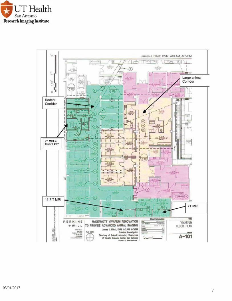
2. MRI Division Information 2.1 Map of MRI Division User Accessibility This map is sectioned off by areas open to MRI Division staff only, MR safety trained individuals, and those who have been MR screened. This map DOES NOT designate the safety zones. All non-safety trained individuals (e.g. participants, visitors, facilities personnel, etc.) should be accompanied by a safety trained individual always during their visit at the RII.
7 05/01/2017

8 05/01/2017
2.2 Zones The MRI Division is divided up into zones based on the American College of Radiology White Paper on MR safety. (AJR:178, June 2002 1335-74).
ZONE 1= Area outside MR environment, accessible to the general public.
ZONE 2= Interface area between publicly accessible uncontrolled Zone I and the strictly controlled Zone III & Zone IV
ZONE 3= Restricted from public access, accessible only to personnel that have completed MR Safety Training; non-trained personnel must be accompanied always. ZONE 4= Synonymous with the scanner room itself, accessible only to those that have completed MR Safety Training and adequately completed the MR screening form
2.3 Phone List
Contact List 210-567- Emergency 7-8911 First Floor Reception Desk (Debbie Espinoza) 7-8115 TIM 1 MRI Console 7-8198 TIM2 MRI Console 7-8199 Bruker 7T Biospec Console 7-8066 Bruker 11.7T Biospec Console 7-8060 Bruker 7T Pharmscan Console 7-8059 John Li’s Office 7-8197 Mock MRI Scanner 7-8016 Radiation Safety (Robert Moreno) 7-8182 Laboratory Animal Resources 7-6166

9 05/01/2017
3. Rules for Working In & Using MRI Division Resources 3.1 Training (see also Training section) Principle Investigators (PIs’) are responsible for ensuring all employees and staff working on MRI projects are trained through the Safety Training Course to access the facility. In addition, it is mandatory that everyone who will be accessing the facility must complete a screening questionnaire to ensure their safety.
3.2 PI Responsibilities
1. Principle Investigators (PI’s) will conduct their research strictly in accordance with and under the oversight of the Institutional Review Board (IRB, for human studies) or the Institutional Animal Care and Use Committee (IACUC, for animal studies). As the principle investigator, you will be accountable for your own research and the protection of human studies. You will ensure, always, that you have the appropriate resources and facilities to conduct this study. You will ensure that all research personnel involved in the conduct of the study have been appropriately trained on the protection of human subjects, in addition to the study procedures.
2. Any adverse or serious adverse event MUST be reported to the IRB per IRB policy as well as to the authorized user in MRI Division.
3. Any changes/additions/revisions to your research plan must be submitted to the IRB for review and approval prior to implementation. This includes changes or additions requested by the sponsor.
4. Your protocol MUST be reviewed annually by submission of the appropriate application to the IRB. Failure to submit renewal documents to the OHRP by the administrative due date indicated on the renewal notice may result in termination of the study by the IRB.
5. Advertisements for the recruitment of subjects must be approved by the IRB prior to implementation.
3.3 Console rooms (SIEMENS MRI Scanners) 1. Before entering the console room:
A. Make certain the research participant is registered in the MRI Division participant registration system.
B. Before entering the console room, you must have provided the scanner operator with completed & signed consent and screening forms, including the ID that will be used for that participant when starting the session on the console computer. Ensure that you have made any necessary photocopies of documents so that the consent and screening forms provided to the scanner operator at this time are the MRI Division copy.
C. The investigator group must limit themselves to three people before entering the console room.

10 05/01/2017
D. Only those individuals that have been through MRI Division’s safety training may enter this area unless prior permission is obtained from the MRI Division Authorized User.
E. There will be NO behavioral testing in MRI Division unless prior permission is given by the Facility Manager or RII Director.
2. During the scanning session: A. There will be absolutely NO food or drink allowed in the console room.
B. One parent may accompany the child that is involved in a pediatric study. Siblings are not allowed in the console room except to view their sibling briefly and then they must return to the waiting area.
C. No strollers or other large, potentially dangerous metallic items may be taken into the console room.
D. When in the console room, please limit your talking to the necessary communication for the scanning to proceed safely in the allotted time frame.
E. The investigator group must initiate the scanning session on the logging computer as soon as they enter the console area.
F. No behavioral data should be copied from the stimulus presentation computer in the console room.
G. The door between the office area and console room should remain closed always, only MRI Division staff may enter this area.
H. At no time, will a group of investigators be allowed in the console area while another group is scanning.
3. Scanning session is completed A. Remove any items that your group brought with them into the console area.
B. End the session on the logging computer only when the participant has been taken out of the scanner and the console/magnet room is ready for the next group.
C. Exit the console area immediately after the area is clean and the participant has returned to the waiting area.
3.4 Waiting Area
A. No food or beverage debris is to be left in the waiting area. It is the responsibility of the PI, NOT the MRI Division, to clean up after their participants after they have left the facility. Those who do not clean up after themselves or participants may lose food/beverage privilege for the conference and waiting areas.
B. Items (Pens, pencils, etc.) left in the rooms are the responsibility of the owner as we do not currently have a lost and found.
C. Chairs are not to be “borrowed” from other rooms in the RII. 3.5 MRI DIVISION Onsite Recruiting Policy To maintain participant confidentiality and compliance with IRB policy MRI Division has outlined the following rules regarding recruitment at the RII. These guidelines are also intended to minimize any conflict of interest that might arise due to recruiting efforts on the RII premises, as well as provide an unbiased access to resources available.

11 05/01/2017
1. Flyers Any IRB approved flyers that include imaging will be allowed in the waiting room only and will be available to any members of the public who enter this facility
2. Verbal Inquiries All staff, when directly asked from a participant or a parent of a participant, who is not directly involved in that staff members research study, may direct them to pick up any of the flyers in the waiting room; NO further efforts to actively recruit these participants are to be made.
3. Other
A. All staff members may recruit participants ONLY with IRB approved methods that are stated explicitly in IRB protocol.
B. All staff members must complete HIPPA and Subject Safety Training annually and file these documents with the appropriate offices.
4. Staff Initiated Recruitment A. Under no circumstances can staff members engage with a subject participating in
another lab's study for recruiting that subject.
B. Under no circumstances can personal information volunteered by a subject participating in another lab's study such as those provided on the MRI Safety Screening form and the logging system be used for recruiting subjects.
3.6 Policies for Imaging Animals on MRI Scanners used for Human Studies Summary:
• At no time should human subjects have any contact with animal subjects. This includes: o No visual contact o They should not be able to hear the animal o They should not be able to identify anything associated with animal experiments
• All waste and supplies brought in and used in the animal experiment must leave with the animal
• All surfaces where animals had contact (this will usually be limited to inside the magnet only) must be disinfected by the MR technologist with an EPA-registered disinfectant
• Imagers may only be operated by the MR faculty and staff trained in the RII procedures • Animals cannot use the same medical gas or scavenging equipment as humans • Everyone involved in animal handling must review and complete the animal handlers’
health questionnaire. • Any person who is required to enter the magnet room must complete the training
requirements for RII safety
3.6.1 General Overview
1. The main principle to safely operating a dual-subject use facility is to ensure physical and temporal separation between human subjects and animals. Human subject and patient use must be the priority. Animal use of the scanners can occur only when it will not conflict with human subjects being scanned.
2. Human subjects should never be able to directly see or hear, or otherwise identify that animals are nearby or were or are about to be used in the facility. This means that all supplies for animal work, and any wastes generated from animal work, must be removed before the facility is returned to human use. It also means that animals, especially non-human primates, must be anesthetized prior to transport into the facility.

12 05/01/2017
3. The MR technicians will coordinate all movement of human subjects and animals in the RII magnet area.
3.6.2 Transport 1. Animals transported into the RII must enter through the rear entrance of the LAR
facility. Human subjects should never be able to directly see or hear, or otherwise identify that animals are nearby or were or are about to be used in the facility. This means that all supplies for animal work, and any wastes generated from animal work, must be removed before the facility is returned to human use. It also means that animals, especially non-human primates, must be anesthetized prior to transport into the facility.
2. The MR technologists must be contacted prior to arrival at 785-6699, or 785-6410. 3. Upon entering the rear hallway in the RII, one transport personal must go and check with the
technologists to ensure that the magnet space is clear of human subjects. 4. Upon getting clearance from the technologist, the animal may be moved into the magnet bay
without delay. 5. At no time, should the animal be station outside the magnet control rooms. 6. No equipment associated with the animal should remain in the hallway outside the control room. 7. The control room doors should remain closed while the animal is in the control room. 8. Upon completion of the MR imaging session the technologist will first ensure that no humans are
in the hallway and that all the other technologists are aware the animal will soon be transported out of the magnet area.
9. When the hall is clear, the animal and all associated equipment can be transport out the rear entrance following the path it came in.
3.6.3 Room air ventilation Yale BS&P/Project Mgt confirms that the entire MR suite in TAC operates on 100% exhaust, meaning that none of the air is re-circulated back to either patient, animal, or other space inside the building.
Air clearance requirements:
a) Sheep: it is required that 99.9% of the air is replaced in the room following a study imaging sheep. This currently requires 41 minutes of clearance time after the animal has been removed. No human subjects can enter the magnet room until that time has elapsed.
b) Dogs, pigs, rabbits, monkeys, and rats: the requirements are not as severe and following animal removal and cleaning as outlined below the room may be used after 20 minutes of circulation.
3.6.4 Surface protection All surfaces that animals encounter inside the facility must be covered with impermeable drapings or covering. Equipment and devices that will regularly be used for animals such as trays and carts should be dedicated to animal use only, labeled as such, and stored elsewhere.
3.6.5 Technologists Technologists must where gowns, masks, and gloves when dealing with sheep. Gowns and gloves are sufficient with dogs, pigs, rabbits, monkeys and rats.
3.6.6 Disinfection All surfaces where animals had contact, even with impermeable coverings, must be disinfected after use with 10% bleach (1 part bleach + 9 parts water, made at least weekly). This is followed by a wipe down with water or 70% alcohol.
Responsibility for the disinfection of used surfaces lies with the MR technologist that ran the scan.

13 05/01/2017
3.6.7 Wastes
Animal excrement, blood or body fluids, hair, and any medical waste generated during the procedure or while in the dual use area must be appropriately packaged and removed from the facility after animal procedure is completed. This also applies to sharps containers, so that no potentially infectious materials from the animal(s) remain after they are removed. If any of these materials are spilled or otherwise contaminate areas inside the MR facility, the surface must be cleaned and decontaminated as for a human blood spill.
**All the above waste shall be transported out of the RII with the animal**
3.6.8 Medical Gases and Vacuum Animals can use the same medical gas, scavenging, or vacuum systems used for humans as if different hoses and apparatus from the wall box out to the subjects are kept separate and dedicated to either human or non-human use. In all cases hoses, must not be re-used and in the case of animals the hoses must be removed from the RII with the animal at the end of the experiment.
3.6.9 MR Equipment Operation Operation of MR equipment must be limited to knowledgeable and authorized persons from the RII. All individuals involved must be trained in MR safety and understand other health and safety issues related to the facility.
3.6.10 MRI Quality Control Procedures Frequency: Daily, Weekly, Monthly, Annually per scanner
Required Equipment: • Daily, Weekly, Monthly Quality Assurance by system operator • Physicist surveys – annually and after major equipment repairs/upgrade • Test phantoms – Siemens MRI phantoms, ACR MRI accreditation phantom, other
phantoms developed in-house • Test images • Logs/forms
Procedure steps: 1. ACR QC tests associated with MRI shall be performed on at least a weekly basis. 2. The MRI Manager will ensure all documentation is readily available for inspector’s review. 3. The designated MRI physicist/scientist will review documentation of QA records weekly. 4. The MRI Manager will address any outstanding failures identified during the records review
with the appropriate MRI Division Staff. 5. QA Deficiencies identified by the performing technologists will immediately be:
a. Documented on the imaging equipment service log b. Reported to the relevant parties e.g. Service, Physicist, MRI Division Chief c. MRI Manager will initiate deficiency corrective action e.g. place service call, contact
manager, etc., d. Upon corrective action completion, MRI Manager, Service Engineer, and/or MRI
physicist/scientist will conclude any documentation, and communicate pertinent outcomes to the relevant parties
e. Copy of service reports will be filed in the MRI system service log for future reference

14 05/01/2017
Precautions: The MRI Manager and primary technologist (modality specific) will review all physicist’s surveys upon receipt, to ensure understanding of the report findings and recommendations and to ensure that any corrective actions are implemented promptly, if needed.
Suggested Performance Criteria and Corrective Action: Each radiologic technologist, regardless of modality, will be informed and trained in the appropriate QC tests for the specific modality. Any concerns or questions will be addressed by the physicist. If additional information or training is required, it will be researched and provided, in accordance with ACR requirements.
3.6.11 Lab Animal Allergy Issues Everyone involved in animal handling must review and complete the animal handlers’ health questionnaire, distributed through either LARC or the Employee Health Office. Since LAA's are common, steps to minimize exposure to potential animal allergens must be considered in all aspects of animal work, including imaging and preparatory steps for imaging.
3.6.12 Non-Human Primates (NHPs) NHPs pose unique and potentially more serious issues than many other lab animals, including physical assault injuries and bites, and infectious disease transmission. Individuals working directly with or around NHPs are encouraged to seek additional training and advice from YARC, especially regarding restraint systems, emergencies, and TB. For the RII, all non-human primates must arrive and leave under anesthesia.
4. Project Protocols Visit the RII Utilization Review Committee webpage: (http://rii.uthscsa.edu/protocolreview.php) to learn how to fill out a project request. 4.1 URC Approved, Funded Projects New Funded Projects New research using the MRI Division core facilities may be initiated with a research proposal. Research proposal forms can be located on the MRI Division website. New projects may be funded, or the goal of the project may be to collect pilot data for a grant application. PI’s are advised to discuss the project with the senior scientists at the RII to help in designing the project finding a RII senior scientist to sponsor the project. All research involving human subjects to be conducted at the Research Imaging Institute must be approved by the UT Health Science Center Institutional Review Board (IRB). Check out the IRB web-site (http://research.uthscsa.edu/irb/ ) for more information on obtaining IRB approval. If you have questions or would like help with your IRB application, contact the UT Health Science Center IRB Office (210) 567-8250. All research involving animals at the Research Imaging Institute must first be approved by the IACUC. (http://research.uthscsa.edu/iacuc/) If you have questions or would like help with your IRB application, contact the UT Health Science Center IRB Office (210) 567-8260. Note that IRB or IACUC approvals must be obtained BEFORE final approval can be granted by the URC.

15 05/01/2017
Application approval is dependent on review and approval by the RII’s Utilization Review Committee (URC), which evaluates the following: • Scientific merit • Feasibility of the research on existing equipment • Availability of equipment • Budget review and ability of the investigator to support the user fees
The URC application shall include the following information: 1. Official study title 2. Project approval number from the IRB or the IACUC 3. PI ‘s name 4. RII sponsor’s name 5. Species being studied 6. Brief description of the scientific goals of the project 7. The estimated session duration, number of subjects and number of sessions per
subject 8. The estimated total scanning costs of the project (including operator and data
archiving charges, assuming all enrolled subjects complete the study 9. Description of the funding source(s) for the project 10. Supporting documents, such as: grant notice of award, IRB approval letter,
sponsor’s brochure (for industry studies), letters of co-funding commitment (e.g., from department Chair).
PI’s on approved proposals will receive email notification and further instructions.
4.2 Development Projects (unfunded) Development Projects can be requested by investigators engaged in research for the development of MRI Division hardware, software, and imaging techniques that will be of general use to the MRI Division community. The PI asks for the scanner time based on a desire to complete some developmental project. The RII encourages development projects that aim to produce a product (device, code or protocol) that is documented. Development projects can be maintained as long as the PI demonstrates regular progress.
1. Development projects should be ongoing and geared to producing a product. The product can be a protocol, device or piece of software.
2. Each development project will have a project name and assigned a DEV number. 3. The development project shall be presented to the URC with clear design goals and
milestones for successful completion. 4. Progress reports on development projects shall be submitted by the PI each month. 5. Although the PI should estimate of the amount of scanner time required for successful
completion of the development project, the PI will not have to return to the URC to request additional time if adequate progress is being demonstrated.
6. A development project that goes three months without significant progress will be automatically terminated.
7. Temporary authorization for a new development project can be given by the MRI Division Chief, but only until the next URC meeting, at which time the URC must review the project’s plan and approve continuation of the project.
Development projects can include the participation of human volunteer subjects under the auspices of IRB protocol # HSC20110120H (PI: Clarke) entitled, “Pilot: Development and Optimization of MRI”. ALL DEVELOPMENT PROJECTS INVOLVING HUMAN SUBJECTS REQUIRE THAT THE MRI DIVISION CHIEF (Dr. Clarke) BE LISTED AS A CO-PI.

16 05/01/2017
4.2.1 Inclusion Criteria for Human Subjects for Development Projects
1. Subjects self-report as being healthy, between ages of 18-75, and able to give written informed consent. OR
2. Medically stable subjects with illness, between ages of 18-75, who may be referred by his/her physician to test if scanning sequence is acceptable prior to writing new protocol to scan specific diseases.
4.2.2 Exclusion Criteria for Human Subjects for Development Projects 1. Subject has cardiac pacemaker or any other implanted electronic device 2. Subject has intracranial clips, metal implants, or external clips within 10 cm of the head 3. Subject has a history of having metal in the eyes or claustrophobia. 4. Subject has medical or other reason leading (i.e. claustrophobia) to difficulty laying quietly
and comfortably motionless in the MRI scanner. 4.2.3 Recruitment Process for Human Subjects for Development Projects
1. Recruitment for medically stable with illness who may be referred by his/her physician. 2. Potential subjects will make initial contact. Initial contact will be made by phone or in
person, the contact person will give a brief explanation about the study. MRI Division staff will inform the volunteers that they cannot be in the study if they have metal in the body. If they are interested MRI Division staff will send them an electronic version or paper copy of the consent so they can prepare questions if they want to. At the first appointment, a staff member will go over the consent with the volunteer, explaining everything.
4.2.4 Screening Process for Human Subjects for Development Projects 1. A member of the MRI Division staff will go over the consent together with the volunteer in a
private setting. All questions will be answered; the volunteer has some time to go over the consent again and afterwards to ask additional questions he/she has. If the volunteer still wants to be in the study, he or she will at that time together with the PI or his designate, and a witness will sign the consent.
2. The volunteer will have an electronic version or paper copy of the consent before coming which provides them with time to consult with family or other people.
3. Potential subjects will have adequate time to decide. A PI may not recruit or enroll students working under the PI’s supervision unless it is the student’s project.
4.2.5 Concurrent Measurements of Human Subjects in MRI Development Projects 1. Physical sensors that are applied either to the surface of the body or at a distance and do
not involve input of significant amounts of energy into the subject or an invasion of the subject’s privacy
2. Weighing or testing sensory acuity 3. Electrocardiography, electroencephalography, thermography, detection of naturally
occurring radioactivity, electroretinography, ultrasound, diagnostic infrared imaging, Doppler blood flow, and echocardiography,
4. Moderate exercise, muscular strength testing, body composition assessment, and flexibility testing where appropriate given the age, weight, and health of the individual.

17 05/01/2017
4.2.6 Agents that can be Administered to Humans during MRI Development Projects 1. 12% O2 inhalation (brief 1-3 min.) 2. 5% CO2 in (air or oxygen)
Use of all other agents, including contrast agents, prescription or non-prescription pharmaceuticals, saline, Ringers lactate, etc. require separate approval through the IRB. ** Development project application forms can be obtained from Debbie Espinoza
([email protected]). After completion, the application may be submitted to the URC for approval.
** Human subject informed consent forms can be obtained from Geoffrey Clarke, PhD ([email protected] ).
4.3 IRB & IACUC Compliance Requirements
Investigators performing human studies at the Research Imaging Institute must submit documentation of compliance with IRB regulations. We cannot assign scan time or allow use of the scanners without IRB documentation. This information will be kept in the investigator's file at MRI Division and will be used by the MRI Division staff to verify adherence to protocol. Please help us keep this information up to date. The following documents must be submitted for each protocol:
• Most recent IRB or IACUC approval letter for protocol • Electronic copy of Detailed Protocol section from approved IRB/IACUC application • Number of subjects approved • Names of persons other than PI or Co-Investigator who are authorized to obtain informed
consent (include documentation of IRB approval) • Copy of any amendments made to subject selection/enrollment (including requests to enroll
additional subjects) or study procedures for IRB/IACUC protocols, and amendment approval letter
4.4 Procedure to Initiate MRI Study 4.4.1 General Use Requirements When submitting a grant proposal for research involving the Research Imaging Institute, the Principal Investigator is responsible for addressing the specific requirements of the granting agency and their home institution. First-time collaborators are encouraged to review their research program with the MRI Division Chief, who can help them find an appropriate MRI Division faculty Project Sponsors to assist them with the application process. Additional requirements of MRI Division are summarized below.
4.4.2 MRI Division Requirements
a. Materials to Submit to the Research Imaging Institute The principal investigator of all grants that propose work at the Research Imaging Institute are required to: • Notify the Chair of the Utilization Review Committee or MRI Division Chief one
month prior to submission • Submit to the MRI Division Chief a copy of the entire grant at the time it is submitted

18 05/01/2017
b. Materials to obtain from the Research Imaging Institute The following documentation must be included in your grant proposal. These materials can be obtained through prior arrangement with the MRI Division staff. o The MRI Division Research Proposal Cover Sheet o Biosketches of MRI Division researcher personnel involved in the proposed project o 'Other Support' document for MRI Division research personnel who will receive
payment from proposed project o Description of MRI Division resources and environment Paragraph describing
the MRI Division cost structure for budget o 'Sign-off' on in-house part of funding application o Imaging Time Approval o Budget Approval o Conflicts of Interest Form
4.5 Billing/ funding / pilot time All scanning time is billable. Pilot time is considered part of scanner time, so be sure to include that into your scanning budget. Time is billed for the time requested for scanning, NOT the time the subject enters the scan room, but the time you have reserved for scanning. If your scan time runs over the requested scan time, you will be billed to the next quarter hour.
5. Time Allocation 5.1 Scheduling MRI time procedures
All requests for scanner time must by URB-approved protocol number or Development Protocol number and shall be submitted to Debbie Espinoza ([email protected]) who will put the appointments on the online calendar. No other requests will be considered. This is being done to streamline and automate the scheduling process. Hours of Operation for 3T MRI scanning:
Monday through Friday: 9am – 5pm Weekends and Evenings can be scheduled by special arrangements.
5.2 Scheduling policies
1. MRI time requests must be submitted within 24 hours of the desired time slot in order to be considered. Special considerations will be taken into account for those submissions made less than 24 hours in advance.
2. An MRI time slot cancellation must be submitted within 24 hours prior to the designated time slot; otherwise, you will be billed for the requested time slot. Please note the reason for canceling a time slot, as this is looked at to see who is abusing the canceling policy (a group with frequent cancellations may face scanning penalties).
3. Three Saturdays a month will be available for scanning. Groups must request at least a 4-hour time block to make it worthwhile for the Authorized User to come in for that day. Once a 4-hour block has been reserved, additional hours may be requested by the same group or other groups within the available time for scheduling on a weekend. Only those Saturdays designated on the calendar with an Authorized User present are available for scheduling.

19 05/01/2017
5.3 Other MRI schedule slots 1. MRI systems may be scheduled for the testing and calibration of accessory MRI
equipment, either developed in-house or purchased from OEM manufacturers. These sessions have low priority and must accommodate previously scheduled projects.
2. MRI systems may be schedule for repairs, upgrades, applications training by the MRI system manufacturer’s employees. These sessions are given high priority, subject to the availability of manufacturer’s personnel schedules.
3. MRI systems may be scheduled for educational purposes, either specific training on the use of the MRI system or general educational sessions involving students in UTHSCSA classes. These sessions have low priority and must accommodate previously scheduled projects.
5.4 MRI Simulator Usage The MRI Simulator (or Mock Scanner) is a resource of the Human Performance Division, which is available to PI’s who need to use it for a URC-approved research protocol. The MRI Simulator can be scheduled by contacting Collin Sauder, Ph.D. (210) 567-8016, [email protected]. All researchers who have scheduled MRI scanning time will have access to the Mock Scanner room 15 minutes prior to scan time to familiarize subjects with the MRI environment prior to the actual scan. For ALL other times, you MUST submit a request to use the Mock scanner room two (2) weeks prior to the desired date to ensure availability. NO behavioral testing are to be done on Research Imaging Institute premises unless prior permission is given by the Utilization Review Committee or Institute Director.
5.5 Requesting Independent Operator and Open Slots Please visit our calendar to find an available time slot. E-mail your time request including the date, the time (A.M. or P.M.), which scanner and the Study ID# to [email protected] . A confirmation e-mail will be returned notifying you that you have the time. Please use e-mail only for requests (or cancellations). Verbal requests are not accepted. This is to ensure that we have a record of your request and you have a record of the confirmation. -Please re-check the calendar to make sure that the slot was entered correctly.
5.6 Special Requests We will make every attempt to accommodate special requests (e.g. requests for daytime scan hours with technical support). However, with the growing number of studies vying for magnet time, we ask that users consider their special requests carefully and try to be as flexible and as reasonable as possible.
5.7 Tours ALL tours requests (including media interviews/photographs) must be submitted two weeks prior to the tour date to [email protected] . In addition, Jon Li must be notified. If there is a scan scheduled at the time of the tour which does not pertain to your study, it is up to you to check with the scheduled user to ensure that the tour will not interfere with the scheduled scan session. For all tours, a MRI Division member MUST be available (not busy or scanning) to accompany the tour. Please avoid bringing tours through the office/cubicle area. All tours with children (under 18 years of age) MUST be accompanied ALWAYS by a MRI Division member.
5.8 Use It or Lose It Past utilization of assigned scan time will be taken into consideration each time the schedule is re-made. Frequent non-use of scheduled scan time indicates a problem that should be discussed before additional time slots are allocated, and may result in a reduction of a user's assigned time in the next iteration of the schedule. After each scheduling period, the history of magnet use will be reviewed carefully for improving efficiency.

20 05/01/2017
5.9 Cancellation Policies
If you give at least 24-hour notice that you are unable to use your scheduled time slot, you will not be billed for that time. Submit your cancellations at [email protected] and the appropriate changes will be made in the calendar. Please make sure to note the reason for the cancellation. If you do not give at least 24-hour notice and you do not show up for your time slot, you will be billed for the entire block of time reserved.
The only exception is if a subject cancels at the last minute (or if there is some emergency). In this case, you must send an e-mail to [email protected] as soon as you know you will not be using the time slot (please provide the reason for cancellation). Please make every effort to keep these incidents to a minimum by calling subjects to confirm appointments and by making sure that subjects know when and where to report for the study, and how they can reach you at any time. It is our opinion that no-shows and last minute cancellations are rare, but if the records indicate that this is a problem for any group, special arrangements will be made.
DO NOT give up your time slot to another group if you are not going to use it. We are often looking for time for the pulse programmers or for time to pay back groups whose time slots we had to take for repairs or updates. If we do not need the time, the slot will be opened on the calendar and then given to the first requester.
6. Training
6.1 Safety Training Successful completion of the MRI Division’s MRI Safety Training Course is required for all investigators working in the facility. The program will include a 1-hr MRI safety lecture, viewing of an MRI safety video, tour of the magnet room, practice in subject screening and a brief exam. Course materials are available …... Course Directors: John Li, MD and Geoffrey Clarke, PhD & John Moreno
6.2 MRI 3T Siemens Training The purpose of this training is to educate the student in the basics of the operation of the MRI scanner. This training will include a combination of lectures and hands-on use of the scanner. Students who pass this training will be qualified to operate the scanner, provided:
a. The student has also successfully completed the MRI Safety Training Course (§ 6.1 Safety Training)
b. An authorized user is present in the MRI facility c. The scanning session has been approved through the appropriate scheduling
procedures
Students will be evaluated based on multiple quizzes, demonstration of competence and other factors based on the instructor’s evaluation of each student’s seriousness and proper respect for the potential dangers involved with MRI. Completion of this training does not automatically entitle the student to operate the scanner. Course Director: John Li, M.D.
21 05/01/2017
6.3 Image Analysis Training
The purpose of this training is to educate the student in the basic analysis of fMRI data. The emphasis will be on the techniques used in the Center. To find out more about when this training is given please visit the website education section of the RII’s website; course materials are available online to be read prior to attending the lectures. Course Director: Filipe Salinas, Ph.D.
7. Safety 7.0 Zone map (see Page 7)
7.1 Hazards/ dangers Magnetic Resonance Imaging (including spectroscopy, conventional, and fast imaging techniques) have been in use for decades, and are viewed as medical procedures associated with acceptable and well controlled risks. However, technological advances in MRI (higher static fields, faster gradients, stronger RF transmitters) have occurred rapidly and many questions regarding the safety of these developments remain unanswered. This document introduces some of the safety concerns associated with MR research. Other related pages address the practical implications of these safety issues.
7.1.1 Static Magnetic Fields
a. Projectiles The most immediate danger associated with the magnet environment is the attraction between the magnet and ferromagnetic objects. Ferromagnetic metal objects can become airborne projectiles when placed in a strong magnetic field. The strength of the field increases super linearly with the distance from the magnet bore, and even hand-held objects can be jerked free very suddenly as the holder moves closer to the magnet. (Small objects, such as paper clips and hairpins, have a terminal velocity of 40 mph when pulled into a 1.5T magnet.) In addition to the possibility of severely injuring someone, it is not good for the magnet to be bombarded with difficult to remove small metal items. Remember, even when you are not scanning, the magnet is ALWAYS “ON”. NEVER bring any metal objects into the scanner rooms.
b. Metal in the Body Metallic objects in the body can also have dangerous effects when placed in a magnetic field. Ferromagnetic metal implants or fragments may twist or move causing internal injury. Even non- ferromagnetic metal (including metal on clothing) can heat up during scanning, causing burns or discomfort. Many of our subject screening criteria are aimed at avoiding these hazards. In addition, metal in or near the body (such as dental implants) can produce artifacts which adversely affect image quality.
7.1.2 RF guidelines/ SAR a. Tissue Heating An RF pulse (a short burst of an electromagnetic wave originating from the RF coils) is used in MRI to "excite" tissue protons by an exchange of energy. This absorption of RF energy can potentially cause heating of the tissue. Absorption of RF power by the tissue is described in terms of Specific Absorption Rate (SAR), which is expressed in Watts/kg. (In the US, the recommended SAR level for head imaging is 3.2 Watts/kg.) SAR in MRI is a function of many variables including pulse sequence and coil parameters and the weight of the region exposed. However, the actual increase in tissue temperature caused by exposure to RF radiation is dependent on the subjects’ thermoregulatory system (e.g. tissue perfusion, etc.). The risk of
22 05/01/2017
exposing subjects with compromised thermoregulatory function (e.g. elderly
patients and patients taking medications that affect thermoregulation, such as calcium-blockers, beta-blockers, diuretics, or vasodilators) to MR procedures that require high SAR levels has not been assessed.
b. Electrical Burns RF fields can cause burns by producing electrical currents in conductive loops. When using equipment such as surface coils, ECG or EEG leads, the investigator must be extremely careful not to allow the wire or cable to form a conductive loop with itself or with the subject. Coupling of a transmitting coil to a receiver coil may also cause severe burns.
7.2 Quenching Quenching refers to the events that occur when the liquid cryogens that cool the magnet coils boil off rapidly, which results in helium escaping very rapidly from the cryogen bath. This means that the coils cease to be superconducting and become resistive. A quench will in general be accompanied by a loud bang or thundering with the cold gas expulsion. Quenching may occur by activation of the magnet STOP button, or spontaneously, caused by a fault in the magnet itself. The magnet emergency stop button should only be used if the magnetic field may possibly be causing patient or personnel injury, and a shutdown of the static field is necessary, or if fire or some other unforeseen occurrence requires the quick access of emergency personnel to the examination room. Note, however, that initiating a quench may not result in total removal of the magnetic field, and a danger may still exist. In case of emergency contact the on-site Authorized User and follow instructions provided by the Authorized User.
7.3 Noise Vibrations of the gradient coil support structure of the MRI scanner create sound waves. These are caused by the interactions of the magnetic field created by pulses of the current through the gradient coil with the main magnetic field in a manner like a loudspeaker coil. The sounds made by the scanner vary in volume and tone with the type of procedure being performed. MRI system noise levels increase with field strength. It is required that ALL participants wear protective headphones during the scan session. Additional ear protection is available if necessary.
7.4 Other Concerns 7.4.1 Pregnancy
There are no known adverse effects of MRI on developing fetuses. Most early studies on pregnant animals were negative for teratogenic effects, and a recent survey found no association between working in the MR environment and several pregnancy outcome variables.* However, given the scarcity of data on the subject and the high susceptibility of the developing fetus to damage in general, we believe it is not worth the risk for pregnant women to participate as subjects in MR research studies unless those studies are directly evaluating conditions related to pregnancy. Most clinical units allow pregnant employees to enter the scan room, but not to remain in the room while the RF and gradient fields are applied during image acquisition. Pregnant researchers at MRI Division are expected to regulate their own exposure to the magnets. For additional and up-to-date information, see MRIsafety.com
*Shellock , FG & E Kanal , Magnetic Resonance: Bioeffects , Safety, and Patient Management, 2nd Edition, Lippincott -Raven, Philadelphia , 1996, pp. 342.

23 05/01/2017
8. Human Scanning 8.1 Forms – screening, consent, etc
[*Portions of the text in this section were excerpted from MR screener documentation provided freely available on the web sites, www.IMRSER.org and www.MRIsafety.com with permission from Frank G. Shellock, Ph.D., FACC, FACSM]
Effective screening procedures of patients and other individuals before entering the MR facility is one of the most critical components for conducting a safe program. This is an important aspect of protecting patients and individuals from MR system-related accidents and injuries. Most MR- related incidents have been due to deficiencies in screening methods and/or a lack of properly controlling access to the MR environment. Hence, persistent vigilance and attention to detail must be a part of every responsible study. Regardless of the duration, every person who enters the Magnet Room should be made aware of the dangers as well as sufficiently cautioned about not bringing in anything into the room under any circumstances. It goes a long way to always advise and oneself be advised that the MR system magnet is ALWAYS on.
Magnetic Resonance (MR) Procedure Screening for Patients
Screening patients for MR using Magnetic Resonance (MR) Procedure Screening for Patients must be done before each scanning session. This should be conducted by a person who has successfully completed the MRI DIVISION safety training course (§6.1 Safety Training). This person is expected be familiar with the information contained on the screening forms for patients and individuals. Subjects must complete IRB approved consent and MRI safety screening forms prior to entering the MR room and being scanned, and copies of these documents must be left at the RII. Comprehensive patient screening involves the use of a printed form to document the screening procedure, a review of the information on the screening form, and a verbal interview to verify the information on the form and to allow discussion of concerns the patient may have. A hard copy of the screener is available at MRI DIVISION, and it can also be downloaded from the web sites, www.IMRSER.org and www.MRIsafety.com This form is used to ascertain if the patient has an implant that may be contraindicated for the MR procedure (e.g., a ferromagnetic aneurysm clip, pacemaker, etc.) or if there is any condition that needs careful consideration (e.g., the patient is pregnant, has a disability, etc.).

24 05/01/2017
Figure 8-1 Screening form for patients
Preliminary screening, fairly in advance to the actual scan session, helps to prevent scheduling participants that may be inappropriate candidates for MR examinations, which can improve efficiency and lower monetary penalties associated with late cancellations. After preliminary screening, the patient must still undergo the comprehensive screening in preparation for scan session.
Quick overview Page one Top section: Helps gather pertinent and up-to-date information about the patient, and contact information. This includes general patient-related information (name, age, sex, height, weight, etc.), as well as a reason for the MR procedure and/or symptoms that may be present.
Bottom Section: Requests information regarding prior surgery or operation, which can help determine if there may be an implant or device present that could create a problem for the patient. Information is also requested pertaining to prior diagnostic imaging studies that may be helpful to review for assessment of the patient’s condition.
Page two Top section: Advises not to enter the MR system room or MR environment if there is any question or concern regarding an implant, device, or object. Middle section: Lists various implants, devices, and objects to identify anything that could be hazardous to the patient undergoing the MR procedure or that may produce an artifact that could interfere with the interpretation of the MR procedure. In general, these items are arranged on the checklist in order of the relative safety hazard (e.g., aneurysm clip, cardiac pacemaker, implantable cardioverter defibrillator, electronic implant, etc.), followed by items that may produce imaging artifacts that could be problematic for the interpretation of the MR procedure. Additionally, questions are posed to determine if the patient has a breathing problem, movement disorder, or claustrophobia. Figures of the human body are included on the second page of the form as a means of showing the location of any object inside of or on the body. This information allows the patient to indicate the approximate position of an object that may be hazardous or that could interfere with the interpretation of the MR procedure due to the occurrence of artifacts.
25 05/01/2017
Lower section: Has Important Instructions for the patients before entering the MR environment. Importantly, undergoing previous MR procedures without incidents does not guarantee a safe subsequent MR examination. A written screening form must be completed each time a patient prepares to undergo an MR procedure.
An MR-safety trained person should review the completed form’s content, and verify/clarify the information provided through a verbal interview, and allow discussion of any question or concern that the patient may have. This allows a mechanism for clarification or confirmation of the answers to the questions posed to the patient so that there is no miscommunication regarding important MR safety issues. In addition, because the patient may not be fully aware of the medical terminology used for a particular implant or device, it is imperative that this particular information on the form be discussed during the verbal interview.
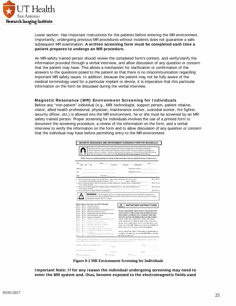
Magnetic Resonance (MR) Environment Screening for Individuals Before any “non-patient” individual (e.g., MRI technologist, support person, patient relative, visitor, allied health professional, physician, maintenance worker, custodial worker, fire fighter, security officer, etc.) is allowed into the MR environment, he or she must be screened by an MR- safety trained person. Proper screening for individuals involves the use of a printed form to document the screening procedure, a review of the information on the form, and a verbal interview to verify the information on the form and to allow discussion of any question or concern that the individual may have before permitting entry to the MR environment.
Figure 8-2 MR Environment Screening for Individuals
Important Note: If for any reason the individual undergoing screening may need to enter the MR system and, thus, become exposed to the electromagnetic fields used

26 05/01/2017
for an MR procedure, this person must be screened using the Magnetic Resonance (MR) Procedure Screening Form for Patients.
In general, magnetic resonance (MR) screening forms were developed with patients in mind and, therefore, tend to pose many questions that are inappropriate or confusing to other individuals that may need to enter the MR environment. Therefore, a screening form was created specifically for individuals that need to enter the MR environment and/or MR system room. A hard-copy of this form, entitled, Magnetic Resonance (MR) Environment Screening Form for Individuals, screener is available at MRI DIVISION, and it can also be downloaded from the web sites, www.IMRSER.org and www.MRIsafety.com
Quick overview:
Top section: Advises not to enter the MR system room or MR environment if there is any question or concern regarding an implant, device, or object. It also gathers pertinent and up-to- date information about the person entering the MR environment, and contact information. This gathers general individual-related information (name, age, date, and contact information).
Middle Section: Requests information regarding prior surgery or operation, which can help determine if there may be an implant or device present that could create a problem for the person preparing to enter the MR environment. It also lists various implants, devices, and objects to identify anything that could be hazardous to the person entering the MR scanner room. In general, these items are arranged on the checklist in order of the relative safety hazard (e.g., aneurysm clip, cardiac pacemaker, implantable cardioverter defibrillator, electronic implant, etc.). To the right side, it has Important Instructions for the patients before entering the MR environment.
Lower section: A written screening form must be completed each time a patient prepares to undergo an MR procedure. An MR-safety trained person should review the completed form’s content, and verify/clarify the information provided through a verbal interview, and allow discussion of any question or concern that the individual may have. This allows a mechanism for clarification or confirmation of the answers to the questions posed to the patient so that there is no miscommunication regarding important MR safety issues. Finally, there is an Important Instructions section on the form that states: “Remove all metallic objects before entering the MR environment or MR system room including hearing aids, beeper, cell phone, keys, eyeglasses, hair pins, barrettes, jewelry (including body piercing jewelry), watch, safety pins, paperclips, money clip, credit cards, bank cards, magnetic strip cards, coins, pens, pocket knife, nail clipper, steel-toed boots/shoes, and tools. Loose metallic objects are especially prohibited in the MR system room and MR environment. Please consult the MRI Technologist or Radiologist if you have any question or concern BEFORE you enter the MR system room.”
The proper use of this written form along with thorough verbal screening of the individual by an MR-safety trained person will prevent accidents and injuries in the MR environment.
REFERENCES
http://www.MRIsafety.com http://www.IMRSER.org

27 05/01/2017
8.2 Set Up and Scanning • Only qualified persons may operate the MRI system (those having gone through the MRI
Siemens training course). • No faculty, staff or students (including researchers and assistants) may enter the
scanner room without completing a screening form and the MR Safety Training course.
• Subjects must sign the appropriate consent and screening forms before they are imaged. • If there are any questions regarding a subject's compatibility with the magnetic field, one of
the technical staff must be notified. • Anyone entering the scanner room must first "de-metal" (empty pockets; remove jewelry,
watches, wallets, beepers, hair clips; pens, clipboards etc.). It is better to have them remove everything from their pockets than to leave the potential for a serious hazard.
• Hearing protection (in the form of earplugs or headphones, or both) must be used when scanning all subjects.
• Double check wires on all equipment you use in the scanning room for loops which can cause serious electrical burns.
• The scanner bed will not support subjects weighing approximately 350 pounds or more. • Don't use paper clips or other small metal objects (staples, etc.) around scanner. They tend
to land on the floor and find their way into the magnet room and into the magnet • In case of an adverse event the session is to be terminated immediately and the incident
should be reported to the GU IRB and a copy submitted to the RII director for evaluation and corrective measures.
8.3 Log in/ out All must sign into the scanning logbook located next to the MRI operating consoles, as soon as they are setting up the room and using the facility. The time start is NOT the same as when they start scanning the participant, but when they start using the MRI suite. (Example: request time from 10:00AM - 11:30AM, billing starts at 10:00AM not 10:05AM) The time is not only representative of using the scanner but the equipment and room as well. The time end represents when you have finished cleaning up and others can come in and start setting up for the next session, not when the participant comes out of the scanner. • It is important to make sure to record in all the columns (date, PI, IRB#, Rx#, Scanner
Operator, Subject ID, Time start, Time end) as failure to do so may result in additional charges or suspension of scanning.
• Subjects must be registered in the logbook prior to scan session • Remember to start scanning session before entering the magnet room
8.4 Clean up • Remember to LOGOUT of your scanning session • Return all equipment to its labeled place on the shelves or in the drawers • Place soiled linens in the laundry hamper outside the magnet room. • Contaminated materials (excluding sharps) must be placed in the contaminated waste box in
the magnet rooms. • All sharps are to be placed in the plastic sharps container located in the magnet room and on
the wall outside the control room. Sharps are never to be placed in any wastebasket.
No food or drink is allowed in the Magnet Console Room; no exceptions. Any researcher who uses the MRI system is required to clean up. If the area is untidy when you arrive, or if equipment has not been returned to its proper position or default state, notify John Li, M.D. by email. If you fail to notify John, you could be held responsible.

28 05/01/2017
8.5 Broken equipment All broken equipment and equipment failures should be reported immediately to John Li, Shiliang Huang or Michael O’Boyle via email (see phone list for contact information). It is understood that equipment in such constant use will occasionally break, and given proper use – taught during Console Operation Training – one may not necessarily be held responsible for the equipment failure. Remember, it cannot be fixed unless it is known to be broken. It should also be remembered that anyone operating the console or using any accessory item located in the vicinity should have successfully completed the console operator training course.
9. Animal Monitoring in the MRI Division These guidelines apply to all animal imaging conducted at RII using imaging facilities, equipment and other resources also used for imaging human patients. These guidelines apply to both RII research staff as well as investigators bringing animals from other institutions for imaging sessions at RII.
9.1 Study Requirements All animal imaging studies and related activities must be performed per established procedures of the UT Health Science Center IACUC and in accordance with applicable federal laws and regulations, which include:
• Public Health Service Policy on Humane Care and Use of Laboratory Animals • USDA-APHIS Animal Welfare Act • Guide for the Care and Use of Laboratory Animals – “The Guide”
9.1.1 IACUC Protocol All protocols and any amendments must be pre-approved by both the UTHSCSA IACUC and RII, in accord with the Policy and Procedure for Animal Transfer to/from UTHSCSA Biomedical Imaging. If a non-UTHSCSA animal or investigator is involved, the UTHSCSA IACUC must review and approve the off-site IACUC protocol before imaging is permitted. Imaging and related activities must adhere to the approved version in the IACUC protocol(s). Evidence that the research staff are qualified to perform the study will be demonstrated by means of his/her having obtained approvals for the IACUC protocol and by completing IACUC and RII training or another institution’s equivalent. The primary responsibility for the conduct of the experiment, for the care of each animal subject during the experiment and the safety of research staff resides with the PI.
9.1.2 MRI Safety Training All personnel must have received MR safety training from the RII before imaging studies commence. During this training, all personnel who will be entering an MR facility will be screened for contraindications to entering a magnetic field (e.g. whether the staff members have pacemakers, cochlear implants, aneurysm clips).
9.1.3 Veterinary Services Meeting A meeting must be scheduled with UTHSCSA Department of Laboratory Animal Resources (LAR) Veterinary Services (210-567-6166; Email: [email protected] ) prior to the first imaging session to address specific issues of safety, animal transportation, anesthesia, monitoring of vital functions, general animal care while in the facility, and cleanup procedures upon completion of the scan. The investigator must provide a clearly written experimental outline to UTHSCSA LAR staff prior to the meeting.

29 05/01/2017
All personnel participating in the imaging activity must have documented training in transporting and handling the pertinent species, the potential hazards associated with such animal handling, and must be current in their occupational health assessment (including evaluation for tuberculosis every six months for non-human primate protocols). Guidelines for such training and safeguards are provided in a CDC publication, Guidelines for Environmental Infection Control in Health-Care Facilities.
9.1.4 Imaging studies Imaging studies at the RII will be coordinated through the RII Director or his designee. The principal investigator is responsible for ensuring that UTHSCSA LAR is informed well in advance of any scans (7 days, if possible). In addition, the RII Director will inform UTHSCSA LAR monthly of animal scans.
9.2 Animal Transportation to and from Imaging Suite Animals must be accompanied always by research staff from the institution of origin or an LAR designee. Research staff must have the approved IACUC protocol number readily accessible. Recent individual medical records, including the most recent tuberculin tests for non-human primates, will also accompany animals. Animal transport to and from RII will be in vehicles that comply with federal laws and regulations and The Guide. Animals will be transported within UTHSCSA buildings in appropriate, secure containers that limit exposure between animals and humans and ensure safety of the animal. Animals will be fully anesthetized prior to imaging (unless a specific exemption was obtained). Animals already at LAR will be transported in an enclosed escape-proof cage or carrying box that is opaque or completely covered by a sheet or drape, and lined with an absorbable pad for the collection of bodily fluids (e.g., urine). Standard operating procedures for transportation within UTHSCSA must be approved by the IACUC. In the case of an escaped animal, the following guidelines must be followed: • Contain the animal as best as possible (e.g. within the room). • Contact UTHSCSA LAR immediately. Contact information is listed on pages 8 and 35.
9.3 Animal Preparation Prior to Entering the Imaging Facility Animal preparation, including hair shaving, catheter placement, anesthesia induction and intubation are to be performed in the prep room by investigators prior to entering the scanner. Research personnel will bring their own routine medical supplies that may be necessary for animal restraint and for the imaging procedure. Research personnel will monitor and document animal anesthesia and recovery in accordance with federal laws and regulations and The Guide. If the investigator prefers veterinary technical assistance to perform these tasks or is interested in receiving training, contact UTHSCSA LAR. All pertinent entries must note the date and time, and be initialed by the person making the entry in the individual animal health record Animals must have completed the required acclimation period or been released from quarantine after review by the designated facility veterinarian. Animals known to have common zoonotic diseases may not enter the suite unless all personnel involved have been properly trained and the risk of infection or cross-contamination can be minimized to an acceptable level. The scanning of any known or suspected infectious animal must be explicitly approved by LAR and by RII, and research staff must inform all personnel who are present in the MRI suite when an infected animal is present.

30 05/01/2017
All personnel (including research staff, RII staff, UTHSCSA LAR staff, etc) will be instructed in and wear appropriate PPE during the entire prep and imaging session. This includes but may not be limited to gowns, face shields or safety glasses, masks, shoe covers and gloves when working with non-human primates. Research staff are responsible for notifying RII staff and visitors of special PPE requirements for their animals. All staff who are present when primates are being scanned must have a negative TB test within the past 6 months. All primate users will ensure the availability in the prep room of monkey bite/scratch kits and post-exposure instructions at each scan, and will notify Occupational Health and LAR Veterinary Services promptly in the event of an exposure.
9.4 Hygiene Disposable absorbent pads (“chucks”) will be used whenever possible to minimize direct contact of animals to surfaces with which human subjects have come into contact. The animal will be placed on top of the chucks for the duration of the study. No one other than persons actively involved in the animal research project will be allowed in the room. Personnel must wear proper PPE (which may include gowns or scrubs, gloves, masks, safety glasses, and shoe covers) when handling animals. Gloves must be removed before touching control panels, video equipment, telephones, doorknobs, elevator buttons, or other objects in shared spaces, including the control room. If surgical manipulation or blood withdrawal is necessary, areas that may be contaminated with blood, body fluids, and animal dander must also be draped. Sanitization supplies must be available for immediate use. At completion of the scan, the room will be cleaned thoroughly. The table, floor, and any instruments in contact with the animals such as surface coils must be cleaned with disinfectant solution, available in the MRI facility, allowing sufficient contact time for agent effectiveness. Sharps containers are readily accessible in the MRI suite and prep area. No human patients will be permitted to enter the area until it has been cleaned. Post-scan sanitization should be arranged immediately after the scan. The research staff will dispose of all biological waste and disposable items in red biohazard bags; RII will consult with building services for disposal of red bag waste. When possible, human patient care equipment which comes in direct contact with the animal must be sterilized before re-use. Steam, gas, or chemical sterilization should be used, as appropriate for each item. Ideally this equipment should be duplicated whenever possible and labeled and stored separately from patient equipment. No food or drink is permitted in the suite or the control room during the study. The designated investigator listed on the IACUC protocol is responsible for adequately cleaning and disinfecting the MR Suite after the animal is removed from the room. All surfaces with which animals have come into contact or have been touched by animal handlers must be disinfected using Quatricide, MB-10, or other approved disinfectant. Personnel handling the animals or sample material must wash their hands prior to leaving the MR suite. All waste material generated during the study will be considered medical-pathological waste and disposed of per current guidelines from the Environmental Health and Safety Office (210-567-2955; [email protected]). The PI or designee will be responsible for ensuring that medical-pathological waste is disposed of properly. All animal associated waste will be red-bagged for disposal.
9.5 Veterinary Oversight LAR Veterinary Services must be notified in advance of any animal MRI scans. LAR veterinary staff or designee (e.g. consulting veterinarian) will be present for the animal preparation and scan, or will be available on call for consultation and attendant care in the event of animal escapes or veterinary emergencies. LAR contact information is listed in Section VII.

31 05/01/2017
9.6 Animal Monitoring Anesthesia monitoring and supportive care is required for all animals that enter the RII. A complete medical record of all procedures and the administration of fluids and drugs will be generated along with an anesthesia monitoring sheet for periodically recording the animal’s vital signs (heart and respiratory rates, etc.) and other vitals where appropriate. Documentation of supportive care (which may include fluid therapy, supplemental heat, etc.) is also required. These records will be kept in the animal’s individual health record. A summary or copy of these records will be made and accompany animals that return to the LAR animal facility. In case of a veterinary medical emergency, the investigative staff must have extra anesthetics and emergency drugs available during the scan. A crash kit and defibrillator are available in RII for veterinary emergencies. The following general guidelines for animal care should be considered and addressed as part of the protocol for each study. Individual protocols might vary from these guidelines as required by the research study, as approved by the IACUC.
9.6.1 Pre-anesthetic evaluation All animals should be in good health prior to the administration of general anesthesia. If the animal has any pre-existing medical conditions or diseases (anemia, high blood pressure, diabetes, kidney problems, etc.), please consult LAR prior to the administration of any drugs.
9.6.2 Handling & Restraint Personal protection equipment should always be worn when handling animal to help prevent the transmission of allergens and zoonotic diseases, and to protect the health status of the animal. The amount of restraint (generally chemical) and its duration should be kept to the minimum necessary to complete the procedure. All necessary equipment and reagents for the procedure should be ready prior to restraint. The use of pre-anesthetic sedatives/tranquilizers will help reduce anxiety and the subsequent doses of other agents.
9.6.3 Ophthalmic ointment Anesthetized animals should have ophthalmic ointment applied to the eyes to prevent drying of the eyes and corneal trauma during manipulation.
9.6.4 Vascular access Placement of an intravenous catheter allows for easy and rapid administration of fluids and drugs during the procedure. This may be a lifesaving factor if an emergency arises. For long procedures (> 30 min.), an intravenous catheter should be placed. The cephalic, saphenous, or femoral veins are most commonly employed for vascular access. For arterial sampling or direct blood pressure monitoring, the femoral artery is most commonly employed. Intramuscular injections are generally given in the thigh or back muscles (parallel to the spine). The smallest possible injection volume should be employed.
9.6.5 Hydration Fluid support should be provided for procedures lasting longer than 30 minutes. Intravenous fluids should be given at an initial rate of 10ml/kg/hr for most procedures. Alternatively, a bolus of subcutaneous (SQ) fluids may be administered at the end of the procedure. For species commonly used in the RII (2 kg – 12 kg body weight), a range of 50 – 100 ml fluids may be given subcutaneously.

32 05/01/2017
9.6.6 Hypothermia
Mammals are prone to hypothermia during anesthesia. Animals that weigh less than 5 kg, those undergoing long procedures and those given Telazol® and/or xylazine are most at risk. Hypothermia can cause bradycardia, low cardiac output, and delay recovery from anesthesia. Serial evaluations of body temperature during anesthesia and recovery should be performed. Warm surgical scrub and warm sterile saline can be used when necessary to prepare the animal. A circulating water blanket covered with a drape should be used as an external heat source during anesthesia. Electric heating pads and hot water bottles may be used during transport and recovery, if close monitoring is utilized to prevent thermal burns and/or hyperthermia; exposed skin should be covered or if a heat lamp is used it should be placed such as to indirectly warm the inside of a cage. The lamp should be kept 3-4 feet away from the cage and the cage surface temperature should be checked with your hand periodically. The surface should be comfortably warm to the touch and the formation of hot spots on the cage surface must be avoided. Warm IV or SQ fluids may also be helpful.
9.6.7 Endotracheal intubation Airway control via endotracheal intubation is highly desirable during anesthesia and required in some situations where controlled ventilation is mandatory, e.g. use of paralytic agents. Administration of inhalation anesthetics is best accomplished via an endotracheal tube. Intubation requires some skill and practice and is best accomplished using a laryngoscope. Personnel must demonstrate proficiency in this technique per the approved IACUC protocol.
9.6.8 Monitoring and Recovery Anesthetic monitoring is of vital importance for proper patient management during the MRI procedure. The goal of monitoring is to maintain adequate anesthetic depth while preserving the normal function of organ systems. Anesthetic depth may be assessed via palpebral and corneal reflexes, jaw tone, and the absence of movement in response to painful stimuli (such as a toe pinch). Reduction and/or loss of these reflexes and responses is associated with an adequate surgical depth of anesthesia. Increased respiratory rate in response to surgical stimuli is a sensitive indication of inadequate depth of anesthesia. Increases in heart rate may be due to pain perception or from increased blood level of carbon dioxide. Blood pressure is a valuable marker for anesthetic depth; for example, the anesthetist could become confused by the paradoxical effect of increased heart rate in response to very low blood pressure, thus making the determination of anesthetic depth (i.e. too deep versus too light) more challenging to decipher. Finally, the desired anesthesia depth for MRI studies may differ from that of surgical procedures (e.g. lack of limb movement may be the only requirement, however the lack of retinal movement may necessitate the use of neuromuscular blocking agents). Animals must be monitored continuously until fully recovered from anesthesia (ambulatory) in their home cage. Vital signs need to be checked and recorded periodically in unconscious or tractable animals until recovery is complete. 1. Cardiovascular monitoring Heart rate and rhythm, pulse quality, mucus membrane color and capillary refill time should be evaluated at regular intervals to access cardiovascular status. The use of an esophageal stethoscope, lead II ECG, and blood pressure monitor can help facilitate cardiovascular monitoring.

33 05/01/2017
2. Respiratory monitoring Proper monitoring of this system involves quantitation of oxygenation and ventilation. For evaluating oxygenation, mucus membrane color is most commonly used, it is however a very insensitive technique. Patients may become dangerously hypoxic by the time a change in mucus membrane color (pink to blue) is noted. Increases in respiratory rate may also indicate low blood oxygen levels; however, this parameter is primarily regulated by blood levels of carbon dioxide. Pulse oximetry is a non-invasive method for continuous measurement of oxygen saturation and heart rate. Other methods include arterial blood gas sampling and end-tidal CO2 monitoring.
9.6.9 Adjunctive drugs 1. Anticholinergics (Atropine, Glycopyrrolate) These drugs are used to decrease salivation and bronchial secretions during anesthesia. Atropine may also be used to treat bradycardia (low heart rate) induced by some drugs (e.g. opioids and xylazine) or by reflex responses. These agents should be used with care as they may cause very rapid heart rates or potentially fatal abnormal electrical activity (arrhythmias) in the heart. Anticholinergics should be administered prior to the use of xylazine and are recommended to help facilitate endotracheal intubation.
2. Neuromuscular Blocking Agents (Pancuronium, Vecuronium, Atracurium, and others) Neuromuscular blocking agents (paralytics) are used in conjunction with general anesthetics to achieve greater control of muscle activity. These drugs produce skeletal muscle relaxation and are very useful when control of ventilation or muscle movement is needed (e.g. ophthalmic scans). These agents do not have any anesthetic or analgesic properties; furthermore, even if animals are not subjected to painful procedures, the inability to move spontaneously when conscious produces unacceptable distress to the animal. Therefore, neuromuscular blocking agents can only be used in anesthetized animals. Extreme care must be taken to ensure that the patient is at a proper level of anesthesia when using these agents. Since the ability of the animal to move is eliminated, parameters such as heart rate and blood pressure must be used to monitor anesthetic depth. These agents paralyze the diaphragm and cause respiratory arrest; therefore, the animal must be intubated and mechanically ventilated. Pancuronium lasts longer than vecuronium and produces mild increases in blood pressure and heart rate. Special attention to recovery from neuromuscular blocking agents must be given: the ability to breathe consistently is regained after the first spontaneous breathe/cough/sigh is observed, so continued ventilatory support is required until certain that the animal can support itself. For more detailed information, see the UTHSCSA IACUC Policy on the Use of Neuromuscular Blocking Agents.
3. Reversal Agents (Nalaxone, Yohimbine, Neostigmine) Anesthesia in general and some anesthetics are prone to cause hypothermia, cardiovascular and respiratory depression. Therefore, reversal agents are recommended to speed recovery from anesthesia following MRI procedures (which are considered non-invasive and non-painful).
• Naloxone (Narcan®) is an opioid antagonist that is used to reverse the effects of morphine, oxymorphone, or fentanyl. It rapidly and effectively reverses opioid sedation, respiratory depression, and bradycardia. Naloxone can be given IV or IM. It should be noted that naloxone will also eliminate the analgesic effects of opioids and its duration of action may be shorter than the opioid it is being used to reverse. Careful monitoring and re-dosing may be needed if the animal begins to relapse because of this difference in duration of action.
34 05/01/2017
• Yohimbine is used to reverse the action of xylazine under similar circumstances as indicated for naloxone. In general, only one dose of yohimbine is needed to reverse the effects of xylazine.
• Neostigmine is used to reverse the effects of neuromuscular blocking agents. It is best administered IV in this indication, although IM and SQ are also acceptable. To avoid cholinergic toxicity, the use of neostigmine should be titrated based on the waning effects of the paralytic agent used. Assurance that the animal has the ability for unaided respiratory function is necessary before discontinuing ventilator support.
9.6.10 Emergency Resuscitation In the event of cardiopulmonary complications, contact LAR Veterinary Services at the numbers listed below. For emergencies after hours (7 pm – 7 am, and Saturdays/Sundays), call the Control Center (x8-4144) who will notify LAR. These instructions are provided as a guideline for emergency treatment prior to the arrival of veterinary staff. Follow the “ABCs” (airway, breathing, cardiovascular support, drugs) for basic CPR: The most important procedure is to ventilate the animal. Establishing control of the airway and breathing should be the priority, do nothing else first!!! A. Tracheal access. If the patient is not already intubated place an endotracheal tube. If an endotracheal tube is not available, use a face mask to ventilate. B. Ventilation. Ventilate with 100% O2 at a rate of 1 breath every 5 seconds. If using inhalant agents, make sure the vaporizer is turned off. An Ambu-bag is available in the crash kit for manual ventilation if the ventilator machine is not easily accessible. C. Chest compressions. After establishing control of ventilation, start regular strong chest compressions (at least 1 per second). It is not necessary to coordinate compressions with ventilation. Rapid infusion of IV fluids should be started to support perfusion. D. Emergency drugs. If no heart beat can be heard or no pulses felt, then epinephrine should be given IV. Reversal drugs should be given if opioids, xylazine, or paralytics have been used. Dopram® may be given to stimulate respiration if spontaneous breathing is not noted. Have emergency drugs and instruments in the suite ready for use.
9.7 Termination of Scan A scan may be terminated by RII or LAR if the procedures outlined in these guidelines and the approved IACUC protocol are not followed or if the animal is at unnecessary risk by the MR staff or LAR staff. In addition, the UTHSCSA IACUC may suspend protocol activities after a convened IACUC meeting if there are concerns of compliance and/or animal welfare. If euthanasia is performed, the methods must be in accordance with guidelines established by the UTHSCSA IACUC and the American Veterinary Medical Association.

35 05/01/2017
10. Emergency Procedures (7-HELP; 7-4357) 10.1 Fire In the event of a fire in the building, stop scan, remove participant from scan room, lock MRI room, and evacuate the RII and building safely. Locking the Magnet Room will ensure that emergency personnel do not enter the room with their non-MR compatible equipment, which poses a great risk. It is important that as soon as the fire alarm is recognized that the scan is stopped immediately, and the MR technologist may NOT attempt to proceed scanning for any reason. This procedure should be adhered to with no exception, and failure to do so will have dire consequences.
10.2 Medical Emergency Contact 7-HELP to send an emergency team to the RII to attend to the patient. Upon calling for help, keep in mind that the emergency teams may not be familiar with the immense forces of the magnet. Be extremely cautious to alert everybody. If possible, remove the participant from the MR room, unless it would cause the participant more harm than good to move them.
10.3 Other 10.3.1 Emergency STOP Scan
Stop the Scanner - To stop a scan quickly, click Stop on the console. Make sure you and your assistants know the location of this control. (Note: There is an additional emergency button near the console which shuts down the whole system. Use this only when necessary. With this shut-down method, scanning cannot resume for approximately fifteen minutes.)
10.4 Emergency Phone Numbers * In case of emergency call: 911
Police, UTHSCSA: 210-567-8911 San Antonio Emergency: 9-911 San Antonio Police Non-Emergency: 210-567-2800
Behavior concerns 210-567-CARE (2273) 24-hour message 210-567-SNOW (7669) Buildings/utilities 210-567-2885 Afterhours 210-567-2947 Computers/email 210-567-7777
Environmental health and safety 210-567-2955 Network/phones 210-567-7777 Geoffrey Clarke, Ph.D. 210-366-8335

36 05/01/2017
10.5 Using the Squeeze-Ball to Signal an Emergency During a scan, due to gradient switching, the scanner makes a loud noise, which may prevent auditory communication of the subject with the operator. The squeeze ball is a small hand-held pneumatic device which is useful in getting the operator’s attention during a scan. It produces a loud sound that can be heard by the magnet operator, as well as anyone in the vicinity of the MR console. While in the magnet, subjects should be informed of and given the Emergency squeeze-ball in an easily accessible location. In case of an emergency during scanning, the squeeze-ball can be used to signal the operator to stop scanning. As this device is intended for emergency, it should be used sparingly.
10.6 Incident reporting In the rare event that subject reports any untoward or unexpected reaction to the scan (e.g. painful cutaneous sensation, nausea, etc.) the experiment is to be terminated and the incident reported to Geoffrey Clarke. Adverse events that meet criteria recognized by OHRP and the FDA should also be reported to the UTHSCSA IRB. A copy of all Adverse Events reports should be submitted to the RII director. It is the PI’s responsibility to take this class and can handle any Adverse Event associated with their study.

37 05/01/2017
A. Glossary of Acronyms and other terms Authorized User (AU)
This is the MRI Division representative who overlooks proper conduct of research scanning session. It is required that one be present at any scan involving human subjects. Adverse effect, equipment malfunction or any other incidents must be immediately reported to the AU
Institutional Review Board (IRB)
On behalf of UT Health Science Center, the Institutional Review Board's (IRB) major role is to safeguard the rights and welfare of all human subjects who participate in research projects conducted at UTHSCSA. All research projects involving human subjects or human material must be reviewed and approved by the IRB. All biomedical, social and behavioral research projects conducted by the faculty, the staff and students of the University are subject to the Policies and Procedures of the Institutional Review Board. Please visit the UT Health Science Center IRB website for further information: http://research.uthscsa.edu/irb/ .
Institute Director
Peter Fox MD is the director of the Imaging Center MRI Division logbook
This is the mechanism used for tracking and detailing scan sessions. Located at each scanner console, the logbook must be utilized for every scan session.
Facilities Manager
This is a MRI Division staff member who is in charge of all research equipment. This individual responds to inquiries about usage, maintenance and replacement.
Faculty attending scan
The RII requires that a faculty representative from a research group conducting a pediatric scan is present at all such scans.
Mock MRI System
MRI simulator used for training subjects to the conditions of MRI scan protocols. (p. 19) MRI Calendar
A mechanism used to book MRI scanning time. MRI Console Operator
An individual who has completed both MRI Division safety training and SIEMENS Console operation training and is authorized by the RII Director to conduct scans.
Principal Investigator (PI) The Principal Investigator is the primary individual in charge of a research grant, cooperative agreement, training or public service project, contract, or other sponsored project.
Pilot study
This is a study that has been approved by the Utilization Review Committee for researching and/or developing new studies/protocols.

38 05/01/2017
Research accounts
1. Funded Account A research account set up with a financial agreement with MRI Division
2. Development Account A research account approved by the URB that keeps track of the time used for a Development (DEV) project.
Zones The ACR Zone system (See Page 7) determines to which areas to which general personnel and the public have and which are only available to staff who have completed appropriate MRI safety training.