Standard Operating Procedure for Benthic Invertebrate Laboratory Analysis · PDF fileStandard...
13
Standard Operating Procedure for Benthic Invertebrate Laboratory Analysis LG407 Revision 09, April 2015
Transcript of Standard Operating Procedure for Benthic Invertebrate Laboratory Analysis · PDF fileStandard...

Standard Operating Procedure for Benthic
Invertebrate Laboratory Analysis
LG407
Revision 09, April 2015
TABLE OF CONTENTS Section
Number Subject Page
1.0 SCOPE AND APPLICATION ................................................................................................................... 1
2.0 SUMMARY OF METHOD ..................................................................................................................... 1
3.0 EQUIPMENT LIST ................................................................................................................................ 1
4.0 SORTING PROCEDURES ...................................................................................................................... 1
5.0 TAXONOMIC ANALYSIS AND WET WEIGHT PROCEDURES .................................................................... 2
6.0 SAMPLE PRESERVATION AND STORAGE .............................................................................................. 7
7.0 DATA HANDLING AND CALCULATIONS ................................................................................................ 8
8.0 SAFETY AND WASTE HANDLING .......................................................................................................... 8
9.0 TRAINING AND QUALITY CONTROL ..................................................................................................... 8
10.0 TAXONOMIC REFERENCES .................................................................................................................. 9

Standard Operating Procedure for Benthic
Invertebrate Laboratory Analysis
LG407, Revision 09, April 2015 Page 1
1.0 SCOPE AND APPLICATION
1.1 The procedures outlined here are for the sorting, identification, and storage of benthic invertebrates in samples
collected from the Great Lakes National Program Office surveys.
2.0 SUMMARY OF METHOD
2.1 This procedure explains how to 1) prepare samples for processing, 2) separate animals from sediment, 3) prepare,
record, and maintain specimens for taxonomic analysis and storage, and 4) perform required QA/QC steps.
3.0 EQUIPMENT LIST
3.1 Equipment
Scintillation Vials (5 mL)
Larger Storage Jars (for larger or numerous specimens)
Preserved Field Samples (1 L)
Bogorov Counting Chamber (90x115mm)
Petri dishes
Dissecting needles
Counter Balance (desired precision ±0.0001 g)
Forceps (Fine Points, 2 pair per worker)
Spoon
Wash bottle Temporary Storage Jars
Dissecting Microscope
Compound Microscope
Slides (25 x 75 mm)
Coverslips (No. 1, 18 x 18 mm or 22 x 22 mm)
Slide Storage Cases
Taxonomic Keys (section 10.0)
Cellulose filter paper (suggested brand: Whatman 11.0 cm Grade 1 Qualitative)
3.2 Reagents
70% to 80% ethanol (EtOH) containing 5% glycerin
Mounting Media (CMC-9, CMC-10, CMCP-9, CMCP-10 or Hoyer's)
Clear Fingernail Polish
5-10% Neutral Buffered Formalin
4.0 SORTING PROCEDURES
4.1 Label a group of scintillation vials for each replicate field sample. Labels for the laboratory sample vials must
include the sample number (e.g. 12GC51S16), lake and station location number (e.g. MI 47), and station
replicate (e.g. RFS, FD1,or FD2). There should be a scintillation vial for each major taxonomic group present
(amphipods, oligochaetes, chironomids, mollusks) for each replicate field sample.
4.2 Sorting benthic invertebrates.
4.2.1 Rinse the formalin out of the sediments by placing the entire sample into a 500 µm sieve within a tray/pan.
Dispose of formalin using proper procedure. Under a fume hood, run tap water over the sample until the
Sampling and Analytical Procedures
for GLNPO’s WQS
Page 2 LG407, Revision 09, April 2015
formalin is gone. If multiple bottles are present for a single sample (check field sample log) they can
be combined at this point, or they can be processed separately; be sure to label all jars properly (e.g. 1
of 3, 2 of 3, 3 of 3). However, keep the replicate samples for each station separate.
4.2.2 Rinse the entire sample from the sieve and into a 16-oz glass jar or other temporary receptacle. Label
the jar exactly as the original jar is labeled.
4.2.3 Under a dissecting microscope, remove organisms from the sediment, and transfer them to the
appropriately labeled vial (see 4.1). It will be necessary to do this by spooning a small amount of
sediment into the Bogorov counting chamber with water and then viewing through the dissecting
microscope to locate all appropriate organisms. If a piece of material does not fit into the counting
chamber, place it separately in a Petri dish and check for organisms.
4.2.4 Place spent sediments back into the sample bottle with 10% neutral buffered formalin solution for
further QC picking (the poured off formalin from initial sample preservation can be used for this). Be
sure the label on the bottle is correct. For samples that have been fractionated, spent sediments from
each fraction should be separately kept until QC checks have been completed.
4.2.6 After removing all benthic organisms except for Dreissenidae from the sample sediment using procedures
described in 4.2.1-4.2.5, Dreissenid organisms will be picked, counted and weighed using the following
procedure. All dreissenid mussels should be sorted by species, and then divided, counted and weighed by size
fractions (0-5mm, 5-10mm, 10-15mm, 15-20mm, etc.) to the nearest 0.001 g after being blotted dry on absorbent
paper to
remove external water (total wet weight, soft tissue plus shell). The number and weight of all dreissenids in each
size category, and the total number and weight of all Dreissenids should be recorded by species. If the sample
contains a
large number of Dreissenid mussels (>200) then the sample can be split using a gridded tray or beaker. The sample will be placed in the beaker or gridded tray, stirred to distribute the organisms, and then split into two parts. Another way to split the sample is subsampling by weight relative to total. The splitting procedure
will be repeated until approximately 100-200 organisms remain in a subsample to be counted. The
subsample fraction of the original sample is to be recorded on the laboratory data sheet and used to
estimate the total density of Dreissenids in the sample based on number of splits. This subsample will be
picked, identified, counted, weighted and the resulting organisms reported by species and size fractions.
Shell size will be divided and reported in 5mm size fractions: 0-5mm, 5-10mm, 10-15mm, 15-20mm, etc.
The remaining sample will be saved and labeled noting the split proportion of the remaining portion.
5.0 TAXONOMIC ANALYSIS AND WET WEIGHT PROCEDURES
5.1 These procedures should be used for wet weight determination.
5.1.1 Before weighing anything on the analytical balance (desired precision ±0.0001 g) make sure that it is
leveled and zeroed.
5.1.2 To check the leveling on the balance, look at the leveling bubble (centered in printed circle) on the floor
of the weighing chamber. If it is not centered, center it by turning the leveling screws on the bottom
toward the back of the balance.
5.1.3 Once the balance is leveled, close all the chamber doors and press the control bar on the front of the
balance. After a few seconds, a row of zeroes will appear. This indicates that the balance is zeroed and
ready for use.
Standard Operating Procedure for Benthic
Invertebrate Laboratory Analysis
LG407, Revision 09, April 2015 Page 3
5.1.4 Separate organisms by taxonomic group or size category, count individuals, and remove foreign material.
5.1.5 Blot dry each individual on cellulose filter paper (can be reused until soiled) to remove external water
until the wet spots left by animal(s) on the absorbent paper disappear. Time drying vary based on the
surface area/volume ratio of the organisms (but should never cause damage to them): approximately one
minute for large and medium chironomids and oligochaetes, less time (0.6 min) for smaller worms. Be
careful not to over dry the organisms.. Larger organisms (like dreissenids) may take longer to dry, and
are done drying when moved to a different part of blotting paper and do not let off excessive water.
5.1.6 Dreissena may take longer to dry and the following procedure should be followed for larger individuals:
5.1.6.1 Dry large individuals on paper towels spaced out. Do not weigh until externally adhering fluids
are blotted off shells.
5.1.6.2 If there is a large number of Dreissena, a petri dish may be used to place on the scale. Place the
petri dish on the balance pan and close the doors. Tare the container by briefly pressing the
control bar. The readout will read zero with the container sitting on the pan. This allows the mass
of your sample to be read directly. Place the sample in the petri dish. Be sure not to press the
control bar again as the readout will be incorrect. With the sample and the petri dish sitting on the
balance, close the chamber doors and read the display to find the mass of your sample after the
readout stabilizes or after one minute.
5.1.7. After blotting dry, weigh all individuals of each species (or size category, e.g. as for Oligochaeta and
Dreissena) and record the total number and weight for the group.
5.1.8. To calculate the total weight for each species that was mounted by size groups for identification (e.g.,
Oligochaeta), multiply the number of individuals of the species found in each size category by the average
weight of worms in that category. If a species was found in more than one size categories, sum the
number and weight of the species across all categories and report as total number, total weight (g), density
(number per m-2) and biomass (weight per m-2) per replicate.
5.2 These procedures should be used for oligochaete worms.
5.2.1 For each sample, place all worms into one or two small watchglasses for viewing through a dissecting
microscope (16-40 X magnification with proper lighting). Identify, count, weigh (§5.1) and remove all
positively identifiable fragments of worms. [A fragment is a piece of worm that lacks its head. If it is
not clear whether a specimen possesses its head, mount the specimen for identification.] All
fragments after counting should be weighed together. Place the fragments in a separately labeled vial
containing ethanol. Record the count and weight on the bench sheet under oligochaete fragments.
5.2.2 Procedures 5.2.2.1 to 5.2.2.8 should be followed by staff who are learning or becoming familiar with
the oligochaete fauna of the Great Lakes region.
5.2.2.1 Count all Oligochaeta species that can be identified without mounting. Weigh Oligochaeta in
groups by species and record the count and weight in bench sheets.
5.2.2.2 Divide all other worms on size categories (if many; for example: extra large, large,
medium, small and extra small), count, weigh (§5.1, by each size category) and record the
number and weight of worms in each category. Make sure to indicate on the bench sheet the
number of slide(s) where the worms from that size category were mounted
Sampling and Analytical Procedures
for GLNPO’s WQS
Page 4 LG407, Revision 09, April 2015
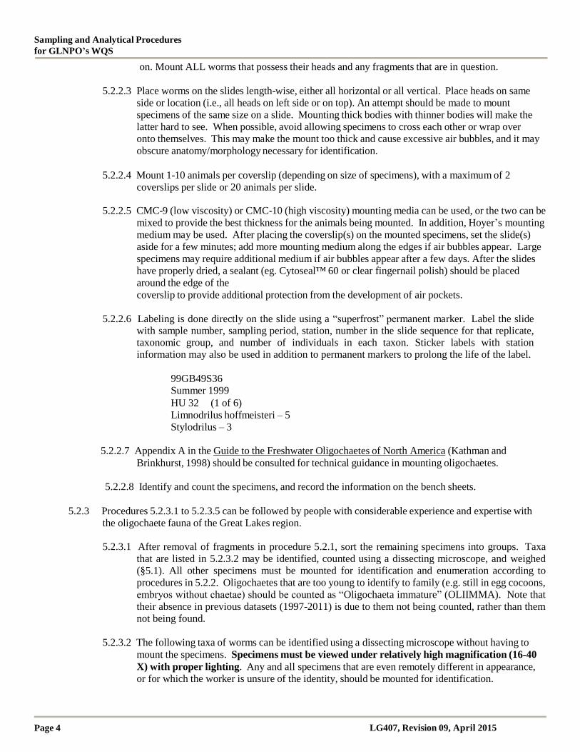
on. Mount ALL worms that possess their heads and any fragments that are in question.
5.2.2.3 Place worms on the slides length-wise, either all horizontal or all vertical. Place heads on same
side or location (i.e., all heads on left side or on top). An attempt should be made to mount
specimens of the same size on a slide. Mounting thick bodies with thinner bodies will make the
latter hard to see. When possible, avoid allowing specimens to cross each other or wrap over
onto themselves. This may make the mount too thick and cause excessive air bubbles, and it may
obscure anatomy/morphology necessary for identification.
5.2.2.4 Mount 1-10 animals per coverslip (depending on size of specimens), with a maximum of 2
coverslips per slide or 20 animals per slide.
5.2.2.5 CMC-9 (low viscosity) or CMC-10 (high viscosity) mounting media can be used, or the two can be
mixed to provide the best thickness for the animals being mounted. In addition, Hoyer’s mounting
medium may be used. After placing the coverslip(s) on the mounted specimens, set the slide(s)
aside for a few minutes; add more mounting medium along the edges if air bubbles appear. Large
specimens may require additional medium if air bubbles appear after a few days. After the slides
have properly dried, a sealant (eg. Cytoseal™ 60 or clear fingernail polish) should be placed
around the edge of the
coverslip to provide additional protection from the development of air pockets.
5.2.2.6 Labeling is done directly on the slide using a “superfrost” permanent marker. Label the slide
with sample number, sampling period, station, number in the slide sequence for that replicate, taxonomic group, and number of individuals in each taxon. Sticker labels with station information may also be used in addition to permanent markers to prolong the life of the label.
99GB49S36
Summer 1999
HU 32 (1 of 6)
Limnodrilus hoffmeisteri – 5
Stylodrilus – 3
5.2.2.7 Appendix A in the Guide to the Freshwater Oligochaetes of North America (Kathman and
Brinkhurst, 1998) should be consulted for technical guidance in mounting oligochaetes.
5.2.2.8 Identify and count the specimens, and record the information on the bench sheets.
5.2.3 Procedures 5.2.3.1 to 5.2.3.5 can be followed by people with considerable experience and expertise with
the oligochaete fauna of the Great Lakes region.
5.2.3.1 After removal of fragments in procedure 5.2.1, sort the remaining specimens into groups. Taxa
that are listed in 5.2.3.2 may be identified, counted using a dissecting microscope, and weighed
(§5.1). All other specimens must be mounted for identification and enumeration according to
procedures in 5.2.2. Oligochaetes that are too young to identify to family (e.g. still in egg cocoons,
embryos without chaetae) should be counted as “Oligochaeta immature” (OLIIMMA). Note that
their absence in previous datasets (1997-2011) is due to them not being counted, rather than them
not being found.
5.2.3.2 The following taxa of worms can be identified using a dissecting microscope without having to
mount the specimens. Specimens must be viewed under relatively high magnification (16-40
X) with proper lighting. Any and all specimens that are even remotely different in appearance,
or for which the worker is unsure of the identity, should be mounted for identification.
Standard Operating Procedure for Benthic
Invertebrate Laboratory Analysis
LG407, Revision 09, April 2015 Page 5
Lumbriculidae:
Tubificinae:
1) Stylodrilus heringianus Claparède, 1862, (a common species, many have their
penes protruding, often the only species present at great depths in the Great
Lakes) 1) immature tubificines without hairs (no sign of tissue density or sexual maturity in
the clitellar region. Warning: some species are small when they are mature, e.g.
Thalassodrilus hallae);
3) mature Potamothrix moldaviensis Vejdovsky and Mrazek, 1902 (with no hairs
and everted penes);
4) Potamothrix vejdovskyi (Hrabe, 1941) (with short, bent hairs. Warning: do
not confuse with Aulodrilus pluriseta);
5) Branchiura sowerbyi Beddard, 1892 (distinctive posterior gills);
6) Quistadrilus multisetosus (Smith, 1900) (very bristly with distinct papillae in
rings around the body); 7) Spirosperma ferox Eisen, 1879 (hairy, with fine whitish particulate matter on nearly
the entire body [often absent in clitellar region], bifid ventral chaetae anteriorly,
retractile prostomium, small papillae throughout body, often occurs in enriched
areas);
8) Spirosperma nikolskyi (Lastochkin and Sokolskaya, 1935) (also hairy, with
fine whitish particulate matter, simple chaetae anteriorly, occurs most often in
deeper oligotrophic areas).
Naidinae: 1) Arcteonais lomondi (Martin, 1907) (with short proboscis and long, bunched hairs);
2) Chaetogaster diaphanus (Gruithuisen, 1828) (short, chunky, anteriorly truncate);
3) Dero sp. (posterior gills);
4) Ophidonais serpentina (Muller, 1773) (eyes, transverse pigment stripes anteriorly,
no dorsal hairs, stout dorsal chaetae); 5) Slavina appendiculata (d’Udekem, 1855) (narrow, debris-laden, with long hairs
on segment VI);
6) Stylaria lacustris (Linnaeus, 1767) (a distinctive species with a proboscis);
7) Ripistes parasita (Schmidt, 1874) (dorsal chaetal bundles on segments VI-VIII
with very long hairs);
8) Pristina leidyi Smith, 1896 (very long dorsal hairs on segment III–unless broken
off).
Enchytraeidae
5.2.3.3 Temporary wet mounts may be done to verify a specimen under the compound microscope (e.g.
Spirosperma ferox vs. Spirosperma nikolskyi, Potamothrix vejdovskyi vs. Aulodrilus pluriseta).
After verification, the specimen(s) must be returned to the labeled vial.
5.2.3.4 Representative specimens from the above list must be initially mounted for verification under a
compound microscope; this should be accomplished when found for first time for each lake. The
remainder of the specimens of these species must be stored in separate, labeled vials containing
ethanol for archival storage; this must be completed for each site. Voucher specimens of these
taxa must be placed in the reference collection both as slide mounts and in vials.
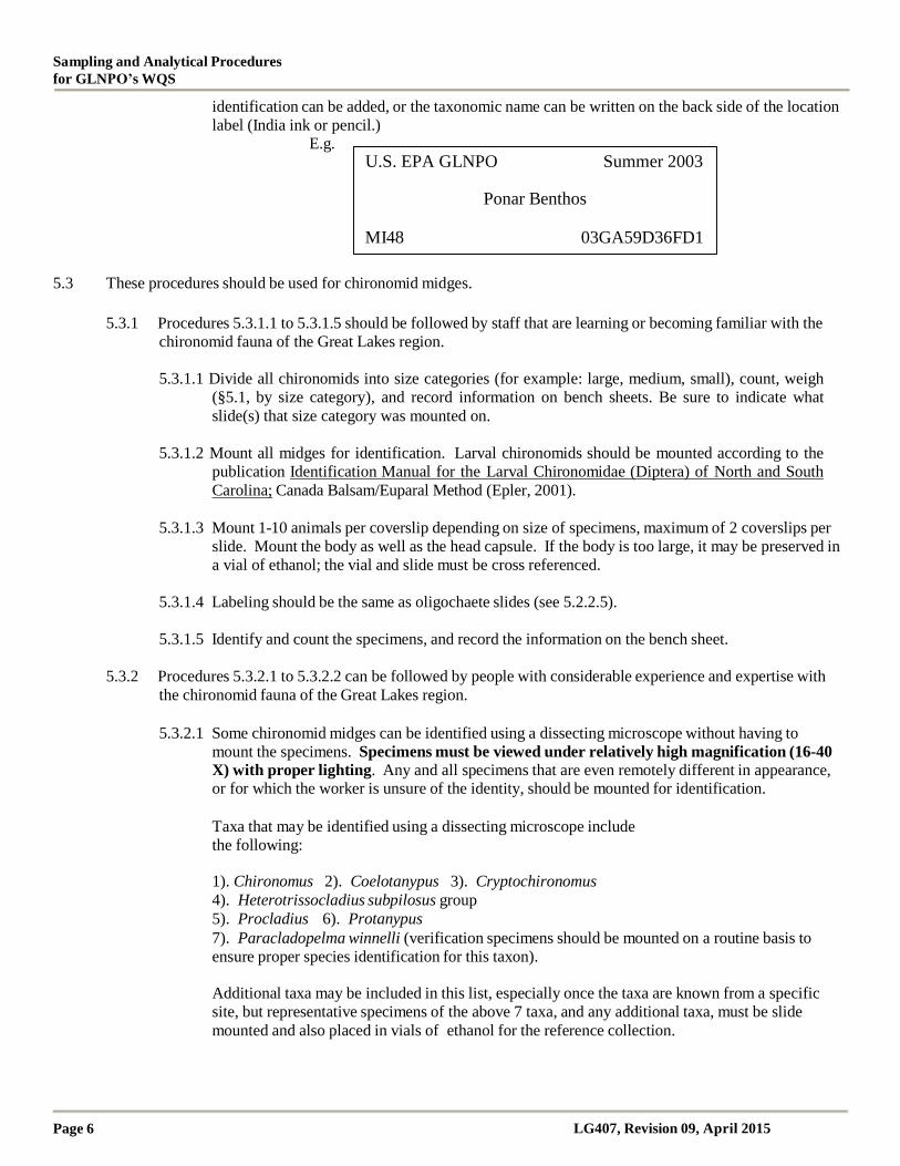
5.2.3.5 The label (Laser printing with acid-free paper containing 25% cotton) for a vial must include at
least the sample number, station number, and the sampling period. A separate label with the
Sampling and Analytical Procedures
for GLNPO’s WQS
Page 6 LG407, Revision 09, April 2015
identification can be added, or the taxonomic name can be written on the back side of the location
label (India ink or pencil.) E.g.
U.S. EPA GLNPO Summer 2003
Ponar Benthos
MI48 03GA59D36FD1
5.3 These procedures should be used for chironomid midges.
5.3.1 Procedures 5.3.1.1 to 5.3.1.5 should be followed by staff that are learning or becoming familiar with the
chironomid fauna of the Great Lakes region.
5.3.1.1 Divide all chironomids into size categories (for example: large, medium, small), count, weigh
(§5.1, by size category), and record information on bench sheets. Be sure to indicate what
slide(s) that size category was mounted on.
5.3.1.2 Mount all midges for identification. Larval chironomids should be mounted according to the
publication Identification Manual for the Larval Chironomidae (Diptera) of North and South
Carolina; Canada Balsam/Euparal Method (Epler, 2001).
5.3.1.3 Mount 1-10 animals per coverslip depending on size of specimens, maximum of 2 coverslips per
slide. Mount the body as well as the head capsule. If the body is too large, it may be preserved in
a vial of ethanol; the vial and slide must be cross referenced.
5.3.1.4 Labeling should be the same as oligochaete slides (see 5.2.2.5).
5.3.1.5 Identify and count the specimens, and record the information on the bench sheet.
5.3.2 Procedures 5.3.2.1 to 5.3.2.2 can be followed by people with considerable experience and expertise with
the chironomid fauna of the Great Lakes region.
5.3.2.1 Some chironomid midges can be identified using a dissecting microscope without having to
mount the specimens. Specimens must be viewed under relatively high magnification (16-40 X) with proper lighting. Any and all specimens that are even remotely different in appearance, or for which the worker is unsure of the identity, should be mounted for identification.
Taxa that may be identified using a dissecting microscope include
the following:
1). Chironomus 2). Coelotanypus 3). Cryptochironomus
4). Heterotrissocladius subpilosus group
5). Procladius 6). Protanypus
7). Paracladopelma winnelli (verification specimens should be mounted on a routine basis to
ensure proper species identification for this taxon).
Additional taxa may be included in this list, especially once the taxa are known from a specific
site, but representative specimens of the above 7 taxa, and any additional taxa, must be slide
mounted and also placed in vials of ethanol for the reference collection.

Standard Operating Procedure for Benthic
Invertebrate Laboratory Analysis
LG407, Revision 09, April 2015 Page 7
5.3.2.3 Temporary wet mounts may be done to verify a specimen under the compound microscope (eg.
Paracladopelma winnelli). After verification, the specimen(s) must be returned to the labeled
vial.
5.3.2.3 Representative specimens from the above list must be initially mounted for verification under a
compound microscope. The remainder of the specimens of these species must be stored in
separate, labeled vials containing ethanol for archival storage. This must be accomplished for
each year and lake. Voucher specimens of these taxa must be placed in the reference collection
both as slide mounts and in vials.
5.4 These procedures should be used for amphipods and all other non-slide mounted animals.
5.4.1 Identify, count and weigh (§5.1, in groups by species) the specimens, and record the information on the
appropriate bench sheet. Only specimens that possess their heads should be included for identification
and enumeration, even if only the head is the piece that is present; all other parts, pieces, exuviae, and
empty shells should be ignored. EXCEPTION: if enough of the body of a specimen (excluding exuviae
and empty shells) is present to make a valid identification, and it is assured that other body parts of the
specimen are not present in the sample (i.e. the specimen is in pieces and is being counted more than
once), then the piece may be included in the taxonomic analysis and enumeration. For example, if there
are two whole amphipods and one amphipod with the head missing (total of only three specimens), but the
technician can identify the headless animal, the specimen should be identified and counted. However, this
is not a common situation.
5.4.2 Return all the specimens for each taxon to their individual, labeled vial for archived sample storage.
5.5 Taxa that do not appear on a current species list for this project are periodically encountered. Specimens of new
taxa should be identified to the lowest possible taxonomic level, counted, weighed (§5.1) and properly preserved
in ethanol, or on a slide, or both. These individuals will serve as voucher specimens and should be sent to an expert
for external confirmation. A notice of confirmation by the outside expert should be submitted to the WAM. The
taxon can be added to the species list after written notification of acceptance by the WAM. The specimen(s)
should then be added to the reference collection.
6.0 SAMPLE PRESERVATION AND STORAGE
6.1 Slide-Mounted Organisms: Mounted chironomid midges and oligochaete worms will be stored as archived
samples.
6.2 Organisms in Vials: All invertebrates can be stored in their properly labeled scintillation vials with 70-80%
ethanol that contains 5% glycerin. The glycerin provides a lubricant to the sample in the event that the alcohol
evaporates from the vial, preventing the specimens from desiccating. The samples should be stored together
according to site, lake, season, and year, and placed into sealed containers. For long-term, archival storage at the
GLNPO facility, the sealed containers with vials should be placed into larger storage receptacles that contain
ethanol.
6.3 Spent Sediment: The remainder of the benthic sample, which may contain sediment, nematodes, and other non-
target organisms should remain preserved in the original sample bottle with formalin until proper QC checks are
completed. After data have been approved by the WAM, the sample may be properly discarded by the laboratory
personnel.
6.4 After the grant ends and all data have been submitted and approved by the WAM, all archived samples will be
shipped to, and stored at, the GLNPO.
Sampling and Analytical Procedures
for GLNPO’s WQS
Page 8 LG407, Revision 09, April 2015
7.0 DATA HANDLING AND CALCULATIONS
7.1 The data must be converted from estimated raw sample counts and weight to abundance and biomass per m2 for each
species by multiplying the totals by the number: 19.12. This conversion factor is derived from dimensions of the
Ponar grab sampler. If a different sampler is used the factor should be recalculated.
7.2 Calculate total abundance and biomass for each species and report the total of each species (or lowest taxonomic
level) per replicate.
8.0 SAFETY AND WASTE HANDLING
8.1 Personal protection equipment (safety glasses, gloves and lab coat) should be worn in the laboratory while
preparing and handling samples for analyses.
8.2 All samples preserved with formalin should be handled under the hood prior to being rinsed for sorting (section
4.2.1.).
8.3 Follow laboratory waste disposal guidelines regarding formalin solution waste. Sample waste should be emptied
into a waste container.
9.0 TRAINING AND QUALITY CONTROL
9.1 New analysts are required to receive formal training in the areas of terminology, anatomy, morphology, and
taxonomy of Great Lakes benthic invertebrates. This can be accomplished in one of two ways: instruction from a
senior benthic analyst in the laboratory, or by attending an external course taught by benthic specialists.
Acceptable training courses can be found by contacting either the North American Benthological Society or the
International Association of Great Lakes Research.
9.2 Quality Control Checks on the processing of benthic macroinvertebrates.
9.2.1 Picking Samples
The senior taxonomist or the lead assistant will “second pick” at least 10% of the samples picked by each
assistant. One sample from a block of 10 consecutively picked samples by each person will be randomly
chosen for second picking. Error percentages in picking should be less than 10%, preferably less than
5%.
However, samples that yield very low numbers of individuals can lead to high percentages of error (eg.
missing 1 specimen from a total of 4 specimens equals a 25% “picking error”). High error percentages
from these types of samples will be taken into consideration when determining if the sample passes or
fails the Quality Control Check. The main criteria in this determination will be deciding whether the error
affects the ecological interpretation of the data.
Samples that do not pass the QC Check will lead to the repicking of another sample within the assistant’s
block of 10 samples. If the second sample fails the QC Check, all of the samples within the block of 10
will be repicked by the same assistant. Another round of QC Checks will be repeated on the block of 10
samples.
During the initial training period of an assistant, additional samples will be 2nd
picked until it is assured
that the assistant is effectively removing 90-100% of the total number of organisms.
Samples containing fingernail clams (Sphaeriidae) will be given special attention during the training process and the random QA/QC checks. Their small size (some appear as large sand grains) and
Standard Operating Procedure for Benthic
Invertebrate Laboratory Analysis
LG407, Revision 09, April 2015 Page 9
resistance to stain (some appear translucent) cause them to sometimes be overlooked.
9.2.2 Taxonomy
Most, if not all, of the taxonomic identifications will be done by the senior or lead taxonomist(s).
Identifications may also be accomplished by a lead assistant(s) provided they have the training and
experience required to identify taxa from the Great Lakes.
Unusual or difficult oligochaetes, chironomids, or other taxa may be sent to acceptable outside taxonomic
experts for identification.
The taxonomy and enumeration for 10% of the samples must be checked for Quality Control. The QC
sample will be randomly chosen from batches of 10 samples. Specimens from the QC samples will be
analyzed by another trained and experienced individual within the same laboratory, or by someone in an
outside laboratory. Percent similarity of the two samples must exceed 90%. Samples that fail the QC
check must be examined to determine the type of problem encountered. The original lab should re-
examine all of the samples in the batch. Further training may be required for the lab personnel depending
on the type of errors encountered. This aspect should be worked out between the original lab, the EPA’s
WAM, and the personnel that conducted the QC check.
Oligochaete fragments will not be included in the QC check because they have the propensity to multiply
every time they are handled. However, they should be checked to ensure no identifiable specimens are
present.
10.0 TAXONOMIC REFERENCES
10.1 Primary Taxonomic Sources for Identification and Nomenclature
Merritt, R.W. and K.W. Cummins (eds.). 1996. An Introduction to the Aquatic Insects of North American. 3rd
Edition. Kendall/Hunt Publishing Co., Dubuque IA.
Pennak, R.W. 1989. Fresh-water Invertebrates of the United States. Porifera to Mollusca. 3rd Edition. John Wiley
& Sons, Inc. New York, NY.
Smith, D.G. 2001. Pennak's Freshwater Invertebrates of the United States. Porifera to Crustacea. 4th Edition.
John Wiley & Sons, Inc. New York, NY.
Thorp, J.H. and A.P. Covich. 2001. Ecology and Classification of North American Freshwater Invertebrates. 2nd
Edition. Academic Press. San Diego, CA.
10.2 Additional Taxonomic Sources for Species Identification
10.2.1 Insecta: Diptera: Chironomidae
Epler, J.H. 1987. Revision of the Nearctic Dicrotendipes Kieffer, 1913 (Diptera: Chironomidae).
Evolutionary Monographs 9. 102 pp + 37 plates.
Epler, J.H. 1995. Identification Manual for the Larval Chironomidae (Diptera) of Florida. Final Report
for DEP Contract Number WM579. Florida Department of Environmental Protection, Tallahassee.
Epler, J.H. 2001. Identification Manual for the Larval Chironomidae (Diptera) of North and South
Carolina. North Carolina Department of Environment and Natural Resources.

Sampling and Analytical Procedures
for GLNPO’s WQS
Page 10 LG407, Revision 09, April 2015
Jackson, G.A. 1977. Nearctic and Palaeartic Paracladopelma Harnisch and Saetheria
n.gen. (Diptera: Chironomidae). Journal of the Fisheries Research Board of Canada 34: 1321-1359.
Maschwitz, D.E. and E.F. Cook. 2000. Revision of the Nearctic species of the genus
Polypedilum Kieffer (Diptera: Chironomidae) in the subgenera P. (Polypedilum) Kieffer and P.
(Uresipedilum) Oyewo and Saether. Bulletin of the Ohio Biological Survey, New Series. Vol. 12, No. 3.
Oliver, D.R, M.E. Dillon. 1994. Corrections and additions to "A Catalog of Nearctic Chironomidae."
Proceedings of the Entomological Society of Washington 96:8-10.
Oliver, D.R, M.E. Dillon , and P.S. Cranston. 1990. A Catalog of Neararctic Chironomidae. Research
Branch, Agriculture Canada Publication No. 1857/B.
Roback, S.S. 1985. The immature chironomids of the eastern United State VI. Pentaneurini— genus
Ablabesmyia. Proceedings of the Academy of Natural Sciences of Philadelphia 137: 153-212.
Saether, O.A. 1973. Taxonomy and ecology of three new species of Monodiamesa Kieffer, with keys to
Nearctic and Palaearctic species of the genus (Diptera: Chironomidae). Journal of the Fisheries Research
Board of Canada 30: 665-679.
Walker, I.R., D.R. Oliver, and M.E. Dillon. 1992. The larva and habitat of Parakiefferiella nigra Brundin
(Diptera: Chironomidae). Netherlands Journal of Aquatic Ecology 26: 527-531.
Wiederholm, T. (ed.). 1983. Chironomidae of the Holarctic region -- keys and diagnoses. Part 1. Larvae.
Entomologica Scandinavica Supplement No.19.
10.2.2 Insecta: Ephemeroptera
McCafferty, W.P. 1975. The burrowing mayflies of the United States (Ephemeroptera: Ephemeroidea).
Transactions of the American Entomological Society 101:447-504.
10.2.3 Insecta: Trichoptera
Wiggins, G.B. 1996. Larvae of the North American Caddisfly Genera (Trichoptera). 2
nd Edition.
University of Toronto Press, Inc. Toronto, Ontario.
10.2.4 Insecta: Heteroptera
Hilsenhoff, W.L. 1984. Aquatic Hemiptera of Wisconsin. The Great Lakes Entomologist 17: 29-50.
Hungerford, H.B. 1948. The Corixidae of the Western Hemisphere (Hemiptera). University of Kansas
Science Bulletin 32: 1-288, 408-827.
10.2.5 Annelida
Kathman, R.D. and Brinkhurst, R.O. 1998 (Revised 1999). Guide to the Freshwater Oligochaetes of
North America. Aquatic Resources Center, College Grove, Tennessee.
Klemm, D.J. 1985. A Guide to the Freshwater Annelida (Polychaeta, Naidid and Tubificid Oligochaeta,
and Hirudinea) of North America. Kendall/Hunt Publishing Co. Dubuque, Iowa.
Krieger, K. A., and A. M. Stearns. 2010. Atlas of the Aquatic Oligochaete Worms (Phylum Annelida:
Standard Operating Procedure for Benthic
Invertebrate Laboratory Analysis
LG407, Revision 09, April 2015 Page
11
Class Clitellata: Superorder Microdrili) Recorded at the Old Woman Creek National Estuarine Research
Reserve and State Nature Preserve, Ohio. National Center for Water Quality Research, Heidelberg
University, Tiffin, Ohio. Available at:
http://www.dnr.state.oh.us/LinkClick.aspx?fileticket=Mu9FGNzuNbk%3D&tabid=15314
10.2.6 Mollusca
Burch, J.B. 1982. Freshwater Snails (Mollusca: Gastropoda) of North America. EPA-600/3-82-026. U.S.
EPA, Cinncinnati, Ohio.
Mackie, G.L., D.S. White, and T.W. Zdeba. 1980. A Guide to the Freshwater Mollusks of the Laurentian
Great Lakes with Special Emphasis on the Genus Pisidium. EPA-600/3-80-068. U.S. EPA, Duluth, MN.
10.2.7 Amphipoda
Bousfield, E.L. 1958. Fresh-water amphipod crustaceans of glaciated North America. The Canadian
Field-Naturalist 72: 55-113
Grigorovich, I.A., M. Kang, and J.J.H. Ciborowski. 2005. Colonization of the Laurentian Great Lakes by
the amphipod Gammarus tigrinus, a native of the North American Atlantic Coast. Journal of Great Lakes
Research 31: 333-342.
Holsinger, J.R. 1972. The Freshwater Amphipod Crustaceans (Gammaridae) of North America. U.S.
EPA Biota of Freshwater Ecosystems, Identification Manual No. 5.
Witt, D.S., P.D.N. Hebert, and W.B. Morton. 1997. Echinogammarus ischnus: another crustacean
invader in the Laurentian Great Lakes basin. Canadian Journal of Fisheries and Aquatic Sciences 54: 264-268.
10.2.8 Isopoda
Williams, W.D. 1972. Freshwater isopods (Asellidae) of North America. U.S. EPA Biota of Freshwater
Ecosystems, Identification Manual No. 7.