STANDARD OPERATING PROCEDURE - ASSIST Performing a rat... · Care should be taken with sharps such...
12
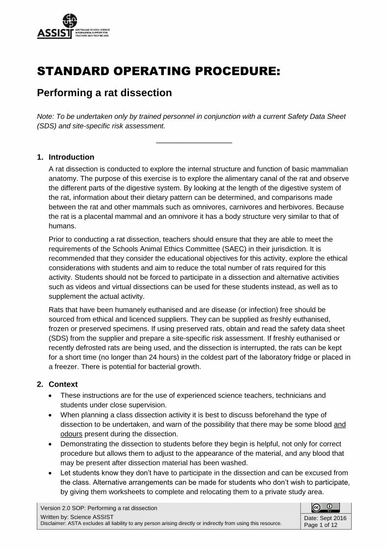
Version 2.0 SOP: Performing a rat dissection Written by: Science ASSIST Disclaimer: ASTA excludes all liability to any person arising directly or indirectly from using this resource. Date: Sept 2016 Page 1 of 12 STANDARD OPERATING PROCEDURE: Performing a rat dissection Note: To be undertaken only by trained personnel in conjunction with a current Safety Data Sheet (SDS) and site-specific risk assessment. ___________________ 1. Introduction A rat dissection is conducted to explore the internal structure and function of basic mammalian anatomy. The purpose of this exercise is to explore the alimentary canal of the rat and observe the different parts of the digestive system. By looking at the length of the digestive system of the rat, information about their dietary pattern can be determined, and comparisons made between the rat and other mammals such as omnivores, carnivores and herbivores. Because the rat is a placental mammal and an omnivore it has a body structure very similar to that of humans. Prior to conducting a rat dissection, teachers should ensure that they are able to meet the requirements of the Schools Animal Ethics Committee (SAEC) in their jurisdiction. It is recommended that they consider the educational objectives for this activity, explore the ethical considerations with students and aim to reduce the total number of rats required for this activity. Students should not be forced to participate in a dissection and alternative activities such as videos and virtual dissections can be used for these students instead, as well as to supplement the actual activity. Rats that have been humanely euthanised and are disease (or infection) free should be sourced from ethical and licenced suppliers. They can be supplied as freshly euthanised, frozen or preserved specimens. If using preserved rats, obtain and read the safety data sheet (SDS) from the supplier and prepare a site-specific risk assessment. If freshly euthanised or recently defrosted rats are being used, and the dissection is interrupted, the rats can be kept for a short time (no longer than 24 hours) in the coldest part of the laboratory fridge or placed in a freezer. There is potential for bacterial growth. 2. Context These instructions are for the use of experienced science teachers, technicians and students under close supervision. When planning a class dissection activity it is best to discuss beforehand the type of dissection to be undertaken, and warn of the possibility that there may be some blood and odours present during the dissection. Demonstrating the dissection to students before they begin is helpful, not only for correct procedure but allows them to adjust to the appearance of the material, and any blood that may be present after dissection material has been washed. Let students know they don’t have to participate in the dissection and can be excused from the class. Alternative arrangements can be made for students who don’t wish to participate, by giving them worksheets to complete and relocating them to a private study area.
Transcript of STANDARD OPERATING PROCEDURE - ASSIST Performing a rat... · Care should be taken with sharps such...
Version 2.0 SOP: Performing a rat dissection
Written by: Science ASSIST Disclaimer: ASTA excludes all liability to any person arising directly or indirectly from using this resource.
Date: Sept 2016 Page 1 of 12
STANDARD OPERATING PROCEDURE:
Performing a rat dissection
Note: To be undertaken only by trained personnel in conjunction with a current Safety Data Sheet
(SDS) and site-specific risk assessment.
___________________
1. Introduction
A rat dissection is conducted to explore the internal structure and function of basic mammalian
anatomy. The purpose of this exercise is to explore the alimentary canal of the rat and observe
the different parts of the digestive system. By looking at the length of the digestive system of
the rat, information about their dietary pattern can be determined, and comparisons made
between the rat and other mammals such as omnivores, carnivores and herbivores. Because
the rat is a placental mammal and an omnivore it has a body structure very similar to that of
humans.
Prior to conducting a rat dissection, teachers should ensure that they are able to meet the
requirements of the Schools Animal Ethics Committee (SAEC) in their jurisdiction. It is
recommended that they consider the educational objectives for this activity, explore the ethical
considerations with students and aim to reduce the total number of rats required for this
activity. Students should not be forced to participate in a dissection and alternative activities
such as videos and virtual dissections can be used for these students instead, as well as to
supplement the actual activity.
Rats that have been humanely euthanised and are disease (or infection) free should be
sourced from ethical and licenced suppliers. They can be supplied as freshly euthanised,
frozen or preserved specimens. If using preserved rats, obtain and read the safety data sheet
(SDS) from the supplier and prepare a site-specific risk assessment. If freshly euthanised or
recently defrosted rats are being used, and the dissection is interrupted, the rats can be kept
for a short time (no longer than 24 hours) in the coldest part of the laboratory fridge or placed in
a freezer. There is potential for bacterial growth.
2. Context
These instructions are for the use of experienced science teachers, technicians and
students under close supervision.
When planning a class dissection activity it is best to discuss beforehand the type of
dissection to be undertaken, and warn of the possibility that there may be some blood and
odours present during the dissection.
Demonstrating the dissection to students before they begin is helpful, not only for correct
procedure but allows them to adjust to the appearance of the material, and any blood that
may be present after dissection material has been washed.
Let students know they don’t have to participate in the dissection and can be excused from
the class. Alternative arrangements can be made for students who don’t wish to participate,
by giving them worksheets to complete and relocating them to a private study area.

Version 2.0 SOP: Performing a rat dissection
Written by: Science ASSIST Disclaimer: ASTA excludes all liability to any person arising directly or indirectly from using this resource.
Date: Sept 2016 Page 2 of 12
3. Safety notes
Fainting: signs and symptoms:
Fainting may occur during this type of activity. Please read the first aid information in
section 7 before conducting the dissection.
Fainting is caused by a sudden drop in blood pressure. Common causes include heat, pain
or distress and the sight of blood.
The possible symptoms include the following.
o ‘Dizziness
o Light-headedness
o A pale face
o Perspiration
o Heightened anxiety and restlessness
o Nausea
o Collapse
o Unconsciousness, for a few seconds
o Full recovery after a few minutes’i
Handling specimens:
If using preserved rats, it is important to take note of the preservative solution that the rats
are in and the recommended precautions.
o Rinse the preserved rats under running water immediately upon removal from the
preservative solution.
o Work in a well-ventilated area.
o It is recommended that people wearing contact lenses should not dissect rats that are in
preservative solution. The fumes from the solution can penetrate between the eye and
contact lens causing irritation to eyes. It is recommended to wear prescription glasses
instead with safety glasses over them OR prescription safety glasses.
Consider issues such as allergies and chemical sensitivities from handling freshly
euthanised or recently defrosted or preserved rats.
Be aware of possible microbial aerosols and unpleasant odours released from freshly
euthanised or recently defrosted rats if the stomach/intestines are accidently cut.
If using frozen rats, defrost overnight in a refrigerator and use within 24 hours. Consistent
with safe food handling procedures, all meat products should be stored below 5ºC prior to
performing any dissections.
Good hygiene practices should be observed at all times: Keep hands away from the mouth,
nose, eyes and face during and after dissection and wash hands immediately after handling
dissection material.
Safety with scalpels and dissecting instruments:
Store all dissecting instruments securely.
Care should be taken with sharps such as scalpel blades and scissors. Some school
science departments restrict the use of scalpels unless specifically requested by a teacher,
and prefer to only issue scissors, probes and forceps to students for dissections.
Ensure students demonstrate responsible behaviour while using scalpels and other
dissecting instruments.
Version 2.0 SOP: Performing a rat dissection
Written by: Science ASSIST Disclaimer: ASTA excludes all liability to any person arising directly or indirectly from using this resource.
Date: Sept 2016 Page 3 of 12
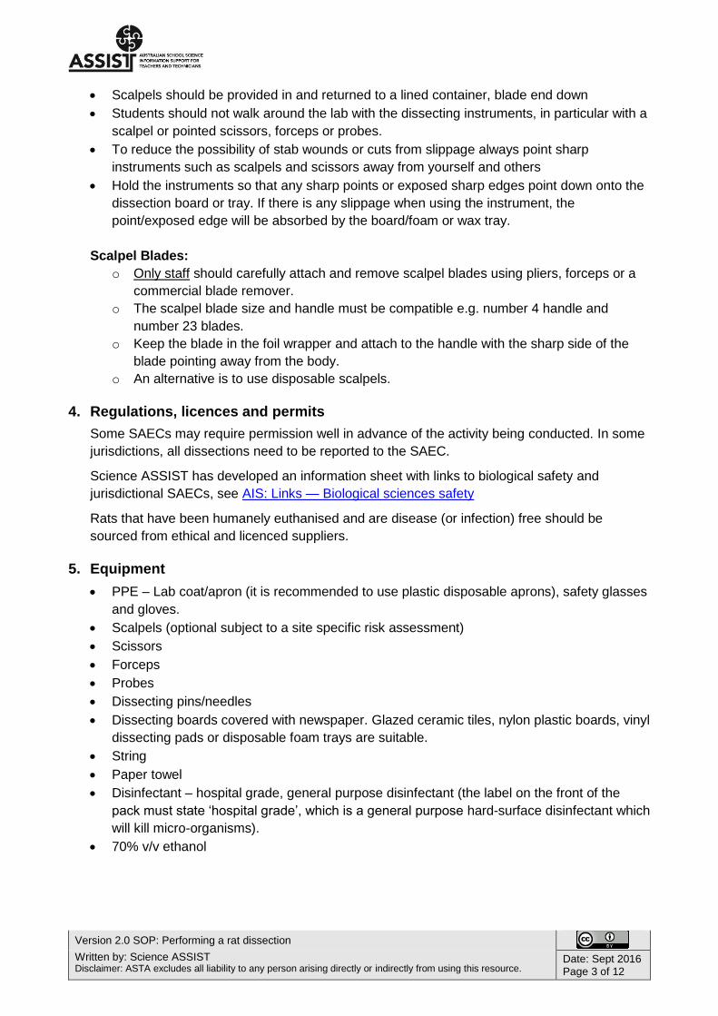
Scalpels should be provided in and returned to a lined container, blade end down
Students should not walk around the lab with the dissecting instruments, in particular with a
scalpel or pointed scissors, forceps or probes.
To reduce the possibility of stab wounds or cuts from slippage always point sharp
instruments such as scalpels and scissors away from yourself and others
Hold the instruments so that any sharp points or exposed sharp edges point down onto the
dissection board or tray. If there is any slippage when using the instrument, the
point/exposed edge will be absorbed by the board/foam or wax tray.
Scalpel Blades:
o Only staff should carefully attach and remove scalpel blades using pliers, forceps or a
commercial blade remover.
o The scalpel blade size and handle must be compatible e.g. number 4 handle and
number 23 blades.
o Keep the blade in the foil wrapper and attach to the handle with the sharp side of the
blade pointing away from the body.
o An alternative is to use disposable scalpels.
4. Regulations, licences and permits
Some SAECs may require permission well in advance of the activity being conducted. In some
jurisdictions, all dissections need to be reported to the SAEC.
Science ASSIST has developed an information sheet with links to biological safety and
jurisdictional SAECs, see AIS: Links — Biological sciences safety
Rats that have been humanely euthanised and are disease (or infection) free should be
sourced from ethical and licenced suppliers.
5. Equipment
PPE – Lab coat/apron (it is recommended to use plastic disposable aprons), safety glasses
and gloves.
Scalpels (optional subject to a site specific risk assessment)
Scissors
Forceps
Probes
Dissecting pins/needles
Dissecting boards covered with newspaper. Glazed ceramic tiles, nylon plastic boards, vinyl
dissecting pads or disposable foam trays are suitable.
String
Paper towel
Disinfectant – hospital grade, general purpose disinfectant (the label on the front of the
pack must state ‘hospital grade’, which is a general purpose hard-surface disinfectant which
will kill micro-organisms).
70% v/v ethanol
Version 2.0 SOP: Performing a rat dissection
Written by: Science ASSIST Disclaimer: ASTA excludes all liability to any person arising directly or indirectly from using this resource.
Date: Sept 2016 Page 4 of 12
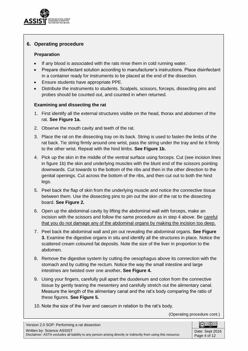
6. Operating procedure
Preparation
If any blood is associated with the rats rinse them in cold running water.
Prepare disinfectant solution according to manufacturer’s instructions. Place disinfectant
in a container ready for instruments to be placed at the end of the dissection.
Ensure students have appropriate PPE.
Distribute the instruments to students. Scalpels, scissors, forceps, dissecting pins and
probes should be counted out, and counted in when returned.
Examining and dissecting the rat
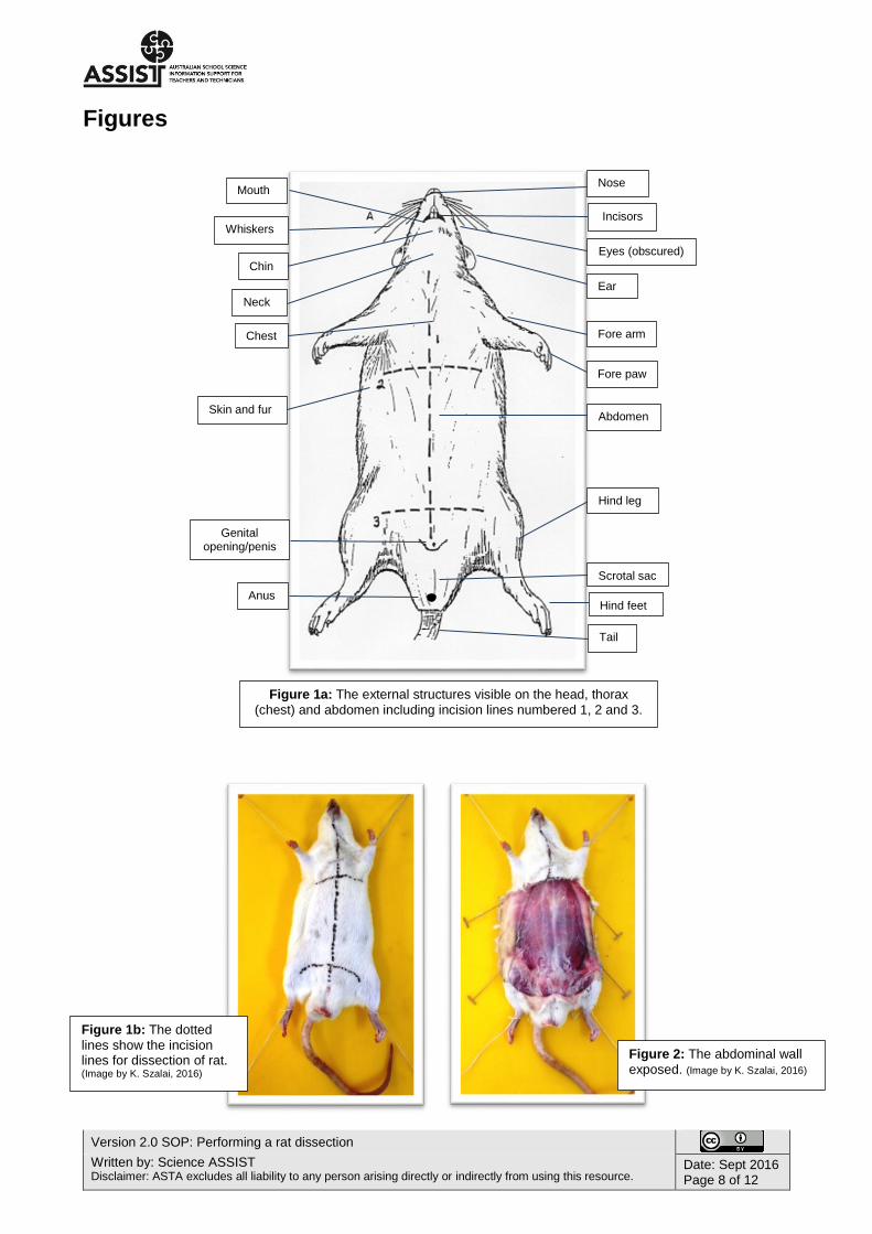
1. First identify all the external structures visible on the head, thorax and abdomen of the
rat. See Figure 1a.
2. Observe the mouth cavity and teeth of the rat.
3. Place the rat on the dissecting tray on its back. String is used to fasten the limbs of the
rat back. Tie string firmly around one wrist, pass the string under the tray and tie it firmly
to the other wrist. Repeat with the hind limbs. See Figure 1b.
4. Pick up the skin in the middle of the ventral surface using forceps. Cut (see incision lines
in figure 1b) the skin and underlying muscles with the blunt end of the scissors pointing
downwards. Cut towards to the bottom of the ribs and then in the other direction to the
genital openings. Cut across the bottom of the ribs, and then cut out to both the hind
legs.
5. Peel back the flap of skin from the underlying muscle and notice the connective tissue
between them. Use the dissecting pins to pin out the skin of the rat to the dissecting
board. See Figure 2.
6. Open up the abdominal cavity by lifting the abdominal wall with forceps, make an
incision with the scissors and follow the same procedure as in step 4 above. Be careful
that you do not damage any of the abdominal organs by making the incision too deep.
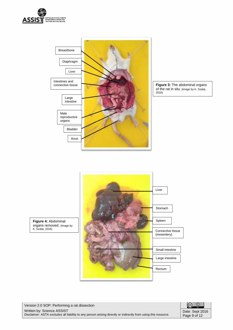
7. Peel back the abdominal wall and pin out revealing the abdominal organs. See Figure
3. Examine the digestive organs in situ and identify all the structures in place. Notice the
scattered cream coloured fat deposits. Note the size of the liver in proportion to the
abdomen.
8. Remove the digestive system by cutting the oesophagus above its connection with the
stomach and by cutting the rectum. Notice the way the small intestine and large
intestines are twisted over one another. See Figure 4.
9. Using your fingers, carefully pull apart the duodenum and colon from the connective
tissue by gently tearing the mesentery and carefully stretch out the alimentary canal.
Measure the length of the alimentary canal and the rat’s body comparing the ratio of
these figures. See Figure 5.
10. Note the size of the liver and caecum in relation to the rat’s body.
(Operating procedure cont.)
Version 2.0 SOP: Performing a rat dissection
Written by: Science ASSIST Disclaimer: ASTA excludes all liability to any person arising directly or indirectly from using this resource.
Date: Sept 2016 Page 5 of 12
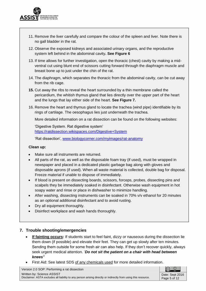
7. Trouble shooting/emergencies
If fainting occurs: If students start to feel faint, dizzy or nauseous during the dissection lie
them down (if possible) and elevate their feet. They can get up slowly after ten minutes.
Sending them outside for some fresh air can also help. If they don’t recover quickly, always
seek urgent medical attention. ‘Do not sit the patient on a chair with head between
knees’ii
First Aid: See latest SDS of any chemicals used for more detailed information.
11. Remove the liver carefully and compare the colour of the spleen and liver. Note there is
no gall bladder in the rat.
12. Observe the exposed kidneys and associated urinary organs, and the reproductive
system left behind in the abdominal cavity. See Figure 6
13. If time allows for further investigation, open the thoracic (chest) cavity by making a mid-
ventral cut using blunt end of scissors cutting forward through the diaphragm muscle and
breast bone up to just under the chin of the rat.
14. The diaphragm, which separates the thoracic from the abdominal cavity, can be cut away
from the rib cage.
15. Cut away the ribs to reveal the heart surrounded by a thin membrane called the
pericardium, the whitish thymus gland that lies directly over the upper part of the heart
and the lungs that lay either side of the heart. See Figure 7.
16. Remove the heart and thymus gland to locate the trachea (wind pipe) identifiable by its
rings of cartilage. The oesophagus lies just underneath the trachea.
More detailed information on a rat dissection can be found on the following websites:
‘Digestive System. Rat digestive system’
https://ratdissection.wikispaces.com/Digestive+System
‘Rat dissection’, www.biologycorner.com/myimages/rat-anatomy
Clean up:
Make sure all instruments are returned.
All parts of the rat, as well as the disposable foam tray (if used), must be wrapped in
newspaper and placed in a dedicated plastic garbage bag along with gloves and
disposable aprons (if used). When all waste material is collected, double bag for disposal.
Freeze material if unable to dispose of immediately.
If blood is present on dissecting boards, scissors, forceps, probes, dissecting pins and
scalpels they be immediately soaked in disinfectant. Otherwise wash equipment in hot
soapy water and rinse or place in dishwasher to minimize handling.
After washing, dissecting instruments can be soaked in 70% v/v ethanol for 20 minutes
as an optional additional disinfectant and to avoid rusting.
Dry all equipment thoroughly.
Disinfect workplace and wash hands thoroughly.
Version 2.0 SOP: Performing a rat dissection
Written by: Science ASSIST Disclaimer: ASTA excludes all liability to any person arising directly or indirectly from using this resource.
Date: Sept 2016 Page 6 of 12
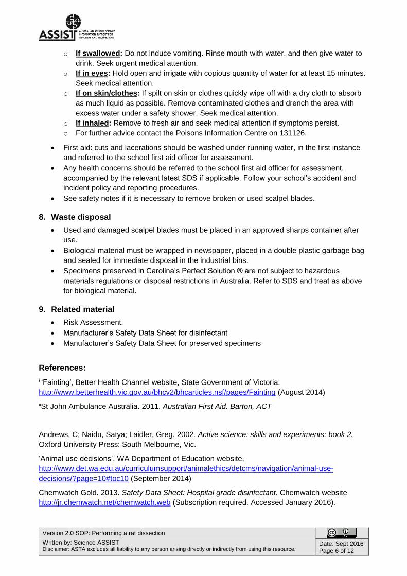
o If swallowed: Do not induce vomiting. Rinse mouth with water, and then give water to
drink. Seek urgent medical attention.
o If in eyes: Hold open and irrigate with copious quantity of water for at least 15 minutes.
Seek medical attention.
o If on skin/clothes: If spilt on skin or clothes quickly wipe off with a dry cloth to absorb
as much liquid as possible. Remove contaminated clothes and drench the area with
excess water under a safety shower. Seek medical attention.
o If inhaled: Remove to fresh air and seek medical attention if symptoms persist.
o For further advice contact the Poisons Information Centre on 131126.
First aid: cuts and lacerations should be washed under running water, in the first instance
and referred to the school first aid officer for assessment.
Any health concerns should be referred to the school first aid officer for assessment,
accompanied by the relevant latest SDS if applicable. Follow your school’s accident and
incident policy and reporting procedures.
See safety notes if it is necessary to remove broken or used scalpel blades.
8. Waste disposal
Used and damaged scalpel blades must be placed in an approved sharps container after
use.
Biological material must be wrapped in newspaper, placed in a double plastic garbage bag
and sealed for immediate disposal in the industrial bins.
Specimens preserved in Carolina’s Perfect Solution ® are not subject to hazardous
materials regulations or disposal restrictions in Australia. Refer to SDS and treat as above
for biological material.
9. Related material
Risk Assessment.
Manufacturer’s Safety Data Sheet for disinfectant
Manufacturer’s Safety Data Sheet for preserved specimens
References:
i ‘Fainting’, Better Health Channel website, State Government of Victoria:
http://www.betterhealth.vic.gov.au/bhcv2/bhcarticles.nsf/pages/Fainting (August 2014)
iiSt John Ambulance Australia. 2011. Australian First Aid. Barton, ACT
Andrews, C; Naidu, Satya; Laidler, Greg. 2002. Active science: skills and experiments: book 2.
Oxford University Press: South Melbourne, Vic.
‘Animal use decisions’, WA Department of Education website,
http://www.det.wa.edu.au/curriculumsupport/animalethics/detcms/navigation/animal-use-
decisions/?page=10#toc10 (September 2014)
Chemwatch Gold. 2013. Safety Data Sheet: Hospital grade disinfectant. Chemwatch website
http://jr.chemwatch.net/chemwatch.web (Subscription required. Accessed January 2016).
Version 2.0 SOP: Performing a rat dissection
Written by: Science ASSIST Disclaimer: ASTA excludes all liability to any person arising directly or indirectly from using this resource.
Date: Sept 2016 Page 7 of 12
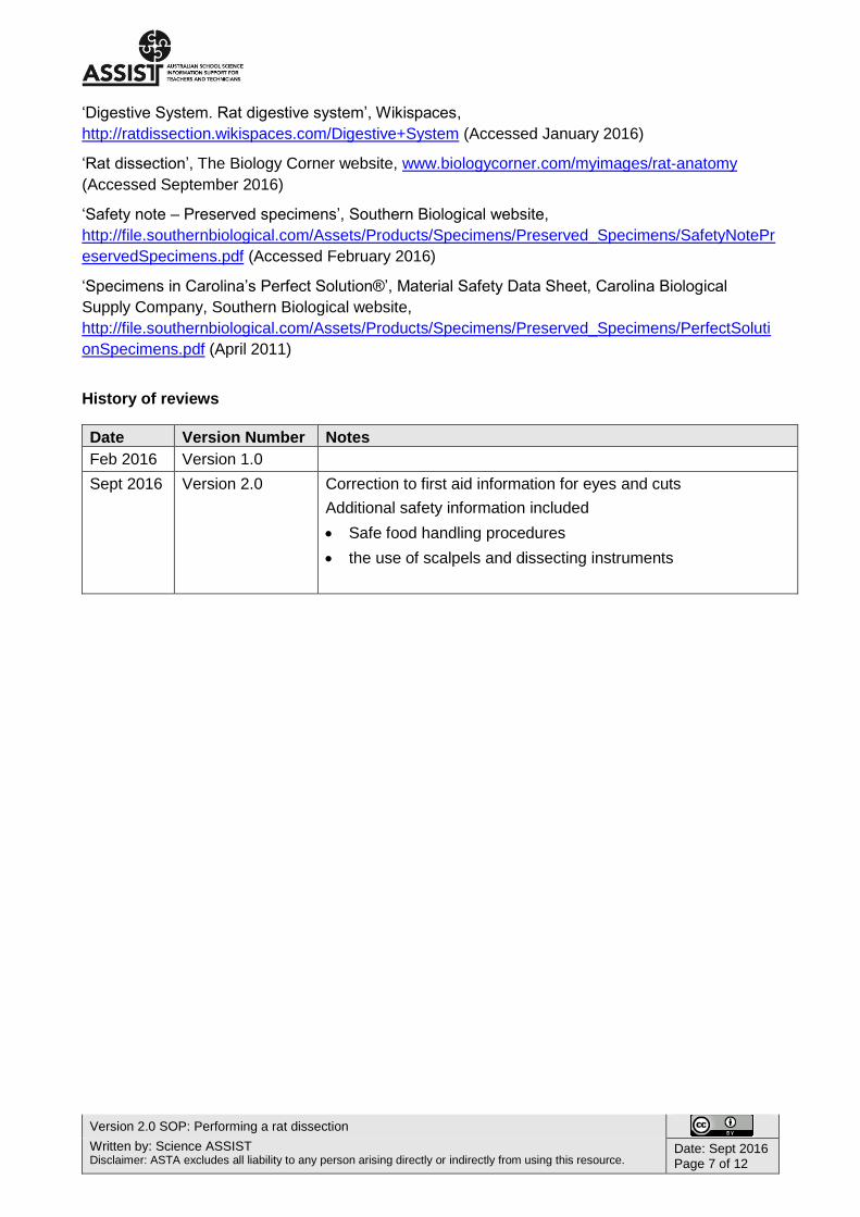
‘Digestive System. Rat digestive system’, Wikispaces,
http://ratdissection.wikispaces.com/Digestive+System (Accessed January 2016)
‘Rat dissection’, The Biology Corner website, www.biologycorner.com/myimages/rat-anatomy
(Accessed September 2016)
‘Safety note – Preserved specimens’, Southern Biological website,
http://file.southernbiological.com/Assets/Products/Specimens/Preserved_Specimens/SafetyNotePr
eservedSpecimens.pdf (Accessed February 2016)
‘Specimens in Carolina’s Perfect Solution®’, Material Safety Data Sheet, Carolina Biological
Supply Company, Southern Biological website,
http://file.southernbiological.com/Assets/Products/Specimens/Preserved_Specimens/PerfectSoluti
onSpecimens.pdf (April 2011)
History of reviews
Date Version Number Notes
Feb 2016 Version 1.0
Sept 2016 Version 2.0 Correction to first aid information for eyes and cuts
Additional safety information included
Safe food handling procedures
the use of scalpels and dissecting instruments
Version 2.0 SOP: Performing a rat dissection
Written by: Science ASSIST Disclaimer: ASTA excludes all liability to any person arising directly or indirectly from using this resource.
Date: Sept 2016 Page 8 of 12
Figures
Mouth
Whiskers
Chin
Neck
Chest
Skin and fur
Genital opening/penis
Anus
Nose
Incisors
Ear
Fore arm
Fore paw
Abdomen
Hind leg
Tail
Scrotal sac
Hind feet
Eyes (obscured)
Figure 1a: The external structures visible on the head, thorax
(chest) and abdomen including incision lines numbered 1, 2 and 3.
Figure 1b: The dotted
lines show the incision lines for dissection of rat. (Image by K. Szalai, 2016)
Figure 2: The abdominal wall
exposed. (Image by K. Szalai, 2016)
Version 2.0 SOP: Performing a rat dissection
Written by: Science ASSIST Disclaimer: ASTA excludes all liability to any person arising directly or indirectly from using this resource.
Date: Sept 2016 Page 9 of 12
Intestines and connective tissue
Breastbone
Diaphragm
Large intestine
Male reproductive organs
Bladder
Anus
Figure 3: The abdominal organs
of the rat in situ. (Image by K. Szalai,
2016)
Liver
Stomach
Spleen
Connective tissue (mesentery)
Small intestine
Large intestine
Rectum
Figure 4: Abdominal
organs removed. (Image by
K. Szalai, 2016)
Liver

Version 2.0 SOP: Performing a rat dissection
Written by: Science ASSIST Disclaimer: ASTA excludes all liability to any person arising directly or indirectly from using this resource.
Date: Sept 2016 Page 10 of 12
Liver
Stomach
Spleen
Pancreas
Duodenum
Large intestine
Caecum
Colon
Small intestine
Appendix
Anus
Figure 5: The alimentary
canal of the rat with the connective tissue removed. (Image by K. Szalai, 2016)
Kidney
Protective fat
Seminal Vesicle
Protective fat layer
Bladder
Testes
Penis
Figure 6: Male Urinogenital
system (Image by K. Szalai, 2016)
Thymus gland
Lungs. Right lobe. Probe points to left lobe.
Heart
Diaphragm
Figure 7: Thorax and neck. (Image by
K. Szalai, 2016)
Thyroid gland and trachea
Rectum

Version 2.0 SOP: Performing a rat dissection
Written by: Science ASSIST Disclaimer: ASTA excludes all liability to any person arising directly or indirectly from using this resource.
Date: Sept 2016 Page 11 of 12
Glossary
Abdominal wall – the lining that encloses the abdominal cavity, consisting mostly of muscle.
Alimentary canal – the tubular passage between the mouth and the anus.
Anus – the last part of the large intestine through which the faeces are excreted.
Appendix – a small tube with only one opening, near the beginning of the large intestine.
Bladder – the organ where urine is stored in the body.
Blood – liquid that circulates in the blood vessels of many animals. It contains plasma, red and
white blood cells, platelets, dissolved gases, nutrients, hormones and salts.
Breast bone – the long flat bone shaped in the middle of the chest, forming the front of the rib
cage.
Caecum – large organ where the small intestine joins the large intestine.
Carnivore – an animal that eats only meat.
Cartilage – flexible connective tissue in animals, including the joints between bones, the rib cage,
the ear, the nose, the bronchial tubes and the intervertebral discs.
Colon – the section of the large intestine that runs from the caecum to the rectum and absorbs
water from faeces.
Connective tissue – animal tissue that supports, connects, and surrounds organs and other body
parts.
Diaphragm – the muscle that runs across the base of the chest cavity and separates the thorax
and the abdomen. It causes the lungs to expand and contract during breathing.
Digestion – the process of converting large, complex organic molecules to smaller, simpler ones
that can pass through cell membranes.
Digestive system – the parts of the body where food is digested.
Duodenum – the top part of the small intestine, where bile and pancreatic juice enter the digestive
system.
Faeces – undigested food and bacteria that is stored in the rectum and expelled through the anus.
Gall Bladder – small organ that is located beneath the liver and drains bile into the duodenum
primarily to break down fat during digestion.
Heart – muscular organ that pumps blood around the body.
Herbivore – an animal that eats only plants.
Incisors – the two front teeth in each jaw of a mammal used for cutting food.
In situ – in its original place.
Kidneys – the major excretory organs that filter the blood, and maintain the concentration of water
and salts in the body. They form urine in which excess salts, water urea and waste products are
removed from the body.

Version 2.0 SOP: Performing a rat dissection
Written by: Science ASSIST Disclaimer: ASTA excludes all liability to any person arising directly or indirectly from using this resource.
Date: Sept 2016 Page 12 of 12
Large intestine – the end section of the alimentary canal reaching from ileum to anus and
consisting of the caecum, colon, and rectum. Its function is to extract water and form faeces.
Liver – a large organ in mammals that controls the level of nutrients and toxic substances in the
blood. Has a digestive function, secretes bile, filters blood, and takes part in many metabolic
functions such as the conversion of sugars into glycogen.
Lungs – inflatable organs used to breathe in oxygen and breathe out carbon dioxide.
Mammal – class of warm-blooded vertebrate animals.
Mechanical digestion – the breaking down of large pieces of food into smaller pieces.
Mesentery – a supportive membrane surrounding and giving structure to the inner organs.
Oesophagus – the passage down which food moves between the throat and the stomach.
Omnivore – an animal that eats both plants and animals.
Pancreas – an organ in the body that produces pancreatic juice that helps to digest food.
Penis – male sex organ used to transfer semen and expel urine from the body.
Pericardium – membrane that forms a sac surrounding the heart and attached portions of the
main blood vessels.
Placental mammal – the embryo develops in the maternal uterus attached to the tissues of the
placenta.
Rectum – the last part of the large intestine where faeces is stored.
Scrotal sac – the sac of skin that holds the testicles.
Seminal vesicle – the gland that secretes seminal fluid in semen.
Small intestine – the part of the intestine between the stomach and the large intestine, consisting
of the duodenum, jejunum, and ileum, where digestion of food and most absorption of nutrients
take place.
Spleen – organ in the left upper abdomen of humans and other vertebrates that helps to destroy
old red blood cells, form lymphocytes, and store blood.
Stomach – a hollow muscular organ where mechanical and chemical digestion takes place.
Testes – male sex organs that produce sperm.
Thoracic –relates to the chest and cavity of an animal.
Thorax – the part of the body between the neck and abdomen, enclosed by the ribs and containing
lungs, heart and diaphragm.
Thymus – the organ, located at the base of the neck, and is involved in development of cells of the
immune system.
Thyroid gland – endocrine gland located in the neck of human beings and other vertebrate
animals that secretes the hormones responsible for controlling metabolism and growth.
Trachea – a tube through which air enters the lungs from the nose.
Ventral surface – the surface of the abdomen or lower body.