Stable DNA Replication: Interplay between DNA Replication ... · Thus, addition of chloramphenicol...
27
MICROBIOLOGY AND MOLECULAR BIOLOGY REVIEWS, 1092-2172/97/$04.0010 June 1997, p. 212–238 Vol. 61, No. 2 Copyright © 1997, American Society for Microbiology Stable DNA Replication: Interplay between DNA Replication, Homologous Recombination, and Transcription TOKIO KOGOMA* Departments of Cell Biology and Microbiology and Cancer Center, University of New Mexico Health Sciences Center, Albuquerque, New Mexico 87131 INTRODUCTION .......................................................................................................................................................213 INDUCIBLE STABLE DNA REPLICATION .........................................................................................................214 Inducing Conditions ...............................................................................................................................................214 Mode of Replication ...............................................................................................................................................214 Origin Usage in iSDR ............................................................................................................................................214 Gene Product Requirement ...................................................................................................................................215 DnaA independence ............................................................................................................................................215 RecA protein ........................................................................................................................................................215 RecBCD enzyme ..................................................................................................................................................215 Other rec gene products .....................................................................................................................................216 Priming proteins .................................................................................................................................................216 DNA polymerases ................................................................................................................................................216 Proposed Models for iSDR Initiation ..................................................................................................................216 Stable-complex model .........................................................................................................................................216 Onion skin model................................................................................................................................................217 D-loop model .......................................................................................................................................................217 Evidence for the D-Loop Model ............................................................................................................................217 Roles of SOS in iSDR Initiation...........................................................................................................................219 RECOMBINATION-DEPENDENT DNA REPLICATION ...................................................................................219 RDR Model ..............................................................................................................................................................219 iSDR Is a Special Type of RDR............................................................................................................................219 POSSIBLE ROLES OF iSDR ...................................................................................................................................219 Damage-Resistant Replication ..............................................................................................................................219 Error-Prone Replication ........................................................................................................................................219 Possible Role in Adaptive Mutation.....................................................................................................................220 RDR AND DOUBLE-STRAND BREAK REPAIR..................................................................................................220 Role of RDR in DSB Repair .................................................................................................................................220 RDR-mediated DSB repair model ....................................................................................................................220 Evidence for the RDR-mediated DSB repair model ......................................................................................221 Efficient DSB Repair Requires SOS Induction ..................................................................................................222 recA polA LETHALITY AND REPLICATION FORK COLLAPSE .....................................................................222 Repair of a Collapsed Replication Fork ..............................................................................................................222 Mechanism of recA polA Lethality ........................................................................................................................222 Replication Fork Collapse and Broth Sensitivity ...............................................................................................223 RecA-INDEPENDENT REPAIR OF DOUBLE-STRAND BREAKS ...................................................................223 Suppression of recA polA Lethality by lexA(Def) ................................................................................................223 Two Pathways for DSB Repair .............................................................................................................................223 Is Srp a RecA Analog? ...........................................................................................................................................224 REPLICATIVE HOMOLOGOUS RECOMBINATION ........................................................................................224 OTHER RECOMBINATION-DEPENDENT REPLICATION SYSTEMS..........................................................225 Bacteriophage T4 ....................................................................................................................................................225 Yeast .........................................................................................................................................................................225 Mammalian Cells....................................................................................................................................................226 Mitochondrial DNA ................................................................................................................................................226 CONSTITUTIVE STABLE DNA REPLICATION..................................................................................................226 SDR Mutants...........................................................................................................................................................226 The Idea of an Alternative Initiation Pathway ...................................................................................................226 sdrA Mutants Are RNase HI Defective ................................................................................................................226 iSDR and cSDR Arise from Two Distinct Initiation Mechanisms ..................................................................227 * Mailing address: Department of Cell Biology, Rm. 217, Cancer Center, University of New Mexico School of Medicine, Albuquerque, NM 87131. Phone: (505) 272-8825. Fax: (505) 272-6943. E-mail: [email protected]. 212 on July 26, 2019 by guest http://mmbr.asm.org/ Downloaded from
Transcript of Stable DNA Replication: Interplay between DNA Replication ... · Thus, addition of chloramphenicol...
MICROBIOLOGY AND MOLECULAR BIOLOGY REVIEWS,1092-2172/97/$04.0010
June 1997, p. 212–238 Vol. 61, No. 2
Copyright © 1997, American Society for Microbiology
Stable DNA Replication: Interplay between DNA Replication,Homologous Recombination, and Transcription
TOKIO KOGOMA*
Departments of Cell Biology and Microbiology and Cancer Center, University of New MexicoHealth Sciences Center, Albuquerque, New Mexico 87131
INTRODUCTION .......................................................................................................................................................213INDUCIBLE STABLE DNA REPLICATION .........................................................................................................214
Inducing Conditions ...............................................................................................................................................214Mode of Replication ...............................................................................................................................................214Origin Usage in iSDR ............................................................................................................................................214Gene Product Requirement ...................................................................................................................................215
DnaA independence ............................................................................................................................................215RecA protein ........................................................................................................................................................215RecBCD enzyme ..................................................................................................................................................215Other rec gene products.....................................................................................................................................216Priming proteins .................................................................................................................................................216DNA polymerases................................................................................................................................................216
Proposed Models for iSDR Initiation ..................................................................................................................216Stable-complex model.........................................................................................................................................216Onion skin model................................................................................................................................................217D-loop model .......................................................................................................................................................217
Evidence for the D-Loop Model............................................................................................................................217Roles of SOS in iSDR Initiation...........................................................................................................................219
RECOMBINATION-DEPENDENT DNA REPLICATION ...................................................................................219RDR Model ..............................................................................................................................................................219iSDR Is a Special Type of RDR............................................................................................................................219
POSSIBLE ROLES OF iSDR...................................................................................................................................219Damage-Resistant Replication ..............................................................................................................................219Error-Prone Replication ........................................................................................................................................219Possible Role in Adaptive Mutation.....................................................................................................................220
RDR AND DOUBLE-STRAND BREAK REPAIR..................................................................................................220Role of RDR in DSB Repair .................................................................................................................................220
RDR-mediated DSB repair model ....................................................................................................................220Evidence for the RDR-mediated DSB repair model ......................................................................................221
Efficient DSB Repair Requires SOS Induction ..................................................................................................222recA polA LETHALITY AND REPLICATION FORK COLLAPSE .....................................................................222
Repair of a Collapsed Replication Fork..............................................................................................................222Mechanism of recA polA Lethality ........................................................................................................................222Replication Fork Collapse and Broth Sensitivity...............................................................................................223
RecA-INDEPENDENT REPAIR OF DOUBLE-STRAND BREAKS ...................................................................223Suppression of recA polA Lethality by lexA(Def) ................................................................................................223Two Pathways for DSB Repair .............................................................................................................................223Is Srp a RecA Analog? ...........................................................................................................................................224
REPLICATIVE HOMOLOGOUS RECOMBINATION ........................................................................................224OTHER RECOMBINATION-DEPENDENT REPLICATION SYSTEMS..........................................................225
Bacteriophage T4 ....................................................................................................................................................225Yeast .........................................................................................................................................................................225Mammalian Cells....................................................................................................................................................226Mitochondrial DNA ................................................................................................................................................226
CONSTITUTIVE STABLE DNA REPLICATION..................................................................................................226SDR Mutants...........................................................................................................................................................226The Idea of an Alternative Initiation Pathway ...................................................................................................226sdrA Mutants Are RNase HI Defective ................................................................................................................226iSDR and cSDR Arise from Two Distinct Initiation Mechanisms ..................................................................227
* Mailing address: Department of Cell Biology, Rm. 217, CancerCenter, University of New Mexico School of Medicine, Albuquerque,NM 87131. Phone: (505) 272-8825. Fax: (505) 272-6943. E-mail:[email protected].
212
on July 26, 2019 by guesthttp://m
mbr.asm
.org/D
ownloaded from

Replication Properties of cSDR............................................................................................................................227Origin Usage in cSDR............................................................................................................................................227Gene Product Requirement ...................................................................................................................................227
DnaA independence ............................................................................................................................................227RecA protein ........................................................................................................................................................227Priming proteins .................................................................................................................................................228DNA polymerases................................................................................................................................................228
R-Loop Model..........................................................................................................................................................228Evidence for R-Loops .............................................................................................................................................229RecA-Catalyzed R-Loop Formation......................................................................................................................229Mechanisms of R-Loop Formation.......................................................................................................................230
sdrT MUTANTS ..........................................................................................................................................................230SDR IN recG MUTANTS...........................................................................................................................................231
rnhA recG Double Mutants Are Inviable .............................................................................................................231recG Mutants Exhibit iSDR and cSDR ...............................................................................................................231Resolution of R-Loops by RecG Helicase............................................................................................................231
WHAT IS cSDR FOR?...............................................................................................................................................231cSDR May Be a Remnant of a Primitive Replication System..........................................................................231nSDR in Wild-Type Stationary-Phase Cells........................................................................................................232Possible Physiological Role of cSDR....................................................................................................................232cSDR as a Research Tool ......................................................................................................................................232
CONCLUDING REMARKS......................................................................................................................................233Is DnaA Protein the Unstable Factor? ................................................................................................................233SDR Studies.............................................................................................................................................................233
ACKNOWLEDGMENTS ...........................................................................................................................................234REFERENCES ............................................................................................................................................................234
INTRODUCTION
“Oh, you are going to talk about the effects of bang-ing on a network!”
Sydney Brenner (151)
Evolution allows room for redundancy in vital functions.Escherichia coli cells possess, in addition to the normal mode,at least three alternative modes of chromosome replication.These modes of replication differ from each other, in essence,in the manner by which they achieve local duplex opening,which is a prerequisite to replication fork assembly. The initialduplex opening that occurs at the normal origin of chromo-some replication, oriC, is accomplished by binding of the ini-tiator protein, DnaA, to the 9-bp repeats (DnaA boxes) clus-tered within the oriC site. In SOS-induced cells, strandseparation can occur by formation of a D-loop, an intermedi-ate in homologous recombination. In yet another circum-stance, strand separation can be induced by hybridizing anRNA transcript to the coding strand displacing the other DNAstrand. The resulting structure is an R-loop, which, when sta-bilized, can become an origin of replication.
One of the legacies of the Copenhagen group led by OleMaaløe is the proposal that “protein and/or RNA synthesis isrequired to initiate but not to sustain DNA replication” (152).Thus, addition of chloramphenicol to an exponentially growingculture or starvation for required amino acids results in thecessation of initiation of a new round of chromosome replica-tion, although the round of replication already under way canbe completed. Subsequently, this requirement for protein andRNA synthesis was shown to be a unique property of DnaA-dependent initiation at oriC (248). The biochemical nature ofthis requirement for concomitant protein synthesis is not un-derstood (but see the end of this review). It suggests that oneor more of the factors essential for the initiation reaction atoriC is “unstable” and must be replenished for each new roundof initiation. When the condition under which this requirementwas circumvented was discovered, the term “stable DNA rep-lication” (SDR) was coined to describe the capacity to undergo
chromosome replication in the absence of concomitant proteinsynthesis (117, 118). The condition that endows cells with thecapacity for SDR is the induction of the SOS response (122).Thus, SDR is normally repressed but can be activated by SOSinduction; it is designated “inducible stable DNA replication”(iSDR). In attempts to gain some insights into the genetic basisof this activity, mutants that constitutively express an SDRactivity were isolated (105, 140). One type of mutation thatconferred this phenotype was that in the rnhA gene encodingRNase HI, an RNase specific to RNA in the RNA-DNA hy-brid form (80, 182). This activity in rnhA mutants was desig-nated “constitutive stable DNA replication” (cSDR) to distin-guish it from the inducible activity (iSDR). Subsequent studieshave revealed that iSDR and cSDR are, despite the superficialsimilarities, distinct activities arising from two different mech-anisms of initiation, which both require no concomitant pro-tein synthesis. More recently, wild-type E. coli cells were shownto exhibit an SDR activity without SOS induction (77). Theactivity, termed nSDR, transiently appears in rapidly growingcells upon entry into the stationary phase.
iSDR is a form of recombination-dependent replication (4,8). Evidence indicates that a replication activity very similar toiSDR is required for homologous recombination and double-strand break (DSB) repair (109, 114). Evidence also suggeststhat iSDR might play a crucial role in adaptive mutation (60,73). cSDR activated in rnhA mutants can compensate for thelack of chromosome replication from oriC. Thus, rnhA mutantscan survive complete inactivation of the dnaA gene or deletionof the oriC site (124). nSDR, which perhaps is mechanisticallysimilar to cSDR, may play important roles in the survival ofstationary phase cells. The salient features of oriC and thesealternative replication systems are compared in Table 1.
In this article, I describe the characteristics of these alter-native replication forms and review the evidence that has led tothe formulation of the proposed models for SDR initiationmechanisms. I attempt to shed light on the interplay betweenDNA replication, homologous recombination, DSB repair, andtranscription in E. coli cells. Brief, condensed reviews of SDR
VOL. 61, 1997 STABLE DNA REPLICATION 213
on July 26, 2019 by guesthttp://m
mbr.asm
.org/D
ownloaded from
and related subjects have already been presented (6, 109). Thereaders are referred to excellent comprehensive reviews byMesser and Weigel (168) for oriC initiation, by Marians (156)for replication fork structures and functions, and by Kowal-czykowski et al. (127) and Lloyd and Low (145) for homolo-gous recombination and to a monograph by Kornberg andBaker (126) for general properties of E. coli and other DNAreplication systems.
INDUCIBLE STABLE DNA REPLICATION
Inducing Conditions
All conditions that induce iSDR also induce the SOS re-sponse; these including thymine starvation, UV irradiation,incubation of dnaB(Ts) mutants at the restrictive temperature,and exposure to genotoxic agents such as mitomycin, methylmethanesulfonate, and nalidixic acid (117, 122, 138, 162). Likeother SOS functions, iSDR induction is blocked by a lexA(Ind2) mutation (122) and rendered temperature sensitive byrecA(Ts) (138). recA(Prtc) mutations, which cause constitutiveactivation of RecA coprotease and thereby chronic derepres-sion of the SOS regulon, do not lead to constitutive expressionof iSDR (122, 257). This is because iSDR requires an activatedform of RecA protein (RecA*) for the activity (see the sectionon Possible Roles of iSDR, below).
Mode of Replication
After SOS induction, semiconservative DNA replication cancontinue in the presence of chloramphenicol for many hours; a16-fold increase in DNA over 20 h has been recorded (117).iSDR can also occur in the presence of rifampin (136). Thus,persisting replication, once it is induced, requires neither trans-lation nor transcription. Since chloramphenicol inhibits the cellmass increase and cell division, a period of iSDR results incells that are packed with DNA (118). The continued DNAsynthesis is not an amplification of selected sequences but areplication of the entire chromosome sequence. Density shiftexperiments indicated that only a part of the chromosomepopulation is engaged in replication at any time during iSDR(117). These are chosen for templates at random from the poolof accumulating chromosomes. The observed random selec-tion of chromosomes for replication indicates the lack of pref-erence either for the most recently replicated chromosomes orfor old chromosomes and rules out the rolling-circle mode ofreplication as the major mechanism of iSDR. In the rolling-circle mode, one strand is being synthesized on the circle andthe other is being synthesized on the strand that has just beendisplaced from the circle and hence is the most recently rep-licated DNA (126). The mechanism of iSDR most probablyinvolves a bidirectional u mode of replication (see the sectionon Evidence for the D-Loop Model, below).
Origin Usage in iSDR
Density shift experiments indicated that initiation of iSDRoccurs around the origin of normal DNA replication (i.e., oriC)(117). This was confirmed by the demonstration that minichro-mosomes, capable of autonomous replication from their oriCsite, can undergo replication in the absence of protein synthesis(153). This effect is specific to SOS-induced cells (153). Sub-sequently, one major origin of iSDR was localized within oriCand designated oriM1 (5). Although oriM1 overlaps with oriC,the intact active oriC is not necessary, because mutations thatinactivate the OriC activity do not block initiation of iSDRfrom oriM1. In fact, the oriC site contains two tandem sites,both of which are independently active as iSDR origins. Theseare designated oriM1A and oriM1B (5) (Fig. 1). Interestingly,the two oriM1 fragments correspond well to the fragments thatwere shown to bind with high affinity to outer membrane prep-arations in vitro (132). This coincidence raises the possibilitythat outer membrane binding plays a role in the regulation ofiSDR initiation. Curiously, up to 10% of amplified RecA pro-tein in SOS-induced cells was found in the membrane fraction(68, 81); RecA protein must be activated to the RecA* form tobecome associated with the membrane (64). iSDR also re-quires RecA* (see below). The relationship, if any, of thisRecA* membrane binding to iSDR is not known.
FIG. 1. Map of oriM and oriK sites. The map locations of oriC, terC, and oriMand oriK sites are shown on the E. coli chromosome. The oriM1A and oriM1Bsites are indicated within the minimal oriC. The hatched and solid rectangles arethe AT-rich cluster and DnaA boxes, respectively.
TABLE 1. Comparison of the replication systems of E. coli
Replicationsystem Type of cells Mode of duplex
opening Origin used
Requirement for:
DnaA RecA RecBC PriA Proteinsynthesis
RNAsynthesis
DnaA/oriC Normal DnaA-ori interaction oriC 1 2 2 2 1 1iSDR SOS induced D-loop oriM sites 2 1 1 1 2 2cSDR rnhA recG mutants R-loop oriK sites 2 1 2 1 2 1nSDR Upshifted R-loop? oriK sites? 2 1 2 ? 2 6
214 KOGOMA MICROBIOL. MOL. BIOL. REV.
on July 26, 2019 by guesthttp://m
mbr.asm
.org/D
ownloaded from
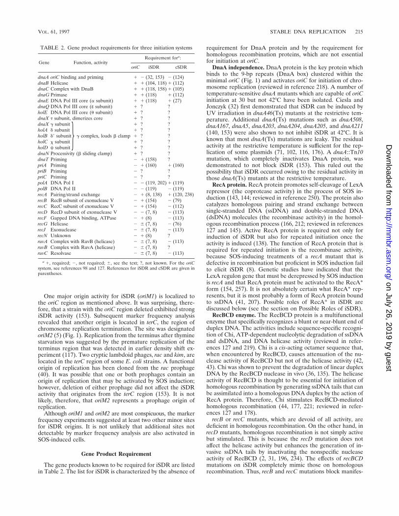
One major origin activity for iSDR (oriM1) is localized tothe oriC region as mentioned above. It was surprising, there-fore, that a strain with the oriC region deleted exhibited strongiSDR activity (153). Subsequent marker frequency analysisrevealed that another origin is located in terC, the region ofchromosome replication termination. The site was designatedoriM2 (5) (Fig. 1). Replication from the terminus after thyminestarvation was suggested by the premature replication of theterminus region that was detected in earlier density shift ex-periment (117). Two cryptic lambdoid phages, rac and kim, arelocated in the terC region of some E. coli strains. A functionalorigin of replication has been cloned from the rac prophage(40). It was possible that one or both prophages contain anorigin of replication that may be activated by SOS induction;however, deletion of either prophage did not affect the iSDRactivity that originates from the terC region (153). It is notlikely, therefore, that oriM2 represents a prophage origin ofreplication.
Although oriM1 and oriM2 are most conspicuous, the markerfrequency experiments suggested at least two other minor sitesfor iSDR origins. It is not unlikely that additional sites notdetectable by marker frequency analysis are also activated inSOS-induced cells.
Gene Product Requirement
The gene products known to be required for iSDR are listedin Table 2. The list for iSDR is characterized by the absence of
requirement for DnaA protein and by the requirement forhomologous recombination proteins, which are not essentialfor initiation at oriC.
DnaA independence. DnaA protein is the key protein whichbinds to the 9-bp repeats (DnaA box) clustered within theminimal oriC (Fig. 1) and activates oriC for initiation of chro-mosome replication (reviewed in reference 218). A number oftemperature-sensitive dnaA mutants which are capable of oriCinitiation at 30 but not 42°C have been isolated. Ciesla andJonczyk (32) first demonstrated that iSDR can be induced byUV irradiation in dnaA46(Ts) mutants at the restrictive tem-perature. Additional dnaA(Ts) mutations such as dnaA508,dnaA167, dnaA5, dnaA203, dnaA204, dnaA205, and dnaA211(140, 153) were also shown to not inhibit iSDR at 42°C. It isknown that most dnaA(Ts) mutations are leaky. The residualactivity at the restrictive temperature is sufficient for the rep-lication of some plasmids (71, 102, 116, 176). A dnaA::Tn10mutation, which completely inactivates DnaA protein, wasdemonstrated to not block iSDR (153). This ruled out thepossibility that iSDR occurred owing to the residual activity inthose dnaA(Ts) mutants at the restrictive temperature.
RecA protein. RecA protein promotes self-cleavage of LexArepressor (the coprotease activity) in the process of SOS in-duction (143, 144; reviewed in reference 250). The protein alsocatalyzes homologous pairing and strand exchange betweensingle-stranded DNA (ssDNA) and double-stranded DNA(dsDNA) molecules (the recombinase activity) in the homol-ogous recombination process (166, 212; reviewed in references127 and 145). Active RecA protein is required not only forinduction of iSDR but also for repeated initiation once theactivity is induced (138). The function of RecA protein that isrequired for repeated initiation is the recombinase activity,because SOS-inducing treatments of a recA mutant that isdefective in recombination but proficient in SOS induction failto elicit iSDR (8). Genetic studies have indicated that theLexA regulon gene that must be derepressed by SOS inductionis recA and that RecA protein must be activated to the RecA*form (154, 257). It is not absolutely certain what RecA* rep-resents, but it is most probably a form of RecA protein boundto ssDNA (41, 207). Possible roles of RecA* in iSDR arediscussed below (see the section on Possible Roles of iSDR).
RecBCD enzyme. The RecBCD protein is a multifunctionalenzyme that specifically recognizes a blunt or near-blunt end ofduplex DNA. The activities include sequence-specific recogni-tion of Chi, ATP-dependent nucleolytic degradation of ssDNAand dsDNA, and DNA helicase activity (reviewed in refer-ences 127 and 219). Chi is a cis-acting octamer sequence that,when encountered by RecBCD, causes attenuation of the nu-clease activity of RecBCD but not of the helicase activity (42,43). Chi was shown to prevent the degradation of linear duplexDNA by the RecBCD nuclease in vivo (36, 135). The helicaseactivity of RecBCD is thought to be essential for initiation ofhomologous recombination by generating ssDNA tails that canbe assimilated into a homologous DNA duplex by the action ofRecA protein. Therefore, Chi stimulates RecBCD-mediatedhomologous recombination (44, 177, 221; reviewed in refer-ences 127 and 178).
recB or recC mutants, which are devoid of all activity, aredeficient in homologous recombination. On the other hand, inrecD mutants, homologous recombination is not simply activebut stimulated. This is because the recD mutation does notaffect the helicase activity but enhances the generation of in-vasive ssDNA tails by inactivating the nonspecific nucleaseactivity of RecBCD (2, 31, 196, 234). The effects of recBCDmutations on iSDR completely mimic those on homologousrecombination. Thus, recB and recC mutations block manifes-
TABLE 2. Gene product requirements for three initiation systems
Gene Function, activityRequirement fora:
oriC iSDR cSDR
dnaA oriC binding and priming 1 2 (32, 153) 2 (124)dnaB Helicase 1 1 (104, 118) 1 (112)dnaC Complex with DnaB 1 1 (118, 158) 1 (105)dnaG Primase 1 1 (118) 1 (112)dnaE DNA Pol III core (a subunit) 1 1 (118) 1 (27)dnaQ DNA Pol III core (ε subunit) 1 ? ?holE DNA Pol III core (u subunit) 1 ? ?dnaX t subunit, dimerizes core 1 ? ?dnaX g subunit 1 ? ?holA d subunit 1 ? ?holB d9 subunit g complex, loads b clamp 1 ? ?holC x subunit 1 ? ?holD c subunit 1 ? ?dnaN Processivity (b sliding clamp) 1 ? ?dnaT Priming 2 1 (158) ?priA Priming 2 1 (160) 1 (160)priB Priming 2 ? ?priC Priming 2 ? ?polA DNA Pol I 2 2 (119, 202) 1 (119)polB DNA Pol II 2 2 (119) 2 (119)recA Pairing/strand exchange 2 1 (8, 138) 1 (120, 238)recB RecB subunit of exonuclease V 2 1 (154) 2 (79)recC RecC subunit of exonuclease V 2 1 (154) 2 (112)recD RecD subunit of exonuclease V 2 2 (7, 8) 2 (113)recF Gapped DNA binding, ATPase 2 1 (8) 2 (113)recG Helicase 2 6 (7, 8) 2 (76)recJ Exonuclease 2 6 (7, 8) 2 (113)recN Unknown 2 1 (8) ?ruvA Complex with RuvB (helicase) 2 6 (7, 8) 2 (113)ruvB Complex with RuvA (helicase) 2 6 (7, 8) ?ruvC Resolvase 2 6 (7, 8) 2 (113)
a 1, required; 2, not required; 6, see the text; ?, not known. For the oriCsystem, see references 98 and 127. References for iSDR and cSDR are given inparentheses.
}
VOL. 61, 1997 STABLE DNA REPLICATION 215
on July 26, 2019 by guesthttp://m
mbr.asm
.org/D
ownloaded from
tation of iSDR after SOS induction (154) whereas recD muta-tions stimulate iSDR induction (8). It is most likely that therole of RecBCD in iSDR is to yield ssDNA for RecA-catalyzedD-loop formation.
The nuclease activity of RecBCD can be inactivated afterSOS induction without significantly inhibiting the helicase ac-tivity. There seem to be two mechanisms that contribute toinactivation of the RecBCD nuclease. First, SOS inductioncauses partial inhibition of Chi activation without affectingoverall recombination levels (197). An SOS-inducible proteinmay modify the RecBCD enzyme by a direct interaction, con-verting it to a recombinase that retains the helicase activity buthas lost the nuclease activity. The induction of the inhibitor ofChi activation is under RecA-LexA control (197). The secondmechanism is the phenomenon that is known as restrictionalleviation (37, 187). SOS induction produces an inhibitor ofdegradation of unmodified duplex DNA by the RecBCD nu-clease and other restriction enzymes. The inhibitor attenuatesthe nuclease activity of RecBCD and blocks the expression ofrecB, recC, and recD (93). Induction of the inhibitor is regu-lated by recA but not lexA (93, 236). Thus, restriction allevia-tion is induced after UV irradiation in lexA3(Ind2) mutantsbut not in recA mutants.
Other rec gene products. RecJ protein is a 59-specific ssDNAexonuclease (149). Like recD mutations, recJ mutations stimu-late iSDR, indicating that RecJ is inhibitory (8). On the otherhand, recF and recN mutations mildly inhibit iSDR, suggestingthat these proteins are partially required for iSDR. TheRuvAB and RecG proteins are helicases that catalyze branchmigration, and the RuvC protein is a nuclease that resolvesHolliday junctions in the late stages of homologous recombi-nation (214, 252). The effects of ruvAB, ruvC, and recG muta-tions on iSDR are twofold. First, these mutations, which blockthe processing of D-loops, greatly stimulate iSDR initiation(for an explanation, see the section on Evidence for the D-Loop Model, below) (8). Second, they severely inhibit theelongation stage of iSDR because Holliday junctions, left onthe chromosome due to abortive recombination, arrest repli-cation fork movement (7).
Priming proteins. The events that must follow strand sepa-ration for initiation of duplex DNA replication are (i) loadingof the replication fork helicase, DnaB, onto ssDNA and (ii)synthesis of primer RNA by DnaG primase. DnaB and DnaGare essential for iSDR (118), indicating that iSDR utilizes thehelicase and primase at the replication fork, as the normalreplication fork does. E. coli possesses several different path-ways for the priming step (reviewed in reference 159). At oriC,DnaA protein delivers the DnaB helicase from a DnaB-DnaCcomplex onto ssDNA (209). Since DnaA is completely dispens-able for iSDR initiation, this is clearly not the choice of prim-ing for iSDR. iSDR utilizes a priming system that was initiallydiscovered in the course of in vitro studies of fX174 DNAreplication (3). This fX174-type priming reaction involves sev-eral proteins including PriA, PriB, PriC, DnaB, DnaC, DnaT,and DnaG. PriA first recognizes and binds a hairpin structurecalled pas (primosome assembly site) in the melted region ofthe duplex. The binding activates the ATPase activity of PriA.PriB then binds to the PriA-DNA complex. DnaT, perhaps inconjunction with PriC, loads a DnaB helicase to the PriA-PriB-DNA complex from a DnaB-DnaC complex. DnaG primasesubsequently interacts with DnaB, completing the assembly ofa primosome.
The first hint of involvement of a fX174-type priming iniSDR came when dnaT, mutations of which block iSDR (136),was identified as the gene encoding the priming protein, i(161). This was followed by the demonstration that DnaC is
also required in iSDR initiation (158). More recently, priA::kannull mutations were shown to completely inhibit the inductionof iSDR (160). PriA protein has, in addition to the primosomeassembly function, an ATPase and a helicase activity. A mutantPriA(K230R) that is deficient in the ATPase and helicase ac-tivities but is capable of catalyzing primosome assembly in vitrowas engineered (266). A plasmid expressing this mutant PriAprotein complements the defect of the priA::kan null mutationin iSDR initiation (114). It is most likely, therefore, that theprimosome assembly function of PriA is essential for iSDR.The requirement for PriB and PriC has not been examined.Thus, most, if not all, of the fX174-type primosome assemblyproteins are involved in the priming of iSDR.
The behavior of the mutant PriA(K230R) described aboveclearly indicates that the ATPase and helicase activities asso-ciated with PriA are nonessential for primosome assembly foriSDR. What, then, is the role(s) played by these activities?Recently, Al-Deib et al. (1) examined suppressor mutationsthat suppress the sensitivity of a recG mutant to DNA damage.A majority of the mutations (srgA) mapped in the region of thehelicase motifs within priA. Therefore, srgA mutations specif-ically inactivate the helicase activity of PriA without affectingthe primosome assembly function. Thus, these PriA mutantproteins are like PriA(K230R). It was proposed that RecGhelicase promotes the conversion of a D-loop into a Hollidayjunction by its branch migration activity and that the PriAhelicase activity opposes this reaction. The balance betweenRecG and PriA helicase activities is suggested to be critical. Inthe absence of RecG helicase, PriA dominates and reduces theefficiency of homologous recombination and DNA repair. Theinactivation of the helicase activity of PriA by srgA mutationscompensates for the absence of RecG, restoring efficient re-combination and DNA repair (1).
The requirement for a pas (primosome assembly site) inSDR priming is not clear. One of the origins of iSDR is locatedat the oriC region (see above). However, no pas site is presentwithin or near this origin. In fact, attempts to identify pas sitesin the E. coli chromosome have thus far been unsuccessful. Itwas suggested that PriA might interact with ssDNA containingno canonical pas sequence (6). PriA protein may recognizesome feature(s) of a D-loop for binding. This view is supportedby two observations. First, the ATPase activity of PriA, whichis activated by binding to pas, is dispensable for iSDR priming(see above). Second, pBR322 plasmid replication absolutelyrequires PriA protein, but the deletion of the pas near theorigin of replication has only a minor effect (157, 160, 240).
DNA polymerases. The dependence of iSDR on DNA poly-merase III (Pol III) is indicated by the requirement for dnaE1,although the genes encoding other subunits of Pol III holoen-zyme have not been examined (Table 1). DNA Pol II, which isSOS inducible (21, 85), is not essential for iSDR. DNA Pol I,which plays a crucial role in cSDR initiation (see the section onthe R-loop model below), is not required for iSDR (119).
Proposed Models for iSDR Initiation
Stable-complex model. The stable-complex model, originallyproposed when SDR was discovered (117, 118) and later mod-ified (138, 140), assumes that the DNA replication complex(replisome) assembled at the origin of replication is pro-grammed to self-destruct at the end of each round of replica-tion. Protein synthesis is postulated to be necessary to replen-ish the unstable factor(s) for replication complex reassembly.The self-destruction is ensured by the presence of a factor (thedestructor) in the complex. Under SOS-inducing conditions, acomplex can be assembled without this factor and thereby
216 KOGOMA MICROBIOL. MOL. BIOL. REV.
on July 26, 2019 by guesthttp://m
mbr.asm
.org/D
ownloaded from
stabilized and reused for the ensuing rounds of replication,giving rise to SDR. Subsequently, two types of mutants, whoseSDR behavior was differently affected, were isolated. One type,sdrT, constitutively expresses SDR without the use of inducingtreatments. It was proposed that sdrT could encode a proteinthat is modified by RecA*. The modified protein, SdrT*, wouldin turn interact with the destructor to inactivate it, leading tothe formation of a stable complex. The sdrT mutation washypothesized to constitutively activate SdrT protein so that theRecA*-mediated modification was dispensable. The secondtype of mutation, dnaT, fails to manifest SDR after the appli-cation of inducing treatments (136). The dnaT mutation mapsvery close to sdrT but not in the same gene. Therefore, dnaTcould be a candidate for the gene encoding the destructionfactor, and the dnaT mutation might render the factor resistantto inactivation by SdrT*, thus leading to obligatory formationof an unstable complex (140).
Although no single line of evidence either proves or dis-proves this model outright, subsequent studies made it ex-tremely improbable that iSDR arises from such a mechanism.For example, implicit in the model is initiation from the nor-mal origin of replication, oriC. iSDR can, in fact, be initiatednot only from an oriC site that is inactive for normal replicationbut also from the origin located in the terC region (5). Fur-thermore, dnaT has been identified as the gene encoding apriming protein, i (161). Finally, artificially induced double-strand breaks can trigger iSDR (4) (see the section on Evi-dence for the D-Loop Model, below).
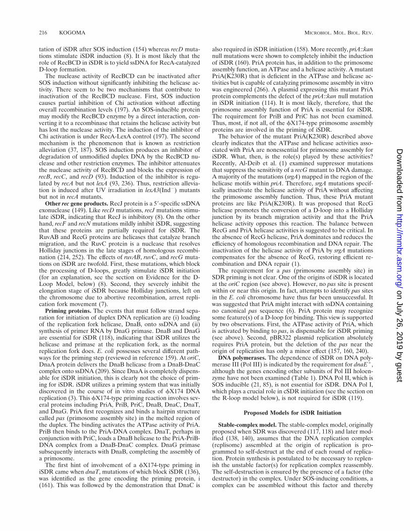
Onion skin model. Conditions that block the movement ofreplication forks, such as thymine starvation and UV irradia-tion, cause accumulation of the initiation potential, includingDnaA protein, which can be used to multiply initiate at theoriC (see, e.g., references 190 and 259). Kuzminov (133) pro-posed that the multiple initiations under the conditions wherethe fork progress is severely restricted result in a chromosomethat has replication forks bunched up near oriC (Fig. 2B to D).Blockade of fork movement would cause replication fork in-stability, and some of the forks would collapse (Fig. 2E). TheRecBCD- and RecA-dependent repair of the collapsed repli-cation forks (see the section on Repair of a Collapsed Repli-
cation Fork, below) “is needed to generate an onion-skin struc-ture with the amplified region of the replication origin” (Fig.2F) (133). This interesting model is clearly inconsistent withseveral of the well-documented properties of iSDR (summa-rized in the section on Mode of Replication, above). First, aperiod of DNA synthesis inhibition, which leads to a 16-foldincrease in DNA during subsequent iSDR (117), elevates thecopy number of the oriC region no more than twofold 15 minafter the release of replication block (153). Second, the modelpredicts both an amplification of the oriC region sequences andthe preferential use of newly synthesized DNA for the tem-plate. During iSDR, no particular sequence is amplified, andthe template is chosen at random from the accumulating chro-mosome pool (117). Third, iSDR can be activated under con-ditions which do not involve the destabilization of replicationforks by replication inhibition, an essential element of themodel. The amplification and activation of RecA protein bygenetic means without DNA synthesis inhibition is sufficientto activate a significant degree of iSDR (165, 257). Finally,DnaA protein which might be used for the multiple initia-tions described in the model is completely dispensable foriSDR (153).
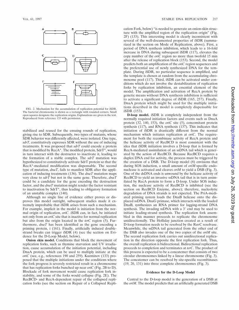
D-loop model. iSDR is completely independent from thenormally required initiation factors and events such as DnaAprotein (32, 140, 153), the oriC site (5), concomitant proteinsynthesis (117), and RNA synthesis (137). This indicates thatinitiation of iSDR is drastically different from the normalmechanism which initiates replication at oriC. The require-ment for both the recombinase activity of RecA protein andthe helicase activity of RecBCD is most consistent with theidea that iSDR initiation involves a D-loop that is formed byRecA-mediated assimilation of an ssDNA tail which is gener-ated by the action of RecBCD. Because RecBCD requires aduplex DNA end for activity, the process must be triggered bythe creation of a DSB. The D-loop model (8) envisions thatduring SOS induction, a small amount of oriM-specific endo-nuclease is activated and cleaves oriM to yield a DSB (Fig. 3).One of the dsDNA ends is unwound by the helicase activity ofRecBCD to yield an invasive ssDNA tail that is in turn assim-ilated by RecA protein to form a D-loop. Under SOS induc-tion, the nuclease activity of RecBCD is inhibited (see thesection on RecBCD Enzyme, above); therefore, nucleolyticdegradation of DNA strands is not expected. The PriA-medi-ated priming system loads the DnaB helicase onto the dis-placed ssDNA. DnaG primase, which interacts with the loadedDnaB, synthesizes an RNA primer for lagging-strand DNAsynthesis. The invading ssDNA with a 39 end may be used toinitiate leading-strand synthesis. The replication fork assem-bled in this manner proceeds to replicate the chromosomeunidirectionally. The Holliday junction created as a result ofD-loop formation needs to be resolved by the RuvC resolvase.Meanwhile, the ssDNA tail generated from the other end ofthe DSB also invades one of the two copies of the oriM site.The second replication fork carries out unidirectional replica-tion in the direction opposite the first replication fork. Thus,the overall replication is bidirectional. Bidirectional replicationproceeds to completion and terminates at terC. The product ofthis process is expected to be a concatemer that consists of twocircular chromosomes linked by a linear chromosome (Fig. 3).The concatemer can be resolved by site-specific recombinases(19, 20, 131) into three complete chromosomes (Fig. 3).
Evidence for the D-Loop Model
Central to the D-loop model is the generation of a DSB atthe oriM. The model predicts that an artificially generated DSB
FIG. 2. Mechanism for the accumulation of replication potential for iSDR.The bacterial chromosome is shown as a rectangle with rounded corners. Smallopen squares designate the replication origins. Explanations are given in the text.Reproduced from reference 133 with permission.
VOL. 61, 1997 STABLE DNA REPLICATION 217
on July 26, 2019 by guesthttp://m
mbr.asm
.org/D
ownloaded from
triggers chloramphenicol- and rifampin-resistant replication,which should occur without the oriM site. This prediction wasdirectly tested by placing l cos site on a plasmid and generatinga DSB at the site with l terminase, which introduces twostaggered nicks, 12 bp apart (reviewed in reference 53). Rep-lication of the plasmid in the presence of chloramphenicol andrifampin, which inhibits plasmid replication from the normalorigin of replication, was monitored by determining the copynumber of the plasmid in SOS-induced cells after controlledsynthesis of l terminase. A limited amount of the terminasesynthesized was expected to cleave part of the plasmid popu-lation, and the ends generated were expected to trigger repli-cation on intact plasmid molecules. DSBs artificially generatedin such a manner indeed triggered origin-independent plasmidreplication (4). The major products of the replication werecovalently closed circular monomers. This suggests that thereplication proceeds by the u mode. The replication dependedboth on the presence of a cos site in the plasmid and onsynthesis of the terminase.
Thus, artificial DSBs trigger an SDR-like replication of aplasmid that lacks oriM sites, providing strong support for themodel. In addition, the following observations support themodel. First, the D-loop that acts as an origin for iSDR is mostprobably structurally identical to the D-loop that is formed asan early intermediate in homologous recombination (166,212). Thus, the iSDR and homologous recombination pro-cesses are expected to compete for D-loops. In homologousrecombination, D-loops are further processed by RuvAB,
RecG, and RuvC proteins to yield recombinant molecules. Asexpected from the model, iSDR activity was found to be sig-nificantly stimulated by ruvA, ruvB, ruvC, and recG mutations,which block the processing of the intermediate (7). Second, themodel predicts that the presence of extra copies of oriM wouldenhance the iSDR initiation frequency because the extra cop-ies could provide additional ends that could trigger replication.This prediction was verified by the demonstration that intro-duction of a plasmid carrying the oriM1 site stimulates theiSDR activity (5). The stimulation is origin specific in that thestimulatory effect of the extra copies of oriM1 cannot be seenin the strain that has the oriM1 site deleted. However, the levelof stimulation is not proportional to the copy number of oriM1introduced (5). This suggests that some trans-acting factor(e.g., the hypothetical endonuclease activity) is limiting in theinitiation reaction. Third, after a period of thymine starvation,a drastic change in the chromosomal DNA structure is inducedsuch that DNA migrates extremely slowly or does not migrateat all through agarose upon electrophoresis (179). The nonmi-grating DNA is enriched with structures containing ssDNAgaps or tails and with highly branched structures. Such struc-tures are largely associated with DNA fragments that containoriC, and their formation depends highly on recA1 (179). It islikely that at least part of such DNA results from chromosomereplication initiated at D-loops formed at oriM1. Fourth, asdescribed previously, iSDR depends very strongly on PriA-catalyzed priming (114, 160).
FIG. 3. D-loop model of iSDR initiation. A partially replicated chromosome has two copies of oriM1 (shaded boxes), one of which is cleaved to yield a DSB. Thesolid box indicates terC, the termination site of normal replication. Newly synthesized DNA strands and replication forks are shown by broken lines and circles,respectively. See the text for details.
218 KOGOMA MICROBIOL. MOL. BIOL. REV.
on July 26, 2019 by guesthttp://m
mbr.asm
.org/D
ownloaded from

Roles of SOS in iSDR Initiation
In the D-loop model, the role of SOS induction is twofold.One role is to activate the hypothetical endonuclease specificto oriM. Previously, thymine starvation was reported to gener-ate DSBs in the chromosome (265) and cause activation of anendonuclease activity (62). The specificity, if any, of the endo-nuclease for certain sequences including oriM has not beenexamined. The second role of SOS induction is inhibition ofthe nuclease activity of RecBCD (93, 187, 197), which results inthe stabilization of invasive ssDNA tails and the consequentialenhancement of iSDR initiation. This role of SOS inductionwas deduced from the following observations. In the modelreplication system where origin-independent plasmid replica-tion is triggered by artificial DSBs (see above), SOS inductionwas still necessary for the replication. The SOS induction wasrendered dispensable by a recD mutation, which inactivates theRecBCD nuclease activity, or by inclusion in the plasmid of aChi site which attenuates the nuclease (4). Furthermore, theorigin-independent replication was detected without SOS in-duction in a recBC sbcA mutant which is devoid of the nucleaseactivity of RecBCD but is proficient for homologous recombi-nation owing to the activation of another recombination path-way, the RecE pathway (4).
RECOMBINATION-DEPENDENT DNA REPLICATION
RDR Model
A crucial observation made in studies with the plasmidmodel system (see the section on Evidence for the D-LoopModel, above) is that an SDR-like activity can be activatedwithout SOS induction when certain specific conditions aremet. These conditions are (i) generation of a duplex DNA endand (ii) attenuation of the RecBCD nuclease activity. Whenencountered by RecBCD, Chi attenuates the nuclease activityof the RecBCD enzyme (42). When the plasmid contains a Chisite, origin-independent plasmid replication can be triggeredby an artificial DSB even in normal cells not induced for SOS.This replication absolutely depends on recA1, recB1, andrecC1 (4). The observation led to the proposal that RecA- andRecBCD-dependent replication could routinely occur in nor-mal cells when a duplex DNA end is generated and the linear-ized duplex DNA is protected from the RecBCD nuclease bythe presence of Chi in the duplex. The homologous recombi-nation function-dependent replication triggered by a duplexDNA end is designated recombination-dependent DNA repli-cation (RDR) (4).
iSDR Is a Special Type of RDR
From the discussions above, it is easy to envision that iSDRand RDR are mechanistically identical. However, there aretwo major differences. First, in iSDR, attenuation of RecBCDnuclease is achieved specifically by the activation of inhibitorsof the RecBCD nuclease as part of the SOS response (93, 197).The second difference is the site where replication is initiated.Clearly, iSDR originates mainly from specific origins (oriM).On the other hand, RDR can be initiated at a site where aD-loop can be formed. In wild-type cells, the site can be at ornear a Chi site where degradation of duplex DNA is stoppedand an invasive ssDNA tail is formed (42). In recBC sbcB sbcCmutants, RecQ helicase, together with RecJ nuclease, mayproduce invasive ssDNA independent of Chi (127). In recBCsbcA cells, exonuclease VIII, activated by the sbcA mutation,may generate an ssDNA tail irrespective of Chi (4). Thus,iSDR is a special type of RDR.
POSSIBLE ROLES OF iSDR
Damage-Resistant Replication
E. coli polymerases are extremely sensitive to pyrimidinedimers (243). DNA replication in cells induced for iSDR isconsiderably more resistant to UV irradiation than is the rep-lication in the presence of chloramphenicol in uninduced cells.Thus, at a UV dose that completely inhibits normal replicationin the absence of protein synthesis, the rate of iSDR, after ashort lag period, recovers to the rate that is found in unirra-diated controls (122). About 80% of the DNA synthesis afterUV irradiation occurs in the semiconservative manner underthese conditions. A similar recovery of DNA synthesis can beseen in a uvrA6 mutant which is excision repair deficient. Thus,iSDR can tolerate more pyrimidine dimers in templates thancan normal replication (122, 208). The mechanism of the tol-erance is not understood. Since a significant fraction of UV-induced damage (e.g., pyrimidine dimers) is converted to DSBs(22, 251) and since iSDR is a type of RDR which is initiated byDSBs as described above, it is likely that at least part of thedimer-resistant replication represents RDR.
There is solid evidence that newly synthesized DNA follow-ing UV irradiation contains gaps opposite dimers (201). It ispossible, therefore, that the replisome of iSDR that is stalled ata dimer is able to restart DNA synthesis downstream of thedimer. It was reported that SOS induction enables stalled rep-lication to restart (50, 99, 258). This induced replisome reac-tivation (IRR) shares some of the characteristics of iSDR: it isinducible upon SOS response, can occur in the presence ofchloramphenicol and rifampin, and requires RecA protein.Furthermore, like in iSDR, recA is the only gene controlled byLexA that must be amplified for IRR (258). Hence, someaspects of SDR were incorporated into a proposed model forIRR (29, 99). However, there are some critical differencesbetween the two activities. Whereas iSDR strictly dependson recB1 and recC1 (154), IRR does not require recB1 (99).Unlike iSDR, amplified RecA protein is not sufficient for IRR:one additional gene product (Irr factor) is required. Despitethe evidence implicating an RNase HI inhibitor in the recoveryprocess of stalled replisomes, no change in RNase HI activitywas detected during or after the SOS response (17, 29). Fur-thermore, little effect of overproduction of RNase HI on theinducibility of iSDR was seen (17). These considerations makeit unlikely that iSDR and IRR are the same activity. It ispossible, however, that the two processes share a basic induc-ible activity.
Error-Prone Replication
iSDR appears to be error prone (138). The proposal is basedmainly on three observations. (i) Cells that are induced foriSDR show a high rate of spontaneous mutations. (ii) This highrate of mutation is drastically reduced by a dnaT mutation,which blocks induction of iSDR. (iii) Mutagenesis with methylmethanesulfonate, which also induces iSDR, is inhibited by thednaT mutation. In general, the conditions that prevent theinduction of iSDR significantly reduce the mutation rate (138).The observations raise the possibility that iSDR is the error-prone replication that is associated with the UmuD9C-depen-dent mutagenesis in SOS-induced cells (reviewed in reference250). Consistent with this proposal is the requirement for theactivated form of RecA protein, RecA*, which parallels therequirements defined for SOS mutagenesis (51, 61, 181, 228).In addition to the two well-characterized roles of RecA proteinin SOS mutagenesis, i.e., cleavage of LexA and conversion ofUmuD to UmuD9, RecA* plays a third role in SOS mutagen-
VOL. 61, 1997 STABLE DNA REPLICATION 219
on July 26, 2019 by guesthttp://m
mbr.asm
.org/D
ownloaded from
esis. The precise nature of the third role is unknown, but theactivated RecA may act directly, perhaps by interacting withand modifying a DNA replication complex. The modificationmay allow efficient replication of damaged DNA at the cost ofincreased infidelity (49). It is possible that this modificationendows iSDR with the characteristics of UV resistance (seeabove) and error-prone DNA replication. However, iSDR canbe induced in umuC mutants, which no longer exhibit mu-tagenesis (unpublished data quoted in reference 249). It couldmean that iSDR is necessary but insufficient for the mutagen-esis. Alternatively, the replisome assembled at a D-loop isinherently error prone (e.g., inhibition of the editing exonucle-ase) or a mismatch repair system associated with the replica-tion is compromised under the condition. In this context, it isnoteworthy that DSB repair has been reported to cause mu-tations in nearby sequences in yeast and mammalian cells (48,223).
Possible Role in Adaptive Mutation
Certain mutations can occur in nongrowing E. coli cells, andthese mutations appear to be adaptive because the only muta-tions recovered are those that permit the cells to grow (re-viewed in references 56 and 199). Analyses of mutations re-covered led to the conclusion that mutations arise as a result ofreplication error in a state in which methyl-directed mismatchrepair is suppressed (59, 148, 200). DNA Pol III is responsiblefor most of the errors, and a DpolB mutation (inactivatingDNA Pol II) stimulates adaptive mutation (57). Adaptive mu-tation depends on recA1 (26). It is severely inactivated by therecB and recC mutations but is stimulated by the recD mutation(72). The shared characteristic in the requirement for RecAprotein and RecBCD enzyme raises the possibility that iSDR isinvolved in adaptive mutation (159). iSDR is a form of repli-cation that can occur in nongrowing cells and appears to bemutagenic (see above). iSDR employs DNA Pol III but notDNA Pol II (119). These characteristics of iSDR are consistentwith the idea. Recently, several models have been proposed forthe mechanism of adaptive mutation (60, 73, 134). These mod-els propose both generation of a D-loop by the actions of RecAprotein and RecBCD enzyme and initiation of semiconserva-tive replication at the D-loop. These schemes are very similarto the D-loop model of iSDR described above. It is likely thatrecombination-dependent replication plays a crucial role in theemergence of adaptive mutation. The predicted requirementfor PriA in adaptive mutation has not been tested.
RDR AND DOUBLE-STRAND BREAK REPAIR
Role of RDR in DSB Repair
In the plasmid model system for RDR described above, it isenvisioned that a donor plasmid that is cleaved by the termi-nase triggers a semiconservative u mode of replication on an-other molecule (4). Completion of the replication would yieldtwo circular plasmid molecules. To gain a net increase in plas-mid yield, it would seem necessary that the donor plasmidmolecule be recovered as an intact circle. This implies that theDSB in the donor plasmid might be repaired by the process.Thus, RDR probably leads to DSB repair.
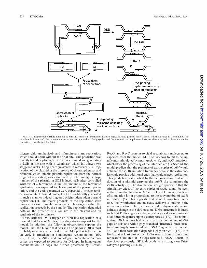
RDR-mediated DSB repair model. When a chromosomesuffers a DSB (Fig. 4a), RecBCD recognizes the ends andbegins degrading the duplex, converting the DSB into a dou-ble-strand gap (route A). Chi sites are overrepresented in theE. coli chromosome (about one every 4 kb on the average)(52), and thus RecBCD is expected to encounter a Chi site
before it degrades the duplex too far. When encountering aChi site, the nuclease activity of RecBCD is attenuated, pro-ducing an invasive ssDNA tail at each end (Fig. 4b). The tailsare assimilated into a homolog by RecA protein to form aD-loop at each end (Fig. 4c). The replication forks assembledat the D-loops after the PriA-mediated priming process repli-cate toward each other (Fig. 4d). Meanwhile, RuvC resolvaseresolves the two Holliday junctions (Fig. 4e). When the twoforks meet and complete the replication, the process yields arepaired chromosome and a homolog (Fig. 4j and k). In thismode of repair, the extent of semiconservative DNA replica-tion is limited to filling the gap generated by RecBCD process-ing.
Efficient DSB repair requires the induction of SOS (seebelow). As discussed above (see the section on Gene ProductRequirement, RecBCD Enzyme), Chi activation is partiallyinhibited in SOS-induced cells (197) and thus the attenuationof RecBCD nuclease activity by Chi may not occur effectively.Instead, inhibitors of the RecBCD nuclease that convert theenzyme into a helicase with no DNA-degrading activity areinduced (93, 197). Because of this, conversion of a DSB into adouble-strand gap is not likely. Thus, in SOS-induced cells, a
FIG. 4. RDR-mediated DSB repair. DNA strands of a chromosome that hasjust suffered DSB and a homolog chromosome are indicated by thick and thinlines, respectively. Newly synthesized DNA strands and replication forks areshown by broken lines and circles, respectively. See the text for details.
220 KOGOMA MICROBIOL. MOL. BIOL. REV.
on July 26, 2019 by guesthttp://m
mbr.asm
.org/D
ownloaded from

more likely sequence of events would be the following (Fig. 4,route B) (110). The altered RecBCD enzyme recognizes theduplex ends and unwinds the duplex to yield recombinogenicssDNA tails (Fig. 4f). A D-loop is formed at one end, and areplication fork assembled at the site begins replication in onedirection (Fig. 4g and h). Subsequently, the second end invadesone of the duplicated chromosomes while the first fork con-tinues to replicate the remainder of the chromosome (Fig. 4i).In this case, the involvement of DNA replication is extensiveand the product would have a complex structure requiring theresolution of Holliday junctions by RuvC resolvase aided byRuvAB and/or RecG helicase. The resulting concatemer canbe eventually resolved into monomers (Fig. 4j and k) in amanner similar to that described for Fig. 3. The possibility thatthe ends generated by DSBs trigger DNA replication leadingto DSB repair has been considered previously (220, 235).
Resnick (193) proposed a model for the repair of radiation-induced DSBs and pointed out that the same process couldaccomplish homologous recombination. Szostak et al. (229) re-fined Resnik’s model to explain meiotic recombination in low-er eukaryotes. A major difference between the two models isthat whereas the former postulates 59339 nucleolytic degrada-tion of one strand to expose 39 ssDNA ends, the latter modelinvolves the generation of a gap flanked by 39 ssDNA ends(Fig. 5). Thus, it can be said that the former is a DSB repairmodel while the latter is a double-strand gap repair model(235). The RDR-mediated DSB repair model described abovediffers from these conventional DSB repair models in at leasttwo very important aspects. First, in the new model, the DSBrepair process could involve extensive semiconservative DNA
replication. In contrast, the conventional models postulate aninvolvement of a limited extent of ssDNA repair synthesis tofill the missing DNA (Fig. 5). Second, in SOS-induced E. colicells, no significant extent of RecBCD processing may occurbecause of inhibition of the nuclease activity of RecBCD.Therefore, in damaged SOS-induced cells, DSB repair is mostlikely to proceed as outlined in Fig. 4, route B.
Evidence for the RDR-mediated DSB repair model. The priAnull mutation blocks iSDR (see the section on Gene ProductRequirement, Priming Proteins, above). The introduction ofplasmid expressing a mutant PriA protein, PriA(K230R), thatis capable of primosome assembly despite the lack of theATPase and helicase activities normally associated with wild-type PriA (266) can rectify this defect of the priA null mutant(114). These results strongly suggest that RDR requires PriA-mediated priming, although the effect of the priA null mutationon RDR has not been tested directly (160). The priA nullmutants were shown to be hypersensitive to gamma rays andmitomycin, which cause DSBs in the chromosome (114). Theresult suggests that priA null mutants are deficient in DSBrepair. It was shown that the mutant PriA(K230R) can com-plement the DSB repair defect of priA null mutants (114).Similarly, an extragenic suppressor mutation, spa-47, of thepriA null mutation, which partially restores iSDR inducibilityto priA null mutants, also suppresses the hypersensitivity tomitomycin. The spa-47 mutation maps at or very close to dnaC(114). Since dnaC and dnaT constitute an operon and bothgene products are involved in the primosome assembly (see thesection on Gene Product Requirement, Priming Proteins,above), it is likely that the suppressor mutation changes thestructure of DnaC or DnaT to allow the assembly of activeprimosomes in the complete absence of PriA. Taken together,these results indicate that a large part of DSB repair proceedsin a manner that involves RDR as described in the abovemodel.
The RDR-mediated DSB repair model is consistent withprevious observations. First, repair of ionizing radiation-in-duced DSBs absolutely depends on recA1, recB1, and recC1
(128, 206) and the availability of duplicated chromosomes(128). Second, RDR (4) and DSB repair (189, 206) both aredependent on RecN. Third, the analysis of DSB repair of theDNA from X-irradiated cells by neutral sucrose gradient cen-trifugation revealed that slow-sedimenting DNA resultingfrom DSBs was converted to fast-sedimenting material as therepair proceeded (205). Two interesting observations were thatthe fast-sedimenting DNA sedimented much faster than non-irradiated DNA and that it took several hours to return to thenormal-sized DNA. It is likely that the fast-sedimenting mate-rial represents DNA that is being replicated by RDR afterrepair of DSBs (see e.g., Fig. 4i).
Kobayashi and coworkers developed an elegant assay systemfor DSB repair in E. coli (103, 264). The pBR322-based con-struct is designed in such a way that when a DSB (or double-strand gap) is repaired, an intact neo gene is generated fromtwo copies of imperfect alleles that each have a small deletionat the opposite ends. Thus, the frequency of DSB repair can bemeasured by selecting for resistance to kanamycin (conferredby the neo gene) upon transformation with the probe. Theprobe also allows easy analysis of the products to determine ifcrossing-over has accompanied the gene conversion (i.e., neoto neo1). However, DSB repair was detectable with this systemonly in recBC sbcA cells, where the RecBCD pathway of ho-mologous recombination is inactive and the RecE pathway isoperating (231). The repair of DSB detected with this systemwas not dependent on the gene products such as RecA andRecN, which are known to be essential for DSB repair (see
FIG. 5. Conventional model of double-strand gap repair. Duplex DNA witha DSB and homolog duplex DNA are shown by thin and thick lines, respectively.Broken lines indicate newly synthesized DNA. Adapted from reference 229.
VOL. 61, 1997 STABLE DNA REPLICATION 221
on July 26, 2019 by guesthttp://m
mbr.asm
.org/D
ownloaded from

above). In fact, DSB repair could not be seen in wild-type cells,where the RecBCD pathway is active (231). This is most prob-ably due to degradation of the probe by RecBCD exonuclease,because the probe did not contain Chi sites for protection.Thus, the repair detected with this system seems to be a subsetof DSB repair which occurs under very special conditions. Anyrelevance to the major DSB repair process as discussed aboveis doubtful.
Efficient DSB Repair Requires SOS Induction
The repair of ionizing radiation-induced DSBs is an induc-ible function (24, 129) and is completely inhibited by alexA(Ind2) mutation (206). These observations indicate thatalthough RDR can occur in normal cells (not induced for SOS)(see the section on RDR Model, above), effective DSB repairrequires additional factors that are inducible by SOS. In nor-mal cells, the RecBCD nuclease is attenuated by the cis-actingChi sites. The action of a Chi site is orientation dependent:only when approached from the 39 side of the octamer (59-GCTGGTGG-39) is Chi recognized (232) and the nuclease ac-tivity of RecBCD attenuated (42). Thus, only properly orientedChi sites can protect duplex DNA from RecBCD degradation.Close examinations of Chi sites in the chromosome showedthat at about 90% of the time, Chi is oriented toward the originof replication, oriC, protecting that portion of the duplex fromRecBCD degradation (25, 167). In other words, when a DSBoccurs, the oriC-proximal chromosome arm can be protectedbut the terC-proximal arm is much less likely to be protected.Even the protection of the oriC-proximal arm is not complete,because the nuclease activity of RecBCD is attenuated only 20to 30% of the time when it encounters a properly oriented Chisite (43, 222, 233, 263). Consequently, repair of DSBs would bevery inefficient. The inhibition of the RecBCD nuclease bySOS induction (93, 197), on the other hand, should result inequal protection of both ends and could promote efficientrepair as outlined in the RDR-mediated DSB repair model(Fig. 4, route B).
Activation of one of the inhibitors of RecBCD nuclease isnot regulated by LexA repressor, and therefore it can be in-duced in lexA(Ind2) mutants (93, 236). Since the DSB repaircapacity cannot be induced in lexA(Ind2) mutants (206), itimplies that efficient DSB repair requires either amplificationof some proteins such as RecA and RuvAB, which are underLexA control (127), or activation of a new gene product(s) thatis repressed by LexA, or both.
recA polA LETHALITY AND REPLICATIONFORK COLLAPSE
Repair of a Collapsed Replication Fork
More than 20 years ago, Skalka (217) proposed that break-age of a chromosome arm could result from replication forkcollapse due to a fork running into a nick or gap left in thetemplate (Fig. 6). The asymmetric distribution of Chi sitesalong the chromosome (25, 167) led to the proposal that Chi isevolutionarily designed to protect the oriC-proximal arm whenit is broken off at the replication fork due to replication forkcollapse (134, 135). A unique feature of RDR-mediated DSBrepair is that it not only repairs DSBs but also regenerates areplication fork at the site. Thus, RDR-mediated repair isideally suited for the repair of a collapsed replication fork (4).The end of the chromosome arm that is broken off could be
recognized by RecBCD, and the arm would be degraded up toa nearest Chi, where an ssDNA tail might be created (Fig. 6).Formation of a D-loop by assimilation of the ssDNA into ahomolog and PriA-mediated priming followed by replicationprotein assembly should effectively restore the replication fork(4, 134).
Mechanism of recA polA Lethality
The polA gene encodes DNA Pol I, which plays an importantrole in the processing of Okazaki fragments in lagging-strandDNA synthesis (126). The 593 39 exonuclease activity of DNAPol I effectively removes the RNA primer, and the polymeraseactivity replaces it with the DNA moiety. The combination ofa polA and a recA mutation is lethal (67, 171). The combinationof polA and recB mutations is also lethal (171). The lethalityhas attracted the attention of a number of molecular geneti-cists, partly because it is thought that solution of the problemmight reveal the elusive relationship between DNA replicationand homologous recombination. Recent studies suggested thatthe defect of polA mutants in Okazaki fragment processingresults in the accumulation of nicks and gaps during lagging-strand synthesis (28, 227). When a replication fork encounters
FIG. 6. Mechanisms of polA recA and polA recB lethality. Thin lines repre-sent DNA strands, and the arrowheads indicate the 39 end. Circles indicate thelocations of replication forks. See the text for details.
222 KOGOMA MICROBIOL. MOL. BIOL. REV.
on July 26, 2019 by guesthttp://m
mbr.asm
.org/D
ownloaded from

a discontinuity in the template, replication fork collapse wouldresult (Fig. 6). The collapsed fork, which could be efficientlyrestored by RDR-mediated repair in wild-type cells, is blockedby the recB or recA mutation, leading to cell death (Fig. 6) (28).The model predicted that the priA mutation, which blocks thepriming step of RDR, should render polA mutants inviable.When the priA mutation was combined with polA12(Ts), thedouble mutant was indeed very temperature sensitive: it couldnot grow even at 37°C (Fig. 7) (141). Growth of the mutant wasalso extremely sensitive to nutritionally rich media, like that ofthe parental priA single mutant (160), and the mutant failed togrow at 30°C in Luria broth (Fig. 7). The results suggest thatrepair of the collapsed replication fork requires PriA-catalyzedpriming and support the above model.
Completion of the repair would require resolution of theHolliday junction near the reestablished replication fork (Fig.6). Holliday junctions are resolved by RuvC resolvase, but theresolution in vitro is inefficient (15, 230). It was suggested thatefficient resolution of Holliday junctions in vivo requiresbranch migration, which is catalyzed by RuvAB helicase andRecG helicase (213, 253). Mutations that block these processesin the late stage of homologous recombination are thereforeexpected to reduce the viability of polA mutants. ruvA, ruvB,ruvC (82), and recG mutations (76) were indeed shown tocause temperature-sensitive growth when combined with thepolA12(Ts) mutation. The genetic requirements of the RecA-dependent, SOS-inducible DSB repair pathway are summa-rized in Fig. 8 (for details on the RecA-independent pathway,see below).
Replication Fork Collapse and Broth Sensitivity
The effects of certain mutations are drastically exacerbatedand may even result in cell death when the mutant is grown innutritionally rich media such as Luria broth. This phenomenonis termed broth sensitivity. Some of the broth sensitivity phe-notypes can now be understood in terms of replication forkcollapse. Deletion of the entire polA gene is lethal in richmedium but not in minimal medium (91). The fact that theDpolA mutant is viable in minimal medium implies that asecondary system can process Okazaki fragments, albeit inef-ficiently. The distance between a leading replication fork andthe following fork depends on how often chromosome repli-cation is initiated at oriC during a cell cycle (247). In minimal
medium containing glucose as the sole carbon source, a newround of replication is initiated when the leading fork travelsabout halfway on the chromosome. Thus, before the next forkcatches up, the secondary processing system could have suffi-cient time to minimize the number of nicks and gaps whichwould cause infrequent DSBs that could still be repaired byRecA-dependent DSB repair. On the other hand, cells growingin rich medium have only a short time for Okazaki fragmentprocessing such that the number of DSBs overwhelms the DSBrepair capacity, leading to cell death. A similar explanation canbe applied to the broth sensitivity seen with rnhA polA mutants(115) and with priA mutants (Fig. 7) (160).
RecA-INDEPENDENT REPAIR OFDOUBLE-STRAND BREAKS
Suppression of recA polA Lethality by lexA(Def)
The lexA(Def) mutation completely inactivates the LexArepressor, derepressing the LexA regulon (250). lexA(Def)was found to suppress the recA polA lethality (28). Thus,recA200(Ts) polA lexA(Def) mutants are temperature resistantat the restrictive temperature for RecA200. This implies thatderepression of one or more LexA regulon genes alleviates thedefect in either RecA recombinase or DNA Pol I. This induc-ible system was designated the Srp (suppression of recA polAlethality) pathway. The suppression does not come from animproved Okazaki fragment processing because the joining ofOkazaki fragments is not significantly faster in lexA(Def) cellsthan in lexA1 cells (28). Rather, the defect in the RecA proteinis alleviated. This conclusion is based on the following obser-vations. First, the lexA(Def) mutation restores homologousrecombination despite the defect in the RecA protein. Forexample, the P1 transduction frequency is reduced 1,000-foldin DrecA mutants compared to recA1, but lexA(Def) elevatesthe level to more than 7% of the wild-type level. Consistentwith this, lexA(Def) desensitizes DrecA mutants to UV light by1,000-fold, suggesting a boosting of the recombinational repaircapacity in DrecA mutants (28). Second, lexA(Def) does notsuppress the recB polA lethality. The suppression is thereforerather specific to the recA defect (92). Thus, the Srp pathway iscapable of DSB repair in the complete absence of the strandexchange activity of RecA protein.
Two Pathways for DSB Repair
The recent analysis of the Srp system has suggested that Srpis another pathway of DSB repair that may play a different rolefrom that of RecA-dependent DSB repair in the survival of E.coli (92). To identify the gene (srp) responsible for the sup-pression, a temperature-sensitive mutant was isolated from apopulation of recA polA lexA(Def) mutant cells randomly mu-tagenized with a mini::Tn10 transposon. This mutation wasdesignated srp-529::miniTn10. The mutation was cloned, and ashort sequence next to the Tn10 insertion was determined. A
FIG. 7. Temperature-sensitive and broth-sensitive growth of polA priA dou-ble mutants. About 105 cells were spotted on CAA (M9 plus glucose supple-mented with Casamino Acids) and LB (Luria broth) plates and spread withtoothpicks. The plates were incubated at 30, 37, and 42°C for 52 h. Sectors: 1,polA12 priA1; 2, polA12 priA1; 3, polA12 priA2; polA1 priA1.
FIG. 8. Two pathways of DSB repair in E. coli. The genetic requirements oftwo inducible DSB repair pathways are compared.
VOL. 61, 1997 STABLE DNA REPLICATION 223
on July 26, 2019 by guesthttp://m
mbr.asm
.org/D
ownloaded from
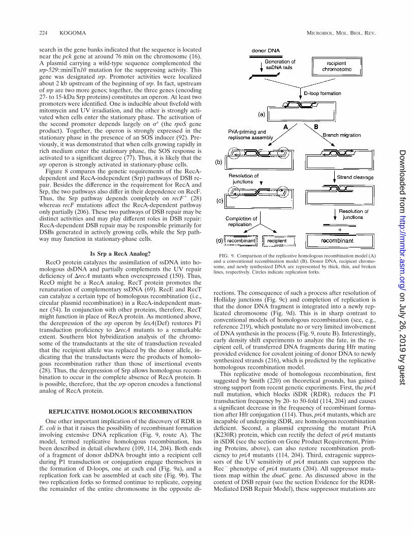
search in the gene banks indicated that the sequence is locatednear the pck gene at around 76 min on the chromosome (16).A plasmid carrying a wild-type sequence complemented thesrp-529::miniTn10 mutation for the suppressing activity. Thisgene was designated srp. Promoter activities were localizedabout 2 kb upstream of the beginning of srp. In fact, upstreamof srp are two more genes; together, the three genes (encoding27- to 15-kDa Srp proteins) constitutes an operon. At least twopromoters were identified. One is inducible about fivefold withmitomycin and UV irradiation, and the other is strongly acti-vated when cells enter the stationary phase. The activation ofthe second promoter depends largely on ss (the rpoS geneproduct). Together, the operon is strongly expressed in thestationary phase in the presence of an SOS inducer (92). Pre-viously, it was demonstrated that when cells growing rapidly inrich medium enter the stationary phase, the SOS response isactivated to a significant degree (77). Thus, it is likely that thesrp operon is strongly activated in stationary-phase cells.
Figure 8 compares the genetic requirements of the RecA-dependent and RecA-independent (Srp) pathways of DSB re-pair. Besides the difference in the requirement for RecA andSrp, the two pathways also differ in their dependence on RecF.Thus, the Srp pathway depends completely on recF1 (28)whereas recF mutations affect the RecA-dependent pathwayonly partially (206). These two pathways of DSB repair may bedistinct activities and may play different roles in DSB repair:RecA-dependent DSB repair may be responsible primarily forDSBs generated in actively growing cells, while the Srp path-way may function in stationary-phase cells.
Is Srp a RecA Analog?RecO protein catalyzes the assimilation of ssDNA into ho-
mologous dsDNA and partially complements the UV repairdeficiency of DrecA mutants when overexpressed (150). Thus,RecO might be a RecA analog. RecT protein promotes therenaturation of complementary ssDNA (69). RecE and RecTcan catalyze a certain type of homologous recombination (i.e.,circular plasmid recombination) in a RecA-independent man-ner (54). In conjunction with other proteins, therefore, RecTmight function in place of RecA protein. As mentioned above,the derepression of the srp operon by lexA(Def) restores P1transduction proficiency to DrecA mutants to a remarkableextent. Southern blot hybridization analysis of the chromo-some of the transductants at the site of transduction revealedthat the recipient allele was replaced by the donor allele, in-dicating that the transductants were the products of homolo-gous recombination rather than those of insertional events(28). Thus, the derepression of Srp allows homologous recom-bination to occur in the complete absence of RecA protein. Itis possible, therefore, that the srp operon encodes a functionalanalog of RecA protein.
REPLICATIVE HOMOLOGOUS RECOMBINATION
One other important implication of the discovery of RDR inE. coli is that it raises the possibility of recombinant formationinvolving extensive DNA replication (Fig. 9, route A). Themodel, termed replicative homologous recombination, hasbeen described in detail elsewhere (109, 114, 204). Both endsof a fragment of donor dsDNA brought into a recipient cellduring P1 transduction or conjugation engage themselves inthe formation of D-loops, one at each end (Fig. 9a), and areplication fork can be assembled at each site (Fig. 9b). Thetwo replication forks so formed continue to replicate, copyingthe remainder of the entire chromosome in the opposite di-
rections. The consequence of such a process after resolution ofHolliday junctions (Fig. 9c) and completion of replication isthat the donor DNA fragment is integrated into a newly rep-licated chromosome (Fig. 9d). This is in sharp contrast toconventional models of homologous recombination (see, e.g.,reference 219), which postulate no or very limited involvementof DNA synthesis in the process (Fig. 9, route B). Interestingly,early density shift experiments to analyze the fate, in the re-cipient cell, of transferred DNA fragments during Hfr matingprovided evidence for covalent joining of donor DNA to newlysynthesized strands (216), which is predicted by the replicativehomologous recombination model.
This replicative mode of homologous recombination, firstsuggested by Smith (220) on theoretical grounds, has gainedstrong support from recent genetic experiments. First, the priAnull mutation, which blocks iSDR (RDR), reduces the P1transduction frequency by 20- to 50-fold (114, 204) and causesa significant decrease in the frequency of recombinant forma-tion after Hfr conjugation (114). Thus, priA mutants, which areincapable of undergoing iSDR, are homologous recombinationdeficient. Second, a plasmid expressing the mutant PriA(K230R) protein, which can rectify the defect of priA mutantsin iSDR (see the section on Gene Product Requirement, Prim-ing Proteins, above), can also restore recombination profi-ciency to priA mutants (114, 204). Third, extragenic suppres-sors of the UV sensitivity of priA mutants can suppress theRec2 phenotype of priA mutants (204). All suppressor muta-tions map within the dnaC gene. As discussed above in thecontext of DSB repair (see the section Evidence for the RDR-Mediated DSB Repair Model), these suppressor mutations are
FIG. 9. Comparison of the replicative homologous recombination model (A)and a conventional recombination model (B). Donor DNA, recipient chromo-some, and newly synthesized DNA are represented by thick, thin, and brokenlines, respectively. Circles indicate replication forks.
224 KOGOMA MICROBIOL. MOL. BIOL. REV.
on July 26, 2019 by guesthttp://m
mbr.asm
.org/D
ownloaded from
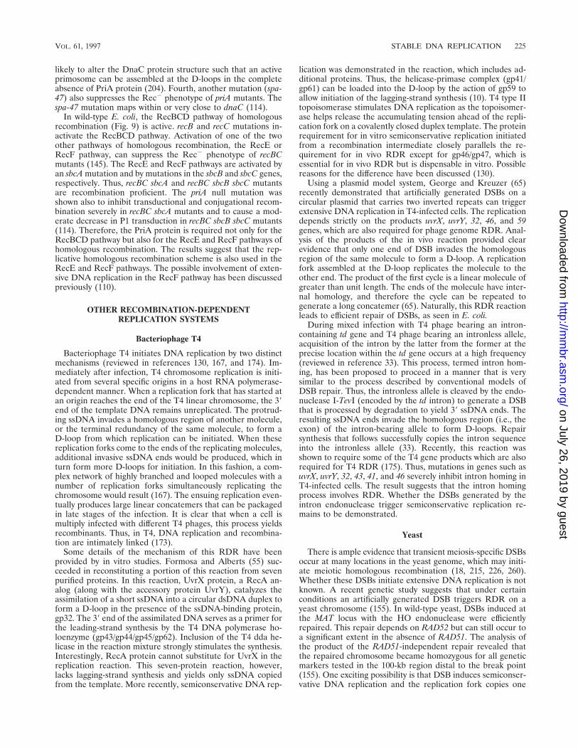
likely to alter the DnaC protein structure such that an activeprimosome can be assembled at the D-loops in the completeabsence of PriA protein (204). Fourth, another mutation (spa-47) also suppresses the Rec2 phenotype of priA mutants. Thespa-47 mutation maps within or very close to dnaC (114).
In wild-type E. coli, the RecBCD pathway of homologousrecombination (Fig. 9) is active. recB and recC mutations in-activate the RecBCD pathway. Activation of one of the twoother pathways of homologous recombination, the RecE orRecF pathway, can suppress the Rec2 phenotype of recBCmutants (145). The RecE and RecF pathways are activated byan sbcA mutation and by mutations in the sbcB and sbcC genes,respectively. Thus, recBC sbcA and recBC sbcB sbcC mutantsare recombination proficient. The priA null mutation wasshown also to inhibit transductional and conjugational recom-bination severely in recBC sbcA mutants and to cause a mod-erate decrease in P1 transduction in recBC sbcB sbcC mutants(114). Therefore, the PriA protein is required not only for theRecBCD pathway but also for the RecE and RecF pathways ofhomologous recombination. The results suggest that the rep-licative homologous recombination scheme is also used in theRecE and RecF pathways. The possible involvement of exten-sive DNA replication in the RecF pathway has been discussedpreviously (110).
OTHER RECOMBINATION-DEPENDENTREPLICATION SYSTEMS
Bacteriophage T4
Bacteriophage T4 initiates DNA replication by two distinctmechanisms (reviewed in references 130, 167, and 174). Im-mediately after infection, T4 chromosome replication is initi-ated from several specific origins in a host RNA polymerase-dependent manner. When a replication fork that has started atan origin reaches the end of the T4 linear chromosome, the 39end of the template DNA remains unreplicated. The protrud-ing ssDNA invades a homologous region of another molecule,or the terminal redundancy of the same molecule, to form aD-loop from which replication can be initiated. When thesereplication forks come to the ends of the replicating molecules,additional invasive ssDNA ends would be produced, which inturn form more D-loops for initiation. In this fashion, a com-plex network of highly branched and looped molecules with anumber of replication forks simultaneously replicating thechromosome would result (167). The ensuing replication even-tually produces large linear concatemers that can be packagedin late stages of the infection. It is clear that when a cell ismultiply infected with different T4 phages, this process yieldsrecombinants. Thus, in T4, DNA replication and recombina-tion are intimately linked (173).
Some details of the mechanism of this RDR have beenprovided by in vitro studies. Formosa and Alberts (55) suc-ceeded in reconstituting a portion of this reaction from sevenpurified proteins. In this reaction, UvrX protein, a RecA an-alog (along with the accessory protein UvrY), catalyzes theassimilation of a short ssDNA into a circular dsDNA duplex toform a D-loop in the presence of the ssDNA-binding protein,gp32. The 39 end of the assimilated DNA serves as a primer forthe leading-strand synthesis by the T4 DNA polymerase ho-loenzyme (gp43/gp44/gp45/gp62). Inclusion of the T4 dda he-licase in the reaction mixture strongly stimulates the synthesis.Interestingly, RecA protein cannot substitute for UvrX in thereplication reaction. This seven-protein reaction, however,lacks lagging-strand synthesis and yields only ssDNA copiedfrom the template. More recently, semiconservative DNA rep-
lication was demonstrated in the reaction, which includes ad-ditional proteins. Thus, the helicase-primase complex (gp41/gp61) can be loaded into the D-loop by the action of gp59 toallow initiation of the lagging-strand synthesis (10). T4 type IItopoisomerase stimulates DNA replication as the topoisomer-ase helps release the accumulating tension ahead of the repli-cation fork on a covalently closed duplex template. The proteinrequirement for in vitro semiconservative replication initiatedfrom a recombination intermediate closely parallels the re-quirement for in vivo RDR except for gp46/gp47, which isessential for in vivo RDR but is dispensable in vitro. Possiblereasons for the difference have been discussed (130).
Using a plasmid model system, George and Kreuzer (65)recently demonstrated that artificially generated DSBs on acircular plasmid that carries two inverted repeats can triggerextensive DNA replication in T4-infected cells. The replicationdepends strictly on the products uvrX, uvrY, 32, 46, and 59genes, which are also required for phage genome RDR. Anal-ysis of the products of the in vivo reaction provided clearevidence that only one end of DSB invades the homologousregion of the same molecule to form a D-loop. A replicationfork assembled at the D-loop replicates the molecule to theother end. The product of the first cycle is a linear molecule ofgreater than unit length. The ends of the molecule have inter-nal homology, and therefore the cycle can be repeated togenerate a long concatemer (65). Naturally, this RDR reactionleads to efficient repair of DSBs, as seen in E. coli.
During mixed infection with T4 phage bearing an intron-containing td gene and T4 phage bearing an intronless allele,acquisition of the intron by the latter from the former at theprecise location within the td gene occurs at a high frequency(reviewed in reference 33). This process, termed intron hom-ing, has been proposed to proceed in a manner that is verysimilar to the process described by conventional models ofDSB repair. Thus, the intronless allele is cleaved by the endo-nuclease I-TevI (encoded by the td intron) to generate a DSBthat is processed by degradation to yield 39 ssDNA ends. Theresulting ssDNA ends invade the homologous region (i.e., theexon) of the intron-bearing allele to form D-loops. Repairsynthesis that follows successfully copies the intron sequenceinto the intronless allele (33). Recently, this reaction wasshown to require some of the T4 gene products which are alsorequired for T4 RDR (175). Thus, mutations in genes such asuvrX, uvrY, 32, 43, 41, and 46 severely inhibit intron homing inT4-infected cells. The result suggests that the intron homingprocess involves RDR. Whether the DSBs generated by theintron endonuclease trigger semiconservative replication re-mains to be demonstrated.
Yeast
There is ample evidence that transient meiosis-specific DSBsoccur at many locations in the yeast genome, which may initi-ate meiotic homologous recombination (18, 215, 226, 260).Whether these DSBs initiate extensive DNA replication is notknown. A recent genetic study suggests that under certainconditions an artificially generated DSB triggers RDR on ayeast chromosome (155). In wild-type yeast, DSBs induced atthe MAT locus with the HO endonuclease were efficientlyrepaired. This repair depends on RAD52 but can still occur toa significant extent in the absence of RAD51. The analysis ofthe product of the RAD51-independent repair revealed thatthe repaired chromosome became homozygous for all geneticmarkers tested in the 100-kb region distal to the break point(155). One exciting possibility is that DSB induces semiconser-vative DNA replication and the replication fork copies one
VOL. 61, 1997 STABLE DNA REPLICATION 225
on July 26, 2019 by guesthttp://m
mbr.asm
.org/D
ownloaded from

arm of the chromosome to the telomere. This would be repli-cative homologous recombination. This replication in yeast,termed break-induced replication, is equivalent to RDR foundin E. coli and bacteriophage T4. An interesting difference isthat while prokaryotic RDR depends on RecA or its analog(UvrX), yeast break-induced replication is independent ofRAD51 (a RecA analog).
Mammalian Cells
Immunoglobulin V(D)J recombination in mice and humansis initiated by double-strand breaks, and repair of the breaksleads to recombination (86, 241). Whether this process in-volves DNA replication is not known. Analyses of the productsof murine immunoglobulin heavy-chain switch recombinationrevealed point mutations, deletions, and duplications in thevicinity of the switch points (47, 48). It was suggested that therecombination events accompany local DNA synthesis, whichis error prone. How widely eukaryotic cells employ RDR-mediated DSB repair and replicative homologous recombina-tion remains to be seen.
Mitochondrial DNA
In contrast to the mitochondrial DNA (mtDNA) of mam-malian cells, which exists as a monomer circle and replicates bythe u mode (34), mtDNA of most other organisms includingyeast, protozoa, and higher plants exists as large concatemersoften of complex structures (reviewed in reference 13). Forexample, newly synthesized mtDNA from tobacco cells is en-riched with multigenomic concatemers of highly branched struc-tures (183). Similar structures have also been observed inmtDNA from fungi including yeast (14). These molecules arespeculated to be mtDNA replicating in a recombination-de-pendent manner or by the rolling-circle mode, which can be ini-iated by recombination (245). Similar multigenomic branchedconcatemers were also detected among replicating mtDNAfrom the malaria parasite. The possibility that these moleculesreplicate in a manner similar to RDR was considered (188).
CONSTITUTIVE STABLE DNA REPLICATION
SDR Mutants
As described above, iSDR is normally repressed but can beactivated by SOS induction. To gain some insights into geneticbases for the activation, mutants that expressed SDR consti-tutively were sought. By a colony autoradiography procedure,two SDR mutants (sdrA2 and sdrA102) were isolated from anE. coli 15 T2 strain (105, 123). Later, additional SDR mutants(e.g., sdrA224 mutants) were isolated from an E. coli K12 strain(238). The mutants could continue DNA replication for atleast 12 h in the presence of chloramphenicol or in the absenceof required amino acids. This type of SDR in sdrA mutants wastermed cSDR (constitutive stable DNA replication). The mu-tations were mapped in the proA-metD region of the chromo-some and were recessive to the wild type.
The Idea of an Alternative Initiation Pathway
An extraordinary characteristic found with cSDR in sdrAmutants was that cSDR completely depends on a RecA func-tion (123, 238). Thus, sdrA recA(Ts) mutants were capable ofcSDR at 30°C, but the replication ceased within 2 h of a shiftto 42°C with an increment of 40 to 45% in DNA content,suggesting that a RecA function was needed for initiation butnot for elongation. Despite the strict requirement for RecA
protein in cSDR, the growth of sdrA (oriC1) mutants was notaffected by recA(Ts), indicating the absence of a requirementfor RecA protein in initiation at oriC in sdrA mutants. Theseobservations led to the hypothesis that cSDR was initiated bya mechanism distinct from that operating at oriC (123). ThesdrA1 gene product was viewed as a repressor of a switch fromthe oriC initiation mechanism to the SDR initiation alterna-tive. This switch model postulated that SOS induction inacti-vated the SdrA repressor, allowing the switch to iSDR, andthat cSDR resulted from inactivation of the repressor by thesdrA mutation.
The notion that SDR initiation is distinct from and alterna-tive to oriC initiation was substantiated by the demonstrationthat sdrA mutants could survive complete inactivation of oriCinitiation (124). Thus, the dnaA gene could be obliterated byinsertion of a Tn10 transposon and the oriC site could beremoved from the chromosome by deletion when cSDR wasactivated by an sdrA mutation. The resulting double mutants,sdrA dnaA::Tn10 and sdrA DoriC, respectively, grew well, albeitslowly, in minimal media. They were, however, unable to growin rich media such as Luria broth: the double mutants arebroth sensitive.
The sdrA mutation can also suppress the temperature sen-sitivity phenotype of many dnaA(Ts) mutations (124). The sdrAmutation is therefore a non-allele-specific suppressor of dnaAmutations. Earlier, Atlung (9) isolated a number of thermore-sistant revertants from a dnaA46(Ts) mutant and mapped theextragenic suppressor mutations to seven distinct loci on thechromosome. These suppressor mutations were designated das(DnaA suppressors). One group of das mutations, the dasFmutations, was concluded to be allelic to sdrA by the followingcriteria (237): (i) dasF mutations impart the cSDR phenotype;(ii) dasF mutants can tolerate deletion of oriC and completeinactivation of dnaA; (iii) dasF and sdrA mutants exhibit brothsensitivity; and (iv) both mutations map between proA and metD.
sdrA Mutants Are RNase HI Defective
It was known that when RNase HI was omitted from certainin vitro DNA replication systems (i.e., phage fd and plasmidColE1), DNA replication was initiated at illegitimate sites (75,84, 242). Thus, the presence of RNase HI ensures the initiationof DNA replication exclusively at the normal origin. This dis-criminatory action of RNase HI was consistent with the repres-sor role of SdrA1 in the switch model described above. Closeproximity of the map positions of sdrA, dasF, and rnhA (en-coding RNase HI) prompted an examination of the possibleidentity of these genes (182). On one hand, sdrA and dasFmutants were found to be devoid of RNase HI activity. On theother hand, rnhA mutants were shown to have a number ofphenotypes in common with sdrA and dasF mutants. For ex-ample, rnhA mutants exhibited cSDR, were broth sensitive,and could tolerate dnaA::Tn10 and DoriC. The shared pheno-types of sdrA, dasF, and rnhA mutants were all complementedby a plasmid carrying a 760-bp segment of the E. coli chromo-some containing an rnhA1 gene (182). Thus, it was establishedbeyond any doubt that sdrA, dasF, and rnhA are allelic to eachother.
How could the lack of RNase HI activity result in activationof the normally repressed alternative DNA replication systemwhich can occur in the absence of DnaA? Along the line of theswitch model, it is easy to see that inactivation of RNase HI(i.e., the SdrA repressor) allows the initiation of replication atother sites as well as at oriC. Normally, RNase HI eliminatesthe RNA transcript hybridizing to the template. In its absence,the RNA is stabilized and used to initiate replication (38, 182).
226 KOGOMA MICROBIOL. MOL. BIOL. REV.
on July 26, 2019 by guesthttp://m
mbr.asm
.org/D
ownloaded from
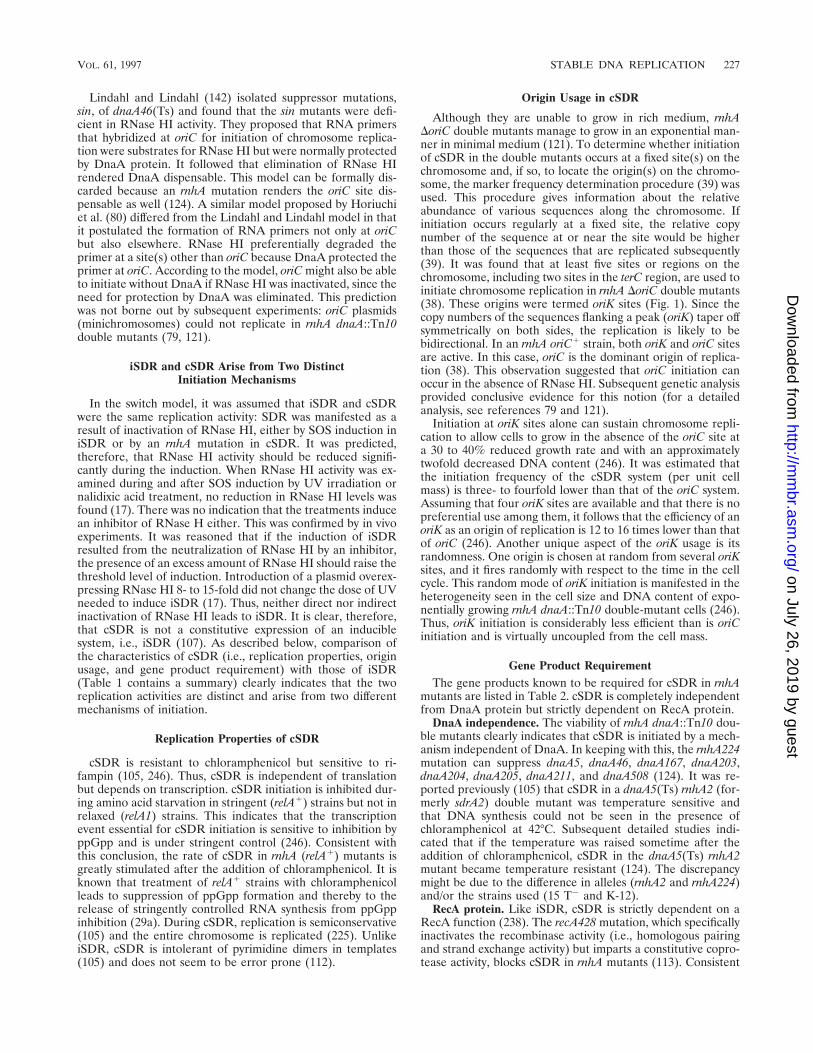
Lindahl and Lindahl (142) isolated suppressor mutations,sin, of dnaA46(Ts) and found that the sin mutants were defi-cient in RNase HI activity. They proposed that RNA primersthat hybridized at oriC for initiation of chromosome replica-tion were substrates for RNase HI but were normally protectedby DnaA protein. It followed that elimination of RNase HIrendered DnaA dispensable. This model can be formally dis-carded because an rnhA mutation renders the oriC site dis-pensable as well (124). A similar model proposed by Horiuchiet al. (80) differed from the Lindahl and Lindahl model in thatit postulated the formation of RNA primers not only at oriCbut also elsewhere. RNase HI preferentially degraded theprimer at a site(s) other than oriC because DnaA protected theprimer at oriC. According to the model, oriC might also be ableto initiate without DnaA if RNase HI was inactivated, since theneed for protection by DnaA was eliminated. This predictionwas not borne out by subsequent experiments: oriC plasmids(minichromosomes) could not replicate in rnhA dnaA::Tn10double mutants (79, 121).
iSDR and cSDR Arise from Two DistinctInitiation Mechanisms
In the switch model, it was assumed that iSDR and cSDRwere the same replication activity: SDR was manifested as aresult of inactivation of RNase HI, either by SOS induction iniSDR or by an rnhA mutation in cSDR. It was predicted,therefore, that RNase HI activity should be reduced signifi-cantly during the induction. When RNase HI activity was ex-amined during and after SOS induction by UV irradiation ornalidixic acid treatment, no reduction in RNase HI levels wasfound (17). There was no indication that the treatments inducean inhibitor of RNase H either. This was confirmed by in vivoexperiments. It was reasoned that if the induction of iSDRresulted from the neutralization of RNase HI by an inhibitor,the presence of an excess amount of RNase HI should raise thethreshold level of induction. Introduction of a plasmid overex-pressing RNase HI 8- to 15-fold did not change the dose of UVneeded to induce iSDR (17). Thus, neither direct nor indirectinactivation of RNase HI leads to iSDR. It is clear, therefore,that cSDR is not a constitutive expression of an induciblesystem, i.e., iSDR (107). As described below, comparison ofthe characteristics of cSDR (i.e., replication properties, originusage, and gene product requirement) with those of iSDR(Table 1 contains a summary) clearly indicates that the tworeplication activities are distinct and arise from two differentmechanisms of initiation.
Replication Properties of cSDR
cSDR is resistant to chloramphenicol but sensitive to ri-fampin (105, 246). Thus, cSDR is independent of translationbut depends on transcription. cSDR initiation is inhibited dur-ing amino acid starvation in stringent (relA1) strains but not inrelaxed (relA1) strains. This indicates that the transcriptionevent essential for cSDR initiation is sensitive to inhibition byppGpp and is under stringent control (246). Consistent withthis conclusion, the rate of cSDR in rnhA (relA1) mutants isgreatly stimulated after the addition of chloramphenicol. It isknown that treatment of relA1 strains with chloramphenicolleads to suppression of ppGpp formation and thereby to therelease of stringently controlled RNA synthesis from ppGppinhibition (29a). During cSDR, replication is semiconservative(105) and the entire chromosome is replicated (225). UnlikeiSDR, cSDR is intolerant of pyrimidine dimers in templates(105) and does not seem to be error prone (112).
Origin Usage in cSDR
Although they are unable to grow in rich medium, rnhADoriC double mutants manage to grow in an exponential man-ner in minimal medium (121). To determine whether initiationof cSDR in the double mutants occurs at a fixed site(s) on thechromosome and, if so, to locate the origin(s) on the chromo-some, the marker frequency determination procedure (39) wasused. This procedure gives information about the relativeabundance of various sequences along the chromosome. Ifinitiation occurs regularly at a fixed site, the relative copynumber of the sequence at or near the site would be higherthan those of the sequences that are replicated subsequently(39). It was found that at least five sites or regions on thechromosome, including two sites in the terC region, are used toinitiate chromosome replication in rnhA DoriC double mutants(38). These origins were termed oriK sites (Fig. 1). Since thecopy numbers of the sequences flanking a peak (oriK) taper offsymmetrically on both sides, the replication is likely to bebidirectional. In an rnhA oriC1 strain, both oriK and oriC sitesare active. In this case, oriC is the dominant origin of replica-tion (38). This observation suggested that oriC initiation canoccur in the absence of RNase HI. Subsequent genetic analysisprovided conclusive evidence for this notion (for a detailedanalysis, see references 79 and 121).
Initiation at oriK sites alone can sustain chromosome repli-cation to allow cells to grow in the absence of the oriC site ata 30 to 40% reduced growth rate and with an approximatelytwofold decreased DNA content (246). It was estimated thatthe initiation frequency of the cSDR system (per unit cellmass) is three- to fourfold lower than that of the oriC system.Assuming that four oriK sites are available and that there is nopreferential use among them, it follows that the efficiency of anoriK as an origin of replication is 12 to 16 times lower than thatof oriC (246). Another unique aspect of the oriK usage is itsrandomness. One origin is chosen at random from several oriKsites, and it fires randomly with respect to the time in the cellcycle. This random mode of oriK initiation is manifested in theheterogeneity seen in the cell size and DNA content of expo-nentially growing rnhA dnaA::Tn10 double-mutant cells (246).Thus, oriK initiation is considerably less efficient than is oriCinitiation and is virtually uncoupled from the cell mass.
Gene Product RequirementThe gene products known to be required for cSDR in rnhA
mutants are listed in Table 2. cSDR is completely independentfrom DnaA protein but strictly dependent on RecA protein.
DnaA independence. The viability of rnhA dnaA::Tn10 dou-ble mutants clearly indicates that cSDR is initiated by a mech-anism independent of DnaA. In keeping with this, the rnhA224mutation can suppress dnaA5, dnaA46, dnaA167, dnaA203,dnaA204, dnaA205, dnaA211, and dnaA508 (124). It was re-ported previously (105) that cSDR in a dnaA5(Ts) rnhA2 (for-merly sdrA2) double mutant was temperature sensitive andthat DNA synthesis could not be seen in the presence ofchloramphenicol at 42°C. Subsequent detailed studies indi-cated that if the temperature was raised sometime after theaddition of chloramphenicol, cSDR in the dnaA5(Ts) rnhA2mutant became temperature resistant (124). The discrepancymight be due to the difference in alleles (rnhA2 and rnhA224)and/or the strains used (15 T2 and K-12).
RecA protein. Like iSDR, cSDR is strictly dependent on aRecA function (238). The recA428 mutation, which specificallyinactivates the recombinase activity (i.e., homologous pairingand strand exchange activity) but imparts a constitutive copro-tease activity, blocks cSDR in rnhA mutants (113). Consistent
VOL. 61, 1997 STABLE DNA REPLICATION 227
on July 26, 2019 by guesthttp://m
mbr.asm
.org/D
ownloaded from
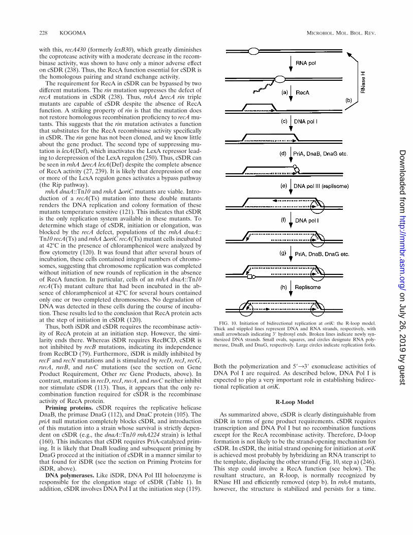
with this, recA430 (formerly lexB30), which greatly diminishesthe coprotease activity with a moderate decrease in the recom-binase activity, was shown to have only a minor adverse effecton cSDR (238). Thus, the RecA function essential for cSDR isthe homologous pairing and strand exchange activity.
The requirement for RecA in cSDR can be bypassed by twodifferent mutations. The rin mutation suppresses the defect ofrecA mutations in cSDR (238). Thus, rnhA DrecA rin triplemutants are capable of cSDR despite the absence of RecAfunction. A striking property of rin is that the mutation doesnot restore homologous recombination proficiency to recA mu-tants. This suggests that the rin mutation activates a functionthat substitutes for the RecA recombinase activity specificallyin cSDR. The rin gene has not been cloned, and we know littleabout the gene product. The second type of suppressing mu-tation is lexA(Def), which inactivates the LexA repressor lead-ing to derepression of the LexA regulon (250). Thus, cSDR canbe seen in rnhA DrecA lexA(Def) despite the complete absenceof RecA activity (27, 239). It is likely that derepression of oneor more of the LexA regulon genes activates a bypass pathway(the Rip pathway).
rnhA dnaA::Tn10 and rnhA DoriC mutants are viable. Intro-duction of a recA(Ts) mutation into these double mutantsrenders the DNA replication and colony formation of thesemutants temperature sensitive (121). This indicates that cSDRis the only replication system available in these mutants. Todetermine which stage of cSDR, initiation or elongation, wasblocked by the recA defect, populations of the rnhA dnaA::Tn10 recA(Ts) and rnhA DoriC recA(Ts) mutant cells incubatedat 42°C in the presence of chloramphenicol were analyzed byflow cytometry (120). It was found that after several hours ofincubation, these cells contained integral numbers of chromo-somes, suggesting that chromosome replication was completedwithout initiation of new rounds of replication in the absenceof RecA function. In particular, cells of an rnhA dnaA::Tn10recA(Ts) mutant culture that had been incubated in the ab-sence of chloramphenicol at 42°C for several hours containedonly one or two completed chromosomes. No degradation ofDNA was detected in these cells during the course of incuba-tion. These results led to the conclusion that RecA protein actsat the step of initiation in cSDR (120).
Thus, both iSDR and cSDR requires the recombinase activ-ity of RecA protein at an initiation step. However, the simi-larity ends there. Whereas iSDR requires RecBCD, cSDR isnot inhibited by recB mutations, indicating its independencefrom RecBCD (79). Furthermore, iSDR is mildly inhibited byrecF and recN mutations and is stimulated by recD, recJ, recG,ruvA, ruvB, and ruvC mutations (see the section on GeneProduct Requirement, Other rec Gene Products, above). Incontrast, mutations in recD, recJ, ruvA, and ruvC neither inhibitnor stimulate cSDR (113). Thus, it appears that the only re-combination function required for cSDR is the recombinaseactivity of RecA protein.
Priming proteins. cSDR requires the replicative helicaseDnaB, the primase DnaG (112), and DnaC protein (105). ThepriA null mutation completely blocks cSDR, and introductionof this mutation into a strain whose survival is strictly depen-dent on cSDR (e.g., the dnaA::Tn10 rnhA224 strain) is lethal(160). This indicates that cSDR requires PriA-catalyzed prim-ing. It is likely that DnaB loading and subsequent priming byDnaG proceed at the initiation of cSDR in a manner similar tothat found for iSDR (see the section on Priming Proteins foriSDR, above).
DNA polymerases. Like iSDR, DNA Pol III holoenzyme isresponsible for the elongation stage of cSDR (Table 1). Inaddition, cSDR involves DNA Pol I at the initiation step (119).
Both the polymerization and 59339 exonuclease activities ofDNA Pol I are required. As described below, DNA Pol I isexpected to play a very important role in establishing bidirec-tional replication at oriK.
R-Loop Model
As summarized above, cSDR is clearly distinguishable fromiSDR in terms of gene product requirements. cSDR requirestranscription and DNA Pol I but no recombination functionsexcept for the RecA recombinase activity. Therefore, D-loopformation is not likely to be the strand-opening mechanism forcSDR. In cSDR, the initial strand opening for initiation at oriKis achieved most probably by hybridizing an RNA transcript tothe template, displacing the other strand (Fig. 10, step a) (246).This step could involve a RecA function (see below). Theresultant structure, an R-loop, is normally recognized byRNase HI and efficiently removed (step b). In rnhA mutants,however, the structure is stabilized and persists for a time.
FIG. 10. Initiation of bidirectional replication at oriK: the R-loop model.Thick and stippled lines represent DNA and RNA strands, respectively, withsmall arrowheads indicating 39 hydroxyl ends. Broken lines indicate newly syn-thesized DNA strands. Small ovals, squares, and circles designate RNA poly-merase, DnaB, and DnaG, respectively. Large circles indicate replication forks.
228 KOGOMA MICROBIOL. MOL. BIOL. REV.
on July 26, 2019 by guesthttp://m
mbr.asm
.org/D
ownloaded from
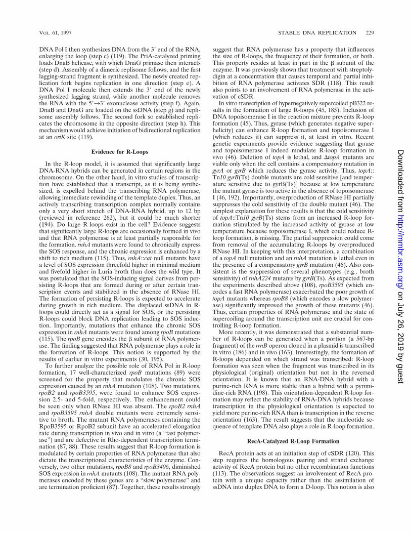
DNA Pol I then synthesizes DNA from the 39 end of the RNA,enlarging the loop (step c) (119). The PriA-catalyzed primingloads DnaB helicase, with which DnaG primase then interacts(step d). Assembly of a dimeric replisome follows, and the firstlagging-strand fragment is synthesized. The newly created rep-lication fork begins replication in one direction (step e). ADNA Pol I molecule then extends the 39 end of the newlysynthesized lagging strand, while another molecule removesthe RNA with the 59339 exonuclease activity (step f). Again,DnaB and DnaG are loaded on the ssDNA (step g) and repli-some assembly follows. The second fork so established repli-cates the chromosome in the opposite direction (step h). Thismechanism would achieve initiation of bidirectional replicationat an oriK site (119).
Evidence for R-Loops
In the R-loop model, it is assumed that significantly largeDNA-RNA hybrids can be generated in certain regions in thechromosome. On the other hand, in vitro studies of transcrip-tion have established that a transcript, as it is being synthe-sized, is expelled behind the transcribing RNA polymerase,allowing immediate rewinding of the template duplex. Thus, anactively transcribing transcription complex normally containsonly a very short stretch of DNA-RNA hybrid, up to 12 bp(reviewed in reference 262), but it could be much shorter(194). Do large R-loops exist in the cell? Evidence suggeststhat significantly large R-loops are occasionally formed in vivoand that RNA polymerase is at least partially responsible forthe formation. rnhA mutants were found to chronically expressthe SOS response, and the chronic expression is enhanced by ashift to rich medium (115). Thus, rnhA::cat null mutants havea level of SOS expression threefold higher in minimal mediumand fivefold higher in Luria broth than does the wild type. Itwas postulated that the SOS-inducing signal derives from per-sisting R-loops that are formed during or after certain tran-scription events and stabilized in the absence of RNase HI.The formation of persisting R-loops is expected to accelerateduring growth in rich medium. The displaced ssDNA in R-loops could directly act as a signal for SOS, or the persistingR-loops could block DNA replication leading to SOS induc-tion. Importantly, mutations that enhance the chronic SOSexpression in rnhA mutants were found among rpoB mutations(115). The rpoB gene encodes the b subunit of RNA polymer-ase. The finding suggested that RNA polymerase plays a role inthe formation of R-loops. This notion is supported by theresults of earlier in vitro experiments (30, 195).
To further analyze the possible role of RNA Pol in R-loopformation, 17 well-characterized rpoB mutations (89) werescreened for the property that modulates the chronic SOSexpression caused by an rnhA mutation (108). Two mutations,rpoB2 and rpoB3595, were found to enhance SOS expres-sion 2.5- and 5-fold, respectively. The enhancement couldbe seen only when RNase HI was absent. The rpoB2 rnhAand rpoB3595 rnhA double mutants were extremely sensi-tive to broth. The mutant RNA polymerases containing theRpoB3595 or RpoB2 subunit have an accelerated elongationrate during transcription in vivo and in vitro (a “fast polymer-ase”) and are defective in Rho-dependent transcription termi-nation (87, 88). These results suggest that R-loop formation ismodulated by certain properties of RNA polymerase that alsodictate the transcriptional characteristics of the enzyme. Con-versely, two other mutations, rpoB8 and rpoB3406, diminishedSOS expression in rnhA mutants (108). The mutant RNA poly-merases encoded by these genes are a “slow polymerase” andare termination proficient (87). Together, these results strongly
suggest that RNA polymerase has a property that influencesthe size of R-loops, the frequency of their formation, or both.This property resides at least in part in the b subunit of theenzyme. It was previously shown that treatment with streptoly-digin at a concentration that causes temporal and partial inhi-bition of RNA polymerase activates SDR (118). This resultalso points to an involvement of RNA polymerase in the acti-vation of cSDR.
In vitro transcription of hypernegatively supercoiled pB322 re-sults in the formation of large R-loops (45, 185). Inclusion ofDNA topoisomerase I in the reaction mixture prevents R-loopformation (45). Thus, gyrase (which generates negative super-helicity) can enhance R-loop formation and topoisomerase I(which reduces it) can suppress it, at least in vitro. Recentgenetic experiments provide evidence suggesting that gyraseand topoisomerase I indeed modulate R-loop formation invivo (46). Deletion of topA is lethal, and DtopA mutants areviable only when the cell contains a compensatory mutation ingyrA or gyrB which reduces the gyrase activity. Thus, topA::Tn10 gyrB(Ts) double mutants are cold sensitive [and temper-ature sensitive due to gyrB(Ts)] because at low temperaturethe mutant gyrase is too active in the absence of topoisomeraseI (46, 192). Importantly, overproduction of RNase HI partiallysuppresses the cold sensitivity of the double mutant (46). Thesimplest explanation for these results is that the cold sensitivityof topA::Tn10 gyrB(Ts) stems from an increased R-loop for-mation stimulated by the increased activity of gyrase at lowtemperature because topoisomerase I, which could reduce R-loop formation, is missing. The partial suppression could comefrom removal of the accumulating R-loops by overproducedRNase HI. In keeping with this interpretation, a combinationof a topA null mutation and an rnhA mutation is lethal even inthe presence of a compensatory gyrB mutation (46). Also con-sistent is the suppression of several phenotypes (e.g., brothsensitivity) of rnhA224 mutants by gyrB(Ts). As expected fromthe experiments described above (108), rpoB3595 (which en-codes a fast RNA polymerase) exacerbated the poor growth oftopA mutants whereas rpoB8 (which encodes a slow polymer-ase) significantly improved the growth of these mutants (46).Thus, certain properties of RNA polymerase and the state ofsupercoiling around the transcription unit are crucial for con-trolling R-loop formation.
More recently, it was demonstrated that a substantial num-ber of R-loops can be generated when a portion (a 567-bpfragment) of the rrnB operon cloned in a plasmid is transcribedin vitro (186) and in vivo (163). Interestingly, the formation ofR-loops depended on which strand was transcribed: R-loopformation was seen when the fragment was transcribed in itsphysiological (original) orientation but not in the reversedorientation. It is known that an RNA-DNA hybrid with apurine-rich RNA is more stable than a hybrid with a pyrimi-dine-rich RNA (198). This orientation-dependent R-loop for-mation may reflect the stability of RNA-DNA hybrids becausetranscription in the physiological orientation is expected toyield more purine-rich RNA than is transcription in the reverseorientation (163). The result suggests that the nucleotide se-quence of template DNA also plays a role in R-loop formation.
RecA-Catalyzed R-Loop Formation
RecA protein acts at an initiation step of cSDR (120). Thisstep requires the homologous pairing and strand exchangeactivity of RecA protein but no other recombination functions(113). The observations suggest an involvement of RecA pro-tein with a unique capacity rather than the assimilation ofssDNA into duplex DNA to form a D-loop. This notion is also
VOL. 61, 1997 STABLE DNA REPLICATION 229
on July 26, 2019 by guesthttp://m
mbr.asm
.org/D
ownloaded from
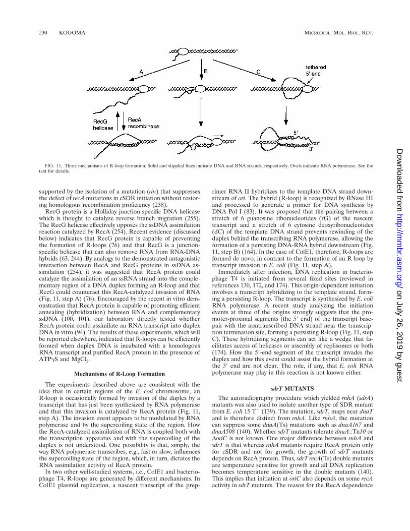
supported by the isolation of a mutation (rin) that suppressesthe defect of recA mutations in cSDR initiation without restor-ing homologous recombination proficiency (238).
RecG protein is a Holliday junction-specific DNA helicasewhich is thought to catalyze reverse branch migration (255).The RecG helicase effectively opposes the ssDNA assimilationreaction catalyzed by RecA (254). Recent evidence (discussedbelow) indicates that RecG protein is capable of preventingthe formation of R-loops (76) and that RecG is a junction-specific helicase that can also remove RNA from RNA-DNAhybrids (63, 244). By analogy to the demonstrated antagonisticinteraction between RecA and RecG proteins in ssDNA as-similation (254), it was suggested that RecA protein couldcatalyze the assimilation of an ssRNA strand into the comple-mentary region of a DNA duplex forming an R-loop and thatRecG could counteract this RecA-catalyzed invasion of RNA(Fig. 11, step A) (76). Encouraged by the recent in vitro dem-onstration that RecA protein is capable of promoting efficientannealing (hybridization) between RNA and complementaryssDNA (100, 101), our laboratory directly tested whetherRecA protein could assimilate an RNA transcript into duplexDNA in vitro (94). The results of these experiments, which willbe reported elsewhere, indicated that R-loops can be efficientlyformed when duplex DNA is incubated with a homologousRNA transcript and purified RecA protein in the presence ofATPgS and MgCl2.
Mechanisms of R-Loop Formation
The experiments described above are consistent with theidea that in certain regions of the E. coli chromosome, anR-loop is occasionally formed by invasion of the duplex by atranscript that has just been synthesized by RNA polymeraseand that this invasion is catalyzed by RecA protein (Fig. 11,step A). The invasion event appears to be modulated by RNApolymerase and by the supercoiling state of the region. Howthe RecA-catalyzed assimilation of RNA is coupled both withthe transcription apparatus and with the supercoiling of theduplex is not understood. One possibility is that, simply, theway RNA polymerase transcribes, e.g., fast or slow, influencesthe supercoiling state of the region, which, in turn, dictates theRNA assimilation activity of RecA protein.
In two other well-studied systems, i.e., ColE1 and bacterio-phage T4, R-loops are generated by different mechanisms. InColE1 plasmid replication, a nascent transcript of the prep-
rimer RNA II hybridizes to the template DNA strand down-stream of ori. The hybrid (R-loop) is recognized by RNase HIand processed to generate a primer for DNA synthesis byDNA Pol I (83). It was proposed that the pairing between astretch of 6 guanosine ribonucleotides (rG) of the nascenttranscript and a stretch of 6 cytosine deoxyribonucleotides(dC) of the template DNA strand prevents rewinding of theduplex behind the transcribing RNA polymerase, allowing theformation of a persisting DNA-RNA hybrid downstream (Fig.11, step B) (164). In the case of ColE1, therefore, R-loops areformed de novo, in contrast to the formation of an R-loop bytranscript invasion in E. coli (Fig. 11, step A).
Immediately after infection, DNA replication in bacterio-phage T4 is initiated from several fixed sites (reviewed inreferences 130, 172, and 174). This origin-dependent initiationinvolves a transcript hybridizing to the template strand, form-ing a persisting R-loop. The transcript is synthesized by E. coliRNA polymerase. A recent study analyzing the initiationevents at three of the origins strongly suggests that the pro-moter-proximal segments (the 59 end) of the transcript base-pair with the nontranscribed DNA strand near the transcrip-tion termination site, forming a persisting R-loop (Fig. 11, stepC). These hybridizing segments can act like a wedge that fa-cilitates access of helicases or assembly of replisomes or both(174). How the 59-end segment of the transcript invades theduplex and how this event could assist the hybrid formation atthe 39 end are not clear. The role, if any, that E. coli RNApolymerase may play in this reaction is not known either.
sdrT MUTANTS
The autoradiography procedure which yielded rnhA (sdrA)mutants was also used to isolate another type of SDR mutantfrom E. coli 15 T2 (139). The mutation, sdrT, maps near dnaTand is therefore distinct from rnhA. Like rnhA, the mutationcan suppress some dnaA(Ts) mutations such as dnaA167 anddnaA508 (140). Whether sdrT mutants tolerate dnaA::Tn10 orDoriC is not known. One major difference between rnhA andsdrT is that whereas rnhA mutants require RecA protein onlyfor cSDR and not for growth, the growth of sdrT mutantsdepends on RecA protein. Thus, sdrT recA(Ts) double mutantsare temperature sensitive for growth and all DNA replicationbecomes temperature sensitive in the double mutants (140).This implies that initiation at oriC also depends on some recAactivity in sdrT mutants. The reason for the RecA dependence
FIG. 11. Three mechanisms of R-loop formation. Solid and stippled lines indicate DNA and RNA strands, respectively. Ovals indicate RNA polymerase. See thetext for details.
230 KOGOMA MICROBIOL. MOL. BIOL. REV.
on July 26, 2019 by guesthttp://m
mbr.asm
.org/D
ownloaded from
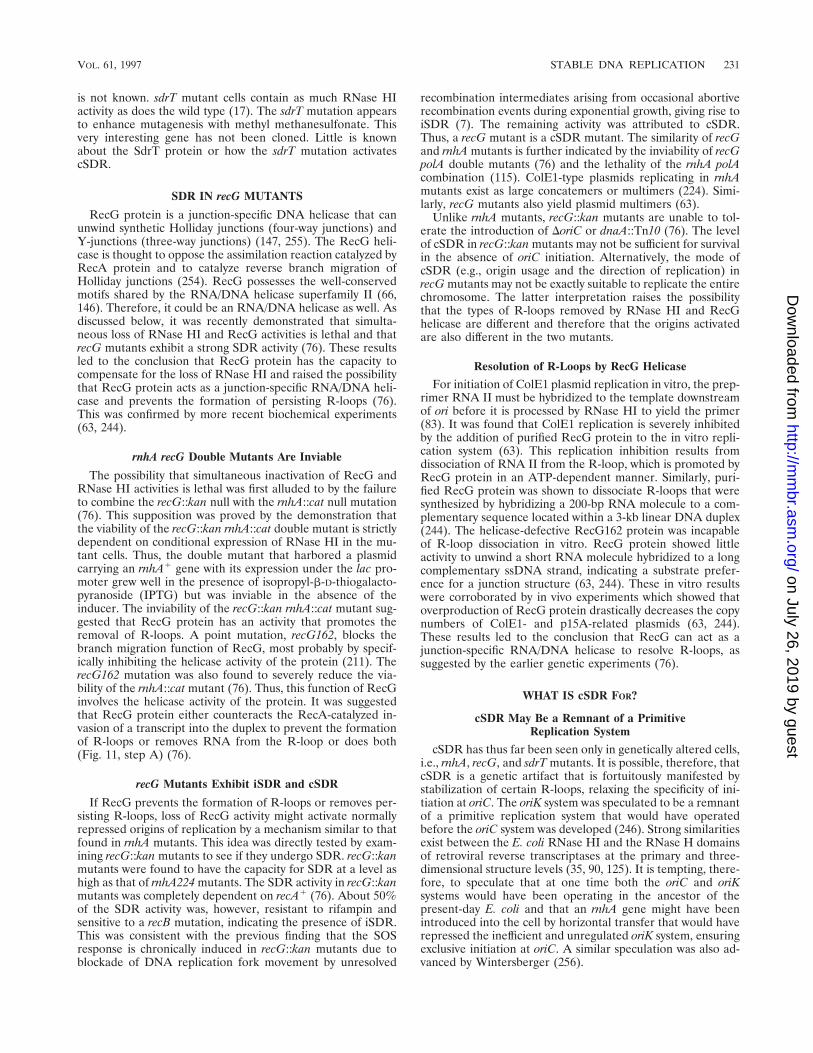
is not known. sdrT mutant cells contain as much RNase HIactivity as does the wild type (17). The sdrT mutation appearsto enhance mutagenesis with methyl methanesulfonate. Thisvery interesting gene has not been cloned. Little is knownabout the SdrT protein or how the sdrT mutation activatescSDR.
SDR IN recG MUTANTS
RecG protein is a junction-specific DNA helicase that canunwind synthetic Holliday junctions (four-way junctions) andY-junctions (three-way junctions) (147, 255). The RecG heli-case is thought to oppose the assimilation reaction catalyzed byRecA protein and to catalyze reverse branch migration ofHolliday junctions (254). RecG possesses the well-conservedmotifs shared by the RNA/DNA helicase superfamily II (66,146). Therefore, it could be an RNA/DNA helicase as well. Asdiscussed below, it was recently demonstrated that simulta-neous loss of RNase HI and RecG activities is lethal and thatrecG mutants exhibit a strong SDR activity (76). These resultsled to the conclusion that RecG protein has the capacity tocompensate for the loss of RNase HI and raised the possibilitythat RecG protein acts as a junction-specific RNA/DNA heli-case and prevents the formation of persisting R-loops (76).This was confirmed by more recent biochemical experiments(63, 244).
rnhA recG Double Mutants Are Inviable
The possibility that simultaneous inactivation of RecG andRNase HI activities is lethal was first alluded to by the failureto combine the recG::kan null with the rnhA::cat null mutation(76). This supposition was proved by the demonstration thatthe viability of the recG::kan rnhA::cat double mutant is strictlydependent on conditional expression of RNase HI in the mu-tant cells. Thus, the double mutant that harbored a plasmidcarrying an rnhA1 gene with its expression under the lac pro-moter grew well in the presence of isopropyl-b-D-thiogalacto-pyranoside (IPTG) but was inviable in the absence of theinducer. The inviability of the recG::kan rnhA::cat mutant sug-gested that RecG protein has an activity that promotes theremoval of R-loops. A point mutation, recG162, blocks thebranch migration function of RecG, most probably by specif-ically inhibiting the helicase activity of the protein (211). TherecG162 mutation was also found to severely reduce the via-bility of the rnhA::cat mutant (76). Thus, this function of RecGinvolves the helicase activity of the protein. It was suggestedthat RecG protein either counteracts the RecA-catalyzed in-vasion of a transcript into the duplex to prevent the formationof R-loops or removes RNA from the R-loop or does both(Fig. 11, step A) (76).
recG Mutants Exhibit iSDR and cSDR
If RecG prevents the formation of R-loops or removes per-sisting R-loops, loss of RecG activity might activate normallyrepressed origins of replication by a mechanism similar to thatfound in rnhA mutants. This idea was directly tested by exam-ining recG::kan mutants to see if they undergo SDR. recG::kanmutants were found to have the capacity for SDR at a level ashigh as that of rnhA224 mutants. The SDR activity in recG::kanmutants was completely dependent on recA1 (76). About 50%of the SDR activity was, however, resistant to rifampin andsensitive to a recB mutation, indicating the presence of iSDR.This was consistent with the previous finding that the SOSresponse is chronically induced in recG::kan mutants due toblockade of DNA replication fork movement by unresolved
recombination intermediates arising from occasional abortiverecombination events during exponential growth, giving rise toiSDR (7). The remaining activity was attributed to cSDR.Thus, a recG mutant is a cSDR mutant. The similarity of recGand rnhA mutants is further indicated by the inviability of recGpolA double mutants (76) and the lethality of the rnhA polAcombination (115). ColE1-type plasmids replicating in rnhAmutants exist as large concatemers or multimers (224). Simi-larly, recG mutants also yield plasmid multimers (63).
Unlike rnhA mutants, recG::kan mutants are unable to tol-erate the introduction of DoriC or dnaA::Tn10 (76). The levelof cSDR in recG::kan mutants may not be sufficient for survivalin the absence of oriC initiation. Alternatively, the mode ofcSDR (e.g., origin usage and the direction of replication) inrecG mutants may not be exactly suitable to replicate the entirechromosome. The latter interpretation raises the possibilitythat the types of R-loops removed by RNase HI and RecGhelicase are different and therefore that the origins activatedare also different in the two mutants.
Resolution of R-Loops by RecG Helicase
For initiation of ColE1 plasmid replication in vitro, the prep-rimer RNA II must be hybridized to the template downstreamof ori before it is processed by RNase HI to yield the primer(83). It was found that ColE1 replication is severely inhibitedby the addition of purified RecG protein to the in vitro repli-cation system (63). This replication inhibition results fromdissociation of RNA II from the R-loop, which is promoted byRecG protein in an ATP-dependent manner. Similarly, puri-fied RecG protein was shown to dissociate R-loops that weresynthesized by hybridizing a 200-bp RNA molecule to a com-plementary sequence located within a 3-kb linear DNA duplex(244). The helicase-defective RecG162 protein was incapableof R-loop dissociation in vitro. RecG protein showed littleactivity to unwind a short RNA molecule hybridized to a longcomplementary ssDNA strand, indicating a substrate prefer-ence for a junction structure (63, 244). These in vitro resultswere corroborated by in vivo experiments which showed thatoverproduction of RecG protein drastically decreases the copynumbers of ColE1- and p15A-related plasmids (63, 244).These results led to the conclusion that RecG can act as ajunction-specific RNA/DNA helicase to resolve R-loops, assuggested by the earlier genetic experiments (76).
WHAT IS cSDR FOR?
cSDR May Be a Remnant of a PrimitiveReplication System
cSDR has thus far been seen only in genetically altered cells,i.e., rnhA, recG, and sdrT mutants. It is possible, therefore, thatcSDR is a genetic artifact that is fortuitously manifested bystabilization of certain R-loops, relaxing the specificity of ini-tiation at oriC. The oriK system was speculated to be a remnantof a primitive replication system that would have operatedbefore the oriC system was developed (246). Strong similaritiesexist between the E. coli RNase HI and the RNase H domainsof retroviral reverse transcriptases at the primary and three-dimensional structure levels (35, 90, 125). It is tempting, there-fore, to speculate that at one time both the oriC and oriKsystems would have been operating in the ancestor of thepresent-day E. coli and that an rnhA gene might have beenintroduced into the cell by horizontal transfer that would haverepressed the inefficient and unregulated oriK system, ensuringexclusive initiation at oriC. A similar speculation was also ad-vanced by Wintersberger (256).
VOL. 61, 1997 STABLE DNA REPLICATION 231
on July 26, 2019 by guesthttp://m
mbr.asm
.org/D
ownloaded from
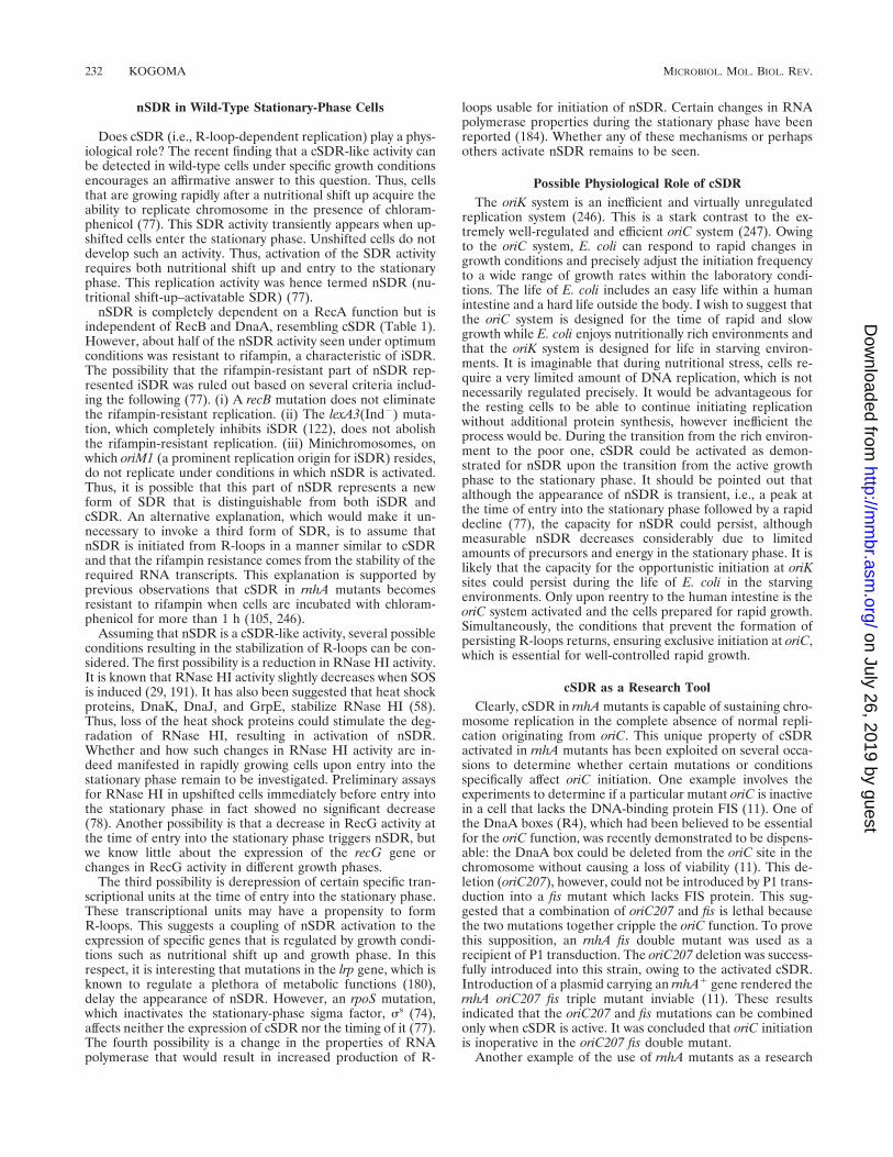
nSDR in Wild-Type Stationary-Phase Cells
Does cSDR (i.e., R-loop-dependent replication) play a phys-iological role? The recent finding that a cSDR-like activity canbe detected in wild-type cells under specific growth conditionsencourages an affirmative answer to this question. Thus, cellsthat are growing rapidly after a nutritional shift up acquire theability to replicate chromosome in the presence of chloram-phenicol (77). This SDR activity transiently appears when up-shifted cells enter the stationary phase. Unshifted cells do notdevelop such an activity. Thus, activation of the SDR activityrequires both nutritional shift up and entry to the stationaryphase. This replication activity was hence termed nSDR (nu-tritional shift-up–activatable SDR) (77).
nSDR is completely dependent on a RecA function but isindependent of RecB and DnaA, resembling cSDR (Table 1).However, about half of the nSDR activity seen under optimumconditions was resistant to rifampin, a characteristic of iSDR.The possibility that the rifampin-resistant part of nSDR rep-resented iSDR was ruled out based on several criteria includ-ing the following (77). (i) A recB mutation does not eliminatethe rifampin-resistant replication. (ii) The lexA3(Ind2) muta-tion, which completely inhibits iSDR (122), does not abolishthe rifampin-resistant replication. (iii) Minichromosomes, onwhich oriM1 (a prominent replication origin for iSDR) resides,do not replicate under conditions in which nSDR is activated.Thus, it is possible that this part of nSDR represents a newform of SDR that is distinguishable from both iSDR andcSDR. An alternative explanation, which would make it un-necessary to invoke a third form of SDR, is to assume thatnSDR is initiated from R-loops in a manner similar to cSDRand that the rifampin resistance comes from the stability of therequired RNA transcripts. This explanation is supported byprevious observations that cSDR in rnhA mutants becomesresistant to rifampin when cells are incubated with chloram-phenicol for more than 1 h (105, 246).
Assuming that nSDR is a cSDR-like activity, several possibleconditions resulting in the stabilization of R-loops can be con-sidered. The first possibility is a reduction in RNase HI activity.It is known that RNase HI activity slightly decreases when SOSis induced (29, 191). It has also been suggested that heat shockproteins, DnaK, DnaJ, and GrpE, stabilize RNase HI (58).Thus, loss of the heat shock proteins could stimulate the deg-radation of RNase HI, resulting in activation of nSDR.Whether and how such changes in RNase HI activity are in-deed manifested in rapidly growing cells upon entry into thestationary phase remain to be investigated. Preliminary assaysfor RNase HI in upshifted cells immediately before entry intothe stationary phase in fact showed no significant decrease(78). Another possibility is that a decrease in RecG activity atthe time of entry into the stationary phase triggers nSDR, butwe know little about the expression of the recG gene orchanges in RecG activity in different growth phases.
The third possibility is derepression of certain specific tran-scriptional units at the time of entry into the stationary phase.These transcriptional units may have a propensity to formR-loops. This suggests a coupling of nSDR activation to theexpression of specific genes that is regulated by growth condi-tions such as nutritional shift up and growth phase. In thisrespect, it is interesting that mutations in the lrp gene, which isknown to regulate a plethora of metabolic functions (180),delay the appearance of nSDR. However, an rpoS mutation,which inactivates the stationary-phase sigma factor, ss (74),affects neither the expression of cSDR nor the timing of it (77).The fourth possibility is a change in the properties of RNApolymerase that would result in increased production of R-
loops usable for initiation of nSDR. Certain changes in RNApolymerase properties during the stationary phase have beenreported (184). Whether any of these mechanisms or perhapsothers activate nSDR remains to be seen.
Possible Physiological Role of cSDRThe oriK system is an inefficient and virtually unregulated
replication system (246). This is a stark contrast to the ex-tremely well-regulated and efficient oriC system (247). Owingto the oriC system, E. coli can respond to rapid changes ingrowth conditions and precisely adjust the initiation frequencyto a wide range of growth rates within the laboratory condi-tions. The life of E. coli includes an easy life within a humanintestine and a hard life outside the body. I wish to suggest thatthe oriC system is designed for the time of rapid and slowgrowth while E. coli enjoys nutritionally rich environments andthat the oriK system is designed for life in starving environ-ments. It is imaginable that during nutritional stress, cells re-quire a very limited amount of DNA replication, which is notnecessarily regulated precisely. It would be advantageous forthe resting cells to be able to continue initiating replicationwithout additional protein synthesis, however inefficient theprocess would be. During the transition from the rich environ-ment to the poor one, cSDR could be activated as demon-strated for nSDR upon the transition from the active growthphase to the stationary phase. It should be pointed out thatalthough the appearance of nSDR is transient, i.e., a peak atthe time of entry into the stationary phase followed by a rapiddecline (77), the capacity for nSDR could persist, althoughmeasurable nSDR decreases considerably due to limitedamounts of precursors and energy in the stationary phase. It islikely that the capacity for the opportunistic initiation at oriKsites could persist during the life of E. coli in the starvingenvironments. Only upon reentry to the human intestine is theoriC system activated and the cells prepared for rapid growth.Simultaneously, the conditions that prevent the formation ofpersisting R-loops returns, ensuring exclusive initiation at oriC,which is essential for well-controlled rapid growth.
cSDR as a Research ToolClearly, cSDR in rnhA mutants is capable of sustaining chro-
mosome replication in the complete absence of normal repli-cation originating from oriC. This unique property of cSDRactivated in rnhA mutants has been exploited on several occa-sions to determine whether certain mutations or conditionsspecifically affect oriC initiation. One example involves theexperiments to determine if a particular mutant oriC is inactivein a cell that lacks the DNA-binding protein FIS (11). One ofthe DnaA boxes (R4), which had been believed to be essentialfor the oriC function, was recently demonstrated to be dispens-able: the DnaA box could be deleted from the oriC site in thechromosome without causing a loss of viability (11). This de-letion (oriC207), however, could not be introduced by P1 trans-duction into a fis mutant which lacks FIS protein. This sug-gested that a combination of oriC207 and fis is lethal becausethe two mutations together cripple the oriC function. To provethis supposition, an rnhA fis double mutant was used as arecipient of P1 transduction. The oriC207 deletion was success-fully introduced into this strain, owing to the activated cSDR.Introduction of a plasmid carrying an rnhA1 gene rendered thernhA oriC207 fis triple mutant inviable (11). These resultsindicated that the oriC207 and fis mutations can be combinedonly when cSDR is active. It was concluded that oriC initiationis inoperative in the oriC207 fis double mutant.
Another example of the use of rnhA mutants as a research
232 KOGOMA MICROBIOL. MOL. BIOL. REV.
on July 26, 2019 by guesthttp://m
mbr.asm
.org/D
ownloaded from
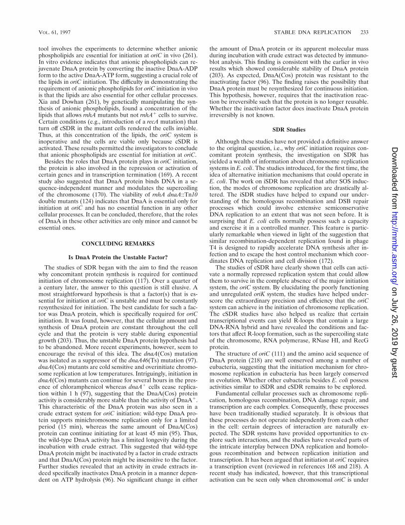
tool involves the experiments to determine whether anionicphospholipids are essential for initiation at oriC in vivo (261).In vitro evidence indicates that anionic phospholipids can re-juvenate DnaA protein by converting the inactive DnaA-ADPform to the active DnaA-ATP form, suggesting a crucial role ofthe lipids in oriC initiation. The difficulty in demonstrating therequirement of anionic phospholipids for oriC initiation in vivois that the lipids are also essential for other cellular processes.Xia and Dowhan (261), by genetically manipulating the syn-thesis of anionic phospholipids, found a concentration of thelipids that allows rnhA mutants but not rnhA1 cells to survive.Certain conditions (e.g., introduction of a recA mutation) thatturn off cSDR in the mutant cells rendered the cells inviable.Thus, at this concentration of the lipids, the oriC system isinoperative and the cells are viable only because cSDR isactivated. These results permitted the investigators to concludethat anionic phospholipids are essential for initiation at oriC.
Besides the roles that DnaA protein plays in oriC initiation,the protein is also involved in the repression or activation ofcertain genes and in transcription termination (169). A recentstudy also suggested that DnaA protein binds DNA in a se-quence-independent manner and modulates the supercoilingof the chromosome (170). The viability of rnhA dnaA::Tn10double mutants (124) indicates that DnaA is essential only forinitiation at oriC and has no essential function in any othercellular processes. It can be concluded, therefore, that the rolesof DnaA in these other activities are only minor and cannot beessential ones.
CONCLUDING REMARKS
Is DnaA Protein the Unstable Factor?
The studies of SDR began with the aim to find the reasonwhy concomitant protein synthesis is required for continuedinitiation of chromosome replication (117). Over a quarter ofa century later, the answer to this question is still elusive. Amost straightforward hypothesis is that a factor(s) that is es-sential for initiation at oriC is unstable and must be constantlyresynthesized for initiation. The best candidate for such a fac-tor was DnaA protein, which is specifically required for oriCinitiation. It was found, however, that the cellular amount andsynthesis of DnaA protein are constant throughout the cellcycle and that the protein is very stable during exponentialgrowth (203). Thus, the unstable DnaA protein hypothesis hadto be abandoned. More recent experiments, however, seem toencourage the revival of this idea. The dnaA(Cos) mutationwas isolated as a suppressor of the dnaA46(Ts) mutation (97).dnaA(Cos) mutants are cold sensitive and overinitiate chromo-some replication at low temperatures. Intriguingly, initiation indnaA(Cos) mutants can continue for several hours in the pres-ence of chloramphenicol whereas dnaA1 cells cease replica-tion within 1 h (97), suggesting that the DnaA(Cos) proteinactivity is considerably more stable than the activity of DnaA1.This characteristic of the DnaA protein was also seen in acrude extract system for oriC initiation: wild-type DnaA pro-tein supports minichromosome replication only for a limitedperiod (15 min), whereas the same amount of DnaA(Cos)protein can continue initiating for at least 45 min (95). Thus,the wild-type DnaA activity has a limited longevity during theincubation with crude extract. This suggested that wild-typeDnaA protein might be inactivated by a factor in crude extractsand that DnaA(Cos) protein might be insensitive to the factor.Further studies revealed that an activity in crude extracts in-deed specifically inactivates DnaA protein in a manner depen-dent on ATP hydrolysis (96). No significant change in either
the amount of DnaA protein or its apparent molecular massduring incubation with crude extract was detected by immuno-blot analysis. This finding is consistent with the earlier in vivoresults which showed considerable stability of DnaA protein(203). As expected, DnaA(Cos) protein was resistant to theinactivating factor (96). The finding raises the possibility thatDnaA protein must be resynthesized for continuous initiation.This hypothesis, however, requires that the inactivation reac-tion be irreversible such that the protein is no longer reusable.Whether the inactivation factor does inactivate DnaA proteinirreversibly is not known.
SDR Studies
Although these studies have not provided a definitive answerto the original question, i.e., why oriC initiation requires con-comitant protein synthesis, the investigation on SDR hasyielded a wealth of information about chromosome replicationsystems in E. coli. The studies introduced, for the first time, theidea of alternative initiation mechanisms that could operate inE. coli. The work on iSDR has revealed that after SOS induc-tion, the modes of chromosome replication are drastically al-tered. The iSDR studies have helped to expand our under-standing of the homologous recombination and DSB repairprocesses which could involve extensive semiconservativeDNA replication to an extent that was not seen before. It issurprising that E. coli cells normally possess such a capacityand exercise it in a controlled manner. This feature is partic-ularly remarkable when viewed in light of the suggestion thatsimilar recombination-dependent replication found in phageT4 is designed to rapidly accelerate DNA synthesis after in-fection and to escape the host control mechanism which coor-dinates DNA replication and cell division (172).
The studies of cSDR have clearly shown that cells can acti-vate a normally repressed replication system that could allowthem to survive in the complete absence of the major initiationsystem, the oriC system. By elucidating the poorly functioningand unregulated oriK system, the studies have helped under-score the extraordinary precision and efficiency that the oriCsystem can achieve in the initiation of chromosome replication.The cSDR studies have also helped us realize that certaintranscriptional events can yield R-loops that contain a largeDNA-RNA hybrid and have revealed the conditions and fac-tors that affect R-loop formation, such as the supercoiling stateof the chromosome, RNA polymerase, RNase HI, and RecGprotein.
The structure of oriC (111) and the amino acid sequence ofDnaA protein (218) are well conserved among a number ofeubacteria, suggesting that the initiation mechanism for chro-mosome replication in eubacteria has been largely conservedin evolution. Whether other eubacteria besides E. coli possessactivities similar to iSDR and cSDR remains to be explored.
Fundamental cellular processes such as chromosome repli-cation, homologous recombination, DNA damage repair, andtranscription are each complex. Consequently, these processeshave been traditionally studied separately. It is obvious thatthese processes do not operate independently from each otherin the cell: certain degrees of interaction are naturally ex-pected. The SDR systems have provided opportunities to ex-plore such interactions, and the studies have revealed parts ofthe intricate interplay between DNA replication and homolo-gous recombination and between replication initiation andtranscription. It has been argued that initiation at oriC requiresa transcription event (reviewed in references 168 and 218). Arecent study has indicated, however, that this transcriptionalactivation can be seen only when chromosomal oriC is under
VOL. 61, 1997 STABLE DNA REPLICATION 233
on July 26, 2019 by guesthttp://m
mbr.asm
.org/D
ownloaded from
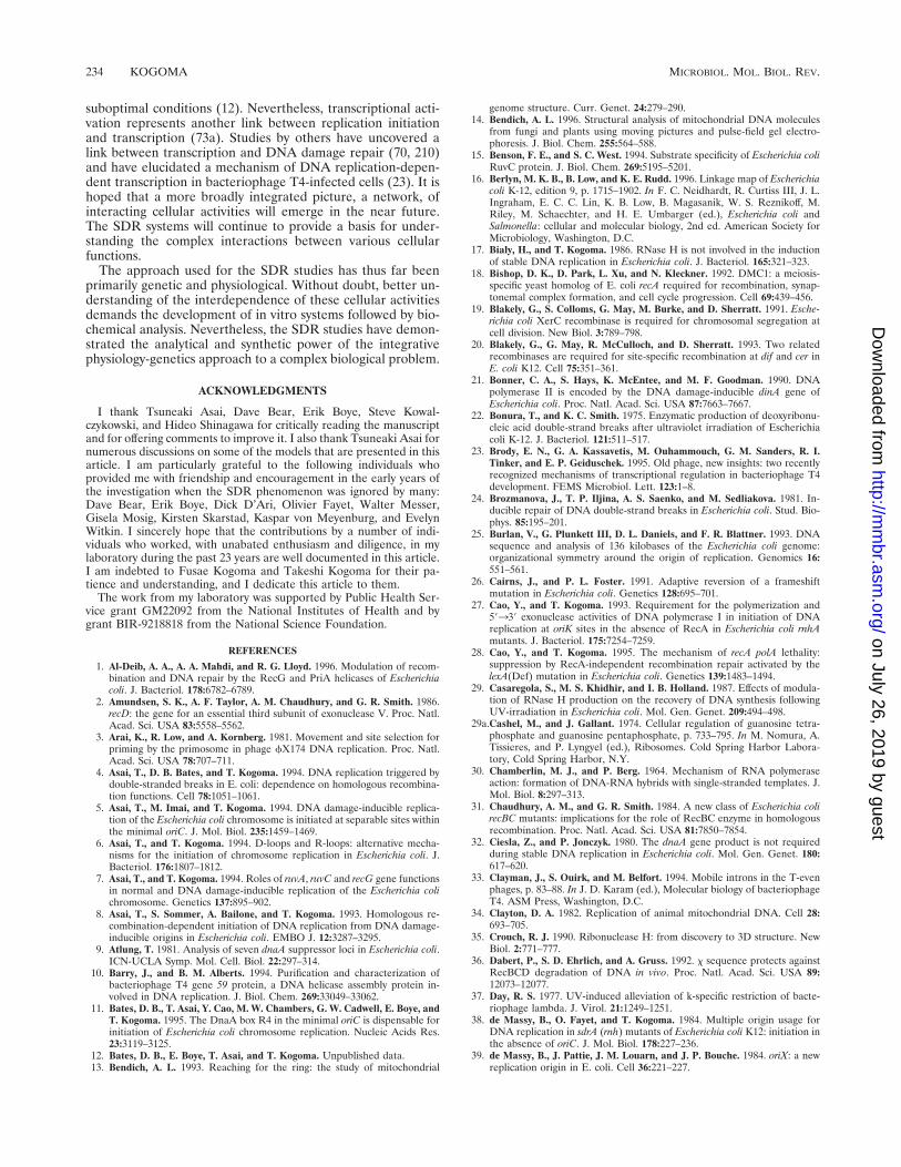
suboptimal conditions (12). Nevertheless, transcriptional acti-vation represents another link between replication initiationand transcription (73a). Studies by others have uncovered alink between transcription and DNA damage repair (70, 210)and have elucidated a mechanism of DNA replication-depen-dent transcription in bacteriophage T4-infected cells (23). It ishoped that a more broadly integrated picture, a network, ofinteracting cellular activities will emerge in the near future.The SDR systems will continue to provide a basis for under-standing the complex interactions between various cellularfunctions.
The approach used for the SDR studies has thus far beenprimarily genetic and physiological. Without doubt, better un-derstanding of the interdependence of these cellular activitiesdemands the development of in vitro systems followed by bio-chemical analysis. Nevertheless, the SDR studies have demon-strated the analytical and synthetic power of the integrativephysiology-genetics approach to a complex biological problem.
ACKNOWLEDGMENTS
I thank Tsuneaki Asai, Dave Bear, Erik Boye, Steve Kowal-czykowski, and Hideo Shinagawa for critically reading the manuscriptand for offering comments to improve it. I also thank Tsuneaki Asai fornumerous discussions on some of the models that are presented in thisarticle. I am particularly grateful to the following individuals whoprovided me with friendship and encouragement in the early years ofthe investigation when the SDR phenomenon was ignored by many:Dave Bear, Erik Boye, Dick D’Ari, Olivier Fayet, Walter Messer,Gisela Mosig, Kirsten Skarstad, Kaspar von Meyenburg, and EvelynWitkin. I sincerely hope that the contributions by a number of indi-viduals who worked, with unabated enthusiasm and diligence, in mylaboratory during the past 23 years are well documented in this article.I am indebted to Fusae Kogoma and Takeshi Kogoma for their pa-tience and understanding, and I dedicate this article to them.
The work from my laboratory was supported by Public Health Ser-vice grant GM22092 from the National Institutes of Health and bygrant BIR-9218818 from the National Science Foundation.
REFERENCES
1. Al-Deib, A. A., A. A. Mahdi, and R. G. Lloyd. 1996. Modulation of recom-bination and DNA repair by the RecG and PriA helicases of Escherichiacoli. J. Bacteriol. 178:6782–6789.
2. Amundsen, S. K., A. F. Taylor, A. M. Chaudhury, and G. R. Smith. 1986.recD: the gene for an essential third subunit of exonuclease V. Proc. Natl.Acad. Sci. USA 83:5558–5562.
3. Arai, K., R. Low, and A. Kornberg. 1981. Movement and site selection forpriming by the primosome in phage fX174 DNA replication. Proc. Natl.Acad. Sci. USA 78:707–711.
4. Asai, T., D. B. Bates, and T. Kogoma. 1994. DNA replication triggered bydouble-stranded breaks in E. coli: dependence on homologous recombina-tion functions. Cell 78:1051–1061.
5. Asai, T., M. Imai, and T. Kogoma. 1994. DNA damage-inducible replica-tion of the Escherichia coli chromosome is initiated at separable sites withinthe minimal oriC. J. Mol. Biol. 235:1459–1469.
6. Asai, T., and T. Kogoma. 1994. D-loops and R-loops: alternative mecha-nisms for the initiation of chromosome replication in Escherichia coli. J.Bacteriol. 176:1807–1812.
7. Asai, T., and T. Kogoma. 1994. Roles of ruvA, ruvC and recG gene functionsin normal and DNA damage-inducible replication of the Escherichia colichromosome. Genetics 137:895–902.
8. Asai, T., S. Sommer, A. Bailone, and T. Kogoma. 1993. Homologous re-combination-dependent initiation of DNA replication from DNA damage-inducible origins in Escherichia coli. EMBO J. 12:3287–3295.
9. Atlung, T. 1981. Analysis of seven dnaA suppressor loci in Escherichia coli.ICN-UCLA Symp. Mol. Cell. Biol. 22:297–314.
10. Barry, J., and B. M. Alberts. 1994. Purification and characterization ofbacteriophage T4 gene 59 protein, a DNA helicase assembly protein in-volved in DNA replication. J. Biol. Chem. 269:33049–33062.
11. Bates, D. B., T. Asai, Y. Cao, M. W. Chambers, G. W. Cadwell, E. Boye, andT. Kogoma. 1995. The DnaA box R4 in the minimal oriC is dispensable forinitiation of Escherichia coli chromosome replication. Nucleic Acids Res.23:3119–3125.
12. Bates, D. B., E. Boye, T. Asai, and T. Kogoma. Unpublished data.13. Bendich, A. L. 1993. Reaching for the ring: the study of mitochondrial
genome structure. Curr. Genet. 24:279–290.14. Bendich, A. L. 1996. Structural analysis of mitochondrial DNA molecules
from fungi and plants using moving pictures and pulse-field gel electro-phoresis. J. Biol. Chem. 255:564–588.
15. Benson, F. E., and S. C. West. 1994. Substrate specificity of Escherichia coliRuvC protein. J. Biol. Chem. 269:5195–5201.
16. Berlyn, M. K. B., B. Low, and K. E. Rudd. 1996. Linkage map of Escherichiacoli K-12, edition 9, p. 1715–1902. In F. C. Neidhardt, R. Curtiss III, J. L.Ingraham, E. C. C. Lin, K. B. Low, B. Magasanik, W. S. Reznikoff, M.Riley, M. Schaechter, and H. E. Umbarger (ed.), Escherichia coli andSalmonella: cellular and molecular biology, 2nd ed. American Society forMicrobiology, Washington, D.C.
17. Bialy, H., and T. Kogoma. 1986. RNase H is not involved in the inductionof stable DNA replication in Escherichia coli. J. Bacteriol. 165:321–323.
18. Bishop, D. K., D. Park, L. Xu, and N. Kleckner. 1992. DMC1: a meiosis-specific yeast homolog of E. coli recA required for recombination, synap-tonemal complex formation, and cell cycle progression. Cell 69:439–456.
19. Blakely, G., S. Colloms, G. May, M. Burke, and D. Sherratt. 1991. Esche-richia coli XerC recombinase is required for chromosomal segregation atcell division. New Biol. 3:789–798.
20. Blakely, G., G. May, R. McCulloch, and D. Sherratt. 1993. Two relatedrecombinases are required for site-specific recombination at dif and cer inE. coli K12. Cell 75:351–361.
21. Bonner, C. A., S. Hays, K. McEntee, and M. F. Goodman. 1990. DNApolymerase II is encoded by the DNA damage-inducible dinA gene ofEscherichia coli. Proc. Natl. Acad. Sci. USA 87:7663–7667.
22. Bonura, T., and K. C. Smith. 1975. Enzymatic production of deoxyribonu-cleic acid double-strand breaks after ultraviolet irradiation of Escherichiacoli K-12. J. Bacteriol. 121:511–517.
23. Brody, E. N., G. A. Kassavetis, M. Ouhammouch, G. M. Sanders, R. I.Tinker, and E. P. Geiduschek. 1995. Old phage, new insights: two recentlyrecognized mechanisms of transcriptional regulation in bacteriophage T4development. FEMS Microbiol. Lett. 123:1–8.
24. Brozmanova, J., T. P. Iljina, A. S. Saenko, and M. Sedliakova. 1981. In-ducible repair of DNA double-strand breaks in Escherichia coli. Stud. Bio-phys. 85:195–201.
25. Burlan, V., G. Plunkett III, D. L. Daniels, and F. R. Blattner. 1993. DNAsequence and analysis of 136 kilobases of the Escherichia coli genome:organizational symmetry around the origin of replication. Genomics 16:551–561.
26. Cairns, J., and P. L. Foster. 1991. Adaptive reversion of a frameshiftmutation in Escherichia coli. Genetics 128:695–701.
27. Cao, Y., and T. Kogoma. 1993. Requirement for the polymerization and59339 exonuclease activities of DNA polymerase I in initiation of DNAreplication at oriK sites in the absence of RecA in Escherichia coli rnhAmutants. J. Bacteriol. 175:7254–7259.
28. Cao, Y., and T. Kogoma. 1995. The mechanism of recA polA lethality:suppression by RecA-independent recombination repair activated by thelexA(Def) mutation in Escherichia coli. Genetics 139:1483–1494.
29. Casaregola, S., M. S. Khidhir, and I. B. Holland. 1987. Effects of modula-tion of RNase H production on the recovery of DNA synthesis followingUV-irradiation in Escherichia coli. Mol. Gen. Genet. 209:494–498.
29a.Cashel, M., and J. Gallant. 1974. Cellular regulation of guanosine tetra-phosphate and guanosine pentaphosphate, p. 733–795. In M. Nomura, A.Tissieres, and P. Lyngyel (ed.), Ribosomes. Cold Spring Harbor Labora-tory, Cold Spring Harbor, N.Y.
30. Chamberlin, M. J., and P. Berg. 1964. Mechanism of RNA polymeraseaction: formation of DNA-RNA hybrids with single-stranded templates. J.Mol. Biol. 8:297–313.
31. Chaudhury, A. M., and G. R. Smith. 1984. A new class of Escherichia colirecBC mutants: implications for the role of RecBC enzyme in homologousrecombination. Proc. Natl. Acad. Sci. USA 81:7850–7854.
32. Ciesla, Z., and P. Jonczyk. 1980. The dnaA gene product is not requiredduring stable DNA replication in Escherichia coli. Mol. Gen. Genet. 180:617–620.
33. Clayman, J., S. Ouirk, and M. Belfort. 1994. Mobile introns in the T-evenphages, p. 83–88. In J. D. Karam (ed.), Molecular biology of bacteriophageT4. ASM Press, Washington, D.C.
34. Clayton, D. A. 1982. Replication of animal mitochondrial DNA. Cell 28:693–705.
35. Crouch, R. J. 1990. Ribonuclease H: from discovery to 3D structure. NewBiol. 2:771–777.
36. Dabert, P., S. D. Ehrlich, and A. Gruss. 1992. x sequence protects againstRecBCD degradation of DNA in vivo. Proc. Natl. Acad. Sci. USA 89:12073–12077.
37. Day, R. S. 1977. UV-induced alleviation of k-specific restriction of bacte-riophage lambda. J. Virol. 21:1249–1251.
38. de Massy, B., O. Fayet, and T. Kogoma. 1984. Multiple origin usage forDNA replication in sdrA (rnh) mutants of Escherichia coli K12: initiation inthe absence of oriC. J. Mol. Biol. 178:227–236.
39. de Massy, B., J. Pattie, J. M. Louarn, and J. P. Bouche. 1984. oriX: a newreplication origin in E. coli. Cell 36:221–227.
234 KOGOMA MICROBIOL. MOL. BIOL. REV.
on July 26, 2019 by guesthttp://m
mbr.asm
.org/D
ownloaded from
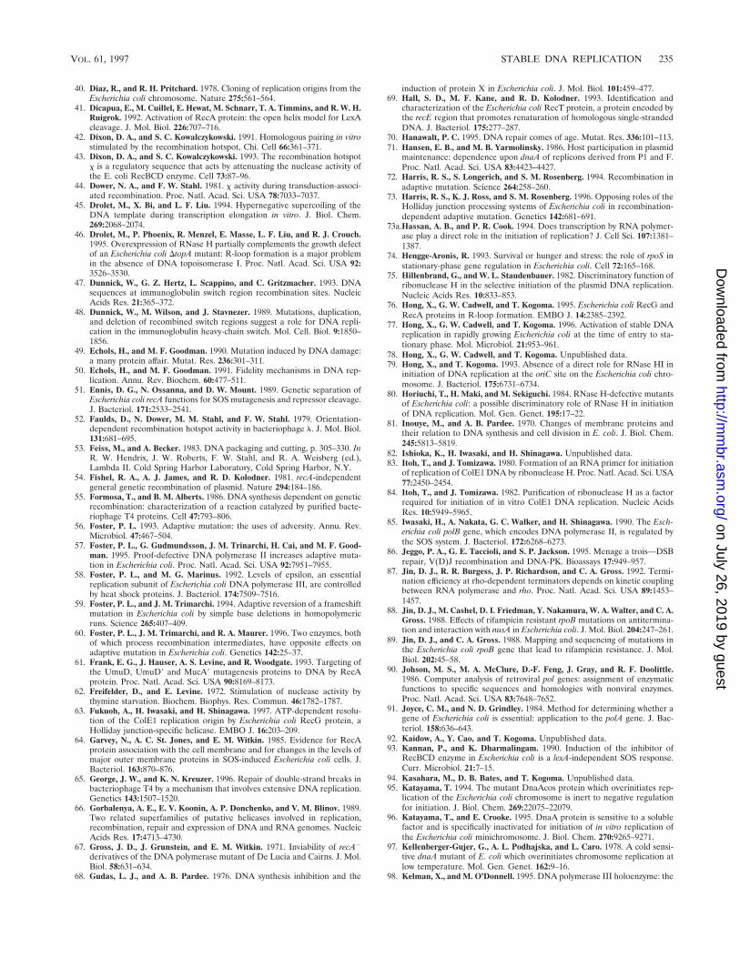
40. Diaz, R., and R. H. Pritchard. 1978. Cloning of replication origins from theEscherichia coli chromosome. Nature 275:561–564.
41. Dicapua, E., M. Cuillel, E. Hewat, M. Schnarr, T. A. Timmins, and R. W. H.Ruigrok. 1992. Activation of RecA protein: the open helix model for LexAcleavage. J. Mol. Biol. 226:707–716.
42. Dixon, D. A., and S. C. Kowalczykowski. 1991. Homologous pairing in vitrostimulated by the recombination hotspot, Chi. Cell 66:361–371.
43. Dixon, D. A., and S. C. Kowalczykowski. 1993. The recombination hotspotx is a regulatory sequence that acts by attenuating the nuclease activity ofthe E. coli RecBCD enzyme. Cell 73:87–96.
44. Dower, N. A., and F. W. Stahl. 1981. x activity during transduction-associ-ated recombination. Proc. Natl. Acad. Sci. USA 78:7033–7037.
45. Drolet, M., X. Bi, and L. F. Liu. 1994. Hypernegative supercoiling of theDNA template during transcription elongation in vitro. J. Biol. Chem.269:2068–2074.
46. Drolet, M., P. Phoenix, R. Menzel, E. Masse, L. F. Liu, and R. J. Crouch.1995. Overexpression of RNase H partially complements the growth defectof an Escherichia coli DtopA mutant: R-loop formation is a major problemin the absence of DNA topoisomerase I. Proc. Natl. Acad. Sci. USA 92:3526–3530.
47. Dunnick, W., G. Z. Hertz, L. Scappino, and C. Gritzmacher. 1993. DNAsequences at immunoglobulin switch region recombination sites. NucleicAcids Res. 21:365–372.
48. Dunnick, W., M. Wilson, and J. Stavnezer. 1989. Mutations, duplication,and deletion of recombined switch regions suggest a role for DNA repli-cation in the immunoglobulin heavy-chain switch. Mol. Cell. Biol. 9:1850–1856.
49. Echols, H., and M. F. Goodman. 1990. Mutation induced by DNA damage:a many protein affair. Mutat. Res. 236:301–311.
50. Echols, H., and M. F. Goodman. 1991. Fidelity mechanisms in DNA rep-lication. Annu. Rev. Biochem. 60:477–511.
51. Ennis, D. G., N. Ossanna, and D. W. Mount. 1989. Genetic separation ofEscherichia coli recA functions for SOS mutagenesis and repressor cleavage.J. Bacteriol. 171:2533–2541.
52. Faulds, D., N. Dower, M. M. Stahl, and F. W. Stahl. 1979. Orientation-dependent recombination hotspot activity in bacteriophage l. J. Mol. Biol.131:681–695.
53. Feiss, M., and A. Becker. 1983. DNA packaging and cutting, p. 305–330. InR. W. Hendrix, J. W. Roberts, F. W. Stahl, and R. A. Weisberg (ed.),Lambda II. Cold Spring Harbor Laboratory, Cold Spring Harbor, N.Y.
54. Fishel, R. A., A. J. James, and R. D. Kolodner. 1981. recA-independentgeneral genetic recombination of plasmid. Nature 294:184–186.
55. Formosa, T., and B. M. Alberts. 1986. DNA synthesis dependent on geneticrecombination: characterization of a reaction catalyzed by purified bacte-riophage T4 proteins. Cell 47:793–806.
56. Foster, P. L. 1993. Adaptive mutation: the uses of adversity. Annu. Rev.Microbiol. 47:467–504.
57. Foster, P. L., G. Gudmundsson, J. M. Trinarchi, H. Cai, and M. F. Good-man. 1995. Proof-defective DNA polymerase II increases adaptive muta-tion in Escherichia coli. Proc. Natl. Acad. Sci. USA 92:7951–7955.
58. Foster, P. L., and M. G. Marinus. 1992. Levels of epsilon, an essentialreplication subunit of Escherichia coli DNA polymerase III, are controlledby heat shock proteins. J. Bacteriol. 174:7509–7516.
59. Foster, P. L., and J. M. Trimarchi. 1994. Adaptive reversion of a frameshiftmutation in Escherichia coli by simple base deletions in homopolymericruns. Science 265:407–409.
60. Foster, P. L., J. M. Trimarchi, and R. A. Maurer. 1996. Two enzymes, bothof which process recombination intermediates, have opposite effects onadaptive mutation in Escherichia coli. Genetics 142:25–37.
61. Frank, E. G., J. Hauser, A. S. Levine, and R. Woodgate. 1993. Targeting ofthe UmuD, UmuD9 and MucA9 mutagenesis proteins to DNA by RecAprotein. Proc. Natl. Acad. Sci. USA 90:8169–8173.
62. Freifelder, D., and E. Levine. 1972. Stimulation of nuclease activity bythymine starvation. Biochem. Biophys. Res. Commun. 46:1782–1787.
63. Fukuoh, A., H. Iwasaki, and H. Shinagawa. 1997. ATP-dependent resolu-tion of the ColE1 replication origin by Escherichia coli RecG protein, aHolliday junction-specific helicase. EMBO J. 16:203–209.
64. Garvey, N., A. C. St. Jones, and E. M. Witkin. 1985. Evidence for RecAprotein association with the cell membrane and for changes in the levels ofmajor outer membrane proteins in SOS-induced Escherichia coli cells. J.Bacteriol. 163:870–876.
65. George, J. W., and K. N. Kreuzer. 1996. Repair of double-strand breaks inbacteriophage T4 by a mechanism that involves extensive DNA replication.Genetics 143:1507–1520.
66. Gorbalenya, A. E., E. V. Koonin, A. P. Donchenko, and V. M. Blinov. 1989.Two related superfamilies of putative helicases involved in replication,recombination, repair and expression of DNA and RNA genomes. NucleicAcids Res. 17:4713–4730.
67. Gross, J. D., J. Grunstein, and E. M. Witkin. 1971. Inviability of recA2
derivatives of the DNA polymerase mutant of De Lucia and Cairns. J. Mol.Biol. 58:631–634.
68. Gudas, L. J., and A. B. Pardee. 1976. DNA synthesis inhibition and the
induction of protein X in Escherichia coli. J. Mol. Biol. 101:459–477.69. Hall, S. D., M. F. Kane, and R. D. Kolodner. 1993. Identification and
characterization of the Escherichia coli RecT protein, a protein encoded bythe recE region that promotes renaturation of homologous single-strandedDNA. J. Bacteriol. 175:277–287.
70. Hanawalt, P. C. 1995. DNA repair comes of age. Mutat. Res. 336:101–113.71. Hansen, E. B., and M. B. Yarmolinsky. 1986. Host participation in plasmid
maintenance: dependence upon dnaA of replicons derived from P1 and F.Proc. Natl. Acad. Sci. USA 83:4423–4427.
72. Harris, R. S., S. Longerich, and S. M. Rosenberg. 1994. Recombination inadaptive mutation. Science 264:258–260.
73. Harris, R. S., K. J. Ross, and S. M. Rosenberg. 1996. Opposing roles of theHolliday junction processing systems of Escherichia coli in recombination-dependent adaptive mutation. Genetics 142:681–691.
73a.Hassan, A. B., and P. R. Cook. 1994. Does transcription by RNA polymer-ase play a direct role in the initiation of replication? J. Cell Sci. 107:1381–1387.
74. Hengge-Aronis, R. 1993. Survival or hunger and stress: the role of rpoS instationary-phase gene regulation in Escherichia coli. Cell 72:165–168.
75. Hillenbrand, G., and W. L. Staudenbauer. 1982. Discriminatory function ofribonuclease H in the selective initiation of the plasmid DNA replication.Nucleic Acids Res. 10:833–853.
76. Hong, X., G. W. Cadwell, and T. Kogoma. 1995. Escherichia coli RecG andRecA proteins in R-loop formation. EMBO J. 14:2385–2392.
77. Hong, X., G. W. Cadwell, and T. Kogoma. 1996. Activation of stable DNAreplication in rapidly growing Escherichia coli at the time of entry to sta-tionary phase. Mol. Microbiol. 21:953–961.
78. Hong, X., G. W. Cadwell, and T. Kogoma. Unpublished data.79. Hong, X., and T. Kogoma. 1993. Absence of a direct role for RNase HI in
initiation of DNA replication at the oriC site on the Escherichia coli chro-mosome. J. Bacteriol. 175:6731–6734.
80. Horiuchi, T., H. Maki, and M. Sekiguchi. 1984. RNase H-defective mutantsof Escherichia coli: a possible discriminatory role of RNase H in initiationof DNA replication. Mol. Gen. Genet. 195:17–22.
81. Inouye, M., and A. B. Pardee. 1970. Changes of membrane proteins andtheir relation to DNA synthesis and cell division in E. coli. J. Biol. Chem.245:5813–5819.
82. Ishioka, K., H. Iwasaki, and H. Shinagawa. Unpublished data.83. Itoh, T., and J. Tomizawa. 1980. Formation of an RNA primer for initiation
of replication of ColE1 DNA by ribonuclease H. Proc. Natl. Acad. Sci. USA77:2450–2454.
84. Itoh, T., and J. Tomizawa. 1982. Purification of ribonuclease H as a factorrequired for initiation of in vitro ColE1 DNA replication. Nucleic AcidsRes. 10:5949–5965.
85. Iwasaki, H., A. Nakata, G. C. Walker, and H. Shinagawa. 1990. The Esch-erichia coli polB gene, which encodes DNA polymerase II, is regulated bythe SOS system. J. Bacteriol. 172:6268–6273.
86. Jeggo, P. A., G. E. Taccioli, and S. P. Jackson. 1995. Menage a trois—DSBrepair, V(D)J recombination and DNA-PK. Bioassays 17:949–957.
87. Jin, D. J., R. R. Burgess, J. P. Richardson, and C. A. Gross. 1992. Termi-nation efficiency at rho-dependent terminators depends on kinetic couplingbetween RNA polymerase and rho. Proc. Natl. Acad. Sci. USA 89:1453–1457.
88. Jin, D. J., M. Cashel, D. I. Friedman, Y. Nakamura, W. A. Walter, and C. A.Gross. 1988. Effects of rifampicin resistant rpoB mutations on antitermina-tion and interaction with nusA in Escherichia coli. J. Mol. Biol. 204:247–261.
89. Jin, D. J., and C. A. Gross. 1988. Mapping and sequencing of mutations inthe Escherichia coli rpoB gene that lead to rifampicin resistance. J. Mol.Biol. 202:45–58.
90. Johson, M. S., M. A. McClure, D.-F. Feng, J. Gray, and R. F. Doolittle.1986. Computer analysis of retroviral pol genes: assignment of enzymaticfunctions to specific sequences and homologies with nonviral enzymes.Proc. Natl. Acad. Sci. USA 83:7648–7652.
91. Joyce, C. M., and N. D. Grindley. 1984. Method for determining whether agene of Escherichia coli is essential: application to the polA gene. J. Bac-teriol. 158:636–643.
92. Kaidow, A., Y. Cao, and T. Kogoma. Unpublished data.93. Kannan, P., and K. Dharmalingam. 1990. Induction of the inhibitor of
RecBCD enzyme in Escherichia coli is a lexA-independent SOS response.Curr. Microbiol. 21:7–15.
94. Kasahara, M., D. B. Bates, and T. Kogoma. Unpublished data.95. Katayama, T. 1994. The mutant DnaAcos protein which overinitiates rep-
lication of the Escherichia coli chromosome is inert to negative regulationfor initiation. J. Biol. Chem. 269:22075–22079.
96. Katayama, T., and E. Crooke. 1995. DnaA protein is sensitive to a solublefactor and is specifically inactivated for initiation of in vitro replication ofthe Escherichia coli minichromosome. J. Biol. Chem. 270:9265–9271.
97. Kellenberger-Gujer, G., A. L. Podhajska, and L. Caro. 1978. A cold sensi-tive dnaA mutant of E. coli which overinitiates chromosome replication atlow temperature. Mol. Gen. Genet. 162:9–16.
98. Kelman, X., and M. O’Donnell. 1995. DNA polymerase III holoenzyme: the
VOL. 61, 1997 STABLE DNA REPLICATION 235
on July 26, 2019 by guesthttp://m
mbr.asm
.org/D
ownloaded from

structure and function of a chromosome replication machine. Annu. Rev.Biochem. 64:171–200.
99. Khidhir, M. A., S. Casaregola, and I. B. Holland. 1985. Mechanism oftransient inhibition of DNA synthesis in ultraviolet-irradiated E. coli: inhi-bition is independent of recA whilst recovery requires RecA protein itselfand an additional, inducible SOS function. Mol. Gen. Genet. 199:133–140.
100. Kirkpatrick, D. P., and C. M. Radding. 1992. RecA protein promotes rapidRNA-DNA hybridization in heterogeneous RNA mixtures. Nucleic AcidsRes. 20:4347–4353.
101. Kirkpatrick, D. P., B. J. Rao, and C. M. Radding. 1992. RNA-DNA hy-bridization promoted by E. coli RecA protein. Nucleic Acids Res. 20:4339–4346.
102. Kline, B. C., T. Kogoma, J. E. Tam, and M. S. Shields. 1986. Requirementof the Escherichia coli dnaA gene product for plasmid F maintenance. J.Bacteriol. 168:440–443.
103. Kobayashi, I., and N. Takahashi. 1988. Double-stranded gap repair ofDNA by gene conversion in Escherichia coli. Genetics 119:751–757.
104. Kogoma, T. 1976. Two types of temperature sensitivity in DNA replicationof an Escherichia coli dnaB mutant. J. Mol. Biol. 103:191–197.
105. Kogoma, T. 1978. A novel Escherichia coli mutant capable of DNA repli-cation in the absence of protein synthesis. J. Mol. Biol. 121:55–69.
106. Kogoma, T. 1984. Absence of RNase H allows replication of pBR322 inEscherichia coli mutants lacking DNA polymerase I. Proc. Natl. Acad. Sci.USA 81:7845–7849.
107. Kogoma, T. 1986. RNase H-defective mutants of Escherichia coli. J. Bac-teriol. 166:361–363.
108. Kogoma, T. 1994. Escherichia coli RNA polymerase mutants that enhanceor diminish the SOS response constitutively expressed in the absence ofRNase HI activity. J. Bacteriol. 176:1521–1523.
109. Kogoma, T. 1996. Recombination by replication. Cell 85:625–627.110. Kogoma, T. 1997. Is RecF a DNA replication protein? Proc. Natl. Acad.
Sci. USA 94:3483–3484.111. Kogoma, T. 1997. Origins of chromosome replication. In F. J. de Bruijin,
J. R. Lupski, and G. Weinstock (ed.), Bacterial genomes: physical structureand analysis, in press. Chapman and Hall, London, United Kingdom.
112. Kogoma, T. Unpublished data.113. Kogoma, T., K. G. Bernard, and X. Hong. 1994. RecA, Tus protein and
constitutive stable DNA replication in Escherichia coli rnhA mutants. Mol.Gen. Genet. 244:557–562.
114. Kogoma, T., G. W. Cadwell, K. G. Barnard, and T. Asai. 1996. The DNAreplication priming protein, PriA, is required for homologous recombina-tion and double-strand break repair. J. Bacteriol. 178:1258–1264.
115. Kogoma, T., X. Hong, G. W. Cadwell, K. G. Barnard, and T. Asai. 1993.Requirement of homologous functions for viability of the Escherichia colicell that lacks RNase HI and exonuclease V activities. Biochimie 75:89–99.
116. Kogoma, T., and B. C. Kline. 1987. Integrative suppression of dnaA(Ts)mutations by plasmid F in Escherichia coli is a DnaA-dependent process.Mol. Gen. Genet. 210:262–269.
117. Kogoma, T., and K. G. Lark. 1970. DNA replication in Escherichia coli:replication in absence of protein synthesis after replication inhibition. J.Mol. Biol. 52:143–164.
118. Kogoma, T., and K. G. Lark. 1975. Characterization of the replication ofEscherichia coli DNA in the absence of protein synthesis: stable DNAreplication. J. Mol. Biol. 94:243–256.
119. Kogoma, T., and R. R. Maldonado. 1997. DNA polymerase I in constitutivestable DNA replication in Escherichia coli. J. Bacteriol. 179:2109–2115.
120. Kogoma, T., K. Skarstad, E. Boye, K. von Meyenburg, and H. B. Steen.1985. RecA protein acts at the initiation of stable DNA replication in rnhmutants of Escherichia coli K-12. J. Bacteriol. 163:439–444.
121. Kogoma, T., N. L. Subia, and K. von Meyenburg. 1985. Function of ribo-nuclease H in initiation of DNA replication in Escherichia coli K-12. Mol.Gen. Genet. 200:103–109.
122. Kogoma, T., T. A. Torrey, and M. J. Connaughton. 1979. Induction ofUV-resistant DNA replication in Escherichia coli: induced stable DNAreplication as an SOS function. Mol. Gen. Genet. 176:1–9.
123. Kogoma, T., T. A. Torrey, N. L. Subia, and G. G. Pickett. 1981. An alter-native DNA initiation pathway in E. coli. ICN-UCLA Symp. Mol. Cell. Biol.22:361–374.
124. Kogoma, T., and K. von Meyenburg. 1983. The origin of replication, oriC,and the dnaA protein are dispensable in stable DNA replication (sdrA)mutants of Escherichia coli K12. EMBO J. 2:463–468.
125. Kohlstaedt, L. A., J. Wang, J. M. Frieman, P. A. Rice, and T. A. Steitz. 1992.Crystal structure at 3.5 Å resolution of HIV-1 reverse transcriptase com-plexed with an inhibitor. Science 256:1783–1790.
126. Kornberg, A., and T. A. Baker. 1992. DNA replication. W. H. Freeman andCompany, New York, N.Y.
127. Kowalczykowski, S. C., D. A. Dixon, A. K. Eggleston, S. D. Lauder, andW. M. Rehrauer. 1994. Biochemistry of homologous recombination inEscherichia coli. Microbiol. Rev. 58:401–465.
128. Krasin, F., and F. Hutchinson. 1977. Repair of DNA double-strand breaksin Escherichia coli, which requires recA function and the presence of aduplicated genome. J. Mol. Biol. 116:81–89.
129. Krasin, F., and F. Hutchinson. 1981. Repair of DNA double-strand breaksin Escherichia coli cells requires synthesis of proteins that can be induced byUV light. Proc. Natl. Acad. Sci. USA 78:3450–3453.
130. Kreuzer, K. N., and S. W. Morrical. 1994. Initiation of DNA replication, p.28–42. In J. D. Karam (ed.), Molecular biology of bacteriophage T4. ASMPress, Washington, D.C.
131. Kuempel, P. L., J. M. Henson, L. Dircks, M. Tecklenburg, and D. F. Lim.1991. dif, a recA-independent recombination site in the terminus region ofthe chromosome of Escherichia coli. New Biol. 3:799–811.
132. Kusano, T., D. Steinmetz, W. G. Hendrickson, J. Murchie, M. King, A.Benson, and M. Schaechter. 1984. Direct evidence for specific binding ofthe replicative origin of the Escherichia coli chromosome to the membrane.J. Bacteriol. 158:313–316.
133. Kuzminov, A. 1995. Instability of inhibited replication forks in E. coli.Bioessays 17:733–741.
134. Kuzminov, A. 1995. Collapse and repair of replication forks in Escherichiacoli. Mol. Microbiol. 16:373–384.
135. Kuzminov, A., E. Schabtach, and F. W. Stahl. 1994. CHI sites in combina-tion with RecA protein increase the survival of linear DNA in Escherichiacoli by inactivating exoV activity of RecBCD nuclease. EMBO J. 13:2764–2776.
136. Lark, C. A., J. Riazi, and K. G. Lark. 1981. dnaT, dominant conditional-lethal mutation affecting DNA replication in Escherichia coli. J. Bacteriol.136:1008–1017.
137. Lark, K. G. 1972. Evidence for the direct involvement of RNA in theinitiation of DNA replication in Escherichia coli 15 T2. J. Mol. Biol. 64:47–60.
138. Lark, K. G., and C. A. Lark. 1978. recA-dependent DNA replication in theabsence of protein synthesis: characteristics of a dominant lethal mutation,dnaT, and requirement for recA1 function. Cold Spring Harbor Symp.Quant. Biol. 43:537–549.
139. Lark, K. G., and C. A. Lark. 1980. recA-dependent DNA replication in E.coli: a new mutation affecting the ability of recA to regulate DNA replica-tion. ICN-UCLA Symp. Mol. Cell. Biol. 19:881–889.
140. Lark, K. G., C. A. Lark, and E. A. Meenen. 1981. Rec-dependent DNAreplication in E. coli: interaction between rec-dependent and normal DNAreplication genes. ICN-UCLA Symp. Mol. Cell. Biol. 22:337–360.
141. Lee, E. H., and A. Kornberg. 1991. Replication deficiencies in priA mutantsof Escherichia coli lacking the primosomal replication n9 protein. Proc. Natl.Acad. Sci. USA 88:3029–3032.
142. Lindahl, G., and T. Lindahl. 1984. Initiation of DNA replication in Esch-erichia coli: RNase H-defective mutants do not require the dnaA function.Mol. Gen. Genet. 196:283–289.
143. Little, J. W. 1984. Autodigestion of lexA and phage lambda repressors.Biochemistry 81:1375–1379.
144. Little, J. W., S. H. Edmiston, L. Z. Pacelli, and D. W. Mount. 1980.Cleavage of the Escherichia coli lexA protein by the recA protease. Proc.Natl. Acad. Sci. USA 77:3225–3229.
145. Lloyd, R. G., and K. B. Low. 1996. Homologous recombination, p. 2236–2255. In F. C. Neidhardt, R. Curtiss III, J. L. Ingraham, E. C. C. Lin, K. B.Low, B. Magasanik, W. S. Reznikoff, M. Riley, M. Schaechter and H. E.Umbarger (ed.), Escherichia coli and Salmonella: cellular and molecularbiology, 2nd ed. American Society for Microbiology, Washington, D.C.
146. Lloyd, R. G., and G. J. Sharples. 1991. Molecular organization and nucle-otide sequence of the recG locus of Escherichia coli K-12. J. Bacteriol.173:6837–6843.
147. Lloyd, R. G., and G. J. Sharples. 1993. Dissociation of synthetic Hollidayjunctions by E. coli RecG protein. EMBO J. 12:17–22.
148. Longerich, S., A. M. Galloway, R. S. Harris, C. Wong, and S. M. Rosenberg.1995. Adaptive mutation sequences reproduced by mismatch repair-defi-ciency. Proc. Natl. Acad. Sci. USA 92:12017–12020.
149. Lovett, S. T., and R. D. Kolodner. 1989. Identification and purification of asingle-stranded DNA-specific exonuclease encoded by the recJ gene ofEscherichia coli. Proc. Natl. Acad. Sci. USA 86:2627–2631.
150. Luise-DeLuca, C. 1995. Homologous pairing of single-stranded DNA andsuperhelical double-stranded DNA catalyzed by RecO protein from Esch-erichia coli. J. Bacteriol. 177:566–572.
151. Maaløe, O. 1985. In retrospect, p. vii–xii. In M. Schaechter, F. C. Neidhardt,J. L. Ingraham, and N. O. Kjeldgaard (ed.), Molecular biology of bacterialgrowth. Jones and Bartlett Publishers Inc., Boston, Mass.
152. Maaløoe, O., and P. C. Hanawalt. 1961. Thymine deficiency and the normalDNA replication cycle. I. J. Mol. Biol. 3:144–155.
153. Magee, T. R., T. Asai, D. Malka, and T. Kogoma. 1992. DNA damage-inducible origins of DNA replication in Escherichia coli. EMBO J. 11:4219–4225.
154. Magee, T. R., and T. Kogoma. 1990. Requirement of RecBC enzyme and anelevated level of activated RecA for induced stable DNA replication inEscherichia coli. J. Bacteriol. 172:1834–1839.
155. Malkova, A., E. L. Ivanov, and J. E. Haber. 1996. Double-strand breakrepair in the absence of RAD51 in yeast: a possible role for break-inducedDNA replication. Proc. Natl. Acad. Sci. USA 93:7131–7136.
156. Marians, K. J. 1996. Replication fork propagation, p. 749–763. In F. C.
236 KOGOMA MICROBIOL. MOL. BIOL. REV.
on July 26, 2019 by guesthttp://m
mbr.asm
.org/D
ownloaded from
Neidhardt, R. Curtiss III, J. L. Ingraham, E. C. C. Lin, K. B. Low, B.Magasanik, W. S. Reznikoff, M. Riley, M. Schaechter and H. E. Umbarger(ed.), Escherichia coli and Salmonella: cellular and molecular biology, 2nded. American Society for Microbiology, Washington, D.C.
157. Masai, H., and K. Arai. 1988. Initiation of lagging-strand synthesis forpBR322 plasmid replication in vitro is dependent on primosome protein iencoded by dnaT. J. Biol. Chem. 263:15016–15023.
158. Masai, H., and K. Arai. 1988. Operon structure of dnaT and dnaC genesessential for normal and stable DNA replication of Escherichia coli chro-mosome. J. Biol. Chem. 263:15083–15093.
159. Masai, H., and K. Arai. 1996. DnaA- and PriA-dependent primosomes: twodistinct replication complexes for replication of Escherichia coli chromo-some. Frontiers Biosci. 1:49–59.
160. Masai, H., T. Asai, Y. Kubota, K. Arai, and T. Kogoma. 1994. Escherichiacoli PriA protein is essential for inducible and constitutive stable DNAreplication. EMBO J. 13:5338–5346.
161. Masai, H., M. W. Bond, and K. Arai. 1986. Cloning of the Escherichia coligene for primosomal protein i: the relationship to dnaT, essential forchromosome DNA replication. Proc. Natl. Acad. Sci. USA 83:1256–1260.
162. Masek, F., and M. Sedliakova. 1992. Inducible stable DNA replication inEscherichia coli uvr1 and uvr2 cells, treated with genotoxic chemicals.Mutat. Res. 281:63–66.
163. Masse, E., P. Phoenix, and M. Drolet. DNA topoisomerases regulate R-loop formation during transcription of the rrnB operon in Escherichia coli.J. Biol. Chem., in press.
164. Masukata, H., and J. Tomizawa. 1990. A mechanism of formation of apersistent hybrid between elongating RNA and template DNA. Cell 62:331–338.
165. McCall, J. O., E. M. Witkin, T. Kogoma, and V. Roegner-Maniscalco. 1987.Constitutive expression of the SOS response in recA718 mutants of Esch-erichia coli requires amplification of RecA718 protein. J. Bacteriol. 169:728–734.
166. McEntee, K., G. M. Weinstock, and I. R. Lehman. 1979. Initiation ofgeneral recombination catalyzed in vitro by the recA protein of Escherichiacoli. Proc. Natl. Acad. Sci. USA 76:2615–2619.
167. Medigue, C., A. Henaut, and A. Danchin. 1993. Colibri: a functional database for the Escherichia coli genome. Microbiol. Rev. 57:623–654.
168. Messer, W., and C. Weigel. 1996. Initiation of chromosome replication, p.1579–1601. In F. C. Neidhardt, R. Curtiss III, J. L. Ingraham, E. C. C. Lin,K. B. Low, B. Magasanik, W. S. Reznikoff, M. Riley, M. Schaechter, andH. E. Umbarger (ed.), Escherichia coli and Salmonella: cellular and molec-ular biology, 2nd ed. American Society for Microbiology, Washington, D.C.
169. Messer, W., and C. Weigel. 1977. DnaA initiator—also a transcriptionfactor. Mol. Microbiol.24:1–6.
170. Mizushima, T., T. Katayama, and K. Sekimizu. 1996. Effect on DNAtopology by DnaA protein, the initiation factor of chromosome DNA rep-lication in Escherichia coli. Biochemistry 35:11512–11516.
171. Monk, M., and J. Kinross. 1972. Conditional lethality of recA and recBderivatives of a strain of Escherichia coli K-12 with a temperature-sensitivedeoxyribonucleic acid polymerase I. J. Bacteriol. 109:971–978.
172. Mosig, G. 1983. Relationship of T4 DNA replication and recombination, p.120–130. In C. K. Mathews, E. M. Kutter, G. Mosig, and P. B. Berget (ed.),Bacteriophage T4. American Society for Microbiology, Washington, D.C.
173. Mosig, G. 1994. Homologous recombination, p. 54–82. In J. D. Karam (ed.),Molecular biology of bacteriophage T4. American Society for Microbiol-ogy, Washington, D.C.
174. Mosig, G., N. Clowick, M. E. Gruidl, A. Chang, and A. J. Harvey. 1995.Multiple initiation mechanisms adapt phage T4 DNA replication to phys-iological changes during T4’s development. FEMS Microbiol. Rev. 17:83–98.
175. Mueller, J. E., J. Clayman, Y.-J. Huang, M. M. Parker, and M. Belfort.1996. Intron mobility in phage T4 occurs in the context of recombination-dependent DNA replication by way of multiple pathways. Genes Dev.10:351–364.
176. Murakami, Y., H. Ohmori, T. Yura, and T. Nagata. 1987. Requirement ofthe Escherichia coli dnaA gene function for ori-2-dependent mini-F plasmidreplication. J. Bacteriol. 169:1724–1730.
177. Myers, R. S., A. Kuzminov, and F. W. Stahl. 1995. The recombinationhotspot x activates RecBCD recombination by converting Escherichia colito a recD mutant phenocopy. Proc. Natl. Acad. Sci. USA 92:6244–6248.
178. Myers, R. S., and F. W. Stahl. 1994. x and the RecBCD enzyme of Esch-erichia coli. Annu. Rev. Genet. 28:49–70.
179. Nakayama, K., K. Kusano, N. Irino, and H. Nakayama. 1994. Thyminestarvation-induced structural changes in Escherichia coli DNA: detection bypulsed field gel electrophoresis and evidence for involvement of homolo-gous recombination. J. Mol. Biol. 243:611–620.
180. Newman, E. B., R. D’Ari, and R. T. Lin. 1992. The leucine-Lrp regulon inE. coli: a global response in search of a raison d’etre. Cell 68:617–619.
181. Nohmi, T., J. R. Battista, L. A. Dodson, and G. W. Walker. 1988. RecA-mediated cleavage activates UmuD for mutagenesis: mechanistic relation-ship between transcriptional derepression and post translational activation.Proc. Natl. Acad. Sci. USA 85:1816–1820.
182. Ogawa, T., G. G. Pickett, T. Kogoma, and A. Kornberg. 1984. RNase Hconfers specificity in the dnaA-dependent initiation of replication at theunique origin of the Escherichia coli chromosome in vivo and in vitro. Proc.Natl. Acad. Sci. USA 81:1040–1044.
183. Oldenburg, D. J., and A. J. Bendich. 1996. Size and structure of replicatingmitochondrial DNA in cultured tobacco cells. Plant Cell 8:447–461.
184. Ozaki, M., A. Wada, N. Fujita, and A. Ishihama. 1991. Growth phase-dependent modification of RNA polymerase in Escherichia coli. Mol. Gen.Genet. 230:17–23.
185. Parada, C. A., and K. J. Marians. 1989. Transcriptional activation ofpBR322 DNA can lead to duplex unwinding catalyzed by the Escherichiacoli preprimosome. J. Biol. Chem. 264:15120–15129.
186. Phoenix, P., M.-A. Raymond, E. Masse, and M. Drolet. 1997. Roles of DNAtopoisomerases in the regulation of R-loop formation in vitro. J. Biol.Chem. 272:1473–1479.
187. Pollard, E. C., and E. P. Randall. 1973. Studies on the inducible inhibitorof radiation-induced DNA degradation of Escherichia coli. Radiat. Res.55:265–279.
188. Preiser, P. R., R. J. M. Wilson, P. W. Moore, S. McCready, M. A. N.Hajibagheri, K. J. Blight, M. Strath, and D. H. Williamson. 1996. Recom-bination associated with replication of malaria mitochondrial DNA. EMBOJ. 15:684–693.
189. Pricksley, S. M., P. V. Attfiels, and R. G. Lloyd. 1984. Repair of DNAdouble-strand breaks in Escherichia coli K12 requires a functional recAproduct. Mol. Gen. Genet. 195:267–274.
190. Pritchard, R. H., and K. G. Lark. 1964. Induction of replication by thyminestarvation at the chromosome origin in Escherichia coli. J. Mol. Biol. 9:288–307.
191. Quinones, A., C. Kucherer, R. Piechocki, and W. Messer. 1987. Reducedtranscription of the rnh gene in Escherichia coli mutants expressing the SOSregulon constitutively. Mol. Gen. Genet. 206:95–100.
192. Raji, A., D. J. Xabel, C. J. Laufer, and R. E. Depew. 1985. Genetic analysisof mutations that compensate for loss of Escherichia coli DNA topoisom-erase I. J. Bacteriol. 162:1173–1179.
193. Resnick, M. A. 1976. The repair of double-strand break in DNA: a modelinvolving recombination. J. Theor. Biol. 59:97–106.
194. Rice, G. A., C. M. Kane, and M. J. Chamberlin. 1991. Footprinting analysisof mammalian RNA polymerase II along its transcript: an alternative viewof transcription elongation. Proc. Natl. Acad. Sci. USA 88:4245–4249.
195. Richardson, J. P. 1975. Attachment of nascent RNA molecules to super-coiled DNA. J. Mol. Biol. 98:565–579.
196. Rinken, R., B. Thomas, and W. Wackernagel. 1992. Evidence that recBC-dependent degradation of duplex DNA in Escherichia coli recD mutantsinvolves DNA unwinding. J. Bacteriol. 174:5424–5429.
197. Rinken, R., and W. Wackernagel. 1992. Inhibition of the recBCD-depen-dent activation of Chi recombinational hotspots in SOS-induced cells ofEscherichia coli. J. Bacteriol. 174:1172–1178.
198. Roberts, R. W., and D. M. Crother. 1992. Stability and properties of doubleand triple helices: dramatic effects of RNA and DNA backbone composi-tion. Science 258:1463–1465.
199. Rosenberg, S. M., R. S. Harris, and J. Torkelson. 1995. Molecular handleson adaptive mutation. Mol. Microbiol. 18:185–189.
200. Rosenberg, S. M., S. Longerich, P. Gee, and R. S. Harris. 1994. Adaptivemutation by deletions in small mononucleotide repeats. Science 265:405–407.
201. Rupp, W. D., and P. Howard-Flanders. 1968. Discontinuities in the DNAsynthesized in an excision defective mutant of E. coli following UV-irradi-ation. J. Mol. Biol. 31:291–304.
202. Ruscitti, T., and S. Linn. 1992. DNA polymerase I modulates induciblestable DNA replication in Escherichia coli. J. Bacteriol. 174:6311–6313.
203. Sakakibara, Y., and S. Yuasa. 1982. Continuous synthesis of the dnaA geneproduct of Escherichia coli in the cell cycle. Mol. Gen. Genet. 186:87–94.
204. Sandler, S. J., H. S. Samra, and A. J. Clark. 1996. Differential suppressionof priA2::kan phenotypes in Escherichia coli K-12 by mutations in priA, lexA,and dnaC. Genetics 143:5–13.
205. Sargentini, N. J., and K. C. Smith. 1985. Growth-medium-dependent repairof DNA single-strand and double-strand breaks in X-irradiated Escherichiacoli. Mutat. Res. 104:109–115.
206. Sargentini, N. J., and K. C. Smith. 1986. Quantitation of the involvementof the recA, recB, recC, recF, recJ, recN, lexA, radA, radB, uvrD, and umuCgenes in the repair of X-ray-induced DNA double-strand breaks in Esche-richia coli. Radiat. Res. 107:58–72.
207. Sassanfar, M., and J. W. Roberts. 1990. Nature of the SOS-inducing signalin Escherichia coli. Involvement of DNA replication. J. Mol. Biol. 212:79–96.
208. Sedliakova, M., V. Slesarikova, F. Masek, and M. Pirsel. 1995. Induciblestable DNA replication of Escherichia coli tolerates unexcised pyrimidinedimers in an uvr-dependent manner. Mutat. Res. 346:9–13.
209. Sekimizu, K., D. Bramhill, and A. Kornberg. 1988. Sequential early stagesin the in vitro initiation of replication at the origin of the Escherichia colichromosome. J. Biol. Chem. 263:7124–7130.
210. Selby, C. P., and A. Sancar. 1994. Mechanisms of transcription-repair
VOL. 61, 1997 STABLE DNA REPLICATION 237
on July 26, 2019 by guesthttp://m
mbr.asm
.org/D
ownloaded from

coupling and mutation frequency decline. Microbiol. Rev. 58:317–329.211. Sharples, G. J., M. C. Whitby, L. Ryder, and R. G. Lloyd. 1994. A mutation
in helicase motif III of E. coli RecG protein abolishes branch migration ofHolliday junctions. Nucleic Acids Res. 22:308–313.
212. Shibata, T., C. DasGupta, R. P. Cunningham, and C. M. Radding. 1979.Purified Escherichia coli recA protein catalyzes homologous pairing of su-perhelical DNA and single-stranded fragments. Proc. Natl. Acad. Sci. USA76:1638–1642.
213. Shinagawa, H., and H. Iwasaki. 1995. Molecular mechanisms of Hollidayjunction processing in Escherichia coli. Adv. Biophys. 31:49–65.
214. Shinagawa, H., and H. Iwasaki. 1996. Processing the Holliday junction inhomologous recombination. Trends Biochem. Sci. 21:107–111.
215. Shinohara, A., and T. Ogawa. 1995. Homologous recombination and theroles of double-strand breaks. Trends Biochem. Sci. 20:387–391.
216. Siddiqi, O., and M. S. Fox. 1973. Integration of donor DNA in bacterialconjugation. J. Mol. Biol. 77:101–123.
217. Skalka, A. 1974. A replicator’s view of recombination (and repair), p.421–432. In R. F. Grell (ed.), Mechanisms in recombination. Plenum Press,New York, N.Y.
218. Skarstad, K., and E. Boye. 1994. The initiator protein DnaA: evolution,properties and function. Biochim. Biophys. Acta 1217:111–130.
219. Smith, G. R. 1988. Homologous recombination in procaryotes. Microbiol.Rev. 52:1–28.
220. Smith, G. R. 1991. Conjugational recombination in E. coli: myths andmechanisms. Cell 64:19–27.
221. Stahl, F. M., J. M. Crasemann, M. M. Stahl. 1974. Rec-mediated recom-binational hotspot activity in bacteriophage lambda. III. Chi mutations aresite-mutations stimulating rec-mediated recombination. J. Mol. Biol. 94:203–212.
222. Stahl, F. W., L. C. Thomason, I. Siddiqi, and M. M. Stahl. 1990. Furthertest of a recombination model in which x removes the RecD subunit fromthe RecBCD enzyme of Escherichia coli. Genetics 126:519–533.
223. Strathern, J. N., B. K. Shafer, and C. B. McGill. 1995. DNA synthesis errorsassociated with double-strand-break repair. Genetics 140:965–972.
224. Subia, N. L., and T. Kogoma. 1986. Concatemer formation of ColE1-typeplasmids in mutants of Escherichia coli lacking RNase H activity. J. Mol.Biol. 189:389–399.
225. Subia, N. L., and T. Kogoma. Unpublished data.226. Sun, H., D. Treco, N. P. Schultes, and J. W. Szostak. 1989. Double-strand
breaks at an initiation site for meiotic gene conversion. Nature (London)338:87–90.
227. Sweasy, J. B., and L. A. Loeb. 1992. Mammalian polymerase b can substi-tute for DNA polymerase I during DNA replication in Escherichia coli.J. Biol. Chem. 267:1407–1410.
228. Sweasy, J. B., E. M. Witkin, N. Shinha, and V. Roegner-Maniscalco. 1990.RecA protein of Escherichia coli has a third essential role in SOS mutatoractivity. J. Bacteriol. 172:3030–3036.
229. Szostak, J. W., T. L. Orr-Weaver, R. J. Rothstein, and F. W. Stahl. 1983.The double-strand break repair model for recombination. Cell 33:25–35.
230. Takahagi, M., H. Iwasaki, and H. Shinagawa. 1994. Structural require-ments of substrate DNA for binding to and cleavage by RuvC, a Hollidayjunction resolvase. J. Biol. Chem. 269:15132–15139.
231. Takahashi, N., K. Kusano, T. Yokochi, Y. Kitamura, H. Yoshikura, and I.Kobayashi. 1993. Genetic analysis of double-strand break repair in Esche-richia coli. J. Bacteriol. 175:5176–5185.
232. Taylor, A. F., D. W. Schultz, A. S. Ponticelli, and G. R. Smith. 1985. RecBCenzyme nicking at Chi sites during DNA unwinding: location and orienta-tion-dependence of the cutting. Cell 41:153–163.
233. Taylor, A. F., and G. R. Smith. 1992. RecBCD enzyme is altered uponcutting DNA at a c recombinational hotspot. Proc. Natl. Acad. Sci. USA89:5226–5230.
234. Thaler, D. S., E. Sampson, I. Siddiqi, S. M. Rosenberg, L. C. Thomason,F. W. Stahl, and M. M. Stahl. 1989. Recombination of bacteriophage l inrecD mutants of Escherichia coli. Genome 31:53–67.
235. Thaler, D. S., and F. W. Stahl. 1988. DNA double-chain breaks in recom-bination of phage l and of yeast. Annu. Rev. Genet. 22:169–197.
236. Thoms, B., and W. Wackernagel. 1984. Genetic control of damage-induc-ible restriction alleviation in Escherichia coli K-12: an SOS function notrepressed by lexA. Mol. Gen. Genet. 197:297–303.
237. Torrey, T. A., T. Atlung, and T. Kogoma. 1984. dnaA suppressor (dasF)mutants of Escherichia coli are stable DNA replication (sdrA/rnh) mutants.Mol. Gen. Genet. 196:350–355.
238. Torrey, T. A., and T. Kogoma. 1982. Suppressor mutations (rin) that spe-cifically suppress the recA1 dependence of stable DNA replication in Esch-erichia coli. Mol. Gen. Genet. 187:225–230.
239. Torrey, T. A., and T. Kogoma. 1987. Genetic analysis of constitutive stableDNA replication in rnh mutants of Escherichia coli K12. Mol. Gen. Genet.208:420–427.
240. van der Ende, A., R. Teertstra, and P. J. Weisbeek. 1983. n9 protein acti-vator sites of plasmid pBR322 are not essential for its DNA replication. J.Mol. Biol. 167:751–756.
241. van Gent, D. C., J. F. McBlane, D. A. Ramsden, M. J. Sadofsky, and M.Gellert. 1996. Initiation of V(D)J recombination in a cell-free system byRAG1 and RAG2 proteins. Curr. Top. Microbiol. Immunol. 217:1–10.
242. Vicuna, R., J. Hurwitz, S. Wallace, and M. Girard. 1977. Selective inhibi-tion of in vitro DNA synthesis dependent on fX174 compared with fdDNA. I. Protein requirements for selective inhibition. J. Biol. Chem. 252:2524–2533.
243. Villani, G., S. Boiteux, and M. Radman. 1978. Mechanisms of ultravioletinduced mutagenesis: extent and fidelity of in vitro DNA synthesis onirradiated templates. Proc. Natl. Acad. Sci. USA 78:3037–3041.
244. Vincent, S. D., A. A. Mahdi, and R. G. Lloyd. 1996. The RecG branchmigration protein of Escherichia coli dissociates R-loops. J. Mol. Biol.264:713–721.
245. Viret, J.-F., A. Bravo, and J. C. Alonzo. 1991. Recombination-dependentconcatemeric plasmid replication. Microbiol. Rev. 55:675–683.
246. von Meyenburg, K., E. Boye, K. Skarstad, L. Koppes, and T. Kogoma. 1987.Mode of initiation of constitutive stable DNA replication in RNase H-defective mutants of Escherichia coli K-12. J. Bacteriol. 169:2650–2658.
247. von Meyenburg, K., and F. G. Hansen. 1987. Regulation of chromosomereplication, p. 1555–1587. In J. L. Ingraham, K. B. Low, B. Magasanik, F. C.Neidhardt, M. Schaechter, and H. E. Umbarger (ed.), Escherichia coli andSalmonella typhimurium: cellular and molecular biology. American Societyfor Microbiology, Washington, D.C.
248. von Meyenburg, K., G. G. Hansen, E. Riise, H. E. N. Bergmans, M. Meijer,and W. Messer. 1978. Origin of replication, oriC, of the Escherichia coli K12chromosome: genetic mapping and minichromosome replication. ColdSpring Harbor Symp. Quant. Biol. 43:121–128.
249. Walker, G. C. 1984. Mutagenesis and inducible responses to deoxyribonu-cleic acid damage in Escherichia coli. Microbiol. Rev. 48:60–93.
250. Walker, G. C. 1996. The SOS response of Escherichia coli, p. 1400–1416. InF. C. Neidhardt, R. Curtiss III, J. L. Ingraham, E. C. C. Lin, K. B. Low, B.Magasanik, W. S. Reznikoff, M. Riley, M. Schaechter, and H. E. Umbarger(ed.), Escherichia coli and Salmonella: cellular and molecular biology, 2nded. American Society for Microbiology, Washington, D.C.
251. Wang, T. V., and K. C. Smith. 1983. Mechanisms for recF-dependent andrecB-dependent pathways of postreplication repair in UV-irradiated Esch-erichia coli uvrB. J. Bacteriol. 156:1093–1098.
252. West, S. C. 1996. The RuvABC proteins and Holliday junction processingin Escherichia coli. J. Bacteriol. 178:1237–1241.
253. Whitby, M. C., E. L. Bolt, S. N. Chan, and R. G. Lloyd. 1996. Interactionbetween RuvA and RuvC at Holliday junctions: inhibition of junctioncleavage and formation of a RuvA-RuvC-DNA complex. J. Mol. Biol.264:878–890.
254. Whitby, M. C., L. Ryder, and R. G. Lloyd. 1993. Reverse branch migrationof Holliday junctions by RecG protein: a new mechanism for resolution ofintermediates in recombination and DNA repair. Cell 75:341–350.
255. Whitby, M. C., S. D. Vincent, and R. G. Lloyd. 1994. Branch migration ofHolliday junctions: identification of RecG protein as a junction-specificDNA helicase. EMBO J. 13:5220–5228.
256. Wintersberger, U. 1990. Ribonucleases H of retroviral and cellular origins.Pharmacol. Ther. 48:259–280.
257. Witkin, E. M., and T. Kogoma. 1984. Involvement of the activated form ofRecA protein in SOS mutagenesis and stable DNA replication in Esche-richia coli. Proc. Natl. Acad. Sci. USA 81:7539–7543.
258. Witkin, E. M., V. Roegner-Maniscalco, J. B. Sweasy, and J. O. McCall.1987. Recovery from ultraviolet light-induced inhibition of DNA synthesisrequires umuDC gene products in recA718 mutant strains but not in recA1
strains of Escherichia coli. Proc. Natl. Acad. Sci. USA 84:6805–6809.259. Worcel, A. 1970. Induction of chromosome re-initiation in a thermosensi-
tive DNA mutant of Escherichia coli. J. Mol. Biol. 52:371–386.260. Wu, T.-C., and M. Lichten. 1994. Meiosis-induced double-strand break sites
determined by yeast chromatin structure. Science 263:515–518.261. Xia, W., and W. Dowhan. 1995. In vivo evidence for the involvement of
anionic phospholipids in initiation of DNA replication in Escherichia coli.Proc. Natl. Acad. Sci. USA 92:83–787.
262. Yager, T. D., and P. H. von Hippel. 1987. Transcript elongation and termi-nation in Escherichia coli, p. 1241–1275. In F. C. Neidhardt, J. L. Ingraham,K. B. Low, B. Magasanik, M. Schaechter, and H. E. Umbarger (ed.),Escherichia coli and Salmonella typhimurium: cellular and molecular biol-ogy. American Society for Microbiology, Washington, D.C.
263. Yagil, E., and I. Shtromas. 1985. Rec-mediated recombinational activity oftwo adjacent x elements in bacteriophage l. Genet. Res. 45:1–8.
264. Yamamoto, K., H. Yoshikura, N. Takahashi, and I. Kobayashi. 1988. Ap-parent gene conversion in an Escherichia coli rec1 strain is explained bymultiple rounds of reciprocal crossing-over. Mol. Gen. Genet. 212:393–404.
265. Yoshinaga, K. 1973. Double-strand scissions of DNA involved in thymine-less death of Escherichia coli 15 TAU. Biochim. Biophys. Acta 294:204–213.
266. Zavitz, K. H., and K. J. Marians. 1992. ATPase-deficient mutants of theEscherichia coli DNA replication protein PriA are capable of catalyzing theassembly of active primosomes. J. Biol. Chem. 267:6933–6940.
238 KOGOMA MICROBIOL. MOL. BIOL. REV.
on July 26, 2019 by guesthttp://m
mbr.asm
.org/D
ownloaded from