Stable coronary syndromes: pathophysiology, diagnostic ... · Stable coronary syndromes:...
9
284 Ford TJ, et al. Heart 2018;104:284–292. doi:10.1136/heartjnl-2017-311446 Stable coronary syndromes: pathophysiology, diagnostic advances and therapeutic need Thomas J Ford, 1,2,3 David Corcoran, 1,2,4 Colin Berry 1,2,4 ABSTRACT The diagnostic management of patients with angina pectoris typically centres on the detection of obstructive epicardial CAD, which aligns with evidence-based treatment options that include medical therapy and myocardial revascularisation. This clinical paradigm fails to account for the considerable proportion (approximately one-third) of patients with angina in whom obstructive CAD is excluded. This common scenario presents a diagnostic conundrum whereby angina occurs but there is no obstructive CAD (ischaemia and no obstructive coronary artery disease—INOCA). We review new insights into the pathophysiology of angina whereby myocardial ischaemia results from a deficient supply of oxygenated blood to the myocardium, due to various combinations of focal or diffuse epicardial disease (macrovascular), microvascular dysfunction or both. Macrovascular disease may be due to the presence of obstructive CAD secondary to atherosclerosis, or may be dynamic due to a functional disorder (eg, coronary artery spasm, myocardial bridging). Pathophysiology of coronary microvascular disease may involve anatomical abnormalities resulting in increased coronary resistance, or functional abnormalities resulting in abnormal vasomotor tone. We consider novel clinical diagnostic techniques enabling new insights into the causes of angina and appraise the need for improved therapeutic options for patients with INOCA. We conclude that the taxonomy of stable CAD could improve to better reflect the heterogeneous pathophysiology of the coronary circulation. We propose the term ’stable coronary syndromes’ (SCS), which aligns with the well-established terminology for ’acute coronary syndromes’. SCS subtends a clinically relevant classification that more fully encompasses the different diseases of the epicardial and microvascular coronary circulation. INTRODUCTION Ischaemic heart disease (IHD) persists as the leading global cause of death and lost life years in adults. 1 Reductions in morbidity and mortality are not consistent across subgroups, with mortality being persistently high in younger women. 2 Overall, stable ischaemic heart disease (SIHD) remains a worldwide public health problem of unmet need. Stable coronary artery disease (CAD), or SIHD, refers to the syndrome of recurrent, transient episodes of chest pain reflecting demand-supply mismatch, that is, angina pectoris. In this article, we reappraise the causes of angina based on new insights into coronary pathophysiology. We focus on disorders of coronary artery function and their clinical relevance. Taxonomy Given the unmet need of IHD, recent advances in diagnostics and the need for further improvements in primary and secondary prevention, we propose the term ‘stable coronary syndromes’ (SCS) to succinctly reflect the heterogeneous pathophysi- ology of epicardial, microvascular and endothe- lial abnormalities in patients with stable angina. SCS aligns with terminology for acute coronary syndromes, and helps to standardise the hierarchy of IHD endotypes, including ischaemia with no obstructive coronary artery disease (INOCA) 3 and myocardial infarction with no obstructive CAD (figure 1). The clinical conundrum of angina Classically, angina is considered to be due to flow-limiting CAD, 4 which by definition results in a supply-demand mismatch in myocardial perfu- sion. Anatomical thresholds for CAD severity vary. A widely used cut-off for obstructive CAD is taken as a stenosis of 70% in a main coronary artery (>2.5 mm) in one angiographic projection, or 50% in two projections, and 50% of the left main coronary artery. 5 The management of patients with angina appropriately centres on the detection of obstructive epicardial CAD, which may be chal- lenging to diagnose objectively (e.g.mild tandem lesions in series may cause flow-limiting disease). Systemic problems including anaemia and aortic stenosis also influence the propensity to angina and should be considered. In patients with obstructive epicardial CAD, the treatment involves optimal medical therapy and consideration of myocardial revascularisation with either percutaneous coro- nary intervention (PCI) or coronary artery bypass grafting (CABG). However, this paradigm fails to account for one-third or more patients with angina in whom obstructive CAD is excluded. A US registry of 398 978 patients referred for coronary angiography demonstrated that 39.2% of patients had no evidence of epicardial CAD. 6 Also, angina may persist following PCI and CABG. The reasons for ‘negative’ coronary angiography are multifacto- rial. However, a growing body of evidence supports the use of coronary function tests, especially since a disorder of coronary artery function may be the unifying diagnosis in a patient with symptoms not explained by anatomical imaging. 7 Historically described as cardiac syndrome X, the term coronary microvascular dysfunction (CMD) is used to describe abnormalities that result in microvascular angina (MVA). CMD is classified into five groups (table 1). 8 The pathophysiology of CMD involves functional and/or structural abnor- malities in the coronary microcirculation. MVA is Review To cite: Ford TJ, Corcoran D, Berry C. Heart 2018;104:284–292. ► Additional material is published online only. To view please visit the journal online (http://dx.doi.org/10.1136/ heartjnl-2017-311446). 1 British Heart Foundation Glasgow Cardiovascular Research Centre, Institute of Cardiovascular and Medical Sciences, University of Glasgow, Glasgow, UK 2 West of Scotland Heart and Lung Centre, Golden Jubilee National Hospital, Clydebank, UK 3 University of New South Wales, Sydney, NSW, Australia 4 British Society of Cardiovascular Research, Glasgow, UK Correspondence to Professor Colin Berry, British Heart Foundation Glasgow Cardiovascular Research Centre, Institute of Cardiovascular and Medical Sciences, University of Glasgow, Glasgow, Scotland, UK; [email protected] TJF and DC contributed equally. Received 15 May 2017 Revised 14 August 2017 Accepted 16 August 2017 Published Online First 13 October 2017 on March 28, 2020 by guest. Protected by copyright. http://heart.bmj.com/ Heart: first published as 10.1136/heartjnl-2017-311446 on 13 October 2017. Downloaded from
Transcript of Stable coronary syndromes: pathophysiology, diagnostic ... · Stable coronary syndromes:...
284 Ford TJ, et al. Heart 2018;104:284–292. doi:10.1136/heartjnl-2017-311446
Stable coronary syndromes: pathophysiology, diagnostic advances and therapeutic needThomas J Ford,1,2,3 David Corcoran,1,2,4 Colin Berry1,2,4
AbStrActThe diagnostic management of patients with angina pectoris typically centres on the detection of obstructive epicardial CAD, which aligns with evidence-based treatment options that include medical therapy and myocardial revascularisation. This clinical paradigm fails to account for the considerable proportion (approximately one-third) of patients with angina in whom obstructive CAD is excluded. This common scenario presents a diagnostic conundrum whereby angina occurs but there is no obstructive CAD (ischaemia and no obstructive coronary artery disease—INOCA). We review new insights into the pathophysiology of angina whereby myocardial ischaemia results from a deficient supply of oxygenated blood to the myocardium, due to various combinations of focal or diffuse epicardial disease (macrovascular), microvascular dysfunction or both. Macrovascular disease may be due to the presence of obstructive CAD secondary to atherosclerosis, or may be dynamic due to a functional disorder (eg, coronary artery spasm, myocardial bridging). Pathophysiology of coronary microvascular disease may involve anatomical abnormalities resulting in increased coronary resistance, or functional abnormalities resulting in abnormal vasomotor tone. We consider novel clinical diagnostic techniques enabling new insights into the causes of angina and appraise the need for improved therapeutic options for patients with INOCA. We conclude that the taxonomy of stable CAD could improve to better reflect the heterogeneous pathophysiology of the coronary circulation. We propose the term ’stable coronary syndromes’ (SCS), which aligns with the well-established terminology for ’acute coronary syndromes’. SCS subtends a clinically relevant classification that more fully encompasses the different diseases of the epicardial and microvascular coronary circulation.
IntroductIonIschaemic heart disease (IHD) persists as the leading global cause of death and lost life years in adults.1 Reductions in morbidity and mortality are not consistent across subgroups, with mortality being persistently high in younger women.2 Overall, stable ischaemic heart disease (SIHD) remains a worldwide public health problem of unmet need.
Stable coronary artery disease (CAD), or SIHD, refers to the syndrome of recurrent, transient episodes of chest pain reflecting demand-supply mismatch, that is, angina pectoris. In this article, we reappraise the causes of angina based on new insights into coronary pathophysiology. We focus on disorders of coronary artery function and their clinical relevance.
taxonomyGiven the unmet need of IHD, recent advances in diagnostics and the need for further improvements in primary and secondary prevention, we propose the term ‘stable coronary syndromes’ (SCS) to succinctly reflect the heterogeneous pathophysi-ology of epicardial, microvascular and endothe-lial abnormalities in patients with stable angina. SCS aligns with terminology for acute coronary syndromes, and helps to standardise the hierarchy of IHD endotypes, including ischaemia with no obstructive coronary artery disease (INOCA)3 and myocardial infarction with no obstructive CAD (figure 1).
the clinical conundrum of anginaClassically, angina is considered to be due to flow-limiting CAD,4 which by definition results in a supply-demand mismatch in myocardial perfu-sion. Anatomical thresholds for CAD severity vary. A widely used cut-off for obstructive CAD is taken as a stenosis of 70% in a main coronary artery (>2.5 mm) in one angiographic projection, or 50% in two projections, and 50% of the left main coronary artery.5 The management of patients with angina appropriately centres on the detection of obstructive epicardial CAD, which may be chal-lenging to diagnose objectively ( e. g. mild tandem lesions in series may cause flow-limiting disease). Systemic problems including anaemia and aortic stenosis also influence the propensity to angina and should be considered. In patients with obstructive epicardial CAD, the treatment involves optimal medical therapy and consideration of myocardial revascularisation with either percutaneous coro-nary intervention (PCI) or coronary artery bypass grafting (CABG). However, this paradigm fails to account for one-third or more patients with angina in whom obstructive CAD is excluded. A US registry of 398 978 patients referred for coronary angiography demonstrated that 39.2% of patients had no evidence of epicardial CAD.6 Also, angina may persist following PCI and CABG. The reasons for ‘negative’ coronary angiography are multifacto-rial. However, a growing body of evidence supports the use of coronary function tests, especially since a disorder of coronary artery function may be the unifying diagnosis in a patient with symptoms not explained by anatomical imaging.7
Historically described as cardiac syndrome X, the term coronary microvascular dysfunction (CMD) is used to describe abnormalities that result in microvascular angina (MVA). CMD is classified into five groups (table 1).8 The pathophysiology of CMD involves functional and/or structural abnor-malities in the coronary microcirculation. MVA is
review
to cite: Ford TJ, Corcoran D, Berry C. Heart 2018;104:284–292.
► Additional material is published online only. To view please visit the journal online (http:// dx. doi. org/ 10. 1136/ heartjnl- 2017- 311446).
1British Heart Foundation Glasgow Cardiovascular Research Centre, Institute of Cardiovascular and Medical Sciences, University of Glasgow, Glasgow, UK2West of Scotland Heart and Lung Centre, Golden Jubilee National Hospital, Clydebank, UK3University of New South Wales, Sydney, NSW, Australia4British Society of Cardiovascular Research, Glasgow, UK
correspondence toProfessor Colin Berry, British Heart Foundation Glasgow Cardiovascular Research Centre, Institute of Cardiovascular and Medical Sciences, University of Glasgow, Glasgow, Scotland, UK; colin. berry@ glasgow. ac. uk
TJF and DC contributed equally.
Received 15 May 2017Revised 14 August 2017Accepted 16 August 2017Published Online First 13 October 2017
on March 28, 2020 by guest. P
rotected by copyright.http://heart.bm
j.com/
Heart: first published as 10.1136/heartjnl-2017-311446 on 13 O
ctober 2017. Dow
nloaded from

285Ford TJ, et al. Heart 2018;104:284–292. doi:10.1136/heartjnl-2017-311446
review
prognostically important, and given the challenges in diagnosing and treating this problem in daily clinical practice, it is a condi-tion of unmet need.9
Pathophysiology of the coronary circulationEpicardial arteries (diameter >500 µm) are predominantly capacitance vessels and offer little resistance to flow in the healthy state. The coronary microvasculature governs resistance to myocardial perfusion. Coronary prearterioles and arterioles (vessels <500 µm) contribute approximately 25% and 50% of coronary resistance, respectively, in response to flow, stretch and metabolic stimuli.10 Myocardial ischaemia may result from pathophysiological processes affecting the epicardial conduit artery, the microvasculature or both (figure 2).
AetiologyCardiovascular risk factors, notably hypertension, are prevalent in patients with INOCA. Hypertension is a cause and conse-quence of endothelial dysfunction, atherosclerosis, microvas-cular remodelling, rarefaction and interstitial fibrosis. Obesity and cigarette smoking may also be relevant. Importantly, many patients with INOCA do not have risk factors for vascular disease. In these patients, the aetiology may involve a genetic abnormality, perturbations in neuroendocrine function (e.g.dys-regulation of the endothelin system), autonomic nervous system abnormalities, or natural changes, such as the menopause.3 Finally, since the natural history of disease is rarely static, the duration and evolution (progression or remission) of disease and ageing are also relevant considerations.
Anatomical abnormalities in the coronary circulationIn addition to evidence-based management of obstructive athero-sclerotic CAD,4 other structural coronary problems (including
anomalous coronary vessels, coronary artery fistula, certain coronary artery bridges or aneurysms) should be considered.
Coronary microvascular disease may reflect anatomical abnor-malities including microvascular remodelling (ie, reductions in capillary luminal size) and number (ie, rarefaction), and there-fore increased microvascular resistance to myocardial blood flow (Poiseuille’s law). Angina may result from systemic disorders, such as hypertension, or myocardial pathology such as hyper-trophic cardiomyopathy, which involves remodelling of intra-mural coronary arterioles, vascular rarefaction and perivascular fibrosis.11
In vivo, the diagnosis of anatomical changes in coronary small vessels is challenging. Yamamoto et al12 performed endomyo-cardial biopsy in 11 patients with angina and no angiographic obstructive CAD, and demonstrated cardiomyocyte hypertrophy and replacement fibrosis compared with a control population. Osamichi et al13 undertook endomyocardial biopsy in 24 patients with MVA and demonstrated smooth muscle cell hypertrophy and narrowed microvasculature due to basement membrane thickening. In contrast, Richardson et al14 performed endo-myocardial biopsy in seven patients with invasively diagnosed MVA and found no significant morphological abnormality.
Functional microvascular abnormalitiesFunctional abnormalities of the epicardial arteries and microves-sels relate to (1) enhanced vasoconstriction, (2) impaired vasodilation secondary to endothelium-independent or endo-thelium-dependent mechanisms, or (3) a combination of these problems. Disorders of coronary vasomotion include epicardial and/or microvascular coronary spasm, impaired coronary artery vasorelaxation and endothelial dysfunction-related reduced myocardial blood flow.15 Various vasoactive substances maybe implicated. For example, endothelin-1 (ET-1) concentrations are elevated in patients with primary CMD; in 1034 patients who underwent stress positron emission tomography (PET) imaging for the investigation of angina, Johnson et al16 identified abnormal diffuse heterogeneous myocardial perfusion that was associated with CMD. In an animal model, this abnormal perfu-sion pattern was recreated using intracoronary infusions of ET-1, implying that ET-1 contributes to abnormal vasoconstriction in patients with CMD.17
The coronary endothelium regulates vascular tone and myocardial blood flow via nitric oxide (NO)-dependent mech-anisms.10 Abnormal vasoconstrictive responses to acetylcholine infusion, consistent with impaired endothelial function, occur in patients with angina and non-obstructive epicardial CAD.15 Abnormal endothelium-independent vasodilator function may involve resistance to NO, adenosine and prostacyclin.18
Angina-myocardial ischaemia discordance and propensity to ischaemiaThe ‘ischaemic threshold’ (the heart rate–blood pressure product at the onset of angina or ECG changes) differs between individuals.19 Innate variations in neurogenic vascular tone and endocrine changes (eg, menopause) dictate the propensity to vasospasm while environmental factors including cold tempera-ture, exertion and mental stress are relevant.20
Silent ischaemia is common and prognostically important.21 Interestingly, the large US CLARIFY registry highlighted the importance of symptoms, showing that angina with or without concomitant ischaemia was more predictive of adverse cardiac events compared with silent ischaemia alone.22 Variations in pain thresholds and cardiac innervation
Figure 1 Hierarchical nomenclature of coronary artery disease endotypes that cause ischaemic heart disease. Modified with permission.2 CAD, coronary artery disease; INOCA, ischaemia and no obstructive coronary artery disease; MINOCA, myocardial infarction with no obstructive coronary artery disease.
table 1 Classification of coronary microvascular dysfunction
coronary microvascular dysfunction (cMd)
Type 1 Primary CMD in the absence of underlying myocardial disease or obstructive epicardial CAD
Type 2 CMD in the presence of myocardial disease (eg, hypertrophic cardiomyopathy, hypertensive heart disease)
Type 3 CMD in the presence of obstructive CAD (either stable CAD or acute coronary syndrome)
Type 4 Iatrogenic CMD secondary to myocardial revascularisation
Type 5 CMD following cardiac transplantation
CAD, coronary artery disease.
on March 28, 2020 by guest. P
rotected by copyright.http://heart.bm
j.com/
Heart: first published as 10.1136/heartjnl-2017-311446 on 13 O
ctober 2017. Dow
nloaded from

286 Ford TJ, et al. Heart 2018;104:284–292. doi:10.1136/heartjnl-2017-311446
review
and diabetic neuropathy are all potential mechanisms for the discordance between symptoms and ischaemia.23 Patients with MVA may have abnormal adrenergic function, increased painful sensitivity to innocuous cardiac stimuli (eg, radio-graphic contrast), and a lower pain threshold and tolerance to the algogenic effects of adenosine (thought to be the main effector of ischaemia-mediated chest pain).24 25
Advances in the diagnosis of disorders of coronary artery functionCoronary angiography has a resolution of 500 µm and the microvasculature is not visible on CT or invasive angiography. Myocardial biopsy is not a feasible diagnostic option. Therefore, currently, the diagnosis of CMD is empirical when specific tests of coronary function are not used (figure 3).
Non-invasive assessment of coronary artery functionMicrovascular disease may be a generalised process resulting in diffuse myocardial perfusion abnormalities. Therefore, traditional non-invasive ischaemia tests may be normal in patients with CMD due to the absence of regional perfusion abnormalities typically seen in obstructive CAD. Myocardial perfusion scintigraphy has comparatively low spatial resolu-tion (~1×1 cm per pixel), and is thus relatively insensitive for detection of subtle perfusion abnormalities secondary to microvascular dysfunction. Stress transthoracic Doppler echocardiography (TTDE) is typically performed in the left anterior descending, and is a potentially feasible and cheap method of assessing flow velocity at rest and during maximal hyperaemia to estimate coronary flow reserve (CFR); however, TTDE lacks accuracy and does not interrogate all myocardial segments.26
The reference-standard non-invasive assessment of myocar-dial blood flow is stress PET imaging, which permits quantitative
flow derivation in mL/g/min. Clinically, PET-derived quanti-fication of myocardial blood flowcan assist in the diagnosis of diffuse and impaired CFR, which is associated with increased risk of major adverse cardiac events (MACE).27 In real-world practice, the use of PET is limited by its availability (including radioisotopes), cost and exposure to ionising radiation.
Cardiac magnetic resonance (CMR) imaging holds most promise as a preferred non-invasive imaging option. Although CMR is also comparatively expensive, it has clear benefits, including lack of ionising radiation, high spatial resolution (ie, ~2.5×2.5 mm at 1.5 Tesla, ~1 ×1 mm at 3.0 Tesla), high sensitivity and specificity for perfusion abnormalities, and multiparametric imaging techniques (reference-standard left ventricular volumes and function, myocardial tissue characterisation with late gadolinium enhancement imaging and parametric mapping). Panting et al28 demonstrated the qualitative detection of inducible circumferential subendocardial perfusion defects in patients with syndrome X. Semiquantitative assessment of CFR (the myocardial perfusion reserve index, MPRi) from CMR has been shown to predict abnormal response to invasive coro-nary reactivity testing, and is an important prognosticator in a large natural history study of women with non-obstructive CAD.29 Novel pixel-wise absolute perfusion quantification of myocardial perfusion by CMR will likely improve the efficiency of absolute quantification of myocardial blood flow by CMR.30
Invasive guidewire-based techniquesInvasive tests of coronary artery function are the reference stan-dard for the diagnosis of CMD.31 We contend that a complete diagnostic evaluation of the coronary circulation should assess structural and functional pathology. Pressure-derived indices, such as fractional flow reserve (FFR), contrast-enhanced FFR,
Figure 2 Structural and functional disorders of the coronary circulation. CFR, coronary flow reserve; FFR, fractional flow reserve; IMR, index of microcirculatory resistance; HMR, hyperaemic microvascular resistance; ACh, Acetycholine; LVH, left ventricular hypertrophy
on March 28, 2020 by guest. P
rotected by copyright.http://heart.bm
j.com/
Heart: first published as 10.1136/heartjnl-2017-311446 on 13 O
ctober 2017. Dow
nloaded from

287Ford TJ, et al. Heart 2018;104:284–292. doi:10.1136/heartjnl-2017-311446
review
instantaneous wave-free ratio (iFR) and resting Pd/Pa, are useful tests to guide revascularisation decisions.32 However, as is the case with coronary angiography, these indices do not inform the clinician about microvascular resistance and or vasodilator potential.
CFR reflects the ratio of hyperaemic flow to basal flow and was first described by Gould et al in 1974.33 Microvascular resistance may be measured by thermodilution (index of microcirculatory resistance, IMR)34 or Doppler (hyperaemic microvascular resis-tance, HMR).35 CFR and IMR/HMR reflect distinct properties of vascular (dys)function and discordance (normal/abnormal) is common.36 CFR reflects the combined vasodilator capacity of the epicardial coronary artery and its subtended microvascula-ture. There are some limitations to using invasively measured CFR in isolation due to its sensitivity to systemic haemody-namics, myocardial contractility and challenges with establishing true resting coronary blood flow during invasive coronary angi-ography. Specific measures of microvascular resistance (i.e., IMR and HMR) are more reproducible, specific and are directly informative about microvascular disease.37
Sezer et al38 assessed coronary physiology in patients with diabetes with INOCA, showing that early reduction in CFR was driven by disturbed coronary regulation and high resting flow. In long-standing diabetes, elevated microvascular resistance may reflect structural remodelling of small vessels. This process paral-lels the paradox of microvascular disease in diabetic nephrop-athy where increased glomerular filtration rate (GFR) typifies the early stages of disease prior to later structural damage and reduction in GFR.
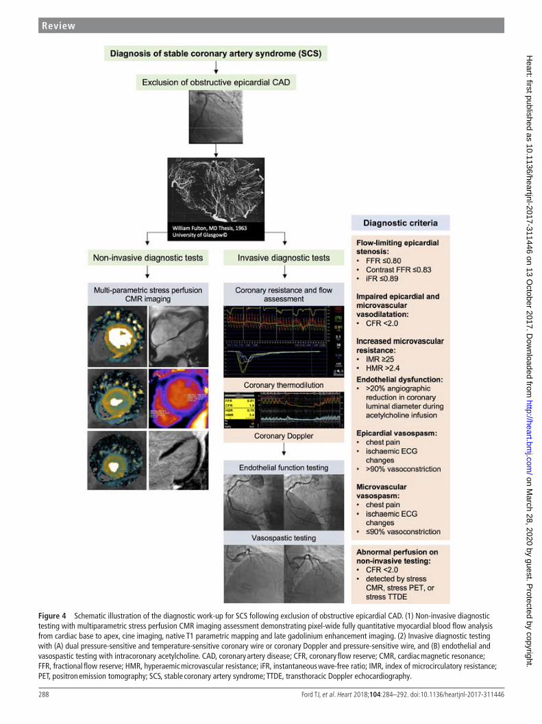
Functional disorders of coronary vasomotionFFR, CFR and IMR are typically derived using intravenous adenosine, which is an endothelium-independent vasodi-lator. Assessment of coronary endothelial function has distinct prognostic utility.39 Abnormal flow response to endothelial agonist can be assessed using symptoms and angiography alone (ie, >20% angiographic reduction in coronary luminal diameter during acetylcholine infusion),31 intracoronary Doppler flow measurement or with thermodilution. Acetylcholine may be used at higher bolus doses (eg, 100–200 µg) in a provocation test to detect abnormal coronary vasoreactivity (ie, vasospasm). A consensus document by the Coronary Vasomotion Disorders International Study Group (COVADIS) defines the criteria for a positive provocative test as meeting the following criteria: (1) reproduction of the usual chest pain, (2) ischaemic ECG changes and (3) >90% vasoconstriction on angiography40 (figure 4).
recent clInIcAl evIdencedetection and incidenceLee et al prospectively enrolled 139 consecutive patients in a single-centre study with angina and no obstructive CAD. During comprehensive invasive multimodality assessment at angiography, all patients had atherosclerosis on intravascular ultrasound, 21% had abnormal IMR, 44% had endothelial dysfunction and only 23% had no explanation for their symptoms.41 Coronary vasore-activity testing with acetylcholine is generally safe and useful for the detection of epicardial and/or microvascular spasm.15 The prevalence of microvascular spasm and vasospastic angina (VSA) is not fully resolved, but these conditions may occur in up to two-thirds of patients with a ‘negative’ angiogram.42
Coronary atherosclerosis and abnormal vasomotion are inextricably linked. A Korean study of CFR and IMR in angi-ographically moderate epicardial lesions demonstrated around
Figure 3 Clinical case demonstrating the utility of non-invasive and invasive diagnostic tests for coronary artery function. A 73-year-old woman presented with a 2-year history of typical Canadian cardiovascular society (CCS) class 2 angina. The patient had type 2 diabetes mellitus, an elevated body mass index and had previously been documented to have a normal invasive coronary angiogram 8 years previously. Invasive coronary angiography (A,B) demonstrated unobstructed epicardial coronary arteries. In the left anterior descending artery, the fractional flow reserve (FFR) value was 0.95, consistent with no epicardial flow-limiting stenosis (C). The coronary flow reserve (CFR) was reduced (1.3, normal >2.0), and the index of microcirculatory resistance (IMR) was elevated (33 units, normal <25), indicative of impaired epicardial and microvascular vasodilation and increased microvascular resistance respectively (C). Coronary endothelial function assessment using graded intracoronary acetylcholine infusion revealed mild vasoconstriction (dashed line) consistent with endothelial dysfunction (D) compared with endothelial-independent function testing with intracoronary glyceryl trinitrate (E). There was inducible coronary vasospasm using 100 µg acetylcholine bolus over 20 s (not shown). The patient subsequently underwent adenosine stress perfusion CMR, which demonstrated an inducible circumferential subendocardial perfusion defect in the basal short axis slice (arrows) with adenosine stress (F), compared with the corresponding rest perfusion imaging (G). A pixel-wide fully quantitative myocardial blood flow analysis confirmed markedly reduced myocardial blood flow in the subendocardium with adenosine stress (H) compared with the corresponding rest perfusion image (I). A diagnosis of coronary microvascular dysfunction was made. The patient was symptomatically improved at 3-month follow-up after treatment with nebivolol, statin and ACE inhibitors was started. The CMR methods were provided by Andrew Arai and Li-Yueh Hsu, National Institutes of Health, MD.
on March 28, 2020 by guest. P
rotected by copyright.http://heart.bm
j.com/
Heart: first published as 10.1136/heartjnl-2017-311446 on 13 O
ctober 2017. Dow
nloaded from
288 Ford TJ, et al. Heart 2018;104:284–292. doi:10.1136/heartjnl-2017-311446
review
Figure 4 Schematic illustration of the diagnostic work-up for SCS following exclusion of obstructive epicardial CAD. (1) Non-invasive diagnostic testing with multiparametric stress perfusion CMR imaging assessment demonstrating pixel-wide fully quantitative myocardial blood flow analysis from cardiac base to apex, cine imaging, native T1 parametric mapping and late gadolinium enhancement imaging. (2) Invasive diagnostic testing with (A) dual pressure-sensitive and temperature-sensitive coronary wire or coronary Doppler and pressure-sensitive wire, and (B) endothelial and vasospastic testing with intracoronary acetylcholine. CAD, coronary artery disease; CFR, coronary flow reserve; CMR, cardiac magnetic resonance; FFR, fractional flow reserve; HMR, hyperaemic microvascular resistance; iFR, instantaneous wave-free ratio; IMR, index of microcirculatory resistance; PET, positron emission tomography; SCS, stable coronary artery syndrome; TTDE, transthoracic Doppler echocardiography.
on March 28, 2020 by guest. P
rotected by copyright.http://heart.bm
j.com/
Heart: first published as 10.1136/heartjnl-2017-311446 on 13 O
ctober 2017. Dow
nloaded from
289Ford TJ, et al. Heart 2018;104:284–292. doi:10.1136/heartjnl-2017-311446
review
tabl
e 2
Trea
tmen
t of S
CS e
ndot
ypes
ScS
endo
type
Inve
stig
atio
nPa
thop
hysi
olog
ytr
eatm
ent
effic
acy
Side
eff
ects
Mic
rova
scul
ar a
ngin
a se
cond
ary
to
impa
ired
vaso
dila
tion
Redu
ced
CFR
and/
or in
crea
sed
mic
rova
scul
ar re
sist
ance
Anat
omic
al re
mod
ellin
g, v
ascu
lar
rare
fact
ion,
dis
turb
ed c
oron
ary
regu
latio
n
ß-bl
ocke
rsRe
duct
ion
in m
yoca
rdia
l oxy
gen
cons
umpt
ion
Fatig
ue, b
lurr
ed v
isio
n,co
ld h
ands
ACE
inhi
bito
rsIm
prov
e CF
R, re
duce
wor
kloa
d, m
ay im
prov
e m
icro
vasc
ular
rem
odel
ling
Coug
h, re
nal i
mpa
irmen
t, hy
perk
alae
mia
Rano
lazi
neIm
prov
es M
PRi i
n pa
tient
s w
ith M
VA a
nd re
duce
d CF
RN
ause
a, d
izzi
ness
, hea
dach
e
Phos
phod
iest
eras
e in
hibi
tors
↓cG
MP
degr
adat
ion,
↑va
scul
ar s
moo
th m
uscl
e re
laxa
tion
and
↑ CF
R fo
r tho
se w
ith b
asel
ine
CFR
<2.
5
Flus
hing
, tin
nitu
s, he
adac
he
Mic
rova
scul
ar a
ngin
a se
cond
ary
to
abno
rmal
vas
ocon
stric
tion
Hype
r-rea
ctiv
ity to
stim
uli (
eg,
acet
ylch
olin
e, e
xerc
ise,
str
ess)
Endo
thel
ial d
ysfu
nctio
n,
inap
prop
riate
pre
arte
riola
r va
soco
nstr
ictio
n
ACE
inhi
bito
rsIm
prov
es e
ndot
helia
l vas
omot
or d
ysfu
nctio
nCo
ugh,
rena
l im
pairm
ent,
hype
rkal
aem
ia
Calc
ium
ant
agon
ists
Vasc
ular
sm
ooth
mus
cle
rela
xatio
n, re
duct
ion
in
myo
card
ial o
xyge
n co
nsum
ptio
nCo
nstip
atio
n, a
nkle
sw
ellin
g, fl
ushi
ng
Nic
oran
dil
Pota
ssiu
m c
hann
el a
ctiv
ator
with
cor
onar
y m
icro
vasc
ular
dila
tory
effe
ctDi
zzin
ess,
flush
ing,
wea
knes
s, na
usea
Stat
ins
Impr
oved
cor
onar
y en
doth
elia
l fun
ctio
n,
plei
otro
pic
effe
cts
incl
udin
g re
duce
d va
scul
ar
infla
mm
atio
n
Mya
lgia
, hea
dach
e, c
ram
ps
Exer
cise
Bene
ficia
l effe
ct o
n en
doth
eliu
m, ↓
rest
ing
bloo
d flo
w a
nd ↑
vas
odila
tory
cap
acity
Mus
cle
fatig
ue, m
yalg
ia
Horm
one
repl
acem
ent t
hera
pyO
estr
ogen
ther
apy
impr
oves
end
othe
lial f
unct
ion
shor
t ter
m in
CM
D↑
Risk
of b
reas
t can
cer,
mar
gina
lly ↑
risk
of
CVD
Mic
rova
scul
ar a
ngin
a se
cond
ary
to
abno
rmal
pai
n pr
oces
sing
Enha
nced
noc
icep
tion
Dysf
unct
iona
l cor
tical
pai
n pr
oces
sing
Tric
yclic
ant
idep
ress
ants
Impr
oved
sym
ptom
bur
den
pote
ntia
lly th
roug
h re
duce
d vi
scer
al p
ain
Blur
red
visi
on, d
ry m
outh
, dro
wsi
ness
, im
paire
d co
ordi
natio
n
Xant
hine
der
ivat
ives
Antia
lgog
enic
effe
ct (d
ue to
the
dire
ct
invo
lvem
ent o
f ade
nosi
ne in
car
diac
pai
n ge
nera
tion)
Nau
sea
and
vom
iting
, pal
pita
tions
Epic
ardi
al a
nd/o
r mic
rova
scul
ar
coro
nary
vas
ospa
smPr
open
sity
to c
oron
ary
vaso
spas
mVa
scul
ar s
moo
th m
uscl
e hy
per-
reac
tivity
Calc
ium
cha
nnel
ant
agon
ists
↓ Sp
onta
neou
s an
d in
duci
ble
coro
nary
spa
sm v
ia
vasc
ular
sm
ooth
mus
cle
rela
xatio
n an
d ↓
oxyg
en
dem
and
Cons
tipat
ion,
ank
le s
wel
ling,
flus
hing
Nitr
ates
↓ Sp
onta
neou
s an
d in
duci
ble
coro
nary
spa
sm v
ia
larg
e ep
icar
dial
vas
odila
tion,
↓ o
xyge
n de
man
d;
lack
of e
ffica
cy in
mic
rova
scul
ar a
ngin
a w
ith
pote
ntia
lly d
elet
erio
us e
ffect
Head
ache
s, di
zzin
ess,
flush
ing
Rho-
kina
se in
hibi
tors
↓ Ca
lciu
m s
ensi
tivity
of s
moo
th m
uscl
e by
↑
phos
phat
ase
activ
ity re
duci
ng p
hosp
hory
late
d (a
ctiv
e) m
yosi
n lig
ht c
hain
s
Rash
, diz
zine
ss; n
ot li
cens
ed fo
r use
in E
urop
e or
USA
Adju
nctiv
e no
n-ph
arm
acol
ogic
al
inte
rven
tions
May
be
usef
ul in
all
endo
type
sM
etab
olic
syn
drom
e, e
ndot
helia
l dy
sfun
ctio
n, c
ardi
ovas
cula
r ris
k fa
ctor
s, an
xiet
y/de
pres
sion
Smok
ing
cess
atio
n, e
xerc
ise,
car
diac
reha
bilit
atio
n, M
edite
rran
ean
diet
, cog
nitiv
e be
havi
oura
l the
rapy
CFR,
cor
onar
y flo
w re
serv
e; C
MD,
cor
onar
y m
icro
vasc
ular
dys
func
tion;
MPR
i, m
yoca
rdia
l per
fusi
on re
serv
e in
dex;
MVA
, mic
rova
scul
ar a
ngin
a; S
CS, s
tabl
e co
rona
ry s
yndr
ome;
CVD
, car
diov
ascu
lar d
isea
se; c
GM
P, Cyclic
gua
nosi
ne m
onop
hosp
hate
on March 28, 2020 by guest. P
rotected by copyright.http://heart.bm
j.com/
Heart: first published as 10.1136/heartjnl-2017-311446 on 13 O
ctober 2017. Dow
nloaded from

290 Ford TJ, et al. Heart 2018;104:284–292. doi:10.1136/heartjnl-2017-311446
review
a quarter of 516 coronary arteries had an elevated IMR and a similar proportion had reduced CFR (<2.0).36 Both low CFR with elevated IMR were associated with poor prognosis.
Prognosis of patients and no obstructive cAdThe prognosis of SCS is linked with the underlying pathophys-iological mechanism and varies depending on the population studied.9 Patients with angiographically normal coronaries and only exercise-induced symptoms may be in a better prognostic group.43 Data from the Women’s Ischemia Syndrome Evalua-tion (WISE) study suggests that there is a worse prognosis; the 5-year annualised risk of MACE was 16.0% in women with non-obstructive CAD, 7.9% in women with normal coronary arteries and 2.4% in an asymptomatic control group (p≤0.002 after adjustment for baseline cardiovascular risk).9 Similarly, a Danish cohort study of 11 223 patients with angina found an increased risk of MACE for patients with diffuse non-ob-structive CAD and those with normal coronaries (adjusted HR of 1.85 and 1.52, respectively), compared with a reference population.
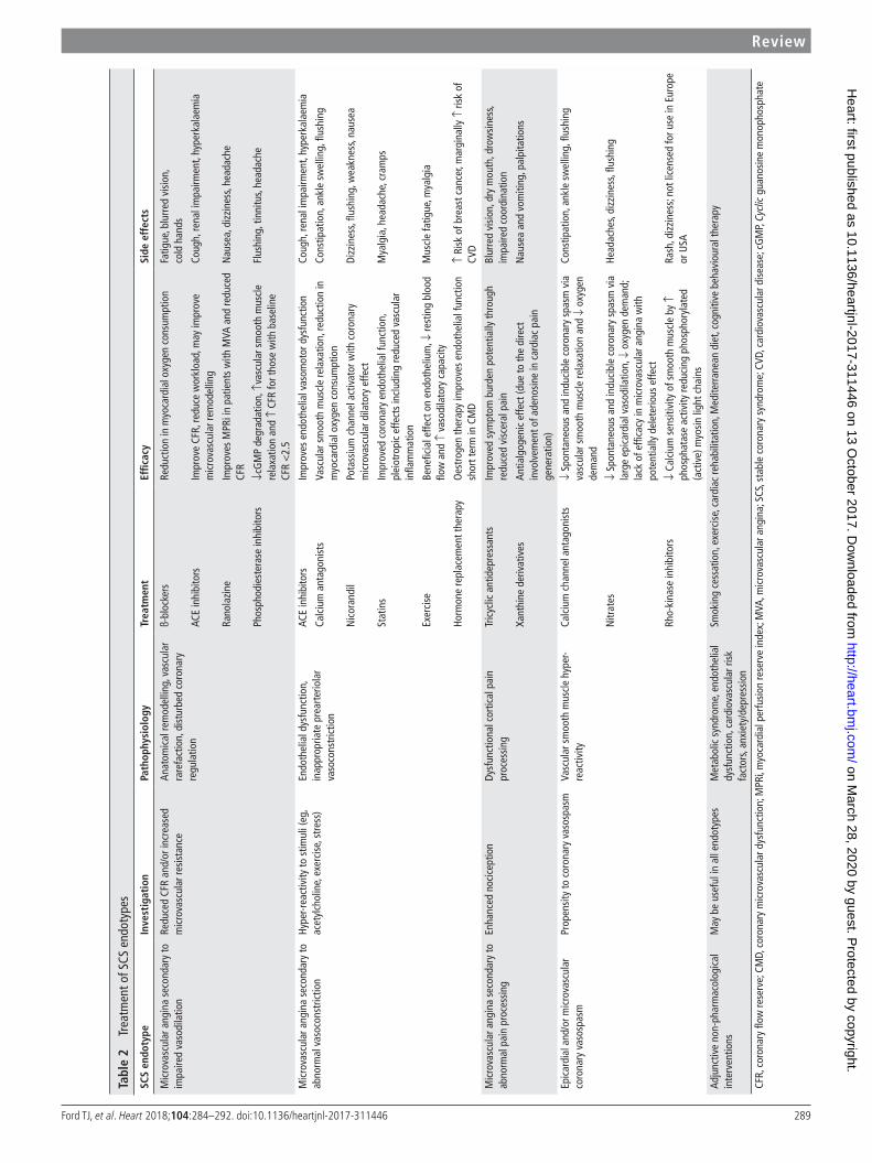
therapyPharmacological symptomatic therapyA detailed review of therapy for the different disorders of coronary artery function is beyond the scope of this review.44 A summary of currently available therapies aligning with the different SCS disease endotypes is shown in table 2 (see additional references in online supplementary file 1). Robust evidence for the treatment of SCS is lacking. The treatment effect in many studies is potentially diluted by enrolment of heterogeneous groups of patients with distinct pathophysio-logical mechanisms of CMD that may respond differently to specific treatment modalities. Current European Society of Cardiology (ESC) guidelines provide recommendations for patients with CMD suggesting ß-blockers as first-line therapy, with calcium antagonists recommended if the former is not tolerated or efficacious.4 Unlike in patients with angina and obstructive CAD, nitrates are not generally effective for treating SCS due to CMD.45 In a randomised, placebo-con-trolled clinical trial of ranolazine led by the WISE investiga-tors, although there were no overall improvements in angina and MPRi with ranolazine, benefit was observed in the subgroup of patients with a reduced CFR (<2.5) at baseline.46 Patients with VSA may benefit symptomatically from treat-ment with both nitrate and calcium channel antagonists while the latter may have prognostic benefit.47 Rho-kinase inhibitors and endothelin-receptor antagonists represent potential future therapeutic options.
Secondary preventionESC guidelines support the use of secondary prevention with aspirin and statin therapy.4 In contrast, the UK National Insti-tute for Health and Care Excellence guideline-95 on manage-ment of stable angina cites the enigmatic term ‘Syndrome X’, but guidance on the diagnosis and management is limited, reflecting the limited evidence base.48 Contemporary guide-lines should be followed for the management of vascular risk factors in patients with INOCA.3 41 In patients with CMD, statin therapy reduces ischaemia as revealed by reduced ST-segment deviation following exercise testing and improves exercise capacity and flow-mediated dilatation in the brachial artery in patients with CMD.49 ACE inhibitors (ACEi) improve endothelial dysfunction and vasoreactivity via NO stimulation
helping to reverse vascular hypertrophy and improve vascular compliance.
Non-pharmacological therapyLifestyle measures promote well-being in patients with INOCA through smoking cessation, healthy eating (eg, Mediterra-nean diet) and physical activity.3 The Comprehensive Treat-ment of Angina in Women With Microvascular Dysfunction trial (NCT02910154) will randomise women with angina and CMD to comprehensive multimodality intervention (dietary, exercise, and statin and ACEi therapy) or control therapy to determine whether angina and microvascular dysfunction can be improved.
Future directionsOverall, there is a critical missing link between the use of rele-vant diagnostic tests of coronary artery function, therapeutic agents with proven efficacy and health outcomes of patients with angina without obstructive CAD. This gap in evidence is currently being addressed in the British Heart Foundation CORonary MICrovascular Angina randomised controlled trial (CorMicA NCT03193294). A personalised approach to therapy is desirable and it is reasonable to target those patients with impairment of microvascular dilation characterised by reduced CFR with anti-anginals that reduce heart rate and oxygen consumption (eg, ß-blockers), whereas vasodilators or ACEi would be more appropriate for patients with evidence of in microvascular constriction.50
‘Stable coronary syndrome’ reflects a disease-based, clin-ically relevant classification that reflects the interaction between structural and functional disorders throughout the coronary circulation. The paucity of pathway-specific therapy presents an opportunity for novel research using stratified medicine. Our vision is for a personalised medicine approach whereby SCS endotypes, defined by the results of coronary function tests, may benefit from targeted therapy. Further research is needed to determine whether this paradigm may lead to patient benefits (table 3).
table 3 Proposed comprehensive research strands for patients with ischaemia and no obstructive coronary artery disease (INOCA)
comprehensive InocA research strands
Stratified medicine trials ► Diagnostic tests (rule-in/rule-out) ► Stratification of endotypes for evidence-based therapy
Vascular science ► Investigations of disease mechanisms, for example, endothelial dysfunction and dysregulation of the endothelin system
Imaging and modelling ► Clinical trials of quantitative perfusion CMR versus standard methods
► Patient-specific computed models of disease to predict responses to novel therapeutics
Molecular pathology and vascular histopathology
► Protein assay-based scores and genetic variants are potential biomarkers for disorders of coronary function.
► Identification of drug targets
Therapeutic trials ► Enhanced system antagonists ► Vasodilating ß-blockers ► Lifestyle interventions, for example, exercise,
weight loss
Health informatics and value assessments
► Assess the cost-effectiveness of innovative stratified approaches
Patient and public involvement
► Ensures relevance of research to patients and carers
on March 28, 2020 by guest. P
rotected by copyright.http://heart.bm
j.com/
Heart: first published as 10.1136/heartjnl-2017-311446 on 13 O
ctober 2017. Dow
nloaded from
291Ford TJ, et al. Heart 2018;104:284–292. doi:10.1136/heartjnl-2017-311446
review
twitter @UofGICAMS
Acknowledgements We acknowledge the patients who have participated in research studies cited in this article.
contributors CB conceived the article. TJF and DC provided the first draft, figures and revisions. All authors have approved the final manuscript.
Funding The British Heart Foundation has supported DC (FS/14/15/30661), TJF (RE/13/5/30177) and CB (RE/13/5/30177; FS/14/15/30661; FS172632744; PG-17-25-32884).
competing interests CB is employed by the University of Glasgow, which holds consultancy and research agreements with companies that have commercial interests in the diagnosis and treatment of angina. The companies include Abbott Vascular, AstraZeneca, Boehringer Ingelheim, Menarini Pharmaceuticals and Siemens Healthcare. None of the other authors have any disclosures.
ethics approval Obtained.
Provenance and peer review Commissioned; externally peer reviewed.
open Access This is an Open Access article distributed in accordance with the terms of the Creative Commons Attribution (CC BY 4.0) license, which permits others to distribute, remix, adapt and build upon this work, for commercial use, provided the original work is properly cited. See: http:// creativecommons. org/ licenses/ by/ 4. 0/
© Article author(s) (or their employer(s) unless otherwise stated in the text of the article) 2018. All rights reserved. No commercial use is permitted unless otherwise expressly granted.
reFerenceS 1 Wang H, Naghavi M, Allen C, et al. Global, regional, and national life expectancy,
all-cause mortality, and cause-specific mortality for 249 causes of death, 1980-2015: a systematic analysis for the global burden of disease study 2015. Lancet 2016;388:1459–544.
2 Berry C. Stable coronary syndromes: the case for consolidating the nomenclature of stable ischemic heart disease. Circulation 2017;136:437 9.
3 Bairey Merz CN, Pepine CJ, Walsh MN, et al. Ischemia and No Obstructive Coronary Artery Disease (INOCA): developing evidence-based therapies and research agenda for the next decade. Circulation 2017;135:1075–92.
4 Montalescot G, Sechtem U, Achenbach S, et al. ESC guidelines on the management of stable coronary artery disease: the Task Force on the management of stable coronary artery disease of the European society of cardiology. Eur Heart J 2013;34:2949–3003.
5 Greenwood JP, Ripley DP, Berry C, et al. Effect of care guided by cardiovascular magnetic resonance, myocardial perfusion scintigraphy, or NICE guidelines on subsequent unnecessary angiography rates: the CE-MARC 2 randomized clinical trial. JAMA 2016;316:1051–60.
6 Patel MR, Peterson ED, Dai D, et al. Low diagnostic yield of elective coronary angiography. N Engl J Med 2010;362:886–95.
7 Camici PG, d’Amati G, Rimoldi O. Coronary microvascular dysfunction: mechanisms and functional assessment. Nat Rev Cardiol 2015;12:48–62.
8 Herrmann J, Kaski JC, Lerman A. Coronary microvascular dysfunction in the clinical setting: from mystery to reality. Eur Heart J 2012;33:2771–82.
9 Gulati M, Cooper-DeHoff RM, McClure C, et al. Adverse cardiovascular outcomes in women with nonobstructive coronary artery disease: a report from the women’s ischemia syndrome evaluation study and the St James women take heart project. Arch Intern Med 2009;169:843–50.
10 Duncker DJ, Koller A, Merkus D, et al. Regulation of coronary blood flow in health and ischemic heart disease. Prog Cardiovasc Dis 2015;57:409–22.
11 Mancini M, Petretto E, Kleinert C, et al. Mapping genetic determinants of coronary microvascular remodeling in the spontaneously hypertensive rat. Basic Res Cardiol 2013;108:316.
12 Yamamoto S, James TN, Kawamura K, et al. Cardiocytic apoptosis and capillary endothelial swelling as morphological evidence of myocardial ischemia in ventricular biopsies from patients with angina and normal coronary arteriograms. Coron Artery Dis 2002;13:25–35.
13 Osamichi S, Kouji K, Yoshimaro I, et al. Myocardial glucose metabolism assessed by positron emission tomography and the histopathologic findings of microvessels in syndrome X. Circ J 2004;68:220–6.
14 Richardson PJ, Livesley B, Oram S, et al. Angina pectoris with normal coronary arteries. Transvenous myocardial biopsy in diagnosis. Lancet 1974;2:677–80.
15 Ong P, Athanasiadis A, Borgulya G, et al. High prevalence of a pathological response to acetylcholine testing in patients with stable angina pectoris and unobstructed coronary arteries. The ACOVA study (Abnormal COronary VAsomotion in patients with stable angina and unobstructed coronary arteries). J Am Coll Cardiol 2012;59:655–62.
16 Johnson NP, Gould KL. Clinical evaluation of a new concept: resting myocardial perfusion heterogeneity quantified by markovian analysis of PET identifies coronary
microvascular dysfunction and early atherosclerosis in 1,034 subjects. J Nucl Med 2005;46:1427–37.
17 Johnson NP, Gould KL. Physiology of endothelin in producing myocardial perfusion heterogeneity: a mechanistic study using darusentan and positron emission tomography. J Nucl Cardiol 2013;20:835–44.
18 Marcus ML, Chilian WM, Kanatsuka H, et al. Understanding the coronary circulation through studies at the microvascular level. Circulation 1990;82:1–7.
19 Garber CE, Carleton RA, Camaione DN, et al. The threshold for myocardial ischemia varies in patients with coronary artery disease depending on the exercise protocol. J Am Coll Cardiol 1991;17:1256–62.
20 Dubois-Randé JL, Dupouy P, Aptecar E, et al. Comparison of the effects of exercise and cold pressor test on the vasomotor response of normal and atherosclerotic coronary arteries and their relation to the flow-mediated mechanism. Am J Cardiol 1995;76:467–73.
21 Davies RF, Goldberg AD, Forman S, et al. Asymptomatic Cardiac Ischemia Pilot (ACIP) study two-year follow-up: outcomes of patients randomized to initial strategies of medical therapy versus revascularization. Circulation 1997;95:2037–43.
22 Steg PG, Greenlaw N, Tendera M, et al. Prevalence of anginal symptoms and myocardial ischemia and their effect on clinical outcomes in outpatients with stable coronary artery disease: data from the International Observational CLARIFY Registry. JAMA Intern Med 2014;174:1651–9.
23 Ambepityia G, Kopelman PG, Ingram D, et al. Exertional myocardial ischemia in diabetes: a quantitative analysis of anginal perceptual threshold and the influence of autonomic function. J Am Coll Cardiol 1990;15:72–7.
24 Lanza GA, Giordano A, Pristipino C, et al. Abnormal cardiac adrenergic nerve function in patients with syndrome X detected by [123I]metaiodobenzylguanidine myocardial scintigraphy. Circulation 1997;96:821–6.
25 Pasceri V, Lanza GA, Buffon A, et al. Role of abnormal pain sensitivity and behavioral factors in determining chest pain in syndrome X. J Am Coll Cardiol 1998;31:62–6.
26 Rigo F, Richieri M, Pasanisi E, et al. Usefulness of coronary flow reserve over regional wall motion when added to dual-imaging dipyridamole echocardiography. Am J Cardiol 2003;91:269–73.
27 Murthy VL, Naya M, Foster CR, et al. Association between coronary vascular dysfunction and cardiac mortality in patients with and without diabetes mellitus. Circulation 2012;126:1858–68.
28 Panting JR, Gatehouse PD, Yang GZ, et al. Abnormal subendocardial perfusion in cardiac syndrome X detected by cardiovascular magnetic resonance imaging. N Engl J Med 2002;346:1948–53.
29 Doyle M, Weinberg N, Pohost GM, et al. Prognostic value of global MR myocardial perfusion imaging in women with suspected myocardial ischemia and no obstructive coronary disease: results from the NHLBI-sponsored WISE (Women’s Ischemia Syndrome Evaluation) study. JACC Cardiovasc Imaging 2010;3:1030–6.
30 Hsu LY, Groves DW, Aletras AH, et al. A quantitative pixel-wise measurement of myocardial blood flow by contrast-enhanced first-pass CMR perfusion imaging: microsphere validation in dogs and feasibility study in humans. JACC Cardiovasc Imaging 2012;5:154–66.
31 Sheikh AR WJ, Bariey Merz N, Beltrame JF. The current state of invasive coronary evaluation and management of patients with angina and nonobstructive coronary arteries: American college of cardiology;2016 [Expert Analysis]. 2017 http://www. acc. org/ latest- in- cardiology/ articles/ 2016/ 05/ 26/ 08/ 31/ the- current- state- of- invasive- coronary- evaluation- and- management- of- patients- with- angina- and- nonobstructive- coronary- arteries? w_ nav= LC (accessed 20th July 2017).
32 Berry C, Corcoran D, Hennigan B, et al. Fractional flow reserve-guided management in stable coronary disease and acute myocardial infarction: recent developments. Eur Heart J 2015;36:3155–64.
33 Gould KL, Lipscomb K, Hamilton GW. Physiologic basis for assessing critical coronary stenosis. Instantaneous flow response and regional distribution during coronary hyperemia as measures of coronary flow reserve. Am J Cardiol 1974;33:87–94.
34 Fearon WF, Balsam LB, Farouque HM, et al. Novel index for invasively assessing the coronary microcirculation. Circulation 2003;107:3129–32.
35 Meuwissen M, Siebes M, Chamuleau SA, et al. Hyperemic stenosis resistance index for evaluation of functional coronary lesion severity. Circulation 2002;106:441–6.
36 Lee JM, Jung JH, Hwang D, et al. Coronary flow reserve and microcirculatory resistance in patients with intermediate coronary stenosis. J Am Coll Cardiol 2016;67:1158–69.
37 Ford TJ, Corcoran D, Berry C. Coronary artery disease: physiology and prognosis. Eur Heart J 2017 (Epub ahead of print 27 May 2017).
38 Sezer M, Kocaaga M, Aslanger E, et al. Bimodal pattern of coronary microvascular involvement in diabetes mellitus. J Am Heart Assoc 2016;5:e003995.
39 Halcox JP, Schenke WH, Zalos G, et al. Prognostic value of coronary vascular endothelial dysfunction. Circulation 2002;106:653–8.
40 Beltrame JF, Crea F, Kaski JC, et al. International standardization of diagnostic criteria for vasospastic angina. Eur Heart J 2017;38:2565-2568.
on March 28, 2020 by guest. P
rotected by copyright.http://heart.bm
j.com/
Heart: first published as 10.1136/heartjnl-2017-311446 on 13 O
ctober 2017. Dow
nloaded from

292 Ford TJ, et al. Heart 2018;104:284–292. doi:10.1136/heartjnl-2017-311446
review
41 Lee BK, Lim HS, Fearon WF, et al. Invasive evaluation of patients with angina in the absence of obstructive coronary artery disease. Circulation 2015;131:1054–60.
42 Sara JD, Widmer RJ, Matsuzawa Y, et al. Prevalence of coronary microvascular dysfunction among patients with chest pain and nonobstructive coronary artery disease. JACC Cardiovasc Interv 2015;8:1445–53.
43 Lanza GA, Filice M, De Vita A, et al. Primary atable microvascular angina: a long-term clinical follow-up study. Circulation 2017;135:1982–4.
44 Ong P, Athanasiadis A, Sechtem U. Treatment of angina pectoris associated with coronary microvascular dysfunction. Cardiovasc Drugs Ther 2016;30:351–6.
45 Russo G, Di Franco A, Lamendola P, et al. Lack of effect of nitrates on exercise stress test results in patients with microvascular angina. Cardiovasc Drugs Ther 2013;27:229–34.
46 Bairey Merz CN, Handberg EM, Shufelt CL, et al. A randomized, placebo-controlled trial of late Na current inhibition (ranolazine) in coronary microvascular dysfunction (CMD): impact on angina and myocardial perfusion reserve. Eur Heart J 2016;37:1504–13.
47 Nishigaki K, Inoue Y, Yamanouchi Y, et al. Prognostic effects of calcium channel blockers in patients with vasospastic angina--a meta-analysis. Circ J 2010;74:1943–50.
48 (NICE) NIfHaCE. Stable angina: management. NICE guideline (CG126). London: NICE, 2011.
49 Kayikcioglu M, Payzin S, Yavuzgil O, et al. Benefits of statin treatment in cardiac syndrome-X1. Eur Heart J 2003;24:1999–2005.
50 Crea F, Lanza GA. Treatment of microvascular angina: the need for precision medicine. Eur Heart J 2016;37:1514–6.
on March 28, 2020 by guest. P
rotected by copyright.http://heart.bm
j.com/
Heart: first published as 10.1136/heartjnl-2017-311446 on 13 O
ctober 2017. Dow
nloaded from