Stability of Eukaryotic Flagellar Beating to Noise and ...
5
Stability of Eukaryotic Flagellar Beating to Noise and Mechanical Perturbations Kirsty Y. Wan and Raymond E. Goldstein Department of Applied Mathematics and Theoretical Physics, Centre for Mathematical Sciences, University of Cambridge, Wilberforce Road, Cambridge CB3 0WA, United Kingdom (Dated: June 12, 2014) The eukaryotic flagellum is a remarkable cellular structure, for it can sustain beating with unfailing rhythmicity, yet is capable of rapid responses to stimuli. Recent studies have revealed flagellar beating to be intrinsically noisy, but detailed understanding of this stochasticity is lacking. Here, we use Chlamydomonas reinhardtii to investigate flagellum sensitivity to noise and perturbations. We find that flagellum oscillations prescribe stable limit cycles and phase-dependent waveform fluctuations, while interbeat intervals exhibit approximate 1/f noise and long-range correlations spanning hundreds of beats. By forcing beating flagella with hydrodynamic loads, we quantify the recovery of periodic breastroke beating from imposed perturbations. These results will help constrain microscopic theories on the origin and regulation of beating. Patterns of coordinated movement in living organisms, such as walking, running, and galloping, may be vari- able yet simultaneously stable. Such repetitive dynamics are distinguished by their reproducibility, sustainability over long times, and stability in the presence of small to moderate perturbations. In the precise, rhythmic beat- ing of the flagella of the alga Chlamydomonas we find remarkable living oscillators that fulfil these three crite- ria. The synchronous beating of its twin flagella allows Chlamydomonas to swim a fast breaststroke [1]. For this alga, oscillations of its flagella are self-sustained – re- peated mechano-chemical cycles continuously supply en- ergy to motor dyneins residing within flagellar axonemes [2]. Stepping action of individual motors is intrinsically stochastic [3], and yet, beating can nevertheless persist, resilient against a cacophony of biochemical and back- ground fluctuations [4]. Often, in assessing the fidelity or robustness of a biological oscillator, the stability and rhythmicity of its oscillations serve as prime indicators: one might identify pathological gaits of human walking from measures of cycle stability [5], determine the phase- dependent response of circadian clocks using external stimuli [6], or infer the health of a human heart from variability of inter-beat intervals [7, 8]. Similarly, peri- odic oscillations of beating flagella are correlated with a cell’s responses and sensitivity to its environment, but study of these features remains inchoate [9–14]. Here, we assess flagellum stability on two complemen- tary levels, drawing on data from a large population (∼100) of cells. First, we evaluate stability to weak fluc- tuations originating from such sources over which the ex- perimenter has little control. These include background thermal noise, intracellular biochemical processes asso- ciated with cell metabolism [15], or even fluctuations in photon irradiance [4]. Flagellar dynamics are found to be inherently stable, but waveform noise displays an in- triguing phase dependence. Measured beat-to-beat in- tervals form a complex timeseries that exhibits fractal structure, and successive beats may remain correlated for many seconds. Second, we measure the recovery of flagellar beating in the aftermath of stronger, externally- derived perturbations imposed by injecting fluid impulses near a beating flagellum. Post-perturbation, the normal breaststroke resumes following a characteristic relaxation dynamics back to the stable limit cycle. This ability to recover readily from disruption to beating may be a cru- cial property of viable ciliary/flagellar structures. To permit long-time, in-focus visualization of flagel- lar dynamics, wildtype cells (strains CC124 and CC125, Chlamydomonas Center) were individually caught and held in place by micropipette micromanipulation (Patch- star, Scientifica, UK) with gentle suction, as described previously [9, 12]. High-speed images (SA3 Fastcam, Photron, USA and Phantom V311, Vision Research, USA) of beating flagella were captured at 1000 - 3000 frames/s – at least one order of magnitude above the maximum flagella beat frequency. Continuous record- ings (1 - 10 minutes) were taken for each cell, from which ∼1 - 5 × 10 3 contiguous beat cycles could be extracted. Recordings were conducted under conditions that appro- priately mimic a wild-type cell’s natural daytime habitat, namely white light illumination (100W halogen lamp) with negligible background lighting, and hence some pho- totactic response is expected [16]. Pixel coordinates that 2 1 4 3 a) b) real (eigenvalue) imag (eigenvalue) θ FIG. 1. (color online) Noisy flagellar limit cycles. a) Tra- jectories in angle (θ) – angular velocity ( ˙ θ) space at a fixed arclength along the flagellum, with four Poincar´ e sections shown. b) Lyapunov exponents at phases 1-4, for 48 cells.
Transcript of Stability of Eukaryotic Flagellar Beating to Noise and ...

Stability of Eukaryotic Flagellar Beating to Noise and Mechanical Perturbations
Kirsty Y. Wan and Raymond E. GoldsteinDepartment of Applied Mathematics and Theoretical Physics, Centre for Mathematical Sciences,
University of Cambridge, Wilberforce Road, Cambridge CB3 0WA, United Kingdom(Dated: June 12, 2014)
The eukaryotic flagellum is a remarkable cellular structure, for it can sustain beating with unfailingrhythmicity, yet is capable of rapid responses to stimuli. Recent studies have revealed flagellarbeating to be intrinsically noisy, but detailed understanding of this stochasticity is lacking. Here,we use Chlamydomonas reinhardtii to investigate flagellum sensitivity to noise and perturbations.We find that flagellum oscillations prescribe stable limit cycles and phase-dependent waveformfluctuations, while interbeat intervals exhibit approximate 1/f noise and long-range correlationsspanning hundreds of beats. By forcing beating flagella with hydrodynamic loads, we quantifythe recovery of periodic breastroke beating from imposed perturbations. These results will helpconstrain microscopic theories on the origin and regulation of beating.
Patterns of coordinated movement in living organisms,such as walking, running, and galloping, may be vari-able yet simultaneously stable. Such repetitive dynamicsare distinguished by their reproducibility, sustainabilityover long times, and stability in the presence of small tomoderate perturbations. In the precise, rhythmic beat-ing of the flagella of the alga Chlamydomonas we findremarkable living oscillators that fulfil these three crite-ria. The synchronous beating of its twin flagella allowsChlamydomonas to swim a fast breaststroke [1]. For thisalga, oscillations of its flagella are self-sustained – re-peated mechano-chemical cycles continuously supply en-ergy to motor dyneins residing within flagellar axonemes[2]. Stepping action of individual motors is intrinsicallystochastic [3], and yet, beating can nevertheless persist,resilient against a cacophony of biochemical and back-ground fluctuations [4]. Often, in assessing the fidelityor robustness of a biological oscillator, the stability andrhythmicity of its oscillations serve as prime indicators:one might identify pathological gaits of human walkingfrom measures of cycle stability [5], determine the phase-dependent response of circadian clocks using externalstimuli [6], or infer the health of a human heart fromvariability of inter-beat intervals [7, 8]. Similarly, peri-odic oscillations of beating flagella are correlated with acell’s responses and sensitivity to its environment, butstudy of these features remains inchoate [9–14].
Here, we assess flagellum stability on two complemen-tary levels, drawing on data from a large population(∼100) of cells. First, we evaluate stability to weak fluc-tuations originating from such sources over which the ex-perimenter has little control. These include backgroundthermal noise, intracellular biochemical processes asso-ciated with cell metabolism [15], or even fluctuations inphoton irradiance [4]. Flagellar dynamics are found tobe inherently stable, but waveform noise displays an in-triguing phase dependence. Measured beat-to-beat in-tervals form a complex timeseries that exhibits fractalstructure, and successive beats may remain correlatedfor many seconds. Second, we measure the recovery of
flagellar beating in the aftermath of stronger, externally-derived perturbations imposed by injecting fluid impulsesnear a beating flagellum. Post-perturbation, the normalbreaststroke resumes following a characteristic relaxationdynamics back to the stable limit cycle. This ability torecover readily from disruption to beating may be a cru-cial property of viable ciliary/flagellar structures.
To permit long-time, in-focus visualization of flagel-lar dynamics, wildtype cells (strains CC124 and CC125,Chlamydomonas Center) were individually caught andheld in place by micropipette micromanipulation (Patch-star, Scientifica, UK) with gentle suction, as describedpreviously [9, 12]. High-speed images (SA3 Fastcam,Photron, USA and Phantom V311, Vision Research,USA) of beating flagella were captured at 1000 − 3000frames/s – at least one order of magnitude above themaximum flagella beat frequency. Continuous record-ings (1−10 minutes) were taken for each cell, from which∼1− 5× 10³ contiguous beat cycles could be extracted.Recordings were conducted under conditions that appro-priately mimic a wild-type cell’s natural daytime habitat,namely white light illumination (100W halogen lamp)with negligible background lighting, and hence some pho-totactic response is expected [16]. Pixel coordinates that
2 1
43
a) b)
real (eigenvalue)
imag (eigenvalue)
θ
FIG. 1. (color online) Noisy flagellar limit cycles. a) Tra-
jectories in angle (θ) – angular velocity (θ) space at a fixedarclength along the flagellum, with four Poincare sectionsshown. b) Lyapunov exponents at phases 1-4, for 48 cells.
2
δF
0
0.2
0.4
0.6
0.8
1.0a)
c)b)
recovery stroke power stroke
1
2
3
4
f
f*
1 2 3 4
δFcis flagellum
α/ 〈α〉
T/〈T
〉
0.8 1 1.2
1
α/ 〈α〉
T/〈T
〉
trans flagellum
d)
0.95
00
8
4
0.2 0.60.4 0.8 1 0.8 1 1.2
1
1.05
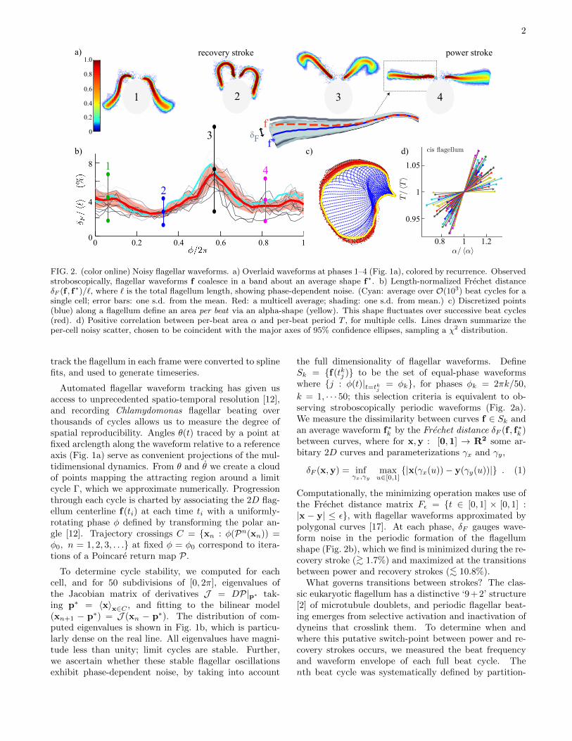
FIG. 2. (color online) Noisy flagellar waveforms. a) Overlaid waveforms at phases 1–4 (Fig. 1a), colored by recurrence. Observedstroboscopically, flagellar waveforms f coalesce in a band about an average shape f∗. b) Length-normalized Frechet distanceδF (f , f∗)/`, where ` is the total flagellum length, showing phase-dependent noise. (Cyan: average over O(103) beat cycles for asingle cell; error bars: one s.d. from the mean. Red: a multicell average; shading: one s.d. from mean.) c) Discretized points(blue) along a flagellum define an area per beat via an alpha-shape (yellow). This shape fluctuates over successive beat cycles(red). d) Positive correlation between per-beat area α and per-beat period T , for multiple cells. Lines drawn summarize theper-cell noisy scatter, chosen to be coincident with the major axes of 95% confidence ellipses, sampling a χ2 distribution.
track the flagellum in each frame were converted to splinefits, and used to generate timeseries.
Automated flagellar waveform tracking has given usaccess to unprecedented spatio-temporal resolution [12],and recording Chlamydomonas flagellar beating overthousands of cycles allows us to measure the degree ofspatial reproducibility. Angles θ(t) traced by a point atfixed arclength along the waveform relative to a referenceaxis (Fig. 1a) serve as convenient projections of the mul-tidimensional dynamics. From θ and θ we create a cloudof points mapping the attracting region around a limitcycle Γ, which we approximate numerically. Progressionthrough each cycle is charted by associating the 2D flag-ellum centerline f(ti) at each time ti with a uniformly-rotating phase φ defined by transforming the polar an-gle [12]. Trajectory crossings C = {xn : φ(Pn(xn)) =φ0, n = 1, 2, 3, . . .} at fixed φ = φ0 correspond to itera-tions of a Poincare return map P.
To determine cycle stability, we computed for eachcell, and for 50 subdivisions of [0, 2π], eigenvalues ofthe Jacobian matrix of derivatives J = DP|p∗ tak-ing p∗ = 〈x〉x∈C , and fitting to the bilinear model(xn+1 − p∗) = J (xn − p∗). The distribution of com-puted eigenvalues is shown in Fig. 1b, which is particu-larly dense on the real line. All eigenvalues have magni-tude less than unity; limit cycles are stable. Further,we ascertain whether these stable flagellar oscillationsexhibit phase-dependent noise, by taking into account
the full dimensionality of flagellar waveforms. DefineSk = {f(tkj )} to be the set of equal-phase waveformswhere {j : φ(t)|t=tkj = φk}, for phases φk = 2πk/50,
k = 1, · · · 50; this selection criteria is equivalent to ob-serving stroboscopically periodic waveforms (Fig. 2a).We measure the dissimilarity between curves f ∈ Sk andan average waveform f∗k by the Frechet distance δF (f , f∗k )between curves, where for x,y : [0,1] → R2 some ar-bitary 2D curves and parameterizations γx and γy,
δF (x,y) = infγx,γy
maxu∈[0,1]
{|x(γx(u))− y(γy(u))|} . (1)
Computationally, the minimizing operation makes use ofthe Frechet distance matrix Fε = {t ∈ [0, 1] × [0, 1] :|x − y| ≤ ε}, with flagellar waveforms approximated bypolygonal curves [17]. At each phase, δF gauges wave-form noise in the periodic formation of the flagellumshape (Fig. 2b), which we find is minimized during the re-covery stroke (>∼ 1.7%) and maximized at the transitionsbetween power and recovery strokes (<∼ 10.8%).
What governs transitions between strokes? The clas-sic eukaryotic flagellum has a distinctive ‘9 + 2’ structure[2] of microtubule doublets, and periodic flagellar beat-ing emerges from selective activation and inactivation ofdyneins that crosslink them. To determine when andwhere this putative switch-point between power and re-covery strokes occurs, we measured the beat frequencyand waveform envelope of each full beat cycle. Thenth beat cycle was systematically defined by partition-
3
F(n)
n
a)
b)
c)
/ /
0
0C
(τ)
τ (beat number)
t (beat number)
ν (H
z)
0.2
500
500
60585654
1000
1000 1500
0 500 1000 1500 0 0.1
2000
10-3
102 103
10-2
α = 0.919
α = 0.511
-0.1
0.1wt
eye2
shuffled
datafit
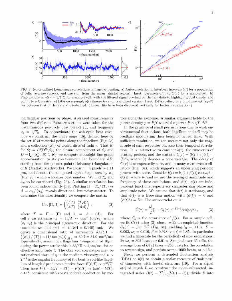
FIG. 3. (color online) Long-range correlations in flagellar beating. a) Autocorrelation in interbeat intervals b(t) for a populationof cells: average (black), and one s.d. from the mean (shaded region). Inset: parametric fit to C(τ) for a sample cell. b)Fluctuations in ν(t) := 1/b(t) for a sample cell, with the filtered signal overlaid on the raw data to highlight global trends, andpdf fit to a Gaussian. c) DFA on a sample b(t) timeseries and its shuffled version. Inset: DFA scaling for a blind mutant (eye2 )lies between that of the wt and wt-shuffled. ( Linear fits have been displaced vertically for better visualization.)
ing flagellar positions by phase. Averaged measurementsfrom two different Poincare sections were taken for theinstantaneous per-cycle beat period Tn, and frequencyνn = 1/Tn. To approximate the nth-cycle beat enve-lope we construct the alpha-shape [18], defined here bythe set K of material points along the flagellum (Fig. 2c)and a collection {Sr} of closed discs of radii r. That is,for Scr = Cl(R2\Sr) the closure complement of Sr andD =
⋃{Scα : Scr ⊃ K} we compute a straight-line graph
approximation to its piecewise-circular boundary ∂D,starting from the (closest-point) Delaunay triangulationof K (Matlab, Mathworks). We chose r = 5 pixels ∼ 1.11µm, and denote the computed alpha-shape area by αn(Fig. 2c), where n indexes beat number. We find Tn andαn to be correlated (Fig. 2d). A similar correlation hasbeen found independently [14]. Plotting Π = Tn/ 〈Tn〉 vsA = αn/ 〈αn〉 reveals directional but noisy scatter. Todetermine this directionality we compute the matrix
Cov [Π, A] =
(〈T T 〉 〈T A〉〈AT 〉 〈AA〉
), (2)
where T = Π − 〈Π〉 and A = A − 〈A〉. Forcell i we estimate γi = Π/A ∼ tan−1(v2/v1) where(v1, v2) is the principal eigenvector direction. For theensemble we find 〈γi〉 ∼ (0.264 ± 0.146) rad. Wederive a dimensional ratio of increments δA/δΠ =⟨⟨αin⟩/⟨T in⟩× (1/ tan(γi))
⟩all i≈ 39.7 ± 31.0 µm2/ms.
Equivalently, assuming a flagellum “wingspan” of 10µmduring the power stroke this is δ`/δΠ ∼ 4µm/ms, for aneffective amplitude `. The observed correlation may berationalized thus: if η is the medium viscosity and ν ∼T−1 is the angular frequency of the beat, a rod-like flagel-lum of length ` produces a motive force F (`, T ) ∼ η`2/T .Then here F (` + δ`, T + δT ) − F (`, T ) ≈ (aδ` − bδT ),a ≈ b, consistent with constant force production by mo-
tors along the axoneme. A similar argument holds for thepower density p = P/` where the power P ∼ ηT−2`3.
In the presence of small perturbations due to weak en-vironmental fluctuations, both flagellum and cell may befeedback modulating their behavior in real-time. Withsufficient resolution, we can measure not only the mag-nitude of such responses but also their temporal correla-tion. It is instructive to consider b(t), the timeseries ofbeating periods, and the statistic C(τ) = 〈b(t+ τ)b(t)−〈b〉2〉, where 〈·〉 denotes a time average. The decay ofC(τ) is unexpectedly slow, and in many cases even oscil-latory (Fig. 3a), which suggests an underlying periodicprocess with noise. Consider b(t) = b0(1+β(t)) cos(ω0t+φ(t)), where b0 and ω0 are the averaged amplitude andfrequency of these oscillations, and β(t), φ(t) are inde-pendent functions respectively characterizing phase andamplitude noise. We assume that β(t) is stationary, andthat φ(t) is a Brownian motion with 〈φ(t)〉 = 0 and⟨φ(t)2
⟩= Dt. The autocorrelation is
C(τ) =b202
[1 + Cb(τ)]e−D|τ | cos(ω0τ) , (3)
where Cb is the covariance of β(t). For a sample cell,we fit C(τ) using (3) above, with an empirical functionCb(τ) = βe−|τ |/ξ (Fig. 3a), yielding b0 = 0.157, D =0.002, ω0 = 0.016, β = 9.928 and ξ = 1.85. In particularwe find a timescale for the periodicity of slow oscillations:2π/ω0 = 392 beats, or 6.01 s. Sampled over 65 cells, theaverage form of C(τ) takes ∼250 beats for the correlationto reverse sign, and persists over ∼1000 beats, or ∼15 s.
Next, we perform a detrended fluctuation analysis(DFA) on b(t) to obtain a scalar measure of ‘noisiness’of timeseries with fractal structure [19]. For a signalb(t) of length L we construct the mean-subtracted, in-
tegrated series B(t) =∑Li=0(b(ti) − 〈b〉), divide B into

4
K non-overlapping sections of size n = L/K, {Ii :=[ti, ti+1], ti = iL/K, i = 1, 2, · · · ,K − 1}, and computethe local trend at the ith section. Let Bn(ti) be the lsqlinear fit to data points B(ti ∈ Ii). We detrend B bysubtracting Bn, and compute
F (n) =
(1
K
K∑i=1
(B(ti)−Bn(ti))2
)0.5
. (4)
A power-law scaling F (n) ∼ nα can be seen to hold(Fig. 3c). For the population, we obtain α = 0.83± 0.10(67 cells, O(103) successive beats for each). Randomlypermuting b(t) (each time averaging over 10 shuffles)yields α = 0.48 ± 0.03, or white noise. DFA on a blindChlamydomonas mutant with wildtype motility (eye2,CC4302 Chlamydomonas Center) gives α = 0.72 ± 0.04(Fig. 3d), consistent with reduced flagellum noise uponremoval of photo-perception. Wildtype Chlamydomonasrespond to single-photon events [4], that alter voltage-gated flagellar Ca2+ channels downstream [20] and elicitcharacteristic flagellar responses in O(ms). For our lightsource we estimate an incident flux of 5 × 10¹⁷ pho-tons s−1 m−2 on an illuminated sample, which falls on amaximum of 3×10⁴ rhodopsins in the eyespot of a singlecell [21], each spanning ∼ 1 A2 in area [22]. A held cellmay therefore only encounter up to 100 photons over thecourse of a single beat, yet the timescale we measure forslow oscillations (Fig. 3a) is orders of magnitude greaterthan that of response to photon fluctuations.
Intracellular calcium is the putative biochemical sourcefor control of flagellar beating [23, 24], and is directlyimplicated in our measured slow oscillations in beat fre-quency. In previous work [9] we found that the flagella offreeswimming Chlamydomonas cells switch stochasticallyfrom synchronous to asynchronous beating (drifts) witha characteristic timescale of ∼ 10 s, and suggested thatthis could be a consequence of fluctuating levels of inter-nal calcium, which is known to affect the cis and transflagella differentially. Here we find oscillatory beat fre-quency correlations in synchronous breaststroke beating,and conjecture that dynamic changes in coupled flagel-lar behavior leading to synchronous/asynchronous tran-sitions corresponds to stochastic crossings of a calciumthreshold. Fluctuations of O(s) have been observed inin vivo measurements of cytosolic calcium in Chlamy-domonas cells ballistically-loaded with calcium dyes [25].
In their native habitats cilia and flagella experiencedisturbances imparted through the fluid: flagella of mi-croalgae must respond to encountered physical obsta-cles, likewise mammalian cilia must withstand changesin viscoelastic loading [26]. To mimic such perturba-tions, pulses of fluid were manually introduced from a2nd pipette placed in the vicinity of a beating flagel-lum, applying forces of O(nN). Such an affected flag-ellum undergoes a number of abnormal beats before re-sumption of normal beating. Greater out-of-plane de-
0
-0.5
0.5
� 2�0
60
80
40
0.2
010 2
beat cycletime (s)
b) c)
1
2
3
t = 0.00s60 80
50
60
70
1
t = 0.00st = 0.33st = 0.67st = 1.00s60 80
50
60
70
2
t = 0.00st = 0.33st = 0.67st = 1.00st = 1.33st = 1.67st = 2.00s60 80
50
60
70
3a) FLOW
ν (H
z)
cistransunperturbed
FIG. 4. (color online) Stability to perturbations. (a) Fluidinjected from a 2nd pipette (arrow) alters beating of bothflagella (1-3). Here waveforms for the cis flagellum only areoverlaid. Insets: x-y coordinates of a reference point at fixedarclength. (b) Deviation from pre-perturbation limit cycleand instantaneous frequency during one perturbation event.(c) The response in the flagellum is phase-dependent.
formations appear when greater perturbative forces areapplied. For small perturbations beating remains con-fined to the plane, enabling dynamic flagellar tracking(Fig. 4a). As in the unperturbed case (Fig. 1), we cannow compute perturbed trajectories r(t) and phases. Toestimate the attractor strength, we define
σ =1
tln
∣∣∣∣r(0)− rL(0)
r(t)− rL(t)
∣∣∣∣ , (5)
for r(t) assumed to contract linearly towards the pre-perturbation limit cycle rL(t). Interestingly perturbingone flagellum can lead to altered beating of both flag-ella in a coupled pair (Fig. 4b), confirming their sharedbiochemical origins. For 57 perturbed-flagella events, wefind σ ∼ 2.94 ± 1.72 s−1 or σ−1 ∼ 20.4 beats, for abeat frequency of 60 Hz. Perturbed oscillations are alsophase-shifted by δφ = φnew − φold (Fig. 4c). Reversingthe forcing direction (sucking fluid back through pipette)produces a qualitatively different response, highlightingthe directional sensitivity of flagellar beating.
These external forces/flows are detected by membranepressure sensors [27], which in conjunction with homeo-static inputs from the cytosol and eyespot, lead to rapidfeedback responses that are difficult to quantify or mon-itor molecularly. Here, we have chosen to address global,generic properties of rhythmic flagellum behaviors, us-ing dynamic high-resolution flagellar tracking to concludethat flagellar beating is (i) noisy but intrinsically sta-ble, (ii) exhibits long-range correlations, and that (iii)

5
flagellum noise is phase-dependent. We attribute flagel-lum noise primarily to fluctuations in calcium. The long-timescale beat frequency correlations we measure may besignatures of spatial and temporal calcium signalling [28];calcium is already known to govern ciliary beat frequen-cies in many different organisms [29–31]. Oscillatory cal-cium dynamics would vastly improve flagellum signallingspecificity, as signals can be integrated over longer timeswithout detrimental, sustained rise. It would be interest-ing to examine the noise spectrum of artificial flagellarbeating, where feedback-regulation would be absent.
We thank M. Polin, K.C. Leptos, and P. Holmes fordiscussions. Financial support is acknowledged from theEPSRC, ERC Advanced Investigator Grant 247333, anda Senior Investigator Award from the Wellcome Trust.
[1] T. J. Racey, R. Hallett, and B. Nickel, Biophys. J. 35,557 (1981).
[2] K. A. Wemmer and W. F. Marshall, Curr. Biol. 14, R992(2004).
[3] C. Shingyoji, H. Higuchi, M. Yoshimura, E. Katayama,and T. Yanagida, Nature 393, 711 (1998).
[4] P. Hegemann and W. Marwan, Photochem. Photobiol.48, 99 (1988).
[5] S. M. Bruijn, O. G. Meijer, P. J. Beek, and J. H. vanDieen, J. R. Soc. Interface 10, 20120999 (2013).
[6] I. Mihalcescu, W. H. Hsing, and S. Leibler, Nature 430,81 (2004).
[7] Y. Ashkenazy, P. C. Ivanov, S. Havlin, C. K. Peng, A. L.Goldberger, and H. E. Stanley, Phys. Rev. Lett. 86, 1900(2001).
[8] P. C. Ivanov, M. G. Rosenblum, C. K. Peng, J. Mietus,S. Havlin, H. E. Stanley, and A. L. Goldberger, Nature383, 323 (1996).
[9] M. Polin, I. Tuval, K. Drescher, J. P. Gollub, and R. E.Goldstein, Science 325, 487 (2009).
[10] R. E. Goldstein, M. Polin, and I. Tuval, Phys. Rev. Lett.103, 168103 (2009).
[11] K. C. Leptos, K. Y. Wan, M. Polin, I. Tuval, A. Pesci,and R. E. Goldstein, Phys. Rev. Lett. 111, 158101(2013).
[12] K. Y. Wan, K. C. Leptos, and R. E. Goldstein, Journalof the Royal Society Interface 11, 20131160 (2014).
[13] V. F. Geyer, F. Juelicher, J. Howard, and B. M.Friedrich, Proceedings of the National Academy of Sci-ences of the United States of America 110, 18058 (2013).
[14] R. Mai, G. S. Klindt, I. H. Riedel-Kruse, F. Julicher, andB. M. Friedrich, arXiv:1401.7036 [q-bio.CB] (2014).
[15] H. Sakakibara, H. Kojima, Y. Sakai, E. Katayama, andK. Oiwa, Nature 400, 586 (1999).
[16] U. Ruffer and W. Nultsch, Cell Motil. Cytoskeleton 41,297 (1998).
[17] T. Eiter and H. Mannila, Technical Report CD-TR94/64, Information systems Department, Technical Uni-versity of Vienna, Austria (1994).
[18] H. Edelsbrunner, D. G. Kirkpatrick, and R. Seidel, IeeeTransactions On Information Theory 29, 551 (1983).
[19] C.-K. Peng, S. V. Buldyrev, S. Havlin, M. Simons, H. E.
Stanley, and A. L. Goldberger, Phys. Rev. E 49, 1685(1994).
[20] R. Uhl and P. Hegemann, Biophysical Journal 58, 1295(1990).
[21] K. W. Foster and R. D. Smyth, Microbiol. Rev. 44, 572(1980).
[22] O. A. Sineschchekov, Light In Biology and Medicine, Vol2 , 523 (1991).
[23] M. Bessen, R. B. Fay, and G. B. Witman, J. Cell Biol.86, 446 (1980).
[24] C. G. DiPetrillo and E. F. Smith, J. Cell Biol. 189, 601(2010).
[25] G. L. Wheeler, I. Joint, and C. Brownlee, Plant Journal53, 401 (2008).
[26] N. T. Johnson, M. Villalon, F. H. Royce, R. Hard, andP. Verdugo, American Review of Respiratory Disease144, 1091 (1991).
[27] K. Fujiu, Y. Nakayama, A. Yanagisawa, M. Sokabe, andK. Yoshimura, Curr. Biol. 19, 133 (2009).
[28] S. Luan, ed., Coding and decoding of calcium signals inplants (Springer, 2011).
[29] J. H. Evans and M. J. Sanderson, Cell Calcium 26, 103(1999).
[30] T. Hayashi, M. Kawakami, S. Sasaki, T. Katsumata,H. Mori, H. Yoshida, and T. Nakahari, Exp. Physiol.90, 535 (2005).
[31] M. Salathe and R. J. Bookman, Journal of Physiology-london 520, 851 (1999).


![Metachronal waves in the flagellar beating of Volvox and ... · colonial alga Volvox carteri is an ideal model organism for the study of flagella-driven flows [35]. Volvox comprises](https://static.fdocuments.in/doc/165x107/5fb2e1b931572466d6768af3/metachronal-waves-in-the-flagellar-beating-of-volvox-and-colonial-alga-volvox.jpg)