Stability and Catechol Modification of Chitosan Hydrogels ... · This Creative Commons licence...
113
Stability and Catechol Modification of Chitosan Hydrogels for Cell Therapy by Sepideh SAMAEI THESIS PRESENTED TO ÉCOLE DE TECHNOLOGIE SUPÉRIEURE IN PARTIAL FULFILLMENT FOR A MASTER'S DEGREE WITH THESIS IN HEALTHCARE TECHNOLOGY ENGINEERING M.A.Sc. MONTREAL, MARCH 28, 2018 ÉCOLE DE TECHNOLOGIE SUPÉRIEURE UNIVERSITÉ DU QUÉBEC Sepideh Samaei, 2018
Transcript of Stability and Catechol Modification of Chitosan Hydrogels ... · This Creative Commons licence...

Stability and Catechol Modification of Chitosan Hydrogels for Cell Therapy
by
Sepideh SAMAEI
THESIS PRESENTED TO ÉCOLE DE TECHNOLOGIE SUPÉRIEURE IN PARTIAL FULFILLMENT FOR A MASTER'S DEGREE
WITH THESIS IN HEALTHCARE TECHNOLOGY ENGINEERING M.A.Sc.
MONTREAL, MARCH 28, 2018
ÉCOLE DE TECHNOLOGIE SUPÉRIEURE UNIVERSITÉ DU QUÉBEC
Sepideh Samaei, 2018
This Creative Commons licence allows readers to download this work and share it with others as long as the
author is credited. The content of this work can’t be modified in any way or used commercially.
BOARD OF EXAMINERS
THIS THESIS HAS BEEN EVALUATED
BY THE FOLLOWING BOARD OF EXAMINERS Mrs. Sophie Lerouge, Thesis Supervisor Mechanical engineering department at École de technologie supérieure Mrs. Marta Cerruti, Thesis Co-supervisor Materials engineering department at McGill University Mrs. Françoise Marchand, President of the Board of Examiners Mechanical engineering department at École de technologie supérieure Mrs. Claudiane Ouellet-Plamondon, Member of the jury Construction engineering department at École de technologie supérieure
THIS THESIS WAS PRESENTED AND DEFENDED
IN THE PRESENCE OF A BOARD OF EXAMINERS AND PUBLIC
ON FEBURARY 5, 2018
AT ÉCOLE DE TECHNOLOGIE SUPÉRIEURE

ACKNOWLEDGMENT
First of all, I would like to thank all my teachers from the first one who taught me how to read
and write to the last, my research director Sophie Lerouge for giving me such a great
opportunity to study and research and welcoming me in her laboratory. I am very grateful for
her kindness, patience and encouragement that made this project possible and practical. I
appreciate her for being so exact in details.
Moreover, I would like to thank my co-director Marta Cerruti, for her advice and guidance
during last two years.
I would like to particularly thank Marion Marie for being present and patient whenever I need
her technical help.
My colleagues at LBeV, especially Capucine, Eve, Fatima, and Yasaman, who helped me with
advices and accompanied me during my project, I really say thank you for sharing your
scientific expertise with me. I have learned a lot from all of you and you have helped me
professionally and personally.
I should mention the name of person who taught me that the science has no border. Dr. Ji Hyun
Ryu, one of the researchers who has several papers on Chitosan-catechol, gave me very
technical points without knowing me.
Last but not least, I would like to thank my father and my mother who I owe them whatever I
have achieved in my life including this project and thank my partner Alireza and my son Emad
for encouraging and supporting me in every single step of the process.


STABILITÉ ET MODIFICATION DU CATÉCHOL DES HYDROGELS DE
CHITOSANE POUR LA THÉRAPIE CELLULAIRE
Sepideh SAMAEI
RÉSUMÉ
Il existe un besoin croissant d’hydrogels injectables pour applications biomédicales,
notamment pour la libération locale et contrôlée de cellules ou de médicaments. Pour ces
applications, une faible viscosité avant et pendant l'injection, une gélification rapide, des
propriétés mécaniques élevées, une adhérence tissulaire et une biodégradation sont requises.
Afin qu’ils soient utilisés pour la thérapie cellulaire, une excellente cytocompatibilité est
également obligatoire. Combiner toutes les propriétés requises dans une même formulation est
toujours un défi à ce jour.
Des hydrogels thermosensibles à base de chitosane (CH), un biopolymère polysaccharidique
soluble dans les conditions acides, ont été récemment conçus au Laboratoire de biomatériaux
endovasculaires (LBeV) en utilisant de nouveaux agents gélifiants. Ils présentent de fortes
propriétés mécaniques, une cytocompatibilité et un temps de gélification ajustable.
En vue d’un potentiel transfert industriel, la stabilité des solutions de CH et d'agent gélifiant
est d'une grande importance. Le premier objectif de cette recherche consistait donc à étudier la
stabilité de la solution de chitosane et des agents gélifiants dans le temps, dans deux conditions
de stockage (temperature pièce et réfrigérateur), ainsi que leur impact sur l'hydrogel de
chitosane formé.
Les résultats ont montré que la solution de CH et les agents gélifiants stockés à basse
température (4-5 °C) avaient moins de changements avec le temps que ceux stockés à
température ambiante.
VIII
Dans un second temps, en vue d’améliorer les propriétés adhésives du gel, le chitosane a été
modifié par greffage covalent de groupements catechol et les propriétés de l’hydrogel obtenu
ont été caractérisées en étudiant notamment l’impact de l’ajout de chlorure d'hydrogène (HCl)
dans la solution de solubilisation du chitosane. Le protocole de greffage du catechol a été
amélioré pour éviter l’oxydation au cours de la fabrication et nous avons montré que des
hydrogels gélifiants à la température du corps humain peuvent être formés par ce procédé. La
concentration de HCl tend à améliorer les propriétés adhésives, mais à diminuer la propriété
mécanique des hydrogels et la cinétique de gélification.
Cette étude constitue un premier pas significatif vers le développement d'un hydrogel
thermosensible, cohésif et adhésif. Les prochaines étapes consisteront à optimiser les
hydrogels, à améliorer la compréhension des mécanismes chimiques impliqués, et à évaluer
leur potentiel d'encapsulation cellulaire.
Mots-clés: Hydrogels de chitosane, stabilité, catéchol, modification, adhésion tissulaire,
biomatériaux

STABILITY AND CATECHOL MODIFICATION OF CHITOSAN HYDROGELS FOR CELL THERAPY
Sepideh SAMAEI
ABSTRACT
Injectable hydrogels based on chitosan (CH), a polysaccharide biopolymer that is soluble in
acidic conditions, are increasingly used for biomedical and pharmaceutical applications. To
achieve the prospective applications, low viscosity before and during injection, rapid gelation,
high mechanical properties, tissue-adhesion and biodegradation are required. In order to be
used for cell therapy, excellent cytocompatibility is also mandatory. Merging all required
properties in one formulation is still an issue as of today.
The recently designed thermosensitive CH hydrogels at Laboratory of Endovascular
Biomaterials (LBeV) by using novel gelling agents, exhibit strong mechanical properties,
cytocompatibility and tunable gelation time. For possible clinical transfer, the stability of the
CH and gelling agent solutions is of great importance. The first objective of this master
research is to study the stability of chitosan solution, gelling agents, and chitosan hydrogel
over time under different storage conditions, to define how long and in which condition the
storage of chitosan and gelling agents is possible while they keep their rheological properties
and gelation kinetic.
The results showed that CH solution and gelling agents that stored at low temperature (4- 5
°C) had less changes in comparison to those stored at room temperature.
In a second step, in order to improve the adhesive properties of the gel, the chitosan was
modified by covalent grafting of catechol groups and the properties of the obtained hydrogel
were characterized by studying, in particular, the impact of the addition of hydrogen chloride
(HCl) in the solution of chitosan. The grafting protocol of catechol has been improved to avoid
oxidation during manufacture and we have shown that hydrogels gelling at human body

X
temperature can be formed by this method. The concentration of HCl tends to improve the
adhesive properties, but to reduce the strength of the hydrogels and gelation kinetic.
This study is a significant first step towards the development of a thermosensitive, cohesive
and adhesive hydrogel. The next steps will be to optimize the hydrogels, to improve the
understanding of the chemical mechanisms involved, and to evaluate their potential for cellular
encapsulation.
Keywords: Chitosan Hydrogels, Stability, Catechol modification, Tissue adhesion,
Biomaterials

TABLE OF CONTENTS
INTRODUCTION .....................................................................................................................1
CHAPTER 1 LITERATURE REVIEW ....................................................................................3 1.1 Hydrogels .......................................................................................................................3
1.1.1 Definition .................................................................................................... 3 1.1.2 Type of hydrogels ....................................................................................... 3
1.2 Chitosan hydrogels.........................................................................................................6 1.2.1 Chitosan ...................................................................................................... 6 1.2.2 Chitosan hydrogels...................................................................................... 9 1.2.3 Previous work in LBeV on chitosan hydrogels ........................................ 10
1.3 Tissue-adhesion ............................................................................................................12 1.3.1 Definition and application......................................................................... 12 1.3.2 Mechanism of tissue adhesion .................................................................. 13 1.3.3 Mucoadhesive property of chitosan .......................................................... 14
1.4 Mussel-inspired mucoadhesion ....................................................................................15 1.4.1 Introduction to marine mussel adhesion ................................................... 15 1.4.2 Catechol chemistry.................................................................................... 18 1.4.3 Catechol-containing hydrogels ................................................................. 19 1.4.4 Previous work on chitosan-catechol hydrogels in Marta Cerruti’s lab ..... 20
1.5 Summary and objectives of this master .......................................................................21
CHAPTER 2 MATERIALS AND METHODS ......................................................................23 2.1 Materials ......................................................................................................................23 2.2 Synthesis of chitosan and chitosan-catechol hydrogels ...............................................23
2.2.1 Purification of chitosan ............................................................................. 23 2.2.2 Synthesis of chitosan-catechol (CH-Cat) .................................................. 25 2.2.3 Gelling agents ........................................................................................... 29 2.2.4 Preparation of chitosan (CH) and chitosan-catechol (CH-cat) hydrogels 29 2.2.5 Storage CH solution and gelling agents to test the stability over the time 31
2.3 Characterization of chitosan-catechol ..........................................................................32 2.3.1 NMR ......................................................................................................... 32 2.3.2 UV-Vis ...................................................................................................... 34
2.4 Mechanical characterization ........................................................................................35 2.4.1 Rheological study ...................................................................................... 35 2.4.2 Compression tests ..................................................................................... 37
2.5 Physico-chemical characterization ...............................................................................37 2.5.1 pH study .................................................................................................... 37 2.5.2 Osmolality ................................................................................................. 38
2.6 Tissue adhesive tests ....................................................................................................38 2.6.1 Tissue adhesive wash off test .................................................................... 38 2.6.2 Tissue adhesive tensile test ....................................................................... 39
2.7 Statistical analysis ........................................................................................................41

XII
CHAPTER 3 RESULTS ..........................................................................................................43 3.1 The stability of Chitosan solution, gelling agents, and hydrogels ...............................43
3.1.1 The stability of chitosan solution .............................................................. 43 3.1.2 The stability of gelling agents and hydrogels ........................................... 45
3.2 Injectable tissue-adhesive chitosan-catechol hydrogel ................................................54 3.2.1 Characterization of chitosan-catechol ....................................................... 54 3.2.2 Characterization of hydrogels ................................................................... 58 3.2.3 Tissue adhesive tests ................................................................................. 67
CHAPTER 4 GENERAL DISCUSSION, LIMITS AND PERSPECTIVES ..........................71 4.1 The stability of Chitosan solution, gelling agents, and hydrogels ...............................71 4.2 Injectable tissue-adhesive chitosan-catechol hydrogel ................................................75
CHAPTER 5 CONCLUSION..................................................................................................81
LIST OF BIBLIOGRAPHICAL REFERENCES ....................................................................87

LIST OF TABLES
Page Table 2.1 Abbreviations and composition of the different hydrogels tested ..........................31
Table 3.1 Degree of catechol conjugation of CH-Cat prepared using various .......................57
Table 3.2 pH of CH-Cat in acidic solutions ............................................................................66
Table 4.1 Effect of HCl concentration on CH-Cat hydrogel properties ..................................77


LIST OF FIGURES
Page
Figure 1.1 Schematic diagram of (a) a chemical hydrogel and (b) a physical one ............5
Figure 1.2 Structure of chitosan (D-glucosamine + N-acetyl-D-glucosamine) .................8
Figure 1.3 Chemical structure of BGP .............................................................................10
Figure 1.4 Mytilus edulis attachment to (a) seaweed, (b) other mussels .........................16
Figure 1.5 Mytilus edulis mussel and byssus structure ....................................................17
Figure 1.6 The structure of DOPA ...................................................................................17
Figure 1.7 Oxidative chemistry of catechol (Wu et al., 2011) .........................................19
Figure 2.1 Purified CH powder ........................................................................................24
Figure 2.2 Grafting hydrocaffeic acid on chitosan by EDC coupling .............................25
Figure 2.3 CH-Cat solution after dialysis (left) and after freeze-drying (right) ..............26
Figure 2.4 CH-Cat solution ..............................................................................................26
Figure 2.5 Catechol oxidation half-reaction ....................................................................27
Figure 2.6 Dissolution tests of the CH-Cat in an acid medium with different pH ..........27
Figure 2.7 CH-Cat (A) solution before dialysis, (B) after dialysis ..................................28
Figure 2.8 Solution of purified CH before sterilization ...................................................30
Figure 2.9 Method of mixing the gelling agent with CH (CH-Cat).................................30
Figure 2.10 The nuclear magnetic resonance (NMR) phenomenon ..................................33
Figure 2.11 CH-Cat structure ............................................................................................35
Figure 2.12 Wash off test: the adhered hydrogels on fresh tissues were glued ................39
Figure 2.13 Sample holder for tissue adhesive tensile test ...............................................40
Figure 2.14 Tensile test to determine the maximum detachment .....................................40

XVI
Figure 3.1 Effect of storage condition (Room temperature (RT) and Fridge temperature (FT)) and time on the viscosity of CH solution. (mean ± SD, n = 3), (*p<0.05 comparing to Day 0) .......................................................................44
Figure 3.2 Effect of storage condition and time on the pH of CH solution. ....................44
Figure 3.3 Effect of storage condition and time on the pH of SHC0075-PB004 ............46
Figure 3.4 Effect of storage condition and time on the pH of SHC0075-PB004. (*, p<0.05 comparing to Day0), (**, p<0.05 comparing RT & FT) ....................46
Figure 3.5 Evolution of storage (G') and loss (G'') modulus for CH/SHC0075-PB004 hydrogels as a function of storage time (Week), for 30 min at 37 °C ............47
Figure 3.6 Evolution of the storage modulus G' for different storage time ...................48
Figure 3.7 Effect of storage condition and time on the pH of SHC0075-PB008. ...........49
Figure 3.8 Evolution of storage (G') and loss (G'') modulus for CH/SHC0075-PB008 hydrogels as a function of storage time (Week), for 30 min at 37 °C ............49
Figure 3.9 Evolution of the storage modulus G' for different storage time .....................50
Figure 3.10 Effect of storage condition and time on the pH of SHC0075-BGP001. ........51
Figure 3.11 Evolution of storage and loss modulus for CH/SHC0075-BGP001 as a hydrogels function of storage time (Week), for 30 min at 37 °C ...................51
Figure 3.12 Evolution of the storage modulus G' for different storage time .....................52
Figure 3.13 Effect of storage condition and time on the pH of BGP04 solution.compare (*, p<0.05 to Day0), (**, p<0.05 comparing RT & FT) ................................53
Figure 3.14 Evolution of storage and loss modulus for CH/BGP04 hydrogels as a function of storage time (Week), for 30 min at 37 °C, (mean, n = 3) .............53
Figure 3.15 Evolution of the storage modulus G' for different storage times ...................54
Figure 3.16 Structure of CH-Cat ........................................................................................55
Figure 3.17 NMR spectra of a) CH and b) CH-Cat ...........................................................56
Figure 3.18 Left) UV-Vis Spectrum of CH and CH-Cat and Right) Hydrocaffeic acid standard curve .................................................................................................57
Figure 3.19 Evolution of storage (G') and loss (G'') modulus for CH-Cat/SHC0.09 hydrogels for 1h at 37 °C, as a function of HCL concentration ( M) (mean, n= 3) ...............................................................................................................59

XVII
Figure 3.20 Evolution of the storage modulus G' for different HCl concentrations .........59
Figure 3.21 Effect of PB on storage modulus G' in CH-Cat-HCl0/SHC009 hydrogels at 37°C, for 1 h (mean ± SD, n = 3), (*p<0.05, compared to SHC 0.09) ......60
Figure 3.22 Effect of PB on storage modulus in CH-Cat-HCl0.05/SHC0.09 hydrogels at 37°C, for 1 h (mean ± SD, n = 3), (*p<0.05, compared to SHC 0.09) ......61
Figure 3.23 Effect of PB concentration (M) on the storage modulus G' of unmodified chitosan hydrogels (CH/SHC0.09) at 37 °C, for 1 h (mean ± SD, n = 3) ......62
Figure 3.24 Typical stress–strain curves in unconfined compression of CH-Cat/SHC hydrogels after 48 h gelation, with method of determination of the secant modulus and ultimate stress and strain when rupture occurs before 50% deformation ...................................................................................................63
Figure 3.25 Effect of HCl concentration on mechanical property of CH-Cat/SHC0.09 hydrogels, (mean ± SD, n = 3), (*, **P<0.05. compared to HCl0) ...............64
Figure 3.26 Effect of PB on mechanical property of CH-Cat-HCl0/SHC0.09 ................64
Figure 3.27 Effect of PB on mechanical properties of CH-Cat-HCl0.05/SHC0.09 .........65
Figure 3.28 pH of hydrogels immediately after mixing the solution and gelling agents (in blue) and 24h after gelation (in red). The black dotted line (7.4) shows the physiological pH ......................................................................................66
Figure 3.29 Osmolality of CH and CH-Cat hydrogels 24h after gelation at 37°C, black dotted line shows the physiological osmolality ..............................................67
Figure 3.30 Adhesion of CH and CH-Cat on sheep intestine as a function of HCl concentrations (in M) in PBS at 37 °C ...........................................................68
Figure 3.31 Simple touch test confirms the effect of HCl concentration .........................69
Figure 3.32 Effect of HCl concentration on mucoadhesive properties of CH-Cat/SHC0.09 hydrogels put in contact with tissue during gelation...............70


LIST OF ABREVIATIONS BGP Beta Glycerophosphate
Cat Catechol
CH Chitosan
DA Degree of acetylation
DDA Degree of deacetylation
DI Deionized
EDC N-(3-dimethylaminopropyl)-N’-ethylcarbodiimide hydrochloride
FT Refrigerator temperature
GA Gelling agent
HCA Hydrocaffeic acid
HCl Hydrochloric acid
LBeV Laboratory of Endovascular Biomaterials
MDF The maximum detachment force
MW Molecular Weight
NaOH Sodium Hydroxide

XX
NMR Nuclear magnetic resonance
PB Phosphate Buffer (tampon phosphate)
PBS Phosphate buffered saline
RT Room temperature
SD Standard deviation
SDS Sodium dodecyl sulfate
SHC Sodium hydrogen carbonate (bicarbonate de sodium)
SPD Sodium phosphate dibasic
SPM Sodium phosphate monobasic

INTRODUCTION
Biomaterials are materials that are created to interact with biological systems for different
purposes, such as replacing or enhancing a body part or function. Hydrogels are particularly
interesting materials for human use. They are water-swollen polymeric materials that maintain
a distinct three-dimensional structure, and therefore contain a large amount of water, as human
tissue and a low amount of material, which limit risks in terms of biocompatibility.
Chitosan, a polycationic polymer derived from chitin, has become a widely used natural
polymer in biomaterials studies and regenerative medicine due to its biocompatibility,
biodegradability, and low toxicity. Hydrogels based on chitosan are increasingly used as
injectable hydrogels in biomedical and pharmaceutical applications. For instance, they can be
used to provide appropriate localization, retention of seeded cells for cell therapy and tissue
engineering, or for local drug delivery.
Low viscosity before and during injection, rapid gelation, high mechanical properties, tissue-
adhesiveness, biodegradation, and excellent cytocompatibility are required to ensure the
benefit of these hydrogels for cell seeding applications. Merging all required properties in one
formulation is challenging. On the other hand, such formulations have to be storable on
extended periods without losing their properties.
Our research team at the Laboratory of Endovascular Biomaterials (LBeV), recently showed
that chitosan can be combined with sodium bicarbonate, phosphate buffer and/or
glycerophosphate in order to design thermosensitive gels without chemical crosslinking. They
exhibit strong mechanical properties, cytocompatibility and tunable gelation time. They are
however limited in terms of mucoadhesion (adhesion to mucus membranes) and tissue-
adhesion in general, while this property is important to extend the drug or cell retention at the
site of application and prolonging its therapeutic effects. There is some functionalization that
can enhance the mucoadhesion of chitosan. Modifying chitosan by catechol is one of them that
attract the attention of biomaterial researchers. This strategy was inspired by the strong

2
adhesion of the Mytilus edulis mussel under the sea. Mytilus edulis produces adhesive proteins
that contain a large amount of an amino acid by the name of 3,4-dihydroxyphenyl-L-alanine
(DOPA). The catechol groups in DOPA are contributing to the adhesion by interacting with
molecules on various surfaces.
Marta Cerruti’s research group at McGill University produced a chitosan-catechol hydrogel by
using genipin as cross linker and used it as a drug delivery system. This hydrogel presents
higher mucoadhesive properties than unmodified chitosan gels, but it has weak mechanical
properties and its gelling time is very slow (more than 2 hours).
We hypothesized that adhesive injectable hydrogels with strong mechanical properties and
rapid gelation can be created by catechol modification of chitosan and using gelling agents
discovered by the LBEV team. In addition, these gels could be compatible with cell
encapsulation, which would be beneficial for cell therapy and tissue engineering application.
The first objective of this master is to study the stability of chitosan solution, gelling agents,
and chitosan hydrogel over time under different storage conditions, in order to define how long
and in which condition the storage of chitosan and gelling agents is possible while they keep
their properties. The second objective of this project is to study the effect of gel compounds,
especially acid concentration, as a first step toward the development of an injectable tissue-
adhesive chitosan-catechol hydrogel with good gelation time, good mechanical strength, and
good compatibility with cells.
The first chapter of this Master's thesis presents the literature review. Hydrogels, and more
particularly injectable hydrogels and chitosan hydrogels will be described in this chapter.
Mucoadhesion and the related topics will be discussed, as well as the previous work at Cerruti’s
lab and LBEV. Chapter 2 presents the materials and methods used for preparation and
characterization of both chitosan and catechol-chitosan hydrogels. The results are presented in
Chapter 3 and discussed in Chapter 4.

CHAPTER 1
LITERATURE REVIEW
1.1 Hydrogels
1.1.1 Definition
Hydrogels are three dimensional polymeric hydrophilic networks capable of absorbing a large
amount of water and biological fluids (Hoffman, 2002; Peppas, Bures, Leobandung &
Ichikawa, 2000). Because of the presence of crosslinks between the polymer chains, the
polymeric network is insoluble in water. They can swell and retain water, thus providing a
water environment like the physiological conditions found in the body. In addition, due to their
hydrophilic nature hydrogels usually have low interfacial free energy in body fluids, thus
proteins and cells cannot bind to them easily (Gibas & Janik, 2010; Gulrez, Al-Assaf &
Phillips, 2011). All these properties make hydrogels good candidates to be used in bio-related
applications. Hydrogels often have very good biocompatibility, thus avoiding significant
immune system reaction or toxicity. They can deliver bioactive drugs or genes.
1.1.2 Type of hydrogels
Hydrogels can be classified based on the polymer origin (natural, synthetic and
synthetic/natural hybrid hydrogels) or the type of crosslinks between polymer chains (chemical
crosslinks (covalent bonds), or physical crosslinks including electrostatic interactions,
hydrophobic interactions, hydrogen bonds, polymer chain entanglement and van der Waals
interactions) (Gulrez et al., 2011). Depending on the charges of the materials, hydrogels can
be classified into cationic, anionic and neutral hydrogels. Changing the degree of crosslinking
or polymer molecular weight can make the hydrogel very soft or very hard to fit the needs of
the applications (Peppas et al., 2000; Qiu & Park, 2001).

4
1.1.2.1 Natural versus synthetic polymers
One of the most attractive options for biomedical applications are natural origin polymers such
as chitosan, collagen, cellulose, etc. Due to their similarities with the extracellular matrix and
other polymers found in the human body, they are biocompatible (Reis et al., 2008). There are
three main types of natural polymers: Polymers derived from living organisms including
carbohydrates (chains of sugar) and proteins (chains of amino acids), and Polynucleotides
(chains of nucleotides) (DNA, RNA).
On the other hand, synthetic polymers can be used to design hydrogels with specific functions
for a specific application. Chemical structures, methods of preparation, water content and
cross-linking degree are parameters which can be changed to make new biomaterials. These
changes can be performed in the chemical composition and the concentration of material or
even in one of the synthesis factors (cross-linking method, cross-linking agent, synthesis
method, conditions of the synthesis (Gibas & Janik, 2010).
However, natural hydrogels synthesised from natural polymers are extensively used in tissue
engineering since they are often more biocompatible, more biodegradable and have less toxic
by-products compared to those synthesized from synthetic constituents (Piai, Rubira & Muniz,
2009).
1.1.2.2 Physical and chemical hydrogels
Hydrogels can be defined as physical and chemical hydrogels, based on the forces involved in
the building of the networks (Figure 1.1).
In chemical gels, polymer chains are covalently cross-linked and make three-dimensional
networks (Hennink & van Nostrum, 2002). In this type of hydrogels, the equilibrium swelling
levels depends on crosslink density and the polymer-water interaction parameters like
hydrophilicity of the polymer chains. (Rosiak & Yoshii, 1999).

5
In physical gels, the polymer chains bond together by physical crosslinks, such as
entanglements or crystallites and/or other weak forces such as van der Waals, hydrogen and
ionic bonding. These links can be broken by applying stress or changing physical conditions.
So, this kind of hydrogels is generally called reversible hydrogels (Rosiak & Yoshii, 1999).
Figure 1.1 Schematic diagram of (a) a chemical hydrogel and (b) a physical
one (Barnett, Hughes, Lin, Arepally & Gailloud, 2009)
1.1.2.3 Injectable hydrogels
Particularly interesting for biomedical and pharmaceutical applications are injectable
hydrogels which are delivered as solutions mixed with drugs, proteins, or cells and form
hydrogels in situ by chemical or physical crosslinking methods. These hydrogels have a lot of
applications in drug delivery, cell therapy, and tissue engineering.
Chemically crosslinked hydrogels are formed by photopolymerization, disulfide bond
formation, or reaction between thiols and acrylate or sulfones methods. Physical crosslinked
hydrogels are formed by the self-assembly in response to environmental stimuli (Nguyen &
Lee, 2010). Therefore, physical hydrogels are more attractive for biomedical applications,

6
because they do not use any organic solvents, crosslinking agents or photo irradiation.
Therefore they have less risk to damage incorporated proteins, embedded cells and surrounding
tissues (Nguyen & Lee, 2010).
1.1.2.4 Environmentally-sensitive hydrogels
Several teams have developed stimuli-sensitive hydrogels, which behave differently upon
environmental changes such as temperature, pH, electric signals, light, pressure, and specific
ions (Qiu & Park, 2001). Environmentally-sensitive hydrogels are also called “smart” or
“Intelligent” hydrogels. They not only can sense external environment stimuli, but also can
respond to them. These responses can be exhibited in various manners like changing in
swelling behavior, network structure, permeability or mechanical strength.
Smart hydrogels are categorized based on the type of their stimuli. Thus hydrogels which can
respond to environmental temperature changes by changing their physical properties are called
temperature-sensitive hydrogels (Fang, Chen, Leu, & Hu, 2008). For example, temperature
increase can break hydrogen bonds in the hydrogel structure. In hydrogels made from
hydrophobic polymers, this will cause the aggregation of polymer chains, leading to shrinkage
of the hydrogels and drug release (Qiu & Park, 2001). In contrast, other materials form
solutions which gel when temperature increases. This is the case of chitosan-based hydrogels
which interest us in this study and will be described in more details below.
1.2 Chitosan hydrogels
1.2.1 Chitosan
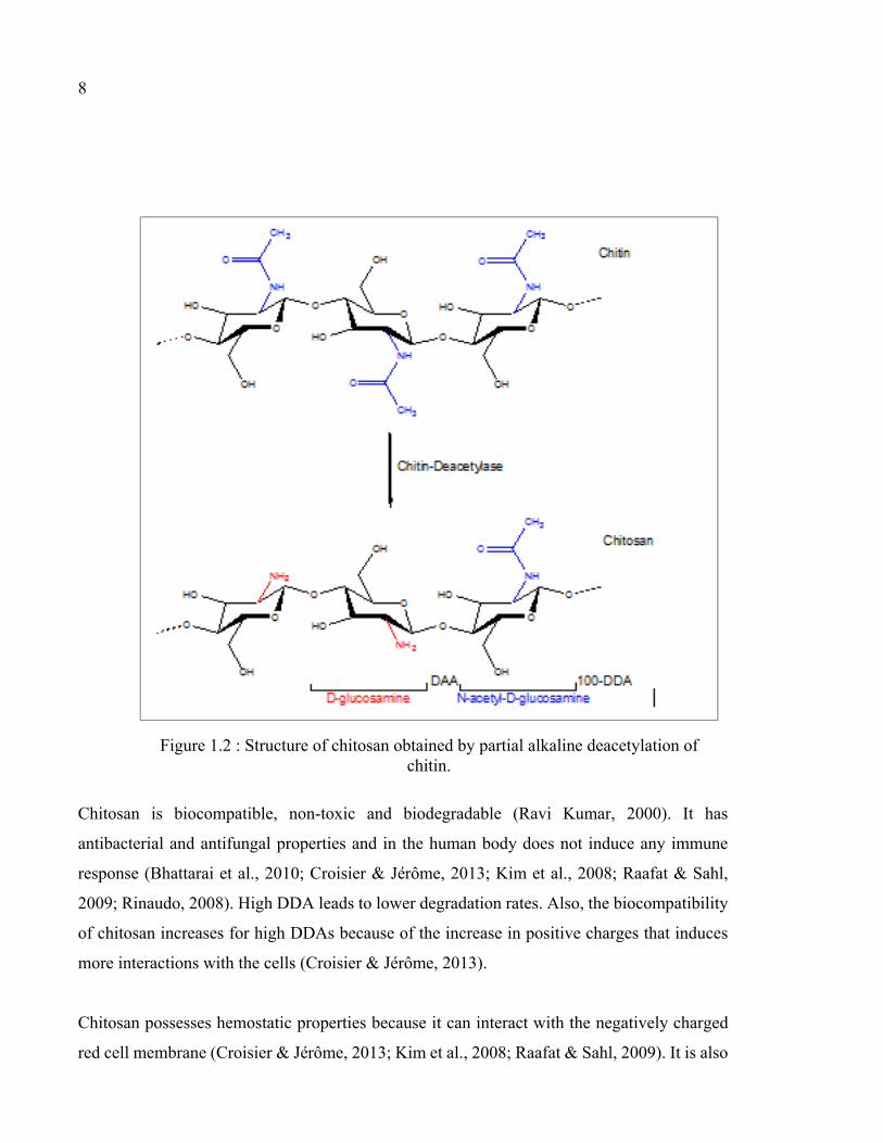
Chitosan is a natural linear polysaccharide composed of two randomly distributed repeating
monomer units of D-glucosamine and N-acetyl-D-glucosamine (acetylated unit) (Figure 1.2)

7
(Bhattarai, Gunn, & Zhang, 2010; Croisier & Jérôme, 2013).The main source of commercial
production of chitosan is deacetylation of chitin, the second most abundant natural polymer
after cellulose. The principal source of chitin is shellfish waste; however, it is widely found in
cell walls of fungi as well as exoskeletons of crustaceans, insects and spiders (Chenite et al.,
2000; Ravi Kumar, 2000). In deacetylation process, strong alkali solutions are used to remove
N-acetyl groups of chitin and form chitosan. The degree of deacetylation (DDA) indicates the
percentage of the deacetylated D-glucosamine units in the chain. Conversely, the degree of
acetylation (DA) is the percentage of acetylated units relative to the total units. The product is
considered as chitosan when the DDA is greater than 50% (Bhattarai et al., 2010; Croisier &
Jérôme, 2013).
The DDA and the molecular weight (MW) are factors that significantly influence chitosan
properties such as the polymer’s solubility, its viscosity, its gelling process and its degradation
kinetics (Berger et al., 2004). For instance, Ganji et al. showed that the gelation time of
hydrogels formed with highly deacetylated chitosan (DDA=98.3%) is less than the gelation
time of chitosan with less DDA (DDA=82.5 %) (Ganji, Abdekhodaie, & Ramazani, 2007).
The molecular weight (indicative of the length of the macromolecular chains) of chitosan
varies between 50 and 2000 kDa (Chenite, Buschmann, Wang, Chaput, & Kandani, 2001) and
directly influences the viscosity of the chitosan solution and therefore the mechanical
properties of the hydrogel made with the chitosan solution.
8
Chitosan is biocompatible, non-toxic and biodegradable (Ravi Kumar, 2000). It has
antibacterial and antifungal properties and in the human body does not induce any immune
response (Bhattarai et al., 2010; Croisier & Jérôme, 2013; Kim et al., 2008; Raafat & Sahl,
2009; Rinaudo, 2008). High DDA leads to lower degradation rates. Also, the biocompatibility
of chitosan increases for high DDAs because of the increase in positive charges that induces
more interactions with the cells (Croisier & Jérôme, 2013).
Chitosan possesses hemostatic properties because it can interact with the negatively charged
red cell membrane (Croisier & Jérôme, 2013; Kim et al., 2008; Raafat & Sahl, 2009). It is also
Figure 1.2 : Structure of chitosan obtained by partial alkaline deacetylation of chitin.

9
mucoadhesive because its positively charged amine groups can interact with mucin, which is
a negatively charged glycoprotein present in the mucus. The higher DDA leads to the better
mucoadhesive properties because of the higher number of positive charges (Bhattarai et al.,
2010; Croisier & Jérôme, 2013; Kim et al., 2008; Raafat & Sahl, 2009; Zhou, Jiang, Cao, Li,
& Chen, 2015).
1.2.2 Chitosan hydrogels
Chitosan has a pKa of ~ 6.5. It is soluble only in an acid medium. The primary amines (-NH2)
in its structure get protonated (-NH3 +) under acidic conditions, making chitosan a cationic
polymer (Chenite et al., 2001). When the molecule is sufficiently ionized, the generation of
repulsive electrostatic forces between the charged groups ensures the solubilization of the
chitosan. However, a change in pH or ionic strength may disturb this balance and induce
deionization and precipitation of chitosan. In other words, when chitosan solution pH is
elevated above 6, the repulsive electrostatic forces between the polymer chains are weakened
due to the neutralization of amine groups. Meanwhile, attractive hydrophobic interaction and
hydrogen bonds dominate, leading to chitosan precipitation (Chenite et al., 2001; Croisier &
Jérôme, 2013; Lavertu, Filion, & Buschmann, 2008; Rinaudo, 2008).
As mentioned in section 1.1.2.3, an interesting characteristic of chitosan is its ability to form
thermosensitive gels, which are liquid state at room temperature and form solid gels at
physiological temperature (37 °C.). This makes it possible to carry out an injection at room
temperature in liquid form and mix the solution with cells and / or drugs, prior to in situ
gelation.
Several methods can be used to make this chitosan solution a rigid gel. For example, the
addition of a mild base, such as β-glycerophosphate (BGP), makes it possible to produce a
thermosensitive chitosan solution. BGP has hydroxyl groups -OH which act as a buffer and
stabilize the chitosan solution. Moreover, BGP is negatively charged and is therefore attracted
by the -NH3 + groups of chitosan (Figure 1.3) (Chenite et al., 2001). The -OH groups of the
BGP control the hydrogen bonds between the chitosan molecules and make it possible to keep

10
the polymer in solution even at a pH above 6.5 (Chenite et al., 2001; Chenite et al., 2000;
Coutu, Fatimi, Berrahmoune, Soulez & Lerouge, 2013). When the temperature increases, the
transfer of the protons (H+) from the chitosan to BGP lead to neutralizing chitosan. The
attracting forces become stronger than the repulsive forces between the chains, which leads to
the creation of a physical gel (Lavertu et al., 2008).
Figure 1.3 Chemical structure of BGP
1.2.3 Previous work in LBeV on chitosan hydrogels
Chitosan hydrogels possess interesting properties for their use in the biomedical field. They
are natural and biocompatible, they have an interconnected porous structure that allows cells
survival and nutrient and waste transfer (Kim et al., 2008). They are biodegradable
(enzymatically or chemically), which ensures natural tissue healing without preserving a
permanent material (Bhattarai et al., 2010; Croisier & Jérôme, 2013; Kim et al., 2008; Raafat
& Sahl, 2009; Rinaudo, 2008).
The low mechanical properties of the chitosan / BGP hydrogels are their main limit. Assaad et
al. have shown that whatever the BGP concentration, the secant modulus of the chitosan / BGP
hydrogels does not exceed 10 kPa. (Assaad, Maire & Lerouge, 2015). Moreover, the use of
high concentrations of BGP required for rapid gelation decreases the biocompatibility of the
gel, due to an increase in the osmolarity of the gel which can cause death of the encapsulated
cells (Ahmadi & de Bruijn, 2008; Monette, Ceccaldi, Assaad, Lerouge & Lapointe, 2016; Riva

11
et al., 2011; Zhou et al., 2015). For these two reasons, the LBeV team has developed novel
gelling agents which offer better biocompatibility, higher mechanical resistance and a suitable
gelling rate. The team has shown that the combination of sodium bicarbonate (Sodium
Hydrogen Carbonate, SHC) with a phosphate buffer (Phosphate Buffer, PB) or BGP
significantly improves the mechanical properties and accelerates gelation at body temperature
(Assaad et al., 2015; Ceccaldi et al., 2017). In addition, in vitro cytocompatibility tests could
demonstrate better biocompatibility of these new gels due to a decrease in salt concentration.
Overall, these hydrogels present several decisive advantages. They are easy to prepare by
simply mixing two solutions. These hydrogels are stable at room temperature and rapidly gel
at 37 °C. They have superior mechanical properties to most hydrogels based on chitosan.
Patent filing has been done to protect these hydrogels which raise great interest for cell therapy
and tissue engineering application. However, for possible clinical transfer, the stability of the
solutions is of great importance. Indeed, chitosan dissolution in acid is a time consuming
process that requires several hours. So, it would be practical to prepare chitosan solutions in
bulk and store them for further use, especially for commercial applications.
However, during storage, specific characteristics of chitosan may be altered. Irreversible loss
of physicochemical properties of chitosan may happen due to the hydrolysis of chitosan and
gradual chain degradation, as it occurs after dissolution and storage at various conditions in
dilute organic acids (No et al., 2006, Nguyen et al., 2008). In particular, possible changes in
viscosity of chitosan solution must be monitored since it may influence other functional
properties of the chitosan solution.
Different internal and external factors can affect the stability of chitosan-based products.
Degree of deacetylation and the pattern of deacetylation, molecular weight, purity, and
moisture level are internal factors and environmental storage conditions, thermal processing,
sterilization, and processing (involving acidic dissolution, type of acid and chitosan
concentration in acidic solution) are external ones (Szymańska & Winnicka, 2015).

12
Overall, it has become a great challenge to establish sufficient shelf-life for chitosan
formulations and the purpose of the stability test is to provide reliable evidence on how the
quality of the chitosan solution may differs upon storage conditions.
It is also important to study the stability of these solutions since short term changes could
explain variability in the results obtained by the different team members.
In addition, one limitation of these hydrogels is their poor mucoadhesive properties, as will be
described in next sections.
1.3 Tissue-adhesion
1.3.1 Definition and application
The term "tissue adhesion" explains the adhesion capability of some natural, biological and
also synthetic materials to biological tissues (Ferreira, Gil & Alves, 2013; Khanlari & Dubé,
2013). The term mucoadhesion is used if the tissue is a mucosal surface (Huang, Leobandung,
Foss, & Peppas, 2000). Tissue-adhesive materials have been applied in different fields, such as
wound closure, and more recently for drug delivery systems. They are so popular for wound
closuring. Comparing to the traditional suture method, the tissue adhesives decrease foreign
body reaction during wound healing. Also, their use is easier and less painful, and there is no
need for removal. Besides, for cosmetic reasons in many cases the adhesive materials are
preferred to traditional methods such as suture. (Tajirian & Goldberg, 2010). (Delibegović,
Iljazović, Katica, & Koluh, 2011; Spotnitz & Burks, 2010).
Nowadays, tissue adhesives are also being used for oral, buccal and rectal (Sosnik, das Neves,
& Sarmento, 2014; Spicer & Mikos, 2010; Vakalopoulos et al., 2013), as well as ocular, nasal,
gingival, and vaginal drug delivery systems (Caló & Khutoryanskiy, 2015).

13
Tissue-adhesive materials have also been proposed for drug delivery to increase the retention time
of the drug at the target site. Otherwise the drug may not have enough time to act on the disease
before being eliminated. For example, the bowel movements can accelerate the elimination of a
rectal drug delivery. In the gastrointestinal tract the ingestion of food and drink may shorten the
retention of an oral drug delivery system. In these cases, tissue adhesive drug delivery systems can
increase the drug retention time, with advantages such as higher drug efficacy (Bernkop-Schnürch,
2005), and reduction of the administrated dose. Moreover, tissue adhesive drug delivery systems
make it possible to release the drugs only in specific targeted sites and avoid adverse effects
(Duchěne, Touchard, & Peppas, 1988), (Nikolaos A. Peppas & Sahlin, 1996). The same
principle could apply to biomaterials for cell therapy in order to increase cell retention on
targeted site.
1.3.2 Mechanism of tissue adhesion
The mechanism of tissue adhesion in general and mucoadhesion specifically, is complex and
not completely elucidated yet. There are different interactions between mucoadhesive
materials and mucin, such as: covalent, ionic, hydrogen bonds, van der Waals, and
hydrophobic interactions (Smart, 2005). The mucoadhesion strength can be affected by the
molecular weight of the polymer, the flexibility of the polymer chains, environmental pH,
charge, and functional groups in the polymer (Khutoryanskiy, 2011), (Smart, 2005).
Based on these interactions, various theories have been proposed to explain the mechanism of
mucoadhesion, including diffusion, wetting, electronic, adsorption and fracture mechanism
(Woertz, Preis, Breitkreutz & Kleinebudde, 2013). The diffusion theory is based on the
diffusion of a polymer into the mucin layer. It is dependent on the concentration gradient and
the diffusion coefficient of the polymer. Voiutskii suggested that mucoadhesion is due to the
semi-permanent adhesive bond formed by inter diffusion between the polymer chains of the
mucoadhesive materials and mucin (Voiutskii, 1963).

14
Peppas and Buri developed the wetting theory based on the spreading of a material, mostly
mucoadhesive liquids or low viscous formulations on the biological tissue. The degree of
spreading can be calculated by an extension of the basic Young’s equation. Better spreading
(i.e. low surface tension) induces better mucoadhesion ( Peppas & Buri, 1985).
The electronic theory explains the electron transfer between adhesive polymer and mucus due
to differences in electronic charge. This mechanism includes the formation of a double layer
due to interactions between the polymer and the mucus layer.
The adsorption theory describes the adhesion caused by primary (ionic, covalent and metallic)
and secondary bonds (van der Waals forces, hydrophobic interactions and hydrogen bonding).
The fracture mechanism is concerned with the strength of the adhesive bond between
mucoadhesive formulation and mucosa and the force which is needed to break this adhesive
bond. Young’s modulus of elasticity, fracture energy and critical crack length upon separation
of two surfaces can be used to calculate the fracture strength (Woertz et al., 2013).
Still, no single theory can fully explain the complex mechanism of mucoadhesion
(Khutoryanskiy, 2011; Smart, 2005). Some researchers used a combination theory to explain
this complicated phenomenon. For instance, a 3-step theory is proposed by Smart: The
mucoadhesives wet and start to swell. Then, they come in contact with mucus and form non-
covalent bonds at the interface. Finally, mucoadhesive polymer chains and mucin chains
interpenetrate each other, and develop further entanglements (Smart, 2005).
1.3.3 Mucoadhesive property of chitosan
There are different types of mucoadhesive materials, including cationic polymers (ex:
polylysine), anionic polymers (ex: Alginate), non-ionic polymers (ex: poly ethylene oxide (PEO)
and PVA), and amphoteric polymers (ex: Gelatin). Cationic polymers such as chitosan can form
electrostatic interactions with the negatively charged mucin at physiological pH, so they present

15
some mucoadhesive properties (Bernkop-Schnürch, 2005; Boddupalli, Mohammed, Nath, & Banji,
2010; Lehr, Bouwstra, Schacht & Junginger, 1992). However, these mucoadhesive properties
remain limited and various efforts have been done to enhance the mucoadhesion of chitosan.
Functionalization of chitosan with catechol groups is one of the most promising approaches and
will be described in detail later.
1.4 Mussel-inspired mucoadhesion
1.4.1 Introduction to marine mussel adhesion
The tissue adhesive should adhere to a wet or moisture surface at approximately body
temperature. The strong underwater adhesion of blue marine mussels (Mytilus edulis) therefore
attracted the attention of material scientists. These mussels stick to many surfaces under the
sea, such as rocks and boats, thus avoiding being removed by the waves. Mussels can adhere
to many different surfaces: organic and inorganic, hydrophilic and hydrophobic, smooth or
rough, and even the inert Teflon (G Silverman & Roberto, 2007).
Figure 1.4 illustrates adhesion of Mytilus edulis to seaweed, other mussels, and a stainless-
steel surface.

16
Figure 1.4 Mytilus edulis attachment to (a) seaweed, (b) other mussels, and
(c) a stainless-steel surface (G Silverman & Roberto, 2007)
To adhere under water, mussels secrete proteins called Mytilus edulis foot proteins (Mefps).
Mefps can rapidly solidify in the seawater and form the byssus. Figure 1.5 shows a schematic
of the Mytilus edulis mussel and byssus structures (G Silverman & Roberto, 2007).
The distal part of the byssus is called the byssal plaque. Mussels use the strong adhesion of
the byssal plaques to attach themselves to various solid surfaces.

17
Figure 1.5 Mytilus edulis mussel and byssus structure
(G Silverman & Roberto, 2007)
At least six Mefps have been identified. Mefp-1 is the key protein of the byssal cuticle while
Mefp-2 through 6 are found within the adhesive plaque. These proteins all share a common
unusual amino acid, 3,4-dihydroxyphenylalanine (DOPA). Figure 1.6 shows DOPA structure.
The DOPA content of Mefps ranges from a few percents to well above 20%. DOPA contains
catechol functional groups (3,4-dihydroxyphenyl), that was found to play a major role in
adhesion ( Lee, Dellatore, Miller & Messersmith, 2007; Waite & Qin, 2001).
Figure 1.6 The structure of DOPA
Catechol
group

18
This discovery inspired many researchers to develop novel catechol-containing adhesives.
1.4.2 Catechol chemistry
The catechol is capable of various catechol-catechol and catechol-surface interactions, leading
to the adhesive property of the catechol-containing materials. In addition, catechol is a unique
molecule capable of forming strong bonds to both inorganic and organic substrates while
utilizing either reversible physical or irreversible covalent crosslinks (H. Lee, Scherer, &
Messersmith, 2006). Understanding catechol chemistry is necessary to understand the
mechanisms of these processes, which are summarized in Figure 1.7.
The benzene ring of catechol can form π- π interaction with another benzyl moiety. This allows
the catechol-containing material to be able to bind to surfaces rich in aromatic compounds such
as polystyrene (Baty et al., 1997). The hydroxyl groups of catechol forms extensive hydrogen
bonds, which allows catechol to compete with water for hydrogen bonding sites and absorb
onto mucosal tissues (Chirdon, O'Brien & Robertson, 2003; Schnurrer & Lehr, 1996).
Catechol is also capable of forming strong complexes with metal ions (such as Fe3+, Ca2+, Cu2+,
Ti3+, Ti4+, Mn2+, Mn3+, Zn2+). Strong and reversible catecholate-metal ion complexation is
responsible for the wear resistance properties, high extensibility and elevated hardness of
mussel byssal cuticles (Holten-Andersen et al., 2009).
In the presence of oxidizing agent (i.e., IO4 -, H2O2, enzyme etc.), catechol is oxidized to its
quinone form. It can also auto-oxidize in a slightly basic aqueous solution (Schweigert,
Zehnder & Eggen, 2001; Yu et al., 2013). Quinone is highly reactive and can form covalent
crosslinks with various functional groups present on tissue surface through three main
pathways: self-crosslinking, involving coupling of two catechol molecules, Michael addition
with –SH or –NH2 group, and Schiff-base reaction with –NH2 (Deming, 1999; Lee, Dalsin &
Messersmith, 2006; Schweigert et al., 2001) (Figure 1.7).

19
Figure 1.7 Oxidative chemistry of catechol (Wu et al., 2011)
1.4.3 Catechol-containing hydrogels
Various biomaterials have been grafted with catechol groups to enhance their adhesive
properties or to form hydrogels.
Chitosan–catechol can be processed into a variety of physical states: films, hydrogels, sponges,
and micro/nanoparticles. As mentioned in the previous section, catechol can create crosslinks
with themselves or other functional groups. This catechol-induced crosslinks can be used to
make hydrogels or films. In one study, Oh et al. synthesized catechol modified hyaluronic acid
and lactose modified chitosan respectively. The mixture of these two polymers formed a re-
moldable hydrogel with interpenetrating network structure. Inter-molecular polyelectrolyte
complexes between the negatively charged hyaluronic acid and the positively charged
chitosan, and covalent bonds between oxidized catechol groups and –NH2 groups were two
types of crosslinks contributed to the interpenetrating network formation (Oh et al., 2012). Lee
et al. developed an alginate-catechol hydrogel that used catechol oxidation for crosslinking,
instead of the conventional calcium ionic crosslinking ( Lee et al., 2013). This catechol-
alginate hydrogel showed excellent biocompatibility, and tunable mechanical properties in

20
contrast to calcium crosslinked alginate hydrogel. In another study by Ryu et al., catechol
modified chitosan was crosslinked with thiolated Pluronic, forming a gel that was adhesive to
soft tissue ( Ryu et al., 2011). Although part of the catechol groups on the polymer chain
participated in crosslinking with –SH by Schiff-base addition, the remaining catechol groups
contributed to the enhancement of bioadhesion at tissue surface.
1.4.4 Previous work on chitosan-catechol hydrogels in Marta Cerruti’s lab
Marta Cerruti’s team at McGill University is another group who worked on chitosan-catechol
hydrogels. They developed three types of catechol-containing chitosan hydrogels as
mucoadhesive drug delivery systems for oral, buccal and rectal drug delivery.
First, they selected DOPA, hydrocaffeic acid (HCA), and dopamine (DA) as three different
catechol-containing compounds (Xu, Soliman, Barralet, & Cerruti, 2012). These three
compounds have the same ortho-dihydroxyphenyl backbone but different functional groups
(both carboxylate and amino group in DOPA, carboxylate group in HCA, and amino group in
DA.
The hydrogels were prepared simply by mixing different catechol compounds with CH and
their adhesion to rabbit intestine were tested. Based on the mucoadhesion result, HCA was
chosen as a catechol compound for further experiment. Besides, their study also demonstrated
that oxidation should be prevented before contact with mucus in order to retain enhanced
mucoadhesion. In the next step of the experiment, this group covalently bonded catechol
functional groups to the backbone of CH, and crosslinked the polymer with a non-toxic
chemical crosslinker, namely genipin (GP) (Xu, Strandman, Zhu, Barralet, & Cerruti, 2015).
Chitosan–catechol adhesives crosslinked by genipin was reported to remain in porcine mucosal
membranes even after 6 h (70% of chitosan–catechol remains), whereas unmodified chitosan
crosslinked by genipin lost contact within 1.5 h.
One unique feature of this hydrogel is the preserving the functionality of catechol groups,
which are responsible for the excellent mucoadhesion enhancement instead of sacrificing them

21
to build the crosslinking. Many studies formed catechol-containing hydrogels by adding
enzymes or oxidizing agents to trigger catechol crosslinking. Or, they added polymers
containing functional groups that could form covalent bonds with the catechols, such as –SH
groups. These strategies sacrificed catechols during the crosslinking, thus limiting their
capability of inducing mucoadhesion. Since GP only crosslinks the amino groups in chitosan,
using it as a crosslinker to form catechol-containing hydrogels, preserved the catechol groups
and contribute to the mucoadhesion enhancement to the greatest possible extent.
Despite the positive points of this study, these hydrogels have low mechanical properties and
slow gelation (about 12 h). This may be a strong limitation for certain applications.
1.5 Summary and objectives of this master
As summarized above, chitosan-based thermosensitive hydrogels are interesting injectable
materials for biomedical and pharmaceutical applications. In addition to low viscosity, rapid
gelation, high mechanical properties, tissue-adhesiveness, and cytocompatibility, these
materials should be storable on extended periods without losing their properties. So, the first
objective of this project is to study the stability of chitosan solution, gelling agents, and
chitosan hydrogel over time under different storage conditions.
Tissue-adhesion is very important for hydrogels which are used in drug delivery and cell
therapy system to extend the drug or cell retention at the site of application and prolonging its
therapeutic effects. Thus, the second objective of this project is to covalently graft catechol
groups on chitosan and study the effect of gel compounds, especially acid concentration, on
gel properties, as a first step towards the development of an injectable tissue-adhesive hydrogel
with good gelation time, good mechanical strength, and good compatibility with cells.


CHAPTER 2
MATERIALS AND METHODS
In this chapter, the preparation and characterization of both chitosan and catechol-chitosan
hydrogels are described.
2.1 Materials
For the two objectives of this project, two different sources of chitosan (CH) were used:
1) CH from Marinard Biotech (Rivière-au-Renard, QC, Canada) (Kitomer, Mw 250 kDa, DDA
94%), that will be named K-CH.
2) CH from Heppe Medical Chitosan (Germany) (HMC+, Mw 250-350kDa, DDA 95%), that
will be named H-CH.
Glycerol phosphate disodium salt penta hydrate C3H7Na2O6P·5H2O (BGP), sodium phosphate
monobasic NaH2PO4 (SPM), sodium phosphate dibasic Na2HPO4 (SPD), Hydrocaffeic acid
(HCA) (≥98%), and N-(3-Dimethylaminopropyl)-N’-ethylcarbodiimide hydrochloride (EDC)
(≥ 98.0%) were purchased from Sigma–Aldrich (Oakville, ON, Canada). Sodium hydrogen
carbonate NaHCO3 (SHC) was purchased from MP Biomedicals (Solon, OH, USA).
2.2 Synthesis of chitosan and chitosan-catechol hydrogels
Hydrogels were prepared in three main steps. First chitosan was purified. Then it was modified
(or not) by grafting catechol groups. Third, a CH (or CH-Cat) solution was mixed with a gelling
agent solution in order to create solutions gelifying around body temperature. These steps are
described in greater details below.
2.2.1 Purification of chitosan
In order to remove the impurities, the commercial powder was first purified as follows (Assaad
et al., 2015). Six (6) grams of CH was dissolved in 600 ml of 0.1 M hydrochloric acid (HCl)

24
solution and stirred overnight at 40° C. The next day, the solution was filtered under vacuum
to remove the insoluble particles. The solubilized CH precipitated with stirring by
incorporating 0.5 M sodium hydroxide (NaOH) until the pH reached between 8 and 9. Then
the mixture was heated to 95 °C with stirring and 6 ml of sodium dodecyl sulfate (SDS) 10%
(w / v) was added. The mixture was kept at 95 °C for 5 min, after which it was cooled to room
temperature. The pH was then adjusted to 10 by adding 0.5 M NaOH. The mixture was filtered
under vacuum and the precipitated CH was recovered and then washed five times in a beaker
containing 600 ml of Milli-Q water, previously heated to 40 ° C, in order to eliminate traces of
SDS. Finally, the CH was frozen overnight, freeze-dried for three days and then ground, sieved
and stored (Figure 2.1)
Figure 2.1 purified CH powder

25
2.2.2 Synthesis of chitosan-catechol (CH-Cat)
Modifying chitosan by catechol was achieved by grafting hydrocaffeic acid to the carbonated
chain of CH (Figure 2.2).
Figure 2.2 Grafting hydrocaffeic acid on chitosan by EDC coupling
As detailed below, the protocol of synthesis of CH-Cat was modified from the work previously
done by Professor Cerruti’s team at McGill University (Xu et al., 2015) .
The steps of this protocol (which will be named old protocol) were as follows:
A. Dissolve 0.6 gr chitosan in 60 ml deionized (DI) water and HCl (pH = 2.5)
B. Add HCA and EDC previously solvated in a water: ethanol 1: 1 mixture in stoichiometric
proportions (1: 0.5: 1.17 of glucosamine: HCA: EDC respectively). Adjust the pH between 5
and 5.5 using 1M NaOH.
C. Let the reaction take place for 12 hours under stirring.
D. Dialyses the solution by using a dialysis membrane tube (MWCO 5,000, Spectrum
Laboratories, USA) for three days against a solution of HCl pH 5.
E. Lyophilize the purified product and store it at -20 ° C.

26
By following the old protocol, the CH-Cat solution obtained after the twelve hours of reaction
exhibited a red color, becoming more intense and darkened during the dialysis stages. After
freeze-drying, the powder obtained formed a porous network, was pink in color. Figure 2.3
shows the solution (in step D) and the final product (after freeze drying) obtained from the old
protocol.
Figure 2.3 CH-Cat solution after dialysis (left) and after freeze-drying (right)
Once dissolved in DI water for gel preparation, CH-Cat formed a brown and dense solution
which was not permeable to light. Moreover, particles remained suspended in the liquid phase
(Figure 2.4)
Figure 2.4 CH-Cat solution

27
This difference between the appearance of the solution before and after lyophilization and
insolubility of CH-Cat in water led us to the hypothesis that catechol oxidation (Quinone)
occurred during the process, following the reaction described in Figure 2.5.
Figure 2.5 Catechol oxidation half-reaction
The solution had the same appearance when Cat-CH was dissolved in acidic media (pH 2, 4
and 6) (Figure 2.6). We first hypothesized that oxidation occurred during solubilisation after
lyophilisation. To control this phenomenon, several dissolution experiments were carried out
in different acidic media (pH 2, 4 and 6). The obtained solutions were more viscous and had
no suspended particles, but were still very brown in color (Figure 2.6). This means that the
oxidation phenomenon had taken place before solubilisation.
Figure 2.6 Dissolution tests of the CH-Cat in an acid medium with different pH
Therefore, various tests were carried out to determine ideal conditions to prevent catechol
oxidation during the steps B, C, and D of the old protocol. Temperature and pH of reaction
during all steps of process were the tested parameters.

28
At the end of the experiments, a protocol was found which enables to avoid catechol oxidation
and leads to a white powder. The changes in modified protocol, which will be named the new
protocol, comparing to the old one, are as follows:
B. Adjust the pH between 4.65- 4.80 using 1M NaOH.
C. Let the reaction take place for 12 hours under stirring in cold room.
D. The dialysis was done against HCl solution (pH 2.5-3) during the first 2 days (10 mM NaCl
solution with 15 mL of 1 N HCl for the first day and 10 mM NaCl solution with 5 mL of 1 N
HCl for the second day) following by dialysis against DI water for 6 h at the last day. Dialysis
solution should be changed at least 4 times in first and second day.
The complete protocol can be found in ANNEX I.
Figure 2.7 shows the solution before and after dialysis and the final product of the new
protocol. All further experiments were done using this optimized protocol.
Figure 2.7 CH-Cat (A) solution before dialysis, (B) after dialysis, and (C) the final product
A B C

29
2.2.3 Gelling agents
For this project, different gelling agents were used, as described in Table 2.1. They were
prepared using phosphate buffer (PB), BGP and SHC. The PB, at a pH of 8, was prepared by
dissolving SPM and SPD salts at molar ratio of 0.073 in Milli-Q water. The SHC and BGP
solutions were prepared by dissolving their salts in Milli-Q water. To prepare SHC-PB and
SHC-BGP solutions, the SHC salt was solubilized in PB and BGP solutions respectively.
2.2.4 Preparation of chitosan (CH) and chitosan-catechol (CH-cat) hydrogels
To prepare the CH (CH-Cat) physical hydrogels, the gelling agent solution was mixed with a
CH or CH-Cat solution prepared as following:
CH hydrogels: The purified CH powder was solubilised in HCl (0.1 M for K-CH and 0.12 M
for the H-CH) at 3.33% (w / v) with intensive stirring for about 3 h. The resulting solution was
sterilized by autoclaving (20 min, 121°C) and then stored at 4 °C (Figure 2.8).
CH-Cat hydrogel: CH-Cat powder was solubilised in DI water at 3.33% (w / v) with intensive
stirring for about 3 h. CH-Cat solution was used freshly to make hydrogel. To study the
influence of the pH on gel properties, CH-Cat was also prepared in aqueous solution containing
various HCl concentrations (from 0 to 0.09 M).

30
Figure 2.8 solution of purified CH before sterilization
(left) and after sterilization (right)
The CH (CH-Cat) hydrogels were prepared at room temperature by mixing one of the gelling
agents with the CH (CH-Cat) solution, by using two syringes and a luer-lock connector (Figure
2.9), at a volume ratio of 0.4: 0.6 respectively.
Figure 2.9 Method of mixing the gelling agent with CH (CH-Cat)

31
All hydrogels contain 2% w/v of CH (Assaad et al., 2015) or CH-Cat. The hydrogels names
express their composition. In addition, the gelling agent names express their final concentration
in hydrogels (SHC0075-PB004 gelling agent solution means in fact the initial concentrations
are 0.19M SHC and 0.1M PB). For example, CH/ SHC0075-PB004 represents a hydrogel
containing 2% (w/v) CH, 0.075 M SHC and 0.04 M PB (Table 2.1).
Table 2.1 Abbreviations and composition of the different hydrogels tested
Sample name Sample composition Initial concentration (M)
PB BGP SHC
Final concentration (M)
PB BGP SHC
CH/ SHC0075-PB004 Chitosan mixed with
(PB + SHC) solutions.
0.1 _ 0.19 0.04 _ 0.075
CH/ SHC0075-PB008 0.2 _ 0.19 0.08 _ 0.075
CH/ SHC0075-BGP001 Chitosan mixed with
(BGP + SHC) solution.
_ 0.025 0.19 _ 0.01 0.075
CH/ BGP04 Chitosan mixed with
BGP solution.
_ 1 _ _ 0.4 _
CH-Cat/ SHC009 Chitosan-Catechol mixed
with SHC solution _ _ 0.225 _ _ 0.09
CH-Cat/ SHC009-PB002 Chitosan-Catechol mixed
with (PB + SHC) solutions. 0.05 _ 0.225 0.02 _ 0.09
CH-Cat/ SHC009-PB004 0.1 _ 0.225 0.04 _ 0.09
CH-Cat/ SHC009-PB008 0.2 _ 0.225 0.08 _ 0.09
2.2.5 Storage CH solution and gelling agents to test the stability over the time
To test the stability of chitosan solution, gelling agents (GA) over time under different storage
conditions and their effect on hydrogel properties, they were stored in two conditions: room
temperature (RT) and refrigerator temperature (FT) (4 to 5 °C). To avoid variability between
chitosan batches, one single batch of chitosan was prepared at Day 0 for all these tests. The
volume of chitosan that was needed for each rheometry test was stored in 3 ml syringes. In
addition, pH measurement was done on one 10 mL sample (which was stored in closed cap
glass bottle) at different time points. Also, the gelling agents that used to mix with chitosan

32
and make hydrogels, were from the same batch that were prepared at day 0 and stored in closed
cap tubes. To test the pH of gelling agents, three different batches of each GA were prepared
so the results of pH measurement of GA come from independent samples (N = 3).
2.3 Characterization of chitosan-catechol
Both nuclear magnetic resonance (NMR) spectroscopy and UV-Vis spectrometry were used to
confirm catechol grafting to chitosan and characterize the degree of conjugation.
2.3.1 NMR
Conjugation of catechol functional groups onto chitosan backbones was confirmed by Nuclear
magnetic resonance (NMR) spectroscopy.
NMR is a technique used to analyze the structure of many chemical molecules, primarily
organic compounds. A typical compound might consist of carbon, hydrogen and oxygen atoms.
The principle of NMR comes from the spin of nucleus. Nuclear spins generate magnetic field
without applied an external magnetic field. The nuclear spins are random in directions. When
an external magnetic field is present the nuclei align themselves either with or against the
external magnet. In this case, an energy transfer is possible between the base energy to a higher
energy level. The energy transfer takes place at a wavelength that corresponds to radio
frequencies and when the spin returns to its base level, energy is emitted at the same frequency.
The signal that matches this transfer is measured in many ways and processed to yield an NMR
spectrum for the nucleus concerned (Figure 2.10).
In its simplest form, an NMR experiment consists of three steps:
1. Place the sample in a static magnetic field.
2. Excite nuclei in the sample with a radio frequency pulse.
3. Measure the frequency of the signals emitted by the sample.

33
From the emitted frequencies, analysts can deduce information about the bonding and
arrangement of the atoms in the sample. 1H and 13C are two of the most widely used NMR
nuclei (Edwards, 2009).
Figure 2.10 The nuclear magnetic resonance (NMR) phenomenon
Taken from utu.fi
NMR signals are usually plotted as spectra and analyzed with respect to two features,
frequency and intensity. It is conventional in NMR to plot frequency on the horizontal axis
and increasing towards the left. Absolute frequencies are measured in Hertz or Megahertz
(MHz). Reporting on measured signals is simplified if all frequency measurements are made
with respect to a reference. The recommended reference is a chemical called tetramethylsilane
(TMS). When a 1H or a 13C spectrum is acquired the presence of TMS gives rise to a single,
easily identifiable peak. This peak is referenced to zero and the frequencies of all other peaks
are given in terms of their frequency relative to the TMS frequency. However, this can be even
more simplified if the ppm unit is used instead of Hertz. The ppm unit represents frequencies
as a fraction of the absolute resonance frequency which will depend on the strength of the
magnet. The advantage of the ppm unit is that frequency measurements are independent of
magnet strength. This greatly simplifies the comparison of spectra acquired on different
spectrometers (Edwards, 2009).

34
In this particular study, 13C CP-MAS NMR spectra were obtained at 100 MHz using a 7.5 mm
rotor spinning at 5 kHz, a 1.5 ms contact time and recycle time of 2 s (Agilent/Varian VNMRS-
400, USA).
2.3.2 UV-Vis
A UV-Vis spectrometer (Carry 5000, USA) was used to determine the degree of conjugation
of catechol to the amine groups of chitosan. This quantitative analysis is the most widely used
technique as a simple and powerful method for measuring catechol conjugation rates ( Ryu,
Hong & Lee, 2015).
The maximum absorption of catechol occurs at a wavelength of 280 nm, while chitosan does
not absorb at this wavelength. Therefore, absorbance at 280 nm was used to quantify the
degree of catechol conjugation. To that purpose, the absorbance of a solution of 5 mg of CH-
Cat dissolved in 10 ml of DI water was compared to a standard curve established with
hydrocaffeic acid (HCA) at five different concentrations (0.1 to 0.5 mmol/L).
To calculate the degree of conjugation of catechol to the amine groups of chitosan (Percentage
of Cat in CH-Cat sample) the degree of deacetylation of chitosan (DDA, 95% in this study),
should be considered as well as the molecular weight of three different monomers in CH-Cat
structure (Figure 2.11), namely the deacetylated section (x), the conjugated section by catechol
(y) and acetylated section (z). The molecular weight of x, y, and z is 161, 325, and 203
respectively.

35
Figure 2.11 CH-Cat structure
By using the following equation, the catechol conjugation rates in sample could be calculated:
5 * 10 -3 = M * 10 -3 * 10 *10 -3 * 325 + [M * (0.95-X) / X] * 10 -3 * 10 * 10 -3 * 161 + [0.05 *
M / X] * 10 -3 * 10 * 10 -3 * 203 (2.1)
Where
X= ratio between conjugated and unconjugated parts of chitosan to catechol (catechol
conjugation rate)
M= the concentration of catechol (mmol / ml) deduced from the standard curve
Then:
X = (0.1631 * M) / (0.5 – 0.164 * M) (2.2)
2.4 Mechanical characterization
2.4.1 Rheological study
The rheological properties of the various formulations of chitosan and chitosan-catechol
hydrogels were evaluated as a first estimate of their mechanical properties, but above all to
study their gelation kinetics. Rapid gelation is one of the important properties for an injectable

36
hydrogel. The gel should remain liquid and stable for storage, preparation and injection at room
temperature, and should rapidly gel upon reaching body temperature in situ to prevent its
migration to undesirable areas.
An Anton Paar instrument (Physica MCR 301, Germany) equiped with coaxial cylinder
geometry (CC10/T200) and connected to a circulating water bath (Julabo AWC100, Germany)
was used to measure the storage modulus (G') and loss modulus (G'') as a function of time. The
storage modulus measures the stored energy (elastic portion) and the loss modulus measures
the energy lost as heat (viscous portion). Immediately after mixing the two solutions (0.6 ml
of gelling agent and 0.9 ml of the chitosan/chitosan-catechol solution), the gel was injected
into the cell and the storage modulus (G'), loss modulus (G'') as well as the complex viscosity
(η) were measured in the linear viscoelastic range (LVR), at a constant shear stress (1 Pa) and
a constant frequency (1 Hz).
It should be mention that to calculate the complex shear modulus (G*) in rheometry test, it is
enough to pre-set τ (motor torque) or γ (deflection angle) and under this pre-set measure the
other value and using the equation: G*= τ /γ, shear modulus will be reached. If we imagine G*
as a vector and determine its angle with X axis (δ, loss or damping factor), then we will have
G' and G''
tan δ = G'' / G' (2.3)
if G' = G'' then tan δ = 1, and it is sol-gel transition point (phase transition) in a material.
tan δ < 1 shows that material is more elastic and tan δ > 1 shows that material is more viscous.
Complex viscosity (η) could be calculated by dividing G* by angular Frequency (Mezger,
2006).
The measurements were carried out at 37 °C (body temperature). Each test was repeated three
times.

37
2.4.2 Compression tests
To evaluate the hydrogels’ strength after 48 h gelation at 37 °C, axial unconfined compression
tests were performed at room temperature using the Anton Paar (Physica MCR 301, Germany)
equipped with parallel geometries (P25 / P2). A volume of 2 mL different formulation of CH-
Cat hydrogels was prepared (n = 3) in small cylindrical molds (14 mm diameter). After 48 h
gelation at 37°C in an incubator, the sample was gently removed from the mold and placed on
the planar geometry. The height of the sample (h) was used to calculate the speed (v) for
applying up to 50% compression at a rate of 100% deformation per minute:
V= h/60 mm.s-1 (2.4)
Load/displacement curves were used to calculate the secant elastic modulus, considered as the
slope of a line connecting the point of zero strain to a point at a specified deformation.
2.5 Physico-chemical characterization
2.5.1 pH study
The pH measurements were carried out at room temperature using a Denver Instrument Ultra
Basic pH meter. For the chitosan stability project, the pH of gelling agents as well as the
chitosan solution were measured after different storage times at 4 °C (in cold room) and room
temperature. For the samples stored in cold room, samples were left to warm up at room
temperature for about 1 h prior to pH measurement.
For the tissue adhesive project, the pH was measured twice. The first time, the pH of mixtures
(CH-Cat + gelling agent) immediately after mixing; the second time, the pH of hydrogel after
putting it for 24h at 37 °C.

38
2.5.2 Osmolality
The Advanced Micro Osmometer (Model 3300, Advanced Instruments), was used to measure
gel osmolality. Different formulations of CH-Cat hydrogels were kept at 37 °C for 24h, then
20 μl of filtrated gel was used for osmolality measurement. It is important to specify that the
osmolality is measured here (in mOsm / kg of solvent) and not the osmolarity (in mOsm / L of
solution).
2.6 Tissue adhesive tests
To evaluate the mucoadhesive properties of CH-Cat hydrogels after and during gelation, wash
off and tensile tests have been done respectively. Fresh tissue (sheep intestine) was obtained
from animal facility of the Centre de recherche du Centre hospitalier de l’Université de
Montréal (CRCHUM). It should be mentioned that these tissues were obtain from animals
sacrificed in animal facility for other experiments.
2.6.1 Tissue adhesive wash off test
Wash off tests were performed as a first assessment of adhesive properties of the hydrogels
after gelation (Xu et al., 2015). The hydrogels were left to gel in cylindrical mold for 48h at 37 °C. Fresh tissue (sheep intestine) was glued to a microscope glass slide and placed into a beaker
vertically. The hydrogels were removed from the mold and gently pressed to adhere on the
tissue surface for 30 seconds. Both the tissue and the hydrogels were then immersed in 30 ml
PBS at 37° C ( Figure 2.12). A magnetic bar was used to generate flow. The adhesive property
of hydrogels after gelation was evaluated by recording the number of remaining hydrogels
adhered to tissue for 27 hours. Five samples from each hydrogel group were tested.
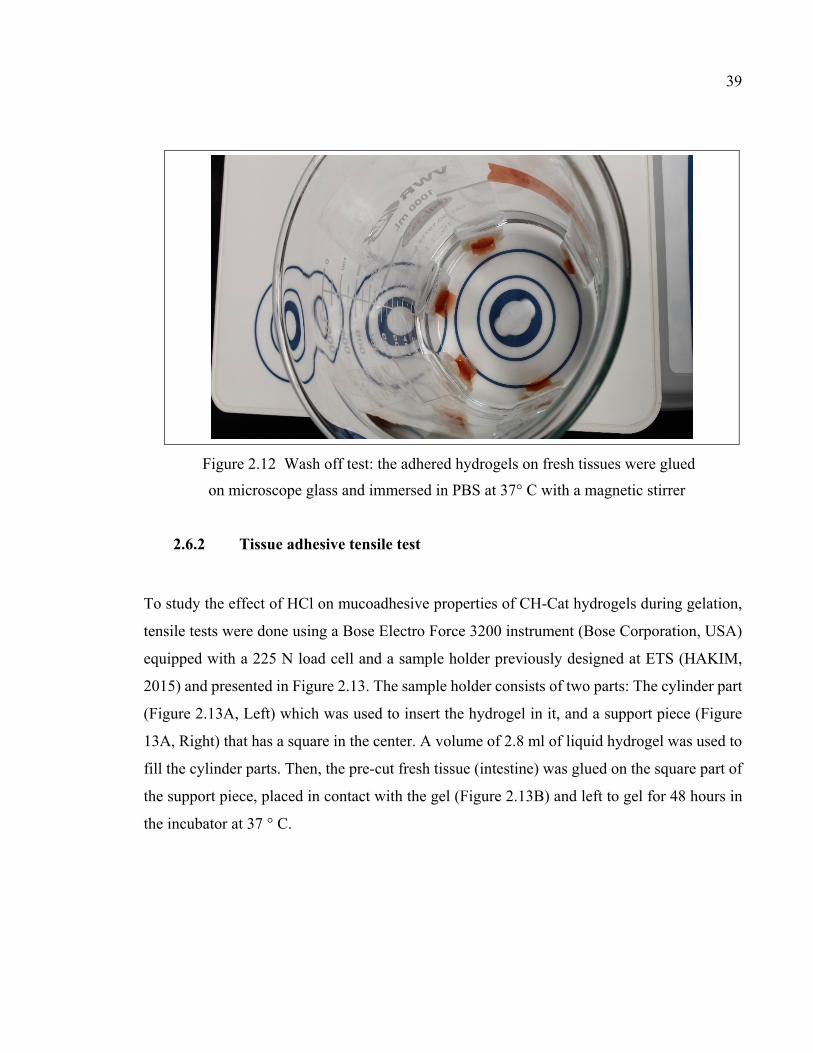
39
Figure 2.12 Wash off test: the adhered hydrogels on fresh tissues were glued
on microscope glass and immersed in PBS at 37° C with a magnetic stirrer
2.6.2 Tissue adhesive tensile test
To study the effect of HCl on mucoadhesive properties of CH-Cat hydrogels during gelation,
tensile tests were done using a Bose Electro Force 3200 instrument (Bose Corporation, USA)
equipped with a 225 N load cell and a sample holder previously designed at ETS (HAKIM,
2015) and presented in Figure 2.13. The sample holder consists of two parts: The cylinder part
(Figure 2.13A, Left) which was used to insert the hydrogel in it, and a support piece (Figure
13A, Right) that has a square in the center. A volume of 2.8 ml of liquid hydrogel was used to
fill the cylinder parts. Then, the pre-cut fresh tissue (intestine) was glued on the square part of
the support piece, placed in contact with the gel (Figure 2.13B) and left to gel for 48 hours in
the incubator at 37 ° C.

40
Figure 2.13 Sample holder for tissue adhesive tensile test
After that time, the whole sample holder was installed on the Bose instrument (Figure 2.14)
and a tensile test was done to determine the maximum detachment force (MDF) between the
tissue and the hydrogels.
Figure 2.14 Tensile test to determine the maximum detachment
force (MDF) between the tissue and the hydrogels
A B

41
2.7 Statistical analysis
All the results are expressed as mean ± standard deviation (SD). Statistical analysis was done
using Statgraphics software (Statgraphic centurion, statpoint technologies Inc., USA). To
determine if a statistical difference between means existed, one-way ANOVA was used when
comparing more than two groups followed by Fisher's least significant difference (LSD)
procedure with a confidence level of 95%. For comparing two conditions, independent t-test
with equal variances was used. A p-value lower than 0.05 was considered significant for all
tests. The number of independent experiments (N), and the total number of samples used for
each independent experiment (n) are indicated in the figure captions.

CHAPTER 3
RESULTS
The results of the two objectives of this project are presented separately. Results of chitosan
(CH) stability tests will be first presented in section 3.1, and then results concerning the
development of a tissue adhesive gel will be presented in section 3.2.
3.1 The stability of Chitosan solution, gelling agents, and hydrogels
The stability of chitosan solution, gelling agents (GA) over time under different storage
conditions and their effect on hydrogel properties were tested by measuring pH, viscosity, and
gelation time in different time points.
3.1.1 The stability of chitosan solution
The stability of chitosan solution (3.33 % chitosan in HCl 0.1M) was tested by measuring its
viscosity and pH. Chitosan solutions were stored in two conditions: room temperature (RT)
and fridge temperature (FT) (4 - 5 °C). At different time points (1, 2, 4, 13, 26, and 52 weeks
after preparation day (Day 0) the pH and complex viscosity were measured.
Figure 3.1 shows that the viscosity of chitosan solution remains relatively stable from Day 0
until 13 weeks, without difference between samples stored in refrigerator and those stored at
room temperature. However, after 1 year, viscosity is significantly higher than day 0 (p<0.05).
Moreover, standard deviations are higher for samples stored at room temperature.
Figure 3.2 shows the pH of the chitosan solution. The pH remained quite stable around 6.3,
with almost no difference between storage chitosan solution in fridge or room temperature
until 26 weeks. The small variations between the time points may be attributed to the precision
of the measurement method.

44
Figure 3.1 Effect of storage condition (Room temperature (RT) and Fridge temperature (FT)) and time on the viscosity of CH solution. (mean ± SD, n = 3), (*p<0.05 comparing to
Day 0)
Figure 3.2 Effect of storage condition and time on the pH of CH solution
0
5
10
15
20
25
Day 0 RT1 FT1 RT2 FT2 RT4 FT4 RT13 FT13 RT26 FT26 RT52 FT52
Com
ple
x V
isco
sity
(P
a.S
)
Time (Week)
*
*
5,85,96,06,16,26,36,46,56,66,76,86,97,0
Day 0 RT 1 FT 1 RT 2 FT 2 RT 4 FT 4 RT 13 FT 13 RT 26 FT 26 RT 52 FT 52
pH
Time (Week)
*

45
3.1.2 The stability of gelling agents and hydrogels
Similarly, GAs (SHC0075-PB004, SHC0075-PB008, SHC0075-BGP001, and BGP04) were
stored in two conditions (room temperature (RT) and fridge temperature (RT)) and their pH
were measured at different time points. Besides, to evaluate the effect of chitosan and GA
changes with time on hydrogel properties, rheological tests have been done to monitor the
evolution of the storage modulus (G') and loss modulus (G'') during gel formation at 37 °C. It
should be mentioned that rheometry tests have been done only for the fridge-stored samples.
Prior to mixing and testing, the chitosan and GAs solution were left in the room for 2 hours to
reach the ambient temperature.
The time at which G'= G'' is considered as gelation time (tgel). Moreover, the rate of increase
of G ' is a good indicator of the gelation kinetics and of the mechanical shear properties and
hence the cohesion of the gel over time. It should be mentioned that in rheometry test, statistical
analysis was only done for G' values at 30 min.
SHC0075-PB004
Figure 3.3 and Figure 3.4 show that the pH of the GA solution used to prepare SHC0075-
PB004 hydrogels (SHC019M-PB01M), originally at 8.2, significantly increased (linearly after
12 weeks, R-squared= 0.98 ) over time (p< 0.05) comparing to Day 0. The pH of RT stored
GA increased more compared to those that were stored at FT, the difference being already
significant at week 2 (p< 0.05).

46
Figure 3.3 Effect of storage condition and time on the pH of SHC0075-PB004
Figure 3.4 Effect of storage condition and time on the pH of SHC0075-PB004. (mean ± SD, N = 3), (*, p<0.05 comparing to Day0), (**, p<0.05 comparing RT & FT)
Figure 3.5 presents the evolution of the storage (G’) and loss (G’’) modulus at 37°C as a
function of time for hydrogels prepared with solutions stored for 0 versus 52 weeks. It shows
7,07,27,47,67,88,08,28,48,68,89,09,29,49,69,8
10,0
Day 0 RT1 FT1 RT2 FT2 RT4 FT4 RT13 FT13 RT26 FT26 RT52 FT52
pH
Time (Week)
** *
* **
*
**
**
*** ** **
**
**
8,08,28,4
8,68,89,09,29,49,69,8
10,0
0 10 20 30 40 50 60
pH
Time (Week)
Room Temperature
Fridge Temperature

47
that the storage time did not affect the gelation time, tgel,is less than 15 s (time required to get
the first measurement) for both gels. However, the initial G’ value is lower when the gel was
prepared with freshly prepared solutions compared to stored GA.
Figure 3.6 summarized the G’ values at different timepoints (initial, after 3 and 30 min). It
shows that storage modulus changes initially. But at the end of test (30 min) it does not
significantly change (comparing to Day 0) for 52 weeks (P= 0.09).
Figure 3.5 Evolution of storage (G') and loss (G'') modulus for CH/SHC0075-PB004
hydrogels as a function of storage time (Week), for 30 min at 37 °C, (mean, n = 3)
0
500
1000
1500
2000
2500
3000
0 10 20 30 40
G`
and
G``
Mod
ulu
s (P
a)
Time (min)
Day 0 (G')
Day 0 (G")
52 Weeks (G`)
52 Weeks (G``)

48
Figure 3.6 Evolution of the storage modulus G' for different storage time (in Week) in
CH /SHC0075-PB004 hydrogels, at 37 °C, for 30 min (mean ± SD, n = 3)
SHC0075- PB008
Figure 3.7 shows that the pH of SHC0075-PB008 solution significantly increased over time
compared to Day 0 (p< 0.05). Besides, there is a significant difference between GA solutions
stored in fridge or room temperature, the pH of RT stored GA increasing more than those
stored at FT (p< 0.05). The pH evolution by time graph could be found in ANNEX II.
The storage time significantly affected the gelation kinetics, as showed by Figure 3.8 and
Figure 3.9, even if tgel is less than 15 seconds at 37 °C for all storage times.
Figure 3.9 indicates the G’ values at different time points. It shows that the storage modulus
after 30 min gelation is significantly decreased, when the storage time prior mixing is longer
(p< 0.05). More importantly, after long time storage, G’ does not increase with time at 37 °C,
as shown by the straight line observed during rheometry test in 30 minutes (Figure 3.8).
0
500
1000
1500
2000
2500
3000
3500
4000
4500
Day 0 1 W 2 W 4 W 13 W 26 W 52 W
Sto
rage
Mod
ulu
s, G
` (P
a)
Time (Week)
0 min
3 min
30 min

49
Figure 3.7 Effect of storage condition and time on the pH of SHC0075-PB008 solution.
(mean ± SD, N = 3), (*, p<0.05 comparing to Day0), (**, p<0.05 comparing RT & FT)
Figure 3.8 Evolution of storage (G') and loss (G'') modulus for CH/SHC0075-PB008 hydrogels as a function of storage time (Week), for 30 min at 37 °C, (mean, n = 3)
7,07,27,47,67,88,08,28,48,68,89,09,29,49,69,8
10,0
Day0
RT1 FT1 RT2 FT2 RT4 FT4 RT13 FT13 RT26 FT26 RT52 FT52
pH
Time (Week)
* * * *
** *
**
** ****
*
*** ****
0500
100015002000250030003500400045005000
0 10 20 30 40G`
and
G``
Mod
ulu
s (P
a)
Time (min)
Day 0 (G')
Day 0 (G")
52 Weeks (G`)
52 Weeks (G``)
* *

50
Figure 3.9 Evolution of the storage modulus G' for different storage time (in Week) in
CH /SHC0075-PB008 hydrogels, at 37 °C, for 30 min (mean ± SD, n = 3), (*, p<0.05
comparing to Day0)
SHC 0075- BGP001
Figure 3.10 shows that the pH of SHC 0075-BGP001, significantly increased over the time
from 8.4 to up to 9.2 after 1year storage at RT (p< 0.05). Besides, there is a significant
difference between storing GA solution in fridge or room temperature, the pH of RT stored
GA increasing more than that stored at FT (p< 0.05).
Figure 3.11 shows that time did not affect on gelation time for CH/SHC0075-BGP001
hydrogels. At day 0 and after 52 weeks, tgel is less than 15 seconds at 37 °C.
Figure 3.12 indicates the G’ values at different time points. It shows that storage modulus
that changes initially, at the end of test (30 min) does not significantly change by time for 52
weeks. (P=0.088).
0
1000
2000
3000
4000
5000
6000
Day 0 1 W 2 W 4 W 13 W 26 W 52 W
Sto
rage
Mod
ulu
s, G
` (P
a)
Time (Week)
0 min 3 min 30 Min
** *
*
*
*

51
Figure 3.10 Effect of storage condition and time on the pH of SHC0075-BGP001 solution.
(mean ± SD, N = 3), (*, p<0.05 comparing to Day0), (**, p<0.05 comparing RT & FT)
Figure 3.11 Evolution of storage (G') and loss (G'') modulus for CH/SHC0075-BGP001
hydrogels as a function of storage time (Week), for 30 min at 37 °C, (mean, n = 3)
7,07,27,47,67,88,08,28,48,68,89,09,29,49,69,8
10,0
Day 0 RT1 FT1 RT2 FT2 RT4 FT4 RT13 FT13 RT26 FT26 RT52 FT52
pH
Time (Week)
* **
**
**
*
****
**** **
**
***
0500
10001500200025003000350040004500
0 10 20 30 40G`
and
G``
Mod
ulu
s (P
a)
Time (min)
Day 0 (G')
Day 0 (G``)
52 Weeks (G`)
52 Weeks (G``)

52
Figure 3.12 Evolution of the storage modulus G' for different storage time (in Week)
in CH /SHC0075-BGP001 hydrogels, at 37 °C, for 30 min (mean ± SD, n = 3)
BGP04
Figure 3.13 shows that, in contrast to the other entire GA, the pH of BGP04 significantly
decreased over time, from 9.8 to 9 for fridge stored sample and 8.7 for room stored one (p<
0.05). The decline is more rapid for GA stored in RT, the difference being significant from
week 4 (p< 0.05). The pH evolution by time graph could be found in ANNEX II.
Figure 3.14 shows that storage time did not affect the gelation time, tgel is less than 15 s for
both gels. However, the G’ value for all time points is higher for the solutions which were
stored for 52 weeks prior to hydrogel preparation.
0
500
1000
1500
2000
2500
3000
3500
4000
4500
5000
Day 0 1 W 2 W 4 W 13 W 26 W 52 W
Sto
rage
Mod
ulu
s, G
` (P
a)
Time (Week)
0 min 3 min 30 Min

53
Figure 3.15 indicates the G’ values at different time points. It shows that storage modulus
that increases initially, does not significantly change after 30 min gelation for 52 weeks (P=
0.127).
Figure 3.13 Effect of storage condition and time on the pH of BGP04 solution. (mean ± SD,
N = 3), (*, p<0.05 comparing to Day0), (**, p<0.05 comparing RT & FT)
Figure 3.14 Evolution of storage (G') and loss (G'') modulus for CH/BGP04 hydrogels as a
function of storage time (Week), for 30 min at 37 °C, (mean, n = 3)
8,0
8,2
8,4
8,6
8,8
9,0
9,2
9,4
9,6
9,8
10,0
Day 0 RT1 FT1 RT2 FT2 RT4 FT4 RT13 FT13 RT26 FT26 RT52 FT52
pH
Time (Week)
* * ** *
**
*
*
*
*******
**
0
500
1000
1500
2000
2500
0 10 20 30 40
G`
and
G``
Mod
ulu
s (P
a)
Time (min)
Day 0 (G')
Day 0 (G``)
52 Weeks (G`)
52 Weeks (G``)

54
Figure 3.15 Evolution of the storage modulus G' for different storage times (in Weeks) in
CH / BGP04 hydrogels, at 37 °C, for 30 min (mean ± SD, n = 3)
3.2 Injectable tissue-adhesive chitosan-catechol hydrogel
3.2.1 Characterization of chitosan-catechol
Both nuclear magnetic resonance (NMR) spectroscopy and UV-Vis spectrometry were used to
confirm catechol grafting to chitosan and characterize the degree of conjugation.
NMR
Catechol functional groups were covalently bonded to the backbone of CH. Figure 3.16 shows
CH-Cat structure. There are three different monomers in CH structure, namely the deacetylated
section (x), the conjugated section by catechol (y) and acetylated section (z).
0
500
1000
1500
2000
2500
3000
3500
4000
Day 0 1 W 2 W 4 W 13 W 26 W 52 W
Sto
rage
Mod
ulu
s, G
` (P
a)
Time (Week)
0 min 3 min 30 Min

55
Figure 3.16 Structure of CH-Cat
Figure 3.17 shows the spectrum of the original CH powder and modified CH by Cat. The
spectra of CH-Cat polymer show a higher intensity of the peak at 175 ppm compared to
unmodified CH. Since this peak is due to C=O bonds, this increase is related to the formation
of extra secondary amide bonds during EDC conjugation. There is also an increase in intensity
in the area due to the presence of aromatic rings (150-110 ppm) and the aliphatic region (50-
20 ppm) which confirms the presence of the catechol groups.

56
Figure 3.17 NMR spectra of a) CH and b) CH-Cat
UV-Vis
The degree of conjugation of CH-Cat was determined using a UV-Vis spectrometer. The
absorbance of aqueous solutions of the CH- Cat polymers was measured at 280. Figure 3.18,
a, shows that CH does not have absorbance at 280 nm while the maximum absorbance of CH-
Cat (HCA) is in this wavelength ( Ryu et al., 2015). Therefore, the degree of conjugation could
be calculated based on a standard curve built using HCA (Figure 3.18, b), as fully described in
the materials and methods section. Table 3.1 shows the absorbance and degree of conjugation
of different CH-Cat samples which were synthesized by using different ratio of glucosamine:
HCA: EDC. CH-Cat 6% was used for further experiments.

57
Figure 3.18 a) UV-Vis Spectrum of CH and CH-Cat and b) Hydrocaffeic acid standard curve
Table 3.1 Degree of catechol conjugation of CH-Cat prepared using various
CH (Glucosamine): HCA: EDC ratios
Samples Glucosamine: HCA: EDC
Absorbance at 280 nm
% Catechol % Catechol (Mean)
SD
A1 1: 0.5: 1.17 0.19
6.68
6.40
0.85
A2 0.16
5.45
A3 0.20
7.07
B1 1: 0.25: 0.585 0.10
3.28
2.96
0.29
B2 0.08
2.73
B3 0.08
2.86
C 1: 1: 1 0.35
12.82
- -
It should be mentioned that J. Xu who used the old protocol, reached 19 and 9% Cat in CH-
Cat samples by using 1: 0.5: 1.17 and 1: 0.25: 0.585 of glucosamine: HCA: EDC respectively
(Xu et al., 2015). The inconsistency of these results will be discussed in the discussion section.
0,00,20,40,60,81,01,2
200 250 300 350 400
Abso
rban
ce
Wavelength (nm)
CH-Cat
CH
a
y = 2,7246x - 0,0131R² = 0,9999
0,00,20,40,60,81,01,21,41,6
0,0 0,2 0,4 0,6
Abso
rban
ce
concentration (mM/L)b

58
3.2.2 Characterization of hydrogels
Rapid gelation, strong mechanical properties, and tissue adhesion are important to ensure good
retention and transfer of drugs or cells from the injectable hydrogel to targeted sites. CH-Cat
is soluble in pure water. However, we hypothesized that the degree of protonation of CH amine
groups in solution could influence the mechanical properties of the final physical gel.
Therefore, these properties were studied for CH-Cat hydrogels prepared with various HCl
concentrations, using SHC0.09M and SHC-PB combination as gelling agents.
Mechanical characterization
Rheological study
The gelation kinetic was evaluated by rheometry. The evolution of the storage modulus (G')
and loss modulus (G'') during gel formation at 37 °C was monitored.
Figure 3.19 and Figure 3.20 show that G’ of CH-Cat gels at 37 °C increases as a function of
time, at a rate which decreases when increasing HCl concentration (p<0.05), and which is
slower than observed with conventional CH-gels. However, for all CH-Cat/SHC0.09
hydrogels, tgel is less than 3 minutes at 37 °C. Compared to gels crosslinked with Genipin (tgel
= 136 min) (Xu et al., 2015), these gels have a significant shorter gelation time.

59
Figure 3.19 Evolution of storage (G') and loss (G'') modulus for CH-Cat/SHC0.09 hydrogels
for 1h at 37 °C, as a function of HCL concentration (in M) (mean, n = 3)
Figure 3.20 Evolution of the storage modulus G' for different HCl concentrations (in M) in
CH-Cat/SHC0.09 hydrogels, at 37 °C, for 1 h (mean ± SD, n = 3), (*, **p<0.05 compared to HCl0)
0
10
20
30
40
50
60
70
80
90
0 10 20 30 40 50 60 70
Stor
age
(G')
& L
oss (
G") M
odul
us (P
a)
Time (min)
HCL 0 (G') HCl 0 (G")
HCL 0.05 (G') HCl 0.05 (G")
HCL 0.075 (G') HCl 0.075 (G")
HCL 0.09 (G') HCl 0.09 (G")
0
20
40
60
80
100
120
HCL0 HCL 0.05 HCL 0.075 HCL 0.09
Stor
age
Mod
ulus
, G' (
Pa)
Concentration (M)
0 min3 min30 min60 min
**
*
**

60
As previous work showed that the addition of PB can accelerate gelation (Ceccaldi et al., 2017),
the effect of adding PB on CH-Cat/SHC 0.09 was studied on hydrogels prepared with HCl=0
and 0.05M. Results are presented in Figure 3.21 and Figure 3.22. In both cases, PB tend to
accelerate gelation.
When there is no HCl (Figure 3.21) there is no significant difference for PB from 0 to 0.02M.
Adding 0.04 as well as 0.08M PB in hydrogel, significantly increases gelatin kinetic.
.
Figure 3.21 Effect of PB on storage modulus G' in CH-Cat-HCl0/SHC009 hydrogels at 37 °C, for 1 h (mean ± SD, n = 3), (*p<0.05, compared to SHC 0.09)
For formulations with 0.05M HCl (Figure 3.22) there is no meaningful difference for PB from
0 to 0.04 M, but adding 0.08M PB in that hydrogel increased the gelation rate. Yet G’ values
remained lower than for the gel without HCl (max =400 Pa compared to 1800 Pa).
0
200
400
600
800
1000
1200
1400
1600
SHC 0.09 SHC-PB 0.02 SHC-PB 0.04 SHC-PB 0.08
Sto
rage
Mod
ulu
s, G
' (P
a)
0 min
3 min
30 min
60 min
*
*

61
Figure 3.22 Effect of PB on storage modulus G' in CH-Cat-HCl0.05/SHC0.09 hydrogels at
37 °C, for 1 h (mean ± SD, n = 3), (*p<0.05, compared to SHC 0.09)
For comparison purpose, the evolution of G’ with time was followed also with unmodified
chitosan gel (CH-SHC0.09M) with different PB concentrations ( Figure 3.23). While a
similar trend was observed, it is important to note that the range of G’ values is different,
maximum being around 5000 and 8000 Pa for CH gels prepared with PB0.04 and PB0.08M
respectively compared to 400 and 1400 Pa for CH-Cat without HCl in formulation.
0
50
100
150
200
250
300
350
400
SHC 0.09 SHC-PB0.02
SHC-PB0.04
SHC-PB0.08
Stor
age
Mod
ulus
, G' (
Pa) 0 min
3 min30 min60 min
*

62
Figure 3.23 Effect of PB concentration (in M) on the storage modulus G' of unmodified
chitosan hydrogels (CH/SHC0.09) at 37 °C, for 1 h (mean ± SD, n = 3)
Compression test
The mechanical properties of the hydrogels were evaluated by unconfined compression test.
Figure 3.24 presents a typical compression curve, showing the nonlinear behavior of the
hydrogels. Therefore, gel rigidity is expressed as the secant modulus at 30 and 50%
deformation. Gel resistance is expressed as the maximal stress during compression tests. For
gel resisting up to 50 % deformation without breakage, this is an underestimation of the
ultimate stress.
0
1000
2000
3000
4000
5000
6000
7000
8000
9000
10000
SHC 0.09 SHC-PB 0.02 SHC-PB 0.04 SHC-PB 0.08
Stor
age
Mod
ulus
, G' (
Pa)
0 min3 min30 min60 min

63
Figure 3.24 Typical stress–strain curves in unconfined compression of CH-Cat/SHC hydrogels after 48 h gelation, with method of determination of the secant modulus and
ultimate stress and strain when rupture occurs before 50% deformation
The rigidity and resistance of the gel drastically decreased as a function of increasing HCl
concentration from 0 to 0.09M. The mean secant modulus at 50% decreased from 53 kPa to
3.4 kPa when increasing HCl concentration from 0 to 0.09M (p<0.05) (Figure 3.25). Hydrogels
formulated without HCl generally broke before 50% deformation, therefore for those
hydrogels the highest secant modulus was observed at 40% deformation.
The effect of PB addition within the gel had various effect on its mechanical properties. For
the hydrogels formulated without HCl, adding PB decreased the secant modulus (Figure 3.26).
In contrast, the secant modulus increased in the presence of HCl by adding PB 0.02M into the
SHC ( Figure 3.27).
It should be mentioned that the mechanical properties in compression of our CH-Cat gels were
significantly improved compared to CH-Cat/genipin hydrogels for which the compression test
was impossible due to their loose structure.
0
5000
10000
15000
20000
25000
30000
35000
0% 10% 20% 30% 40% 50% 60%
Stre
ss (P
a)
Strain (%)
E41%
E30%

64
Figure 3.25 Effect of HCl concentration on mechanical properties of CH-Cat/SHC0.09
hydrogels, (mean ± SD, n = 3), (*, **P<0.05. compared to HCl0)
Figure 3.26 Effect of PB on mechanical properties of CH-Cat-HCl0/SHC0.09 hydrogels,
mean ± SD, n = 3, (*P<0.05, compared to SHC)
0
20000
40000
60000
80000
100000
0 0.025 0.05 0.075 0.09
Sec
ant
Mod
ulu
s (P
a)
HCL concentrations (M)
Secant Modulus 30%
Secant Modulus 50%
* ** *
**
0
20000
40000
60000
80000
100000
SHC SHC-PB0.02 SHC-PB0.04
Sec
ant
Mod
ulu
s(P
a)
Secant Modulus 30%
Secant Modulus 50%
* *

65
Figure 3.27 Effect of PB on mechanical properties of CH-Cat-HCl0.05/SHC0.09
hydrogels, mean ± SD, n = 3, (*P<0.05, compared to SHC)
Physico-chemical characterization
To better understand how HCl, SHC and PB concentration could influence gel properties, and
as a preliminary assessment of their biocompatibility, pH and osmolality of hydrogels were
studied.
pH study
Table 3.2 shows the pH of CH-Cat solutions, which decreased from 4.8 when CH-Cat was
added in pure water to 1.3 or less as soon as a small HCl concentration was added. It should
be mentioned that CH is not soluble in pure water and that the pH of CH in HCl 0.12 M is 6.14,
quite close to chitosan pKa (6.5). Figure 3.3.28 shows the pH of CH and CH-Cat hydrogels
immediately after mixing the polymer solutions with different gelling agents (SHC and SHC-
PB), as well as after 24 h of gelation at 37 °C. A pH close to physiological pH (7.4) is important
0
20000
40000
60000
80000
100000
SHC SHC-PB0.02 SHC-PB0.04
Sec
ant
Mod
ulu
s (P
a)
Secant Modulus 30%
Secant Modulus 50%*
*

66
to allow viability of encapsulated cells. For CH-Cat hydrogels the pH tends to be slightly lower
than CH gels, and as expected, more HCl in composition led to lower pH. Moreover, in all
formulations, the pH of hydrogels increased when adding PB. The pH after 24h gelation is
higher than immediately after mixing for all hydrogels.
Table 3.2 pH of CH-Cat in acidic solutions
Solutions pH of solutions pH of CH-Cat in solutions
HCl 0 - 4.77
HCl 0.025 M 1.3 1.63
HCl 0.05 M 1.2 1.15
HCl 0.075 M 1.08 1.01
HCl 0.09 M 0.99 0.93
Figure 3.3.28 pH of hydrogels immediately after mixing the solution and gelling agents and 24 h after gelation. The black dotted line (7.4) shows the physiological pH
4,0
4,5
5,0
5,5
6,0
6,5
7,0
7,5
0 0.02 0.04 0.08 0 0.02 0.04 0.08 0 0.02 0.04 0.08 0 0.02 0.04 0.08 0 0.02 0.04 0.08
0 0.05 0.075 0.09 0.12
Cat-CH CH
pH aft. mixing
pH aft. gelation
PBHCl

67
Osmolality
As shown in Figure 3.29, most of the formulations studied have osmolality close to
physiological values (300 mOsm/kg). As expected, adding PB increases the osmolality of
hydrogels.
Figure 3.29 Osmolality of CH and CH-Cat hydrogels 24 h after gelation at 37 °C. The black
dotted line shows the physiological osmolality
3.2.3 Tissue adhesive tests
Wash off and tensile tests have been done as a first estimation of the mucoadhesive properties
of CH-Cat hydrogels, after and during gelation respectively. Indeed, we hypothesized that
mucoadhesive properties of the pregelled material may be different than that of the hydrogel
solution during the gelation process.
Tissue adhesive wash off test
The mucoadhesion of CH/SHC0.09 and CH-Cat-HCl/SHC0.09 hydrogels were evaluated by
recording the number of hydrogel samples remaining adhered to tissue (intestine) after
different times in PBS while stirring (Figure 3.30). Hydrogels were left to gel in molds for 48
h gelation at 37 °C, then put in contact with the tissue.
0
50
100
150
200
250
300
350
400
450
0 0.02 0.04 0.08 0 0.02 0.04 0.08 0 0.02 0.04 0.08 0 0.02 0.04 0.08 0 0.02 0.04 0.08
0 0.05 0.075 0.09 0.12
Cat-CH CH
mO
sm/k
g
PBHCl

68
After 27 hours, only 1/5 (20%) or 2/5 (40%) samples were detached for CH-cat prepared with
HCl=0.05 and 0.09M. In contrast 5/5 (100%) of CH-Cat prepared without HCl were detached,
as well as 4/5 of CH gel samples (80%). Detachment generally occurred rapidly since the same
trend was observed after 6 hours.
While the number of samples is not sufficient to draw statistically significant conclusion, this
test suggests better mucoadhesive property of CH-Cat comparing to unmodified CH, as long
as they are prepared with HCl≥0.05M. This confirms observations made by simple touch test
(Figure 3.31). It also confirms that HCl concentration influence the mucoadhesive properties
of CH-Cat.
Figure 3.30 Adhesion of CH and CH-Cat on sheep intestine as a function of HCl concentrations (in M) in PBS at 37 °C
0
1
2
3
4
5
6
0 5 10 15 20 25 30
No
of s
tick
ing
Hyd
roge
ls
Time (hours)CH Cat-CH HCl 0MCat-CH HCl 0.05M Cat-CH HCl 0.09M

69
Figure 3.31 Simple touch test confirms the effect of HCl concentration on
adhesive property of CH-Cat hydrogels
Tissue adhesive tensile test
Figure 3.32 shows the maximum detachment force (MDF) required to detach the hydrogels
from tissue during tensile tests. For this test, hydrogels and tissues were in contact during the
gelation process. MDF tend to increase for hydrogels prepared with increasing concentration
of HCl, but the difference between the various groups is not significant (P= 0.42).
This can be explained by different reasons. First the large variation of results. Seconds, MDF
can be influenced by both the adhesion force (breakage at the interface) and the cohesion within
the gel (breakage within the gel). Therefore, since mechanical properties of hydrogels decrease

70
when increasing HCl concentration, the gel could break at lower level and therefore prevent to
observe increased tissue adhesion.
The higher adhesive properties of hydrogels which have more HCl in their formulation during
gelation, was observed in simple qualitative tests. For instance, removing those hydrogels from
the mold where they were left to gel prior to the compression test was more difficult comparing
to gels which had less HCl.
Figure 3.32 Effect of HCl concentration on mucoadhesive properties of CH-Cat/SHC0.09 hydrogels put in contact with tissue during gelation
0,0
0,5
1,0
1,5
2,0
2,5
3,0
3,5
4,0
4,5
0 0,05 0,09
MD
F (
N)
HCl concentration (M)

CHAPTER 4
GENERAL DISCUSSION, LIMITS AND PERSPECTIVES
4.1 The stability of Chitosan solution, gelling agents, and hydrogels
Due to its unique biological properties, chitosan is regarded as a useful compound in medical
and pharmaceutical technology. Its biocompatibility, biodegradability, low toxicity and
mucoadhesion properties are some noticeable features which recently attract the attention of
researchers and considerable research effort has been made to develop safe and efficient
chitosan products. At LBeV, researchers aim to develop chitosan hydrogels for cell therapy
and drug delivery systems. The new hydrogel formulations developed by the team have been
subjected to patent filing and raise interest from the industry. However, for any industrial
transfer, it is important to verify the stability of the solutions and reproducibility of the
hydrogels as a function of storage time of these solutions. Therefore, the first objective of this
project was to study the stability of chitosan and gelling agent solutions under different storage
conditions and evaluate their impact on the rheological properties of chitosan hydrogels.
Chitosan solution
The problem of poor stability of chitosan-based systems is an obstacle that restricts its practical
applicability; thus, it has become a great challenge to establish sufficient shelf-life for chitosan
formulations (Szymańska & Winnicka, 2015). Based on literature, upon storage, chitosan
undergoes gradual chain degradation followed by destruction of its functional groups. As a
consequence, it leads to irreversible loss of its physicochemical properties. Different internal
(degree of deacetylation, molecular weight, purity, and moisture level) and external
(environmental storage conditions, thermal processing, sterilization, and processing involving
acidic dissolution) factors can affect the stability of chitosan-based products (Szymańska &
Winnicka, 2015).

72
The purpose of the stability test in this project was to provide reliable evidence on how the
quality of the chitosan solution differs with time under the influence of the temperature as an
environmental factor. Therefore, the stability of chitosan was tested by measuring the viscosity
and the pH of chitosan solution at different time points during a year (52 weeks). The results
(Figure 3.1) show that the viscosity remain relatively stable from day 0 until 13 weeks, after
26 weeks 100% of increasing and after 52 weeks 150% of increasing is noticeable. In addition,
due to higher standard deviations for samples stored at room temperature after 26 weeks, the
best storage condition is at 4- 5 °C.
These results are not in accordance to most results from the literature. Indeed, other studies
showed that viscosity of chitosan solution decreases with increased storage time and
temperature. For instance, No et al., noticed the differences in viscosity of chitosan solutions
at 4 °C and 25 °C after 15-week storage. They reported that a drop of viscosity was more
pronounced in solutions stored at ambient temperature (No, Kim, Lee, Park, &
Prinyawiwatkul, 2006). As well, Nguyen et al. reported decrease in viscosity of chitosan
solution during storage at 28 °C over a period of 60 days. But they noticed the stability of
chitosan when stored at 5 °C ( Nguyen, Hein, Ng, & Stevens, 2008). Conversely, Khangtragool
et al. reported that chitosan solutions remained stable during storage, at 2-8 °C, as well as
storage at 30 °C with only slow and relatively small further decreases in viscosity over a period
of 60 days (Khangtragool, Ausayakhun, Leesawat, Molloy & Laokul, 2008).
Variation in viscosity of stored chitosan among these studies might be due to differences in
chitosan product used and storage conditions. Chitosan material extracted from various sources
differs significantly in terms of its molecular weight (MW), degree of deacetylation (DDA),
and purity level. We used chitosan with DDA of 94% and Mw of 250 kDa., but, in the
mentioned studies, chitosan with lower DDA and lower MW have been used. Generally, high
molecular weight chitosan is regarded as more stable. (Mucha & Pawlak, 2002). In addition,
there are several studies devoted to chitosan hydrolysis using several types of acids. A faster
rate of chain damage was noticed when chitosan with lower DDA was used in the studies. This
phenomenon can be explained by the fact that chitosan with low DDA possesses a more porous

73
structure and electrostatic repulsion between protonated amino groups is more pronounced
thus promoting penetration of acid solution inside the polymer structure. Moreover, storage of
chitosan solution at ambient temperatures is regarded as accelerating the degradation rate of
the polymer (Szymańska & Winnicka, 2015).
In our study, no decrease in viscosity indicative of chitosan degradation was observed, but
rather viscosity increase. This suggests that our chitosan solution may have be less degraded
during this storage period due to its high MW, DDA, and higher concentration (3.33% w/v)
compared to all other studies that used 0.1% to 1% w/v solutions. Besides, the ambient
temperature was not the same (22 °C vs 25- 30 °C).
The increase in viscosity could be explained by evaporation of the liquid, which would lead to
increase of the chitosan concentration. Moreover, since evaporation could mask the effect of
chitosan degradation, it would be interesting to confirm the absence of chitosan degradation
during storage, by studying molecular weight of chitosan.
Finally, the pH values of chitosan solutions stored for 52 weeks was almost stable upon the
time. Also, there is almost no difference between storage chitosan solution in fridge or room
temperature. However, the pH measurement was done just for one sample that is one limitation
of this study. The pH stability of chitosan is reported in other study (Khangtragool et al., 2008).
Gelling agents
Similarly, to test the stability of gelling agents (SHC0075-PB004, SHC0075-PB008,
SHC0075-BGP001, and BGP04), they were stored in two conditions (room temperature (RT)
and fridge temperature (RT)) and their pH were measured at different time points.
The results (Figure 3.4, Figure 3.7, Figure 3.10, and Figure 3.13) of pH measurement show
that the pH of all gelling agents significantly changed over time compared to Day 0 (p< 0.05).
The pH of BGP04 decreased over the time. But it increased for all others GA tested. The effect

74
of time was already observed after only 1 week storage, and was less pronounced for samples
stored in the fridge compared to room temperature.
Chitosan hydrogels
To test the effect of these changes due to storage of chitosan solution and gelling agents on the
kinetic of gelation of the resulting hydrogels, rheological tests have been done consisting in
monitoring the evolution of the storage modulus (G') and loss modulus (G'') during gel
formation at 37 °C. It should be mentioned that rheometry tests have been done for the fridge
stored samples only, since this storage condition was chosen for our solution in the future.
The storage did not impair gelation and all gels formed rapidly, as shown by a tgel of less than
15 seconds (Figure 3.5, Figure 3.8, Figure 3.11, and Figure 3.14). Moreover, the analysis of
G’ values at different time points indicates that CH/SHC0075-PB008 is the only formulation
in which the storage modulus significantly decreased by time for 52 weeks (p< 0.05). In
addition, 52 weeks after storage CH solution and gelling agents, storage modulus of hydrogel
does not increase during rheometry test in 30 minutes and the G’ curve is more like straight
line. In other formulation no significant change in storage modulus by time (comparing to Day
0) for 52 weeks was found.
However, the storage time affected the initial G’ value and the kinetic of gelation; in all but
CH/SHC0075-PB008 formulation, G’0 was higher and G’ values were higher than their
counterpart with fresh solutions. These trends are probably linked with the changes in pH of
gelling agent.
Since theses gelling agents are those that are produced by LBeV, to best of our knowledge,
there is not similar researches in literature to compare the results of this study with them. Even
for chitosan/BGP hydrogels (which is the novel attractive hydrogel for biomaterial researchers)
the condition of experiments reported by other groups is not the same as our research. It should
be mentioned that type of stability studies and storage conditions should be selected with
respect to the application.

75
Study limitations
The limitation of this study is mainly the low number of studied samples. Due to the large
amount of purified chitosan that was needed to start all tests at the same times, four different
batch of chitosan were purified, and mixed to ensure the uniformity of material for all
experiments. For pH of chitosan, just one sample was tested, so the SD is not present (Figure
3.2). For all other tests, three samples were used. It made possible to have SD, but another
limiting factor was in statistical analysis. Three samples may not be sufficient for statistic
validation.
4.2 Injectable tissue-adhesive chitosan-catechol hydrogel
The second objective of this master project was to study the effect of gel compounds, especially
acid concentration, as a first step toward the development of an injectable tissue-adhesive
chitosan-catechol hydrogel with good gelation time, good mechanical strength, and good
compatibility with cells. The objective was divided into 2 specific objectives: 1) synthesize the
chitosan-catechol, and 2) study the effect of gel compounds, especially acid concentration on
gelation time, mechanical strength, tissue adhesive property, and compatibility with cells.
In the following text, for each specific objective, the main results will be briefly summarized,
then further detailed and discussed.
Synthesis of chitosan-catechol
In this study, numerous tests were done to find the best physicochemical parameters for the
reaction of catechol grafting to chitosan. A protocol was developed to avoid oxidation of
catechol during grafting process.
While chitosan is normally soluble only at acidic pH, catechol functionalization converts it
into a highly water soluble polymer at physiological pH ( Kim, Ryu, Lee & Lee, 2013). The

76
CH-Cat formulated in our study is white and powdery, and dissolves perfectly at neutral pH.
It is the first point that confirms the catechol grafting to chitosan in our sample. To ensure the
conjugation, nuclear magnetic resonance (NMR) spectroscopy was used. Then, the degree of
conjugation of CH-Cat was determined using a UV-Vis spectrometer. The result of this test
shows the presence of 6% catechol in the CH-Cat samples which were synthesized by using
glucosamine: HCA: EDC in the ratio of 1: 0.5: 1.17. Xu et al., who used another protocol to
synthesis CH-Cat but the same ratio of glucosamine, HCA, and EDC, reported 19% Cat in
CH-Cat samples (Xu et al., 2015). Comparing the ratio of glucosamine, HCA, and EDC, which
determine the amount of catechol in samples, in other studies shows that by using this ratio it
is not possible to reach 19% catechol in samples. For instance, Ryu et al. reported the degree
of catechol conjugation for the chitosan–catechol was 8.4% by using the molar equivalent ratio
of 1: 1.2: 1.15 (Ryu, Jo, Koh, & Lee, 2014).
Future work should include increasing Cat grafting efficiency by changing the amount of
hydrocaffeic acid and EDC used to synthesize the samples.
Characterization of hydrogels
Rapid gelation, strong mechanical properties, and tissue adhesion are important to ensure good
retention and transfer of drugs or cells from the injectable hydrogel to targeted sites. These
properties were studied for CH-Cat hydrogels prepared with various HCl concentration, using
SHC and SHC-PB as gelling agents.
Effect of HCl
The gelation kinetic was evaluated by rheometry at 37 °C and the mechanical properties by
unconfined compression tests.

77
Table 4.1 Effect of HCl concentration on CH-Cat hydrogel properties
HCl
concentration
(0- 0.09 M)
pH of CH-Cat
solution
Gelation kinetic Mechanical
strength
Adhesive
properties
Table 4.1 summarizes the results obtained when increasing the HCl concentration from 0 to
0.09 M. The pH of the CH-Cat solution decreases, and the gelation kinetic once mixed with
the GA at 37 °C is slow down compared to hydrogels without HCl in their formulation. In
addition, the rigidity and resistance of the gel drastically decreased as a function of increasing
HCl concentration. However, compared to gels crosslinked with genipin, all these new
hydrogels have a significant shorter gelation time and much better mechanical strength.
In contrast, HCl increased the adhesive property of hydrogels, according to the preliminary
testing performed in this study using wash off and tensile tests.
In both cases, however, the number of samples was not sufficient to draw statistically
significant conclusion and a high variability was observed during tensile tests.
The lower mechanical strength and gelation rate when increasing HCl concentration in CH-
Cat/SHC hydrogels, may be due to an increase in CO2, which slowed down the sol–gel
transition. Indeed, using the weak SHC base as a gelling agent leads to CO2 generation after
mixing with the acidic solution of chitosan.
HCO3− H2O + CO2 (pKa = 6.3)
This means increasing HCl concentration (lower pH) and generation more CO2, leads to
decreasing HCO3− in solution and prevent crosslinking of the gel through NH2
+ groups and
thus decrease secant Modulus and gelation kinetic. CO2 transformation was observed during
78
experiments. When mixing CH-Cat solution and gelling agent, the reaction produced gas in
large quantities and many bubbles appeared in the medium. The higher the acid concentration,
the more the resulting gas was observed.
In addition, changes from a transparent color (just after casting the mixed solution) to orange
(at the end of the time allotted to the formation of the gel) was observed, which is a distinctive
sign of the oxidation of the catechol groups. This oxidation, inhibited at acidic pH, is triggered
spontaneously in the presence of bicarbonate, affects on enhancing the pH of the medium. The
oxidation of catechol makes the group sensitive to intramolecular nucleophilic addition. In the
absence of H + ions in the medium, the free amine groups of chitosan add to the quinone
(oxidized catechol), resulting in the formation of the network. The gel obtained is relatively
flexible and of good behavior, but the catechol groups are mobilized in covalent bonds with
the network, thus preventing them from intervening in mucoadhesive bonds. This phenomenon
could explain the better mucoadhesive property of hydrogels when increasing HCl
concentration.
Rheometry and compression results of these Cat-CH gel shows that the mechanical properties
achieved for Cat-CH gels are still far below those of non-grafted chitosan gels (Figure 3.20,
Figure 3.25). Even for the 0M HCl formulation, which demonstrates the highest stress
resistance. In addition, this neutral pH hydrogel does not exhibit particularly mucoadhesive
properties.
Further optimization work is needed to reach stronger mechanical properties.
Study limitations and future perspectives
This project presented several limitations. First of all, the concentration of SHC (0.09M) was
chosen based on the best concentration for unmodified chitosan and based on the supposition
that we had 10% catechol in our samples when we started the project (according to the results
of Xu et al.) (Xu et al., 2015). In future study, the concentration of SHC could be modified. In
addition, our preliminary study shows that increasing the degree of catechol from 3 to 6%,

79
could increase the mechanical strength (data are not presented). It is recommended to increase
the degree of catechol more than 6% and study the effect on gelation kinetic, as well as
mechanical strength and adhesive property.
The effect of adding PB on mucoadhesive property could be tested. In addition, other gelling
agent combinations, especially SHC-BGP, would be interesting to study.
Mucoadhesive test is another limitation of our study. Du to different problems such as lack of
tissue, sufficient samples were not tested in wash off test. Besides, the tensile test needs to be
modified since it led to large variability and since both the mechanical and adhesive properties
of hydrogels impact on the results. Therefore, since mechanical properties of hydrogels
decrease when increasing HCl concentration, the gel could break at lower level and therefore,
prevent to see increased tissue adhesion.
Finally, the mucoadhesive property of CH-cat/SHC and CH-Cat/Genipin could be compared
in future study.


CHAPTER 5
CONCLUSION
This project took place in the framework of development of injectable chitosan-based
hydrogels for cell therapy and drug delivery.
In the preparation process, it is particularly important to establish the shelf-life of the product
by conducting stability studies. In the first part of this work, we studied the stability of chitosan
solution, gelling agents, and their impact on chitosan thermosensitive hydrogel over 1 year
under two storage conditions. Since samples stored at low temperature (4- 5 °C) showed less
changes in comparison to those stored at room temperature, in the future all samples will be
stored in the refrigerator and time of storage will be controlled to avoid possible variability in
the results due to storage time.
In the second part of this work, the protocol of catechol grafting to chitosan was optimized and
the effect of HCl concentration on gel properties was studied, as a first step towards the
development of an injectable tissue-adhesive chitosan-catechol hydrogel with good gelation
time, good mechanical strength, and good compatibility with cells.
HCl showed positive effect on adhesive properties but decreased the gelation kinetic and
mechanical strength of hydrogels. Further optimization is therefore needed. For example,
preliminary results suggest that adding PB to hydrogel formulations could improve both the
mechanical properties and the kinetic of gelation. Its effect on mucoadhesive could be studied
in future researches.
Despite their limitation, the catechol-chitosan physical hydrogels developed in this work show
interesting results compared to chitosan- catechol hydrogels crosslinked by genipin.
These results open the way to make tissue adhesive hydrogel with good gelation kinetic and
tunable mechanical strength. Optimize the hydrogels, improve the understanding of the
chemical mechanisms, and evaluate their potential for cellular encapsulation will be the next
steps.


ANNEX I
PROTOCOL OF SYNTHESIS OF CHITOSAN-CATECHOL
1. Dissolve 0.6 gr chitosan in 60 ml deionized (DI) water and HCl (pH = 2.5)
2. Add HCA and EDC previously solvated in a water: ethanol 1: 1 mixture in stoichiometric
proportions (1: 0.5: 1.17 of glucosamine: HCA: EDC respectively). Adjust the pH between
4.65- 4.80 using 1M NaOH
3. Let the reaction take place for 12 hours under stirring in cold room.
4. Dialyses the solution by using a dialysis membrane tube (MWCO 5,000, Spectrum
Laboratories, USA) for three days. The dialysis should be done against HCl solution (pH 2.5-
3) during the first 2 days (10 mM NaCl solution with 15 mL of 1 N HCl for the first day and
10 mM NaCl solution with 5 mL of 1 N HCl for the second day) following by dialysis against
DI water for 6 h at the last day. Dialysis solution should be changed at least 4 times in first and
second day.
5. Lyophilize the purified product and store it at -20 ° C.


ANNEX II
EVOLUTION OF pH BY TIME FOR GELLING AGENTS
SHC0075-PB008
Figure-A II- 1 Effect of storage condition and time on the pH of SHC0075-PB008
(mean; n=3)
7,8
8,0
8,2
8,4
8,6
8,8
9,0
9,2
9,4
0 10 20 30 40 50 60
pH
Time (Week)
Room Temperature
Fridge Temperature
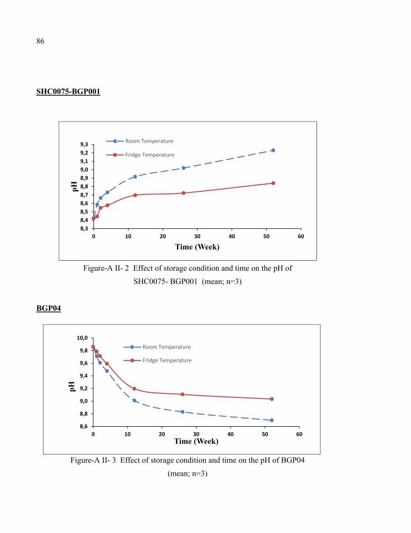
86
SHC0075-BGP001
Figure-A II- 2 Effect of storage condition and time on the pH of
SHC0075- BGP001 (mean; n=3)
BGP04
Figure-A II- 3 Effect of storage condition and time on the pH of BGP04
(mean; n=3)
8,38,48,58,68,78,88,99,09,19,29,3
0 10 20 30 40 50 60
pH
Time (Week)
Room Temperature
Fridge Temperature
8,6
8,8
9,0
9,2
9,4
9,6
9,8
10,0
0 10 20 30 40 50 60
pH
Time (Week)
Room Temperature
Fridge Temperature

LIST OF BIBLIOGRAPHICAL REFERENCES
Ahmadi, R., & de Bruijn, J. D. (2008). Biocompatibility and gelation of chitosan-glycerol phosphate hydrogels. J Biomed Mater Res A, 86(3), 824-832.
Assaad, E., Maire, M., & Lerouge, S. (2015). Injectable thermosensitive chitosan hydrogels
with controlled gelation kinetics and enhanced mechanical resistance. Carbohydrate polymers, 130, 87-96.
Barnett, B. P., Hughes, A. H., Lin, S., Arepally, A., & Gailloud, P. H. (2009). In vitro
assessment of EmboGel and UltraGel radiopaque hydrogels for the endovascular treatment of aneurysms. Journal of Vascular and Interventional Radiology, 20(4), 507-512.
Baty, A. M., Leavitt, P. K., Siedlecki, C. A., Tyler, B. J., Suci, P. A., Marchant, R. E. & Geesey,
G. G. (1997). Adsorption of adhesive proteins from the marine mussel, Mytilus edulis, on polymer films in the hydrated state using angle dependent X-ray photoelectron spectroscopy and atomic force microscopy. Langmuir, 13(21), 5702-5710.
Berger, J., Reist, M., Mayer, J. M., Felt, O., Peppas, N., & Gurny, R. (2004). Structure and
interactions in covalently and ionically crosslinked chitosan hydrogels for biomedical applications. European Journal of Pharmaceutics and Biopharmaceutics, 57(1), 19-34.
Bernkop-Schnürch, A. (2005). Mucoadhesive systems in oral drug delivery. Drug Discovery
Today: Technologies, 2(1), 83-87. Bhattarai, N., Gunn, J. & Zhang, M. (2010). Chitosan-based hydrogels for controlled, localized
drug delivery. Advanced Drug Delivery Reviews, 62(1), 83-99. Boddupalli, B. M., Mohammed, Z. N., Nath, R. A. & Banji, D. (2010). Mucoadhesive drug
delivery system: An overview. Journal of advanced pharmaceutical technology & research, 1(4), 381.
Caló, E., & Khutoryanskiy, V. V. (2015). Biomedical applications of hydrogels: A review of
patents and commercial products. European Polymer Journal, 65, 252-267. Ceccaldi, C., Assaad, E., Hui, E., Buccionyte, M., Adoungotchodo, A. & Lerouge, S. (2017).
Optimization of Injectable Thermosensitive Scaffolds with Enhanced Mechanical Properties for Cell Therapy. Macromolecular Bioscience, 1600435.
Chenite, A., Buschmann, M. D., Wang, D., Chaput, C. & Kandani, N. (2001). Rheological
characterisation of thermogelling chitosan/glycerol-phosphate solutions. Carbohydrate Polymers, 46, 39-47.

88
Chenite, A., Chaput, C., Wang, D., Combes, C., Buschmann, M. D., Hoemann, C., Selmani, A. (2000). Novel Injectable neutral solutions of chitosan form biodegradable
gels in situ. Biomaterials, 21, 2155-2161. Chirdon, W. M., O'Brien, W. J. & Robertson, R. E. (2003). Adsorption of catechol and
comparative solutes on hydroxyapatite. Journal of Biomedical Materials Research Part B: Applied Biomaterials, 66(2), 532-538.
Coutu, J. M., Fatimi, A., Berrahmoune, S., Soulez, G., & Lerouge, S. (2013). A new radiopaque
embolizing agent for the treatment of endoleaks after endovascular repair: influence of contrast agent on chitosan thermogel properties. J Biomed Mater Res B Appl Biomater, 101(1), 153-161.
Croisier, F. & Jérôme, C. (2013). Chitosan-based biomaterials for tissue engineering.
European Polymer Journal, 49(4), 780-792. Delibegović, S., Iljazović, E., Katica, M. & Koluh, A. (2011). Tissue reaction to absorbable
endoloop, nonabsorbable titanium staples, and polymer Hem-o-lok clip after laparoscopic appendectomy. JSLS: Journal of the Society of Laparoendoscopic Surgeons, 15(1), 70-76. Deming, T. J. (1999). Mussel byssus and biomolecular materials. Current Opinion in Chemical
Biology, 3(1), 100-105. Duchěne, D., Touchard, F. & Peppas, N. A. (1988). Pharmaceutical and medical aspects of
bioadhesive systems for drug administration. Drug Development and Industrial Pharmacy, 14(2-3), 283-318.
Edwards, J. C. (2009). Principles of NMR. Process NMR Associates LLC, 87A Sand Pit Rd,
Danbury CT, 6810. Fang, J.-Y., Chen, J.-P., Leu, Y.-L. & Hu, J.-W. (2008). Temperature-sensitive hydrogels
composed of chitosan and hyaluronic acid as injectable carriers for drug delivery. European Journal of Pharmaceutics and Biopharmaceutics, 68(3), 626-636.
Ferreira, P., Gil, M., & Alves, P. AN OVERVIEW IN SURGICAL ADHESIVES. G Silverman, H., & Roberto, F. (2007). Understanding Marine Mussel Adhesion (Vol. 9). Ganji, F., Abdekhodaie, M. & Ramazani S.A., A. (2007). Gelation time and degradation rate
of chitosan-based injectable hydrogel. Journal of sol-gel science and technology, 42(1), 47-53.

89
Gibas, I., & Janik, H. (2010). Review: synthetic polymer hydrogels for biomedical applications.
Gulrez, Syed K. H., Al-Assaf, S. & Phillips, G. O. (2011). Hydrogels: methods of preparation,
characterisation and applications. Progress in Molecular and Environmental Bioengineering-From Analysis and Modeling to Technology Applications.
HAKIM, A. (2015). Conception d'un banc d'essai de mucoadesion. Montreal: ÉCOLE DE
TECHNOLOGIE SUPÉRIEURE (ETS). Hennink, W. E. & van Nostrum, C. F. (2002). Novel crosslinking methods to design hydrogels.
Adv Drug Deliv Rev, 54(1), 13-36. Hoffman, A. S. (2002). Hydrogels for biomedical applications. Advanced Drug Delivery
Reviews, 54(1), 3-12. Holten-Andersen, N., Mates, T. E., Toprak, M. S., Stucky, G. D., Zok, F. W. & Waite, J. H.
(2009). Metals and the integrity of a biological coating: the cuticle of mussel byssus. Langmuir: the ACS journal of surfaces and colloids, 25(6), 3323.
Huang, Y., Leobandung, W., Foss, A. & Peppas, N. A. (2000). Molecular aspects of muco-
and bioadhesion:: Tethered structures and site-specific surfaces. Journal of controlled release, 65(1), 63-71.
Khangtragool, A., Ausayakhun, S., Leesawat, P., Molloy, R. & Laokul, C. (2008). Stability of
chitosan solutions for potential use in ocular drug delivery. CHIANG MAI UNIVERSITY JOURNAL, 209.
Khanlari, S. & Dubé, M. A. (2013). Bioadhesives: a review. Macromolecular Reaction
Engineering, 7(11), 573-587. Khutoryanskiy, V. V. (2011). Advances in mucoadhesion and mucoadhesive polymers.
Macromolecular Bioscience, 11(6), 748-764. Kim, I. Y., Seo, S. J., Moon, H. S., Yoo, M. K., Park, I. Y., Kim, B. C. & Cho, C. S. (2008).
Chitosan and its derivatives for tissue engineering applications. Biotechnol Adv, 26(1), 1-21.
Kim, K., Ryu, J. H., Lee, D. Y. & Lee, H. (2013). Bio-inspired catechol conjugation converts
water-insoluble chitosan into a highly water-soluble, adhesive chitosan derivative for hydrogels and LbL assembly. Biomater Sci, 1(7), 783-790.
Lavertu, M., Filion, D. & Buschmann, M. D. (2008). Heat-induced transfer of protons from
chitosan to glycerol phosphate produces chitosan precipitation and gelation. Biomacromolecules, 9, 11.

90
Lee, B. P., Dalsin, J. L. & Messersmith, P. B. (2006). Biomimetic adhesive polymers based on
mussel adhesive proteins. Biological Adhesives (pp. 257-278). Springer. Lee, C., Shin, J., Lee, J. S., Byun, E., Ryu, J. H., Um, S. H. & Cho, S.-W. (2013). Bioinspired,
calcium-free alginate hydrogels with tunable physical and mechanical properties and improved biocompatibility. Biomacromolecules, 14(6), 2004-2013.
Lee, H., Dellatore, S. M., Miller, W. M. & Messersmith, P. B. (2007). Mussel-inspired surface
chemistry for multifunctional coatings. science, 318(5849), 426-430. Lee, H., Scherer, N. F. & Messersmith, P. B. (2006). Single-molecule mechanics of mussel
adhesion. Proceedings of the National Academy of Sciences, 103(35), 12999-13003. Lehr, C.-M., Bouwstra, J. A., Schacht, E. H. & Junginger, H. E. (1992). In vitro evaluation of
mucoadhesive properties of chitosan and some other natural polymers. International journal of Pharmaceutics, 78(1-3), 43-48.
Mezger, T. G. (2006). The rheology handbook: for users of rotational and oscillatory rheometers. Vincentz Network, Hannover.
Monette, A., Ceccaldi, C., Assaad, E., Lerouge, S. & Lapointe, R. (2016). Chitosan thermogels
for local expansion and delivery of tumor-specific T lymphocytes towards enhanced cancer immunotherapies. Biomaterials, 75, 237-249.
Mucha, M. & Pawlak, A. (2002). Complex study on chitosan degradability. POLIMERY-
WARSAW, 47(7/8), 509-516. Nguyen, & Lee, D. S. (2010). Injectable Biodegradable Hydrogels. Macromolecular
Bioscience, 10(6), 563-579. Nguyen, T. T. B., Hein, S., Ng, C. H. & Stevens, W. F. (2008). Molecular stability of chitosan
in acid solutions stored at various conditions. Journal of applied polymer science, 107(4), 2588-2593.
No, H. K., Kim, S. H., Lee, S. H., Park, N. Y. & Prinyawiwatkul, W. (2006). Stability and
antibacterial activity of chitosan solutions affected by storage temperature and time. Carbohydrate polymers, 65(2), 174-178.
Oh, Y. J., Cho, I. H., Lee, H., Park, K.-J., Lee, H. & Park, S. Y. (2012). Bio-inspired catechol
chemistry: a new way to develop a re-moldable and injectable coacervate hydrogel. Chemical Communications, 48(97), 11895-11897.

91
Peppas, Bures, P., Leobandung, W. & Ichikawa, H. (2000). Hydrogels in pharmaceutical formulations. European Journal of Pharmaceutics and Biopharmaceutics, 50(1), 27-46.
Peppas, N. A., & Buri, P. A. (1985). Surface, interfacial and molecular aspects of polymer
bioadhesion on soft tissues. Journal of controlled release, 2, 257-275. Peppas, N. A., & Sahlin, J. J. (1996). Hydrogels as mucoadhesive and bioadhesive materials:
a review. Biomaterials, 17(16), 1553-1561. Piai, J. F., Rubira, A. F., & Muniz, E. C. (2009). Self-assembly of a swollen
chitosan/chondroitin sulfate hydrogel by outward diffusion of the chondroitin sulfate chains. Acta Biomater, 5(7), 2601-2609.
Qiu, Y., & Park, K. (2001). Environment-sensitive hydrogels for drug delivery. Advanced
Drug Delivery Reviews, 53(3), 321-339. Raafat, D. & Sahl, H. G. (2009). Chitosan and its antimicrobial potential--a critical literature
survey. Microb Biotechnol, 2(2), 186-201. Ravi Kumar, M. N. (2000). A review of chitin and chitosan applications. Reactive and
functional polymers, 46(1), 1-27. Reis, R. L., Neves, N. M., Mano, J. F., Gomes, M. E., Marques, A. P. & Azevedo, H. S. (2008).
Natural-based polymers for biomedical applications. Woodhead Pub. Rinaudo, M. (2008). Main properties and current applications of some polysaccharides as
biomaterials. Polymer International, 57(3), 397-430. Riva, R., Ragelle, H., des Rieux, A., Duhem, N., Jérôme, C. & Préat, V. (2011). Chitosan and
Chitosan Derivatives in Drug Delivery and Tissue Engineering. Chitosan for Biomaterials II.
Advances in Polymer Science, 244, 19-44. Springer, Berlin, Heidelberg.
Rosiak, J. M. & Yoshii, F. (1999). Hydrogels and their medical applications. Nuclear
Instruments and Methods in Physics Research Section B: Beam Interactions with Materials and Atoms, 151(1–4), 56-64.
Ryu, J. H., Hong, S., & Lee, H. (2015). Bio-inspired adhesive catechol-conjugated chitosan
for biomedical applications: A mini review. Acta Biomater, 27, 101-115. Ryu, J. H., Jo, S., Koh, M. Y. & Lee, H. (2014). Bio- Inspired, Water- Soluble to Insoluble
Self- Conversion for Flexible, Biocompatible, Transparent, Catecholamine Polysaccharide Thin Films. Advanced Functional Materials, 24(48), 7709-7716.

92
Ryu, J. H., Lee, Y., Kong, W. H., Kim, T. G., Park, T. G. & Lee, H. (2011). Catechol-
Functionalized Chitosan/Pluronic Hydrogels for Tissue Adhesives and Hemostatic Materials. Biomacromolecules, 12(7), 2653-2659.
Schnurrer, J. & Lehr, C.-M. (1996). Mucoadhesive properties of the mussel adhesive protein.
International journal of Pharmaceutics, 141(1-2), 251-256. Schweigert, N., Zehnder, A. J. & Eggen, R. I. (2001). Chemical properties of catechols and
their molecular modes of toxic action in cells, from microorganisms to mammals. Environmental microbiology, 3(2), 81-91.
Silverman, H. & Roberto, F. (2007). Understanding Marine Mussel Adhesion. Marine
Biotechnology, 9(6), 661-681. Smart, J. D. (2005). The basics and underlying mechanisms of mucoadhesion. Advanced Drug
Delivery Reviews, 57(11), 1556-1568. Sosnik, A., das Neves, J. & Sarmento, B. (2014). Mucoadhesive polymers in the design of
nano-drug delivery systems for administration by non-parenteral routes: a review. Progress in Polymer Science, 39(12), 2030-2075.
Spicer, P. P., & Mikos, A. G. (2010). Fibrin glue as a drug delivery system. Journal of
controlled release, 148(1), 49-55. Spotnitz, W. D., & Burks, S. G. (2010). Hemostats, sealants, and adhesives II: update as well
as how and when to use the components of the surgical toolbox. Clinical and Applied Thrombosis/Hemostasis, 713-713.
Szymańska, E. & Winnicka, K. (2015). Stability of chitosan a challenge for pharmaceutical
and biomedical applications. Marine drugs, 13(4), 1819-1846. Tajirian, A. L. & Goldberg, D. J. (2010). A review of sutures and other skin closure materials.
Journal of Cosmetic and Laser Therapy, 12(6), 296-302. Vakalopoulos, K. A., Daams, F., Wu, Z., Timmermans, L., Jeekel, J. J., Kleinrensink, G.-J.,
Lange, J. F. (2013). Tissue adhesives in gastrointestinal anastomosis: a systematic review. Journal of Surgical Research, 180(2), 290-300.
Voiutskii, S. S. (1963). Autohesion and adhesion of high polymers. Wiley, New York. Waite, J. H., & Qin, X. (2001). Polyphosphoprotein from the adhesive pads of Mytilus edulis.
Biochemistry, 40(9), 2887-2893.

93
Woertz, C., Preis, M., Breitkreutz, J. & Kleinebudde, P. (2013). Assessment of test methods evaluating mucoadhesive polymers and dosage forms: an overview. European Journal of Pharmaceutics and Biopharmaceutics, 85(3), 843-853.
Wu, J., Zhang, L., Wang, Y., Long, Y., Gao, H., Zhang, X., Xu, J. (2011). Mussel-inspired
chemistry for robust and surface-modifiable multilayer films. Langmuir, 27(22), 13684-13691.
Xu, J., Soliman, G. M., Barralet, J. & Cerruti, M. (2012). Mollusk glue inspired mucoadhesives
for biomedical applications. Langmuir, 28(39), 14010-14017. Xu, J., Strandman, S., Zhu, J. X. X., Barralet, J. & Cerruti, M. (2015). Genipin-crosslinked
catechol-chitosan mucoadhesive hydrogels for buccal drug delivery. Biomaterials, 37, 395-404.
Yu, J., Wei, W., Menyo, M. S., Masic, A., Waite, J. H. & Israelachvili, J. N. (2013). The
adhesion of mussel foot protein-3 to TiO2 surfaces: the effect of pH. Biomacromolecules, 14(4), 1072.
Zhou, H. Y., Jiang, L. J., Cao, P. P., Li, J. B. & Chen, X. G. (2015). Glycerophosphate-based
chitosan thermosensitive hydrogels and their biomedical applications. Carbohydrate Polymers, 117, 524-536.


![General Aspects of Chitosan · 2020. 2. 2. · hydrogels. Carreira et al. [4] addressed smart polymers derived from chitosan, including particulate carrier systems, hydrogels, and](https://static.fdocuments.in/doc/165x107/60ff3bed77d00b7ab74500bc/general-aspects-of-chitosan-2020-2-2-hydrogels-carreira-et-al-4-addressed.jpg)