Optimisation of fungal cellulase production from textile ...

Vol. 53, No. 9APPLIED AND ENVIRONMENTAL MICROBIOLOGY, Sept. 1987, p. 2175-21820099-2240/87/092175-08$02.00/0Copyright © 1987, American Society for Microbiology
Sporotrichum thermophile Growth, Cellulose Degradation, andCellulase Activity
K. M. BHATt AND RAMESH MAHESHWARI*
Department of Biochemistry, Indian Institute of Science, Bangalore 560 012, India
Received 30 January 1987/Accepted 28 May 1987
The activity of components of the extracellular cellulase system of the thermophilic fungus Sporotrichumthermophile showed appreciable differences between strains; I-glucosidase (EC 3.2.1.21) was the most variablecomponent. Although its endoglucanase (EC 3.2.1.4) and exoglucanase (EC 3.2.1.91) activities were markedlylower, S. thermophile degraded cellulose faster than Trichoderma reesei. The production of I-glucosidaselagged behind that of endoglucanase and exoglucanase. The latter activities were produced during activegrowth. When growth was inhibited by cycloheximide treatment, the hydrolysis of cellulose was lower than inthe control in spite of the presence of both endoglucanase and exoglucanase activities in the culture medium.Degradation of cellulose was a growth-associated process, with cellulase preparations hydrolyzing cellulose onlyto a limited extent. The growth rate and cell density of S. thermophile were similar in media containing celluloseor glucose. A distinctive feature of fungal development in media incorporating cellulose or lactose (inducers ofcellulase activity) was the rapid differentiation of reproductive units and autolysis of hyphal cells to liberatepropagules which were capable of renewing growth immediately.
Thermophilic fungi are one of the major components of themicroflora which develops in self-heating masses of vegeta-ble matter where they contribute to decomposition of plantcell wall polysaccharides (4, 5). Tansey (27) found that thecellulolytic rates of some thermophilic fungi (Chaetomiumthermophile, Sporotrichum thermophile, and Thermoascusaurantiacus) were two or three times that of Trichodermaviride, one of the most cellulolytic mesophilic fungi (15).Romanelli et al. (20) reported that, of the three thermophilicfungi, S. thermophile degraded cellulose fastest in liquidshake cultures. Mandels (12) also noted that thermophilicfungi, including S. thermophile, degraded cellulose rapidly,but she found that the cellulase activity of their culturefiltrates was low. This was repudiated by Tansey (M. R.Tansey, ASM News 45:417, 1979) on the grounds thatsuboptimal temperatures for growth and enzyme assayswere used for the thermophilic fungi. Coutts and Smith (6)reported that S. thermophile produced cellulase yields com-parable to those produced by T. viride and in one-fourth asmuch time.
In view of the contradictory reports regarding cellulaseactivity of S. thermophile, we have examined several strainsof the fungus and made comparisons with T. reesei. We havealso studied the interrelationship among growth, celluloseutilization, and cellulase production in shake flask culturesand features of growth and development when the funguswas grown under cellulase-inductive conditions.
MATERIALS AND METHODSIsolation of S. thermophile. The procedure used for isola-
tion of the fungus from soil has been described byMaheshwari et al. (11). Strains were maintained on YpSsagar (5) slants at 40°C.
Cultivation techniques. Batch cultures of S. thermophile ina medium containing cellulose were prepared as follows.
* Corresponding author.t Present address: Department of Microbial Biochemistry,
Rowett Research Institute, Bucksburn, Aberdeen AB2 9 SB, UnitedKingdom.
Spore suspensions were obtained by harvesting growth onYpSs agar medium in bottles (12 by 5 by 3 cm) which hadbeen incubated at 40°C for 10 to 15 days in a humidifiedincubator. Mycelial inocula were prepared by incubating aspore suspension in a glucose-ammonium dihydrogen phos-phate medium (pH 6.0) which contained, per liter: glucose,10.0 g; NH4H2PO4, 2.0 g; KH2PO4, 3.0 g; K2HPO4, 2.0 g;MgSO4 7H20, 0.5 g; CaCI2. 2H20, 0.1 g; FeSO4- 7H20,5.0 mg; MnSO4 4H20, 1.6 mg; ZnSO4 7H20, 1.4 mg;CoCl2 6H20, 2.0 mg; yeast extract, 1.0 g; and peptone, 1.0g. The fungus was grown in 150 ml of medium in a 500-mlErlenmeyer flask at 50°C for 24 h on a gyratory shaker.Finally, the cellulose cultures were initiated by adding 5 mlof the homogeneous mycelial suspension to the above me-dium in which cellulosic material (1%) replaced glucose. Thecellulosic substrate was blotting paper (manufactured mainlyfrom bamboo pulp). This was pretreated with alkali beforeuse: 20 g was autoclaved with 360 ml of 1% NaOH for 1 h,washed free of alkali, blended in distilled water, and dried at70°C. In some experiments untreated Whatman no. 1 filterpaper, cellulose powder, or absorbent cotton was alsotested.
T. reesei was grown in the medium of Mandels andSternberg (14), containing the cellulose described above, at26 to 28°C on a gyratory shaker (240 rpm). When stated,batch cultures of T. reesei and S. thermophile were alsoinitiated by using 5 ml of a conidial suspension containingapproximately 106 spores m-1'.Enzyme assays. Unless stated otherwise, all assays of S.
thermophile enzymes were carried out in sodium acetatebuffer (pH 5.6) at 50°C for 30 min, which were optimalconditions. In some experiments the cellulolytic activity ofculture filtrates was assayed by the release of glucose fromfilter paper. A reaction mixture (1 ml) containing 50-mgWhatman no. 1 filter paper (1 by 6 cm) was incubated with0.1 ml of culture filtrate, and the glucose released wasquantified by the glucose oxidase-peroxidase method (16).Filter paper activity was assayed according to Mandels et al.(13), using 0.5 ml of culture filtrate in a 1.0-ml reactionmixture. After 1 h of incubation, reducing sugar was esti-
2175
on Decem
ber 25, 2019 by guesthttp://aem
.asm.org/
Dow
nloaded from

2176 BHAT AND MAHESHWARI
mated by the dinitrosalicylic acid method (17). The activity,calculated as reducing sugar (milligrams of glucose) x 0.185,was expressed as filter paper units (13). Endoglucanase (EC3.2.1.4) activity was determined on sodium carboxymethylcellulose (medium viscosity), using 0.5 ml of 1% (wt/vol)substrate in a 2-ml reaction mixture. The liberation ofreducing end groups was measured by the Nelson-Somogyimethod (25). Exoglucanase (EC 3.2.1.91) activity was deter-mined on microcrystalline cellulose (Sigmacell; SigmaChemical Co.) pretreated by boiling for 30 min in 1 M HCI(to remove any frayed ends in particles) and washed free ofacid. The reaction mixture (2 ml) contained 1 ml of a 5%suspension of the substrate. The reducing sugar was mea-sured by the Nelson-Somogyi method. P-Glucosidase (EC3.2.1.21) activity was assayed by using p-nitrophenyl P-D-glucopyranoside. The reaction mixture (1 ml) containedculture filtrate and 0.5 ml of 1 mM substrate solution. Thereaction was stopped by adding 2 ml of 5% Na2CO3, and theabsorbance of the solution was measured at 400 nm.
Cellobiase activity was measured by estimating the glucoseproduced from the hydrolysis of cellobiose.For T. reesei, cellulase assays were carrried out as de-
scribed above except 0.05 M sodium citrate buffer, pH 4.8(13), was used.One unit of enzyme activity on a cellulosic substrate was
defined as 1 jxmol of reducing end group (glucose) releasedper min at 50°C. One unit of ,-glucosidase activity wasdefined at that amount of enzyme which produced 1 ,umol ofp-nitrophenol per min under the stated assay conditions.
Protein measurement. Soluble protein in culture filtratewas precipitated by trichloroacetic acid and estimated by theLowry method (10), using bovine serum albumin as stan-dard.Reducing sugar. Reducing sugar in culture fluids was
determined colorimetrically by the dinitrosalicylic acidmethod (17), with glucose as standard.
Cellulose estimation. For studying the utilization of cellu-lose during growth, a 10-ml culture mass (mycelia pluscellulose) was removed from duplicate flasks and filteredthrough a Whatman glass fiber circle (2.5 cm) by suction.The insoluble material was washed with distilled water anddried at 70°C. Cellulose in the material was estimated by themethod of Updegraff (28). The finely dispersed mycelialgrowth permitted sampling with a broad-tipped pipette.Growth measurement. Growth of the fungus was followed
as the increase in insoluble nitrogen by the Kjeldahl methodor as an increase in mycelial dry weight. The latter was
determined by subtracting the cellulose content from theweight of the dried insoluble material. Specific growth rate(,u) in media incorporating cellulosic material or solublesugar was determined from the exponential portion of semi-logarithmic plots of growth curves and calculated as follows:,u = [2.303 (log x2 - log xj)]h-1/[t2 - tl, where x2 and xl are
mycelial protein contents at times t2 and t1, respectively.Samples (5 ml) of culture were removed from duplicateflasks at 3-h intervals, filtered through glass fiber, andwashed three times with 10 ml of distilled water. Theinsoluble material was extracted with 5 ml of 0.5 M NaOH at90°C for 30 min. The extract was clarified by centrifugationand protein was measured by the Lowry method (10).
Cellulase preparation. Culture suspensions from 6-day-oldcultures were filtered through glass wool. Culture filtrateprotein was precipitated with 80% saturated ammoniumsulfate, desalted by chromatography on a Sephadex G-25column, and lyophilized before use.
Saccharification and solubilization of cellulose. The lyophil-
ized cellulase powder was dissolved in 50 mM sodiumacetate buffer (pH 5.6) and the solution was sterilized bymembrane filtration (0.45-,um pore size; Millipore Corp.).Suspensions of cellulosic material (1%) were incubated withthe cellulase preparation in sterile Sporotrichum medium at50°C on a shaker. At intervals, aliquots of the reactionmixture were removed and glucose was estimated (16). Theresidual cellulose was estimated gravimetrically. Percentsaccharification was calculated as follows: [milligrams ofreducing sugar x (162/180) x 100]/(milligrams of initialsubstrate). (In this calculation, the factor 162/180 normalizesthe conversion for the weight gain caused by addition ofwater to the glycosyl moiety on hydrolysis.) Percent solubi-lization was calculated as follows: [(milligrams of totalcellulose - milligrams of remaining cellulose) x 100]/milli-grams of total cellulose.
Light microscopy. For examination of fungal growth andstructural changes in cellulosic material during culture,samples were mounted in lactophenol and examined byphase-contrast and polarized-light microscopy.
Quantitative expression of results. All estimations weremade on samples pooled from duplicate or triplicate cultureflasks. Experiments were repeated at least once and resultswere quite reproducible. Data are average or typical valuesfrom 2 to 3 experiments.
Biochemicals. Biochemicals were purchased from SigmaChemical Co., St. Louis, Mo. Yeast extract and malt extractwere from Difco Laboratories, Detroit, Mich. Peptone andcellulose powder were from Hindustan Dehydrated Media,Bombay, India. Blotting paper was purchased from a localpaper merchant and the same lot was used throughout.
RESULTS
Morphological characteristics of S. thermophile. Most iso-lates were buff or cinnamon brown and powdery (e.g., IIS 72and IIS 101), but a few isolates were white and floccose (e.g.,IIS 125 and IIS 220). Despite visible differences, the culturesresembled one another in conidiophore morphology (Fig. 1)and conformed to the description of S. thermophile Apinisgiven by Semeniuk and Carmichael (23).
Cellulase activity in different strains. Eleven strains weregrown in a cellulose-NH4H2PO4 medium to determine theircellulolytic potential and production of extracellularcellulase enzymes and to examine possible differences intheir cellulase systems. Microscopic examination of cultures(day 6) showed absence of cellulose particles. The final (day6) cellulase activity (measured as release of glucose fromfilter paper) varied from 0 to 0.16 U ml-'.Four strains, IIS 101, IIS 220, ATCC 42464, and UAMH
2015, were compared for activity of endoglucanase,exoglucanase, and 3-glucosidase. Two of these strains, IIS101 and IIS 220, were selected because their culture filtratesproduced, respectively, the lowest and highest amounts ofglucose in the above cellulase assay. Strain ATCC 42464 wasinvestigated because it was reported to lack extracellularP-glucosidase activity (3). UAMH 2015 was included be-cause this strain was reported to produce cellulase activitycomparable to that of T. viride (6). The activities ofendoglucanase and exoglucanase between strains varied 1.4-to 2-fold, respectively, while that of 3-glucosidase varied upto 5.3-fold (Table 1). The filter paper activity of the mostproductive strain, IIS 220, was comparable to that of UAMH2015 but was <10% of that reported for T. viride 9414 (14).
Additional differences in the cellulase systems of isolateswere found when two strains, IIS 101 and IIS 220, were
APPL. ENVIRON. MICROBIOL.
on Decem
ber 25, 2019 by guesthttp://aem
.asm.org/
Dow
nloaded from

CELLULOSE DEGRADATION BY S. THERMOPHILE 2177
_ _~~~~~~0 tQfql- 4 E- 9 ww *i&4% MA
FIG. 1. S. thermophile, phase-contrast micrographs. (A) Strain IIS 72, young conidiophores. (B) Strain IIS 72, mature conidiophores. (C)Strain IIS 220, dendritic branching of a fertile hypha and conidiophores. (D) Strain IIS 220, mature conidiophores. (E) Strain IIS 220, prostratehyphae bearing lateral conidia. Bars, 50 ,um.
TABLE 1. Activities of primary cellulase components in some strains of S. thermophileaActivity (U ml-') + SE
ExtracellularStrain protein (mg ml-') Filter paper Endoglucanase Exoglucanase 3-Glucosidaseactivity
IIS 101 0.07 ± 0.002 0.05 ± 0.01 0.85 ± 0.18 0.09 ± 0.03 0.31 ± 0.09IIS 220 0.17 ± 0.005 0.11 + 0.01 1.20 ± 0.09 0.18 ± 0.01 1.65 ± 0.22ATCC 42464 0.13 ± 0.003 0.07 ± 0.006 1.05 ± 0.08 0.10 ± 0.03 0.26 ± 0.01UAMH 2015 0.41 ± 0.01 0.11 ± 0.01 1.00 + 0.10 0.16 ± 0.02 0.45 ± 0.10
a All strains were grown in cellulose-NH4H2PO4 medium for 6 days.
VOL. 53, 1987
on Decem
ber 25, 2019 by guesthttp://aem
.asm.org/
Dow
nloaded from
2178 BHAT AND MAHESHWARI
TABLE 2. Cellulase activities of S. thermophile IIS 110 and IIS 220 in media containing soluble and insoluble inducers"
Activity (U ml-') + SECarbon Strain Extracellularsource protein (mg ml-') Filter paper Endoglucanase Exoglucanase P-Glucosidase
activity
Lactose IIS 101 0.01 ± 0.005 0.03 ± 0.00 0.03 ± 0.005 0.02 ± 0.005 0.23 ± 0.00IIS 220 0.07 ± 0.00 0.08 ± 0.01 0.39 ± 0.005 0.12 ± 0.005 1.25 ± 0.03
Cellulose IIS 101 0.07 ± 0.02 0.07 ± 0.008 0.89 ± 0.07 0.10 ± 0.01 0.16 ± 0.018IIS 220 0.19 ± 0.01 0.12 ± 0.007 1.44 ± 0.09 0.22 ± 0.01 1.29 ± 0.05
Cultures were grown in media containing 1% lactose or 1% cellulose (blotting paper, Whatman filter paper, cellulose powder, or absorbent cotton). The datafor cellulases are average values of enzyme activities on all types of cellulosic substrates tested.
grown in media containing lactose or cellulosic materials assoluble (present observation) and insoluble inducers ofcellulase, respectively (Table 2). Strain IIS 220 secretedmore protein than IIS 101, 7- and 2.5-fold more in lactoseand cellulose media, respectively. The enhanced proteinsecretion was reflected in the increased absolute levels of allenzymes. Microscopic examination of cultures showed thatpractically complete conversion of cellulose had occurred byday 2 in cultures of both strains. A qualitative difference inthe composition of the cellulase system was also revealed;the specific activities of endoglucanase and exoglucanase incellulosic media in IIS 101 were 1.7- and 1.2-fold higher,respectively, than in IIS 220. The ratio of endoglucanase toexoglucanase activity was 1.5 in IIS 101 and 3.3 in IIS 220 inlactose-grown cultures and 8.9 in IIS 101 versus 6.5 in IIS220 in cellulose-grown cultures.
Cultural conditions for cellulose degradation. Microscopicexamination at 12-h intervals of cultures of some strainstested (IIS 72, IIS 125, IIS 220, and ATCC 42464) showedthat degradation of cellulose in shaken flasks also occurredduring growth at 30°C, although slowly. Some celluloseparticles remained insolubilized in the culture flasks. Incontrast, in parallel cultures at 50°C, virtually no trace of
SPOROTRICHUM THERMOPHILE
1-
z
x
tr
LI
J,
x
z
TRICHODERMA REESEI
T ME (DAYS)
FIG. 2. Time course of cellulose utilization, growth, and extra-cellular cellulase activity in S. thermophile lIS 220 and T. reesei inshake flask cultures. Symbols: 0, growth; 0, cellulose; O,endoglucanase; *, exoglucanase, A, ,-glucosidase. Cultures were
initiated with spore inocula.
cellulose particles remained by 48 h. Sporulation was in-creased at 30°C. The filter paper activity (units per milliliter)in cultures grown at 30°C for 6 days ranged from 0.08 to 0.10,whereas at 50°C it ranged from 0.10 to 0.13.
Parallel cultures of the above four strains in stationaryflasks at 30 and 50°C showed the development of a whitemycelial mat on the surface of the liquid. Appreciableamounts of cellulose remained insolubilized until day 12.This indicated that adequate aeration of cultures was essen-tial for the rapid and complete degradation of cellulose.
S. thermophile versus T. reesei. Strain IIS 220 was the bestcellulase producer among S. thermophile strains tested (Ta-ble 1). Its cellulolytic activity was compared with that of T.reesei (Fig. 2).Growth curves based on estimations of insoluble nitrogen
or mycelial dry weight in culture flasks containing cellulosicmaterial were almost identical in S. thermophile IIS 220(data not shown). The agreement between growth parame-ters in T. reesei was also good, except that after maximalgrowth (day 4) the growth curve based on mycelial dryweight remained stationary with culture age whereas thatbased on mycelial nitrogen content showed a small declinebefore becoming stationary. Both fungi degraded the cellu-losic substrate completely, but their cellulolytic behaviordiffered in a number of ways. S. thermophile showed a fastergrowth rate and a higher culture density than T. reesei.Whereas the time of maximal growth and complete celluloseconversion coincided in S. thermophile, in T. reesei they didnot. Estimations revealed that 20 to 40% cellulose remainedat the time when the culture density of T. reesei attained amaximum value (days 4 to 5). In S. thermophile, soon aftermaximal growth had been attained, the culture densitydeclined sharply and was reduced by 50% at day 4. Incontrast, the culture density of T. reesei showed a slowerand smaller reduction with time.
T. reesei was superior to S. thermophile in terms ofextracellular protein, endoglucanase, and exoglucanase ac-tivities (Table 3). However, in spite of its lower titer ofcellulase in the culture broth, the thermophilic fungusachieved a complete conversion of the cellulosic substrate ata faster rate than T. reesei.Growth and development in cellulosic media. Microscopic
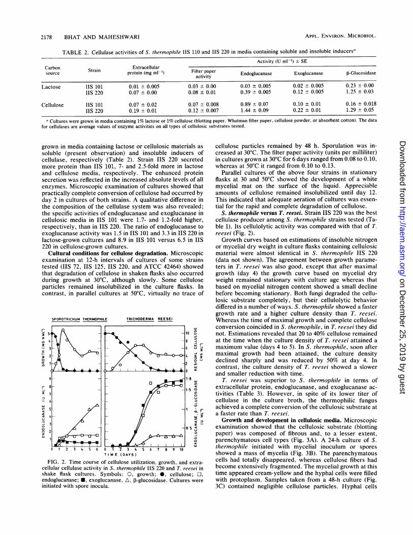
examination showed that the cellulosic substrate (blottingpaper) was composed of fibrous and, to a lesser extent,parenchymatous cell types (Fig. 3A). A 24-h culture of S.thermophile initiated with mycelial inoculum or sporesshowed a mass of mycelia (Fig. 3B). The parenchymatouscells had totally disappeared, whereas cellulose fibers hadbecome extensively fragmented. The mycelial growth at thistime appeared cream-yellow and the hyphal cells were filledwith protoplasm. Samples taken from a 48-h culture (Fig.3C) contained negligible cellulose particles. Hyphal cells
APPL. ENVIRON. MICROBIOL.
on Decem
ber 25, 2019 by guesthttp://aem
.asm.org/
Dow
nloaded from

CELLULOSE DEGRADATION BY S. THERMOPHILE 2179
TABLE 3. Comparison of growth, cellulose utilization, and cellulase activities of S. thermophile IIS 220 and T. reeseia
Growthrate (mg Cellulose Cellulase activity (U mi-')Organism Growth rate (mg utilization Soluble protein Enolucase acoivucanasml-GIucosidaseOrganism ~[dry wt] h'1) (mg h'1) (mg ml-') Edguaae Eolcns -lcsds
S. thermophile 0.16 40 0.15 0.65 0.14 0.20T. reesei 0.03 7 1.5 7.20 1.61 0.33
a Cultures were initiated with spore inocula in media containing blotting paper. Protein and cellulase activity were measured at the times of complete celluloseutilization, which were 48 and 168 h for S. thermophile and T. reesei, respectively.
were long and vacuolated and the mycelia appeared brittle.Cultures at 72 h of all strains examined invariably showed avisible transformation; the culture had turned greenish-brown and had become thin in consistency. The mycelia hadbecome fragmented (Fig. 3D), and some had spores attachedto them. In 96-h cultures, some of the hyphal fragments hadrenewed growth ("secondary growth phase") by protrudingfine, unbranched hyphae of limited length (Figs. 3E and F),presumably because nutrients were exhausted. Asexual re-production by differentiation of conidia was more abundantin cultures of IIS 101 and ATCC 42464 than in IIS 220. Inthese cultures, the secondary growth phase was largelycomposed of germinating conidia.Time of cellulase secretion. An attempt was made to
determine the crucial time during which the amount ofcellulase enzymes required for cellulose hydrolysis wasformed. The approach taken was to inhibit growth at dif-ferent ages by the addition of cycloheximide, an inhibitor ofeucaryotic protein synthesis.
Figure 4A shows the pattern of growth, cellulose utiliza-tion, and cellulase (endoglucanase) activity in control flasksof S. thermophile. The appearance of cellulase activity inculture broth lagged behind growth apparently because theenzymes were adsorbed on the culture substrate, a well-established phenomenon. In the second set of culture flasks(Fig. 4B) to which cycloheximide was added at 12 h and thenagain at 12-h intervals, there was neither measurable growthnor cellulose utilization or cellulase production. When cy-cloheximide additions were begun after 24 h (Fig. 4C), thegrowth was arrested and the utilization of cellulose pro-ceeded slowly. However, the activities of cellulase enzymeswere detected later when approximately 75% cellulose hadbeen solubilized. In the fourth set of culture flasks to whichcycloheximide addition was begun at 48 h (Fig. 4D), celldensity and cellulose utilization were comparable to those ofthe control. The exoglucanase (not shown) and endoglu-canase activities, however, were 60 and 70% those of thecontrol. From the above observations, the interval between12 and 24 h appeared to be the crucial time for conversion ofthe bulk substrate.Growth on soluble and insoluble carbon sources. The spe-
cific growth rate and the cell density (mycelial protein) of S.thermophile IIS 220 on glucose were marginally higher thanon cellulose (blotting paper) (Table 4). Other cellulosicsubstrates such as Whatman filter paper or cellulose powderwere also extensively degraded but an appreciable amount ofcotton remained in fibrous form. Whether the utilization of agiven type of celllulosic material was complete or not, thefinal cellulase activity of the culture broths was in the rangeof 0.10 to 0.14 filter paper units ml-1', showing that thecellulase enzyme was induced to similar extents by all typesof cellulosic materials. In contrast, culture filtrates of S.thermophile grown on glucose, maltose, sucrose, starch, orsodium carboxymethyl cellulose as carbon source did notpossess filter paper cellulase or ,-glucosidase activities.
Contrary to that in S. thermophile ATCC 42464 studied byCanevascini et al. (2), cellobiose did not induce these en-zyme in strain IIS 220. Cell-associated cellobiase activitywas detected, but the specific activity of the enzyme inmycelia grown with glucose or cellobiose was low (0.13 and0.8 U mg of protein-1, respectively) compared to that incellulose- or lactose-grown mycelia (4.7 and 5.0 U mg ofprotein-', respectively).Under noninductive conditions, with either glucose or
cellobiose as carbon source, the mycelia remained in avegetative state and lysis did not occur until day 10. Incontrast, under inductive conditions, with either cellulose(blotting paper, Whatman filter paper, and cellulose powder)or lactose as the carbon source, the mycelia formed sporesand autolysis of hyphal cells occurred after 48 h, as de-scribed before. Mycelial growth on cotton, however, re-mained in a vegetative state up to day 10, when the experi-ment was terminated. It was noted that in cellulosic mediathe level of soluble sugars at all times was very low,indicating that cellulose hydrolysis and uptake of hydrolysisproducts occurred concurrently.
Cellulolytic activity of culture filtrates. In experiments todetermine cellulolytic activity, the same type (blotting pa-per) and quantity (1%) of substrate was incubated with thequantity of cellulase estimated to be secreted by S.thermophile. For example, on day 6 the total cellulaseactivity (glucose produced ml-' min-') in 100 ml of culturefiltrate was nearly 10 U. However, hydrolysis of cellulose bycellulase preparations containing 20 U was only 51%. Theresults were similar when hydrolysis of cellulose was per-formed with an amount of cellulase preparation equivalent tothe protein secreted in culture broth (18 mg 100 ml-'). Thesame quantity of cellulase preparation, based on enzymeunit or protein value, hydrolyzed Whatman filter paper to 21to 25%, although this substrate was also solubilized nearlycompletely under cultural conditions.
Cellulase may be inactivated under in vitro conditions,whereas it is renewed continuously by the growing fungus invivo. The effect of a single dose and of multiple doses of thesame quantity of cellulase (20 U) was investigated. Theinitial rate of cellulose hydrolysis was slower when cellulasewas added intermittently in portions at 12-h intervals (datanot shown). However, the extent of hydrolysis of celluloseafter 120 h by both treatments was comparable (70%).The difference in solubilization of cellulose under in vitro
conditions could be due to the accumulation of solubleproducts, which would be continuously removed by assim-ilation. However, glucose, the principal product of cellulosehydrolysis by culture filtrates of S. thermophile IIS 220 in 24h (determined by paper chromatography of reaction mix-tures), even at 1% caused only 14% inhibition of cellulosesolubilization after 96 h, as determined by gravimetric esti-mation. Therefore, end-product inhibition was discarded asthe primary reason for the weak activity of culture filtrates.The results of these experiments, using rather large quanti-
VOL. 53, 1987
on Decem
ber 25, 2019 by guesthttp://aem
.asm.org/
Dow
nloaded from

2180 BHAT AND MAHESHWARI
i,
I1
FIG. 3. Growth and development of S. thermophile IIS 220 in medium containing blotting paper in shake cultures examined byphase-contrast microscopy (except panel A). (A) 0 h; initial appearance of cellulosic substrate under polarized light optics. (B) 24 h; primarygrowth phase consisting of vegetative mycelium. Extensive fragmentation and solubilization of cellulose has occurred. Some insolublecellulose particles ( T ) are visible. (C) 48 h; cellulose particles are no longer visible. Hyphae with vacuolated cells. (D) 72 h; fragmentationof mycelia. (E) 96 h; secondary growth phase. Some separated hyphal cells have renewed growth by protruding fine hyphae at one or bothends ( T ). (F) 96 h; cells showing renewed growth ( T ) at higher magnification. Bars, 50 ,um.
APPL. ENVIRON. MICROBIOL.
on Decem
ber 25, 2019 by guesthttp://aem
.asm.org/
Dow
nloaded from

VOL. 53, 1987
100
75
so
25
-i
LA
100
75
50
25
_ 4
3
-2
O
iTI
3
o 2 09- I1 >a
_ O 3
24 . 48 72 96 120 144
T M E IHOURS)
FIG. 4. Effect of cycloheximide addition on growth, celluloseutilization, and cellulase production in S. thermophile IIS 220. (A)Control. Cycloheximide (20 jig ml-') added after 12, (B), 24 (C), and48 (D) h. Cycloheximide (20 ,ug ml-') was readded at 12-h intervalsfollowing the initial addition (indicated by arrow). Cultures wereinitiated with a uniform spore inoculum. Estimations were done onpooled samples from duplicate flasks. Symbols: A, growth; 0,
percent cellulose utilized; 0, endoglucanase. The pattern ofexoglucanase activity (not shown) was similar to that of endoglu-canase activity.
ties of cellulase (more than in physiological concentrations),showed that culture filtrates, unlike the organism itself,brought about only a modest conversion of cellulose.
DISCUSSION
Quantitative and qualitative differences, comparable tothose observed in some laboratory-produced mutants ofTrichoderma sp. (24), were found in S. thermophile strains.Extracellular ,B-glucosidase was the principal variable com-ponent of the cellulase system in the latter fungus. It appearsthat the production and levels of this enzyme are stronglyinfluenced by cultural conditions. For example, Canevasciniand Meyer (3) found that S. thermophile ATCC 42464 did notproduce extracellular 3-glucosidase. The level of I-
glucosidase in an enzyme preparation strongly affects cellu-
CELLULOSE DEGRADATION BY S. THERMOPHILE 2181
lase assays, in particular, the assay of filter paper activity (1,9). It was therefore necessary to compare the cellulaseproductivity of different strains of S. thermophile by using auniform cultural regimen and assay procedure to obtain atrue representation of cellulase activity of this fungus.
In spite of its distinctly lower cellulase activity, S. thermo-phile grew more rapidly and degraded cellulose faster thanthe hyperproducing cellulase mutant T. reesei (Table 3).Such behavior has also been reported for the anaerobicbacterium Clostridium thermocellum (19). The substrateutilization rate of S. thermophile is approximately 2.5 timesfaster than that of C. thermocellum (18). In spite of differ-ences in the cellulase composition among strains, theydegraded cellulose at the same rate, as judged by micro-scopic examination of cultures. These observations raise thequestion as to whether the levels of cellulase produced are ofprime importance in determining the rate or extent ofcellulolysis. This point was also brought out by the inabilityof enzyme preparations from S. thermophile or from someother cellulolytic microorganisms (22, 26) to extensivelyhydrolyze cellulose.Few attempts have been made to determine the precise
relationship among growth, cellulase production, and cellu-lose utilization. Presumably because of the tedium of mea-suring growth on an insoluble substrate, investigators haveused certain soluble inducers (7, 21) and the experimentalresults have been interpreted to show that cellulase forma-tion is not directly correlated with mycelial growth, but theenzymes are produced during idiophase. We have found thata close interrelationship exists between cellulase productionand cellulose utilization during the trophophase, in both S.thermophile and T. reesei. Therefore, unlike lignin degrada-tion by the white-rot fungi (8), cellulose degradation is nottemporally separated from primary growth. The activegrowth of the fungus is crucial in cellulolysis. When growthwas inhibited (Fig. 4), cellulolysis remained weak, althoughcellulase enzymes were present in culture broth. Vaheri (29)proposed the participation of an oxidative reaction which isbelieved to disrupt the hydrogen bonds in crystalline cellu-lose, rendering it susceptible to attack by endoglucanase. Hefound that this activity was associated with cell wall in youngcells of T. reesei in both induced and noninduced conditions.Thus, activities associated with growing cells appear to playa crucial role in the degradation of crystalline cellulose.Growth of fungi on a polysaccharide is simply viewed as a
problem of converting the substrate into soluble sugars fortransport within the mycelium for biosynthesis. The obser-vations in this paper illustrate that the physiology of themycelium is influenced differently by a polymeric substrateand its depolymerized forms. A distinctive feature of devel-opment of S. thermophile on cellulosic substrates was theprecocious differentiation of reproductive units and exten-
TABLE 4. Some characteristics of the growth of S. thermophile in media containing soluble and insoluble carbon sources"
Extracellular (mg ml-')content of:
Carbon Specific growth Mycelial proteinsource rate (h-) (mg 10 ml-,) Protein Reducing Cultural characteristic
sugar
Glucose 0.10 0.75b 0.00 1.9 Asexual reproduction absentCellobiose ND' ND 0.01 2.3 Asexual reproduction absentLactose ND ND 0.01 0.38 Asexual reproduction presentCellulose 0.09 0.72' 0.17 0.02 Asexual reproduction present
a Carbon sources were present at 1% protein, and reducing sugars in culture filtrates were estimated after 144 h.b Sampled after 24 h.C ND, Not determined.d Sampled after 28 h.
A
l
^ 1 A
1 T
5
on Decem
ber 25, 2019 by guesthttp://aem
.asm.org/
Dow
nloaded from

2182 BHAT AND MAHESHWARI
sive autolysis of hyphal cells to liberate propagules capableof renewing growth immediately. This type of developmentwas reflected in the rapid reduction in cell density with time;the initial dense mycelium which filled the liquid was re-duced to a thin mass of propagules which, on standing,settled at the bottom of the culture flask. This observationshows the strong stimulus that cellulosic substrates can haveon reproduction by fungi. The observation may serve toexplain how vast quantities of vegetable matter are decom-posed in nature but with only a sparse accumulation offungal mycelia.
ACKNOWLEDGMENTS
We acknowledge the valuable contributions and help of P. T.Kamalam, P. V. Balasubramanyam, and Joel S. Gaikwad. Culturesof some fungi used in this study were kindly provided by M.Mandels, U.S. Army Research Development and Command,Natick, Mass. (T. reesei), G. Canevascini, University of Fribourg,Fribourg, Switzerland (S. thermophile ATCC 42464), and LynneSigler, Microfungus Collection and Herbarium, University ofAlberta, Edmonton, Canada (S. thermophile UAMH 2015).
This work was supported by the Department of Science andTechnology, Government of India.
LITERATURE CITED1. Bailey, M. J. 1981. The effect of ,-glucosidase on some assays
for cellulolytic enzymes. Biotechnol. Lett. 3:695-700.2. Canevascini, G., M. R. Coudray, J. P. Rey, R. J. G. Southgate,
and H. Meier. 1979. Induction and catabolite repression ofcellulase synthesis in the thermophilic fungus, Sporotrichumthermophile. J. Gen. Microbiol. 110:291-303.
3. Canevascini, G., and H. P. Meyer. 1979. 13-Glucosidase in thecellulolytic fungus Sporotrichum thermophile Apinis. Exp.Mycol. 3:203-214.
4. Chang, Y. 1967. The fungi of wheat straw compost. II. Bio-chemical and physiological studies. Trans. Br. Mycol. Soc.50:667-677.
5. Cooney, D. G., and R. Emerson. 1964. Thermophilic fungi. Anaccount of their biology, activities and classification. W. H.Freeman & Co., San Francisco.
6. Coutts, A. D., and R. E. Smith. 1976. Factors influencing theproduction of cellulases by Sporotrichum thermophile. Appl.Environ. Microbiol. 31:819-825.
7. Hulme, M. A., and D. W. Stranks. 1971. Regulation of cellulaseproduction by Myrothecium verrucaria grown on non-cellulosicsubstrates. J. Gen. Microbiol. 69:145-155.
8. Jeffries, T. W., S. Choi, and T. K. Kirk. 1981. Nutritionalregulation of lignin degradation by Phanerochaete chryso-sporium. Appl. Environ. Microbiol. 42:290-296.
9. Joglekar, A. V., N. G. Karanth, and M. C. Srinivasan. 1983.Significance of P-D-glucosidase in the measurement of exo-P-D-glucanase activity of cellulolytic fungi. Enzyme Microb.Technol. 5:25-29.
10. Lowry, 0. H., N. J. Rosebrough, A. L. Farr, and R. J. Randall.1951. Protein measurement with the Folin phenol reagent. J.Biol. Chem. 193:265-275.
11. Maheshwari, R., P. T. Kamalam, and P. V. Balasubramanyam.1987. The biogeography of thermophilic fungi. Cuff. Sci. 56:151-155.
12. Mandels, M. 1975. Microbial sources of cellulase. Biotechnol.Bioeng. Symp. 5:81-105.
13. Mandels, M., R. Andreotti, and C. Roche. 1976. Measurement ofsaccharifying cellulase. Biotechnol. Bioeng. Symp. 6:17-23.
14. Mandels, M., and D. Sternberg. 1976. Recent advances incellulase technology. J. Ferment. Technol. 54:267-286.
15. Mandels, M., and J. Weber. 1969. The production of cellulases.Adv. Chem. 95:391-414.
16. McComb, R. B., and W. D. Yushok. 1957. Colorimetric estima-tion of D-glucose and 2-deoxy-D-glucose with glucose oxidase.J. Franklin Inst. 265:417-422.
17. Miller, G. L. 1959. Use of dinitrosalicyclic acid reagent fordetermination of reducing sugars. Anal. Chem. 31:426-428.
18. Ng, T. K., P. J. Weimer, and J. G. Zeikus. 1977. Cellulolytic andphysiological properties of Clostridium thermocellum. Arch.Microbiol. 14:1-7.
19. Ng, T. K., and J. G. Zeikus. 1981. Comparison of extracellularcellulase activities of Clostridium thermocellum IQRI andTrichoderma reesei QM 9414. Appl. Eviron. Microbiol. 42:231-240.
20. Romanelli, R. A., C. W. Houston, and S. M. Barnett. 1975.Studies on thermophilic cellulolytic fungi. Appl. Microbiol.30:276-281.
21. Ryu, D., R. Andreotti, M. Mandels, B. Gallo, and E. T. Reese.1979. Studies on the quantitative physiology of Trichodermareesei with two-stage continuous culture for cellulase produc-tion. Biotechnol. Bioeng. 21:1887-1903.
22. Selby, K. 1963. The effect of cellulolytic enzymes on someproperties of cotton fibers, p. 33-70. In E. T. Reese (ed.),Advances in enzymatic hydrolysis of cellulose and relatedmaterials. Pergamon Press, New York.
23. Semeniuk, G., and J. W. Carmichael. 1966. Sporotrichumthermophile in North America. Can. J. Bot. 44:105-108.
24. Shoemaker, S. P., J. C. Raymond, and R. Bruner. 1981.Cellulases: diversity amongst improved Trichoderma strains, p.89-109. In A. Hollaender (ed.), Trends in the biology of fermen-tation for fuels and chemicals. Plenum Publishing Corp., NewYork.
25. Somogyi, M. 1952. Notes on sugar determination. J. Biol. Chem.195:19-23.
26. Stutzenberger, F. J. 1972. Cellulolytic activity of Ther-momonospora curvata: optimal assay conditions, partial purifi-cation, and product of the cellulase. Appl. Microbiol. 24:83-90.
27. Tansey, M. R. 1971. Agar-diffusion assay of cellulolytic abilityof thermophilic fungi. Arch. Mikrobiol. 77:1-11.
28. Updegraff, D. M. 1969. Semimicro determination of cellulose inbiological materials. Anal. Biochem. 32:420-424.
29. Vaheri, M. P. 1983. Formation of oxidative activity for theinitial degradation of crystalline cellulose by Trichodermareesei. J. Appl. Biochem. 5:66-74.
APPL. ENVIRON. MICROBIOL.
on Decem
ber 25, 2019 by guesthttp://aem
.asm.org/
Dow
nloaded from