Spongiotic (Eczematous) Dermatitis Catherine … Objectives •Review the clinical findings in...
87
Spongiotic (Eczematous) Dermatitis Catherine Barry, D.O. Dermatopathologist
Transcript of Spongiotic (Eczematous) Dermatitis Catherine … Objectives •Review the clinical findings in...
Spongiotic (Eczematous) Dermatitis
Catherine Barry, D.O.Dermatopathologist

Learning Objectives
• Review the clinical findings in patients
with eczema or hypersensitivity dermatitis
• Review the histological findings of
eczematous (spongiotic) diseases and
their diagnostic challenges
• Review the work up and
helpful treatments

What information is essential to
provide to the pathologist when
performing a skin biopsy for a rash?
• A. Clinical description of the rash
• B. Duration of the lesion
• C. Previous therapy
• D. All of the above

Prototype
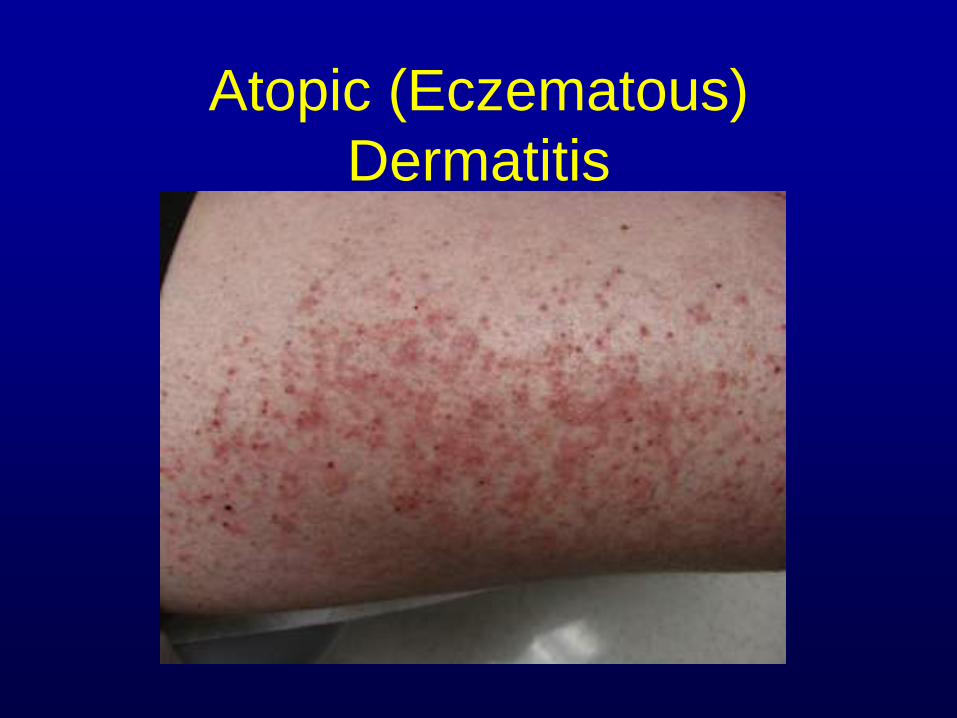
Atopic (Eczematous)
Dermatitis

Histological Pattern
• Acute - microvesical formation
• Subacute – spongiosis where bridging
between keratinocytes is conspicuous
at low power
• Chronic – mild spongiosis

Stratum Corneum:
The Permeability Barrier
• Keeps the water in
• Keeps the world out
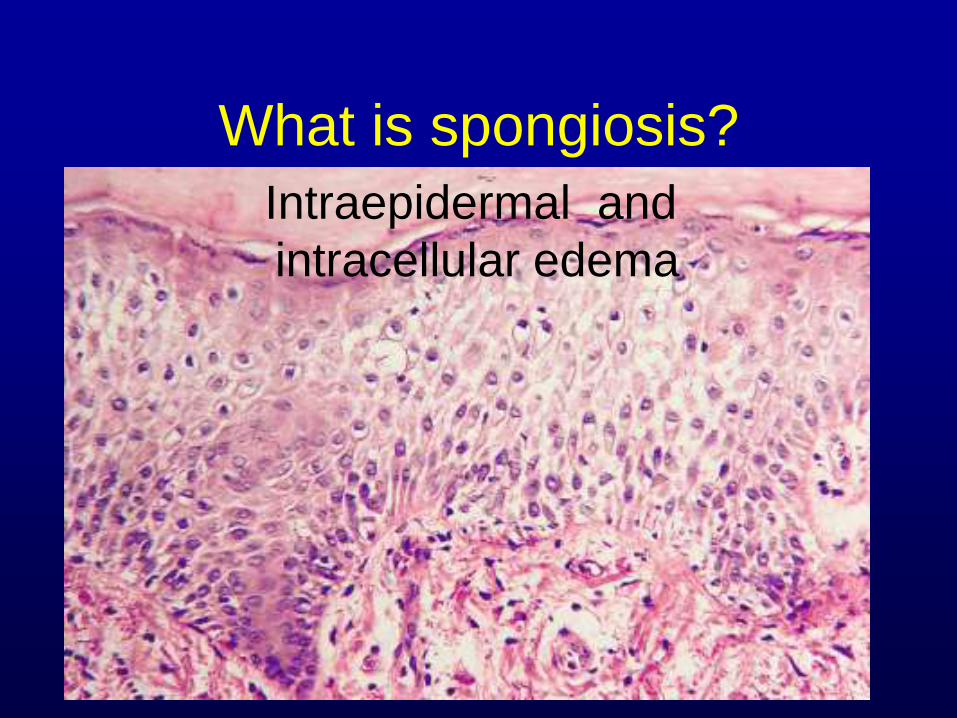
What is spongiosis?
Intraepidermal and
intracellular edema
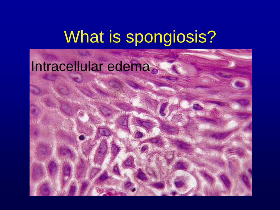
What is spongiosis?
Intracellular edema

Acute Spongiotic Dermatitis
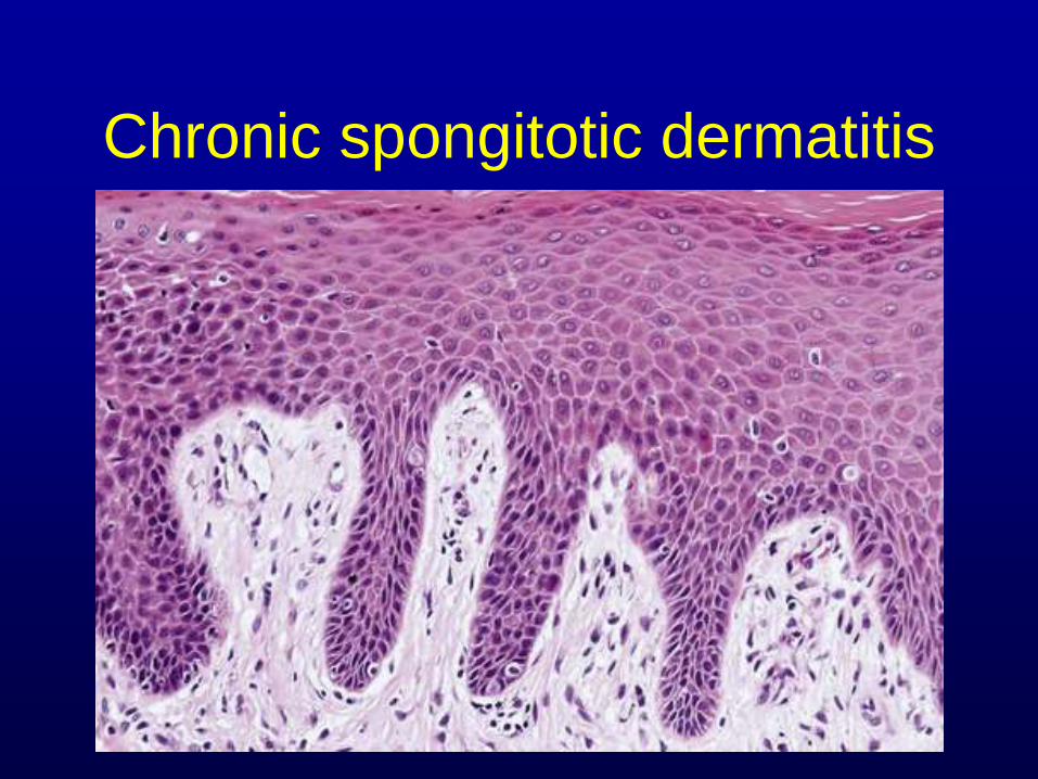
Chronic spongitotic dermatitis

Two Types of Classification
• Pathological – spongiosis under the
microscope
• Clinical presentation
– Endogenous dermatitis - related to major
constitutional or hereditary factors
– Exogenous dermatitis - involving
environmental factors.
Important
• Provide clinical history
– Description of the rash
– Distribution
– Associated medications and prior
treatment
– Differential diagnosis helps when possible
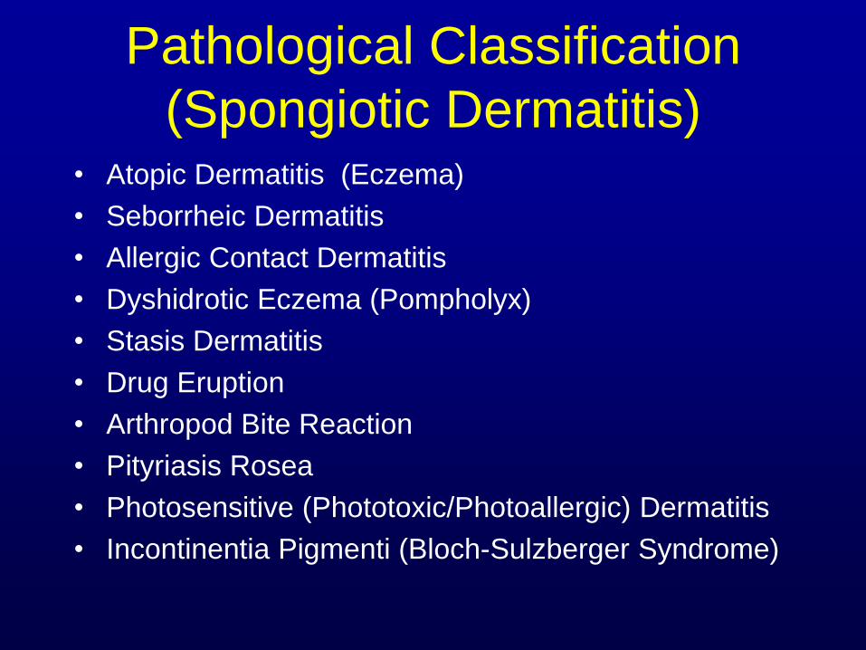
Pathological Classification
(Spongiotic Dermatitis)• Atopic Dermatitis (Eczema)
• Seborrheic Dermatitis
• Allergic Contact Dermatitis
• Dyshidrotic Eczema (Pompholyx)
• Stasis Dermatitis
• Drug Eruption
• Arthropod Bite Reaction
• Pityriasis Rosea
• Photosensitive (Phototoxic/Photoallergic) Dermatitis
• Incontinentia Pigmenti (Bloch-Sulzberger Syndrome)

Clinical Classification
(Endogenous)
• Atopic dermatitis
• Seborrheic dermatitis
• Discoid dermatitis (nummular eczema)
• Hand eczema (dyshidrotic eczema,
palmoplantar eczema, pompholyx)
• Autosensitization (Id reaction)

Clinical Classification
(Exogenous)
• Allergic Contact dermatitis – poison ivy
• Irritant dermatitis – topical damage
• Infectious – ie. fungus
• Asteatotic dermatitis - elderly, in winter
and in those with minor degrees of
ichthyosis, asteatotic dermatitis
(eczema craquelé)
Pathological Classification
(Spongiotic Dermatitis)• Atopic Dermatitis (Eczema)
• Seborrheic Dermatitis
• Allergic Contact Dermatitis
• Dyshidrotic Eczema (Pompholyx)
• Stasis Dermatitis
• Drug Eruption
• Arthropod Bite Reaction
• Pityriasis Rosea
• Photosensitive (Phototoxic/Photoallergic) Dermatitis
• Incontinentia Pigmenti (Bloch-Sulzberger Syndrome)
Establish the Diagnosis
• No objective diagnostic lab test
• No specific histopathology
• Numerous clinical presentations
• Complex pathophysiology
• Multiple, often unknown triggers

Definitive diagnosis is difficult
• Pathologists usually cannot render a
more specific diagnosis other than
– Spongiotic dermatitis consistent with
eczematous dermatitis etc.
• Can offer a limited differential diagnosis
when given some clinical information.

Chronic spongitotic dermatitis

Subacute spongiotic
dermatitis

Acute Spongiotic Dermatitis
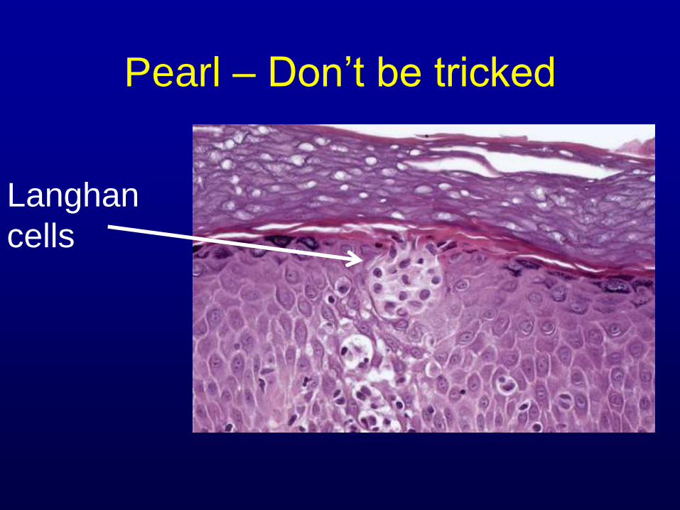
Pearl – Don’t be tricked
Langhan
cells

If we see parakeratosis?

Order a fungal stain
Atopic dermatitis
• Complex inflammatory skin disorder
– intense pruritus
– cutaneous hyperreactivity
– immune dysregulation
• Exacerbations and remissions
• Affects all ages, but more common in
kids

Atopic dermatitis
• Pathogenesis: immune mediated
• Epidemiology:
– 10% of children
– Most present before age 7
– Atopic diathesis: 75% have a personal or
family history of allergic disease

Atopic dermatitis
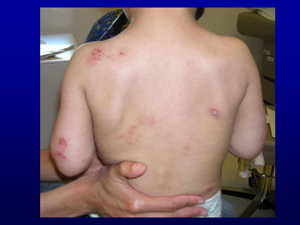
• Clinical: “the itch that rashes”
– Lesions:
• Acute: erythema and vesiculation
• Subacute: papular
• Chronic: brown/red, lichenification
– Distribution:
• Infancy: face, extensors of extremities
• Childhood: neck, antecubital and popliteal
fossae
• Adulthood: fossae, hands/feet
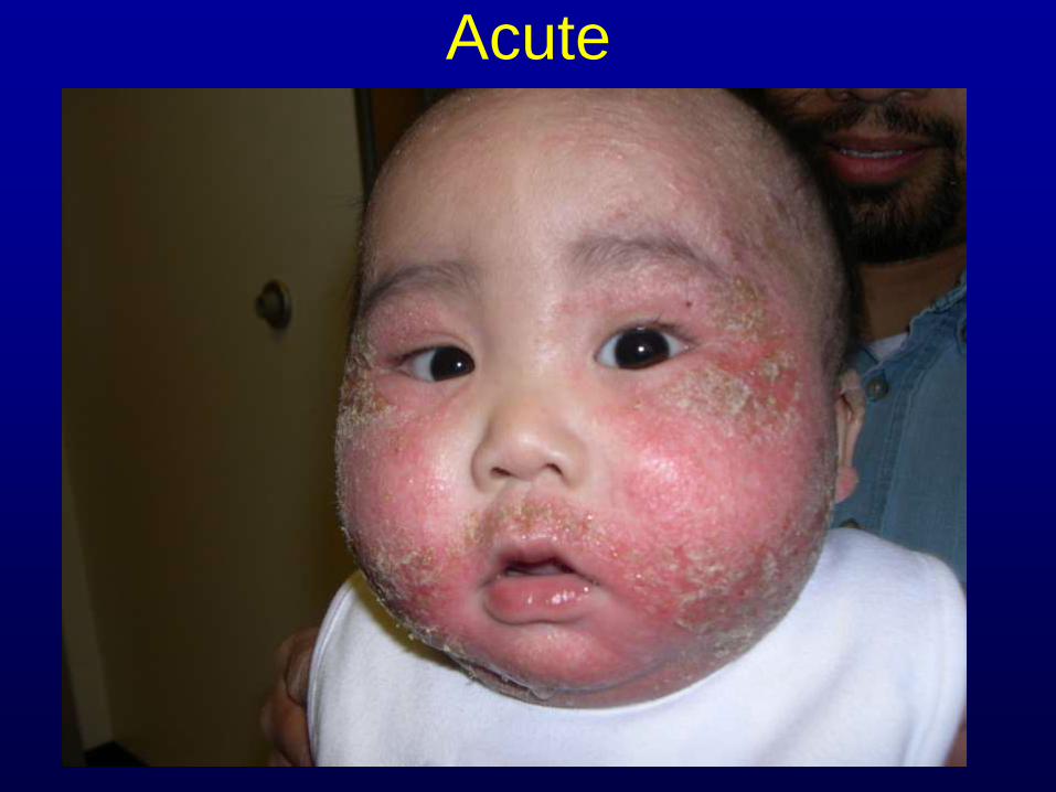
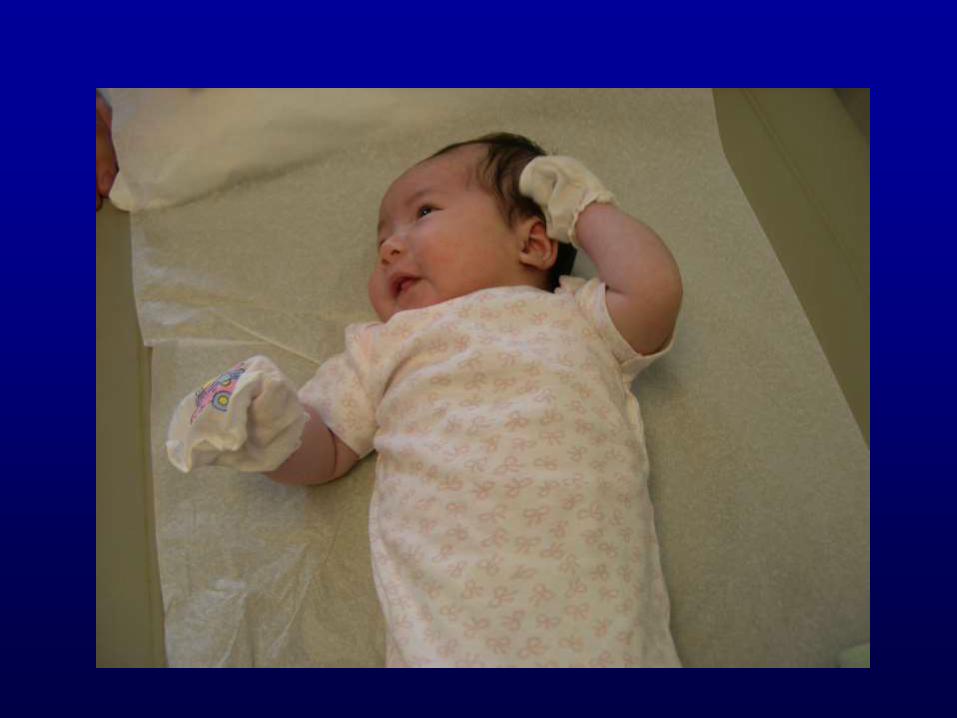
Acute
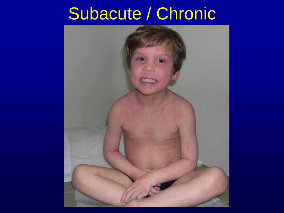
Subacute / Chronic


Atopic dermatitis
• Clinical:
– Other findings:
• Pityriasis alba
• Dennie-Morgan lines, allergic shiners
• Keratosis Pilaris
• Icthyosis Vulgaris
• Hyperlinear palms

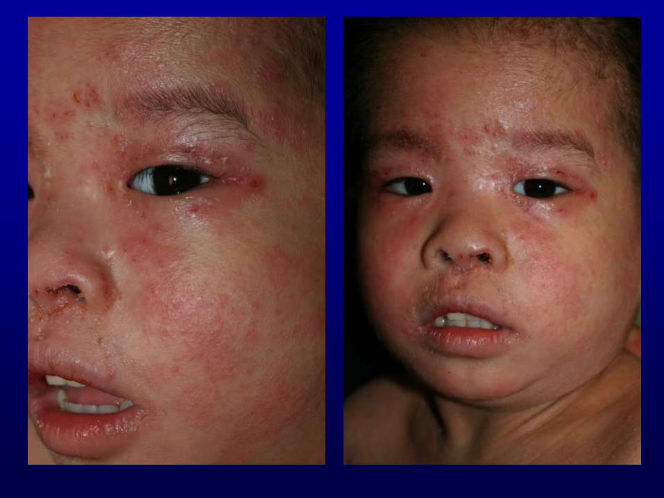
Infantile Distribution
• Face
• Elbows
• Knees

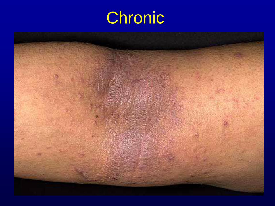
Chronic
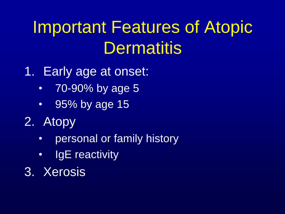
Important Features of Atopic
Dermatitis
1. Early age at onset:
• 70-90% by age 5
• 95% by age 15
2. Atopy
• personal or family history
• IgE reactivity
3. Xerosis

Common overlapping features
Asthma Excema
Hayfever

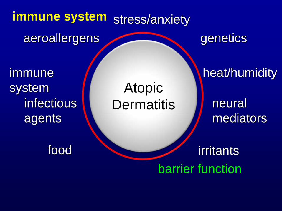
immune
system
heat/humidity
stress/anxiety
aeroallergens
food
genetics
irritants
infectious
agents
neural
mediators
Atopic
Dermatitis
immune
system
heat/humidity
stress/anxiety
aeroallergens
food
genetics
irritants
infectious
agents
neural
mediators
immune system
barrier function
Atopic
Dermatitis

The bottom line…
the diagnosis is
clinical


Exclusionary Conditions
• Scabies
• Psoriasis
• Seborrheic dermatitis
• Allergic contact dermatitis
• Cutaneous lymphoma
• Immunodeficiency diseases
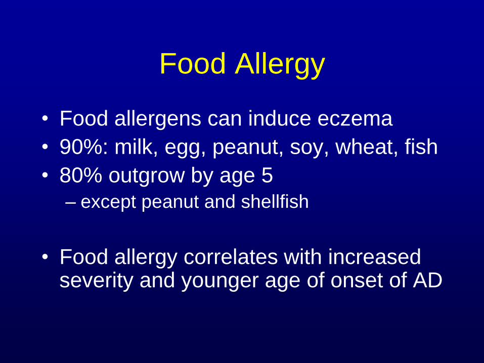
Food Allergy
• Food allergens can induce eczema
• 90%: milk, egg, peanut, soy, wheat, fish
• 80% outgrow by age 5
– except peanut and shellfish
• Food allergy correlates with increased severity and younger age of onset of AD

Scratch testing
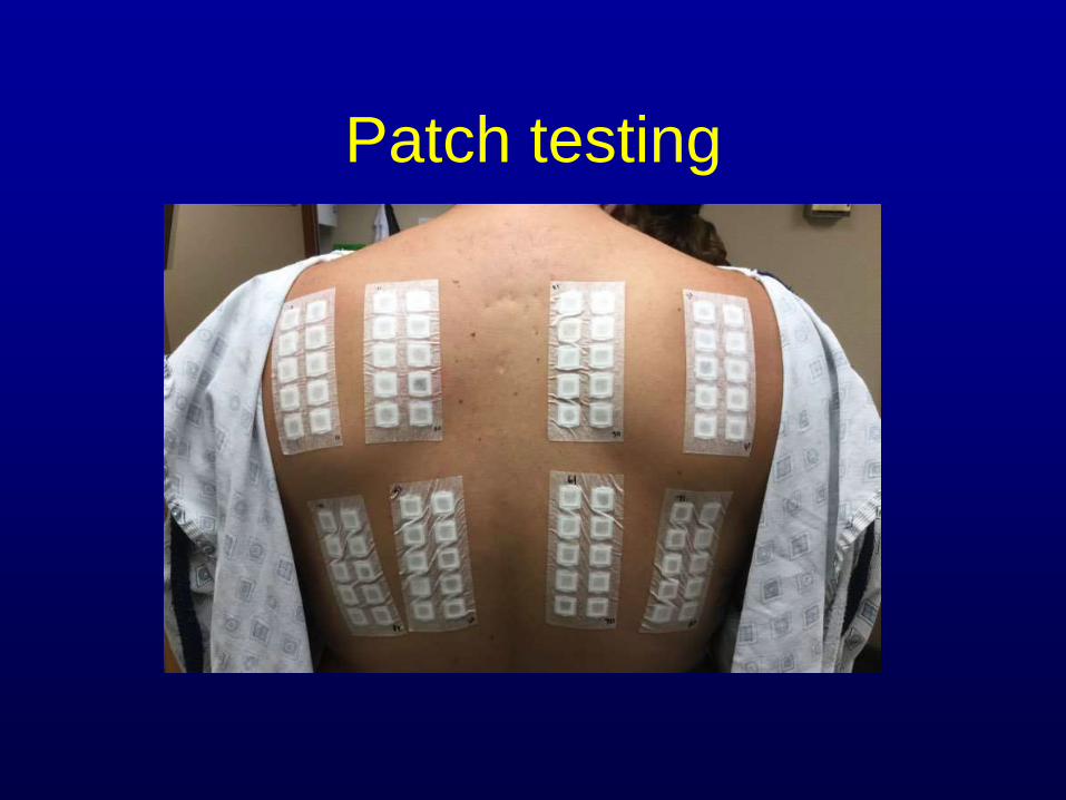
Patch testing
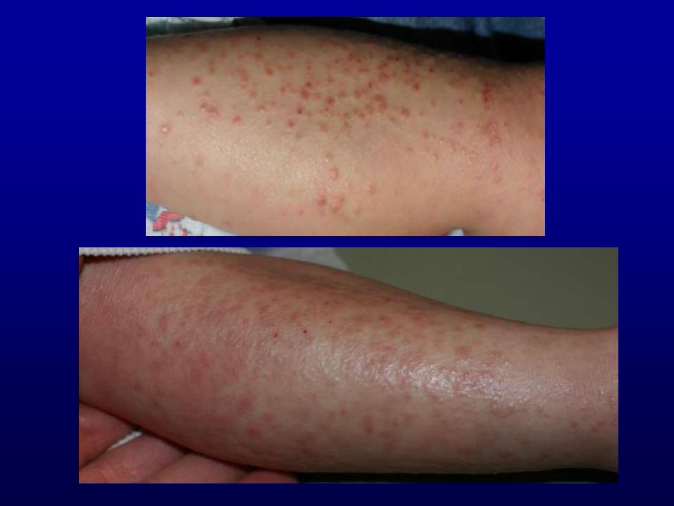


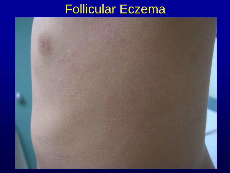
Follicular Eczema
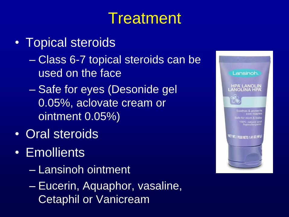
Treatment
• Topical steroids
– Class 6-7 topical steroids can be
used on the face
– Safe for eyes (Desonide gel
0.05%, aclovate cream or
ointment 0.05%)
• Oral steroids
• Emollients
– Lansinoh ointment
– Eucerin, Aquaphor, vasaline,
Cetaphil or Vanicream
Treatment
• Anithistamines
– Sedating – diphenhydramine, hydroxyzine,
cyprohepatine
– Nonsedating fexofenadine, cetirizine,
loratadine - useful, especially when there is
an urticarial component (doxepin topical or
10mg QD -tricyclic antidepressant with
potent H1 and H2 blocking properties) or
concurrent allergic rhinoconjunctivitis

Treatment
• Topical calcineurin inhibitors
– pimecrolimus1% cream or tacrolimus 0.03% to 0.1%
ointment
• Crisaborole – expensive, helpful in children
• Phototherapy– helpful in dyshidrotic eczema in adults and
severe cases
• Cyclosporin – moderate to severe cases
• Methotrexate – once a week dosing, monitor LFTs, CBC
• Mycophenolate mofetil (Cellcept) - immunosuppression
• Dupixent – IL-4 alpha antagonist, expensive, moderate to
severe cases

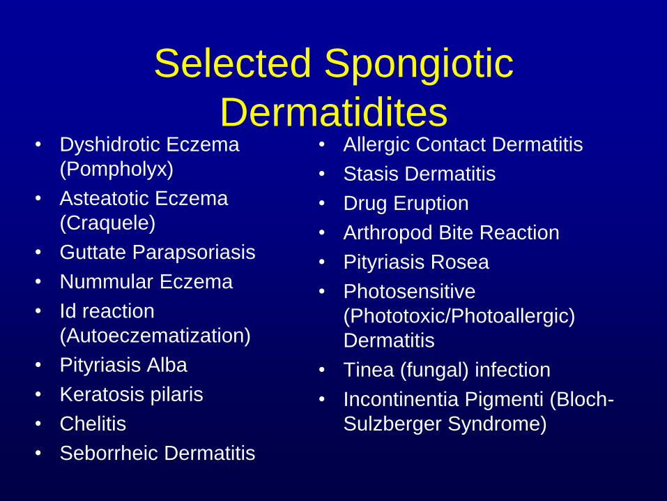
Selected Spongiotic
Dermatidites• Dyshidrotic Eczema
(Pompholyx)
• Asteatotic Eczema
(Craquele)
• Guttate Parapsoriasis
• Nummular Eczema
• Id reaction
(Autoeczematization)
• Pityriasis Alba
• Keratosis pilaris
• Chelitis
• Seborrheic Dermatitis
• Allergic Contact Dermatitis
• Stasis Dermatitis
• Drug Eruption
• Arthropod Bite Reaction
• Pityriasis Rosea
• Photosensitive
(Phototoxic/Photoallergic)
Dermatitis
• Tinea (fungal) infection
• Incontinentia Pigmenti (Bloch-
Sulzberger Syndrome)
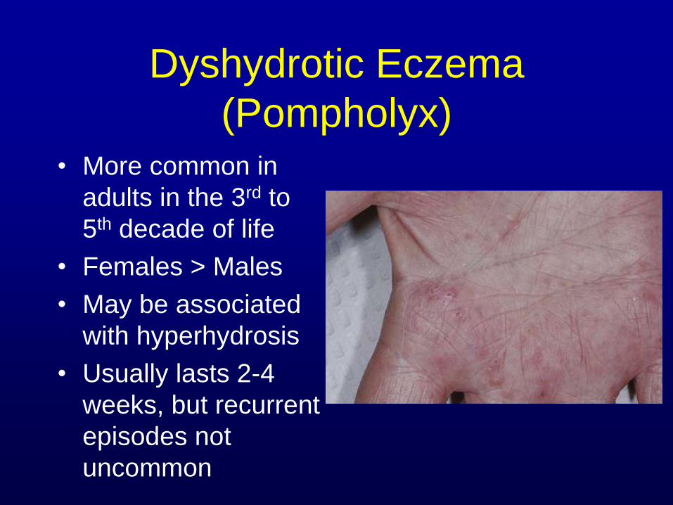
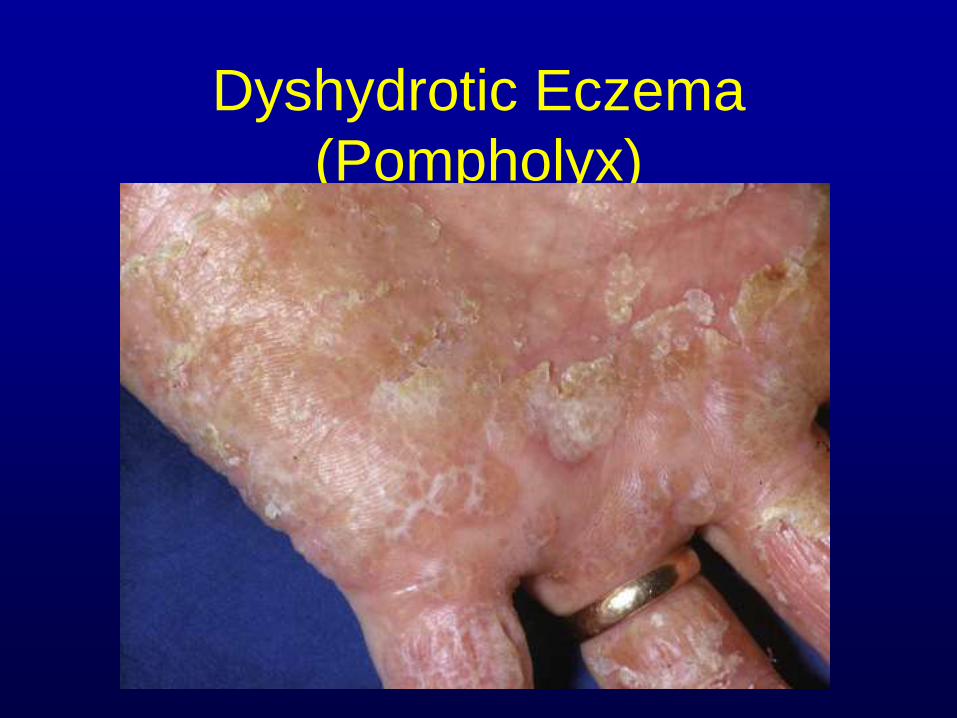
Dyshydrotic Eczema
(Pompholyx) • More common in
adults in the 3rd to
5th decade of life
• Females > Males
• May be associated
with hyperhydrosis
• Usually lasts 2-4
weeks, but recurrent
episodes not
uncommon
Dyshydrotic Eczema
(Pompholyx)

Asteatotic Eczema (Craquelé)
• Elderly, bilateral,
winter months
• Can be associated
with an underlying
malignancy
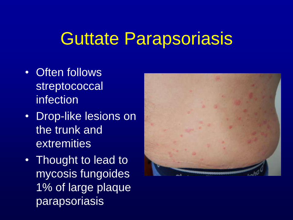
Guttate Parapsoriasis
• Often follows
streptococcal
infection
• Drop-like lesions on
the trunk and
extremities
• Thought to lead to
mycosis fungoides
1% of large plaque
parapsoriasis

Nummular Eczema
• Coin shaped tiny
papules and
papulovesicles that
become confluent
• Not related to atopic
dermatitis
• Associated with cold
dry weather, infection,
predisposing
medication
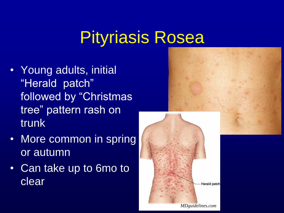
Pityriasis Rosea
• Young adults, initial
“Herald patch”
followed by “Christmas
tree” pattern rash on
trunk
• More common in spring
or autumn
• Can take up to 6mo to
clear
MDguidelines.com

Id reaction
(Autoeczematization)
• Dissemination of a
previously localized
‘eczematous’
process such as
fungal infection or
stasis dermatitis
• Commonly seen as
a reaction to foods,
look at the feet and
nails for fungus

An id reaction is an eczematous skin
reaction that develops in response to a
distant unknown antigen. Which of the
following is a known and common cause
of “id reaction”?
• A. Tinea pedis
• B. Food allergens
• C. Stasis dermatitis
• D. All of the above

Pityriasis Alba
• Hypopigmented scaly
patches with predilection for
face, neck and shoulders of
darker skinned atopic
individuals
• Usually between 6-16 years
• Topical 1% hydrocortisone
(or other low-potency steroid
cream or ointment) may be
used sparingly for 3-7 days
to abate any ongoing
inflammation.
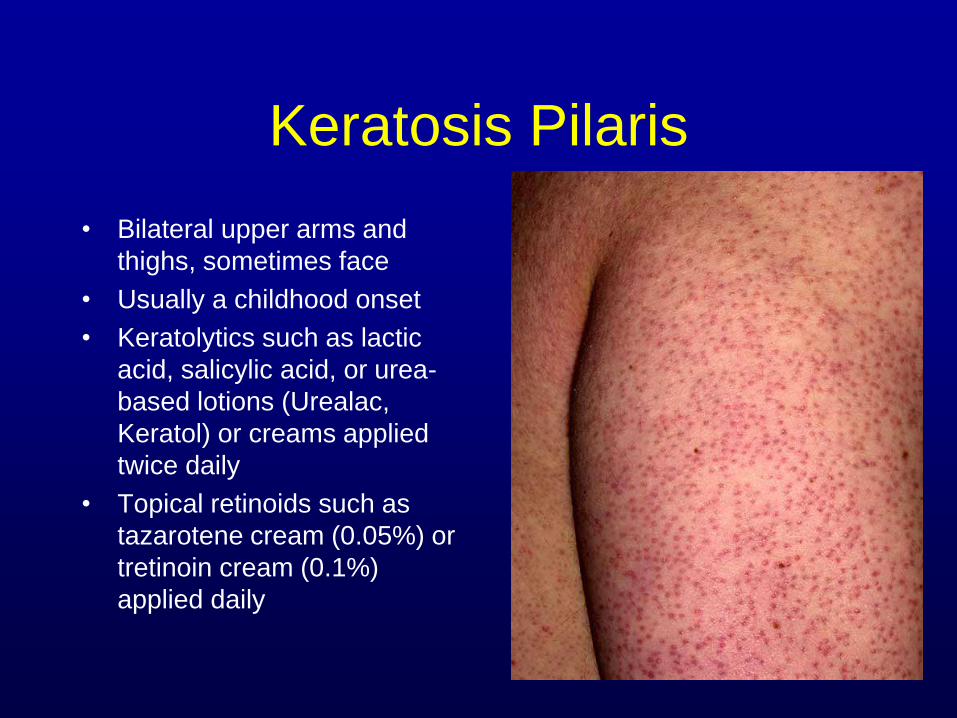
Keratosis Pilaris
• Bilateral upper arms and
thighs, sometimes face
• Usually a childhood onset
• Keratolytics such as lactic
acid, salicylic acid, or urea-
based lotions (Urealac,
Keratol) or creams applied
twice daily
• Topical retinoids such as
tazarotene cream (0.05%) or
tretinoin cream (0.1%)
applied daily
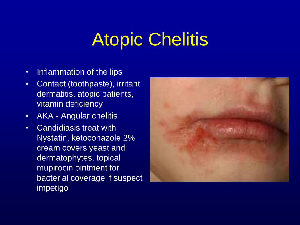
Atopic Chelitis
• Inflammation of the lips
• Contact (toothpaste), irritant
dermatitis, atopic patients,
vitamin deficiency
• AKA - Angular chelitis
• Candidiasis treat with
Nystatin, ketoconazole 2%
cream covers yeast and
dermatophytes, topical
mupirocin ointment for
bacterial coverage if suspect
impetigo

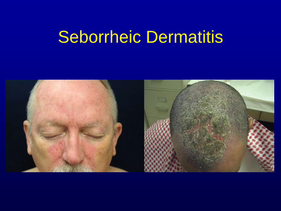
Seborrheic Dermatitis
• Affects sebum rich areas of
the skin
• Adult, caucasian, male
prediliction, AIDS,
neurological disorders
• Scalp, eyebrows, perinasal,
beard, presternal
• OTC treatment – alternate
over the counter shampoos
– Demodex mites – selenium
sulfide 1% shampoo
– Yeast-like species – Nizoral 1%
shampoo
Seborrheic Dermatitis
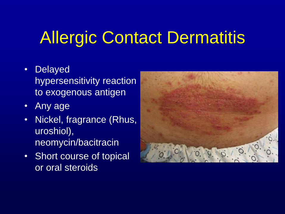
Allergic Contact Dermatitis
• Delayed
hypersensitivity reaction
to exogenous antigen
• Any age
• Nickel, fragrance (Rhus,
uroshiol),
neomycin/bacitracin
• Short course of topical
or oral steroids

Allergic Contact Dermatitis
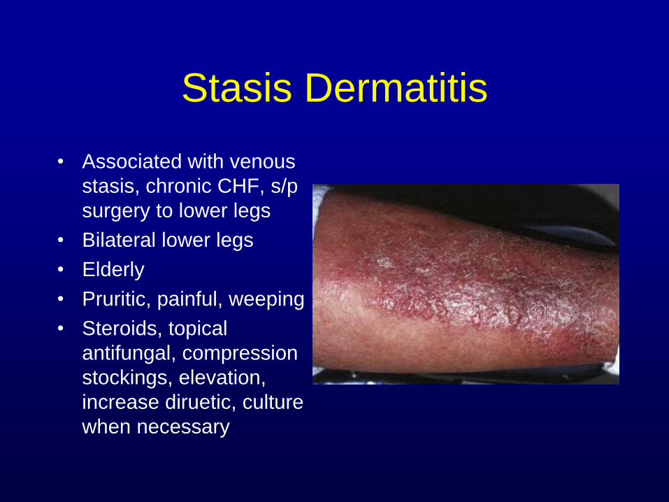
Stasis Dermatitis
• Associated with venous
stasis, chronic CHF, s/p
surgery to lower legs
• Bilateral lower legs
• Elderly
• Pruritic, painful, weeping
• Steroids, topical
antifungal, compression
stockings, elevation,
increase diruetic, culture
when necessary
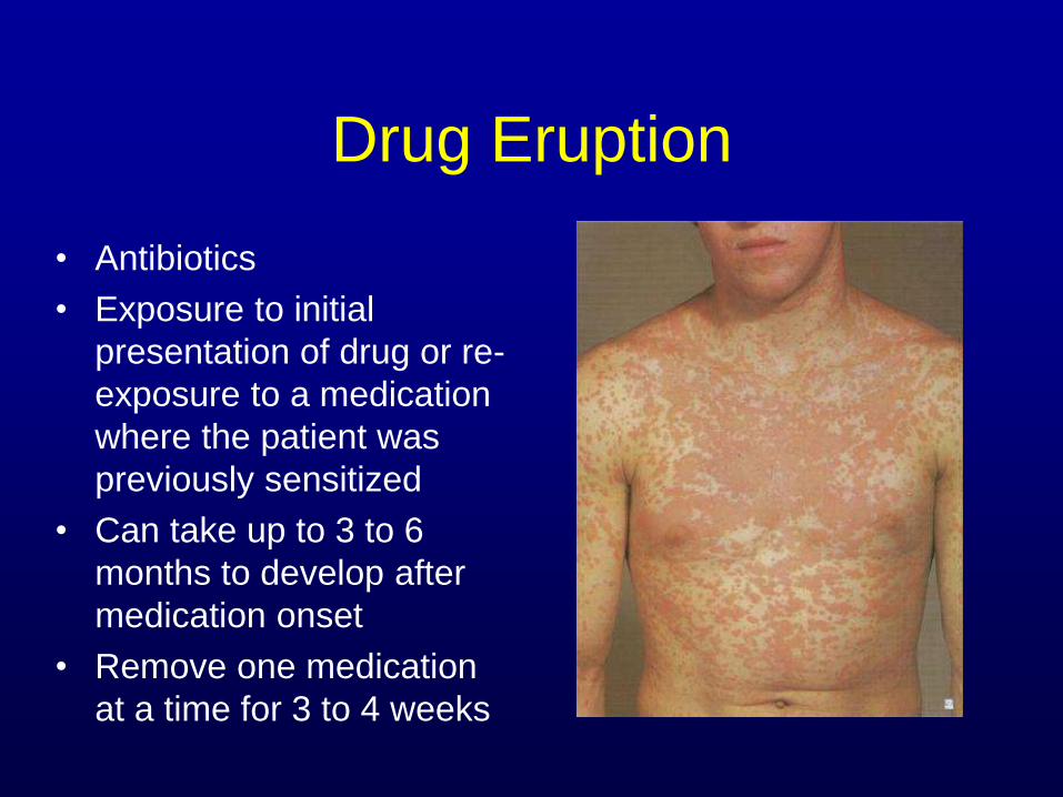
Drug Eruption
• Antibiotics
• Exposure to initial
presentation of drug or re-
exposure to a medication
where the patient was
previously sensitized
• Can take up to 3 to 6
months to develop after
medication onset
• Remove one medication
at a time for 3 to 4 weeks
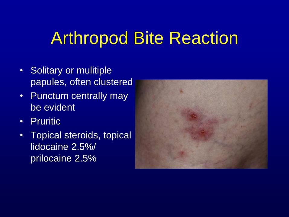
Arthropod Bite Reaction
• Solitary or mulitiple
papules, often clustered
• Punctum centrally may
be evident
• Pruritic
• Topical steroids, topical
lidocaine 2.5%/
prilocaine 2.5%

Photosensitive
(Phototoxic/Photoallergic)
Dermatitis• Can begin within
minutes of light
exposure
• Tender macular
erythema and
edema in sun
exposed areas
• r/o photo drug,
dermatomyositis,
lupus

Tinea (fungal) infection
• Infectious organisms
Trichophyton, Microsporum,
Epidermophyton species
• Children or adults
• Mimics eczema, psoriasis,
gyrate erythemas
• Topical azole creams
(ketoconazole 2%,
econazole 1%)
• Oral for severe reactions
lamisil 250mg QD x 14 days,
oral sporonox 100mg BID x
14 days

Incontinentia Pigmenti
(Bloch-Sulzberger Syndrome)
• Genodermatosis noted
at birth
• Progressive cutaneous
blistering along the lines
of Blaschko
• Mutation in the NEMO
gene
• X-linked dominant
nearly exclusively in
females

When spongiosis and parakeratosis
are present, what histochemical stain
should be ordered”?
• A. AFB
• B. GMS or PAS
• C. Gram
• D. All of the above
If eosinophils are present in the
dermis
• A. the diagnosis is eczema.
• B. the diagnosis is a medication
reaction.
• C. the diagnosis is arthropod insult.
• D. a hypersensitivity dermatitis cannot
be excluded.

When eosinophils are found in
association with neutrophils, fibrin
thrombi and leukocytoclasis, which of
the following should be considered?
• A. mastocytosis
• B. bullous pemphigoid
• C. leukocytoclastic vasculitis
• D. sarcoidosis

Treatment Summary• Elimination of exacerbating factors
– Avoid trigger factors such as heat, low humidity
– Treat skin infections such as Staphylococcus aureus and herpes simplex
Use antihistamines for sedation and control of itching
– Treat stress and anxiety
• Elimination of aeroallergens and food allergens
• Elimination of contact allergens
• Maintaining skin hydration
– Emollients and moisturizers
– Bathing practices
• Controling pruritus
• Topical/Oral steroids
Treatment Summary
• Topical calcineurin inhibitors
– pimecrolimus1% cream or tacrolimus 0.03% to 0.1%
ointment
• Crisaborole – expensive, helpful in children
• Phototherapy– helpful in dyshidrotic eczema in adults and
severe cases
• Cyclosporin – moderate to severe cases
• Methotrexate – once a week dosing, monitor LFTs, CBC
• Mycophenolate mofetil (Cellcept) - immunosuppression
• Dupixent – IL-4 alpha antagonist, expensive, moderate to
severe cases
Selected Spongiotic
Dermatidites• Dyshidrotic Eczema
(Pompholyx)
• Asteatotic Eczema
(Craquele)
• Guttate Parapsoriasis
• Nummular Eczema
• Id reaction
(Autoeczematization)
• Pityriasis Alba
• Keratosis pilaris
• Chelitis
• Seborrheic Dermatitis
• Allergic Contact Dermatitis
• Stasis Dermatitis
• Drug Eruption
• Arthropod Bite Reaction
• Pityriasis Rosea
• Photosensitive
(Phototoxic/Photoallergic)
Dermatitis
• Tinea (fungal) infection
• Incontinentia Pigmenti (Bloch-
Sulzberger Syndrome)
Pathology Report
• Chronic/subacute/acute spongiotic
dermatitis with eosinophils, see note
• NOTE:– Discribe histological features from the top down
– The findings are not diagnostic for a specific
disease process but can be identified in a variety
of forms of eczematous (hypersensitivity)
dermatidites.
– Offer a differential if possible
– Answer the clinician's question
Thank you!• Spergel JM. From atopic dermatitis to asthma: the atopic march. Ann Allergy Asthma
Immunol 2010; 105:99.
• Eichenfield LF, Tom WL, Chamlin SL, et al. Guidelines of care for the management of atopic
dermatitis: section 1. Diagnosis and assessment of atopic dermatitis. J Am Acad Dermatol
2014; 70:338.
• Williams H, Flohr C. How epidemiology has challenged 3 prevailing concepts about atopic
dermatitis. J Allergy Clin Immunol 2006; 118:209.
• Williams H, Robertson C, Stewart A, et al. Worldwide variations in the prevalence of
symptoms of atopic eczema in the International Study of Asthma and Allergies in Childhood.
J Allergy Clin Immunol 1999; 103:125.
• Shaw TE, Currie GP, Koudelka CW, Simpson EL. Eczema prevalence in the United States:
data from the 2003 National Survey of Children's Health. J Invest Dermatol 2011; 131:67.
• Weidinger S, Novak N. Atopic dermatitis. Lancet 2016; 387:1109.
• Eichenfield LF, Tom WL, Berger TG, et al. Guidelines of care for the management of atopic
dermatitis: section 2. Management and treatment of atopic dermatitis with topical therapies.
J Am Acad Dermatol 2014; 71:116.
• Sidbury R, Davis DM, Cohen DE, et al. Guidelines of care for the management of atopic
dermatitis: section 3. Management and treatment with phototherapy and systemic agents. J
Am Acad Dermatol 2014; 71:327.
• Tollefson MM, Bruckner AL, Section On Dermatology. Atopic dermatitis: skin-directed
management. Pediatrics 2014; 134:e1735.