
sponging hsa-miR-483-3p in lupus nephritis endogenous RNA ...
22
Page 1/22 Hsa_circ_0123190 acts as a competitive endogenous RNA to regulate APLNR expression by sponging hsa-miR-483-3p in lupus nephritis Chunyi Zhang Zhengzhou University First Aliated Hospital Congcong Gao Zhengzhou University First Aliated Hospital Xueqi Di Zhengzhou University First Aliated Hospital Siwan Cui Zhengzhou University First Aliated Hospital Wenfang Liang Zhengzhou University First Aliated Hospital Wenbo Sun Zhengzhou University First Aliated Hospital Menghui Yao Zhengzhou University First Aliated Hospital Shengyun Liu Zhengzhou University First Aliated Hospital Zhaohui Zheng ( [email protected] ) Zhengzhou University First Aliated Hospital https://orcid.org/0000-0003-3817-0171 Research article Keywords: circRNAs, microRNAs, competitive endogenous RNAs, lupus nephritis, systemic lupus erythematosus Posted Date: November 20th, 2020 DOI: https://doi.org/10.21203/rs.3.rs-26778/v2 License: This work is licensed under a Creative Commons Attribution 4.0 International License. Read Full License
Transcript of sponging hsa-miR-483-3p in lupus nephritis endogenous RNA ...

Page 1/22
Hsa_circ_0123190 acts as a competitiveendogenous RNA to regulate APLNR expression bysponging hsa-miR-483-3p in lupus nephritisChunyi Zhang
Zhengzhou University First A�liated HospitalCongcong Gao
Zhengzhou University First A�liated HospitalXueqi Di
Zhengzhou University First A�liated HospitalSiwan Cui
Zhengzhou University First A�liated HospitalWenfang Liang
Zhengzhou University First A�liated HospitalWenbo Sun
Zhengzhou University First A�liated HospitalMenghui Yao
Zhengzhou University First A�liated HospitalShengyun Liu
Zhengzhou University First A�liated HospitalZhaohui Zheng ( [email protected] )
Zhengzhou University First A�liated Hospital https://orcid.org/0000-0003-3817-0171
Research article
Keywords: circRNAs, microRNAs, competitive endogenous RNAs, lupus nephritis, systemic lupuserythematosus
Posted Date: November 20th, 2020
DOI: https://doi.org/10.21203/rs.3.rs-26778/v2
License: This work is licensed under a Creative Commons Attribution 4.0 International License. Read Full License

Page 2/22
Version of Record: A version of this preprint was published on January 13th, 2021. See the publishedversion at https://doi.org/10.1186/s13075-020-02404-8.
Page 3/22
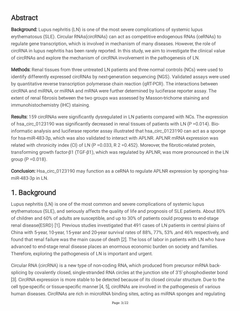
AbstractBackground: Lupus nephritis (LN) is one of the most severe complications of systemic lupuserythematosus (SLE). Circular RNAs(circRNAs) can act as competitive endogenous RNAs (ceRNAs) toregulate gene transcription, which is involved in mechanism of many diseases. However, the role ofcircRNA in lupus nephritis has been rarely reported. In this study, we aim to investigate the clinical valueof circRNAs and explore the mechanism of circRNA involvement in the pathogenesis of LN.
Methods: Renal tissues from three untreated LN patients and three normal controls (NCs) were used toidentify differently expressed circRNAs by next-generation sequencing (NGS). Validated assays were usedby quantitative reverse transcription polymerase chain reaction (qRT-PCR). The interactions betweencircRNA and miRNA, or miRNA and mRNA were further determined by luciferase reporter assay. Theextent of renal �brosis between the two groups was assessed by Masson-trichome staining andimmunohistochemistry (IHC) staining.
Results: 159 circRNAs were signi�cantly dysregulated in LN patients compared with NCs. The expressionof hsa_circ_0123190 was signi�cantly decreased in renal tissues of patients with LN (P =0.014). Bio-informatic analysis and luciferase reporter assay illustrated that hsa_circ_0123190 can act as a spongefor hsa-miR-483-3p, which was also validated to interact with APLNR. APLNR mRNA expression wasrelated with chronicity index (CI) of LN (P =0.033, R 2 =0.452). Moreover, the �brotic-related protein,transforming growth factor-β1 (TGF-β1), which was regulated by APLNR, was more pronounced in the LNgroup (P =0.018).
Conclusion: Hsa_circ_0123190 may function as a ceRNA to regulate APLNR expression by sponging hsa-miR-483-3p in LN.
1. BackgroundLupus nephritis (LN) is one of the most common and severe complications of systemic lupuserythematosus (SLE), and seriously affects the quality of life and prognosis of SLE patients. About 80%of children and 60% of adults are susceptible, and up to 30% of patients could progress to end-stagerenal disease(ESRD) [1]. Previous studies investigated that 491 cases of LN patients in central plains ofChina with 5-year, 10-year, 15-year and 20-year survival rates of 88%, 77%, 53% ,and 46% respectively, andfound that renal failure was the main cause of death [2]. The loss of labor in patients with LN who haveadvanced to end-stage renal disease places an enormous economic burden on society and families.Therefore, exploring the pathogenesis of LN is important and urgent.
Circular RNA (circRNA) is a new type of non-coding RNA, which produced from precursor mRNA back-splicing by covalently closed, single-stranded RNA circles at the junction site of 3’5’-phosphodiester bond[3]. CircRNA expression is more stable to be detected because of its closed circular structure. Due to thecell type-speci�c or tissue-speci�c manner [4, 5], circRNAs are involved in the pathogenesis of varioushuman diseases. CircRNAs are rich in microRNA binding sites, acting as miRNA sponges and regulating

Page 4/22
gene transcription, which is called competitive endogenous RNA (ceRNA) molecules [6, 7]. In the studieson renal diseases, Wang et al. reported that androgen receptor enhanced migration and invasion of renaltransparent cell carcinoma by inhibiting the expression of circHTAT1 regulating miR-195-5p/29a-3p/29c-3p [8]. In addition, circRNA ZNF609 was found to regulate fork head box P4 (FOXP4) expression bytargeting miR-138-5p in renal carcinoma[9].However, the role and mechanism of circRNAs in LN had beenrarely reported.
Renal �brosis is a common pathological feature of progressive LN, which is closely related to ESRD[10,11].Apelin and its receptor (apelin receptor, APLNR) can be widely distributed in heart, lung, pancreas,kidney and other tissues [12]. Numerous evidences indicate that apelin and APLNR play a key role invarious kidney diseases, such as renal �brosis, renal ischemia/reperfusion injury, polycystic kidneydisease and diabetic nephropathy[13].Particularly, apelin and APLNR could inhibit the deposition ofextracellular matrix (ECM) and attenuate renal �brosis by acting on TGF-β [14]. In the present study, we�rstly established the circRNAs expression pro�le in kidney tissues of patients with LN. And then, wefurther explored that hsa_circ_0123190 was a novel biomarker of peripheral blood for LN and could actas a sponge for hsa-miR-483-3p to regulate APLNR expression involved in renal �brosis in LN.
2. Methods2.1 Subjects and samples
A total of 10 LN patients with renal biopsy were enrolled in this study between May 2018 and December2018 from the Department of Rheumatology and Immunology of the First A�liated Hospital ofZhengzhou University. Five patients with renal tumor were from the Urology Department at the samehospital. Ten peripheral blood samples were collected from the volunteers as healthy controls (HCs). Allpatients and volunteers were female, between 18 to 60 years old. The following were exclusion criteria:(1) patients with serious infection within one month before admission; (2) patients with malignanttumors; (3) patients with other autoimmune diseases; (4) patients with pregnancy; and (5) patients witheGFR lower than 30 mL/min/1.73m2.
Kidney tissues from LN patients were obtained from renal biopsies before the treatment with steroidand/or immunosuppressant. Renal normal controls (NCs) were kidney tissues at least 5 cm from theedge of tumor from patients with renal cancer, and then con�rmed to be normal histological morphologyunder microscopy. All fresh tissues were stored in RNAlater® Solution (Thermo Fisher Scienti�c, CA, USA)and then frozen in -80℃ until RNA extraction. All peripheral blood samples (2mL) were drawn from themedian cubital vein with an PAXgene Blood RNA Tube (Qiagen, Hilden, Germany).
2.2 NGS pro�ling analysis
Total RNA was extracted from the frozen renal tissues with using Trizol LS reagent (Invitrogen, CA, USA).Total RNA from fresh peripheral blood samples are isolated by PAXgene Blood RNA Kit (Qiagen, Hilden,Germany). Total RNA was quanti�ed and quali�ed by an Agilent 2100 Bioanalyzer (Agilent Technologies,
Page 5/22
CA, USA), NanoDrop™ 2000 spectrophotometer (Thermo Fisher Scienti�c, CA, USA) and 1% agarose gel.The criteria of total RNA was used for subsequent library preparation: (1) the value of OD260/280 wasbetween1.8~2.2 and OD260/230 was above 2.0;(2) the value of RIN was above seven.
NGS library preparation was performed by Genesky Biotechnologies Inc (Shanghai, China). The rRNA wasdepleted from total RNA by Ribo-ZeroTM rRNA removal kit (Human/Mouse/Rat) (Illumina, CA, USA) beforebuilding the RNA-seq library. After puri�cation, divalent cations at higher temperatures were applied formaking small pieces of fragments of the residual RNA fractions. The reverse transcription of all thecleaved RNA fragments was used to construct the complementary DNA (cDNA) library with TruSeqStranded Total RNA Library Prep Kit (Illumina, CA, USA) according to the manufacturer’s instructions. Thelibrary quality was evaluated with Agilent 2100 Bioanalyzer (Agilent Technologies, CA, USA). RNA librarieswere denatured as single-stranded DNA molecules. Finally, sequencing was carried out using a 2×150 base paired-end con�guration with Illumina Hiseq 2500 (Illumina, CA, USA).
2.3 GO and KEGG pathway analysis
The predicted functions of the differentially expressed circRNAs between LN and NC were conducted bygene ontology (GO, http://geneontology.org) and Kyoto Encyclopedia of Genes and Genomes (KEGG,http://www.kegg.jp) analysis. Hierarchical clustering of the differentially expressed circRNA according tothree categories, the biological process (BP), cellular component (CC), and molecular function (MF) wasused by GO analysis. Pathway analysis of circRNA was performed by KEGG database.
2.4 QRT-PCR validation
The candidate circRNAs were selected according to the following: Firstly, the top 20 differently expressedcircRNAs between the LN group and the control group were selected. Secondly, according to the results ofGO and KEGG enrichment analysis and literature, the circRNAs with most likelihood to be related to thepathogenesis of LN was analyzed. Thirdly, the primers of circRNAs for qRT-PCR could be designed.Finally, every of the candidate circRNAs had only one circBase ID. QRT-PCR with SYBR green analysis wasused to validate the expression of the selected circRNAs from circRNA pro�les. Total RNA was extractedby using Trizol reagent (Invitrogen, Carlsbad, CA, USA). The cDNA was synthesized by ReverseTranscriptase M-MLV (Takara, Tokyo, Japan). In addition, qRT-PCR was performed with SYBR® Premix ExTaq™ II (Takara, Tokyo, Japan). GAPDH was used as endogenous control for circRNA qRT-PCR, and U6 formiRNA. 1 μg of RNA was mixed with 1 μL of RNase R (20 U/μL) at 37°C for 20 min, and then, washedwith RNeasy cleaning agent. All of the primers are listed in Table 1. The relative circRNAs expression wascalculated using 2−ΔΔCt, with ΔCt = Ct target – Ct β-actin, −ΔΔCt = − (ΔCt sample − ΔCt control).
2.5 Dual-luciferase reporter assay
Luciferase reporter assays were used to detect the direct binding between selected RNAs. Thehsa_circ_0123190 wild‐type (WT) 3′‐UTR, APLNR WT 3′‐UTR and mutant sequence hsa_circ_0123190(MUT) 3′‐UTR, APLNR MUT 3′‐UTR were constructed. Subsequently, we inoculated HEK293T cells into 96‐

Page 6/22
well plates and co-transfected 5 pmol hsa-miR-483-3p mimics or negative control with the 0.16 μghsa_circ_0123190 and APLNR wild‐type or mutant type plasmids. We tested the �uorescence intensityusing the dual‐luciferase reporter gene assay system (Promega, Madison, USA) after 48 hours oftransfection.
2.6 Histology and immunostaining
Para�n-embedded kidney tissues from human were cut at 2 μm thickness, depara�nized, rehydrated.Then, for histological examination, kidney sections were stained with Masson-trichrome reagent toexplore the degree of �brosis. Moreover, for immunohistochemistry (IHC) staining, kidney sections wereincubated in citrate buffer for 20 min at 95 °C to retrieve antigen. Non- speci�c binding was blocked with10% normal goat serum for 30 min at RT. The slides were incubated with antibodies of TGF-β1, α-smoothmuscle antibody (α-SMA) overnight at 4 °C, followed by incubation with biotin-conjugated goat anti-mouse/rabbit immunoglobulin IgG for 30 min at RT, and then reacted with streptavidin-conjugatedperoxidase for 30 min at RT. On IHC staining, we semi-quanti�ed the stained slides using a speci�cimmunohistochemical histological score technique, H-score[15]. The H-score was obtained by multiplyingthe staining intensity by a constant to adjust the mean to the strongest staining, to produce a score in therange of 0‐300. Speci�cally, H‐score=scale x percentage of strong staining (0‐100%). H‐score=1.0indicated a weak percentage; 2.0 indicated a moderate percentage; and 3.0 indicated a strong percentage.
2.7 Western blot
RIPA buffer was used for tissues lysis, according to the manufacturer’s instructions, and the proteinconcentration was determined using a Bicinchoninic Acid (BCA) Protein Assay Kit. Proteins wereseparated by SDS-PAGE using Tris-Glycine 10% polyacrylamide gels in SDS page running buffer andtransferred to PVDF membrane (Servicebio, Wuhan, China). Membranes were immunoblotted withantibodies against APLNR (1:500, Proteintech, 20341-1-AP) after blocking in 5% milk. Following primaryantibody incubation, membranes were probed with HRP-conjugated mouse anti-rabbit secondaryantibody (1:5000, Santa Cruz, sc-374015) and imaged using the Chemidoc system (BioRad).
2.8 Statistical analysis
Statistical software SPSS 25.0 (IBM Corporation, USA) was used for statistical analysis. Scatterdiagrams were drawn by GraphPad Prism version 6.0 (GraphPad Software, Inc, CA, USA). Differencesbetween two groups were analyzed for statistical signi�cance by t test. The correlation between circRNAslevel and clinical parameters of LN were analyzed by Pearson’s linear correlation. ROC curves wereperformed, and the speci�city and sensitivity of predictive power was assessed by area under curve(AUC). A P value < 0.05 was considered as statistically signi�cant.
3. Results3.1 Pro�ling and characteristics of circRNAs in renal tissues of LN patients

Page 7/22
Firstly, we analyzed the circRNAs pro�ling in renal tissues of three LN patients and three NCs by next-generation sequencing. The clinical information for the 3 patients and NCs for circRNA pro�ling wereshown in Supplement Table 1 and 2. Results showed that total 159 circRNAs were identi�ed to beabnormally expressed in renal tissues of LN patients compared with NCs (log2|fold change| ≥ 1, P<0.05),of which 73 circRNAs were signi�cantly upregulated and 86 circRNAs were remarkably downregulated.The top 10 upregulated and downregulated circRNAs were summarized in Table 2. Hierarchical clusteringheatmap and volcano plots were used to show the different expression levels of circRNAs between LNand NC (Addition �le 1: Supplement Figure 1).
3.2 GO and KEGG analysis in LN
Gene Ontology (GO) analysis predicted the functions of differentially expressed circRNAs through thehost genes based on three common aspects, including biological processes (BP), cellular components(CC), and molecular functions (MF). We selected the top 10 GO terms where circRNAs were signi�cantlyenriched in each of three biological functions. In the MF and CC category, we found that lysophosphatidicacid acyltransferase activity and apical plasma membrane were signi�cantly regulated by thedifferentially expressed circRNAs in LN kidneys. Moreover, in the BP category, enriched terms includedbiological regulation, cellular process, and metabolic process. Positive regulation of autophagy, whichplays an important role in the pathogenesis of LN, was signi�cantly regulated by these circRNAs in the BPcategory (Addition �le 1: Supplement Figure 2).
Correspondingly, the Kyoto Encyclopedia of Genes and Genomes (KEGG) analysis can de�ne themolecular function pathway of target host genes of the 159 dysregulated circRNAs. Target genes ofdifferentially expressed circRNAs were enriched in pathways, such as apelin signaling pathway andtumor necrosis factor (TNF) signaling pathway. The molecular function of the associated apelinsignaling pathway involves in regulating the expression of beclin1 and LC3, which are both importantfactors in autophagy, one of well-known pathogenesis in LN[16]. TNF-α contributes to the development ofT cells, B cells, and dendritic cells in pathogenesis of SLE[17]. Both pathways were associated withactivation of PI3K, which is an important factor in the pathogenesis of LN (Addition �le 1: SupplementFigure 2).
3.3 Validation of the selected circRNAs in renal tissues
Since females are predominantly susceptible to lupus than males, ten female patients with LN wereenrolled in this study, except for males. Characteristics of all 10 LN patients are shown in Table 3. Withage and gender match, �ve females with renal tumor were used as control group of normal kidneys. In LNpatients, urinary analysis showed 24-hr urinary total protein excretion and numbers of red blood cell,white blood cell and serology displayed levels of complements, erythrocyte sedimentation rate, serumcreatine, albumin and routine blood indexes, the increased score of systemic lupus erythematous activityof diseases indices (SLEDAI), and different severity of renal dysfunction. Ten LN patients presented

Page 8/22
various pathological classi�cations (class III n=2, class IV n=5, class III+V n=1, class IV+V n=2), whenactivity index (AI) and chronicity index (CI) were both assessed.
We selected three downregulated circRNAs (hsa_circ_0000660, hsa_circ_0007379, andhsa_circ_0123190) and one upregulated circRNAs (hsa_circ_0003302) for the further validation in renalsamples of 10 LN patients and 5 NC subjects by qRT-PCR. All four circRNAs showed similar expressionpatterns between the qRT-PCR and the circRNA sequencing. However, only hsa_circ_0123190 wassigni�cantly dysregulated between the renal tissues of patients with LN and NCs (P=0.014) (Fig.1a).Therefore, hsa_circ_0123190 was considered as a candidate for further analysis. Moreover,hsa_circ_0123190 was resistant to RNase R digestion (P>0.05), suggesting that hsa_circ_0123190 iscircular (Fig.1b). The clinical signi�cance of hsa_circ_0123190 in LN patients was investigated. Therewas no association of hsa_circ_0123190 with clinical parameters which we collected (all P>0.05).
3.4 Hsa_circ_012319 serves as a sponge for hsa-miR-483-3p
In view of the fact that circRNA can act as a miRNA sponge, the potential targets miRNAs ofhsa_circ_0123190 were predicted using bioinformatics (CircInteractome, and miRanda). As a result, wefound that hsa-miR-483-3p was the most likely complementary miRNA, which had a perfect matchsequence to bind hsa_circ_0123190. We examined the expression of hsa-miR-483-3p in renal tissuesusing qRT-PCR. The results revealed that the expression of hsa-miR-483-3p was signi�cantly increased inLN compared with NCs (P=0.0498) (Fig.2a). To further verify the hypothesis that hsa-miR-483-3p directlytargets hsa_circ_0123190, we performed the dual-luciferase reporter assay with HEK293T cells, and theresults showed that the relative luciferase activity was signi�cantly reduced in cells co-transfected withhsa_circ_0123190 WT and hsa-miR-483-3p mimics compared with control. The relative luciferase activitywas unchanged in co-transfected cell with hsa_circ_0123190 MUT and hsa-miR-483-3p mimicscompared with control group (Fig. 2b). In a word, all results indicate that hsa_circ_0123190 targets hsa-miR-483-3p.
3.5 APLNR is the target gene of hsa-miR-483-3p
The potential targets genes of hsa-miR-483-3p was searched with bioinformatics (targetScan andmiRanda). Based on the NGS pro�ling of mRNA in this study, we found APLNR had perfect bindingrelationship with hsa-miR-483-3p. The expression of APLNR in kidneys were tested using qRT-PCR andwestern blot (Fig.3a, b). It was found that the mRNA level of APLNR showed a remarkable downregulationin the LN renal tissues compared with NCs. However, the APLNR protein expression was slightlydecreased with no signi�cance in kidney of LN(n=3) (P>.0.05). To further determine the interactionbetween hsa-miR-483-3p and APLNR, we transfected plasmids of h-APLNR-3UTR-WT and h-APLNR-3UTR-MUT into HEK293T cells. The luciferase activity is signi�cantly decreased in the cells co-transfected withh-APLNR-3UTR-WT and hsa-miR-483-3p mimics compared with control group. But the luciferase activityin co-transfected cells with h-APLNR-3UTR-MUT and hsa-miR-483-3p mimics had no signi�cantdifference compared with the control group (Fig.3c). In addition, the level of APLNR in peripheral bloodwas detected but failed.

Page 9/22
3.6 Renal �brosis may correlate with the expression of APLNR
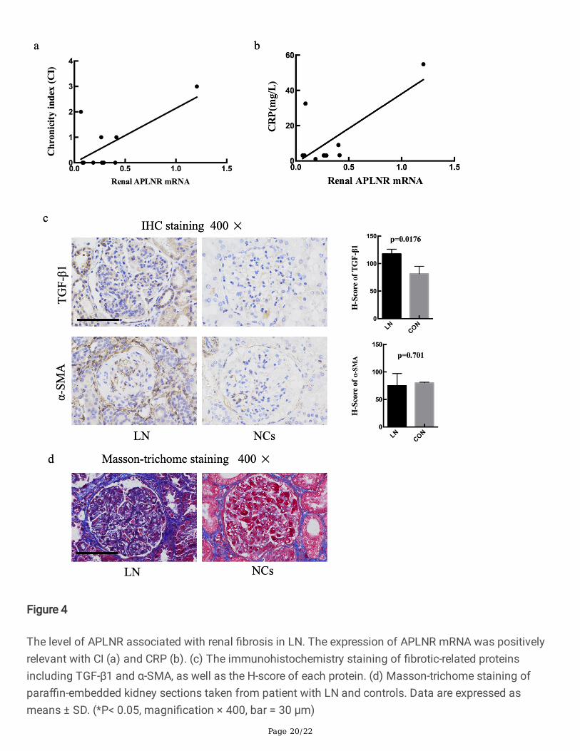
The relationship between the mRNA level of APLNR and the clinical characteristics of LN patients wasexplored. The results showed that the expression of APLNR was positively relevant with chronicity index(CI) (P=0.033, R2=0.45) and C-reactive protein (CRP) (P=0.015, R2=0.54) (Fig.4a, b). APLNR, as apelinreceptor, was involved in renal �brosis by regulating the expression of TGF-β1[13]. Pro-�brotic proteinTGF-β1 and α-SMA are central features of renal �brosis[18]. Therefore, we assessed the extent of TGF-β1and α-SMA IHC staining using H-score to semi-quantify. LN renal tissues showed signi�cant highexpression of TGF-β1(P=0.0176) compared with NCs (Fig.4c). In addition, Masson-trichome staining wasfurther demonstrated that �brosis was signi�cantly promoted in LN group (Fig. 4d).
3.7 Validation of hsa_circ_0123190 expression in peripheral blood of LN patients
The recent studies revealed that circRNA had tissue-speci�c manner, we identi�ed hsa_circ_0123190 fromrenal tissues. However, renal biopsy is an invasive and expensive operation, hsa_circ_0123190 wassupposed to be validated by qRT-PCR in peripheral blood of ten LN patients and ten healthy controls. As aresult, the expression of hsa_circ_0123190 was remarkably decreased in LN patients (P=0.0005) (Fig.5a). In addition, we assessed the correlation between circRNA levels and clinical characteristics in LNpatients. The expression of hsa_circ_0123190 was negatively correlated with leukocyte levels (P=0.0123,R2=0.5638), complement 4 (C4) levels (P=0.0099, R2=0.5855). Moreover, hsa_circ_0123190 expressionwas signi�cantly associated with serum creatine levels (P=0.044, R2=0.4151) (Fig. 5b).
The important utility of hsa_circ_0123190 was further explored by ROC curve analysis. The area underthe curve (AUC) for hsa_circ_0123190 in peripheral blood when distinguishing LN patients from NCs was0.900 (95% CI 0.7659-1.034, P= 0.0025). The maximum Youden’s J index (sensitivity and speci�city) was70% (90% and 80%) for hsa_circ_0123190 to differentiate LN patients from controls, the correspondingoptimal cut-off values was 0.6773 (Fig. 5c).
4. DiscussionThe poor prognosis of LN is still a seriously clinical and economic problem, as the mechanisms of LNremain indistinct[19]. Thus, the identi�cation of novel treatment targets for LN would be very desirable.CircRNA is a special class of endogenous RNAs with multiple functions[20]. Many studies have revealedthe abnormal circRNAs were associated with several renal diseases ,such as AKI[21], carcinoma[8, 22].However, the role of circRNA in LN was rarely reported. In the present study, we conducted integrativeanalysis using circRNA sequencing in renal tissues and identi�ed 159 circRNAs with signi�cantlydifferential expression, of which 73 were upregulated and 86 downregulated. Hsa_circ_0123190 maybeact as a sponge for hsa-miR-483-3p, which regulates APLNR expression in LN. In addition, peripheralblood hsa_circ_0123190 would be a biomarker for patients with LN.
Special attention must be paid to the type of specimen since circRNAs were highly expressed in a celltype-speci�c or tissue-speci�c manner[4, 5]. Ouyang et al discovered that upregulated plasma
Page 10/22
circRNA_002453 level in LN patients was associated with the severity of renal involvement and served asa novel biomarker for LN patient diagnosis [23]. Luan et al, showed that circHLA-C played an importantrole in the pathogenesis by sponging miR-150 in LN [24]. These data are not consistent across researchesdue to different samples and methods. For instance, Luan et al reported a circRNA pro�ling in single classIV of LN and found seven differentially expressed circRNAs. The reasons why the results were differentfrom our study may be as follows: different sample sources, different sample sizes and different renalpathological types. Compared with study by Luan et al, the present study covered the commonproliferative classi�cation of LN to explore the role of circRNA and validated differentially expressedcircRNAs both in peripheral blood and kidney tissues. In addition, we performed ROC curve analysis toensure the clinical value of the circRNAs as diagnostic biomarkers for LN. The pro�ling of circRNAs fromthis study may provide a novel database and new view to study mechanisms of LN.
Over the last few years, many functions of circRNAs have been elucidated. CircRNAs can act as geneexpression regulators via different regulatory modes[25],such as “sponges” miRNAs resulting in theexpression of target mRNAs[7]. The interaction network of circRNA in LN was slightly shed in the currentstudy. The circRNA hsa_circ_0123190 was downregulated in renal tissues of patients with LN, and theexpression of hsa-miR-483-3p had a signi�cantly opposite direction. Bioinformatic analysis andluciferase reporter assay discovered that hsa_circ_0123190 directly bind to hsa-miR-483-3p, which servesas a miRNA sponge. There have been several reports that increased hsa-miR-483-3p impair endothelialcell survival to limit vascular repair capacity upon injury in cancer and metabolic diseases [26-28].However, the role of hsa-miR-483-3p has been unreported in LN, which also has vascular lesion. Thissuggests that the downregulated hsa_circ_0123190 might sponge hsa-miR-483-3p in LN to promotekidney vascular damage.
In addition, hsa-miR-483-3p was only interacted with APLNR in this study, which was decreased in renaltissues of LN. APLNR is the orphan G protein-coupled apelin receptor, which could be expressed invarious organ and tissues[13]. Hus-Citharel et al discovered that the APLNR mRNA were widely expressedin rat kidney, and the level of APLNR in all nephron segments was lower than the glomeruli[29]. There aremultiple functions of APLNR and apelin, such as regulation of blood pressure, immune response and anti-in�ammatory effect[30]. They also play an important role in organ �brosis[14]. Renal �brosis is a majorfeature of chronic kidney disease including LN. Collagen, α-SMA, TGF-β are considered to be important�brosis-related proteins. In the present study, Masson-trichome staining laterally demonstrated that renal�brosis is a typical pathological manifestation of LN kidney. A large amount of studies on kidneydiseases indicates that APLNR and apelin can improve renal interstitial �brosis by restraining theexpression of TGF-β1[14].TGF-β participates in chronic renal in�ammation and renal �brosis through theSmad signaling pathway, protein kinase C pathway and mitogen-activated protein kinase pathway. Aresearch on mice with complete unilateral ureteral obstruction (UUO) illustrated that apelin treatmentcould signi�cantly reduce the expression of α-SMA, TGF-β1 and its receptor[31]. In this study, we foundthat the expression of TGF-β1 was signi�cantly increased in LN, which was opposite to APLNR.Therefore, we tentatively hypothesized APLNR may be involved in renal �brosis of LN by regulating TGF-β1.

Page 11/22
The expression of peripheral blood hsa_circ_0123190 was downregulated and negatively associated withserum creatine level in LN, which indicated that it might be involved in renal injury in patients with LN.Moreover, we ensured the clinal value of this circRNA as a diagnostic biomarker for LN throughperforming ROC curve analysis. These results indicated that hsa_circ_0123190 in peripheral blood couldbe a novel promising diagnostic and non-invasive biomarker of LN patients.
There are some limitations in our study. First of all, our sample sizes were comparatively small. Theexamination of circRNAs in kidneys and blood samples in larger cohorts of LN patients may de�ne itsclinical value as a diagnostic biomarker. Secondly, the mechanisms of hsa_circ_0123190 in thedevelopment and pathogenesis of LN has not yet been completely studied. Thus, further experiments invitro and in vivo are needed. Thirdly, the expression of hsa_circ_0123190 in T cells, B cells and otherimmune cells will be detected and compared in the next step.
5. ConclusionIn a conclusion, the study provided a pro�le of renal circRNAs for patients with LN. Hsa_circ_0123190 canregulate APLNR expression involved in renal �brosis by sponging hsa-miR-483-3p in LN (Fig.6).Hsa_circ_0123190 in peripheral blood was a non-invasive and novel biomarker for LN. Further work needsinvestigation of hsa_circ_0123190 in larger cohorts of LN patients and mechanisms of hsa_circ_0123190in LN in vitro and in vivo experiments.
6. AbbreviationsLN: lupus nephritis; SLE: systemic lupus erythematosus; CircRNAs: circular RNAs; NCs: normal controls;APLNR: Apelin receptor; ceRNA: competitive endogenous RNA; NGS: next-generation sequencing; qRT-PCR: quantitative reverse transcription polymerase chain reaction; IHC: immunohistochemistry; CI:chronicity index; AI: activity index; TGF-β1: transforming growth factor-β1; α-SMA : α-smooth muscle;ESRD : end-stage renal disease; ROC: receiver operating characteristic; AUC: area under the curve ; GO:Gene Ontology; BP ,biological processes; CC: cellular components; MF: molecular functions; KEGG: KyotoEncyclopedia of Genes and Genomes; TNF: tumor necrosis factor; CRP: C-reactive protein; C4:complement 4; AKI: acute kidney injury; FOXP4: fork head box P4.
DeclarationsAcknowledge
We thank the technical assistance and expertise of Genesky Biotechnologies Inc. (Shanghai, China) fortheir perfect technical assistance.
Funding

Page 12/22
This work was supported by the Medical Science and Technology Research Project of Henan in China[No. LHGJ20190260].
Availability of data and materials
The datasets analyzed during the current study are available from the corresponding author onreasonable request.
Authors’ contributions
All authors have contributed su�ciently to the project. C.Z. participated in kidney sample collection,clinical data collection, data analysis, and manuscript writing. Z.Z. designed and conducted the wholeexperiment and �nalized the manuscript. D.D., W.S. and W.L. participated in data collection. M.Y. and Q.W.collected kidney tissues from patients. C.G. participated in the design of experiments.
Ethics approval and consent to participate
The study was approved by the Ethics Committee of the First A�liated Hospital of Zhengzhou University(2018-KY-22). All patients provided written informed consent.
Consent for publication
Not applicable.
Competing interests
The authors declare no competing interests.
References1. Aljaberi N, Bennett M, Brunner HI, Devarajan P. Proteomic pro�ling of urine: implications for lupus
nephritis. Expert review of proteomics. 2019;16(4):303-13.
2. Zheng ZH, Zhang LJ, Liu WX, Lei YS, Xing GL, Zhang JJ, et al. Predictors of survival in Chinesepatients with lupus nephritis. Lupus. 2012;21(10):1049-56.
3. Chen LL. The biogenesis and emerging roles of circular RNAs. Nature reviews Molecular cell biology.2016;17(4):205-11.
4. Liang D, Wilusz JE. Short intronic repeat sequences facilitate circular RNA production. Genes &development. 2014;28(20):2233-47.
5. Starke S, Jost I, Rossbach O, Schneider T, Schreiner S, Hung LH, et al. Exon circularization requirescanonical splice signals. Cell reports. 2015;10(1):103-11.
�. Jeck WR, Sorrentino JA, Wang K, Slevin MK, Burd CE, Liu J, et al. Circular RNAs are abundant,conserved, and associated with ALU repeats. RNA (New York, NY). 2013;19(2):141-57.

Page 13/22
7. Mahmoudi E, Cairns MJ. Circular RNAs are temporospatially regulated throughout development andageing in the rat. Scienti�c reports. 2019;9(1):2564.
�. Wang K, Sun Y, Tao W, Fei X, Chang C. Androgen receptor (AR) promotes clear cell renal cellcarcinoma (ccRCC) migration and invasion via altering the circHIAT1/miR-195-5p/29a-3p/29c-3p/CDC42 signals. Cancer letters. 2017;394(unde�ned):1-12.
9. Xiong Y, Zhang J, Song C. CircRNA ZNF609 functions as a competitive endogenous RNA to regulateFOXP4 expression by sponging miR-138-5p in renal carcinoma. Journal of cellular physiology.2019;234(7):10646-54.
10. Austin HA, Muenz LR, Joyce KM, Antonovych TT, Balow JE. Diffuse proliferative lupus nephritis:identi�cation of speci�c pathologic features affecting renal outcome. Kidney international.1984;25(4):689-95.
11. Zhou D, Liu Y. Renal �brosis in 2015: Understanding the mechanisms of kidney �brosis. Naturereviews Nephrology. 2016;12(2):68-70.
12. Antushevich H, Wójcik M. Review: Apelin in disease. Clinica chimica acta; international journal ofclinical chemistry. 2018;483:241-8.
13. Huang Z, Wu L, Chen L. Apelin/APJ system: A novel potential therapy target for kidney disease.Journal of cellular physiology. 2018;233(5):3892-900.
14. Huang S, Chen L, Lu L, Li L. The apelin-APJ axis: A novel potential therapeutic target for organ�brosis. Clinica chimica acta; international journal of clinical chemistry. 2016;456(unde�ned):81-8.
15. Batu ED, Erden A, Seyhoğlu E, Kilic L, Büyükasık Y, Karadag O, et al. Assessment of the HScore forreactive haemophagocytic syndrome in patients with rheumatic diseases. Scandinavian journal ofrheumatology. 2017;46(1):44-8.
1�. Wang L, Law HK. The Role of Autophagy in Lupus Nephritis. International journal of molecularsciences. 2015;16(10):25154-67.
17. Postal M, Appenzeller S. The role of Tumor Necrosis Factor-alpha (TNF-α) in the pathogenesis ofsystemic lupus erythematosus. Cytokine. 2011;56(3):537-43.
1�. Djudjaj S, Boor P. Cellular and molecular mechanisms of kidney �brosis. Molecular aspects ofmedicine. 2019;65:16-36.
19. Anders HJ, Saxena R, Zhao MH, Parodis I, Salmon JE, Mohan C. Lupus nephritis. Nature reviewsDisease primers. 2020;6(1):7.
20. Kristensen LS, Andersen MS, Stagsted LVW, Ebbesen KK, Hansen TB, Kjems J. The biogenesis,biology and characterization of circular RNAs. Nature reviews Genetics. 2019;20(11):675-91.
21. Kölling M, Seeger H, Haddad G, Kistler A, Nowak A, Faulhaber-Walter R, et al. The Circular RNAPredicts Survival in Critically Ill Patients With Acute Kidney Injury. Kidney international reports.2018;3(5):1144-52.
22. Jin C, Shi L, Li Z, Liu W, Zhao B, Qiu Y, et al. Circ_0039569 promotes renal cell carcinoma growth andmetastasis by regulating miR-34a-5p/CCL22. American journal of translational research.

Page 14/22
2019;11(8):4935-45.
23. Ouyang Q, Huang Q, Jiang Z, Zhao J, Shi GP, Yang M. Using plasma circRNA_002453 as a novelbiomarker in the diagnosis of lupus nephritis. Molecular immunology. 2018;101(unde�ned):531-8.
24. Luan J, Jiao C, Kong W, Fu J, Qu W, Chen Y, et al. circHLA-C Plays an Important Role in LupusNephritis by Sponging miR-150. Molecular therapy Nucleic acids. 2018;10(unde�ned):245-53.
25. Han B, Chao J, Yao H. Circular RNA and its mechanisms in disease: From the bench to the clinic.Pharmacology & therapeutics. 2018;187(unde�ned):31-44.
2�. Kuschnerus K, Straessler ET, Müller MF, Lüscher TF, Landmesser U, Kränkel N. Increased Expressionof miR-483-3p Impairs the Vascular Response to Injury in Type 2 Diabetes. Diabetes. 2019;68(2):349-60.
27. Abue M, Yokoyama M, Shibuya R, Tamai K, Yamaguchi K, Sato I, et al. Circulating miR-483-3p andmiR-21 is highly expressed in plasma of pancreatic cancer. International journal of oncology.2015;46(2):539-47.
2�. Pepe F, Visone R, Veronese A. The Glucose-Regulated In�uences Key Signaling Pathways in Cancer.Cancers. 2018;10(6):unde�ned.
29. Hus-Citharel A, Bouby N, Frugière A, Bodineau L, Gasc JM, Llorens-Cortes C. Effect of apelin onglomerular hemodynamic function in the rat kidney. Kidney international. 2008;74(4):486-94.
30. Wu L, Chen L, Li L. Apelin/APJ system: A novel promising therapy target for pathologicalangiogenesis. Clinica chimica acta; international journal of clinical chemistry. 2017;466:78-84.
31. Wang LY, Diao ZL, Zhang DL, Zheng JF, Zhang QD, Ding JX, et al. The regulatory peptide apelin: anovel inhibitor of renal interstitial �brosis. Amino acids. 2014;46(12):2693-704.
TablesTable 1 The sequences of primers
Gene Forward primer sequence Reverse primer sequence
hsa_circ_0123190 TGAGGATGGAGAACCCACCAA CCCCCATCACATGAGCACAA
hsa_circ_0000660 TGCTTCCAGTGGGAATCCACAT TCAGAGAGCCGTAGGTTGCGTAT
hsa_circ_0007379 TCTCTTTCTCCAAGGAGCTCCACA TGCTGATGAAGCTGAGCAGGGA
hsa_circ_0003302 TGGATGTTCCACAGGAAGAAGTGC GGGCCACGGCGATAAGGAAAAT
APLNR AGGCAGCAGGGCTGATGAATGG TGCAGACACCCCTCCATCCTCT
hsa-miR-483-3p TCACTCCTCTCCTCCCGTCTT

Page 15/22
Table 2 The top 10 abnormally expressed circRNAs
circBaseID circRNA ID P value typeNA chr2:153431650-153437563:+ 0.038640885Uphsa_circ_0008683chr14:102486230-102489217:+0.0035304 Uphsa_circ_0002980chr7:141336760-141349133:+ 0.004624716Uphsa_circ_0002153chrX:107083900-107097934:+ 0.005655858Uphsa_circ_0000734chr17:1746097-1756483:+ 0.040425419Uphsa_circ_0003757chr1:24140680-24147083:- 0.039814259Uphsa_circ_0004958chr17:58711214-58725443:+ 0.039891741Uphsa_circ_0002319chr3:197592983-197598333:+ 0.028417612UpNA chr1:246890194-246903603:+ 0.032809392Uphsa_circ_0003302chr11:120916383-120930794:+0.031378282UpNA chr2:40655613-40673788:- 0.014846209DownNA chr4:122411146-122446439:+ 0.009160292Downhsa_circ_0005806chr16:30675536-30677862:+ 0.004353445DownNA chr4:166960491-166999182:+ 0.015788499Downhsa_circ_0004284chr1:71304482-71320883:+ 0.002193657Downhsa_circ_0003602chr3:47702784-47719801:- 0.005016822Downhsa_circ_0000994chr2:40655613-40657444:- 0.027239613Downhsa_circ_0000660chr15:94899366-94945248:+ 0.019867591Downhsa_circ_0007379chr14:35020920-35024118:- 0.005379896Downhsa_circ_0123190chr3:195415404-195435712:+ 6.1567E-06 Down
NA: Not applicable. *P value < 0.05 was considered as statistically significant. Table 3 Clinical characteristics of LN patients

Page 16/22
SerumPt Age
yearsDisease duration months C3
g/LC4g/L
ESR(mm/h)
CRP(mg/L)
WBC(*10^9/L)
RBC(*10^12/L)
Hb(g/L)
PLT(*10^9/L)
1 39 144 0.43 0.06 37 3.06 2.80 3.09 89.0 1042 38 48 0.65 0.11 27 3.13 3.64 4.15 116.8 209
3 34 19 0.59 0.10 60 54.83 6.40 2.69 81.0 96
4 20 36 0.19 0.07 3 3.23 2.32 3.90 115.9 128
5 23 6 1.03 0.15 12 3.13 5.56 5.46 117.8 221
6 35 36 0.25 0.07 38 3.13 2.13 3.30 103.3 106
7 19 60 0.56 0.11 10 3.13 4.60 2.37 74.0 110
8 48 156 0.34 0.08 14 1.00 5.40 2.84 85.0 80
9 36 1 0.33 0.05 11 9.00 2.10 3.45 105.0 136
10 46 120 0.62 0.05 74 32.52 3.50 3.14 98.0 227
Table 3 Continued Serum Urine Renal biopsy SLEDAI
(score)Pt Creatinine(umol/L)
Urea(mmol/L)
eGFRmL/min
/1.73m2
UTP(g/24h)
RBC(/uL)
WBC(/uL)
Classification AI(score)
CI(score)
1 59 5.03 111.223 8.30 95 56 IV-S(A)+V 9 0 92 63 4.50 107.899 2.52 26 4 III-(A) 8 1 8
3 130 12.44 46.224 6.03 252 94 IV-G(A)+V 19 3 11
4 51 3.85 133.345 2.07 50 0 III-(A/C) 6 1 12
5 58 7.11 109.314 2.03 50 0 III-(A)+V 0 0 6
6 135 12.40 38.299 6.25 92 22 IV-G(A/C) 10 2 4
7 77 4.90 96.472 2.82 350 12 IV-S(A) 9 0 8
8 56 10.36 98.682 6.39 45 22 IV-G(A) 15 0 5
9 69 4.31 98.028 2.35 184 50 IV-S(A) 9 0 4
10 59 6.91 93.006 1.40 37 28 IV-S(A) 7 0 4
Pt, patient; WBC, white blood cells; RBC, red blood cells; Hb, hemoglobin; PLT, Platelet; C3, complement3; C4, complement 4; AI, activity index; CI, chronicity index; eGFR, estimated glomerular filtration ratebased on CKD-EPI formula; SLEDAI, systemic lupus erythematosus disease activity index; UTP, urinarytotal proteinuria per day
Figures

Page 17/22
Figure 1
The expression of selected circRNAs in renal tissues. (a)The renal levels of circRNA from qRT-PCR wereexpressed as a ratio of average 2−ΔΔCt, of each circRNA form LN to NC. Hsa_circ_0123190 wassigni�cantly decreased in renal tissues. (b) Hsa_circ_0123190 was resistant to RNase R digestion. therewas no remarkable difference in the expression of hsa_circ_0123190 before and after RNase R digestion(P>0.05). Data are expressed as means ± SD (*P < 0.05).

Page 18/22
Figure 2
The interaction of renal hsa_circ_0123190 and hsa-miR-483-3p in LN. (a) The expression of hsa-miR-483-3p in renal tissues was signi�cantly increased in LN patients compared with controls. (b) Bindingsequence prediction of hsa_circ_0123190 WT and hsa-miR-483-3p and sequence construction ofhsa_circ_0123190 MUT. Dual‐luciferase reporter assay showed the binding relationship betweenhsa_circ_0123190 and hsa-miR-483-3p in HEK293T cells.

Page 19/22
Figure 3
The expression of APLNR and the interaction of hsa-miR-483-3p and APLNR in LN. (a) The level ofAPLNR mRNA in renal tissues was decreased in LN patients. (b)Western blot results showed the proteinlevel of APLNR. (c) Binding sequence prediction of APLNR WT and hsa-miR-483-3p and sequenceconstruction of APLNR MUT. Dual‐luciferase reporter assay to validate the binding relationship betweenAPLNR and hsa-miR-483-3p in HEK293T cells.
Page 20/22
Figure 4
The level of APLNR associated with renal �brosis in LN. The expression of APLNR mRNA was positivelyrelevant with CI (a) and CRP (b). (c) The immunohistochemistry staining of �brotic-related proteinsincluding TGF-β1 and α-SMA, as well as the H-score of each protein. (d) Masson-trichome staining ofpara�n-embedded kidney sections taken from patient with LN and controls. Data are expressed asmeans ± SD. (*P< 0.05, magni�cation × 400, bar = 30 μm)

Page 21/22
Figure 5
The expression and clinical relevance of hsa_circ_0123190 in peripheral blood of LN compared with NC.The levels of hsa_circ_0123190 from qRT-PCR were expressed as a ratio of average 2−ΔΔCt. Data areexpressed as means ± SD (*P< 0.05). (a) Hsa_circ_0123190 was signi�cantly decreased in peripheralblood samples. (b) Hsa_circ_0123190 was negatively associated with serum creatine levels. (c) Thereceiver operating characteristic (ROC) curve analysis of hsa_circ_0123190.

Page 22/22
Figure 6
The role of hsa_circ_0123190 in LN. Hsa_circ_012319 can regulate APLNR expression by sponging hsa-miR-483-3p, and then APLNR is involved in renal �brosis by inhibiting TGF-β1.
Supplementary Files
This is a list of supplementary �les associated with this preprint. Click to download.
Addistion�le1.pdf
SupplementTable2.xls
SupplementTable1.xls