
Splinting for radial nerve palsy - BraceLab · Splinting for Radial Nerve Palsy Judy c. Colditz,...
6
Splinting for Radial Nerve Palsy Judy c. Colditz, O.T.R./L. From the Raleigh Hand Rehabilitation Center, Inc., Raleigh, North Carolina. ETIOLOGY OF RADIAL PALSY T he radial nerve is the most frequently injured of the three major peripheral nerves in the upper extremity.u It is most vulnerable to injury at the mid-humeral level, since it lies superficially and wraps around the spiral groove of the humerus. The most common causes of radial palsy are fractures of the humerus, elbow dislocations, and Monteggia frac- ture-dislocations. Barton states that one in every ten humeral shaft fractures is complicated by radial nerve palsy.! Neuropathy of the radial nerve is also fre- quently seen as a result of direct pressure from an external source, such as pressure in the axilla from a crutch or pressure over the nerve at the mid-humeral level (often referred to as Saturday night or drunk- ard's palsy). Although direct trauma is the most com- mon cause of radial palsy, other etiological factors may cause the loss of radial nerve function, e.g., sys- temic disease such as diabetes mellitus, polyarteritis nodosa, alcoholism, and serum sickness; or the loss of function may result from a general neurological disorder. Dysfunction may also result from internal anatomical compression aSSOciated. with spontaneous compression syndromes or the-presence of a tumor. ANATOMY Injury commonly occurs as the radial nerve courses around the spiral groove of the humerus (Fig. 1). Injury at this level is described as high radial palsy. Innervation of the triceps muscle is spared, leaving elbow function intact. Loss of innervation of the bra- chioradialis and supinator muscles (which'results in little functional loss since both provide accessory mo- tions), all wrist extensors, all extrinsic finger exten- sors and all extrinsic thumb abductors characterize this lesion. Before the nerve plunges below the su- pinator muscle, it bifurcates, dividing into the su- perficial sensory branch, which innervates the dor- soradial aspect of the hand, and a motor branch, the posterior interosseous nerve. In posterior interos- Presented at the .Seventh Annual Meeting of the American Society of Hand Therapists, Atlanta, Georgia, February 1984. Reprint requests to Ms. Colditz, Raleigh Hand Rehabilitation Cen- ter, Inc., P.O. Box 3062, Raleigh, NC 27622. 18 JOURNAL OF HAND THERAPY The radial nerve is the most frequently injured ma- Jor nerve of the upper extremity. Splinting to maintain joint mo- tion and functional use of the hand is usually required until nerve or tendon transfer procedures are performed. A review of the literature reveals numerous splint designs for radial palsy. The biomechanics of "harnessing" the hand with radial nerve palsy are reviewed and splint designs are analyzed to determine appropriate use. A design is recommended that reestablishes the normal tenodesis pattern of the hand. seous palsy, radial wrist extension is spared, since the extensor carpi radialis longus and brevis muscles are innervated more proximally and the brachioradialis will always be present.!,3The clinical picture IS one of radially directed wrist extension, but absent finger and thumb extension. DEFORMITY jlOSS OF FUNCTION The deformity in radial palsy is classic: inability to extend the wrist, loss of finger extension at the metacarpophalangeal joints, and inability to extend and abduct the thumb (Fig. 2). This is commonly re- ferred to as the wrist-drop deformity. With radial palsy functional impairment to the hand is significant (Fig. 3). The inability to extend and stabilize the wrist causes the patient to be unable to use his long flexors adequately in making a fist. Since palmar extrinsic muscles and all intrinsic mus- cles are intact in isolated radial palsy, there is the problem of the absent antagonistic muscles being un- a.ble to position the normal muscles so they can func- tIon. The sensibility of the palmar surface of the hand is uninvolved; therefore, the loss of active extension robs the otherwise normal palmar surface of its use- fulness: For this reason appropriate splinting during the penod of recovery has the potential of establish- ing almost normal functional use of the hand. This is in sharp contrast to median or ulnar palsy, both of which rob the hand simultaneously of portions of palmar sensibility and intrinsic function. Splinting to preserve movement and prevent overs.tretching of the denervated muscles is particu- larly Important when the recovery is prolonged. With complete lesions recovery of radial nerve function either spontaneously or following neurorrhaphy, be quite lengthy. Barton states that onset of recovery occurs at an average of five weeks, but it may be seen as late as the eighth month.' Green reports that one can expect to wait five or six months following neu- rorrhaphy before one begins to see return in proximal musculature. 4 Bevin's series showed an average of 7.5 months until full functional recovery.s Packer, et al. observed that complete recovery may take from 1 to 24 months. 6 The loss of power in the wrist and finger exten- sors destroys the reciprocal tenodesis action that is
Transcript of Splinting for radial nerve palsy - BraceLab · Splinting for Radial Nerve Palsy Judy c. Colditz,...
Splinting for Radial Nerve Palsy
Judy c. Colditz, O.T.R./L. From the Raleigh Hand Rehabilitation Center, Inc., Raleigh, North Carolina.
ETIOLOGY OF RADIAL PALSY
T he radial nerve is the most frequently injured of the three major peripheral nerves in the
upper extremity.u It is most vulnerable to injury at the mid-humeral level, since it lies superficially and wraps around the spiral groove of the humerus. The most common causes of radial palsy are fractures of the humerus, elbow dislocations, and Monteggia fracture-dislocations. Barton states that one in every ten humeral shaft fractures is complicated by radial nerve palsy.! Neuropathy of the radial nerve is also frequently seen as a result of direct pressure from an external source, such as pressure in the axilla from a crutch or pressure over the nerve at the mid-humeral level (often referred to as Saturday night or drunkard's palsy). Although direct trauma is the most common cause of radial palsy, other etiological factors may cause the loss of radial nerve function, e.g., systemic disease such as diabetes mellitus, polyarteritis nodosa, alcoholism, and serum sickness; or the loss of function may result from a general neurological disorder. Dysfunction may also result from internal anatomical compression aSSOciated. with spontaneous compression syndromes or the-presence of a tumor.
ANATOMY Injury commonly occurs as the radial nerve
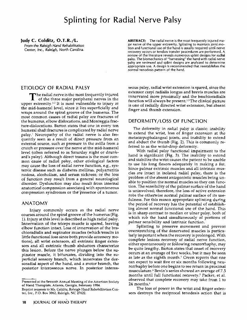
courses around the spiral groove of the humerus (Fig. 1). Injury at this level is described as high radial palsy. Innervation of the triceps muscle is spared, leaving elbow function intact. Loss of innervation of the brachioradialis and supinator muscles (which'results in little functional loss since both provide accessory motions), all wrist extensors, all extrinsic finger extensors and all extrinsic thumb abductors characterize this lesion. Before the nerve plunges below the supinator muscle, it bifurcates, dividing into the superficial sensory branch, which innervates the dorsoradial aspect of the hand, and a motor branch, the posterior interosseous nerve. In posterior interos-
Presented at the .Seventh Annual Meeting of the American Society of Hand Therapists, Atlanta, Georgia, February 1984. Reprint requests to Ms. Colditz, Raleigh Hand Rehabilitation Center, Inc., P.O. Box 3062, Raleigh, NC 27622.
~
18 JOURNAL OF HAND THERAPY
~BSTRACT: The radial nerve is the most frequently injured maJor nerve of the upper extremity. Splinting to maintain joint motion and functional use of the hand is usually required until nerve rec~)Very occu~s or tendon transfer procedures are performed. A review of the literature reveals numerous splint designs for radial palsy. The biomechanics of "harnessing" the hand with radial nerve palsy are reviewed and splint designs are analyzed to determine appropriate use. A design is recommended that reestablishes the normal tenodesis pattern of the hand.
seous palsy, radial wrist extension is spared, since the extensor carpi radialis longus and brevis muscles are innervated more proximally and the brachioradialis ~unction will always be present.!,3The clinical picture IS one of radially directed wrist extension, but absent finger and thumb extension.
DEFORMITY jlOSS OF FUNCTION
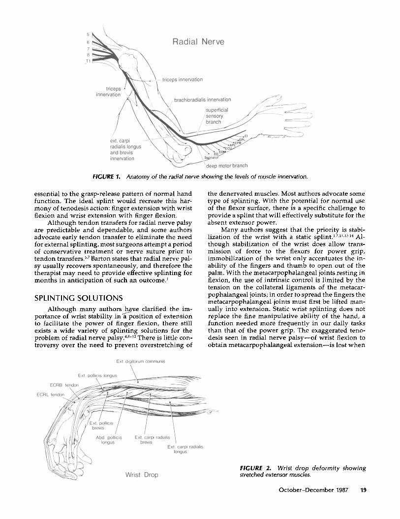
The deformity in radial palsy is classic: inability to extend the wrist, loss of finger extension at the metacarpophalangeal joints, and inability to extend and abduct the thumb (Fig. 2). This is commonly referred to as the wrist-drop deformity.
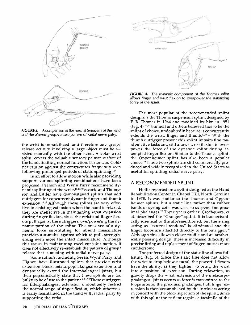
With radial palsy functional impairment to the hand is significant (Fig. 3). The inability to extend and stabilize the wrist causes the patient to be unable to use his long flexors adequately in making a fist. Since palmar extrinsic muscles and all intrinsic muscles are intact in isolated radial palsy, there is the problem of the absent antagonistic muscles being una.ble to position the normal muscles so they can functIon. The sensibility of the palmar surface of the hand is uninvolved; therefore, the loss of active extension robs the otherwise normal palmar surface of its usefulness: For this reason appropriate splinting during the penod of recovery has the potential of establishing almost normal functional use of the hand. This is in sharp contrast to median or ulnar palsy, both of which rob the hand simultaneously of portions of palmar sensibility and intrinsic function.
Splinting to preserve movement and prevent overs.tretching of the denervated muscles is particularly Important when the recovery is prolonged. With complete lesions recovery of radial nerve function either spontaneously or following neurorrhaphy, ma~ be quite lengthy. Barton states that onset of recovery occurs at an average of five weeks, but it may be seen as late as the eighth month.' Green reports that one can expect to wait five or six months following neurorrhaphy before one begins to see return in proximal musculature.4 Bevin's series showed an average of 7.5 months until full functional recovery.s Packer, et al. observed that complete recovery may take from 1 to 24 months.6
The loss of power in the wrist and finger extensors destroys the reciprocal tenodesis action that is
5
6
7 _~,_-----..
Radial Nerve
:1 ~="~
brachioradialis innervation
ext. carpi radialis longus and brevis innervation
superficial sensory branch
FIGURE 1. Anatomy of the radial nerve showing the levels of muscle innervation.
essential to the grasp-release pattern of normal hand function. The ideal splint would recreate this harmony of tenodesis action: finger extension with wrist flexion and wrist extension with finger flexion.
Although tendon transfers for radial nerve palsy are predictable and dependable, and some authors advocate early tendon transfer to eliminate the need for external splinting, most surgeons attempt a period of conservative treatment or nerve suture prior to tendon transfers.5,7Barton states that radial nerve palsy usually recovers spontaneously, and therefore the therapist may need to provide effective splinting for months in anticipation of such an outcome. 1
SPLINTING SOLUTIONS Although many authors hJI,1r.e clarified the im
portance of wrist stability ina position of extension to facilitate the power of finger flexion, there still exists a wide variety of splinting solutions for the problem of radial nerve palsy.4,8-12 There is little controversy over the need to prevent overstretching of
Ext dlgltorum communiS
Ext poll lcis lOngus \ =--======-=-=~~:;:::: I,.. ~
ECRB tend~ ~V=~ ECRL tendon ' ::~::::~J-iiiI:I:l:I;-~S;;:-;~:==-::;;:""-:;::;:;;'~~~
Wrist Drop
the denervated muscles. Most authors advocate some type of splinting. With the potential for normal use of the flexor surface, there is a specific challenge to provide a splint that will effectively substitute for the absent extensor power.
Many authors suggest that the priority is stabilization of the wrist with a static splint.1,7,1l,13,14 Although stabilization of the wrist does allow transmission of force to the flexors for power grip, immobilization of the wrist only accentuates the inability of the fingers and thumb to open out of the palm. With the metacarpophalangeal joints resting in flexion, the use of intrinsic control is limited by the tension on the collateral ligaments of the metacarpophalangeal joints; in order to spread the fingers the metacarpophalangeal joints must first be lifted manually into extension. Static wrist splinting does not replace the fine manipulative ability of the hand, a function needed more frequently in our daily tasks than that of the power grip. The exaggerated tenodesis seen in radial nerve palsy-of wrist flexion to obtain metacarpophalangeal extension-is lost when
FIGURE 2. Wrist drop deformity showing stretched extensor muscles.
October-December 1987 19
radial palsy
fiGURE 3. A comparison of the normal tenodesis of the hand and the altered grasp/release pattern of radial nerve palsy.
the wrist is immobilized, and therefore any grasp / release actiVity involving a large object must be assisted manually with the other hand. A volar wrist splint covers the valuable sensory palmar surface of the hand, limiting normal function. Barton and Goldner caution against the contractures frequently seen following prolonged periods of static splinting.1,9
In an effort to allow motion while also providing support, various splinting combinations have been proposed. Pearson and Wynn Parry recommend dynamic splinting of the wrist. 13,15 Peacock, and Thompson and Littler have demonstrated splints that add outriggers for concurrent dynamic finger and thumb extension.16,17 Although these splints are very effective in assisting extension when the hand is relaxed, they are ineffective in maintaining wrist extension during finger flexion, since the wrist and finger flexors pull against the outriggers, overpowering the dynamic portion of the splint. The presence of a dynamic force substituting for absent musculature provides a stimulus against which to pull, strengthening even more the intact musculature. Although this assists in maintaining excellent joint motion, it does not effectively re-establish the pattern of grasp/ release that is missing with radial nerve palsy.
Some authors, including Green, Wynn Parry, and Highet, have illustrated splints that provide wrist extension, block metacarpophalangeal extension, and dynamically extend the interphalangeal joints, but then pe~simistically state that these splints are too bulky to be of use to the patient.4,15,18These outriggers for interphalangeal extension undoubtedly restrict the normal range of finger flexion, which otherwise is easily maintained in the hand with radial palsy by supporting the wrist,
20 JOURNAL OF HAND THERAPY
. ~-
fiGURE 4. The dynamic component of the Thomas splint allows finger and wrist flexion to overpower the stabilizing force of the splint.
The most popular of the recommended splint designs is the Thomas suspension splint, designed by F. B. Thomas in 1944 and modified by him in 1951 (Fig. 4).12,19 Bunnell and others believed this to be the splint of choice, undoubtedly because it concurrently extends the wrist, finger and thumb. I ,20-23 With the thumb outrigger present this splint impairs fine manipulative tasks and still allows wrist flexion to overpower the force of the dynamic splint during attempted finger flexion. Similar to the Thomas splint, the Oppenheimer splint has also been a popular choice.24 These two splints are still commercially produced and widely recognized in the United States as useful for splinting radial nerve palsy.
A RECOMMENDED SPLINT Hollis reported on a splint designed at the Hand
Rehabilitation Center in Chapel Hill, North Carolina in 1978. It was similar to the Thomas and Oppenheimer splints, but a static line rather than rubber bands or spring coils was used to suspend the proximal phalanges.25 Three years earlier, Crochetiere, et al. described the "Granger" splint. It is biomechanically identical to the aforementioned, but the string acting as "external tendons" is eliminated and the finger loops are attached directly to the outrigger.26
Although this allows a closer profile and an aesthetically pleasing design, there is increased difficulty in precise fitting and replacement of finger loops is more cumbersome.
The preferred design of the static line allows full fisting (Fig. 5). Since the static line does not allow the wrist to drop below neutral, the powerful flexors have the ability, as they tighten, to bring the wrist into a position of extension. During relaxation, as gravity drops the wrist, extension of the metacarpophalangeal joints occurs as force is transmitted to the loops around the proximal phalanges. Full finger extension is then accomplished by the intrinsics acting in concert with the blocking action of the splint. Since with this splint the patient regains a facsimile of the
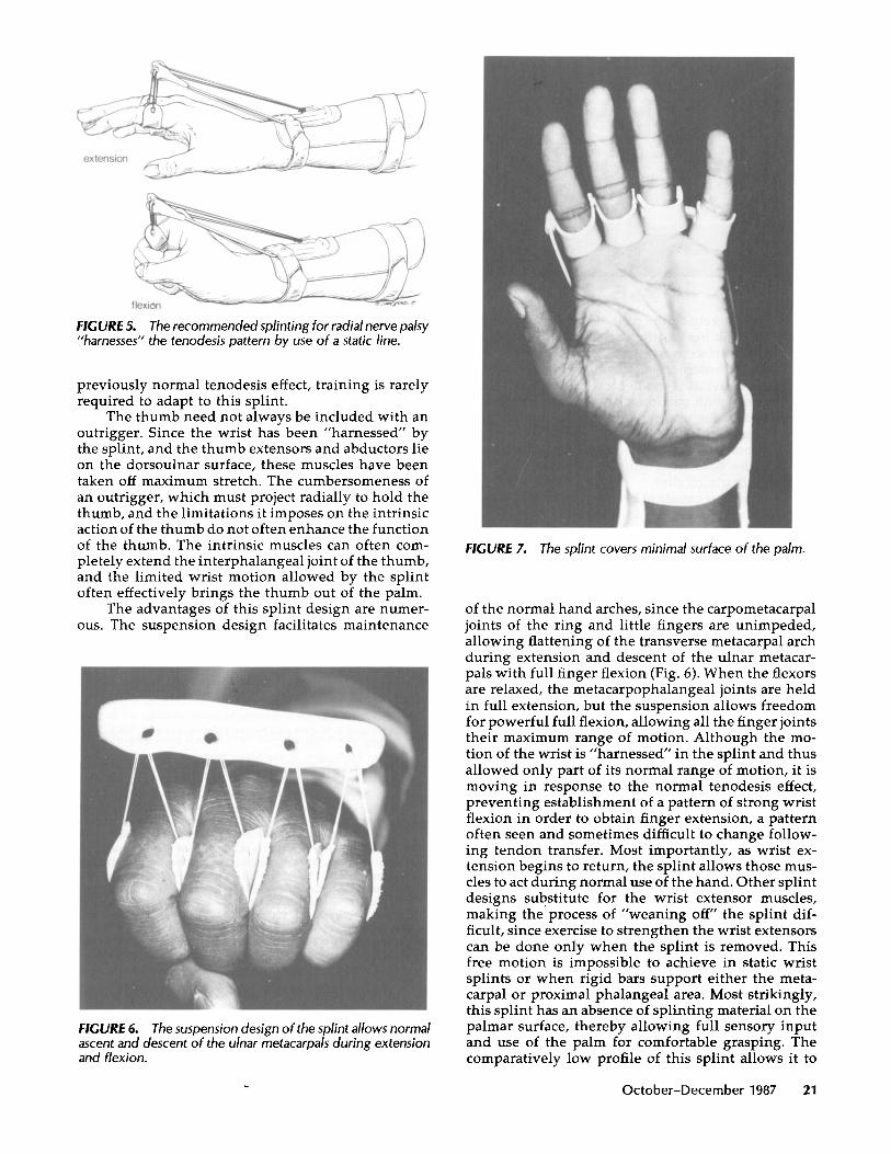
fiGURE 5. The recommended splinting for radial nerve palsy "harnesses" the tenodesis pattern by use of a static line.
previously normal tenodesis effect, training is rarely required to adapt to this splint.
The thumb need not always be included with an outrigger. Since the wrist has been "harnessed" by the splint, and the thumb extensors and abductors lie on the dorsoulnar surface, these muscles have been taken off maximum stretch. The cumbersomeness of an outrigger, which must project radially to hold the thumb, and the limitations it imposes on the intrinsic action of the thumb do not often enhance the function of the thumb. The intrinsic muscles can often completely extend the interphalangeal joint of the thumb, and the limited wrist motion allowed by the splint often effectively brings the thumb out of the palm.
The advantages of this splint design are numerous. The suspension design facilitates maintenance
fiGURE 6. The suspension design of the splint allows normal ascent and descent of the ulnar metacarpals during extension and flexion.
fiGURE 7. The splint covers minimal surface of the palm.
of the normal hand arches, since the carpometacarpal joints of the ring and little fingers are unimpeded, allowing flattening of the transverse metacarpal arch during extension and descent of the ulnar metacarpals with full finger flexion (Fig. 6). When the flexors are relaxed, the metacarpophalangeal jOints are held in full extension, but the suspension allows freedom for powerful full flexion, allowing all the finger jOints their maximum range of motion. Although the motion of the wrist is "harnessed" in the splint and thus allowed only part of its normal range of motion, it is moving in response to the normal tenodesis effect, preventing establishment of a pattern of strong wrist flexion in order to obtain finger extension, a pattern often seen and sometimes difficult to change following tendon transfer. Most importantly, as wrist extension begins to return, the splint allows those muscles to act during normal use of the hand. Other splint designs substitute for the wrist extensor muscles, making the process of "weaning off" the splint difficult, since exercise to strengthen the wrist extensors can be done only when the splint is removed. This free motion is impossible to achieve in static wrist splints or when rigid bars support either the metacarpal or proximal phalangeal area. Most strikingly, this splint has an absence of splinting material on the palmar surface, thereby allowing full sensory input and use of the palm for comfortable grasping. The comparatively low profile of this splint allows it to
October-December 1987 21
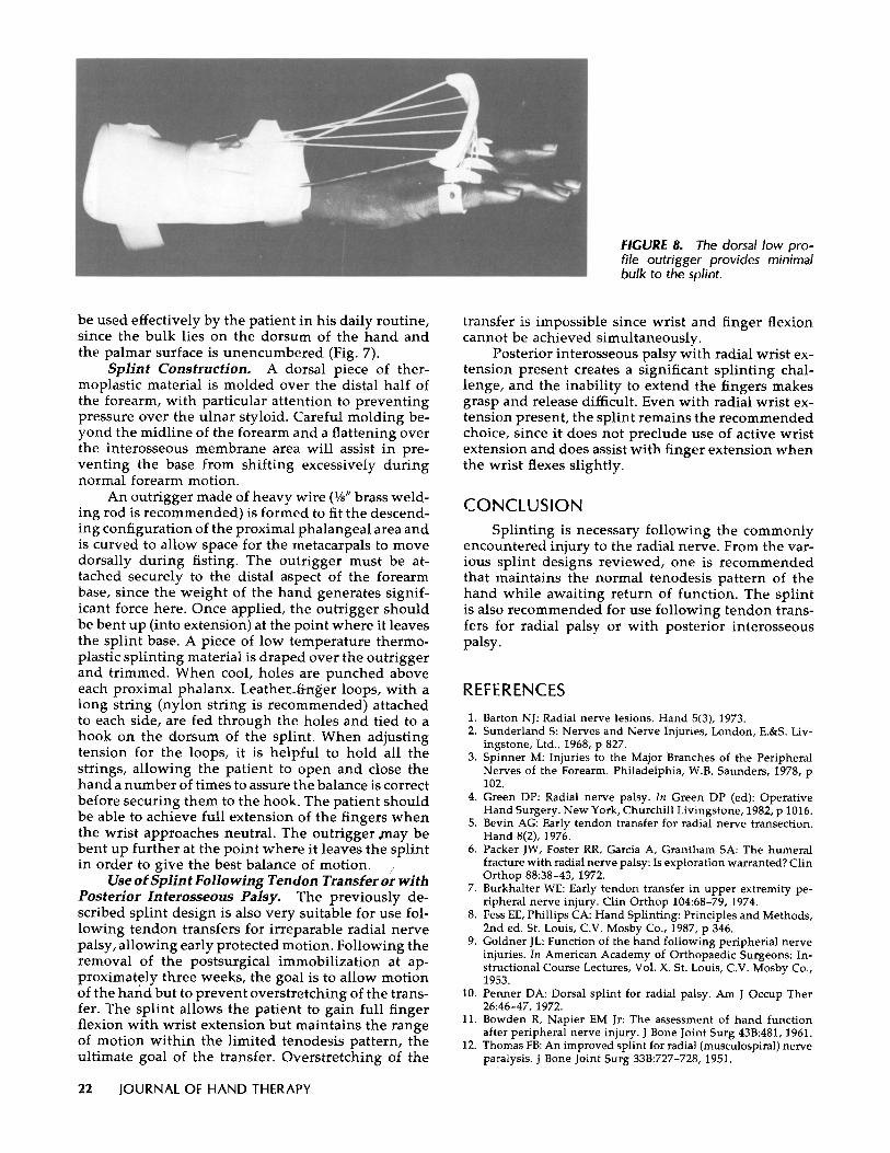
be used effectively by the patient in his daily routine, since the bulk lies on the dorsum of the hand and the palmar surface is unencumbered (Fig. 7).
Splint Construction. A dorsal piece of thermoplastic material is molded over the distal half of the forearm, with particular attention to preventing pressure over the ulnar styloid. Careful molding beyond the midline of the forearm and a flattening over the interosseous membrane area will assist in preventing the base from shifting excessively during normal forearm motion.
An outrigger made of heavy wire (VB" brass welding rod is recommended) is formed to fit the descending configuration of the proximal phalangeal area and is curved to allow space for the metacarpals to move dorsally during fisting. The outrigger must be attached securely to the distal aspect of the forearm base, since the weight of the hand generates significant force here. Once applied, the outrigger should be bent up (into extension) at the point where it leaves the splint base. A piece of low temperature thermoplastic splinting material is draped over the outrigger and trimmed. When cool, holes are punched above each proximal phalanx. Leatheunger loops, with a long string (nylon string is recommended) attached to each side, are fed through the holes and tied to a hook on the dorsum of the splint. When adjusting tension for the loops, it is helpful to hold all the strings, allowing the patient to open and close the hand a number of times to assure the balance is correct before securing them to the hook. The patient should be able to achieve full extension of the fingers when the wrist approaches neutral. The outrigger .may be bent up further at the point where it leaves the splint in order to give the best balance of motion.
Use of Splin t Following Tendon Transfer or with Posterior Interosseous Palsy. The previously described splint design is also very suitable for use following tendon transfers for irreparable radial nerve palsy, allowing early protected motion. Following the removal of the postsurgical immobilization at apprOXimately three weeks, the goal is to allow motion of the hand but to prevent overstretching of the transfer. The splint allows the patient to gain full finger flexion with wrist extension but maintains the range of motion within the limited tenodesis pattern, the ultimate goal of the transfer. Overstretching of the
22 JOURNAL OF HAND THERAPY
FIGURE 8. The dorsal low profile outrigger provides minimal bulk to the splint
transfer is impossible since wrist and finger flexion cannot be achieved simultaneously.
Posterior interosseous palsy with radial wrist extension present creates a significant splinting challenge, and the inability to extend the fingers makes grasp and release difficult. Even with radial wrist extension present, the splint remains the recommended choice, since it does not preclude use of active wrist extension and does assist with finger extension when the wrist flexes slightly.
CONCLUSION Splinting is necessary following the commonly
encountered injury to the radial nerve. From the various splint designs reviewed, one is recommended that maintains the normal tenodesis pattern of the hand while awaiting return of function. The splint is also recommended for use following tendon transfers for radial palsy or with posterior interosseous palsy.
REFERENCES
1. Barton NJ: Radial nerve lesions. Hand 5(3),1973. 2. Sunderland S: Nerves and Nerve Injuries, London, E.&S. Liv
ingstone, Ltd., 1968, P 827. 3. Spinner M: Injuries to the Major Branches of the Peripheral
Nerves of the Forearm. Philadelphia, W.B. Saunders, 1978, p 102.
4. Green DP: Radial nerve palsy. In Green DP (ed): Operative Hand Surgery. New York, Churchill Livingstone, 1982, p 1016.
5. Bevin AG: Early tendon transfer for radial nerve transection. Hand 8(2),1976.
6. Packer JW, Foster RR, Garcia A, Grantham SA: The humeral fracture with radial nerve palsy: Is exploration warranted? Clin Orthop 88:38-43, 1972.
7. Burkhalter WE: Early tendon transfer in upper extremity peripheral nerve injury. Clin Orthop 104:68-79, 1974.
8. Fess EE, Phillips CA: Hand Splinting: Principles and Methods, 2nd ed. St. Louis, C.V. Mosby Co., 1987, P 346.
9. Goldner J1: Function of the hand following peripherial nerve injuries. In American Academy of Orthopaedic Surgeons: Instructional Course Lectures, Vol. X. St. Louis, C.V. Mosby Co., 1953.
10. Penner DA: Dorsal splint for radial palsy. Am J Occup Ther 26:46-47,1972.
11. Bowden R, Napier EM Jr: The assessment of hand function after peripheral nerve injury. J Bone Joint Surg 43B:481, 1961.
12. Thomas FB: An improved splint for radial (musculospiral) nerve paralysis. J Bone Joint Surg 33B:727-728, 1951.
13. Pearson S: Splinting the nerve injured hand. In Hunter JW, Schneider LH, Mackin EJ, Callahan A: Rehabilitation of the Hand. St. Louis, C.V. Mosby Co., 1984, P 455.
14. Ellis M: Orthoses for the hand. In Lamb DW, Kuczynski K: The Practice of Hand Surgery. London, Blackwell Scientific Publications, 1981.
15. Wynn Parry CB: Rehabilitation of the Hand. London, Butterworths, 1978, pp 91-92.
16. Peacock EE Jr: Dynamic splinting for the prevention and correction of hand deformities. J Bone Joint Surg 43A(4), 1952.
17. Thompson JS, Littler JW: Dressings and splints. In Converse JM (ed): Reconstructive Plastic Surgery, Vol. 6. Philadelphia, W.B. Saunders, 1977, pp 2991-2999.
18. Highet WB: Splintage of peripheral nerve injuries. Lancet 1: 555-558, 1942.
19. Thomas FB: A splint for radial (musculospiral) nerve palsy. J Bone Joint Surg 26:602-605, 1944.
SURGERY AND REHABILITATION OF THE HAND - 88
Symposium and Workshop Sponsored by
Hand Rehabilitation Foundation and Thomas Jefferson University
Honored Senior Professor Daniel C. Riordan, M.D. New Orleans, Louisiana
MARCH 13-16, 1988 Philadelphia, Pennsylvania
Course Chairmen: James M. Hunter, M.D. Lawrence H. Schneider, M.D.
Evelyn J. Mackin, L.P. T.
Wyndham Franklin Plaza Hotel Vine Street between 16th & 17th Streets, amade1phia, PA 19103
Anatomy of the upper 11mb demonstrated In J-dlmenslonal projection
Live Surgery using c1osed-clrcult television demonstrations
A symposium and workshop designed to present to the surgeon, resident, physiatrist, physical and occupational therapist, and registered nurse, a unique opportunity to correlate the concepts, indications, surgical techniques, and pre- and post-operative care of the injured and disabled upper extremity. "Hands-on" workshops and panel discussions will complement the didactic sessiol1s.
Information: Evelyn J. Mackin, L.P. T. Hand Rehabilitation Foundation 901 Walnut Street, Philadelphia, PA 19107 (215) 925-4579
20. Bunnell S: Splinting the hand. In American Academy of Orthopaedic Surgeons: Instructional Course Lectures, Vol. IX. St. Louis, C.V. Mosby Co. 1952, pp 233-243.
21. Bunnell S: Active splinting of the hand. J Bone Joint Surg 28(4), 1946.
22. Von Werssowitz OF: Analysis of functional bracing of the hand. Am J Occup Ther 11:4, 1957.
23. Von Werssowitz, OF: Biophysical principles in selection of hand splints. Am J Occup Ther 9:2,1955.
24. Boyes JH: Bunnell's Surgery of the Hand (4th ed). Philadelphia, J.B. Lippincott Co., 1964, p 175.
25. Hollis I: Innovative splinting ideas. In Hunter JM, Schneider LH, Mackin EJ, Bell JA: Rehabilitation of the Hand. S. Louis, C.V. Mosby Co., 1978, P 641.
26. Crochetiere W, Goldstein S, Granger CV, Ireland J: The Granger orthosis for radial nerve palsy. Orthot Prosthet 29(4), 1975.
VON'T MISS WASHINGTON, V.C. AT CHERRY BLOSSOM TIME
The FoUftteenth Annual WIUlh.i.n.g.ton Hand and WILW.t SympO.6.i.wn
(Upp~ E~emi.ty)
A~ 14-16, 1988
Pcvr.k. Hya.t.t Ho.tel. (LuxwUoUA)
Fa.c.u1..ty .inci.u.du EYII!.OUmen.t
1 n:te/Ll'lation.all.y k.nown ex.peJr;tA .in Upp~ E~emliy Swrg ~y and Hand Th~py
Send cheek pa.ya.ble .to: WIUlh.i.n.g.ton Hand SympO.6.i.wn Phy.6~~ $425: Allied
SympO.6.i.wn Foromat Lec;twr.u Panel. V~c.u..6.6~On.6 Th~py Wo~hop.6
H e.a.Uh P Jt.O 6 U.6M na.to $ 3 0 0
GloJUa. ChJU.6tia.n P.O. Box 32073 WIUlh.i.ng.ton, V.C. 20007 (202)342-1779
Noroman J. Cowen, M.V. M~y K. SO~en.6on, R.P.T. SympO.6.i.wn Co-Chairomen
Inci.u.du one-ha£.6 da.y Rev~ew COUMe 6M BOMd Exam
C~erli:t.4 19 HOUM CME CategMY I
Spon.6Med By
The Na.:t.£onai. Hand Ru~ch and Reha.b..i..U.ta.t.Wn Fund, Inc.. a.nd
The C0n.60~ Me~c.ctI'. Educ.a.;Uon
October-December 1987 23