Human Physiology (Part I) Neurons and Nervous System Brain Spinal Chord Muscles Heart.

Spinal Neurons that Possess the Substance P Receptor AreRequired for the Development of Central Sensitization
Sergey G. Khasabov,1,2 Scott D. Rogers,1 Joseph R. Ghilardi,1 Christopher M. Peters,1 Patrick W. Mantyh,1,3,4
and Donald A. Simone2,3,4
Departments of 1Preventive Sciences, 2Oral Science, 3Psychiatry, and 4Neuroscience, University of Minnesota,Minneapolis, Minnesota 55455
In previous studies, we have shown that loss of spinal neuronsthat possess the substance P receptor (SPR) attenuated painand hyperalgesia produced by capsaicin, inflammation, andnerve injury. To determine the role of SPR-expressing neuronsin modulating pain and hyperalgesia, responses of superficialand deep lumbar spinal dorsal horn neurons evoked by me-chanical and heat stimuli and by capsaicin were made afterablation of SPR-expressing neurons using the selective cyto-toxin conjugate substance P-saporin (SP-SAP). Morphologicalanalysis and electrophysiological recordings were made afterintrathecal infusion of vehicle, saporin alone, or SP-SAP. SP-SAP, but not vehicle or SAP alone, produced an �62% de-crease in SPR-expressing neurons in the dorsal horn. Loss ofSPR-expressing neurons diminished the responses of remain-ing neurons to intraplantar injection of capsaicin. Peak re-
sponses to 10 �g of capsaicin were �65% lower in animalspretreated with SP-SAP compared with controls. Additionally,sensitization to mechanical and heat stimuli that normally fol-lows capsaicin was rarely observed. Importantly, responses tomechanical and heat stimuli in the absence of capsaicin werenot altered after SP-SAP treatment. In addition, nociceptiveneurons did not exhibit windup in the SP-SAP-treated group.These results demonstrate that SPR-expressing neurons lo-cated in the dorsal horn are a pivotal component of the spinalcircuits involved in triggering central sensitization and hyperal-gesia. It appears that this relatively small population of neuronscan regulate the physiological properties of other nociceptiveneurons and drive central sensitization.
Key words: hyperalgesia; capsaicin; electrophysiology; spinalcord; substance P-saporin; windup
Chronic pain and hyperalgesia are symptoms associated withtissue injury and inflammation. The neural mechanisms underly-ing persistent pain and hyperalgesia are not fully understood butare known to involve sensitization of primary afferent nocicep-tors and central sensitization. Previous studies have shown thatexcitability of nociceptive dorsal horn neurons is enhanced aftertissue injury, and responses evoked by both innocuous and nox-ious stimuli are increased after injury and are believed to con-tribute to allodynia and hyperalgesia (Treede et al., 1992; Millan,1999; Mannion and Woolf, 2000).
It is well established that substance P (SP) participates innociceptive transmission in the spinal cord. SP is synthesized insmall-caliber afferent fibers (McCarthy and Lawson, 1989) andreleased into the spinal cord after noxious stimulation (Duggan etal., 1987; Schaible et al., 1990), excites nociceptive dorsal hornneurons (Radhakrishnan and Henry, 1991), and contributes tothe development of hyperalgesia (Moochhala and Sawynok,1984). When released into the spinal cord, SP interacts with thesubstance P receptor (SPR), also referred to as the neurokinin-1(NK-1) receptor, to produce its postsynaptic effects. Although�10% of lamina I neurons possess the SPR (Brown et al., 1995;Littlewood et al., 1995), the majority belong to the spinothalamictract (STT) and spinoparabrachial tract and are involved in theascending transmission of nociceptive information. We have dem-
onstrated that SPR-expressing neurons have a unique role inprocessing nociceptive information, and their intact function maybe critical for the development of chronic pain and hyperalgesia.Using internalization of the SPR as a portal of entry to the cell,intrathecal application of a conjugate of SP and the ribosome-inactivating toxin saporin (SP-SAP) resulted in a dramatic loss oflamina I SPR-expressing neurons and attenuated the nocifensivebehavior and hyperalgesia produced by capsaicin (Mantyh et al.,1997), inflammation, and nerve injury (Nichols et al., 1999).Importantly, basal pain reactivity was not affected. Althoughthese studies showed that SPR-expressing neurons are necessaryfor the development of hyperalgesia, the exact role of theseneurons in pain processing is unclear. One possibility is that theseare the neurons excited by and sensitized after injury or inflam-mation. This is supported by studies demonstrating that STTneurons, including those located in lamina I, become sensitizedafter injury and contribute to hyperalgesia (Simone et al., 1991).To determine further the role of SPR-containing neurons innociceptive transmission, we examined the response propertiesand sensitization of superficial and deep dorsal horn neurons inrats pretreated intrathecally with SP-SAP. Our results suggestthat SPR-possessing neurons in the dorsal horn are an integralcomponent of a spinal and/or supraspinal circuit that is crucial forthe development of central sensitization after capsaicin.
MATERIALS AND METHODSSubjectsSeventy-two adult male Sprague Dawley rats (Harlan Industries, India-napolis, IN) weighing 290–470 gm have been housed and used underapproval of the Animal Care Committee at the University of Minnesota.
Received May 20, 2002; revised July 29, 2002; accepted Aug. 5, 2002.This work was supported by National Institutes of Health Grant DA11986.
Saporin and SP-SAP were obtained from Advanced Targeting Systems.Correspondence should be addressed to Dr. Donald A. Simone, Department of
Oral Science, University of Minnesota, 515 Delaware Street Southeast, 17-252 Moos,Minneapolis, MN 55455. E-mail: [email protected] © 2002 Society for Neuroscience 0270-6474/02/229086-13$15.00/0
The Journal of Neuroscience, October 15, 2002, 22(20):9086–9098

Experiments were conducted according to the guidelines set forth by theInternational Association for the Study of Pain.
Intrathecal injectionRats were anesthetized by intramuscular injection of ketamine (100mg/kg) and acepromazine (45 mg/kg) and were placed into a stereotaxicframe. An incision was made in the atlanto–occipital membrane, and apolyethylene catheter (Intramedic, Sparks, MD) (inner diameter, 0.28mm; outer diameter, 0.61 mm) was inserted into the intrathecal space tothe area of lumbar enlargement. Animals were given one intrathecalinjection of normal saline (n � 27), 5 � 10 �5 M saporin (n � 12), or 5 �10 �5 M SP-SAP (n � 33). All injections were given in a volume of 10 �lfollowed by a 5 �l flush with saline. After injection, the catheter wasremoved, and the incision was closed by suture. Experiments wereperformed 10 or 30 d after injection.
Immunohistochemistry and quantificationAnimals pretreated with an intrathecal injection of saline vehicle, SAP,or SP-SAP (n � 5 per group) were deeply anesthetized with sodiumpentobarbital (50 mg/kg, i.p.) and perfused intracardially with 12 ml of0.1 M PBS followed by 25 ml of 4% formaldehyde in 0.1 M PBS. Spinalcord segments L1-S2 were removed, postfixed for 16 hr in the perfusionfixative, and cryoprotected for 24 hr in 30% sucrose in 0.1 M PBS. Serialfrozen spinal cord sections, 60 �m thick, were cut on a sliding microtome,collected in PBS, and processed as free-floating sections. Tissue sectionswere incubated for 30 min at room temperature in a blocking solution of
1% normal donkey or goat serum in PBS with 0.3% Triton X-100 andthen incubated overnight at room temperature in the primary antiserumfor the substance P receptor (rabbit anti-SPR, 1:5000; raised in ourlaboratory). After incubation, tissue sections were washed three times for10 min in PBS and incubated in the secondary antibody solution for 2 hrat room temperature. Secondary antibodies conjugated to the fluorescentmarker Cy3 (Jackson ImmunoResearch, West Grove, PA) were used at1:600. Finally, the sections were washed three times for 10 min in PBS,mounted on gelatin-coated slides, air dried, dehydrated via an alcoholgradient (70, 90, and 100%), cleared in xylene, and coverslipped. Toconfirm the specificity of the primary antibody, controls included preab-sorption with the corresponding synthetic peptide or omission of theprimary antibody.
Slides were viewed through a 1 cm 2 eyepiece grid, which was dividedinto 100 1 � 1 mm units, and the total number of immunofluorescent cellbodies per unit area was counted. The mean numbers of SPR-immunoreactive (SPR-IR) cell bodies located in the superficial (laminasI and II) and deep (laminas III-V) dorsal horn were obtained from threeto eight sections per animal.
Electrophysiological recordingsRats were anesthetized by intramuscular injection of ketamine (100mg/kg) and acepromazine (45 mg/kg). The trachea was cannulated toprovide unobstructed ventilation, and a catheter was inserted into theexternal jugular vein for supplemental anesthesia with sodium pentobar-bital (10 mg � kg �1 � hr �1). Areflexia was maintained by monitoring the
Figure 1. Loss of SPR-expressing neurons after intrathecal infusion of SP-SAP. A, Confocal images showing representative examples of SPR-IR inanimals pretreated intrathecally with vehicle, SAP alone, or SP-SAP. A dramatic reduction in SPR-IR is evident after SP-SAP. B, Mean � SEMpercentage of neurons that express the SPR after intrathecal vehicle, SAP, or SP-SAP. The number of SPR-expressing cells was obtained from individualanimals, and a mean � SEM was calculated. This mean value was designated as 100%, and the SEM was proportionately adjusted (as a percentage) toprovide a measure of variability. Data for SAP- and SP-SAP-treated groups represent the percentage of cells compared with the vehicle-treated group.�Significant differences from vehicle.
Khasabov et al. • SPR-Expressing Spinal Neurons and Sensitization J. Neurosci., October 15, 2002, 22(20):9086–9098 9087
corneal reflex at frequent intervals throughout the experiment. Thecarotid artery was cannulated, and mean blood pressure was monitoredcontinuously with a pressure transducer (World Precision Instruments,Sarasota, FL). Experiments were terminated if mean pressure droppedbelow 60 mmHg. The lumbar enlargement was exposed by laminectomy,and the animal was secured in a spinal frame. The spinal cord wascontinually bathed in a pool of warm (37°C) mineral oil. Core bodytemperature was maintained at 37°C by a feedback-controlled heatingpad.
Extracellular recordings of single dorsal horn neurons with receptivefields (RFs) located on the plantar surface of the hindpaw were obtainedusing stainless steel microelectrodes (Frederick Haer and Co., Bruns-wick, ME) (10 m�). Recording electrodes were lowered into the spinalcord at the L4 and L5 segments using an electronic micromanipulator(Burleigh Instruments, Fishers, NY) in 5 �m steps. Recordings weremade only from single neurons whose amplitude could be easily discrim-inated. Electrophysiological activity was amplified using an alternatingcurrent amplifier (model DAM80; World Precision Instruments), audiomonitored (Grass AM8 audiomonitor; Grass Instruments, West War-wick, RI), and displayed on a storage oscilloscope before being sent to acomputer for data collection using a customized version of Lab View(National Instruments, Austin, TX) software that enabled storage of rawdata, discriminated impulses, and stimulus temperature. In most exper-iments, recordings were obtained from two neurons, one on each side ofthe spinal cord.
Functional classification of spinal neurons. Search stimuli consisted ofmechanical stimulation (stroking the skin and mild pinching with theexperimenter’s fingers) of the rat hindpaw. The RFs of isolated neuronswere mapped with a suprathreshold von Frey monofilament. Each spinalneuron was characterized based on its response to graded intensities ofmechanical stimulation applied to the RF. Innocuous stimuli consisted ofstroking the skin with a cotton swab. Noxious stimulation included mildpinching with the experimenter’s fingers and with serrated forceps, butthis latter stimulus was applied sparingly to avoid neuronal sensitization.Neurons were classed functionally according to responses evoked bymechanical stimuli as: (1) low threshold if they were excited maximally byinnocuous stimulation, (2) wide dynamic range (WDR) if they respondedin a graded manner to increasing intensity of stimulation, and (3) highthreshold (HT) if responses were evoked by noxious stimulation only.Only WDR and HT neurons were studied.
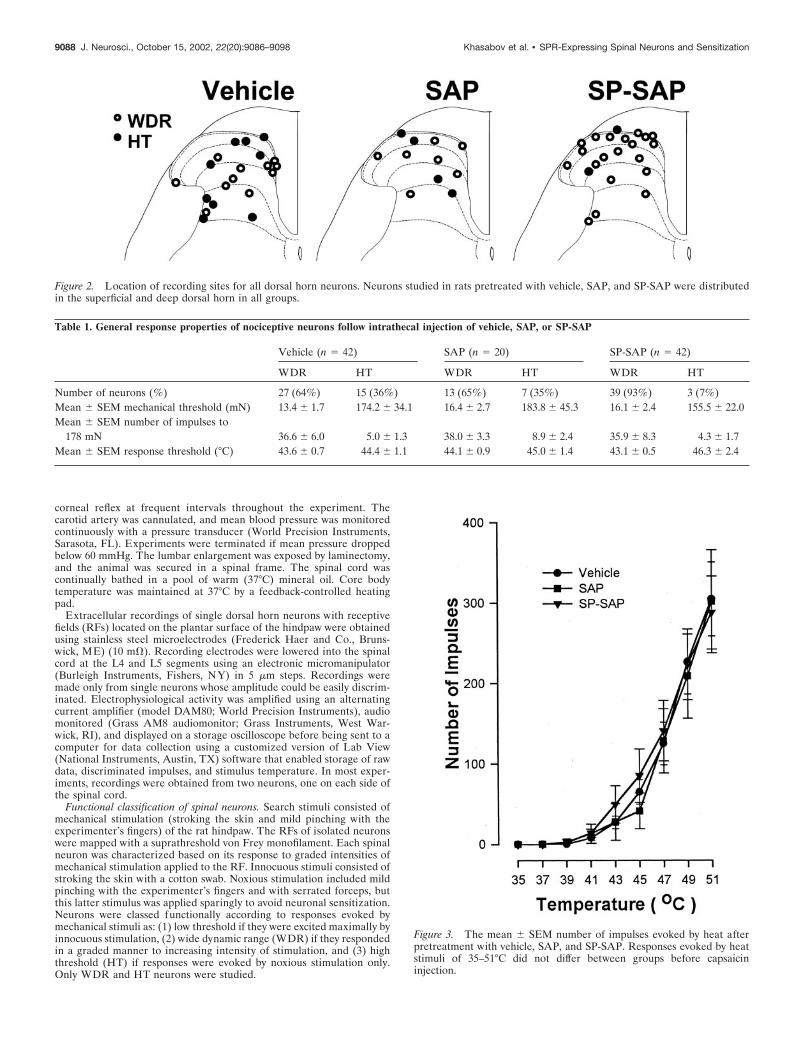
Figure 2. Location of recording sites for all dorsal horn neurons. Neurons studied in rats pretreated with vehicle, SAP, and SP-SAP were distributedin the superficial and deep dorsal horn in all groups.
Figure 3. The mean � SEM number of impulses evoked by heat afterpretreatment with vehicle, SAP, and SP-SAP. Responses evoked by heatstimuli of 35–51°C did not differ between groups before capsaicininjection.
Table 1. General response properties of nociceptive neurons follow intrathecal injection of vehicle, SAP, or SP-SAP
Vehicle (n � 42) SAP (n � 20) SP-SAP (n � 42)
WDR HT WDR HT WDR HT
Number of neurons (%) 27 (64%) 15 (36%) 13 (65%) 7 (35%) 39 (93%) 3 (7%)Mean � SEM mechanical threshold (mN) 13.4 � 1.7 174.2 � 34.1 16.4 � 2.7 183.8 � 45.3 16.1 � 2.4 155.5 � 22.0Mean � SEM number of impulses to
178 mN 36.6 � 6.0 5.0 � 1.3 38.0 � 3.3 8.9 � 2.4 35.9 � 8.3 4.3 � 1.7Mean � SEM response threshold (°C) 43.6 � 0.7 44.4 � 1.1 44.1 � 0.9 45.0 � 1.4 43.1 � 0.5 46.3 � 2.4
9088 J. Neurosci., October 15, 2002, 22(20):9086–9098 Khasabov et al. • SPR-Expressing Spinal Neurons and Sensitization
Evoked response measures and experimental design. After identificationand general functional characterization of a neuron as WDR or HT, theRF was mapped by stroking and mildly pinching with forceps andoutlined on the skin with a felt-tip pen. Mechanical threshold (in mil-liNewtons) was determined using calibrated von Frey monofilamentsapplied to the most sensitive area of the RF. To obtain responses evokedby mechanical stimuli before and after capsaicin, four test sites within theRF were marked on the skin and stimulated with a von Frey monofila-ment (178 mN bending force applied for 2 sec). Each test site wasstimulated three times with a 10 sec interval between stimuli. To deter-mine response evoked by heat, stimuli of 35–51°C were applied inascending order of 2°C increments from a base temperature of 32°C usinga Peltier thermode (contact area of 1 cm 2). Stimuli were of 5 sec durationand were delivered at a ramp rate of 18°C/sec with an interstimulusinterval of 60 sec. Capsaicin (10 or 100 �g in 10 �l) was injectedintradermally into the middle of the RF. Responses evoked by capsaicinwere recorded for 5 min, and responses evoked by mechanical and heatstimuli were again determined as described above. At the end of theexperiment, the recording site was marked by passing current (10 �A for20 sec) through the recording electrode.
In separate experiments, we determined whether nociceptive neuronsin vehicle-treated animals (n � 8) and SP-SAP-treated animals (n � 8)exhibited windup. Neurons were activated by 12 successive electricalstimuli applied to the RF via fine needle electrodes. Stimuli of 1 msecduration were applied at the rate of 0.5 Hz and at a current intensity thatwas 150% of threshold intensity that produced a long latency (110–450msec) C-fiber-evoked response.
Histolog ical localization of recording sites. Animals were perfused with
normal saline followed by 10% formalin containing 1% potassium ferro-cyanide. Serial transverse sections (50 �m) were cut using a vibratomeand stained with neutral red. Recording sites were identified by PrussianBlue marks or small lesions.
Data analysesThe numbers of SPR-IR cell bodies, impulses evoked by capsaicin, andimpulses evoked by mechanical and heat stimuli before and after capsa-icin were compared between groups using ANOVA and Bonferroni posthoc comparisons. Evoked responses were determined by subtracting thespontaneous discharge rate from the response that occurred during thestimulus. The proportion of neurons that were classed as HT and WDRneurons was compared between the groups using the � 2 test. The numberof SPR-expressing neurons after SAP and SP-SAP was normalized to thenumber of neurons found in vehicle-treated animals. For all statisticaltests, a p value of �0.05 was considered significant.
RESULTSAblation of SPR-expressing neurons after intrathecalSP-SAP: morphological characteristicsIn animals pretreated with vehicle or SAP, SPR immunoreactiv-ity is observed on cell bodies and dendrites located primarily inlamina I; however, distinct SPR immunoreactivity was also foundon neurons located in the deep dorsal horn. No differencesoccurred in the number of SPR-expressing neurons in animals
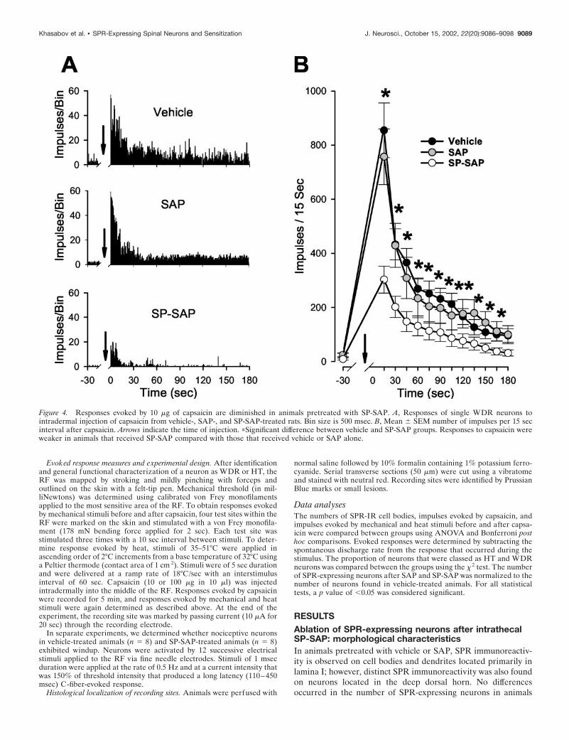
Figure 4. Responses evoked by 10 �g of capsaicin are diminished in animals pretreated with SP-SAP. A, Responses of single WDR neurons tointradermal injection of capsaicin from vehicle-, SAP-, and SP-SAP-treated rats. Bin size is 500 msec. B, Mean � SEM number of impulses per 15 secinterval after capsaicin. Arrows indicate the time of injection. �Significant difference between vehicle and SP-SAP groups. Responses to capsaicin wereweaker in animals that received SP-SAP compared with those that received vehicle or SAP alone.
Khasabov et al. • SPR-Expressing Spinal Neurons and Sensitization J. Neurosci., October 15, 2002, 22(20):9086–9098 9089
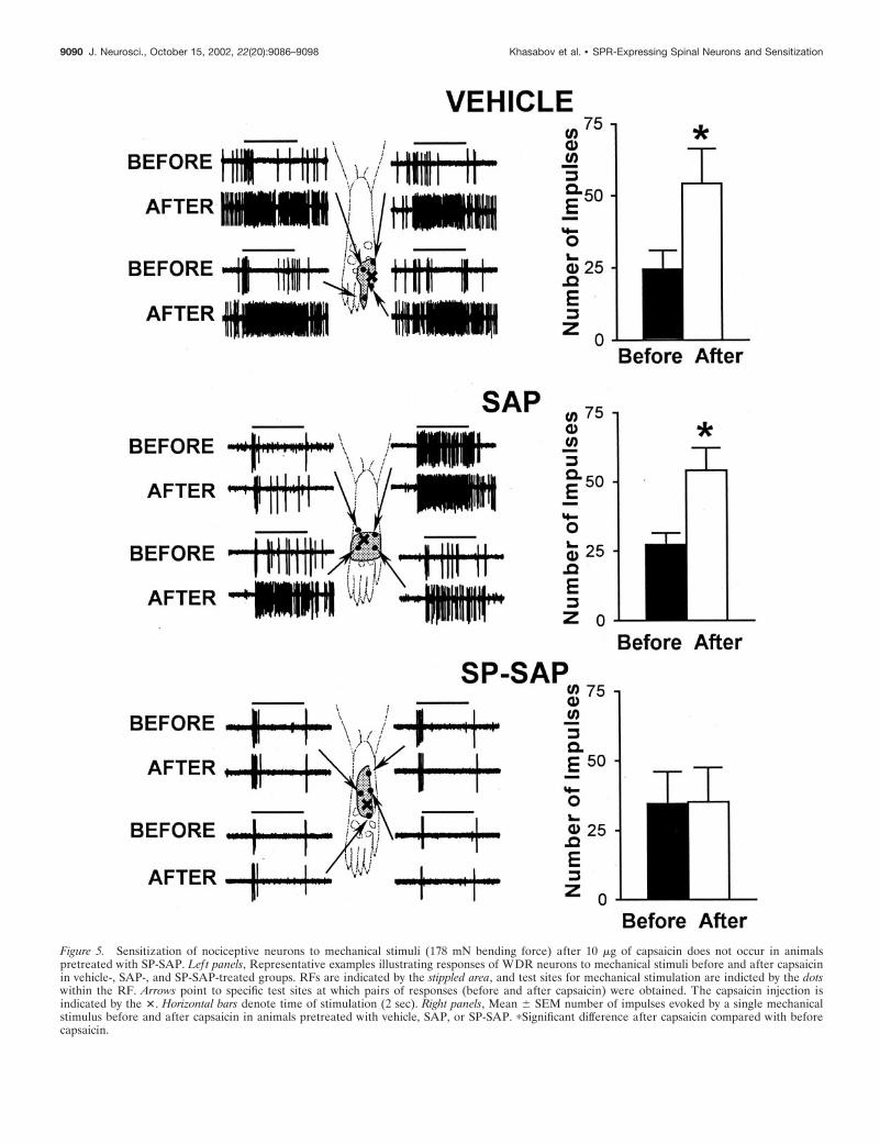
Figure 5. Sensitization of nociceptive neurons to mechanical stimuli (178 mN bending force) after 10 �g of capsaicin does not occur in animalspretreated with SP-SAP. Left panels, Representative examples illustrating responses of WDR neurons to mechanical stimuli before and after capsaicinin vehicle-, SAP-, and SP-SAP-treated groups. RFs are indicated by the stippled area, and test sites for mechanical stimulation are indicted by the dotswithin the RF. Arrows point to specific test sites at which pairs of responses (before and after capsaicin) were obtained. The capsaicin injection isindicated by the �. Horizontal bars denote time of stimulation (2 sec). Right panels, Mean � SEM number of impulses evoked by a single mechanicalstimulus before and after capsaicin in animals pretreated with vehicle, SAP, or SP-SAP. �Significant difference after capsaicin compared with beforecapsaicin.
9090 J. Neurosci., October 15, 2002, 22(20):9086–9098 Khasabov et al. • SPR-Expressing Spinal Neurons and Sensitization
pretreated with vehicle or with SAP alone (Fig. 1). Animalspretreated with SAP alone exhibited 80 � 16% and 79 � 13% ofSPR-IR neurons in lamina I/II and laminas III-V, respectively,compared with animals pretreated with vehicle. In contrast, asignificant reduction in the number of SPR-IR neurons was ob-served in animals pretreated with SP-SAP. These animals exhib-ited only 32 � 13% and 42 � 9.9% of SPR-IR neurons in laminaI/II and in laminas III-V, respectively, compared with the vehicle-treated group. Thus, animals pretreated with SP-SAP exhibited adecrease in SPR-expressing neurons in the superficial and deepdorsal horn of 65 and 58%, respectively.
Ablation of SPR-expressing neurons after intrathecalSP-SAP: electrophysiological responsesGeneral response propertiesSeventy-two rats were pretreated intrathecally with saline (vehi-cle; n � 27), SAP alone (n � 12), or SP-SAP (n � 33). Electro-physiological responses were obtained from a total of 104 noci-ceptive dorsal horn neurons. Forty-two cells (27 WDR and 15
HT) were studied in animals pretreated with vehicle, 20 cells (13WDR and 7 HT) were studied in animals pretreated with SAP,and 42 cells (39 WDR and 3 HT) were studied after SP-SAPtreatment. Within each group, no differences were observed inresponses of neurons studied 10 or 30 d after treatment, and datafrom these time points were therefore combined. Receptive fieldsof all neurons included the plantar surface of the hindpaw.Recording sites were recovered for 53 neurons and were found tobe located in the superficial and deep dorsal horn. Recording sitesof WDR and HT neurons in vehicle-, SAP-, and SP-SAP-treatedgroups are illustrated in Figure 2, which shows that recording siteswere distributed throughout the dorsal horn in all treatmentgroups. Similarly, the mean recording depth from the spinal cordsurface for all neurons did not differ between groups and was547 � 63, 502 � 77, and 438 � 44 �m for the vehicle, SAP, andSP-SAP groups, respectively.
The proportion of WDR and HT neurons studied in eachtreatment group, as well as their general response characteristics
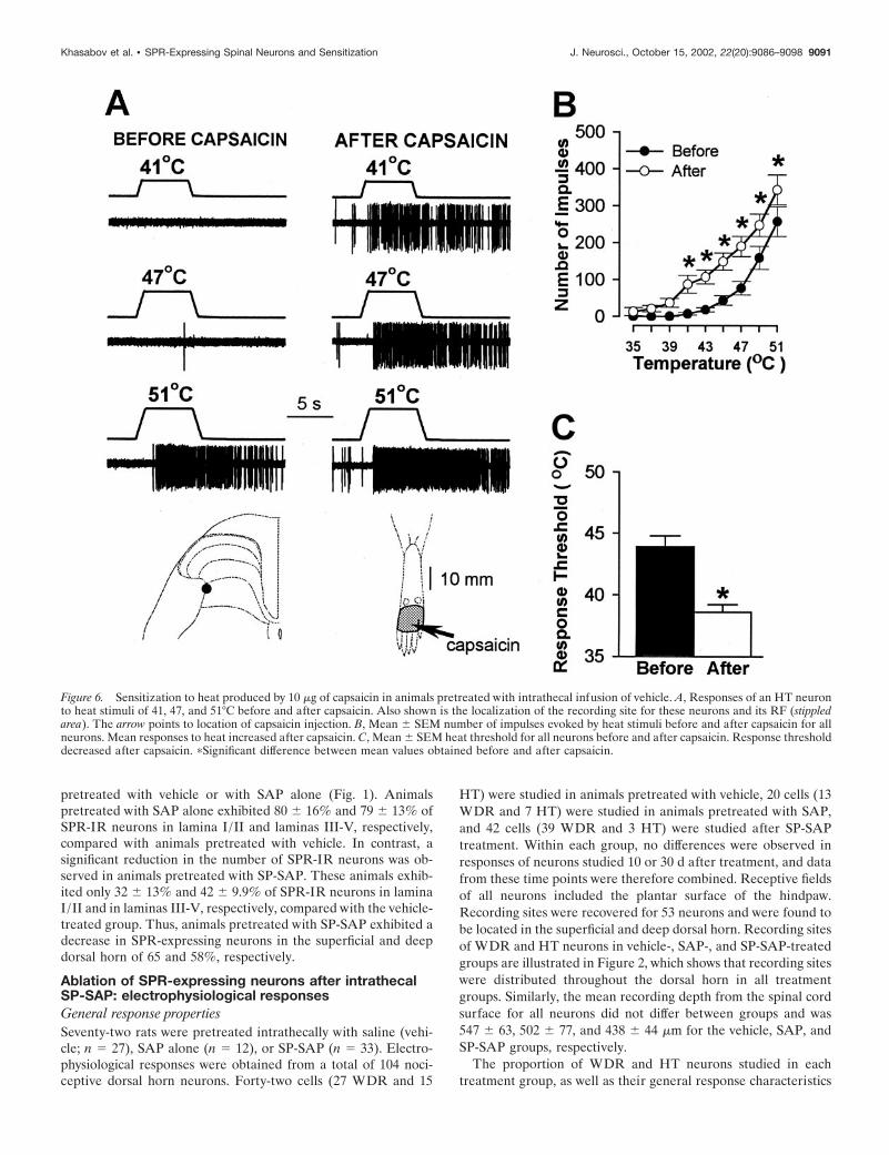
Figure 6. Sensitization to heat produced by 10 �g of capsaicin in animals pretreated with intrathecal infusion of vehicle. A, Responses of an HT neuronto heat stimuli of 41, 47, and 51°C before and after capsaicin. Also shown is the localization of the recording site for these neurons and its RF (stippledarea). The arrow points to location of capsaicin injection. B, Mean � SEM number of impulses evoked by heat stimuli before and after capsaicin for allneurons. Mean responses to heat increased after capsaicin. C, Mean � SEM heat threshold for all neurons before and after capsaicin. Response thresholddecreased after capsaicin. �Significant difference between mean values obtained before and after capsaicin.
Khasabov et al. • SPR-Expressing Spinal Neurons and Sensitization J. Neurosci., October 15, 2002, 22(20):9086–9098 9091
(mean mechanical threshold, mean number of impulses evoked bya suprathreshold von Frey monofilament with a bending force of178 mN applied for 2 sec, and mean heat threshold) beforecapsaicin injection, are provided in Table 1. Responses of WDRand HT neurons showed quantitative differences in their re-sponses to mechanical stimuli in that HT neurons exhibited ahigher response threshold and were less responsive to the su-prathreshold von Frey monofilament. However, responses ofWDR and HT neurons to mechanical stimuli before capsaicin didnot differ between treatment groups. Additionally, no differenceswere found between WDR and HT neurons in their responses toheat before capsaicin. As shown in Figure 3, mean responsefunctions of nociceptive neurons for heat stimuli of 35–51°Cbefore capsaicin did not differ between groups. The mean cumu-lative numbers of impulses evoked across all heat stimuli were759.3 � 124.1, 726.9 � 179.7, and 805.5 � 172.1 after vehicle,SAP, and SP-SAP, respectively.
Although the response properties of nociceptive neurons didnot differ among the groups, a difference was found in the pro-
portion of WDR and HT neurons encountered between groups.Neurons classed as HT were 36 and 35% in vehicle- and SAP-treated animals, respectively, whereas the number of HT neuronsencountered in SP-SAP-treated animals was significantly lower.Only three neurons (7%) in SP-SAP-treated rats were classed asHT ( p � 0.003) (Table 1).
Responses to capsaicin and central sensitizationResponses evoked by intraplantar capsaicin injections were de-creased in animals pretreated with SP-SAP. Injection of 10 �g ofcapsaicin (in 10 �l) produced a vigorous and long-lasting dis-charge in all WDR and HT neurons recorded from animalspretreated with vehicle (n � 34) or SAP alone (n � 20). Dis-charge rates were highest soon after injection, decreased to amoderate level within �1 min after injection, and typically per-sisted for �3 min (Fig. 4A). Responses of WDR and HT neuronswere similar and did not differ between vehicle- and SAP-treatedgroups. In contrast, capsaicin-evoked responses of WDR and HTneurons (n � 29) were much weaker in animals pretreated with
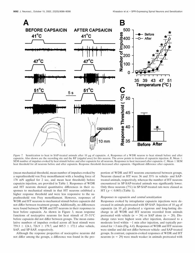
Figure 7. Sensitization to heat in SAP-treated animals after 10 �g of capsaicin. A, Responses of a WDR neuron to heat stimuli before and aftercapsaicin. Also shown are the recording site and the RF (stippled area) for this neuron. The arrow points to location of capsaicin injection. B, Mean �SEM number of impulses evoked by heat stimuli before and after capsaicin for all neurons. Responses to heat increased after capsaicin. C, Mean � SEMheat threshold for all neurons before and after capsaicin. Response threshold decreased after capsaicin. �Significant difference after capsaicin.
9092 J. Neurosci., October 15, 2002, 22(20):9086–9098 Khasabov et al. • SPR-Expressing Spinal Neurons and Sensitization
SP-SAP, and the duration of response was often shorter. Beforeinjection, mean discharge rates of spontaneous activity did notdiffer between the groups. Because no differences in responsesoccurred between WDR and HT neurons within each group,responses of these neurons were combined. The mean number ofimpulses evoked during the first 15 sec after capsaicin was 855 �101 and 757 � 102 for the vehicle- and SAP-treated groups,respectively, but was only 303 � 50 impulses in animals treatedwith SP-SAP ( p � 0.01) (Fig. 4B). Thus, the peak response tocapsaicin was �65% less in animals pretreated with SP-SAPcompared with vehicle. Although the mean number of impulsesthat occurred in each consecutive 15 sec interval after capsaicinwas similar for the vehicle- and SAP-treated groups, capsaicin-evoked discharges were less in SP-SAP-treated rats during theentire 3 min period after capsaicin injection.
Before and at 10 min after injection of capsaicin, responsesevoked by a suprathreshold von Frey monofilament (178 mNbending force) were determined at selected sites within the RFthat were �2 mm away from the capsaicin injection. In animalspretreated with vehicle or with SAP alone, all WDR and HT
neurons located in the superficial or deep dorsal horn exhibitedan increase in mechanically evoked responses after capsaicin.Responses to mechanical stimuli were increased similarlythroughout the RF. In vehicle-treated animals, the mean numberof impulses evoked by the von Frey monofilament increased from24.6 � 6.4 to 54.2 � 12.1 (or 120%; p � 0.001) after capsaicin. Asimilar increase of 98% in evoked responses was found in theSAP-treated group; the mean number of impulses increased from27.3 � 4.3 to 54.2 � 8.2 ( p � 0.001). In contrast, none of theWDR or HT neurons in SP-SAP-treated rats exhibited sensiti-zation to mechanical stimuli after capsaicin injection (Fig. 5). Themean number of impulses evoked by the monofilament was 34.6 �11.5 before capsaicin and 35.3 � 12.4 after capsaicin.
Pretreatment with SP-SAP also prevented the sensitization toheat stimuli that normally occurs after capsaicin. Before capsa-icin, heat thresholds and responses evoked by suprathreshold heatstimuli did not differ between WDR and HT neurons, and nodifferences in responses of these neurons were observed betweentreatment groups. In animals pretreated with vehicle, WDR andHT neurons exhibited sensitization to heat after capsaicin as
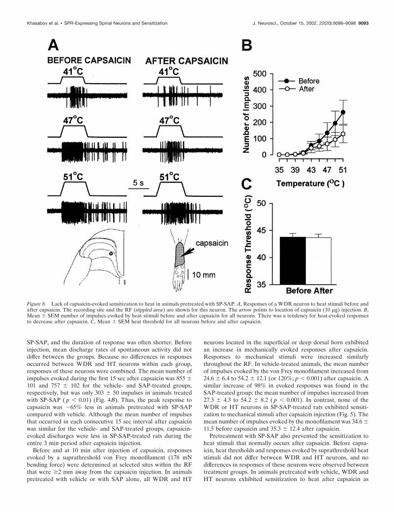
Figure 8. Lack of capsaicin-evoked sensitization to heat in animals pretreated with SP-SAP. A, Responses of a WDR neuron to heat stimuli before andafter capsaicin. The recording site and the RF (stippled area) are shown for this neuron. The arrow points to location of capsaicin (10 �g) injection. B,Mean � SEM number of impulses evoked by heat stimuli before and after capsaicin for all neurons. There was a tendency for heat-evoked responsesto decrease after capsaicin. C, Mean � SEM heat threshold for all neurons before and after capsaicin.
Khasabov et al. • SPR-Expressing Spinal Neurons and Sensitization J. Neurosci., October 15, 2002, 22(20):9086–9098 9093

indicated by a mean decrease of 5.3°C in response threshold (from43.9 � 0.9°C to 38.6 � 0.6°C; p � 0.001) and increased responsesto suprathreshold stimuli (Fig. 6). The mean cumulative numberof impulses evoked by all heat stimuli increased from 663 � 96before capsaicin to 1297 � 158 after capsaicin (96% increase; p �0.001). A similar degree of sensitization to heat was observed forWDR and HT neurons in SAP-treated animals (Fig. 7). Themean response threshold decreased 5.7°C (from 44.1 � 0.8°C to38.4 � 0.8°C after capsaicin; p � 0.001), and the mean cumulativenumber of impulses increased from 727 � 180 to 1441 � 277(98% increase; p � 0.001). In contrast, sensitization to heat aftercapsaicin injection did not occur in SP-SAP-treated animals (Fig.8). Mean heat thresholds were unchanged after capsaicin (43.8 �0.7°C before capsaicin and 43.7 � 0.6°C after capsaicin), and themean, cumulative number of impulses evoked by all heat stimulidecreased from 749 � 217 before capsaicin to 397 � 127 aftercapsaicin (47% decrease; p � 0.01). The decrease in the cumu-lative responses to heat was attributed to a decrease in responsesevoked by the higher stimulus temperatures (Fig. 8B).
Because the excitation of nociceptive neurons evoked by 10 �gof capsaicin was weak in animals pretreated with SP-SAP, it wasunclear whether central sensitization failed to occur because thecapsaicin-evoked response was not strong enough to induce sen-sitization or whether there was a disruption of the mechanismsthat drive central sensitization. To address this issue, we repeatedthe experiments above but used a capsaicin dose of 100 �g toincrease the capsaicin-evoked response in SP-SAP-treated ani-mals. In vehicle-treated animals, intraplantar injection of 100 �gof capsaicin produced a strong and long-lasting discharge of eightneurons (seven WDR and one HT). The mean number of im-pulses during the first 15 sec after capsaicin was 898 � 82 and wassimilar to that produced by the 10 �g dose of capsaicin in controlanimals. A similar level of excitation was produced in 13 neurons(12 WDR and 1 HT) of animals pretreated with SP-SAP (Fig. 9).
The mean number of impulses evoked during the first 15 sec after100 �g of capsaicin was 836 � 118 and was significantly greaterthan the 303 � 50 impulses evoked during the first 15 sec after 10�g of capsaicin. Thus, mean responses of nociceptive neuronsevoked by 100 �g of capsaicin in vehicle- and SP-SAP-treatedanimals were similar to responses produced by injection of 10 �gof capsaicin in control animals.
Although nociceptive neurons in animals pretreated with vehi-cle and SP-SAP exhibited a similar degree of capsaicin-evokedexcitation, sensitization did not occur in the SP-SAP group. Asillustrated in Figure 10, the mean number of impulses evoked bythe suprathreshold von Frey monofilament increased from 31.1 �9 to 57.1 � 11.3 (or 83%; p � 0.001) in animals pretreated withvehicle. However, responses of neurons in SP-SAP-treated ani-mals were unchanged after capsaicin (30 � 6.8 impulses beforecapsaicin and 33.8 � 8.8 impulses after capsaicin). In addition,neurons in vehicle-treated animals, but not in SP-SAP-treatedanimals, exhibited sensitization to heat after capsaicin. In animalspretreated with vehicle, capsaicin decreased the mean heatthreshold from 43.8 � 1.1°C to 39.3 � 0.9°C ( p � 0.001), and themean cumulative number of impulses evoked by all heat stimuliincreased from 755 � 228 to 1444 � 204 (an increase of 91%; p �0.05). In contrast, response thresholds for heat in animals pre-treated with SP-SAP remained unchanged after capsaicin (42.3 �1°C before injection and 43 � 1.6°C after injection), and the meancumulative number of impulses evoked by all heat stimuli aftercapsaicin decreased from 892 � 244 to 499 � 208 (or 44%; p �0.01).
Effect of SP-SAP on windupWe also examined the ability of nociceptive neurons in vehicle-and SP-SAP-treated animals to develop windup. Responses toconsecutive electrical stimuli were obtained for 13 WDR neuronsin the vehicle-treated group and 10 WDR neurons in the SP-SAP-treated group. Electrical stimulation of the RF at a fre-quency of 0.5 Hz induced a significant increase in the C-fiber-evoked response of 187 � 21% ( p � 0.01) by the 12th stimuluscompared with the response evoked by the first stimulus. Windupfailed to occur in animals pretreated with SP-SAP, as shown inFigure 11.
DISCUSSIONElimination of spinal neurons that possess the SPR using SP-SAPoffers the unique opportunity to determine the role of SPR-expressing neurons, as well as other neurons, in pain processing.SP-SAP induced specific degeneration of neurons expressing SPRreceptors. This observation was made previously (Mantyh et al.,1997; Nichols et al., 1999) and is supported by present data. Aninteresting finding of the present study was the proportionalchange in the functional classification of spinal neurons encoun-tered in animals pretreated with SP-SAP. The proportion of HTneurons encountered in control animals was �36%, whereas only7% of the neurons identified in SP-SAP-treated animals were HT.This suggests that SP-SAP targeted primarily HT neurons. It hasbeen shown in cats that HT neurons possess more SPRs thanWDR neurons (Ma et al., 1996, 1997), suggesting that HT neu-rons are more vulnerable to SP-SAP because of the greaternumber of SPRs on these neurons.
The absence of sensitization and windup after SP-SAP resultsfrom elimination of SPR-expressing spinal neurons within thearea of intrathecal application for the following reasons. First,SPR immunoreactivity was not altered in thoracic or cervical
Figure 9. Neuronal discharges evoked by 100 �g of capsaicin do notdiffer between groups pretreated with vehicle or with SP-SAP. The meannumber of impulses and the temporal profile of the capsaicin-evokedresponses were nearly identical for both groups. Responses shown are themean � SEM number of impulses evoked during each consecutive 15 secinterval after capsaicin. The arrow indicates the time of injection.
9094 J. Neurosci., October 15, 2002, 22(20):9086–9098 Khasabov et al. • SPR-Expressing Spinal Neurons and Sensitization

segments after application of SP-SAP to the lumbar spinal cord,and its administration did not disrupt daily behavior patterns orlocomotion (Nichols et al., 1999). These data indicate that thetoxic effect of SP-SAP is localized to treated segments. Second,cell loss was not observed in the DRG (Mantyh et al., 1997;Nichols et al., 1999) after intrathecal SP-SAP, indicating that theeffects of SP-SAP on central sensitization are not attributed to apresynaptic effect on primary afferent terminals.
It is particularly intriguing that a relatively small population ofneurons can have such a dramatic effect on the behavioral andphysiological responses of nociceptive neurons located through-out the dorsal horn. Our previous studies demonstrated thatSPR-expressing neurons were necessary for the full expression ofhyperalgesia after capsaicin, inflammation, and nerve injury(Mantyh et al., 1997; Nichols et al., 1999). One explanation toaccount for these changes is that neurons that express the SPRare the neurons that undergo central sensitization and transmitnociceptive information rostrally to account for hyperalgesia.This has been proposed to account for decreased hyperalgesiaafter ablation of SPR-expressing neurons using SP–diphtheriatoxin conjugates (Benoliel et al., 1999). In the present study, we
found that loss of �60% of SPR-expressing neurons, which con-stitute a small population of ascending tract neurons locatedprimarily in the superficial dorsal horn, resulted in the inability ofremaining neurons to respond vigorously to capsaicin and toexhibit sensitization and windup. This percentage of neuron lossmay represent a threshold effect related to the concentration ofSP-SAP, suggesting that relatively few SPR-containing neuronsare required for the development of central sensitization. Re-markably, neurons located in the deep as well as the superficialdorsal horn underwent this change in their capacity to becomesensitized. This indicates that SPR-expressing neurons, eitherdirectly or indirectly, modulate excitability of other superficial aswell as deep dorsal horn neurons. It is possible that only SPR-expressing neurons become sensitized after capsaicin, and thatthe apparent sensitization of other neurons results from direct orindirect synaptic connections with these neurons. This is sup-ported by our findings that increasing capsaicin-evoked neuronalactivity in SP-SAP-treated animals to that evoked in controlanimals, by using a higher dose of capsaicin, failed to inducesensitization. This hypothesis can be tested by using iontophoreti-cally applied SP and determining whether only SP-responsive
Figure 10. Capsaicin (100 �g) produces sensitization after pretreatment with vehicle, but this sensitization fails to occur after SP-SAP pretreatment.Top row, Data from animals pretreated with vehicle. A, Mean � SEM number of impulses evoked by a von Frey monofilament (178 mN) before and aftercapsaicin. B, Mean � SEM number of impulses evoked by heat stimuli before and after capsaicin. C, Mean � SEM heat response threshold before andafter capsaicin. Bottom row, Responses to mechanical ( D) and heat (E, F ) stimuli before and after capsaicin in animals pretreated with SP-SAP.�Significant difference after capsaicin.
Khasabov et al. • SPR-Expressing Spinal Neurons and Sensitization J. Neurosci., October 15, 2002, 22(20):9086–9098 9095

neurons become sensitized. The absence of central sensitizationand windup recorded electromyographically from hindlimb mus-cles (De Felipe et al., 1998) or from single spinal neurons (Wenget al., 2001) in mice lacking the SPR also supports this concept.Regarding their role in transmission of acute pain, SPR-expressing neurons may have a minor function, because loss ofthese neurons in the present study, or deletion of the SPR (Wenget al., 2001), did not alter responses of nociceptive neurons toacute noxious stimuli. These findings are consistent with behav-ioral studies showing that antagonists of the SPR (Garces et al.,1993), knock-out of the SPR (De Felipe et al., 1998; Mansikka etal., 1999; Weng et al., 2001), or ablation of SPR-IR neurons bySP-SAP (Mantyh et al., 1997; Nichols et al., 1999) did not alterwithdrawal responses to acute stimuli. Rather, it appears thatneurons possessing the SPR play a pivotal role in central sensiti-zation in that they are capable of driving the sensitization of othernociceptive neurons after injury. Unfortunately, clinical studieshave shown that SPR (NK-1) antagonists were not very effective
in reducing hyperalgesia (Hill, 2000; Urban and Fox, 2000). Onepossibility to account for this disappointing finding is that theSPR may be important for the development but not the mainte-nance of central sensitization (Ma and Woolf, 1995). Activationof the SPR may play a pivotal role in initiating sensitization ofSPR-expressing neurons, which would influence excitability ofother neurons. However, once central sensitization is developed,it is maintained by circuitry involving sensitized SPR-expressingneurons. The inability of sensitization to occur after deletion ofthe SPR (De Felipe et al., 1998; Weng et al., 2001) or ablation ofSPR-expressing neurons by SP-SAP (Nichols et al., 1999) sup-ports this concept.
The precise mechanisms by which the small population ofSPR-expressing neurons contributes to the development of cen-tral sensitization are unclear. One possibility is that these neuronsare part of a supraspinal circuit that activates descending facili-tation mechanisms to increase the excitability of nociceptivedorsal horn neurons. Accumulating evidence shows that in addi-tion to the well known descending antinociceptive pathways (Bas-baum and Fields, 1978; Fields and Basbaum, 1978), other de-scending pathways exist that facilitate spinal nociceptiveresponses (Urban and Gebhart, 1999; Porreca et al., 2001; Van-derah et al., 2001a,b). Pronociceptive actions have been proposedfor descending noradrenergic projections (Martin et al., 1999)and serotoninergic neurons (Calejesan et al., 1998) using spinal5-HT1A (Millan and Colpaert, 1991a,b) and possibly 5-HT3 (Aliet al., 1996) receptors. Interestingly, descending facilitation andinhibition could be produced from the rostral ventromedial me-dulla (RVM) depending on intensity of activation. Low and highlevels of RVM stimulation correspondingly produced pronoci-ceptive and antinociceptive effects on spinal neurons (Smith et al.,1997; Urban and Gebhart, 1997; Zhuo and Gebhart, 1997;Calejesan et al., 1998). Moreover, lesion of the RVM (Urban etal., 1996, 1999) or injection of lidocaine into the RVM (Mansikkaand Pertovaara, 1997; Kauppila et al., 1998; Pertovaara, 1998)eliminated descending facilitation and inhibited certain typesof hyperalgesia. Importantly, neurons in the superficial spinallaminas project to the periaqueductal gray matter (PAG) (Keayet al., 1997), a large proportion of which possesses the SPR(Todd et al., 2000), and from the PAG to RVM (Li et al., 1990;Zeng et al., 1991). Thus, activation of ascending SPR-expressingneurons may activate those structures involved in descendingfacilitation. Interestingly, it is likely that SPR-expressing neuronsdo not play a prominent role in activation of descending tonicinhibition from the RVM (Li et al., 1998), because no differenceswere observed in the neuronal responses evoked by mechanicaland heat stimuli between SP-SAP and control animals beforecapsaicin. If SPR-expressing neurons contributed to tonic activa-tion of descending inhibitory pathways, responses of spinal neu-rons would be expected to increase in SP-SAP-treated animals.Blockade of descending inhibitory pathways has been shown toincrease the spontaneous and evoked activity of nociceptive dor-sal horn neurons (Vanegas et al., 1997; Budai et al., 1998; Li et al.,1998) and to increase receptive field area (Pubols et al., 1991).
A second possible mechanism by which SPR-expressing neu-rons promote central sensitization is through segmental processeswhereby these neurons can modulate the excitability of neighbor-ing neurons. Central sensitization can occur through local pro-cesses and without bulbo–spinal influences, as demonstratedusing an isolated rat spinal cord (Woolf, 1992; Baba et al., 1999;Nakatsuka et al., 1999). Anatomical studies have shown that manydeep dorsal horn neurons possess dorsally directed dendrites that
Figure 11. Nociceptive spinal neurons in SP-SAP-treated animals do notexhibit windup. A, Responses to electrical stimulation of a WDR neuronrecorded from a vehicle- (top) and an SP-SAP (bottom)-pretreated ani-mal. Responses are shown for the 1st, 6th, and 12th electrical stimulus.The time frame of the C-fiber component is within the dashed line. TheC-fiber response was facilitated in the vehicle-treated animal on the 6thand 12th trial but not in the SP-SAP-treated animal. Arrows indicate timeof electrical stimulation. B, Mean � SEM normalized C-fiber responsesevoked by 12 successive electrical stimuli in rats pretreated with vehicle orSP-SAP. Responses were normalized to the response evoked by the firststimulus.
9096 J. Neurosci., October 15, 2002, 22(20):9086–9098 Khasabov et al. • SPR-Expressing Spinal Neurons and Sensitization

receive information from lamina I neurons (Honore et al., 1999;Nichols et al., 1999). Thus, activation and sensitization of SPR-expressing neurons may provide at least part of the input forother ascending tract cells. We also found that in SP-SAP-treatedrats, responses to the most intense heat stimuli (47–51°C) tendedto decrease after capsaicin. This may be caused by the disruptionof segmental neuronal circuits normally driven by SPR-expressing neurons and is supported by studies demonstrating aunique function for SP in transmission of moderate to intensepain (Cao et al., 1998; Mansikka et al., 1999).
In conclusion, dorsal horn neurons that possess the SPR play apivotal role in the development of central sensitization and hy-peralgesia. Activity of these neurons is capable of modifyingresponse properties of remaining nociceptive neurons througheither direct or indirect circuitry. Understanding the mechanismsby which SPR-expressing neurons can influence excitability ofother neurons, as well as the molecular changes that occur inthese cells after their activation, may identify new targets fortreating chronic pain and hyperalgesia.
REFERENCESAli Z, Wu G, Kozlov A, Barasi S (1996) The role of 5HT3 in nociceptive
processing in the rat spinal cord: results from behavioural and electro-physiological studies. Neurosci Lett 208:203–207.
Baba H, Doubell TP, Woolf CJ (1999) Peripheral inflammation facili-tates A� fiber-mediated synaptic input to the substantia gelatinosa ofthe adult rat spinal cord. J Neurosci 19:859–867.
Basbaum AI, Fields HL (1978) Endogenous pain control mechanisms:review and hypothesis. Ann Neurol 4:451–462.
Benoliel R, Eliav E, Mannes AJ, Caudle RM, Leeman S, Iadarola MJ(1999) Actions of intrathecal diphtheria toxin-substance P fusion pro-tein on models of persistent pain. Pain 79:243–253.
Brown JL, Liu H, Maggio JE, Vigna SR, Mantyh PW, Basbaum AI(1995) Morphological characterization of substance P receptor-immunoreactive neurons in the rat spinal cord and trigeminal nucleuscaudalis. J Comp Neurol 356:327–344.
Budai D, Harasawa I, Fields HL (1998) Midbrain periaqueductal gray(PAG) inhibits nociceptive inputs to sacral dorsal horn nociceptive neu-rons through alpha2-adrenergic receptors. J Neurophysiol 80:2244–2254.
Calejesan AA, Ch’ang MH, Zhuo M (1998) Spinal serotonergic recep-tors mediate facilitation of a nociceptive reflex by subcutaneous forma-lin injection into the hindpaw in rats. Brain Res 798:46–54.
Cao YQ, Mantyh PW, Carlson EJ, Gillespie AM, Epstein CJ, BasbaumAI (1998) Primary afferent tachykinins are required to experiencemoderate to intense pain. Nature 392:390–394.
De Felipe C, Herrero JF, O’Brien JA, Palmer JA, Doyle CA, Smith AJ,Laird JM, Belmonte C, Cervero F, Hunt SP (1998) Altered nocicep-tion, analgesia and aggression in mice lacking the receptor for sub-stance P. Nature 392:394–397.
Duggan AW, Morton CR, Zhao ZQ, Hendry IA (1987) Noxious heatingof the skin releases immunoreactive substance P in the substantiagelatinosa of the cat: a study with antibody microprobes. Brain Res403:345–349.
Fields HL, Basbaum AI (1978) Brainstem control of spinal pain-transmission neurons. Annu Rev Physiol 40:217–248.
Garces YI, Rabito SF, Minshall RD, Sagen J (1993) Lack of potentantinociceptive activity by substance P antagonist CP-96,345 in the ratspinal cord. Life Sci 52:353–360.
Hill R (2000) NK1 (substance P) receptor antagonists–why are they notanalgesic in humans? Trends Pharmacol Sci 21:244–246.
Honore P, Menning PM, Rogers SD, Nichols ML, Basbaum AI, BessonJM, Mantyh PW (1999) Spinal substance P receptor expression andinternalization in acute, short-term, and long-term inflammatory painstates. J Neurosci 19:7670–7678.
Kauppila T, Kontinen VK, Pertovaara A (1998) Influence of spinaliza-tion on spinal withdrawal reflex responses varies depending on thesubmodality of the test stimulus and the experimental pathophysiolog-ical condition in the rat. Brain Res 797:234–242.
Keay KA, Feil K, Gordon BD, Herbert H, Bandler R (1997) Spinalafferents to functionally distinct periaqueductal gray columns in the rat:an anterograde and retrograde tracing study. J Comp Neurol385:207–229.
Li HS, Monhemius R, Simpson BA, Roberts MH (1998) Supraspinalinhibition of nociceptive dorsal horn neurones in the anaesthetized rat:tonic or dynamic? J Physiol (Lond) 506:459–469.
Li YQ, Rao ZR, Shi JW (1990) Collateral projections from the midbrainperiaqueductal gray to the nucleus raphe magnus and nucleus accum-
bens in the rat. A fluorescent retrograde double-labeling study. Neuro-sci Lett 117:285–288.
Littlewood NK, Todd AJ, Spike RC, Watt C, Shehab SA (1995) Thetypes of neuron in spinal dorsal horn which possess neurokinin-1receptors. Neuroscience 66:597–608.
Ma QP, Woolf CJ (1995) Involvement of neurokinin receptors in theinduction but not the maintenance of mechanical allodynia in rat flexormotoneurones. J Physiol (Lond) 486:769–777.
Ma W, Ribeiro-Da-Silva A, De Koninck Y, Radhakrishnan V, Henry JL,Cuello AC (1996) Quantitative analysis of substance P-immunoreactiveboutons on physiologically characterized dorsal horn neurons in the catlumbar spinal cord. J Comp Neurol 376:45–64.
Ma W, Ribeiro-da-Silva A, De Koninck Y, Radhakrishnan V, Cuello AC,Henry JL (1997) Substance P and enkephalin immunoreactivities inaxonal boutons presynaptic to physiologically identified dorsal hornneurons. An ultrastructural multiple-labeling study in the cat. Neuro-science 77:793–811.
Mannion RJ, Woolf CJ (2000) Pain mechanisms and management: acentral perspective. Clin J Pain 16:S144–S156.
Mansikka H, Pertovaara A (1997) Supraspinal influence on hindlimbwithdrawal thresholds and mustard oil-induced secondary allodynia inrats. Brain Res Bull 42:359–365.
Mansikka H, Shiotani M, Winchurch R, Raja SN (1999) Neurokinin-1receptors are involved in behavioral responses to high-intensity heatstimuli and capsaicin-induced hyperalgesia in mice. Anesthesiology90:1643–1649.
Mantyh PW, Rogers SD, Honore P, Allen BJ, Ghilardi JR, Li J, Daugh-ters RS, Lappi DA, Wiley RG, Simone DA (1997) Inhibition of hy-peralgesia by ablation of lamina I spinal neurons expressing the sub-stance P receptor. Science 278:275–279.
Martin WJ, Gupta NK, Loo CM, Rohde DS, Basbaum AI (1999) Dif-ferential effects of neurotoxic destruction of descending noradrenergicpathways on acute and persistent nociceptive processing. Pain80:57–65.
McCarthy PW, Lawson SN (1989) Cell type and conduction velocity ofrat primary sensory neurons with substance P-like immunoreactivity.Neuroscience 28:745–753.
Millan MJ (1999) The induction of pain: an integrative review. ProgNeurobiol 57:1–164.
Millan MJ, Colpaert FC (1991a) 5-hydroxytryptamine (HT)1A recep-tors and the tail-flick response. II. High efficacy 5-HT1A agonistsattenuate morphine-induced antinociception in mice in a competitive-like manner. J Pharmacol Exp Ther 256:983–992.
Millan MJ, Colpaert FC (1991b) 5-hydroxytryptamine (HT)1A recep-tors and the tail-flick response. III. Structurally diverse 5-HT1A partialagonists attenuate mu- but not kappa-opioid antinociception in miceand rats. J Pharmacol Exp Ther 256:993–1001.
Moochhala SM, Sawynok J (1984) Hyperalgesia produced by intrathecalsubstance P and related peptides: desensitization and cross desensiti-zation. Br J Pharmacol 82:381–388.
Nakatsuka T, Park JS, Kumamoto E, Tamaki T, Yoshimura M (1999)Plastic changes in sensory inputs to rat substantia gelatinosa neuronsfollowing peripheral inflammation. Pain 82:39–47.
Nichols ML, Allen BJ, Rogers SD, Ghilardi JR, Honore P, Luger NM,Finke MP, Li J, Lappi DA, Simone DA, Mantyh PW (1999) Trans-mission of chronic nociception by spinal neurons expressing the sub-stance P receptor. Science 286:1558–1561.
Pertovaara A (1998) A neuronal correlate of secondary hyperalgesia inthe rat spinal dorsal horn is submodality selective and facilitated bysupraspinal influence. Exp Neurol 149:193–202.
Porreca F, Burgess SE, Gardell LR, Vanderah TW, Malan TP, OssipovMH, Lappi DA, Lai J (2001) Inhibition of neuropathic pain by selec-tive ablation of brainstem medullary cells expressing the �-opioidreceptor. J Neurosci 21:5281–5288.
Pubols LM, Simone DA, Bernau NA, Atkinson JD (1991) Anestheticblockade of the dorsolateral funiculus enhances evoked activity ofspinal cord dorsal horn neurons. J Neurophysiol 66:140–152.
Radhakrishnan V, Henry JL (1991) Novel substance P antagonist, CP-96,345, blocks responses of cat spinal dorsal horn neurons to noxiouscutaneous stimulation and to substance P. Neurosci Lett 132:39–43.
Schaible HG, Jarrott B, Hope PJ, Duggan AW (1990) Release of immu-noreactive substance P in the spinal cord during development of acutearthritis in the knee joint of the cat: a study with antibody microprobes.Brain Res 529:214–223.
Simone DA, Sorkin LS, Oh U, Chung JM, Owens C, LaMotte RH, WillisWD (1991) Neurogenic hyperalgesia: central neural correlates in re-sponses of spinothalamic tract neurons. J Neurophysiol 66:228–246.
Smith DJ, Hawranko AA, Monroe PJ, Gully D, Urban MO, Craig CR,Smith JP, Smith DL (1997) Dose-dependent pain-facilitatory and-inhibitory actions of neurotensin are revealed by SR 48692, a nonpep-tide neurotensin antagonist: influence on the antinociceptive effect ofmorphine. J Pharmacol Exp Ther 282:899–908.
Todd AJ, McGill MM, Shehab SA (2000) Neurokinin 1 receptor expres-sion by neurons in laminae I, III and IV of the rat spinal dorsal hornthat project to the brainstem. Eur J Neurosci 12:689–700.
Khasabov et al. • SPR-Expressing Spinal Neurons and Sensitization J. Neurosci., October 15, 2002, 22(20):9086–9098 9097

Treede RD, Meyer RA, Raja SN, Campbell JN (1992) Peripheral andcentral mechanisms of cutaneous hyperalgesia. Prog Neurobiol38:397–421.
Urban LA, Fox AJ (2000) NK1 receptor antagonists–are they reallywithout effect in the pain clinic? Trends Pharmacol Sci 21:462–464.
Urban MO, Gebhart GF (1997) Characterization of biphasic modulationof spinal nociceptive transmission by neurotensin in the rat rostralventromedial medulla. J Neurophysiol 78:1550–1562.
Urban MO, Gebhart GF (1999) Supraspinal contributions to hyperalge-sia. Proc Natl Acad Sci USA 96:7687–7692.
Urban MO, Jiang MC, Gebhart GF (1996) Participation of central de-scending nociceptive facilitatory systems in secondary hyperalgesiaproduced by mustard oil. Brain Res 737:83–91.
Urban MO, Zahn PK, Gebhart GF (1999) Descending facilitatory in-fluences from the rostral medial medulla mediate secondary, but notprimary hyperalgesia in the rat. Neuroscience 90:349–352.
Vanderah TW, Ossipov MH, Lai J, Malan TP, Porreca F (2001a) Mech-anisms of opioid-induced pain and antinociceptive tolerance: descend-ing facilitation and spinal dynorphin. Pain 92:5–9.
Vanderah TW, Suenaga NM, Ossipov MH, Malan TP, Lai J, Porreca F(2001b) Tonic descending facilitation from the rostral ventromedial
medulla mediates opioid-induced abnormal pain and antinociceptivetolerance. J Neurosci 21:279–286.
Vanegas H, Tortorici V, Eblen-Zaj jur A, Vasquez E (1997) PAG-microinjected dipyrone (metamizol) inhibits responses of spinal dorsalhorn neurons to natural noxious stimulation in rats. Brain Res759:171–174.
Weng HR, Mansikka H, Winchurch R, Raja SN, Dougherty PM (2001)Sensory processing in the deep spinal dorsal horn of neurokinin-1receptor knockout mice. Anesthesiology 94:1105–1112.
Woolf CJ (1992) Excitability changes in central neurons following pe-ripheral damage: role of central sensitization in the pathogenesis ofpain. In: Hyperalgesia and allodynia (Willis WD, ed). New York:Raven.
Zeng SL, Li YQ, Rao ZR, Shi JW (1991) Projections from serotonin-and substance P-like immunoreactive neurons in the midbrain periaq-ueductal gray onto the nucleus reticularis gigantocellularis pars alpha inthe rat. Neurosci Lett 131:205–209.
Zhuo M, Gebhart GF (1997) Biphasic modulation of spinal nociceptivetransmission from the medullary raphe nuclei in the rat. J Neurophysiol78:746–758.
9098 J. Neurosci., October 15, 2002, 22(20):9086–9098 Khasabov et al. • SPR-Expressing Spinal Neurons and Sensitization