Setaria Seed Life History - Agronomy Courses - Iowa State University

TECHNICAL ADVANCE
Spike-dip transformation of Setaria viridis
Prasenjit Saha and Eduardo Blumwald*
Department of Plant Sciences, University of California, One Shields Avenue, Davis, CA 95616, USA
Received 16 November 2015; revised 9 February 2016; accepted 15 February 2016; published online 1 March 2016.
*For correspondence (e-mail [email protected])
SUMMARY
Traditional method of Agrobacterium-mediated transformation through the generation of tissue culture had
limited success for Setaria viridis, an emerging C4 monocot model. Here we present an efficient in planta
method for Agrobacterium-mediated genetic transformation of S. viridis using spike dip. Pre-anthesis devel-
oping spikes were dipped into a solution of Agrobacterium tumefaciens strain AGL1 harboring the b-glucur-onidase (GUS) reporter gene driven by the cauliflower mosaic virus 35S (CaMV35S) promoter to standardize
and optimize conditions for transient as well as stable transformations. A transformation efficiency of
0.8 � 0.1% was obtained after dipping of 5-day-old S3 spikes for 20 min in Agrobacterium cultures contain-
ing S. viridis spike-dip medium supplemented with 0.025% Silwet L-77 and 200 lM acetosyringone. Repro-
ducibility of this method was demonstrated by generating stable transgenic lines expressing
b-glucuronidase plus (GUSplus), green fluorescent protein (GFP) and Discosoma sp. red fluorescent protein
(DsRed) reporter genes driven by either CaMV35S or intron-interrupted maize ubiquitin (Ubi) promoters
from three S. viridis genotypes. Expression of these reporter genes in transient assays as well as in T1
stable transformed plants was monitored using histochemical, fluorometric GUS activity and fluorescence
microscopy. Molecular analysis of transgenic lines revealed stable integration of transgenes into the gen-
ome, and inherited transgenes expressed in the subsequent generations. This approach provides opportuni-
ties for the high-throughput transformation and potentially facilitates translational research in a monocot
model plant.
Keywords: Agrobacterium-mediated spike dip, in planta, monocots, Setaria viridis, transient and stable
transformation, technical advance.
INTRODUCTION
Genetic transformation is a ‘hallmark’ technique for trans-
lational research in transgenic plants. Out of several
genetic transformation methods available, Agrobacterium-
mediated genetic transformation is the preferred choice for
introducing genes of interest into target plant species (Gel-
vin, 2003). This gene transfer method possess several
advantages over others; a relative high transformation effi-
ciency, integration of large segments of transfer DNA
(T-DNA) into the host genome, low copy number of
transgene, stable inheritance, fewer rearrangements of
T-DNA with less transgene silencing in subsequent genera-
tions (Birch, 1997; Gelvin, 2003). The conventional method
of Agrobacterium-mediated genetic transformation of rela-
tively recalcitrant monocots involves labor-intensive and
time-consuming in vitro tissue cultures where transformed
undifferentiated callus tissue becomes organogenic to
regenerate whole T0 plants by highly skilled personnel
(Birch, 1997; Sood et al., 2011; Hiei et al., 2014). These
in vitro tissue culture phases are usually genotype depen-
dent and frequently result in somaclonal variations (Cheng
et al., 2004; Wang and Wang, 2012). In addition, the pro-
duction of genetic chimeras with morphological abnormal-
ities and reduced fertility including significant epigenetic
changes occur (Wang and Wang, 2012). It is therefore
highly desirable to develop an Agrobacterium-mediated
transformation procedure for monocot plants that obviates
these obstacles by eliminating the tissue culture and
regeneration phases, and also accelerating the time
needed for the generation of the transgenic plants.
A simple and rapid Agrobacterium-mediated transforma-
tion using in planta floral dip in Arabidopsis thaliana, a
dicot model species, is in common use (Clough and Bent,
© 2016 The AuthorsThe Plant Journal © 2016 John Wiley & Sons Ltd
89
The Plant Journal (2016) 86, 89–101 doi: 10.1111/tpj.13148

1998). This routine transformation system for Arabidopsis
has contributed significantly to the knowledge obtained
from this model plant (Bent, 2000). A similar floral transfor-
mation approach was developed for few dicot plant spe-
cies (Curtis and Nam, 2001; Yasmeen et al., 2009; Liu et al.,
2012; Bastaki and Cullis, 2014) including the model legume
Medicago truncatula (Trieu et al., 2000), although not
reproducible.
Several parameters like: (i) developmental growth stage
of target tissue or explant; (ii) genetic background of the
Agrobacterium strains; (iii) composition of culture medium
and surfactants; and (iv) cell density and co-cultivation
time, significantly influence the efficiency of Agrobac-
terium-mediated transient and stable transformations in
dicots (Cheng et al., 2004; Kim et al., 2009; Ghedira et al.,
2013). The young immature flowers of Arabidopsis (Clough
and Bent, 1998) and the pre-anthesis spikes of wheat at the
early or mid uni-nucleate microspore stage (Zale et al.,
2009) are the most susceptible target tissues amenable to
Agrobacterium. In addition, depending on the target plant
species some Agrobacterium strains are more virulent than
others (Ghedira et al., 2013). The GV3101 strain (Koncz and
Schell, 1986) is one of the commonly used laboratory
strains useful for several dicot transformations, whereas
the AGL1 strain (Lazo et al., 1991), is the most widely used
Agrobacterium strain in monocot transformations (Wu
et al., 2008; Alves et al., 2009; Zale et al., 2009). Applica-
tions of several surfactants like Tween 20, Triton X-100 and
Silwet L-77 to improve efficiency of the Agrobacterium-
mediated transformations have been reported for many
plant species (Clough and Bent, 1998; Kim et al., 2009).
The culture medium composition, cell density and co-culti-
vation time are also critical factors for successful Agrobac-
terium-mediated transient and stable transformations
(Clough and Bent, 1998; Kim et al., 2009; Chen et al., 2010).
The Setaria viridis with its small diploid genome (ap-
proximately 510 Mb) (Bennetzen et al., 2012), little size,
simple growth requirements, short life cycle, C4 photosyn-
thesis and is a close relative of major food (maize and sor-
ghum) and biofuel (switchgrass) monocots of the
Panicoideae subfamily represents an ideal emerging
monocot model (Brutnell et al., 2010, 2015; Li and Brutnell,
2011). Several resources, for example, genome sequencing
(Bennetzen et al., 2012); a method for performing crosses
(Jiang et al., 2013); seed germination (Sebastian et al.,
2014); feedstock characterization (Petti et al., 2013); molec-
ular diversity of populations (Jia et al., 2013); and genomic
(Huang et al., 2014) and transcriptomic (Xu et al., 2013;
John et al., 2014) data sets including a traditional way of
Agrobacterium-mediated transformation through interven-
tion of tissue culture (Brutnell et al., 2010), have been
developed or underway for this model plant. Recently, the
feasibility of the floral-dip transformation of S. viridis was
suggested (Martins et al., 2015), unfortunately the method
was not optimized, the transformation efficiency was not
determined and the method reproducibility was not estab-
lished. Although, an analogous method has been applied
to transform a few large and polyploid genome monocot
species, they had limited success (Zale et al., 2009; Mu
et al., 2012). Therefore, due to the lack of a well established
in planta Agrobacterium-mediated transformation protocol
for any small diploid genome monocot model, establishing
a similar transformation protocol of S. viridis would enable
significant progress in the field.
Here we report the development and optimization of
conditions for in planta Agrobacterium-mediated genetic
transformation of S. viridis using spike-dip method. Molec-
ular analysis of transgenic lines showed stable integration
of transgenes into the genome, and inherited transgenes
expressed in the subsequent generations. The repro-
ducibility of this method was demonstrated by generating
stable transgenic lines from three S. viridis genotypes
using five reporter gene constructs that produced fertile
transgenic plants within 8–10 weeks’ time. This protocol
provides an in planta monocot transformation system, and
will be widely applicable to study gene function and gene
silencing.
RESULTS
Selection of an appropriate medium for spike-dip
transformation
To optimize different parameters for optimal transforma-
tion, we initially used an A. tumefaciens strain AGL1 har-
boring a 35S::GUS (pCAMBIA1201) reporter gene construct
(Figure S1a) to transform S. viridis spikes. The pCAM-
BIA1201 vector includes a b-glucuronidase (GUS) reporter
gene from Escherichia coli with an intron from the castor
bean catalase gene within the coding sequence to ensure
that expression of glucuronidase activity was derived from
plant cells, not from expression by residual A. tumefaciens
cells. Transient transformation was measured by monitor-
ing the expression of GUS using fluorometric GUS assays
and/or histochemical GUS activity after 3 days post dipped
(DPD), while the efficiency of stable transformations was
calculated by germinating T1 seeds on plates containing
hygromycin. Five media constituents, S. viridis infiltration
(SvI), S. viridis spike dip (SvSD), Murashige and Skoog
(MS), sucrose (5%) and a combination of MS + sucrose,
previously shown to be useful for plant transformations
were tested. Relatively low transformation rates of both
transient and stable transformations were found in SvI and
MS media (Figure 1a). Sucrose (5%) and addition of
sucrose to MS medium increased two to five-fold the
transformation rates as compared with MS alone, suggest-
ing that simple media had no effect on transformation effi-
ciencies. The complex SvSD medium showed significantly
higher transient as well as stable [0.7% � 0.1 mean
© 2016 The AuthorsThe Plant Journal © 2016 John Wiley & Sons Ltd, The Plant Journal, (2016), 86, 89–101
90 Prasenjit Saha and Eduardo Blumwald

transformation rate � standard deviation (SD)] transforma-
tion among all five different combinations of media tested
(Figure 1a). Glycerol and sucrose were found to be critical
components of the SvSD medium and elimination of either
component from the SvSD medium resulted in no transfor-
mation event. The pH of the medium was adjusted to 5.8
and the surfactant Silwet L-77 (0.025%) was used. The
addition of BAP (10 mg L�1) to SvSD had no effect on
spike-dip transformation of S. viridis, therefore SvSD alone
was used in subsequent experiments.
Identification of the optimal foxtail spike developmental
stage for Agrobacterium transformation
To identify the ideal S. viridis spikes growth stage amen-
able to Agrobacterium transformation, we categorized the
spike development process from pre-anthesis to post-
anthesis into seven distinct successive stages (S1 to S7)
based on the first visualization of its emergence among
leaf sheath, auricle and flag leaf (Figure S2). The spike
lengths and the time required for each developmental
stage of S. viridis accession A10.1 is given in Figure S2. At
the early booting phase, primary tillers bearing spikes
needed an average of 1.8 � 0.8 days to reach a length of
3.7 � 0.3 cm at S1, while it reached to maximum size of
5.9 � 0.30 cm in 7.4 � 1.3 days at S4 (Figure S2). We
found that pre-anthesis spikes at S1 to S5 stages were sus-
ceptible to transformation, whereas early (S6) or late (S7)
post-anthesis spikes were recalcitrant (Figures 1b,c and
S2). We also observed that spikes of 5.5 � 0.3 cm size after
5.4 � 0.8 days of emergence at S3 were the most
(a)
(b)
(c)
Figure 1. Optimized conditions for spike-dip trans-
formation.
(a) Effect of media composition on transformation
efficiency.
(b) Histochemical GUS assay of untransformed,
mock transformed control, transformed spikes
expressing GUS at different developmental stages
(S3 to S6). The inset panel shows an enlarged view
of untransformed and transformed florets after
GUS assays.
(c) Spike development stages (S1 to S6) most
amenable to high transformation efficiency.
The A. tumefaciens strain AGL1 harboring 35S::
GUS (pCAMBIA1201) reporter gene construct was
used for these experiments. Histochemical staining
and fluorometric GUS assays were conducted after
3 DPD. SvI, S. viridis infiltration; SvSD, S. viridis
spike dip; MS, Murashige and Skoog; Suc (5%),
sucrose (5% w/v); and MS + Suc (5%), MS medium
supplemented with 5% sucrose. Efficiency of stable
transformation was calculated by germinating T1
seeds on hygromycin plates.
Data are the mean � SD (n = 3). a,b,cDifferent letters
above the bars indicate significant differences at
the P ≤ 0.05 level as tested by Tukey–Kramer HSD.
Primary and secondary y-axes represent stable (%
of transformation) and transient (fluorometric GUS
assays) transformations, respectively. See Experi-
mental procedures and Figures S1–4 for more
details.
© 2016 The AuthorsThe Plant Journal © 2016 John Wiley & Sons Ltd, The Plant Journal, (2016), 86, 89–101
Spike-dip transformation of Setaria viridis 91

amenable to Agrobacterium infection in both transient and
stable transformations. Histochemical staining and fluoro-
metric GUS assays revealed that, as the spikes approached
anthesis, the susceptibility of spikes to Agrobacterium
transformation decreased from S2 to S6 (Figure 1b,c).
Suitable dipping time and the effect of surfactants
The minimum dipping time required for the highest trans-
formation efficiency was determined. S. viridis transient
transformation did not change when the spikes were
dipped between 10 and 30 min (Figure 2a). For stable
transformations, the highest efficiency was attained with
20 min of dipping, whereas the transformation efficiency
decreased at 40 min (Figure 2a). Levels of transient expres-
sion was higher in the presence of 0.01% Silwet L-77, while
highest transformation was obtained in the presence of
0.025% Silwet L-77 and both transient and stable transfor-
mations decreased in the presence of amounts of surfac-
tants higher than 0.1% (Figure 2b).
Transformation efficiency of Agrobacterium strains
The efficiency of four A. tumefaciens strains (AGL1,
EHA105, GV3101 and LBA4404) in S. viridis transformation
was evaluated by measuring GUS activity (Figure 2c). All
the Agrobacterium strains were equally effective in trans-
forming S. viridis transiently (Figure 2c). Nevertheless, for
stable transformation the order of efficiency was
EHA105 = AGL1 > LBA4404 = GV3101 (Figure 2c). We
assessed the effect of the Agrobacterium growth phase on
transformation efficiency using the strain EHA105 and four
growth phases representing lag phase (OD600 = 0.2), early
log phase (OD600 = 0.5), late log phase (OD600 = 1.0), and
stationary phase (OD600 = 1.5) (Figure 2d). Low transforma-
tion was attained at the lag phase, higher rates were seen
at the log phases and significant lower rates were obtained
at the stationary phase (Figure 2d). Agrobacterium at the
stationary phase induced severe spike yellowing, wilting
and bleaching. Although, Agrobacterium at early or late
log phases were equally effective in transiently transform,
the late log phase was more efficient than the early log
phase for stable transformation of S. viridis (Figure 2d).
Validation of spike-dip transformation and inheritance of
marker genes in the T1 generation
To validate the optimized protocol established with the A.
tumefaciens strain AGL1 harboring the 35S::GUS binary
vector (Figure S1a), we transformed S. viridis using
EHA105 harboring four more constructs, namely, 35S::
GUSplus, Ubi::GUSplus, 35S::GFP and Ubi::DsRed (Fig-
ure S1b–e). The GUSplus gene was originally isolated from
a Staphylococcus species and is more stable at higher tem-
peratures and in fixatives than the GUS gene. The Aequo-
ria victoria green fluorescent protein (GFP) and the
Discosoma sp. red fluorescent protein (DsRed) reporter
Figure 2. Standardization of spike-dip transformation.
(a) Minimum time requirement for highest transformation efficiency.
(b) Effect of Silwet-L77 concentration on stable and transient transformation.
(c) Efficiency of Agrobacterium strains in spike-dip transformation.
(d) Growth stage and concentration of A. tumefaciens strain EHA105, harboring 35S::GUS (pCAMBIA1201) reporter gene construct that was used for figure (a, b,
d) experiments. Transformation efficiency and transient transformation were conducted as described in Figure 1 (see Experimental procedures for more detail).
Data are the mean � SD (n = 3). a,b,cDifferent letters on the bars indicate significant differences at the P ≤ 0.05 level as tested by Tukey–Kramer HSD test. Pri-
mary and secondary y-axes represent stable (% of transformation) and transient (fluorometric GUS assays) transformations, respectively.
© 2016 The AuthorsThe Plant Journal © 2016 John Wiley & Sons Ltd, The Plant Journal, (2016), 86, 89–101
92 Prasenjit Saha and Eduardo Blumwald

genes were under the control of CaMV35S and Ubi pro-
moters, respectively. In addition, the transformation with
these vectors allowed us to examine the efficacy and
strength of the CaMV35S and Ubi promoters for in planta
transformation of S. viridis. One day before dipping, plants
with single primary tiller bearing a spike at S3 were identi-
fied. The spikes were gently unsheathed and labelled for
appropriate construct (Figure S3a) and the remaining sec-
ondary spikes were clipped off from the plants. Agrobac-
terium cultures containing the plasmids were prepared a
day before dipping by inoculating 500 ll of glycerol stocksinto 50 ml of YEB medium. The following day, the
Agrobacterium culture density was adjusted to OD600 = 1.0
with SvSD medium after addition of 200 lM acetosy-
ringone. Following pre-induction for 1 h, the spikes were
immersed into the Agrobacterium culture for 20 min with
occasional gentle agitation (Figure S3b), before placing
them under a plastic dome for 24 h (to retain humidity),
either in a growth chamber or in a culture room under the
low light intensity. DsRed was highly expressed under the
control of the Ubi promoter (Figure 3c) as compared with
GFP driven by CaMV35S (Figure 3d). Fluorometric mea-
surements also showed four to six-fold higher GUS activity
from GUSplus lines driven by Ubi promoter as compared
with lines driven by CaMV35S (Figure 3e). As the trans-
formed spikes approached anthesis, the plants were placed
inside arecones to prevent cross pollination and were kept
under normal greenhouse conditions for seed setting (Fig-
ure S3c). Mature T1 seeds were harvested and screened
for stable transformation events (Figure S4). The expres-
sion and inheritance of the marker genes in the T1 genera-
tion was monitored (Figure 4). We observed expression of
GUS and GUSplus reporter genes in mature T1 seeds, T1
seedlings of 3 and 5 days post germination (DPG) old, and
mature leaves of T1 plants (Figure 4a–d). In addition, GFP
and DsRed fluorescence was also seen in stable trans-
formed T1 lines (Figure 4e–h).
Selection of putative transformants and transformation
efficiency
A range of transformation efficiency of 0.5–0.7% was
obtained using Agrobacterium strain AGL1 expressing
35S::GUS (Figures 1a, 2c and Figure S1a). For further vali-
dation, we tested several transgenic lines generated from
all reporter gene constructs using Agrobacterium strain
EHA105 and obtained a range of 0.5–0.8% efficiency of
stable transformation with the genotype A10.1 (PI 66942/
Ames 31045), based on the selection of T1 seeds on hygro-
mycin plates (Figure 2c and Table 1). The germination of
false positives during the selection of T1 seeds on hygro-
mycin-containing plates was observed after 7 DPG (Fig-
ure S4c–d). These seedlings grew slowly and turned
yellow to brown and eventually died. On the other hand,
the hygromycin-tolerant transformed seedlings remained
(a) (b)
(c)
(e)
(d)
Figure 3. Expression of fluorescent reporter genes in transient transforma-
tion assays.
(a, b) Untransformed control and mock (pCAMBIA1201) transformed spikes
measured through DsRed and GFP filters.
(c, d) Transient expression of DsRed and GFP reporter genes from spikes
transformed with Ubi::DsRed (pGWB17-UbiDsRED-UbiBAR) and 35S::GFP
(pH7m24GW35Sp-GFP) constructs, respectively. Pictures were taken using a
Leica MZFLIII fluorescence stereomicroscope coupled with respective filters
and CCD camera after 5 DPD. Scale = 1 mm. See Experimental procedures
and Figure S1 for further detail.
(e) Transient fluorometric measurement of GUS expression from S. viridis
spikes transformed with Ubi::GUSplus and 35S::GUSplus reporter gene
constructs after 3 DPD. Data are the mean � SD (n = 3). a,bDifferent letters
on the bars indicate significant differences at the P ≤ 0.05 level as tested by
the Tukey–Kramer HSD test.
© 2016 The AuthorsThe Plant Journal © 2016 John Wiley & Sons Ltd, The Plant Journal, (2016), 86, 89–101
Spike-dip transformation of Setaria viridis 93

green with well developed roots (Figure S5). When Ubi::
DsRed T1 seeds were selected on plates containing both
hygromycin and bialaphos, the transformation efficiency
was 0.5% and no false positives were developed (Table 1).
To further evaluate the reproducibility of this protocol, two
other genotypes, 132 (PI Ames 28193) and 98HT-80 (PI
649320) were transformed using A. tumefaciens strain
EHA105 containing 35S::GUS construct and obtained 0.7
and 0.8% of stable transformation rates, respectively
(Table 1).
Molecular analysis of transgenic plants
To assess the stable integration of transgenes in the S. viri-
dis genome, we used polymerase chain reaction (PCR) and
gene-specific primer pairs (Table S1) to test the presence
of the reporter GUS, GUSplus, GFP and DsRed, and the
selectable marker HptII genes (Figure 5). Agarose gel elec-
trophoresis analysis of the PCR products obtained from
randomly selected independent transgenic plants revealed
the presence of the respective size bands, specific for each
reporter gene and selectable marker (Figure 5a,b). No
amplification was found in the PCR products of untrans-
formed control (UT-C) plant DNA under the identical condi-
tions. Amplification of a native endogenous tubulin (TUB)
gene from genomic DNA of S. viridis further validates our
experiment (Figure 5c).
We confirmed transgene integration and determine the
copy number of the integrated T-DNA in the genome by
(a)
(e) (f)
(g) (h)
(d)
(c)
(b)
Figure 4. Stable expression of reporter genes in the T1 generation.
(a) Histochemical GUS assay of mature seeds of untransformed or mock transformed controls and T1 lines transformed with the 35S::GUS (pCAMBIA1201)
reporter gene.
(b, c) Histochemical assay for GUSplus reporter gene activity in 3 DPG and 5 DPG T1 seedling transformed with the Ubi::GUSplus (pH7m24GW pUbi-GUSplus)
and the 35S::GUSplus (pH7m24GWp35S-GUSplus) constructs, respectively.
(d) Histochemical GUS assay of mature leaves from untransformed control plant and T1 plants transformed with the 35S::GUS (pCAMBIA1201) construct.
(e–g) Expression of the GFP reporter gene in three DPG seedling, seven DPG plantlets and 10 DPG leaf of T1 lines transformed with the 35S::GFP
(pH7m24GW35Sp-GFP) construct.
(h) Expression of the DsRed reporter gene in five DPG T1 seedling transformed with the Ubi::DsRed (pGWB17-UbiDsRED-UbiBAR) construct. Histochemical GUS
staining photos were captured using a Nikon COOLPIX P530 digital camera while fluorescence pictures were taken using a Leica MZFLIII fluorescence stereomi-
croscope coupled with respective filters and a CCD camera. Arrows indicate roots. Scale bar represents 1 mm.
© 2016 The AuthorsThe Plant Journal © 2016 John Wiley & Sons Ltd, The Plant Journal, (2016), 86, 89–101
94 Prasenjit Saha and Eduardo Blumwald
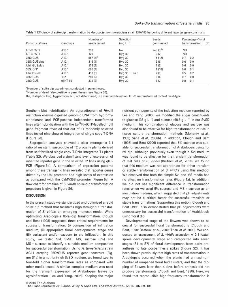
Southern blot hybridization. An autoradiogram of HindIII
restriction enzyme-digested genomic DNA from hygromy-
cin-tolerant and PCR-positive independent transformed
lines after hybridization with the [a-32P]-dCTP-labelled hptII
gene fragment revealed that out of 11 randomly selected
lines tested nine showed integration of single copy T-DNA
(Figure 5d).
Segregation analyses showed a clear monogenic 3:1
ratio of resistant: susceptible of T2 progeny plants derived
from self-fertilized single copy T-DNA integrated T1 plants
(Table S2). We observed a significant level of expression of
inherited reporter gene in the selected T2 lines using qRT-
PCR (Figure 5d). A comparison of expression patterns
among these transgenic lines revealed that reporter genes
driven by the Ubi promoter had high levels of expression
as compared with the CaMV35S promoter (Figure 5e). A
flow chart for timeline of S. viridis spike-dip transformation
procedure is given in Figure S6.
DISCUSSION
In the present study we standardized and optimized a rapid
spike-dip method that facilitates high-throughput transfor-
mation of S. viridis, an emerging monocot model. While
optimizing Arabidopsis floral-dip transformation, Clough
and Bent (1998) suggested three critical requirements for
successful transformation: (i) composition of infiltration
medium; (ii) appropriate floral developmental stage and
(iii) surfactant and/or vacuum to aid infiltration. In this
study, we tested SvI, SvSD, MS, sucrose (5%) and
MS + sucrose to identify a suitable medium composition
for successful transformation. Using A. tumefaciens strain
AGL1 carrying 35S::GUS reporter gene construct (Fig-
ure S1a) in a nutrient-rich SvSD medium, we found two- to
four-fold higher transformation rates as compared with
other media tested. A similar complex medium was used
for the transient expression of Arabidopsis leaves by
agroinfiltration (Lee and Yang, 2006). Keeping the major
nutrient components of the induction medium reported by
Lee and Yang (2006), we modified the sugar constituents
to glucose (36 g L�1) and sucrose (68.5 g L�1) in our SvSD
medium. This combination of glucose and sucrose was
also found to be effective for high transformation of rice in
tissue culture transformation methods (Mohanty et al.,
1999; Saha et al., 2006b). In addition, Clough and Bent
(1998) and Bent (2006) reported that 5% sucrose was suit-
able for successful transformation of Arabidopsis using flo-
ral dip. Although previously even though a SvI medium
was found to be effective for the transient transformation
of leaf cells of S. viridis (Brutnell et al., 2010), we found
that this medium was not appropriate for either transient
or stable transformation of S. viridis using this method.
We observed that both the simple SvI and MS media had
no effect on transformation rates (Figure 1a). In addition,
we did not see significant difference in transformation
rates when we used 5% sucrose and MS + sucrose as an
inoculation medium, which suggested that pH adjustments
may not be a critical factor for successful transient or
stable transformations. Supporting this notion, Clough and
Bent (1998) also demonstrated that pH adjustments were
unnecessary for successful transformation of Arabidopsis
using floral dip.
Developmental stage of the flowers was shown to be
crucial for successful floral transformations (Clough and
Bent, 1998; Desfeux et al., 2000; Trieu et al. 2000). We con-
ducted an assessment of S. viridis accession A10.1 foxtail
spikes developmental stage and categorized into seven
stages (S1 to S7) of floral development, from early pre-
anthesis to late post-anthesis spikes (Figure S2). It has
been shown previously that high rates of transformation in
Arabidopsis occurred when the plants had a maximum
number of unopened floral bud clusters, and that the dip-
ping of flowers later than 4 days before anthesis did not
produce transformants (Clough and Bent, 1998). Here, we
found that reproducible high-frequency transformation is
Table 1 Efficiency of spike-dip transformation by Agrobacterium tumefaciens strain EHA105 harboring different reporter gene constructs
Constructs/lines GenotypeNumber ofseeds tested
Selection(mg L�1)
Seedsgerminated
Percentage (%) oftransformation � SD
UT-C (WT) A10.1 252 No 245 (3)b NDUT-C (WT) A10.1 120 Hyg 30 0 (2) ND35S::GUS A10.1 567 (4)a Hyg 30 4 (12) 0.7 � 0.235S::GUSplus A10.1 316 (1) Hyg 30 2 (6) 0.6 � 0.0Ubi::GUSplus A10.1 176 (1) Hyg 30 1 (3) 0.6 � 0.035S::GFP A10.1 606 (4) Hyg 30 4 (10) 0.6 � 0.1Ubi::DsRed A10.1 413 (3) Hyg 30 + Bia 3 2 (0) 0.5 � 0.235S::GUS 132 289 (2) Hyg 30 2 (4) 0.7 � 0.035S::GUS 98HT-80 372 (3) Hyg 30 3 (7) 0.8 � 0.1
aNumber of spike dip experiment conducted in parentheses.bNumber of dead false positive in parentheses (see Figure S5).Bia, Bialaphos; Hyg, hygromycin; ND, not determined; SD, standard deviation; UT-C, untransformed control (wild-type).
© 2016 The AuthorsThe Plant Journal © 2016 John Wiley & Sons Ltd, The Plant Journal, (2016), 86, 89–101
Spike-dip transformation of Setaria viridis 95
achieved when the spikes were approximately 5.5 cm long
over 6–8 days before anthesis at S3 (Figures 1 and S2).
These results corroborate those found in wheat, in which
6–7 cm long spikes, which did not emerge from the sheath
before 4–7 days of anthesis, were ideal for transformation
(Zale et al., 2009). Although a significant high transient
GUS expression was seen at the early S2 stage, low stable
transformants were obtained. In general, we noticed a
gradual decrease in transformation rates with the progres-
sion of spike development towards anthesis (S4 to S6)
(Figures 1c and S2). Moreover, a high histochemical GUS
staining was seen at the bottom of each floret where the
ovule is located (Figure 1b), indicating that the ovule is the
probable target tissue amenable to T-DNA transfer upon
Agrobacterium infection. In agreement with this notion,
Bechtold and Pelletier (1998), Desfeux et al. (2000) and Ye
et al. (1999) showed that the Arabidopsis ovule is the likely
target for T-DNA transfer. To address the question of
whether multiple Agrobacterium inoculations over the
course of spike development might result in increased
numbers of transformants, we performed three consecu-
tive dipping of the same spikes on every alternate day.
Multiple Agrobacterium inoculations caused bleaching of
spikes with no seed setting as most plant tissues died.
Successful monocot transformation depends on the abil-
ity of the Agrobacterium cells to attach to the plant target
cells (Shrawat and Lorz, 2006). We reasoned that increas-
ing the infection time would allow a better access of
Agrobacterium to the target tissues and result in higher
transformation rates. Here, we determined that 20 min
infection was the best time for an efficient spike-dip trans-
formation. Furthermore, surfactants are known to function
either as enhancers of cuticle penetration by making the
plant cuticular membrane more susceptible to Agrobac-
terium, or by acting as co-solvents, thereby enhancing the
movement of Agrobacterium into the plant cells (Madhou
et al., 2006). The supplementation of surfactants to
improve the efficiency of the Agrobacterium-mediated
genetic transformation of diverse plant tissues was appar-
ent from previous studies (Clough and Bent, 1998; Cheng
et al., 2004; Zhang et al., 2006; Kim et al., 2009), although
high surfactant levels in the infection medium could cause
the necrosis of plant tissues (Clough and Bent, 1998). We
determined that the rate of transformation did not change
significantly when Tween 20 and Triton X-100 were used at
concentrations between 0.01 and 1.0%. Conversely, Silwet
L-77 at 0.025% produced about a four-fold higher stable
transformants when spikes were inoculated with Agrobac-
terium stain AGL1 containing the 35S::GUS reporter gene
(Figure 2b). Silwet L-77 is a preferred surfactant because of
its low phytotoxicity, reduces surface tension compared
with most surfactants and it improves penetration of bacte-
ria into relatively inaccessible plant tissues (Whalen et al.,
1991). However, we observed that inoculation medium
Figure 5. Molecular analyses of stable transformants from T1 and T2 gener-
ations.
(a–c) PCR analysis of putative T1 transformants for the presence of (a)
reporter genes (GUS, GUSplus, GFP and DsRed), (b) selectable marker
(HptII) gene, and (c) an endogenous TUB gene.
(d) Southern blot analysis of HindIII restriction enzyme-digested genomic
DNA isolated from leaves of 11 independent T1 transgenic lines.
(e) Expression patterns of reporter genes (GUS, GUSplus, GFP and DsRed)
in selected T2 lines as determined by qRT-PCR. M, Thermo Scientific
O’Gene Ruler 1 kb Plus DNA ladder. Plasmid DNA of 35S::GUS, 35S::GUS-
plus, Ubi::GUSplus, 35S::GFP and Ubi::DsRed were used as positive (+) con-trol while untransformed control (UT-C) plant DNA was taken as negative
(�) control to amplify with each reporter gene-specific primer for PCR.
Restriction enzyme digested genomic DNAs from T1 transgenic lines and
UT-C (�control) including PCR purified HptII gene (+ control) were sepa-
rated in a 1% agarose gel, and hybridized with a [a-32P]-dCTP-labelled 578-
bp HptII gene probe. Position of molecular weight markers are indicated on
the left side. qRT-PCR results are presented as mean relative expression
(2�DDCT) with SE from three progeny plants of same T1 parent after normal-
ization using an ACT endogenous reference gene. UT-C plant RNA was used
as the negative (�) control to amplify with each reporter gene-specific pri-
mers for qRT-PCR. a–oDifferent letters on the bars indicate significant differ-
ences at the P ≤ 0.05 level as tested by the Tukey–Kramer HSD test. See
Table S1 for primer sequences and product size.
© 2016 The AuthorsThe Plant Journal © 2016 John Wiley & Sons Ltd, The Plant Journal, (2016), 86, 89–101
96 Prasenjit Saha and Eduardo Blumwald

containing 1.0% Silwet L-77 was deleterious because it
caused necrosis of plant tissues.
The different Agrobacterium strains used in this study
differed in their virulence, with strains EHA105 and AGL1
showing a superior transformation rate. Both EHA105 and
AGL1 contain succinamopine type Ti-plasmid with C58 ori-
gin (Hamilton and Fall, 1971) and were shown previously
to be suitable for monocot transformation (Hiei and
Komari, 2008; Chen et al., 2010).
Further to validate our transformation protocol and to
examine the appropriate promoter to conduct future trans-
formation in S. viridis, we conducted several transforma-
tions to overexpress GUSplus, GFP and DsRed driven by
either CaMV35S or maize Ubi promoters. Chen et al. (2010)
demonstrated that the GUSplus reporter gene was useful
for high-throughput transient gene expression in switch-
grass, while fluorescent (GFP and DsRed) reporter genes
were known to be suitable for Agrobacterium transforma-
tions (Sheen et al., 1995; Jach et al., 2001). We monitored
significantly higher expression of reporter genes in tran-
sient expression (Figure 3c,e) as well as subsequent T1
(Figure 4) and T2 (Figure 5e) generations when driven by
the Ubi promoter. The maize Ubi promoter has been exten-
sively employed for enhanced constitutive expression of
target gene in monocot cereals (Toki et al., 1992; Hiei et al.,
2014). Although the choice and the selection agent concen-
tration are also important considerations when using this
protocol, the concentration of selection agent should not
be as high as to be lethal to low copy number T-DNA
transformants (Wilmink and Dons, 1993). We chose HptII
because it is a suitable selection marker with relatively low
escapes from hygromycin antibiotic selection. The devel-
opment of false positives in our study could be attributed
to the stress induced by dipping, as similar results were
reported in wheat by Zale et al. (2009), and demanded the
use of a secondary screen. Thus the selection with a com-
bination of hygromycin and bialaphos allowed us to iden-
tify putative transformants (Figures 5, S4 and S5). We
further demonstrated the stable integration of the T-DNA
cassette into the nuclear genome by PCR (Figure 5a,b) of
genomic DNA isolated from hygromycin-resistant and
GUS positive T1 lines (Figure 4 and Table 1) with respec-
tive primers specific for each reporter and selectable mar-
ker genes (Table S1). We confirmed the stable integration
and determined the copy number of T-DNA by Southern
blot analysis (Figure 5d). We also demonstrated a mono-
genic 3:1 Mendelian pattern of transgene inheritance
(Table S2) and the expression of inherited transgenes in
the subsequent T2 generation (Figure 5e).
In conclusion, we developed an alternative method for
the Agrobacterium-mediated transformation of S. viridis
using a spike dip that avoids the traditional in vitro culture
steps. Several genotypes of S. viridis were transformed
following the optimized conditions for transient gene
expression assay as well as stable transformation. Fertile
transgenic T1 plants can be obtained within 8–10 weeks
and the expression and inheritance of transgenes can be
monitored over generations. In vitro culture, plant tissue
culture media, growth regulators and vacuum infiltration
were not essential to obtain stable transformants at a rate
of 0.8 � 0.1% (Table 1), a transformation efficiency rate
that is comparable to the reported transformation effi-
ciency by floral dip in Arabidopsis (Clough and Bent,
1998). The application of the method reported here will
facilitate the high-throughput transformation of S. viridis.
EXPERIMENTAL PROCEDURE
Plant materials and growth conditions
Three Setaria viridis (L.) Thell. genotypes, A10.1 (PI 669942/Ames31045), 132 (PI Ames 28193) and 98HT-80 (PI 649320), were usedin this study (Table 1). Seeds were obtained from the GermplasmResources Information Network (GRIN, http://www.ars-grin.gov/),United States Department of Agriculture (USDA). Seeds from eachgenotype were sowed in germination trays (27.9 9 54.3 cm)(McConkey, Sumner, WA, USA) containing moist agronomy mix(equal parts of redwood compost, sand and peat moss) and ver-nalized at 4°C for 2 days in a Isotemp incubator (Fisher Scientific,Pittsburgh, PA, USA). Trays were kept in greenhouse at 28 � 2°Cwith 50% relative humidity for a 16 h day/8 h night photoperiodfor seed germination. After 7 days, seedlings were transferred topots (10.2 9 8.2 cm) (McConkey) containing moist agronomy mixand allowed to grown under greenhouse conditions until flower-ing. Plants were watered every alternate day with deionized waterand fertilized using a solution of 50% N:phosphorus:potassium(20:10:20) and 50% ammonium sulfate (total of 0.5 g of N) once ina week until spike initiation.
Transformation vectors and Agrobacterium strains
Five binary vectors, pCAMBIA1201, pH7m24GWp35S-GUSplus,pH7m24GW pUbi-GUSplus, pH7m24GW35Sp-GFP and pGWB17-UbiDsRED-UbiBAR, containing GUS (b-glucuronidase), GUSplus(b-glucuronidase plus), GFP and DsRed (Discosoma sp. red fluo-rescent protein) reporter genes were used in this study. The sche-matic representation of 35S::GUS, 35S::GUSplus, Ubi::GUSplus,35S::GFP and Ubi::DsRed transformation vectors are shown in Fig-ure S1. The reporter gene constructs were driven by either thecauliflower mosaic virus 35S (CaMV35S) or an intron-interruptedmaize ubiquitin (Ubi) promoters, and CaMV35S or nopaline syn-thase (NOS) terminators within the right (RB) and left (LB) bordersof the T-DNA cassette region. These binary vectors contain HptII(hygromycin phosphotransferase II) and Bar (bialaphos) genes asplant selection markers. Four Agrobacterium tumefaciens strainsused were AGL1(Lazo et al., 1991), EHA105 (Hood et al., 1993),GV3101 (Koncz and Schell, 1986) and LBA4404 (Hoekema et al.,1983). The plasmids for each set of experiments were electrotrans-formed into the respective A. tumefaciens strains using a GenePulser XcellTM Electroporation Systems (Bio-Rad, Hercules, CA,USA) following the method described before (Lin, 1995).
Bacterial culture and dipping medium
A single colony of each A. tumefaciens strain harboring one of thefive binary vectors was cultured overnight in 5 ml liquid YEB med-ium (Vervliet et al., 1975), containing appropriate antibiotics, at
© 2016 The AuthorsThe Plant Journal © 2016 John Wiley & Sons Ltd, The Plant Journal, (2016), 86, 89–101
Spike-dip transformation of Setaria viridis 97

28°C with an agitation of 250 rpm. The following day, 500 ll ofthis starter culture was added to 50 ml of culture medium contain-ing 5 g L�1 tryptone, 2.5 g L�1 yeast extract, 5 g L�1 NaCl, 5 g L�1
mannitol, 100 mg L�1, MgSO4, 250 mg L�1 K2HPO4, 1.2 g L�1 glu-tamic acid, 15 g L�1 sucrose, pH 7.2 and antibiotics, and the bacte-ria was grown for 16 h at the same conditions as mentionedabove. The next day, after adjusting the OD600 of the culture to1.0, the culture was collected by centrifugation at 2400 g for 5 minand re-suspended in dipping medium.
The five dipping media tested were as follows: (i) Setaria viridisInfiltration (SvI) medium consisting of 50 mM (N-morpholino)methanesulfonic acid (MES), 2 mM Na3PO4, 12H2O, 0.025% (w/v)glucose; pH 5.8 (Brutnell et al., 2010); (ii) Setaria viridis spike-dip(SvSD) medium composed of 10.5 g L�1 K2HPO4, 4.5 g L�1
KH2PO4, 1 g L�1 (NH4)2SO4, 0.5 g L�1 Na citrate, 4 g L�1 glycerol,1 mM MgSO4, 15 g L�1 ascorbic acid, 10 mM MES; pH to 5.8 modi-fied from Lee and Yang (2006) and supplemented with 68.5 g L�1
sucrose, 36 g L�1 glucose (Mohanty et al., 1999); (iii) MS fullstrength basal medium; pH 5.8 (Murashige and Skoog, 1962); (iv)sucrose (5% w/v) (Bent, 2006); (v) MS full strength basal mediumfortified with 5% (w/v) sucrose; pH 5.8 (Clough and Bent, 1998).
Agrobacterium-mediated transformation and selection of
putative transformants
Prior to spike dip, the bacteria harboring the reporter gene con-struct were re-suspended in 40 ml dipping medium supplementedwith 200 lM acetosyringone (PhytoTechnology Laboratories, Over-land Park, KS, USA) in a 50 ml Falcon tube and pre-induced byshaking at 180 rpm for 1 h. Plants bearing spikes at suitable devel-opmental stage (Figure S2) were labelled (Figure S3a) and 15 minbefore to transformation all spikes were made wet by applying1 ml of infiltration solution (10 mM MgSO4, 10 mM MES; pH to 5.8)(Lee and Yang, 2006) containing Silwet-L77 (Lehle Seeds, RoundRock, TX, USA) in the range of 0.01–0.10% (v/v). Inflorescenceswere then immersed into the pre-induced Agrobacterium culturefor 10–40 min with an occasional gentle agitation at 5-min inter-vals (Figure S3b). After dipping, plants were kept under the domeat low light intensity for overnight at 22°C. Next day plants werereturned at 25°C for 16 h day/8 h night photoperiod with 50% rela-tive humidity in the growth chamber or in the growth room wherethey grew for 5–7 days, and during this time transient transforma-tion assay was performed on three or five DPD spikes. Finally, theplants were put inside the arecones (arabase and aratubes, LehleSeeds) and grown under greenhouse conditions mentioned abovefor 2–4 weeks until maturity, when spikes turned brown and dry,and set seeds (Figure S3c). Seeds were harvested by gentlyremoving the arecones and collected in microfuge tubes andstored at 4°C under desiccation.
Seeds obtained from T1 plants and subsequent generationswere dehusked, and surface sterilized using 10% commercialbleach with 0.1% Tween 20 for 5 min followed by three to fivewashes in sterile deionized water. Seeds were blotted dry on ster-ile filter papers for 5 min and 20–25 seeds were transferred toplates containing half strength MS medium fortified with either30 mg L�1 hygromycin (PhytoTechnology Laboratories, ShawneeMission, KS, USA) or 3 mg L�1 bialaphos (PhytoTechnology Labo-ratories) as described before (Saha et al., 2006b). Petri plates andlids were sealed with parafilm tape and kept at 4°C for 2 days forseed vernalization. Plates were then kept horizontally and incu-bated in the growth chamber (Percival Scientific, Perry, IA, USA)at 25°C with 16 h day/8 h night photoperiod and 50% relativehumidity. Putative transformants were identified as hygromycin-resistant seedlings with well-developed green leaves showing
profuse rooting after 10 DPG for selection on hygromycin (FiguresS4 and 5). Selected putative transformants were transferred to soilin pots and grown under greenhouse conditions until maturity.The percentage (%) of transformation efficiencies were calculatedas (number of hygromycin-resistant seedlings)/(total numberseeds tested) 9 100.
b-Galactosidase activity (GUS) assay and fluorescence
microscopy
Histochemical staining and fluorometric measurements of the b-galactosidase (GUS) activity were performed according to themethod described by Jefferson et al. (1987). The transformed tis-sues were soaked in the GUS staining buffer (100 mM sodiumphosphate, 1 mM 5-bromo-4-chloro-3-indolyl-b-D-glucuronide,0.5 mM potassium ferrocyanide, 0.5 mM potassium ferricyanide,10 mM Na2EDTA; pH 7.0) and incubated for 16 h at 37°C. Next day,the stained tissues were washed by repeated rinsing with 3:1 (v/v)absolute alcohol: acetic acid until completely cleared the chloro-phylls and photographed.
For fluorometric measurements of the GUS activity, trans-formed spikes after dipping were homogenized in the extractionbuffer (50 mM sodium phosphate, 10 mM Na2EDTA, 0.1% TritonX-100, 0.1% sarcosyl, 10 mM b-mercaptoethanol; pH 7.0) and cen-trifuged for 15 min at 16 100 g to collect the supernatants. Theprotein concentrations were determined following the protocoldescribed before Bradford (1976) using bovine serum albumin(BSA) as standard. The GUS activity was carried out using 10 lgof total protein after incubation with the substrate 4-methyl-umbelliferyl-b-D-glucuronide (4-MUG; Sigma, St. Louis, MO, USA)for 1 h at 37°C. The reaction was terminated by adding 0.2 M
Na2CO3, and the fluorescence was measured at 365 nm excita-tion and 455 nm emission using a 96-well BioTek SynergyMxmicroplate reader (BioTek Instruments, Inc., Winooski, VT, USA).Specific GUS enzyme activity was determined based on the stan-dard curve of 4-methylumbelliferone (4MU; Sigma) standardsfrom the same microtiter plate. Data were recorded using Gen5(version 1.11.5) software (BioTek) and relative GUS activity ineach sample was calculated after normalization of the fluores-cence signal value by subtracting of the background fluorescencesignal detected from equal amount of proteins in the mock con-trol.
Expression of GFP and red fluorescent protein (DsRed) wasobserved using a Leica MZFLIII fluorescence stereomicroscope(Leica Microsystems Inc., Buffalo Grove, IL, USA) equipped withfilters for GFP (excitation 395 nm and emission 509 nm) andDsRed (excitation 554 nm and emission 586 nm) respectively, anda SPOT Insight charge coupled device (CCD) camera. Photographswere captured using SPOT Advanced (version 4.1) software (Diag-nostic Instruments, Inc., Sterling Heights, MI, USA).
Nucleic acids analysis
Genomic DNA was isolated from transformed and untransformedcontrol tissue followed by the cetyltrimethylammonium bromide(CTAB) extraction method (Doyle and Doyle, 1987). PCR analysisfor detection of the transgenes (GUS, GUSplus, GFP, DsRed, HptIIand Bar) was carried out as described before (Saha et al., 2006a)using pair of primer specific for each gene (Table S1). The five bin-ary vector plasmids were used as a positive control for PCR. Theamplicons were separated by electrophoresis on 1% (w/v) agarosegel.
Southern blot analysis was carried out according to Sambrooket al. (1989). Genomic DNA (10 lg) from transformed and
© 2016 The AuthorsThe Plant Journal © 2016 John Wiley & Sons Ltd, The Plant Journal, (2016), 86, 89–101
98 Prasenjit Saha and Eduardo Blumwald

untransformed control plants were digested with HindIII (NewEngland BioLabs, Inc., Ipswich, MA, USA) restriction enzyme,fractionated on a 1% (w/v) agarose gel, denatured and trans-ferred onto a N + -nylon membrane (Amersham Biosciences, GEHealthcare Life Sciences, Pittsburgh, PA, USA). Hybridization ofmembrane with the [a-32P] dCTP (Molecular Probe, Life Technolo-gies) labelled PCR amplified HptII gene probe, followed by wash-ings under stringent conditions, was carried out according toprocedures described earlier (Saha et al., 2007). Blot wasexposed to phosphor imaging screen (Molecular Dynamics, GEHealthcare Life Sciences, Pittsburgh, PA, USA). Phosphor imageswere scanned on a Typhoon 8600 Variable Mode Imager (Molec-ular Dynamics, GE Healthcare Life Sciences, Pittsburgh, PA, USA)and the scans were analyzed using Typhoon Scanner Control(version 1.0) software (GE Healthcare Life Sciences). ImageQuantTL Array (version 8.1) software (GE Healthcare LifeSciences) and Adobe Photoshop CS5 (version 12.0) softwarewere used to adjust the digital image.
For quantitative real-time PCR (qRT-PCR), gene-specific primerpair was designed using the Primer Express (version 3.0) soft-ware (Applied Biosystems, Foster City, CA) following the defaultparameters according to the manufacturer’s recommendation(Table S1). Total RNA was isolated from 100 mg of frozenground tissue using the RNeasy plant mini kit (Qiagen, Valencia,CA, USA) according to the manufacturer’s procedure. The RNAsample was assessed by NanoDrop ND-1000 (NanoDrop Tech-nologies, Wilmington, DE, USA) and the first strand cDNA syn-thesis was carried out according to the protocol described earlier(Saha and Blumwald, 2014) using the QuantiTect reverse tran-scription kit (Qiagen). The qRT-PCR reactions were performed ina reaction volume of 5 ll containing 1 ll of diluted cDNA,200 nM of each gene-specific primer and 2.5 ll of 29 Fast SYBRGreen PCR Master Mix (Applied Biosystems, Foster City, CA,USA) in an optical 96 well plate (Applied Biosystems) using aStepOnePlusTM Real-Time PCR System (Applied Biosystems)equipment as previously described (Saha et al., 2013). An ACT(actin) gene was used as an endogenous reference gene(Table S1) to normalize gene expression and the fold change oftransgenes expression were calculated by the 2�DDCT equation asdescribed before (Saha et al., 2013).
Segregation analysis
Segregation analysis of T2 seeds was carried out according toDutta et al. (2005) on hygromycin (30 mg L�1) amended half-strength MS medium and 10 DPG seedling were scored as hygro-mycin resistance (HygR) or hygromycin sensitive (HygS).
Statistical analysis
All experiments were repeated three times. Mean with SD for eachexperiment was calculated using Microsoft Excel (version 2010)software. Tukey–Kramer honest significant difference (HSD) testwas performed for multiple comparisons to evaluate significantdifferences at the P ≤ 0.05 level within experiments using JMP(version 7.0.2) software (SAS Institute Inc. Cary, NC, USA). Segre-gation patterns of the T2 progeny plants were validated using thechi-squared test.
ACKNOWLEDGEMENTS
Authors are thankful to Dr Ellen Tumimbang, Dr Hiromi Tajima,Elham Abed, Yrian Hong and Hye Won Kim for technical support.This work is funded by the United States Agency for InternationalDevelopment (USAID) to support the Feed the Future Innovation
Laboratory for Climate-Resilient Millet under Grant No. APS M/OAA/GRO/EGAS-11–002011.
SUPPORTING INFORMATION
Additional Supporting Information may be found in the online ver-sion of this article.Figure S1. Schematic representation of a linearized map of planttransformation vectors.
Figure S2. Developmental stages of S. viridis accession A10.1 fox-tail spikes.
Figure S3. Steps of spike dip transformation.
Figure S4. Selection of putative transformants at the T1 plantgeneration.
Figure S5. Representation of stable transformants and hygromycinsusceptible false positive seedlings after 10 DPG growing on halfMS + hygromycin (30 mg L�1) plates and hygromycin-resistantputative transformed seedlings with well-developed green leavesshowing profuse rooting in the presence of hygromycin.
Figure S6. Flow chart for timeline of S. viridis spike dip transfor-mation procedure.
Table S1 Details of PCR and qRT-PCR primer sequences used forS. viridis spike dip method.
Table S2 Segregation analyses of T2 progeny plants derived fromself-fertilized T1 plants.
REFERENCES
Alves, S.C., Worland, B., Thole, V., Snape, J.W., Bevan, M.W. and Vain, P.
(2009) A protocol for Agrobacterium-mediated transformation of Brachy-
podium distachyon community standard line Bd21. Nat. Protoc. 4, 638–649.
Bastaki, N.K. and Cullis, C.A. (2014) Floral-dip transformation of flax (Linum
usitatissimum) to generate transgenic progenies with a high transforma-
tion rate. J. Vis. Exp. 94, 1–10.Bechtold, N. and Pelletier, G. (1998) In planta Agrobacterium-mediated
transformation of adult Arabidopsis thaliana plants by vacuum infiltra-
tion. Methods Mol. Biol. 82, 259–266.Bennetzen, J.L., Schmutz, J., Wang, H. et al. (2012) Reference genome
sequence of the model plant Setaria. Nat. Biotechnol. 30, 555–561.Bent, A.F. (2000) Arabidopsis in planta transformation. Uses, mechanisms,
and prospects for transformation of other species. Plant Physiol. 124,
1540–1547.Bent, A. (2006) Arabidopsis thaliana floral dip transformation method. Meth-
ods Mol. Biol. 343, 87–103.Birch, R.G. (1997) PLANT TRANSFORMATION: problems and strategies for
practical application. Annu. Rev. Plant Physiol. Plant Mol. Biol. 48, 297–326.
Bradford, M.M. (1976) A rapid and sensitive method for the quantitation of
microgram quantities of protein utilizing the principle of protein-dye
binding. Anal. Biochem. 72, 248–254.Brutnell, T.P., Wang, L., Swartwood, K., Goldschmidt, A., Jackson, D., Zhu,
X.G., Kellogg, E. and Van Eck, J. (2010) Setaria viridis: a model for C4
photosynthesis. Plant Cell, 22, 2537–2544.Brutnell, T.P., Bennetzen, J.L. and Vogel, J.P. (2015) Brachypodium dis-
tachyon and Setaria viridis: model genetic systems for the grasses.
Annu. Rev. Plant Biol. 66, 465–485.Chen, X., Equi, R., Baxter, H., Berk, K., Han, J., Agarwal, S. and Zale, J.
(2010) A high-throughput transient gene expression system for
switchgrass (Panicum virgatum L.) seedlings. Biotechnol. Biofuels, 3,
1–10.Cheng, M., Lowe, B.A., Spencer, T.M., Ye, X. and Armstrong, C.L. (2004)
Factors influencing Agrobacterium-mediated transformation of mono-
cotyledonous species. In Vitro Cell. Dev. Biol. Plant, 40, 31–45.Clough, S.J. and Bent, A.F. (1998) Floral dip: a simplified method for
Agrobacterium-mediated transformation of Arabidopsis thaliana. Plant J.
16, 735–743.
© 2016 The AuthorsThe Plant Journal © 2016 John Wiley & Sons Ltd, The Plant Journal, (2016), 86, 89–101
Spike-dip transformation of Setaria viridis 99

Curtis, I. and Nam, H. (2001) Transgenic radish (Raphanus sativus L. longip-
innatus Bailey) by floral-dip method–plant development and surfactant
are important in optimizing transformation efficiency. Transgenic Res.
10, 363–371.Desfeux, C., Clough, S.J. and Bent, A.F. (2000) Female reproductive tissues
are the primary target of Agrobacterium-mediated transformation by the
Arabidopsis floral-dip method. Plant Physiol. 123, 895–904.Doyle, J.J. and Doyle, J.L. (1987) A rapid DNA isolation procedure for small
quantities of fresh leaf tissue. Phytochem. Bull. 19, 11–15.Dutta, I., Saha, P., Majumder, P., Sarkar, A., Chakraborti, D., Banerjee, S.
and Das, S. (2005) The efficacy of a novel insecticidal protein, Allium sati-
vum leaf lectin (ASAL), against homopteran insects monitored in trans-
genic tobacco. Plant Biotechnol. J. 3, 601–611.Gelvin, S.B. (2003) Agrobacterium-mediated plant transformation: the biol-
ogy behind the “gene-jockeying” tool. Microbiol. Mol. Biol. Rev. 67, 16–37.
Ghedira, R., De Buck, S., Nolf, J. and Depicker, A. (2013) The efficiency
of Arabidopsis thaliana floral dip transformation is determined not
only by the Agrobacterium strain used but also by the physiology and
the ecotype of the dipped plant. Mol. Plant Microbe Interact. 26, 823–832.
Hamilton, R.H. and Fall, M.Z. (1971) The loss of tumor-initiating ability in
Agrobacterium tumefaciens by incubation at high temperature. Experien-
tia, 27, 229–230.Hiei, Y. and Komari, T. (2008) Agrobacterium-mediated transformation of
rice using immature embryos or calli induced from mature seed. Nat.
Protoc. 3, 824–834.Hiei, Y., Ishida, Y. and Komari, T. (2014) Progress of cereal transformation
technology mediated by Agrobacterium tumefaciens. Front. Plant Sci. 5,
628.
Hoekema, A., Hirsch, P.R., Hooykaas, P.J.J. and Schilperoort, R.A.
(1983) A binary plant vector strategy based on separation of vir- and
T-region of the Agrobacterium tumefaciens Ti-plasmid. Nature, 303,
179–180.Hood, E., Gelvin, S., Melchers, L. and Hoekema, A. (1993) New Agrobac-
terium helper plasmids for gene transfer to plants. Transgenic Res. 2,
208–218.Huang, P., Feldman, M., Schroder, S., Bahri, B.A., Diao, X., Zhi, H., Estep,
M., Baxter, I., Devos, K.M. and Kellogg, E.A. (2014) Population genetics
of Setaria viridis, a new model system. Mol. Ecol. 23, 4912–4925.Jach, G., Binot, E., Frings, S., Luxa, K. and Schell, J. (2001) Use of red fluo-
rescent protein from Discosoma sp. (dsRED) as a reporter for plant gene
expression. Plant J. 28, 483–491.Jefferson, R.A., Kavanagh, T.A. and Bevan, M.W. (1987) GUS fusions: beta-
glucuronidase as a sensitive and versatile gene fusion marker in higher
plants. EMBO J. 6, 3901–3907.Jia, G., Shi, S., Wang, C., Niu, Z., Chai, Y., Zhi, H. and Diao, X. (2013) Molec-
ular diversity and population structure of Chinese green foxtail [Setaria
viridis (L.) Beauv.] revealed by microsatellite analysis. J. Exp. Bot. 64,
3645–3656.Jiang, H., Barbier, H. and Brutnell, T. (2013) Methods for performing crosses
in Setaria viridis, a new model system for the grasses. J. Vis. Exp. 80, 1–8.
John, C.R., Smith-Unna, R.D., Woodfield, H., Covshoff, S. and Hibberd, J.M.
(2014) Evolutionary convergence of cell-specific gene expression in inde-
pendent lineages of C4 grasses. Plant Physiol. 165, 62–75.Kim, M.J., Baek, K. and Park, C.M. (2009) Optimization of conditions for
transient Agrobacterium-mediated gene expression assays in Arabidop-
sis. Plant Cell Rep. 28, 1159–1167.Koncz, C. and Schell, J. (1986) The promoter of TL-DNA gene five controls
the tissue-specific expression of chimaeric genes carried by a novel type
of Agrobacterium binary vector. Mol. Gen. Genet. 204, 383–396.Lazo, G.R., Stein, P.A. and Ludwig, R.A. (1991) A DNA transformation-com-
petent Arabidopsis genomic library in Agrobacterium. Biotechnology, 9,
963–967.Lee, M.W. and Yang, Y. (2006) Transient expression assay by agroinfiltration
of leaves. Methods Mol. Biol. 323, 225–229.Li, P. and Brutnell, T.P. (2011) Setaria viridis and Setaria italica, model
genetic systems for the Panicoid grasses. J. Exp. Bot. 62, 3031–3037.Lin, J.J. (1995) Electrotransformation of Agrobacterium. Methods Mol. Biol.
47, 171–178.
Liu, X., Brost, J., Hutcheon, C. et al. (2012) Transformation of the oilseed
crop Camelina sativa by Agrobacterium-mediated floral dip and simple
large-scale screening of transformants. In Vitro Cell. Dev. Biol. Plant, 48,
462–468.Madhou, P., Raghavan, C., Wells, A. and Stevenson, T.W. (2006) Genome-
wide microarray analysis of the effect of a surfactant application in Ara-
bidopsis. Weed Res. 46, 275–283.Martins, P.K., Nakayama, T.J., Ribeiro, A.P., Cunha, B.A.D.B.d., Nepomu-
ceno, A.L., Harmon, F.G., Kobayashi, A.K. and Molinari, H.B.C. (2015)
Setaria viridis floral-dip: a simple and rapid Agrobacterium-mediated
transformation method. Biotechnol. Rep. 6, 61–63.Mohanty, A., Sarma, N.P. and Tyagi, A.K. (1999) Agrobacterium-mediated
high frequency transformation of an elite indica rice variety Pusa Bas-
mati 1 and transmission of the transgenes to R2 progeny. Plant Sci. 147,
127–137.Mu, G., Chang, N., Xiang, K., Sheng, Y., Zhang, Z. and Pan, G. (2012) Dip
method mediated by Agrobacterium. Biotechnology, 11, 178–183.Murashige, T. and Skoog, F. (1962) A revised medium for rapid growth
and bio assays with tobacco tissue cultures. Physiol. Plant. 15, 473–497.
Petti, C., Shearer, A., Tateno, M., Ruwaya, M., Nokes, S., Brutnell, T. and
Debolt, S. (2013) Comparative feedstock analysis in Setaria viridis L. as a
model for C4 bioenergy grasses and Panicoid crop species. Front. Plant
Sci. 4, 181.
Saha, P. and Blumwald, E. (2014) Assessing reference genes for accurate
transcript normalization using quantitative real-time PCR in pearl millet
[Pennisetum glaucum (L.) R. Br.]. PLoS ONE, 9, e106308.
Saha, P., Dasgupta, I. and Das, S. (2006a) A novel approach for develop-
ing resistance in rice against phloem limited viruses by antagonizing
the phloem feeding hemipteran vectors. Plant Mol. Biol. 62, 735–752.Saha, P., Majumder, P., Dutta, I., Ray, T., Roy, S.C. and Das, S. (2006b)
Transgenic rice expressing Allium sativum leaf lectin with enhanced
resistance against sap-sucking insect pests. Planta, 223, 1329–1343.Saha, P., Chakraborti, D., Sarkar, A., Dutta, I., Basu, D. and Das, S. (2007)
Characterization of vascular-specific RSs1 and rolC promoters for their
utilization in engineering plants to develop resistance against hemi-
pteran insect pests. Planta, 226, 429–442.Saha, P., Ray, T., Tang, Y., Dutta, I., Evangelous, N.R., Kieliszewski,
M.J., Chen, Y. and Cannon, M.C. (2013) Self-rescue of an EXTENSIN
mutant reveals alternative gene expression programs and candidate
proteins for new cell wall assembly in Arabidopsis. Plant J. 75, 104–116.
Sambrook, J., Fritsch, E.F. and Maniatis, T. (1989) Molecular Cloning: A Lab-
oratory Manual. Cold Spring Harbor, NY: Cold Spring Harbor laboratory
Press.
Sebastian, J., Wong, M.K., Tang, E. and Dinneny, J.R. (2014) Methods to
promote germination of dormant Setaria viridis seeds. PLoS ONE, 9,
e95109.
Sheen, J., Hwang, S., Niwa, Y., Kobayashi, H. and Galbraith, D.W. (1995)
Green-fluorescent protein as a new vital marker in plant cells. Plant J. 8,
777–784.Shrawat, A.K. and Lorz, H. (2006) Agrobacterium-mediated transformation
of cereals: a promising approach crossing barriers. Plant Biotechnol. J. 4,
575–603.Sood, P., Bhattacharya, A. and Sood, A. (2011) Problems and possibilities of
monocot transformation. Biol. Plant. 55, 1–15.Toki, S., Takamatsu, S., Nojiri, C., Ooba, S., Anzai, H., Iwata, M., Chris-
tensen, A.H., Quail, P.H. and Uchimiya, H. (1992) Expression of a maize
ubiquitin gene promoter-bar chimeric gene in transgenic rice plants.
Plant Physiol. 100, 1503–1507.Trieu, A.T., Burleigh, S.H., Kardailsky, I.V. et al. (2000) Transformation of
Medicago truncatula via infiltration of seedlings or flowering plants with
Agrobacterium. Plant J. 22, 531–541.Vervliet, G., Holsters, M., Teuchy, H., Van Montagu, M. and Schell, J. (1975)
Characterization of different plaque-forming and defective temperate
phages in Agrobacterium. J. Gen. Virol. 26, 33–48.Wang, Q.M. and Wang, L. (2012) An evolutionary view of plant tissue
culture: somaclonal variation and selection. Plant Cell Rep. 31, 1535–1547.
Whalen, M.C., Innes, R.W., Bent, A.F. and Staskawicz, B.J. (1991) Identifica-
tion of Pseudomonas syringae pathogens of Arabidopsis and a bacterial
© 2016 The AuthorsThe Plant Journal © 2016 John Wiley & Sons Ltd, The Plant Journal, (2016), 86, 89–101
100 Prasenjit Saha and Eduardo Blumwald

locus determining avirulence on both Arabidopsis and soybean. Plant
Cell, 3, 49–59.Wilmink, A. and Dons, J.J.M. (1993) Selective agents and marker genes for
use in transformation of monocotyledonous plants. Plant Mol. Biol. Rep.
11, 165–185.Wu, H., Doherty, A. and Jones, H.D. (2008) Efficient and rapid Agrobac-
terium-mediated genetic transformation of durum wheat (Triticum turgi-
dum L. var. durum) using additional virulence genes. Transgenic Res. 17,
425–436.Xu, J., Li, Y., Ma, X., Ding, J., Wang, K., Wang, S., Tian, Y., Zhang, H. and
Zhu, X.-G. (2013) Whole transcriptome analysis using next-generation
sequencing of model species Setaria viridis to support C4 photosynthe-
sis research. Plant Mol. Biol. 83, 77–87.
Yasmeen, A., Mirza, B., Inayatullah, S., Safdar, N., Jamil, M., Ali, S. and
Choudhry, M.F. (2009) In planta transformation of tomato. Plant Mol.
Biol. Rep. 27, 20–28.Ye, G.N., Stone, D., Pang, S.Z., Creely, W., Gonzalez, K. and Hinchee, M.
(1999) Arabidopsis ovule is the target for Agrobacterium in planta vac-
uum infiltration transformation. Plant J. 19, 249–257.Zale, J.M., Agarwal, S., Loar, S. and Steber, C.M. (2009) Evidence for stable
transformation of wheat by floral dip in Agrobacterium tumefaciens.
Plant Cell Rep. 28, 903–913.Zhang, X., Henriques, R., Lin, S.S., Niu, Q.W. and Chua, N.H. (2006)
Agrobacterium-mediated transformation of Arabidopsis thaliana using
the floral dip method. Nat. Protoc. 1, 641–646.
© 2016 The AuthorsThe Plant Journal © 2016 John Wiley & Sons Ltd, The Plant Journal, (2016), 86, 89–101
Spike-dip transformation of Setaria viridis 101


![Vol 3[1] 2 Setaria 13-34.pdf - Tropical Grassland Society](https://static.fdocuments.in/doc/165x107/6207552449d709492c3064e2/vol-31-2-setaria-13-34pdf-tropical-grassland-society.jpg)