Title Chemical Structures of Polymyxin Series Antibiotics ...
Upload
mahendra-kumarCategory
view
212download
0
Specificity for the Exchange of Phospholipids Through Polymyxin B MediatedIntermembrane Molecular Contacts†
Yolanda Cajal,*,‡ Jiothy Ghanta,‡,§ Kalpathy Easwaran,§ Avadhesha Surolia,§ and Mahendra Kumar Jain*,‡
Department of Chemistry and Biochemistry, UniVersity of Delaware, Newark, Delaware 19716
ReceiVed NoVember 13, 1995; ReVised Manuscript ReceiVed March 7, 1996X
ABSTRACT: Structural specificity for the direct vesicle-vesicle exchange of phospholipids through stablemolecular contacts formed by the antibiotic polymyxin B (PxB) is characterized by kinetic and spectroscopicmethods. As shown elsewhere [Cajal, Y., Rogers, J., Berg, O. G., & Jain, M. K. (1996)Biochemistry 35,299-308], intermembrane molecular contacts between anionic vesicles are formed by a small number ofPxB molecules, which suggests that a stoichiometric complex may be responsible for the exchange ofphospholipids. Larger clusters containing several vesicles are formed where each vesicle can make multiplecontacts if sterically allowed. In this paper we show that the overall process can be dissected into threefunctional steps: binding of PxB to vesicles, formation of stable vesicle-vesicle contacts, and exchangeof phospholipids. Polycationic PxB binds to anionic vesicles. Formation of molecular contacts andexchange of monoanionic phospholipids through PxB contacts does not depend on the chain length of thephospholipid. Only monoanionic phospholipids (with methanol, serine, glycol, butanol, or phosphati-dylglycerol as the second phosphodiester substituent in the head group) exchange through these contacts,whereas dianionic phosphatidic acid does not. Selectivity for the exchange was also determined withcovesicles of phosphatidylmethanol and other phospholipids. PxB does not bind to vesicles of zwitterionicphosphatidylcholine, and its exchange in covesicles is not mediated by PxB. Vesicles of dianionicphospholipids, like phosphatidic acid, bind PxB; however, this phospholipid does not exchange. Thestructural features of the contacts are characterized by the spectroscopic and chemical properties of PxBat the interface. PxB in intermembrane contacts is readily accessible from the aqueous phase to quenchersand reagents that modify amino groups. Results show that PxB at the interface can exist in two formsdepending on the lipid/PxB ratio. Additional studies show that stable PxB-mediated vesicle-vesiclecontacts may be structurally and functionally distinct from “stalks”, the putative transient intermediatefor membrane fusion. The phenomenon of selective exchange of phospholipids through peptide-mediatedcontacts could serve as a prototype for intermembrane targeting and sorting of phospholipids during theirbiosynthesis and trafficking in different compartments of a cell. The protocols and results described herealso extend the syllogistic foundations of interfacial equilibria and catalysis.
Polymyxin B (PxB)1 is a cyclic amphipathic decapeptidewith five positively charged side chains and an acyl chainat the N-terminus (Storm et al., 1977):
We have shown that PxB mediates direct intervesicleexchange of DMPM by establishing stable intermembranemolecular contacts between vesicles without vesicle fusionor solubilization (Jain et al., 1991; Cajal et al., 1995, 1996).The PxB-mediated exchange of phospholipids occurs withoutnet transfer, because integrity of vesicles in contact ismaintained. This is expected in a contact between identicalvesicles: even though the surface energy of small vesiclesis not favorable, mixing of inner monolayers is not mediatedby PxB.Operationally, it is possible to dissociate at least three steps
that lead to the formation of a functional contact betweentwo vesicles: binding of PxB to the surface, apposition oftwo bilayers to form a stable vesicle-vesicle contact, andselective exchange of phospholipids through the contact. Inthis paper we describe criteria and protocols to dissect thesesteps and show that PxB promotes exchange of monoanionic
† This work was supported by Grant GM29703 from NationalInstitutes of Health. Y.C. was supported by a postdoctoral fellowshipfrom the Ministry of Science and Education (Spain).* To whom correspondence should be addressed.‡ University of Delaware.§ Permanent address: Molecular Biophysics Unit, Indian Institute
of Science, Bangalore, India.X Abstract published inAdVance ACS Abstracts,April 15, 1996.1 Abbreviations: CL, Tetralauroylcardiolipin; Dab,R,γ-diaminobu-
tyric acid; DMPX, 1,2-dimyristoylglycero-sn-3-phosphatidic acid(DMPA), or the corresponding phosphodiester with -choline (DMPC),-butanol (DMPBut), -glycerol (DMPG), -glycol (DMPGly), -methanol(DMPM), -serine (DMPS); DPPM, 1,2-dipalmitoylglycero-sn-3-phos-phomethanol; DSPM, 1,2-distearoylglycero-sn-3-phosphomethanol;DTPA, 1,2-ditetradecylglycero-sn-3-phosphate; DTPM, 1,2-ditetradec-ylglycero-sn-3-phosphomethanol; E*, enzyme bound to the interface;KSV, Stern-Volmer quenching constant; LPA, lysophosphatidic acid;LPC, lysophosphatidylcholine; LPM, lysophosphatidylmethanol; NBD-PE,N-(7-nitro-2,1,3-benzoxadiazol-4-yl)dioleoylphosphatidylethanol-amine; PLA2, phospholipase A2 from pig pancreas; PxB, polymyxinB; PxE, polymyxin E or colistin sulfate; PyPX, 1-hexadecanoyl-2-(1-pyrenedecanoyl)glycero-sn-3-phosphate (PyPA) and the corresponding-sn-3-phosphodiesters (PyPM or PyPC); Rh-PE, N-(lissamine rhodamineB sulfonyl)dioleoylphosphatidylethanolamine; TNBS, 2,4,6-trinitroben-zenesulfonic acid.
5684 Biochemistry1996,35, 5684-5695
S0006-2960(95)02703-6 CCC: $12.00 © 1996 American Chemical Society
+ +
+ +
phospholipids such as DMPM but not of dianionic phos-phatidic acid, although vesicle-vesicle contacts are formedin both cases. Results with covesicles containing DMPMshow that DMPA or DMPC do not exchange across PxB-mediated contacts between covesicles, although DMPM does.Structural and organizational constraints of PxB-mediatedcontacts are explored by chemical and spectroscopic meth-ods. There are differences in the spectroscopic propertiesand chemical reactivity of PxB bound to DMPM or DMPAvesicles. Results also show that PxB in contacts is readilyaccessible from the aqueous phase and that the chemicalreactivity and spectroscopic properties of PxB in the interfaceare different compared to those of PxB in the aqueous phase.Spectroscopic signatures of two forms of PxB at the interfacecan be dissected which suggests that the properties of PxBin the vesicle-vesicle contacts are different compared tothose of PxB bound to the interface but not in an intervesiclecontact. These differences are emphasized by the modelscompared in Figure 12. Broader significance of intermem-brane interactions is discussed in the context of phospholipidtransfer through peptide mediated contacts. It may also beemphasized for the benefit on the uninitiated reader that, insuch a system, valid inference can be drawn only from asyllogistic interpretation of the sequence of addition. Thisis necessitated by the fact that the local and global conditionsin a microscopically heterogeneous system can be verydifferent, and the valid inference can only be drawn fromthe microscopic relations.
EXPERIMENTAL PROCEDURES
Materials. Pyrene-labeled phospholipids (with PC, PM,and PA head group) were purchased from Molecular Probes.DMPA (monosodium salt), DMPS (sodium salt), DMPG(sodium salt), DMPC, and the NBD- and Rhodamine-phospholipids labeled at the amino group of phosphatidyleth-anolamines were from Avanti. CL was kindly provided byDr. J. Marecek (Stony Brook). LPC was from Calbiochem.Polymyxin B sulfate (PxB) and colistin methanesulfonatewere from Sigma, and polymyxin E sulfate (PxE or colistinsulfate) was fromWaco Ltd. (Japan). The purity and identityof these peptides was confirmed by analytical HPLC, aminoacid analysis, and fast atom bombardment mass spectrometry(FAB-MS). Preparation of PLA2 from pig pancreas and allother phospholipids and lysophospholipids (lithium or sodiumsalts) used in this study have been described before (Jain etal., 1986a).Vesicle Preparation.All spectroscopic and kinetic studies
reported in this paper were carried out on small unilamellarvesicles obtained by sonication of the phospholipid powderdispersed in water above their gel-fluid transition temper-ature. Co-vesicles of DMPM containing other phospholipidsor vesicles containing fluorescent probes were prepared byevaporation of a mixture of the lipid solutions in CHCl3/CH3OH (3:2). The dried film was hydrated and thensonicated (Lab Supplies, Hickesville, NY, Model G112SPIT)until a clear dispersion was obtained (typically 2-4 min).Kinetic Measurements.Experimental protocols and condi-
tions are adopted from those described elsewhere (Cajal etal., 1996). Protocols for monitoring the time course ofhydrolysis of vesicles in the highly processive scooting mode(Jain et al., 1986, 1995; Berg et al., 1991) and for theinterpretation of the reaction progress in the presence of PxB(Jain et al., 1991; Cajal et al., 1996) have been described in
detail. Specific experimental conditions are given in the textor the figure captions; however, general protocols may benoted. Progress curves for the hydrolysis of phospholipidvesicles were obtained on a (Radiometer) pH-stat titrationassembly where the stirrer speed is about 2000 rpm.Protocols for obtaining the size of vesicles and the amountof substrate available in the reaction mixture for thehydrolysis by PLA2 are as before (Cajal et al., 1995). Basedon the total substrate present in the reaction mixture, it wasfound that typically about 65% of the total substrate wasaccessible to the enzyme added to the reaction mixture.
Precautions for handling PxB and for monitoring its effectson the reaction progress on DMPM vesicles are describedin detail for DMPM (Cajal et al., 1996). The sameprecautions are necessary with other anionic phospholipidsbecause the enzyme and PxB bind rapidly and do notexchange between vesicles. Vesicle-vesicle contacts medi-ated by PxB are stable for at least several minutes such thatPxB or vesicles in contact do not exchange. The substrateand the products of hydrolysis do not exchange betweenvesicles except through the PxB contacts whose specificityis investigated in this paper. All studies reported here werecarried out at low mole fractions of PxB where there is noindication of fusion or solubilization of vesicles induced byPxB. To avoid complications due to fusion during thereaction progress by PLA2 on DMPA (co-)vesicles, it wasnecessary to keep the calcium concentration below 0.1 mM;in all other cases calcium concentration could be kept at 0.5mM without any detectable fusion for at least 30 min. Itmay be noted that calcium is not required for the PxB-mediated vesicle-vesicle exchange of phospholipids nordoes it mediate exchange of phospholipids at the concentra-tions used for the kinetic studies (Cajal et al., 1996): theapparent dissociation constant for calcium for the catalyticturnover of PLA2 on DMPM vesicles is about 0.09 mM (Yuet al., 1993).
Fluorescence Measurements.Spectroscopic measure-ments were carried out at 25°C in 10 mM Tris at pH 8.0 onan AB-2 spectrofluorimeter (SLM-Aminco) with constantstirring. All spectral manipulations were also carried outwith the software provided with the instrument. Typically,the slit widths were kept at 4 nm each, and the sensitivity(PMT voltage) was adjusted to 1% for the Raman peakcorresponding to the same excitation wavelength from thebuffer blank. The relative change in the fluorescence,δF,is defined as: (F - Fo)/Fo, whereFo andF are the intensitywithout and in the presence of PxB, respectively. Theexchange of lipid between vesicles on the addition of PxBwas assessed by monitoring transfer of pyrene-labeledphospholipids as donor vesicles to an 100-fold excess ofunlabelled phospholipid vesicles as acceptors. Fluorescenceemission was monitored at 395 nm (with excitation at 346nm) corresponding to the monomer emission.
Vesicle preparations used for the resonance energy transfermeasurements contained 0.6% of NBD-PE or Rh-PE codis-persed with suitable unlabeled lipids. Excitation was at 460nm, and fluorescence emission from rhodamine was moni-tored at 592 nm. The change in fluorescence was calculatedas [F - Fo]/[F∞ - Fo], with Fo andF corresponding to thefluorescence intensities before and after adding PxB, andF∞as the fluorescence after total mixing of lipids, as measuredwith co-vesicles containing 0.3 mol % of each of the probesat the same total bulk lipid concentration. This protocol was
Specificity for Phospholipid Exchange by PxB Biochemistry, Vol. 35, No. 18, 19965685
+ +
+ +

also used to monitor (hemi)fusion-induced dilution of theprobes in excess unlabeled vesicles.
Quenching Experiments.Changes in phenylalanine fluo-rescence spectra of PxB were recorded at the excitationwavelength of 256 nm using 4 nm bandwidths, in 10 mMTris at pH 8.0 and 25°C with constant stirring. Appropriateamounts of lipid vesicles were added to a solution of 50µMPxB in the cuvette; spectra were recorded after equilibration(about 30 s). To monitor quenching of phenylalaninefluorescence signal by iodide or acrylamide, the quencherwas added in increasing amounts from a stock solution inwater: 1 M KI containing 0.25 mM Na2S2O3 (to prevent I2and I3- formation) or 3.3 M acrylamide. Since the fluores-cence emission intensity from phenylalanine is quite low,several corrections were necessary after subtracting the darkcurrent of the instrument. The emission maximum was at282 nm after correction for the light scattering and Ramanpeak of the buffer. These corrections were made bysubtracting the spectra obtained under identical conditionsusing the non-fluorescent analogue PxE (Leu6 instead ofPhe6), which shows the same phospholipid exchange behav-ior as PxB. The fluorescence intensity readings were alsocorrected for the inner filter effect due to quencher absorp-tion, according toF ) FmfQ, where F is the correctedfluorescence intensity,Fm is the measured intensity aftercorrection for scattering, andfQ is the correction factordetermined in a parallel titration with PxE, defined as theratio of the signal in the absence of quencher with the signalobtained at a specified quencher concentration. Quenchingresults were analyzed according to Stern-Volmer equationfor collisional quenching:Fo/F ) 1 + KSV[Q], whereFoand F are the fluorescence intensities in the absence andpresence of quencher, [Q] is the molar concentration ofquencher, andKSV is the Stern-Volmer quenching constant.Values of the Stern-Volmer constant for free phenylalanineor for PxB were of the same magnitude. Since thefluorescence life-time for phenylalanine is about 5 ns (Loreyet al., 1971), the quenching constant is in the range of 1010
M-1 s-1 which suggests that the quenching mechanism iscollisional.
Light Scattering.The change in turbidity was measuredas the change in the 90° scattered intensity at 360 nm and 1nm slit widths on a SLM-Aminco AB-2 spectrofluorimeter.The relative change in the intensity of the scattered light,δI, is defined as for the fluorescence intensity.
Circular Dichroism Measurements.The CD spectra weretypically recorded on a Jasco J700 spectropolarimeter in 1or 10 mm cells with 1 nm slit width, at 25°C in 10 mMTris at pH 8.0. Five scans were recorded and averaged toimprove the signal to noise ratio. The spectrum of bufferor blank vesicles was subtracted from the CD spectrum ofthe sample. For calculations of molar ellipticities, theconcentration of the peptide was calculated by amino acidanalysis of the acid hydrolysate on a Beckman System 6300Autoanalyzer. Special care was taken to avoid possibleartifacts due to light scattering from the vesicles in thepresence of PxB. There is no noticeable path lengthdependence (the same CD signal is obtained in cells of 10and 1 mm, with the exception that with 10 mm cell it is notpossible to scan below 190 nm). Also in experiments withPxB in the presence of sodium dodecyl sulfate, essentiallythe same CD signal was obtained as in the presence ofDMPM vesicles.
Determination of the Number of Amino Groups Modifiedwith TNBS. Reactivity of the fiveγ-amino groups of Dabresidues in PxB (cationic at physiological pH) to TNBS wasdetermined according to Fields (1972) on a UV spectropho-tometer (Hewlett-Packard, Model 8452A). In short, thereaction in the cuvette was initiated by adding excess TNBS(10 mM) to a solution of 10µM PxB in 0.025 M boratebuffer at pH 9.5 containing 1 mM Na2SO3 with appropriateamount of lipid vesicles, if present. The solution was rapidlymixed (<10 s), and the formation of the TNB-amino groupcomplexes was monitored by continuously recording the UVabsorption at 420 nm. Each reaction progress curve wascorrected for the time-dependent change in the reagent bysubtracting the corresponding curve obtained under identicalconditions but in the absence of peptide. Modification peramino group was calibrated with 1,8-diaminooctane, withthe experimentally measured extinction coefficient of 19 000M-1 cm-1 per amino group. This value is in accord withthe reported values (Fields, 1972). Experimental curves wereanalyzed by least square nonlinear regression fitting to oneor more exponential processes (GraphPad Inplot, San Diego,CA).
RESULTS
Effect of the Phospholipid Structure on the PxB-MediatedExchange.The effect of PxB on the hydrolysis of vesiclesof DMPG or DMPA by PLA2 is shown in Figure 1A. Inboth cases hydrolysis starts immediately after the additionof the enzyme. With less than one enzyme per six vesicles,
FIGURE1: (A) Reaction progress curves for the hydrolysis of smallsonicated vesicles of (top) DMPG (1µmol in 4 mL of 0.5 mMCaCl2 and 1 mM NaCl at pH 8.0) and (bottom) DMPA (1.1µmolin 4 mL of 0.1 mM CaCl2 and 100 mM NaCl at pH 8.0). Thereaction was initiated with (20 pmol) PLA2 from pig pancreas, andafter cessation of the catalysis PxB (11 nmol) was added to thereaction mixture. (B) Reaction progress curve for the hydrolysisof vesicles of DMPC (295 nmol) by PLA2 (26.5 pmol) in 4 mL of0.5 mM CaCl2 and 1 mM NaCl at pH 8.0. At the end of thehydrolysis, a fresh aliquot of DMPC (295 nmol) was added,followed by PxB (55 nmol).
5686 Biochemistry, Vol. 35, No. 18, 1996 Cajal et al.
+ +
+ +

the reaction progresses until a constant number of phospho-lipids per enzyme are hydrolyzed; excess substrate vesiclespresent in the reaction mixture are not accessible to the boundenzyme. According to Poisson distribution, under theseconditions each enzyme containing vesicle has at most oneenzyme molecule; therefore, the extent of hydrolysis perenzyme is the number of accessible substrate molecules ()NS), which also corresponds to the number of phospholipidspresent in the outer monolayer of a vesicle (Berg et al., 1991).As discussed in detail elsewhere for DMPM vesicles, thisbehavior is characteristic for the hydrolysis of vesicles inthe scooting mode where the enzyme bound to vesicles doesnot exchange with excess vesicles, nor do the substrate orproducts of hydrolysis (Jain et al., 1995). Based on suchcriteria, hydrolysis of DMPG and DMPA vesicles, as wellas that of DMPS and DMPGly (results not shown), occursin the highly processive scooting mode.As shown in Figure 1A, at the end of the reaction progress
for DMPG vesicles, hydrolysis of excess vesicles is reini-tiated immediately after the addition of PxB. The extent ofhydrolysis in the second phase depends on the amount ofPxB added, and at about 40 PxB molecules per vesiclevirtually all the substrate present in the outer monolayer ofDMPG vesicles (about 65% of the total) is hydrolyzed. Thiseffect of PxB on the reaction progress for the hydrolysis ofDMPG is similar to that reported for DMPM vesicles (Cajalet al., 1996). Essentially the same effect of PxB wasobserved with DMPS, DMPGly, and DMPBut vesicles(results not shown). In analogy with the results with DMPM,such observations show that PxB promotes vesicle-vesicleexchange of these monoanionic phospholipids, i.e., at leastqualitatively, PxB promoted exchange has little head groupspecificity for monoanionic phospholipids. Since the initialrate of hydrolysis with the large vesicles in the absence ofPxB is the same as that in the presence of PxB, these resultsalso show that for phospholipids with different head groupsthe rate of exchange mediated by PxB is significantly fasterthan the rate of their hydrolysis (≈300 s-1) which, at leastin the case of DMPM vesicles, is limited by the chemicalstep of the catalytic turnover cycle (Jain et al., 1992).PxB mediates the exchange of monoanionic phospholipids
of different fatty acid composition. The rate of hydrolysisis inversely proportional to the length of the fatty acids(DMPM > DPPM> DSPM), and the chemical step of thecatalytic cycle remains rate limiting. PxB has no effect onthe hydrolysis of DMPA and DMPC vesicles. The basisfor the effect of PxB on DMPA or DMPC vesicles isappreciably different, although in both cases the intervesicleexchange is not promoted. For example, as shown in Figure1A, PxB added at the end of the reaction progress for thehydrolysis of DMPA vesicles does not initiate the hydrolysisof excess vesicles. By the same criterion, the acidicphospholipid cardiolipin (diphosphatidylglycerol) is trans-ferred through PxB-mediated vesicle-vesicle contacts (re-sults not shown). Although each molecule of CL haseffectively two negative charges, the charges are apparentlysufficiently farther apart so as to be recognized as a pair ofmolecules of monoanionic phospholipids by PxB rather thanas a dianionic phosphate like DMPA.As shown in Figure 1B, hydrolysis of DMPC vesicles
follows a complex course of reaction progress (Upreti & Jain,1980; Apitz et al., 1982; Jain et al., 1989; Jain & Berg, 1989).Initially, the affinity of PLA2 for DMPC vesicles is poor,and the rate of hydrolysis is low during the latency period;
as the products of hydrolysis accumulate more enzyme bindsand virtually all accessible DMPC is hydrolyzed. As shownin Figure 1B, DMPC vesicles added at the end of the reactionprogress are not hydrolyzed even after the addition of PxB,which suggests that the substrate replenishment for the PLA2bound to vesicles of the products of DMPC is not promotedby PxB.Possible origin of the inability of PxB to promote
intervesicle exchange on DMPA or DMPC was investigatedin detail by experiments designed on the basis of thefollowing rationale. The effect of PxB on the exchange ofDMPM (Cajal et al., 1996) showed that the transfer ofphospholipids occurs through molecular contacts formed byPxB between the vesicles. This requires that PxB must bindto vesicles, that bound PxB forms the vesicle-vesiclecontact, and that the exchange occurs through such contacts.A defect in any of these three steps would result in the lossof phospholipid exchange as monitored by the substratereplenishment assay (cf. Figure 1).As elaborated below by kinetic and spectroscopic methods,
it is possible to discern the origin of the differences in theeffects of PxB described above. It may be remarked that,for the interpretation of the kinetic results, the order ofaddition of components is critically important for a properdistribution of the components on vesicles; random mixingof the components in the reaction mixture would lead toentirely different results. In fact, a strict sequence of additionof the components to a vigorously stirred reaction mixtureforms a basis for several experiments described in this paper.Virtually a syllogistic interpretation of the sequence ofaddition of components is necessary to arrive at a specificinference about the properties of the vesicle-vesicle ex-change of phospholipids mediated by PxB. This is anecessary consequence of the fact that, in a microscopicallyheterogeneous system, the local and global conditions canbe very different, and the valid inference can only be derivedfrom the microscopic relations.PxB Does Not Bind to DMPC Vesicles. Binding of PxB
to DMPC vesicles could not be demonstrated under a widerange of conditions. For example, as shown in Figure 2(top), the progress curve for the hydrolysis of DMPMvesicles (present as clusters in the presence of PxB) is not
FIGURE 2: Effect of PxB on the reaction progress curves for thehydrolysis of DMPM and DMPC vesicles under different sequencesof addition of the essential components. (Top) Reaction progresscurve for the hydrolysis of DMPM (952 nmol) vesicles in 4 mL of0.5 mM CaCl2, 1 mM NaCl, and PxB (1.37 nmol) in the presence(a) or absence (b) of DMPC (295 nmol) vesicles. Reaction wasinitiated by PLA2 (5 pmol). (Bottom) DTPM (300 nmol)+ E (36pmol) + PxB (3 nmol)+ DMPM (1.3 µmol).
Specificity for Phospholipid Exchange by PxB Biochemistry, Vol. 35, No. 18, 19965687
+ +
+ +

altered in the presence of DMPC vesicles. In this sequenceof addition, DMPC vesicles have no effect because all theenzyme and PxB is taken up by DMPM vesicles. If PxBcould bind to DMPC vesicles, it would not be available forthe binding to DMPM vesicles added afterwards. On theother hand, PxB and PLA2 bound to DTPM vesicles are notavailable for the hydrolysis of DMPM vesicles (Figure 2,bottom). Under these conditions, if PxB was not bound toDTPM, or if it could exchange between vesicles, it wouldestablish contacts between DMPM and DTPM vesicles, andresulting exchange would initiate hydrolysis of DMPM. Foradditional controls, see Cajal et al. (1996). Results in Figures1B and 2 show that PxB does not promote transfer of DMPCto DMPC vesicles and that PxB added to DMPC vesicles isreadily accessible to DMPM vesicles added subsequently.Other relevant controls for these experiments come from thespectroscopic studies described later; however, results de-scribed so far collectively show that cationic PxB does notbind to zwitterionic DMPC vesicles, thus implying thathydrophobic interactions are not critical as such. This is anexpected result, and it is also supported by independentevidence (Zidovetzki et al., 1988; Sixl & Watts, 1985). Inthis context, it should be noted that colistin methanesulfonatederivative does not support exchange of anionic or zwitter-ionic phospholipids; it has only three positively chargedγ-amino groups of Dab.PxB Binds to DMPA Vesicles. As shown in Figure 1A,
hydrolysis of DMPA vesicles occurs in the processivescooting mode; however, PxB does not promote transfer ofexcess DMPA to the enzyme-containing vesicles. This effectis not due to a lack of binding of PxB to DMPA vesicles.As shown in Figure 3, the extent of hydrolysis of a mixtureof DMPM vesicles and PLA2 added to PxB is largely dueto formation of clusters (compare sequence d with a). Theextent of hydrolysis is relatively modest when the mixtureis added to DTPA in the absence of PxB (sequence a) or inthe presence of PxB (sequences b and c). Control experi-ments (not shown but carried out under the conditions ofsequence d) also showed that DTPA vesicles added duringthe reaction progress had no effect on the time course, whichsuggested that DTPA is not spontaneously accessible to theenzyme-containing DMPM cluster. These results indicatethat PxB effectively binds to DTPA vesicles and that mostof the bound PxB is not available for making contactsbetween DMPM vesicles where hydrolysis occurs. Basedon these results alone, it is not possible to discern whether
or not PxB bound to DTPA forms contacts with DMPMvesicles.PxB Does Not Form Functional Contacts between DMPA
Vesicles. The effect of PxB on the Poisson distribution ofPLA2 on DMPA vesicles is shown in Figure 4. Both in theabsence and in the presence of 30 PxB/vesicle, a slope of 1and 1.4, respectively, is obtained, which shows that in bothcases the random Poisson distribution of PLA2 leads to thehydrolysis of one vesicle per enzyme (Berg et al., 1991).The slope of 1 in the absence of PxB indicates that themechanism of action or the state of aggregation of theenzyme on DMPA interface is not noticeably different thanthat seen with DMPM vesicles (Cajal et al., 1995, 1996). Incontrast to results with DMPM vesicles, the fact that the slopeof the Poisson plots does not change significantly in thepresence of PxB indicates that functional contacts betweenDMPA vesicles leading to substrate replenishment are notformed.DMPC or DMPA Do Not Exchange through Functional
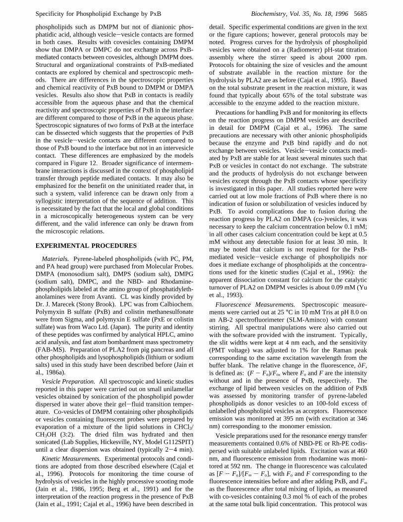
PxB Contacts between Co-Vesicles Containing DMPM.SincePxB mediates contacts between vesicles of DMPM or itsproducts of hydrolysis by PLA2, the ability of such contactsto mediate exchange of other phospholipids present in co-vesicles containing the contact-forming DMPM was exam-ined. For example, as shown in Figure 5, co-vesicles ofDMPA containing 12 mol % DMPM form functionalcontacts with other co-vesicles or with DMPM vesicles.Sequence A shows that all the available substrate inco-vesicles (both DMPA and DMPM) is hydrolyzed by theexcess enzyme added initially. At the end of the reactionprogress, all the enzyme is bound and it is not available forthe hydrolysis of DMPM vesicles added afterward; however,with PxB added last, DMPM from the excess vesicles istransferred across the PxB contact formed with the PLA2-containing co-vesicles, where both DMPM and DMPA havebeen hydrolyzed.Sequence B in Figure 5 shows that only DMPM is
transferred across PxB contacts between co-vesicles of 12%DMPM in DMPA. PLA2 added to excess co-vesicleshydrolyzes only a small fraction of vesicles, as expected onthe basis of Poisson distribution of enzyme over vesicles.However, PxB added after the cessation of hydrolysis causesadditional hydrolysis which corresponds to the amount ofDMPM present in the outer layer of the excess vesicles(curve a). As shown by curve b, the same amount of PxB
FIGURE 3: Effect of PxB on the hydrolysis of DMPM vesicles inthe presence of DTPA vesicles. DTPA (340 nmol)+ PxB (0, 0.825,and 1.65 nmol, curves a, b, and c, respectively). Control: curve dis as in c but no DTPA was added.
FIGURE 4: Poisson plot of lnPo versus enzyme/vesicle ratio forDMPA vesicles in the (O) absence, slope 1.007( 0.073, or (4)presence of 30 PxB/vesicle, slope 1.415( 0.168.Po is defined asthe fraction of the total hydrolyzable substrate that is not hydrolyzed.
5688 Biochemistry, Vol. 35, No. 18, 1996 Cajal et al.
+ +
+ +
added to DMPM vesicles allows hydrolysis of all theaccessible substrate. These results show that DMPA presentin excess co-vesicles is not transferred to the enzyme-containing co-vesicles. This interpretation is based on theassumption that vesicles of DMPM and DMPA behave thesame as those of their products of hydrolysis. The fact thatexchange of DMPM with DMPM or with products of itshydrolysis is indistinguishable has been shown independently(Jain et al., 1991; Cajal et al., 1996).Selectivity for the exchange of DMPM, but not of DMPA,
through PxB-mediated contacts is also supported by sequenceC. PLA2 added to DTPM vesicles is not available for the
hydrolysis of DMPM vesicles or of 12% DMPM in DMPAcovesicles; however, in both cases the extent of hydrolysisafter the addition of PxB corresponds only to the amount ofDMPM present in the reaction mixture, that is, DMPA isnot transferred to the enzyme containing vesicles.Specificity for the exchange of DMPC across PxB-
mediated vesicle-vesicle contacts formed between DTPMvesicles and co-vesicles of 12% DMPM in DMPC is shownby sequence D (Figure 5). PLA2 on DTPM vesicles is notavailable for the hydrolysis of the co-vesicles. If 0.1 mol% PxB (relative to DMPM) is added, hydrolysis is initiatedimmediately and only DMPM in co-vesicles is hydrolyzed(curve a). Surprisingly, however, at>0.3% PxB, DMPC isalso hydrolyzed with a time delay. Such a behavior isexpected if (hemi)fusion of vesicle has occurred under theseconditions. In this paper we have not examined thispossibility further. Control experiments showed that enzymeadded directly to co-vesicles hydrolyzed both DMPM andDMPC [see also Ghomashchi et al. (1991)].Spectroscopic EVidence for Apposition of Vesicles. Kinetic
results summarized thus far are unique in the sense that theyprovide a rapid and continuous method for monitoringselective and direct vesicle-vesicle transfer of phospholipidsthat are substrates for PLA2. This method therefore providesa steady-state measure of the phospholipid substrate that theenzyme “sees” during the course of reaction progress.Independent spectroscopic protocols were developed that notonly provide a measure of transfer of the probe phospholipidbut also could dissect the steps for the formation of vesicle-vesicle contact and the transfer of phospholipids.A change in the size of the particles is accompanied by a
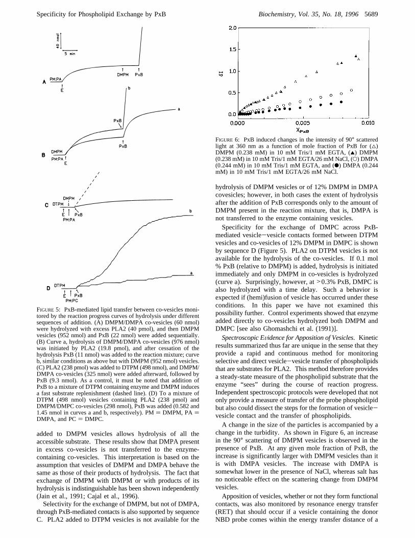
change in the turbidity. As shown in Figure 6, an increasein the 90° scattering of DMPM vesicles is observed in thepresence of PxB. At any given mole fraction of PxB, theincrease is significantly larger with DMPM vesicles than itis with DMPA vesicles. The increase with DMPA issomewhat lower in the presence of NaCl, whereas salt hasno noticeable effect on the scattering change from DMPMvesicles.Apposition of vesicles, whether or not they form functional
contacts, was also monitored by resonance energy transfer(RET) that should occur if a vesicle containing the donorNBD probe comes within the energy transfer distance of a
FIGURE 5: PxB-mediated lipid transfer between co-vesicles moni-tored by the reaction progress curves of hydrolysis under differentsequences of addition. (A) DMPM/DMPA co-vesicles (60 nmol)were hydrolyzed with excess PLA2 (40 pmol), and then DMPMvesicles (952 nmol) and PxB (22 nmol) were added sequentially.(B) Curve a, hydrolysis of DMPM/DMPA co-vesicles (976 nmol)was initiated by PLA2 (19.8 pmol), and after cessation of thehydrolysis PxB (11 nmol) was added to the reaction mixture; curveb, similar conditions as above but with DMPM (952 nmol) vesicles.(C) PLA2 (238 pmol) was added to DTPM (498 nmol), and DMPM/DMPA co-vesicles (325 nmol) were added afterward, followed byPxB (9.3 nmol). As a control, it must be noted that addition ofPxB to a mixture of DTPM containing enzyme and DMPM inducesa fast substrate replenishment (dashed line). (D) To a mixture ofDTPM (498 nmol) vesicles containing PLA2 (238 pmol) andDMPM/DMPC co-vesicles (298 nmol), PxB was added (0.582 and1.45 nmol in curves a and b, respectively). PM) DMPM, PA )DMPA, and PC) DMPC.
FIGURE 6: PxB induced changes in the intensity of 90° scatteredlight at 360 nm as a function of mole fraction of PxB for (4)DMPM (0.238 mM) in 10 mM Tris/1 mM EGTA, (2) DMPM(0.238 mM) in 10 mM Tris/1 mM EGTA/26 mM NaCl, (O) DMPA(0.244 mM) in 10 mM Tris/1 mM EGTA, and (b) DMPA (0.244mM) in 10 mM Tris/1 mM EGTA/26 mM NaCl.
Specificity for Phospholipid Exchange by PxB Biochemistry, Vol. 35, No. 18, 19965689
+ +
+ +

vesicle containing the acceptor Rh probe. Results in Figure7 were obtained by mixing vesicles containing NBD-PE(0.6%) with vesicles containing Rh-PE (0.6%). The energytransfer distance of this pair is about 50 Å. No noticeableincrease in the RET intensity at 592 nm (rhodamineemission) is seen on mixing of the vesicles. As shown inFigure 7, an increase in the energy transfer signal wasobserved in the presence of PxB, and a correspondingdecrease (not shown) in the intensity was also observed atthe emission from NBD (540 nm). The magnitude of theincrease depends upon the mole fraction of PxB; however,essentially the same magnitude of energy transfer is inducedby PxB whether the probes are codispersed in the DMPMor DMPA matrix. The fact that a comparable increase inthe RET intensity is observed in the two pairs of matrices(PM-PM, PA-PA) suggests that close apposition of vesiclesis induced by PxB between DMPM or DMPA vesicles.Formation of large clusters containing several vesicles at∼0.02 mole fraction PxB also accounts for a relatively largemagnitude of the RET intensity (∼40% compared to thatseen with the total mixing of the probes). These results areentirely consistent with the kinetic results.The possibility that the RET probes are mixed between
vesicles in close apposition is eliminated by results shownin the inset to Figure 7, where ternary co-vesicles containingboth the donor and acceptor probes (0.3% each) with a matrixlipid were mixed with an excess of DMPM or DMPAvesicles. In both cases, little change in the RET intensity at592 nm was observed. A decrease is expected if the probesare diluted by lipid mixing (Struck et al., 1981), and suchan effect was indeed seen at higher mole fraction of PxBwhere (hemi)fusion occurs.Spectroscopic EVidence for Specificity for the Phospholipid
Exchange. Having shown that PxB binds to DMPA vesicles,and that these vesicles are apposed in the presence of PxB,the spectroscopic evidence described next shows that func-tional vesicle-vesicle contacts are not formed betweenvesicles of phosphatidic acid. Transfer of phospholipidsacross PxB contacts was monitored as the change in thefluorescence intensity of pyrene-labeled phospholipids. Emis-sion from vesicles of pyrene-phospholipid is dominated bythe excimer band (480 nm), and the intensity of the monomer
band (395 nm) increases as the probe is diluted due to itsexchange with phospholipids from vesicles in contact. Asshown in Figure 8, such an increase was observed on theaddition of PxB to a mixture of vesicles containing PyPMwith vesicles of DMPM, but not with pairs of vesicles ofPyPA/DMPA, or PyPC/DMPC, or (PyPA+DMPM)/DMPM.These results are entirely consistent with the conclusion
that PxB-mediated functional vesicle-vesicle contacts areformed between DMPM and PyPM vesicles, whereas, in thecase of DMPA, PxB induces close apposition betweenvesicles (Figure 7), but functional contacts leading tophospholipid exchange (Figure 8) are not formed. Morestriking is the observation that PyPA in co-vesicles with 12%DMPM does not transfer the PyPA probe to DMPM vesiclesin the presence of PxB contacts. This is in agreement withthe kinetic results with co-vesicles of DMPA and 12%DMPM shown in Figure 5.The presence of two negative charges in the head group
of DMPA, instead of only one in DMPM, apparently is notentirely responsible for the inability of DMPA to exchangethrough PxB-mediated contacts. The PxB-mediated lipidexchange between DMPM and 12% DMPM/PyPA vesicleswas monitored at pH 5.2. Even under these conditions,where DMPA is monoanionic, there was no significanttransfer of PyPA (data not shown). This suggests a morecomplex nature for the mechanism of selectivity in thefunctional PxB contacts, possibly due to a pKa change inthe phosphate head group complexed with PxB.Collectively, these results clearly show that phospholipids
with dianionic head group such as in DMPA or PyPA donot exchange through vesicle-vesicle contacts mediated byPxB. Also, DMPC and other lipids with probes (dansyl-,NBD-, or rhodamine-) in the head group region of thephospholipid do not cross the contact. On the other hand,probes (NBD, pyrene) attached to the acyl chain of monoan-ionic phospholipids are transferred (results for NBD are notshown).Lysophospholipids Do Not Perturb the PxB-Mediated
Exchange of Phospholipids.The possible effect of lyso-phospholipids in the PxB-mediated phospholipid exchangewas evaluated by means of the fluorescence assay using amixture of PyPM and DMPM vesicles. Exogenous lyso-phospholipids (LPC, LPM, or LPA) were added to DMPM
FIGURE7: Increase in the resonance energy transfer (RET) intensityas a function of the mole fraction of PxB added to an equimolarmixture of vesicles containing 0.6% NBD-PE or Rh-PE in DMPM(O), DMPA (4), and DMPA with 26 mM NaCl (b). The total lipidconcentration was 81.6µM in Tris 10 mM. (Inset) Change in RETafter mixing vesicles containing 0.3% of both probes (16.32µM)with excess unlabeled vesicles (816µM) in 10 mM Tris, uponaddition of PxB: (O) DMPM, (4) DMPA.
FIGURE8: Fluorescence intensity of pyrene monomer as a functionof PxB mole fraction in a mixture vesicles of PyPM with DMPM(2), PyPA with DMPA (4), co-vesicles of 12% DMPM in PyPAwith DMPM (0), and vesicles of PyPC with DMPC (O). Bulk lipidconcentration was 1.25µM for Py-vesicles and 159µM forunlabeled acceptor vesicles, in 10 mM Tris, pH 8.0.
5690 Biochemistry, Vol. 35, No. 18, 1996 Cajal et al.
+ +
+ +

vesicles 5 min before the addition of PyPM vesicles, so thatthe lysophospholipid is incorporated mainly in the outermonolayer of the DMPM vesicles (Jain et al., 1985). In otherexperiments, DMPM was sonicated in presence of thelysophospholipid to ensure its incorporation in both innerand outer monolayers. At 4.2 mol % lysophospholipid, thetime course of the change in the pyrene fluorescence wasrecorded after the addition of PxB (0.43 mol %), with a timeresolution of 0.5 s. No differences in the rate of fluorescenceincrease were observed with any of the lysophospholipidsassayed; in all cases, the increase of fluorescence after PxBaddition was virtually instantaneous and indistinguishablefrom the controls without additive. These results show thatthe lipids with propensity to change the surface curvaturedo not influence the kinetics of exchange and therefore thefunctional properties of the PxB-mediated intervesicle con-tacts.Bound PxB Is Accessible from the Aqueous Phase. The
fluorescence emission spectrum of PxB does not changesignificantly on binding to anionic vesicles. The degree ofexposure of bound PxB was monitored as quenching offluorescence emission from phenylalanine at 282 nm by twoaqueous quenchers, iodide and acrylamide. An exampleof typical spectra of PxB bound to DMPM vesicles beforeand after addition of iodide is shown in Figure 9A. Stern-Volmer plots in Figure 9B show the effect of iodide on the
emission from PxB on DMPM vesicles. A key observationis that the fluorescence of PxB in buffer is readily quenchedby iodide (KSV is higher than that for free phenylalanine;see Table 1). This is expected because polycationic PxBwould facilitate the binding of iodide. On the other hand,PxB bound to DMPM vesicles is 3-7-fold less accessibleto iodide. There are two possible reasons for this differ-ence: either the phenylalanine residue is shielded in thebound form of PxB or the negatively charged iodide isshielded from the bound PxB at the negatively chargedsurface of the vesicle. Results on quenching of phenylalanineby acrylamide support the second possibility. As sum-marized in Table 1, the Stern-Volmer constant for quench-ing by acrylamide does not change significantly on thebinding of PxB to the interface. These results show thatPxB bound to DMPM vesicles and in vesicle-vesiclecontacts remains accessible from the aqueous phase; thepossibility that the quencher promotes the removal of PxBfrom the vesicles was ruled out by kinetic protocols of thetype shown in Figure 5. Thus it can be concluded that PxBin the contacts is not included inside a micellar or anhexagonal phase particle where it will become inaccessiblefrom the bulk aqueous phase.The magnitude of iodide quenching appears to depend on
the lipid/peptide ratio (Figure 9B). At DMPM to PxB moleratio of 200:1 (or at 30 PxB/V, where all PxB is in vesicle-vesicle contacts: PxBc form), quenching is less efficient thanthat at 60:1 mole ratio (or at 100 PxB/V, where some PxBwould be in contacts and the rest bound to the vesicle butnot in contact: PxBb form). This difference was not seenwith acrylamide quenching. An interpretation that reconcilesboth results is that phenylalanine of PxB bound to DMPMvesicles remains completely accessible to aqueous phase (seeacrylamide results), but PxB at the interface exists in twoforms (PxBc and PxBb) neutralized by the anionic surfaceof the vesicles to a different extent. This will reflect indifferent iodide quenching efficiencies, simply because iodideis shielded from the peptide by the negatively chargedphospholipid headgroups.When the same experiment was carried out with DMPA
vesicles (Table 1), essentially the same results were ob-tained: bound PxB remains accessible to acrylamide andiodide quenching, but in this case the Stern-Volmerconstants for iodide seem to be independent of the lipid toPxB ratio, as if only one form of PxB exists in DMPAvesicles.The Chemical ReactiVity of PxB-Amino Groups is Different
in DMPM or DMPA Vesicles.Additional information aboutthe accessibility of PxB at the interface was obtained bymonitoring the chemical reactivity of its free amino groupstoward TNBS from the bulk aqueous phase. The progress
FIGURE 9: Quenching of PxB fluorescene. (A) Fluorescenceemission spectra of PxB (50µM) in the presence of DMPM vesicles(2.75 mM) before (a) and after (b) addition of KI (13.3 mM).Spectra are corrected for turbidity and Raman contribution of thebuffer as described in Experimental Procedures. (B) Stern-Volmerplots showing the phenylalanine quenching of 50µM PxB in buffer(4) and in the presence of DMPM vesicles at DMPM/PxB moleratio 200:1 (O) and 60:1 (b), by the aqueous quencher I-. Excitationwas set at 256 nm, and emission was recorded at 282 nm; excitationand emission band passes were 4 nm.
Table 1: KSV, the Stern-Volmer Quenching Constants (M-1) forthe Quenching of Phenylalanine (50µM) Fluorescence by Iodideand Acrylamide in Different Environments
conditions KIa acrylamidea
Phe (free) 63.3 43.8PxB (free) 177.3 53.460:1 DMPM/PxB 26.2 87.5200:1 DMPM/PxB 57.0 70.060:1 DMPA/PxB 46.5 47.6200:1 DMPA/PxB 68.8 70.8
a All intensity values are corrected for turbidity and for inner filtereffect before plotting (Figure 9B).
Specificity for Phospholipid Exchange by PxB Biochemistry, Vol. 35, No. 18, 19965691
+ +
+ +

curves for the modification of PxB under different conditionsare shown in Figure 10. Such curves were analyzed byiterative least-squares fit to
HereB andC are the extinction coefficients correspondingto two different classes of amino groups with half times (t1/2)0.69/D and 0.69/E, respectively. The residuals were exam-ined to ascertain goodness of fit and systematic departure.Fits were judged to be good within 1%. In the case of freePxB (Figure 10, curve a), the progress curve could beresolved into three components: the first component (speciesA), corresponds to the modification of about two aminogroups (ε ) 37 700 M-1 cm-1) with t1/2 of less than 10 s.The rest of the progress curve showed the presence of twoother classes of modifiable amino groups: one (ε ) 23 200M-1 cm-1) with a t1/2 of less than 32 s (species B), and 1.6amino groups (ε ) 31 942 M-1 cm-1) with t1/2 ) 195 s(species C). Such parameters obtained under a variety ofconditions at different PxB/lipid ratios are summarized inTable 2, and the most significant conclusions are developedbelow.TNBS does not penetrate lipid membranes; however, the
reactivity of the amino groups to TNBS changes with theenvironment and accessibility. PxB bound to vesicles ofDMPM does not show the fast-reacting amino groups witht1/2 below 10s. seen with PxB in buffer, and only two ofthe five amino groups are derivatized (Figure 10, curve c).Under the conditions where all PxB is present in intervesiclecontacts, i.e., the PxBc form, (DMPM/PxB) 300:1 and 150:
1, or 20 PxB/V, and 40 PxB/V, respectively), only two ofthe five amino groups are available to TNBS, with differentreactivities: one (species B) around 30 or 50 s, and the other(species C) witht1/2 around 280 s. Also under the conditionswhere excess PxB is present, and thus at least some PxBcan be in the bound form but not involved in intervesiclecontacts (PxBb) as at DMPM/PxB 40:1, only two aminogroups are derivatized with TNBS; apparently both areequally exposed, witht(1/2)C ) 200 s.When the same experiment was carried out with DMPA
vesicles, where PxB cannot form functional intervesiclecontacts for the transfer of phospholipids, the number ofsusceptible amino groups was three (Figure 10, curve b).The additional amino group that is not modified in PxBbound to DMPM belongs to the slow-reacting species C,with t(1/2)C≈ 1000 s (Table 2). These results clearly showthat PxB in its two bound forms is accessible from the bulkaqueous phase; however, the chemical reactivities of theamino groups are appreciably altered.Bound PxB Exists in Two Forms.CD spectra of 134µM
PxB in 10 mM Tris at pH 8 is shown in Figure 11A. It isdominated by a cross point at 188 nm, a strong band centeredat 198 nm, and a shoulder at 217 nm. The magnitude ofellipticity and the presence of the shoulder indicate an orderedstructure of PxB in aqueous solution, with a significantcontribution of random coil or another rigid conformation(Shinnar et al., 1993). The same spectrum was obtained atpH 12, where all the amino groups are deprotonated. Asignificant induction of secondary structure in PxB is seenupon binding to negatively charged DMPM vesicles: theminima shifts to 203 nm, and the cross point to 194 nm. Inaddition, the magnitude of ellipticity of the spectra varies asa function of the lipid concentration, decreasing withincreasing lipid ratio from 24:1 to 238:1 (Figure 11A). Thisdecrease in spectral intensity upon dilution of the peptide atthe bilayer surface is indicative of a conformational changeaffecting the peptide backbone. According to previousobservations (Cajal et al., 1995, 1996), the ability of PxB toform intervesicle molecular contacts for the exchange ofphospholipids (PxBc) is maximum at around 30 PxBmolecules per vesicle, which corresponds to a lipid/peptidemole ratio of 200:1. Accordingly, the CD spectra obtainedat 238:1 mole ratio corresponds to the PxBc species, whereas,with decreasing lipid ratio, the contribution from PxB boundto the interface (PxBb), but not in contacts, increases. Thedifference spectrum corresponding to PxBc (curve e-b inFigure 11B) was generated by subtracting the spectrum ofPxB/DMPM at 1:24 mole ratio from the one in PxB/DMPMin 238:1 ratio. Similarly, a spectrum for the PxBb form (b-a) was obtained by subtracting the spectrum of PxB in bufferfrom that in PxB/DMPM at 1:24 ratio.Although we cannot yet assign a secondary structure
responsible for the spectra shown in Figure 11, they certainlyare not due to a random coil structure. The band corre-sponding to the PxBc form (e-b) is reminiscent of aâ-turnstructure, with a maxima around 202 nm and a minima at184 nm, whereas the band corresponding to PxBb form(b-a) has a maximum at around 191 nm and a minima at207 nm. No changes in the CD spectra are observed on theaddition of PxB to DMPM vesicles at pH 12, thus indicatingthat the positive charges are necessary for the structuralchanges in PxB associated with its binding to anionicvesicles. It may also be noted that the CD signal of colistinmethanesulfonate derivative corresponds to a random coil
FIGURE 10: Reaction progress for the modification of PxB aminogroups by TNBS: (a) PxB in buffer, (b) PxB with DMPA vesicles,(c) PxB with DMPM vesicles. Lipid concentration, 1.5 mM; PxB,10 µM.
Table 2: Changes in the Extinction Coefficient (halftime forModification in Seconds) at 420 nm on Incubation of PxB (10µM)with TNBS (10 mM) at 25°C
Aa B C no.b
PxB 37700(<10s)
23 204 (32s) 31 942 (196s) 4.64
1:300 PxB/DMPM 16 896 (29 s) 20 186 (260 s) 1.851:150 PxB/DMPM 20 973 (49 s) 18 501 (294 s) 1.971:40 PxB/DMPM 39 393 (199 s) 1.961:300 PxB/DMPA 28 620 (47 s) 32 209 (1017 s) 3.041:150 PxB/DMPA 21 475 (56 s) 40 623 (1144 s) 3.11:40 PxB/DMPA 56 882 (806 s) 2.8
a A, B, andC are the extinction coefficients (M) for the three differentspecies of amino groups in PxB with different reactivity to TNBS.bNumber of amino groups that have reacted with TNBS.
y) A+ B[1 - e-DX] + C[1 - e-EX]
5692 Biochemistry, Vol. 35, No. 18, 1996 Cajal et al.
+ +
+ +

peptide in buffer, and no changes are observed in presenceof DMPM.
The CD difference spectra resulting from the binding ofPxB to DMPA vesicles are shown in Figure 11C. At lowlipid to PxB ratios (e.g., 24:1), the difference spectrum(b-a) is comparable to those obtained in DMPM undercomparable conditions. On the other hand, the differencespectra (e-b) corresponding to high lipid to PxB ratio (238:1) with these two lipids are noticeably different in shapeand magnitude, indicating that PxBc form in DMPM doesnot adopt the same structure in DMPA. As expected, nochange in the secondary structure of PxB is induced byzwitterionic DMPC (result not shown). These results clearlyshow that the molecular conformation of PxB changes indifferent ways under different conditions at the interface.
DISCUSSION
Studies in this paper are designed to dissect three stepsthat lead to the formation of a functional intermembranemolecular contact mediated by PxB: binding of PxB,apposition of vesicles which may or may not involveformation of a functional contact between vesicles, andexchange of phospholipids through the molecular contact.As summarized in Table 3, results obtained with differentprotocols are useful in dissecting steps that lead to theformation of a functional contact. A striking conclusion ofthis study is that PxB contacts formed between co-vesiclescontaining the contact-forming DMPM with DMPA orDMPC permit exchange of DMPM but not of the otherphospholipid. This raises interesting possibilities about themechanistic significance and the structural basis for theselectivity. The reason for a lack of binding of polycationicPxB to zwitterionic interface is most probably the lack ofthe necessary electrostatic interaction. That PxB binds to,but cannot transfer, DMPA is particularly striking, becauseit implies that PxB bound to DMPA vesicles is in a differentmolecular state than that on vesicles of monoanionic phos-pholipids like DMPM, as supported by chemical andspectroscopic evidence. Besides extending the syllogisticfoundations of interfacial catalysis and equilibria, the novelmolecular phenomenon underlying the properties of PxBcontacts has broad implications. As developed elsewhere(Cajal et al., 1995; Cajal & Jain, 1996), properties of PxBcontacts provide a novel basis for its antibiotic activity byscrambling the phospholipid composition of the membranessurronding the periplasmic space in Gram-negative organ-isms. Such a mode of action is not expected to be overcomeby stable genetic resistance. Two additional implicationsof the peptide-mediated contacts are discussed below.Properties of PxB-Contact Are Different Than Those of
“Stalk”. Our working model for the structure of the PxBcontact is shown in Figure 12, where its structural featuresare compared with those of a “stalk”, the putative intermedi-ate of membrane fusion (Chernomordik & Zimmerberg,1995; Markin et al., 1984; Siegel, 1993). Despite obvioussimilarity in their structural conceptualization, functionaldifferences may be emphasized. The model for contact isconsistent with observations that the contacts are formed bya small number of PxB molecules, possibly six per contact(Cajal et al., 1996), that PxB in the contact remains exposedto the solvent (quenching and chemical modification experi-ments), and that the bilayer structure is maintained (kineticand fluorescence results). The fact that the bilayer organiza-
FIGURE 11: (A) Circular dichroism spectra of PxB (134µM) inthe presence of DMPM vesicles at lipid to PxB mole ratios: 0:1(a), 24:1 (b), 60:1 (c), 119:1 (d), and 238:1 (e). (B) Differencespectra for PxB in DMPM at high lipid ratio (e-b) (PxB is in thecontact form) and at low lipid ratio (b-a) (PxB is in the bound form,see text). (C) Difference spectra for PxB in DMPA at the samemole ratios as in panel B. Mean residue molar ellipticity [θ] is indeg cm2 dmol-1.
Table 3: Specificity for the PxB-Mediated Exchange ofPhospholipids: Three Sequential Steps That Lead to Formation of aFunctional Intervesicle Contact Are Dissected
PM/PMd PA/PAPM:PA/PM
PM:PA/PM:PA PC/PCPM:PC/PM
bindinga + + + - +appositionb + + + - +exchangec + - + PM- PA - + PM- PCe
a Binding of PxB to the lipid vesible determined by kinetic andspectroscopic experiments.b Apposition between vesicles determinedby 90° light scattering and RET.c Exchange of phospholipids betweenvesicles is measured by kinetics of hydrolysis by PLA2 and fluorescencespectroscopy (pyrene).dResults are extensible to different monoanionicphospholipids (DMPM, DPPM, DSPM, DMPG, DMPGly, DMPBut,DMPS). ePC can be transferred when PxB mole fraction is>0.3%(relative to DMPM).
Specificity for Phospholipid Exchange by PxB Biochemistry, Vol. 35, No. 18, 19965693
+ +
+ +

tion is not altered by PxB suggests that all mechanismsinvoking formation of soluble complexes of PxB with lipidmay be eliminated from consideration. Similarly, all mech-anisms based on fusion or hemifusion of vesicles arediscounted because the intermembrane transfer of phospho-lipid through these contacts is selective, and lipid-mixingdoes not occur. Results reported here also show that thepresence of additives (e.g., lysophospholipids) that preventthe formation of nonbilayer structures (Chernomordik et al.,1993; Vogel et al., 1993; Kozlov & Markin, 1983) do notplay a role in the formation or function of the PxB contacts.The fact that PxB in the contacts is accessible to quenchersand chemical reagents from the aqueous phase also rules outthe possibilities that invoke formation of intermembraneparticles and related structures based on inverted micellesand hexagonal phase. This is because PxB sequestered insuch particles would not be readily accessible from the bulkaqueous phase. These results along with the difference CDspectra (Figure 11) clearly show that specific molecularinteractions of several PxB molecules with vesicles is mostprobably responsible for the formation of a complex thatmediates selective exchange of phospholipids. Withinconstraints of these observations, an attractive possibility isthat a torus of six PxB molecules forms the contact (Cajalet al., 1996). As implied in Figure 12, PxB is not a “catalyst”but an integral part of a stable functional contact. Such arole of PxB-mediated vesicle-vesicle contacts is dramaticallydifferent than that invoked in the properties of a transient“stalk” or TMC (transmonolayer contact), whose formationis driven by the curvature propensities of the componentlipids. We are pursuing the mechanistic basis of suchpossibilities.
The heuristic and multistep character of the process offusion between two membranes is emphasized by the variousmodels that are being actively debated (White, 1992; Siegel,1993; Schweizer et al., 1995; Vogel et al., 1993). The stepsthat have been described by consensus are the apposition ordocking of membranes and the final fused membrane.Sincethe intermediates of fusion are believed to be of a highlytransient nature, there is little experimental data on themolecular organization of the structures involved. Neverthe-less, two types of intermediates have been proposed in theliterature: the inverted micellar intermediate (Siegel, 1984,1986a,b; Verkleij et al., 1979) and the stalk fusion intermedi-ate (Chernomordik & Zimmerberg, 1995; Markin et al., 1984;Siegel, 1993). The inverted micellar intermediate cannotaccount for the observed fusion behavior of most lipidsystems, whereas there is a great deal of data that favors thestalk as a likely intermediate for fusion. The stalk is asemitoroidal structure that forms between the two apposedmembranes and makes the contacting monolayers continuous,so that there is lipid mixing or hemifusion. The stalk isthermodynamically transient, and it expands radially leadingto the formation of a fusion pore. While emphasizing suchdifferences, we propose that PxB contacts are new speciesdifferent than any of the fusion intermediates postulated thusfar. First, as shown experimentally, the PxB-mediatedcontacts are thermodynamically stable, and the exchange ofmolecules present in the apposing monolayers through thesecontacts is selective. Another critical difference is that thestalk is a rate-limiting intermediate of membrane fusion,whereas the formation of PxB contacts is very rapid.Differences between stalk and peptide contact do not stophere. Formation of the stalk intermediate is influenced bythe membrane lipid composition (Chernomordik & Zimmer-berg, 1995). For example, micelle-forming lipids likelysophospholipids inhibit fusion (Chernomordik et al., 1993;Vogel et al., 1993), probably due to the fact that their positivespontaneous curvature is unfavorable for the formation ofthe negatively curved stalk (Kozlov & Markin, 1983). Wehave observed that the rate of lipid exchange through PxB-mediated contacts between DMPM vesicles is not affectedby the presence of 4 mol % lysophospholipids, thus sug-gesting that the transfer through PxB contacts is relativelyindependent of the spontaneous curvature of the bulk lipidpopulation.Proteins as Agents for Direct Intermembrane Transfer of
Phospholipids.The phenomenon described here has somebroad implications with significant bearing on several cellularprocesses. Peptide-mediated selective intermembrane trans-fer or exchange of phospholipids without (hemi)fusion ofmembranes is implicated in several cellular processes. Ineukaryotic cells, there are different intracellular membraneswith different composition and function. Only a part of thisdiversity is due to asymmetric distribution of proteins andlipids (Krebs et al., 1979; Op den Kamp, 1979). Thisdiversity is necessary for the viability of the cell, and,therefore, it must be regulated. Nevertheless, production ofphospholipids and proteins is often centralized in fewlocations inside the cell: for example, most of the phospho-lipids are formed in the endoplasmic reticulum. Obviously,the cell must have an efficient mechanism for traffickingand sorting of proteins and lipids (Moreau & Cassagne, 1994)to maintain this diversity. Two nonexclusive processes havebeen implicated (Longmuir, 1987): specific phospholipid-transfer proteins (Dawidowicz, 1987; Wirtz, 1991) and
FIGURE 12: Schematic models for the hypothetical intermembraneinteractions through PxB contacts or through “stalk”. The cross-section of bilayers are shown with two different species ofphospholipids to emphasize exchange through contacts and mixingthrough stalks. (a) Two membranes come in close apposition. (b)Working model for the molecular structure of PxB contacts wherea complex formed by six PxB molecules brings together the outermonolayers of two vesicles, and phospholipids from the apposedmonolayers selectively exchange through the opening in the middleof the contact. (c) Stalk, putatively the first intermediate for(hemi)fusion, brings in contact the two apposed monolayers. (d) Afusion pore formed from an expanded stalk as the probability of achance contact between the inner monolayers increases.
5694 Biochemistry, Vol. 35, No. 18, 1996 Cajal et al.
+ +
+ +
membrane flow either by diffusion through connectionsbetween membranes or by vesicle transport (Morre 1979).Vesicular membrane traffic and fusion between membranesis also implicated in many other physiological functions, i.e.,intracellular membrane fusion events occur during movementof secretory material between the stacks of the Golgiapparatus, exocytosis of neurotransmitters and hormones,endocytosis, viral infection, cell division, fertilization, etc.For example, secretory vesicles are retrieved after exocytosis,without intermixing between vesicles and plasma membranecomponents; this is possibly due to the rapidity of theretrieval or to the absence of complete lipid fusion (Valtorta& Befenati, 1995; Bergeron, 1973).To recapitulate, PxB-mediated molecular contacts are
attractive prototype for selective intermembrane transfer ofphospholipids. Specificity for the structure of the lipid headgroup is particularly interesting in this context because lipidssorted for head group could be tailored for the acylchainsby acyltransferases present in specific organelles. Themechanisms conferring regulation and specificity for theintracellular transfer of phospholipids have not been articu-lated, yet it is widely accepted that proteins play a funda-mental role in membrane fusion by fulfilling two basic re-quirements: cross-linking and destabilization of bilayers(White, 1992; Zimmerberg, 1993; Hoekstra, 1990). Specificproteins are involved in facilitation of merging of lipidmembranes, target selection, and fusion regulation (Ferro-Novick & Jahn, 1994; Hughson, 1995; Schweizwer et al.,1995). We do not know yet which step in the membranefusion cascade, if any, corresponds to the PxB contacts, nordo we know if a contact is a precursor or analog of the stalk.
ACKNOWLEDGMENT
We thank Drs. M. Bruch, L. V. Chernomordik, R. Epand,V. Markin, D. P. Siegel, and J. Vanderkooi for usefulsuggestions, stimulating discussions, and critical comments.M.K.J. acknowledges the hospitality of the Nehru Center(Bangalore), where some of this work was conceived.
REFERENCES
Apitz-Castro, R. J., Jain, M. K., & DeHaas, G. H. (1982)Biochim.Biophys. Acta 688, 349-356.
Berg, O. G., Yu, B.-Z., Rogers, J., & Jain, M. K. (1991)Biochemistry 30, 7283-7297.
Bergeron, J. J. M., Ehrenreich, J. H., Siekevitz, P., & Palade, E.(1973)J. Cell. Biol. 59, 73.
Cajal, Y., & Jain, M. K. (1996)J. NIH Res.(in press, May issue).Cajal, Y., Berg, O. G., & Jain, M. K. (1995)Biochem. Biophys.Res. Commun. 210, 746-752.
Cajal, Y., Rogers, J., Berg, O. G., & Jain, M. K. (1996)Biochemistry 35, 299-308.
Chernomordik, L. V., & Zimmerberg, J. (1995)Curr. Opin. Struct.Biol. 5, 541-547.
Chernomordik, L. V., Vogel, S. S., Sokoloff, A., Onaran, H. O.,Leikina, E. A., & Zimmerberg, J. (1993)FEBS Lett. 318, 71-76.
Craig, W. A., Turner, J. H., & Junin, C. M. (1974)Infect. Immun.10, 287-292.
Dawidowicz, E. A. (1987)Curr. Top. Membr. Transp. 29, 175-202.
Ferro-Novick, S., & Jahn, R. (1994)Nature 370, 191-193.Fields, R. (1972)Methods Enzymol. 25, 464-468.Ghomashchi, F., Yu, B.-Z., Berg, O. G., Jain, M. J., & Gelb, M.H. (1991)Biochemistry 30, 7318-7329.
Hoekstra, D. (1990)J. Bioenerg. Biomembr. 22, 121-125.Hughson, F. M. (1995)Curr. Opin. Struct. Biol. 5, 507-513.Jain, M. K., & Berg, O. (1989)Biochim. Biophys. Acta 1002, 127-156.
Jain, M. K., Jahagirdar, D. V., VanLinde, M., Roelofsen, B., &Eibl, H. (1985)Biochim. Biophys. Acta 818, 356-364.
Jain, M. K., Rogers, J., Jahagirdar, D. V., Marecek, J. F., &Ramirez, F. (1986a)Biochim. Biophys. Acta 860, 435-447.
Jain, M. K., Maliwal, B. P., de Haas, G. H., & Slotboom, A. J.(1986b)Biochim. Biophys. Acta 860, 448-461.
Jain, M. K., Rogers, J., Marecek, J. F., Ramirez, F., & Eibl, H.(1986c)Biochim. Biophys. Acta 860, 462-474.
Jain, M. K., Yu, B.-Z., & Kozubek, A. (1989)Biochim. Biophys.Acta 980, 23-32.
Jain, M. K., Rogers, J., Berg, O., & Gelb, M. H. (1991)Biochemistry 30, 7340-7348.
Jain, M. K., Yu, B.-Z., Rogers, J., Gelb, M. H., Tsai, M.Hendrickson, E. K., & Hendrickson, S. (1992)Biochemistry 32,7841-7847.
Jain, M. K., Gelb, M. H., Rogers, J., & Berg, O. G. (1995)MethodsEnzymol. 249, 567-614.
Kozlov, M. M., & Markin, V. S. (1983)Biofizika 28, 255-261.Krebs, J. J. R., Hauser, H., & Carafoli, E. (1979)J. Biol. Chem.254, 5308-5316.
Kubesch, P., Boggs, J., Luciano, L., Maass, G., & Tu¨mmler, B.(1987)Biochemistry 26, 2139-2149.
Longmuir, K. J. (1987)Curr. Top. Membr. Transp. 29, 129-174.Lorey, E., Lami, H., & Laustrait, G. (1971)Photochem. Photobiol.14, 411-421
Markin, V. S., Kozlov, M. M., & Borovjagin, V. L. (1984)Gen.Physiol. Biophys. 5, 361-370.
Moreau, P., & Cassagne, C. (1994)Biochim. Biophys. Acta 1197,257-290.
Morre, D. J., Kartenbeck, J., & Franke, W. W. (1979)Biochim.Biophys. Acta 559, 71-152.
Op den Kamp, J. A. F. (1979)Annu. ReV. Biochem. 48, 47-71.Schweizer, F. E., Betz, H., & Augustine, G. J. (1995)Neuron 14,689-696.
Siegel, D. P. (1986a)Biophys. J. 49, 1155-1170.Siegel, D. P. (1986b)Biophys. J. 49, 1171-1183.Siegel, D. P. (1984)Biophys. J. 45, 399-420.Siegel, D. P. (1993)Biophys. J. 65, 2124-2140.Shinnar, A. E., Olsen, L., Shinnar, M., Reed, G., & Leig, J. S.(1993)Biophys. J. 64, 377.
Sixl, F., & Watts, A. (1985)Biochemistry 24, 7906-7910.Storm, D. R., Rosenthal, K. S., & Swanson, P. E. (1977)Annu.ReV. Biochem. 4, 723-763.
Struck, D. K., Hoekstra, D., & Pagano, R. E. (1981)Biochemistry20, 4093-4099.
Upreti, G. C., & Jain, M. K. (1980)J. Membr. Biol. 55, 113-123.Valtorta, F., & Befenati, F. (1995)AdV. Pharmacol. 32, 505-557.Verkleij, A. J., Mombers, C., Leunissen-Bijvelt, J., & Verbergaert,P. H. (1979)Nature (London) 279, 162-163.
Vogel, S. S., Leikina, E. A., & Chernomordik, L. V. (1993)J. Biol.Chem. 268, 25764-25768.
White, J. M. (1992)Science 258, 917-924.Wirtz, K. W. A. (1991)Annu. ReV. Biochem. 60, 73-99.Yu, B.-Z., Berg, O. G., & Jain, M. K. (1993)Biochemistry 32,6485-6492.
Zidovetzki, R., Banerjee, U., Harrington, D. W., & Chan, S. I.(1988)Biochemistry 27, 5686-5692.
Zimmerberg, J., Vogel, S. S., & Chernomodik, L. V. (1993)Annu.ReV. Biomol. Struct. 22, 433-466.
BI952703C
Specificity for Phospholipid Exchange by PxB Biochemistry, Vol. 35, No. 18, 19965695
+ +
+ +