Species ofDasyaceae (Rhodophyta) from Hawaii · Dasyaceae (Rhodophyta) from Hawaii-SCHLECHAND...
20
Pacific Science (1989), vol. 43, no. 4 © 1989 by University of Hawaii Press. All rights reserved Species of Dasyaceae (Rhodophyta) from Hawaii 1,2 KRISTEN E. SCHLECH 3 AND ISABELLA A. ABBorr 3 ,4 ABSTRACT: Eight species of Dasyaceae (Ceramiales, Rhodophyta) are re- ported from the Hawaiian Islands, extending the geographic range for six of the species into the central North Pacific. The species are Heterosiphonia crispella, Eupogodon anastomosans, Eupogodon iridescens, Eupogodon pilosus, Dasya bail- louviana, Dasya collinsiana, Dasya corymbtfera, and Dasya iyengarii. Heterosi- phonia crispella (as H. wurdemannii var. laxa) and D. baillouviana were previously listed from Hawaii. Two SPECIES of the Dasyaceae were among the earliest collections of marine algae from Hawaii (Abbott 1980), but no systematic study of Hawaiian Dasyaceae has been made pre- viously. This study was part of the Master's thesis of the first author (Schlech 1983) and is being put into publication form in anticipation of using this information in a forthcoming marine flora of the Hawaiian Islands. Addi- tional information is provided with respect to a wider distribution range owing to more collections since 1983. The descriptions and comments on species of Dasya, except D. baillouviana, and all Eupogodon species are entirely those of Schlech, as are the descrip- tions of Heterosiphonia. Comments on and interpretations of Heterosiphonia, however, and some added distribution ranges were contributed by the second author, who had additional material. Members of the Dasyaceae are not com- mon in Hawaiian waters and in general collec- I We dedicate this paper to the memory of Munenao Kurogi, professor emeritus, Hokkaido University (Sap- poro), who died unexpectedly in October 1988. Although known widely as an experimental ecologist, Dr. Kurogi spent many of his later years teaching students the intri- cacies of systematics, in just the same spirit that the sec- ond author is glad to help the first. 2 Manuscript accepted 18 February 1989. 3Department of Botany, University of Hawaii at Manoa, Honolulu, Hawaii 96822. 4To whom correspondence should be sent. tions it is possible to miss them altogether. A careful examination of subtidal macroalgae will usually yield Heterosiphonia crispella (C. Agardh) Wynne, and intertidal mixed turf may show Dasya collinsiana Howe, where it may be attached to rock or epiphytically. The most frequently collected taxa, Eupogodon iridescens Schlech and Dasya corymbifera J. Agardh, are from the shallow subtidal. The remaining species reported here are relatively rare: Eupo- godon pilosus (Weber van Bosse) Silva, E. an- astomosans (Weber van Bosse) Silva, Dasya iyengarii B0rgesen, and· Dasya baillouviana (Gmelin) Montagne. The structure that distinguishes the Das- yaceae is the characteristic tetrasporangial branch, a stichidium, which is fusiform to lanceolate, tapering at one or both ends and in which the tetrasporangia mature. Distinc- tions between some genera, and species in some cases, may be made by the number and shape of the cover cells of the tetrasporangium. These cells are cut off either before the sporangium is initiated (Heterosiphonia) or afterwards (Dasya and Eupogodon) , and there may be three (Heterosiphonia), two to four (Dasya), or two cover cells (Eupogodon). Their shapes may be cuboidal to rounded to oval. An important vegetative feature is the pre- sence or absence of cortication, which are cells, one or two layers thick, that add to the cir- cumference of the axes and branches by forming outside the pericentral cells. In Ha- waiian species of Heterosiphonia there are no cortications, but many species elsewhere (Par- 332
Transcript of Species ofDasyaceae (Rhodophyta) from Hawaii · Dasyaceae (Rhodophyta) from Hawaii-SCHLECHAND...

Pacific Science (1989), vol. 43, no. 4© 1989 by University of Hawaii Press. All rights reserved
Species of Dasyaceae (Rhodophyta) from Hawaii1,2
KRISTEN E. SCHLECH3 AND ISABELLA A. ABBorr3 ,4
ABSTRACT: Eight species of Dasyaceae (Ceramiales, Rhodophyta) are reported from the Hawaiian Islands, extending the geographic range for six of thespecies into the central North Pacific. The species are Heterosiphonia crispella,Eupogodon anastomosans, Eupogodon iridescens, Eupogodon pilosus, Dasya baillouviana, Dasya collinsiana, Dasya corymbtfera, and Dasya iyengarii. Heterosiphonia crispella (as H. wurdemannii var. laxa) and D. baillouviana were previouslylisted from Hawaii.
Two SPECIES of the Dasyaceae were amongthe earliest collections of marine algae fromHawaii (Abbott 1980), but no systematic studyof Hawaiian Dasyaceae has been made previously. This study was part of the Master'sthesis of the first author (Schlech 1983) and isbeing put into publication form in anticipationof using this information in a forthcomingmarine flora of the Hawaiian Islands. Additional information is provided with respect toa wider distribution range owing to morecollections since 1983. The descriptions andcomments on species of Dasya, except D.baillouviana, and all Eupogodon species areentirely those of Schlech, as are the descriptions of Heterosiphonia. Comments on andinterpretations of Heterosiphonia, however,and some added distribution ranges werecontributed by the second author, who hadadditional material.
Members of the Dasyaceae are not common in Hawaiian waters and in general collec-
I We dedicate this paper to the memory of MunenaoKurogi, professor emeritus, Hokkaido University (Sapporo), who died unexpectedly in October 1988. Althoughknown widely as an experimental ecologist, Dr. Kurogispent many of his later years teaching students the intricacies of systematics, in just the same spirit that the second author is glad to help the first.
2 Manuscript accepted 18 February 1989.3Department of Botany, University of Hawaii at
Manoa, Honolulu, Hawaii 96822.4To whom correspondence should be sent.
tions it is possible to miss them altogether. Acareful examination of subtidal macroalgaewill usually yield Heterosiphonia crispella (C.Agardh) Wynne, and intertidal mixed turfmayshow Dasya collinsiana Howe, where it may beattached to rock or epiphytically. The mostfrequently collected taxa, Eupogodon iridescensSchlech and Dasya corymbifera J. Agardh, arefrom the shallow subtidal. The remainingspecies reported here are relatively rare: Eupogodon pilosus (Weber van Bosse) Silva, E. anastomosans (Weber van Bosse) Silva, Dasyaiyengarii B0rgesen, and· Dasya baillouviana(Gmelin) Montagne.
The structure that distinguishes the Dasyaceae is the characteristic tetrasporangialbranch, a stichidium, which is fusiform tolanceolate, tapering at one or both ends andin which the tetrasporangia mature. Distinctions between some genera, and species in somecases, may be made by the number and shapeof the cover cells of the tetrasporangium. Thesecells are cut off either before the sporangiumis initiated (Heterosiphonia) or afterwards(Dasya and Eupogodon) , and there may bethree (Heterosiphonia), two to four (Dasya),or two cover cells (Eupogodon). Their shapesmay be cuboidal to rounded to oval.
An important vegetative feature is the presence or absence ofcortication, which are cells,one or two layers thick, that add to the circumference of the axes and branches byforming outside the pericentral cells. In Hawaiian species of Heterosiphonia there are nocortications, but many species elsewhere (Par-
332
Dasyaceae (Rhodophyta) from Hawaii-SCHLECH AND ABBOTI' 333
sons 1975) are corticated. In Hawaii, Dasyaspecies are corticated, but elsewhere there aresome that are not. All Eupogodon species arecorticated.
The pericentral cells themselves are anotherimportant taxonomic feature; their number isthought to be fixed for each species. Following the lead provided by Parsons (1975), attention is given to the pigmented monosiphonousfilaments, sometimes called hairs, that arecharacteristic of Dasyaceae; their angle ofbranching, shape, and attachment may behelpful taxonomic features.
MATERIALS AND METHODS
Specimens were preserved in 3% formaldehyde-seawater, and permanent microscopeslides were prepared using 1% aqueous ani-
line blue, followed by 1% Hel and mountingin a glucose solution. When necessary, smallpieces of dried herbarium specimens weresoaked using a variety ofliquids, none ofwhichwas highly successful. For some specimens,the colored monosiphonous filaments that arecharacteristic of the species were trimmed before slides were made because in some casesthey were excessively matted or too numerousand interfered with observation.
Herbarium specimens, which are standarddried specimens, are cited with the major location first, followed by a specific locality, habitat notes, collector and date if known, andparenthetically where the specimen is. Specimens deposited in the Bishop Museum,Honolulu, are cited BISH; those retained bythe authors, as Schlech or Abbott. In the latter case, only microscopic slides have beenretained.
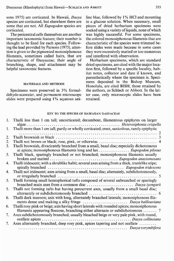
KEY TO THE SPECIES OF HAWAIIAN DASYACEAE
1. Thalli less than 1 em tall; uncorticated; decumbent, filamentous epiphytes on largeralgae Heterosiphonia crispella
1. Thalli more than 1em tall; partly or wholly corticated; erect, saxicolous, rarely epiphytic....................................................................... 2
2. Thalli brownish or black . . . . . . . . . . . . . . . . . . . . . . . . . . . . . . . . . . . . . . . . . . . . . . . . . . .. 32. Thalli not brown or black; rose, pink, or otherwise . . . . . . . . . . . . . . . . . . . . . . . . . . . . .. 43. Thalli brownish, divaricately branched from a small, basal disc; especially dichotomous
at apices; monosiphonous filaments long and lax Eupogodon pilosus3. Thalli black, sparingly branched or not branched; monosiphonous filaments usually
broken and matted Eupogodon anastomosans4. Thalli iridescent; with a shrublike habit; several axes arising from a thick, trunklike stipe;
spirally branched Eupogodon iridescens4. Thalli not iridescent; axes arising from a small, basal disc; alternately, subdichotomously,
or irregularly branched 55. Thalli forming small hemispherical tufts composed of several unbranched or sparingly
branched main axes from a common disc . . . . . . . . . . . . . . . . . . . . . . .. Dasya iyengarii5. Thalli not forming tufts but having percurrent axes, usually from a small basal disc;
alternately or subdichotomously branched . . . . . . . . . . . . . . . . . . . . . . . . . . . . . . . . . .. 66. Thalli dark maroon; axis with long, alternately branched laterals; monosiphonous fila-
ments dense and making a silky fringe Dasya baillouviana6. Thalli rosy pink or beige; axis having short laterals with rounded apices; monosiphonous
filaments appearing floccose, branching either alternate or subdichotomous . . . . . .. 77. Axes subdichotomously branched, usually bleached beige or very pale pink, with round,
ocellate apices Dasya collinsiana7. Axes alternately branched, deep rosy pink, apices tapering and not ocellate .
. . . . . . . . . . . . . . . . . . . . . . . . . . . . . . . . . . . . . . . . . . . . . . . . . . . . . . .. Dasya 'corymbifera

334
DESCRIPTIONS OF SPECIES
For convenience, species are listed alphabetically within the genus. Localities are listedfrom north to south within the Hawaiian Islands (Midway in the northwest to HawaiiIsland in the southeast) and where practicalfrom the north to the south within each island.
Genus Dasya
Dasya baillouviana (Gmelin) Montagne
Figures 1,2Montagne, 1841: 164.BASIONYM: Fucus baillouviana S. G. Gmelin,
1768:165.SYNONYMS: Sphaerococcus pedicel/ala C.
Agardh, 1822: 321; Dasya elegans (Martens)C. Agardh, 1828: 117.
Specimens are between 8 and 17 cm tall withirregular to alternate branching (Figures 1,2),reddish brown to purplish red on drying. Axes1 to 2 mm in diameter, branches 0.75 mm attheir attachment, tapering at their apices, ulti-
PACIFIC SCIENCE, Volume 43, October 1989
mate branchlets 3-7 mm long. Holdfast absent in material. Main axis with five pericentral cells, densely corticated by a layer of elongated rhizoidal cells 5-8 ,urn in diameter. Axisand lateral branches clothed with fine monosiphonous filaments that arise spirally fromthe cortical layer and do not have a distinctivebasal cell. They are dichotomously branchedonce near the base and divided a further threeto four times. Monosiphonous filament cells20 to 55 ,urn wide by 70 ,urn long and taper onlyslightly to their apices.
Tetrasporangial stichidia positioned at thedichotomies, the oldest at the first and youngerones more distal. They have a one- or twocelled pedicel. Stichidia lanceolate to spindleshaped, somewhat rounded at the base andacute at the apex. Mature stichidia 300450 ,urn long and have two cover cells persporangium.
Spermatangial plants have monosiphonousfilaments that are more slender than those ontetrasporangial plants; the linear-Ianceolatespermatangial branches are borne in the samepositions as tetrasporangial stichidia. Cystocarpic plants were not seen, but cystocarps aredescribed as having a narrow ostiolate neck.
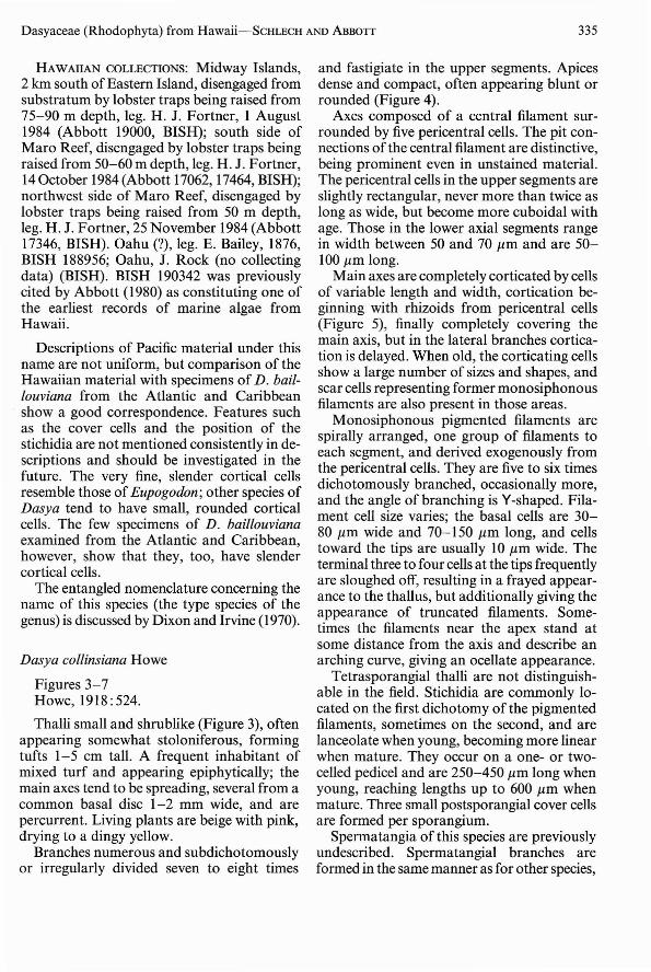
FIGURES 1-2. Dasya baillouviana. FIGURE I. Specimen disengaged from substratum by lobster trap set at 75-90 mdepth off Midway Island, Northwest Hawaiian Islands. FIGURE 2. Specimen disengaged from substratum by lobstertrap set at 50-60 m depth off Maro Reef, Northwest Hawaiian Islands.
334
DESCRIPTIONS OF SPECIES
For convenience, species are listed alphabetically within the genus. Localities are listedfrom north to south within the Hawaiian Islands (Midway in the northwest to HawaiiIsland in the southeast) and where practicalfrom the north to the south within each island.
Genus Dasya
Dasya baillouviana (Gmelin) Montagne
Figures 1,2Montagne, 1841: 164.BASIONYM: Fucus baillouviana S. G. Gmelin,
1768:165.SYNONYMS: Sphaerococcus pedicel/ala C.
Agardh, 1822: 321; Dasya elegans (Martens)C. Agardh, 1828: 117.
Specimens are between 8 and 17 cm tall withirregular to alternate branching (Figures 1,2),reddish brown to purplish red on drying. Axes1 to 2 mm in diameter, branches 0.75 mm attheir attachment, tapering at their apices, ulti-
PACIFIC SCIENCE, Volume 43, October 1989
mate branchlets 3-7 mm long. Holdfast absent in material. Main axis with five pericentral cells, densely corticated by a layer of elongated rhizoidal cells 5-8 ,urn in diameter. Axisand lateral branches clothed with fine monosiphonous filaments that arise spirally fromthe cortical layer and do not have a distinctivebasal cell. They are dichotomously branchedonce near the base and divided a further threeto four times. Monosiphonous filament cells20 to 55 ,urn wide by 70 ,urn long and taper onlyslightly to their apices.
Tetrasporangial stichidia positioned at thedichotomies, the oldest at the first and youngerones more distal. They have a one- or twocelled pedicel. Stichidia lanceolate to spindleshaped, somewhat rounded at the base andacute at the apex. Mature stichidia 300450 ,urn long and have two cover cells persporangium.
Spermatangial plants have monosiphonousfilaments that are more slender than those ontetrasporangial plants; the linear-Ianceolatespermatangial branches are borne in the samepositions as tetrasporangial stichidia. Cystocarpic plants were not seen, but cystocarps aredescribed as having a narrow ostiolate neck.
FIGURES 1-2. Dasya baillouviana. FIGURE I. Specimen disengaged from substratum by lobster trap set at 75-90 mdepth off Midway Island, Northwest Hawaiian Islands. FIGURE 2. Specimen disengaged from substratum by lobstertrap set at 50-60 m depth off Maro Reef, Northwest Hawaiian Islands.
Dasyaceae (Rhodophyta) from Hawaii-SCHLECH AND ABBOTT 335
HAWAIIAN COLLECTIONS: Midway Islands,2 km south ofEastern Island, disengaged fromsubstratum by lobster traps being raised from75-90 m depth, leg. H. J. Fortner, 1 August1984 (Abbott 19000, BISH); south side ofMaro Reef, disengaged by lobster traps beingraised from 50-60 m depth, leg. H. J. Fortner,14 October 1984 (Abbott 17062, 17464, BISH);northwest side of Maro Reef, disengaged bylobster traps being raised from 50 m depth,leg. H. J. Fortner, 25 November 1984 (Abbott17346, BISH). Oahu (?), leg. E. Bailey, 1876,BISH 188956; Oahu, J. Rock (no collectingdata) (BISH). BISH 190342 was previouslycited by Abbott (1980) as constituting one ofthe earliest records of marine algae fromHawaii.
Descriptions of Pacific material under thisname are not uniform, but comparison of theHawaiian material with specimens of D. baillouviana from the Atlantic and Caribbeanshow a good correspondence. Features suchas the cover cells and the position of thestichidia are not mentioned consistently in descriptions and should be investigated in thefuture. The very fine, slender cortical cellsresemble those of Eupogodon; other species ofDasya tend to have small, rounded corticalcells. The few specimens of D. baillouvianaexamined from the Atlantic and Caribbean,however, show that they, too, have slendercortical cells.
The entangled nomenclature concerning thename of this species (the type species of thegenus) is discussed by Dixon and Irvine (1970).
Dasya collinsiana Howe
Figures 3-7Howe, 1918: 524.
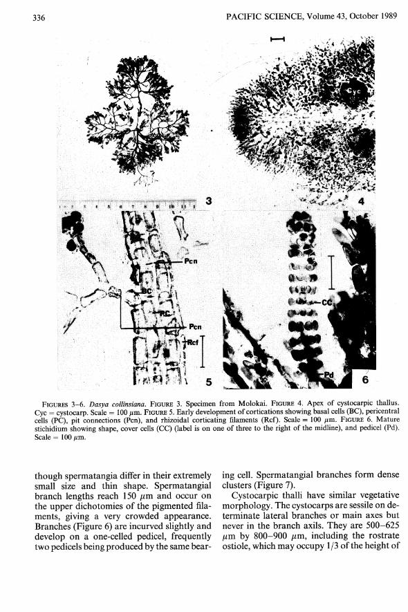
Thalli small and shrublike (Figure 3), oftenappearing somewhat stoloniferous, formingtufts 1-5 cm tall. A frequent inhabitant ofmixed turf and appearing epiphytically; themain axes tend to be spreading, several from acommon basal disc 1-2 mm wide, and arepercurrent. Living plants are beige with pink,drying to a dingy yellow.
Branches numerous and subdichotomouslyor irregularly divided seven to eight times
and fastigiate in the upper segments. Apicesdense and compact, often appearing blunt orrounded (Figure 4).
Axes composed of a central filament surrounded by five pericentral cells. The pit connections of the central filament are distinctive,being prominent even in unstained material.The pericentral cells in the upper segments areslightly rectangular, never more than twice aslong as wide, but become more cuboidal withage. Those in the lower axial segments rangein width between 50 and 70 J1.m and are 50100 J1.m long.
Main axes are completely corticated by cellsof variable length and width, cortication beginning with rhizoids from pericentral cells(Figure 5), finally completely covering themain axis, but in the lateral branches cortication is delayed. When old, the corticating cellsshow a large number of sizes and shapes, andscar cells representing former monosiphonousfilaments are also present in those areas.
Monosiphonous pigmented filaments arespirally arranged, one group of filaments toeach segment, and derived exogenously fromthe pericentral cells. They are five to six timesdichotomously branched, occasionally more,and the angle of branching is Y-shaped. Filament cell size varies; the basal cells are 3080 J1.m wide and 70-150 J1.m long, and cellstoward the tips are usually 10 J1.m wide. Theterminal three to four cells at the tips frequentlyare sloughed off, resulting in a frayed appearance to the thallus, but additionally giving theappearance of truncated filaments. Sometimes the filaments near the apex stand atsome distance from the axis and describe anarching curve, giving an ocellate appearance.
Tetrasporangial thalli are not distinguishable in the field. Stichidia are commonly located on the first dichotomy of the pigmentedfilaments, sometimes on the second, and arelanceolate when young, becoming more linearwhen mature. They occur on a one- or twocelled pedicel and are 250-450 J1.m long whenyoung, reaching lengths up to 600 J1.m whenmature. Three small postsporangial cover cellsare formed per sporangium.
Spermatangia of this species are previouslyundescribed. Spermatangial branches areformed in the same manner as for other species,
336 PACIFIC SCIENCE, Volume 43, October 1989
FIGURES 3-6. Dasya collinsiana. FIGURE 3. Specimen from Molokai. FIGURE 4. Apex of cystocarpic thallus.Cyc = cystocarp. Scale = 100 /lm. FIGURE 5. Early development of cortications showing basal cells (BC), pericentralcells (PC), pit connections (Pen), and rhizoidal corticating filaments (Ref). Scale = 100 /lm. FIGURE 6. Maturestichidium showing shape, cover cells (CC) (label is on one of three to the right of the midline), and pedicel (Pd).Scale = 100/lm.
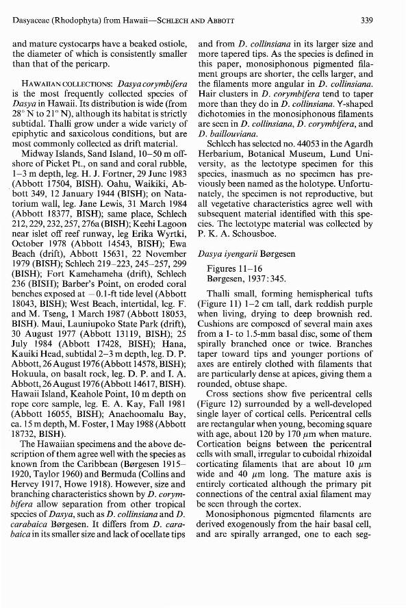
though spermatangia differ in their extremelysmall size and thin shape. Spermatangialbranch lengths reach 150 /lm and occur onthe upper dichotomies of the pigmented filaments, giving a very crowded appearance.Branches (Figure 6) are incurved slightly anddevelop on a one-celled pedicel, frequentlytwo pedicels being produced by the same bear-
ing cell. Spermatangial branches form denseclusters (Figure 7).
Cystocarpic thalli have similar vegetativemorphology. The cystocarps are sessile on determinate lateral branches or main axes butnever in the branch axils. They are 500-625/lm by 800-900 /lm, including the rostrateostiole, which may occupy 1/3 of the height of
336 PACIFIC SCIENCE, Volume 43, October 1989
FIGURES 3-6. Dasya collinsiana. FIGURE 3. Specimen from Molokai. FIGURE 4. Apex of cystocarpic thallus.Cyc = cystocarp. Scale = 100 /lm. FIGURE 5. Early development of cortications showing basal cells (BC), pericentralcells (PC), pit connections (Pen), and rhizoidal corticating filaments (Ref). Scale = 100 /lm. FIGURE 6. Maturestichidium showing shape, cover cells (CC) (label is on one of three to the right of the midline), and pedicel (Pd).Scale = 100/lm.
though spermatangia differ in their extremelysmall size and thin shape. Spermatangialbranch lengths reach 150 /lm and occur onthe upper dichotomies of the pigmented filaments, giving a very crowded appearance.Branches (Figure 6) are incurved slightly anddevelop on a one-celled pedicel, frequentlytwo pedicels being produced by the same bear-
ing cell. Spermatangial branches form denseclusters (Figure 7).
Cystocarpic thalli have similar vegetativemorphology. The cystocarps are sessile on determinate lateral branches or main axes butnever in the branch axils. They are 500-625/lm by 800-900 /lm, including the rostrateostiole, which may occupy 1/3 of the height of

Dasyaceae (Rhodophyta) from Hawaii-SCHLECH AND ABBOTT 337
the cystocarp. The pericarp cells are similar tothe cortical cells in their irregularity, exceptaround the ostiole where they maintain a catenate orientation. A few carpogonial branchesexamined showed four cells.
HAWAllAN COLLECTIONS: Kauai, Lawai Kai,Schlech 235 (BISH). Oahu, Laie Bay, Abbott34a, 16 March 1941 (BISH); Laie Bay, Abbott1354, 5 May 1946 (BISH); Kahala Beach,Schlech 226, 234, 238 (BISH); Waikiki, Abbott332, 12 January 1944 (BISH); Abbott 1091, 8April 1946 (BISH); Abbott 1138, 19 April1946 (BISH); Schlech 274-276, 213 (BISH);Ala Moana Beach Park, Schlech 277 (BISH);Ewa Beach (drift), Schlech 200 (BISH); WestBeach, leg. F. and M. Tseng (Abbott 18051,BISH). This species is common in turf.
The most distinctive feature at the grossmorphological level is the subdichotomousbranching pattern and the rounded or bluntbranch tips. Additional features that may aididentification in the field are the frequent beigeor pale pink color and the small size of thisspecies compared to D. corymbifera. Microscopically, D. collinsiana may be separatedfrom D. corymbifera by its more prominentscar cells, its cuboidal pericentral cells that canbe clearly seen even in unstained material thatis not yet corticated, and in the less taperedmonosiphonous filaments. Spermatangialbranches in D. collinsiana are less tapered andmore compact and more numerous. Cystocarps differ in their placement in D. corymbifera by being borne in a branch axil insteadof directly on a lateral as in D. collinsiana.
Dasya corymbifera J. Agardh
Figures 8-10J. Agardh, 1841 :31.
Thalli are up to 15 em tall, although mostare between 7 and 10 em, and have a soft,flexible habit (Figure 8). Plants are often pyramidal in outline with shorter branches atthe base, and when living, have a deep rosecolor, drying to a bright magenta or brownishred. Apices are often ragged in appearance,as the younger monosiphonous filaments arestrongly incurved initially, becoming less sowith age.
Main axes lightly to entirely corticated, depending on age, by small, thin and irregularrhizoidal cells, in this way resembling the cortex of D. baillouviana. Cross sections near theapex reveal a central filament surrounded byfive pericentral cells and a cortical layer. Mature pericentral cells may reach a length of200.urn or more. Axes secondarily bilaterally organized and alternately branched, althoughin some cases two branches may occur on thesame side successively.
Branches and upper segments of axescovered with spirally and loosely arrangedmonosiphonous pigmented filaments, one toeach segment. Filaments are derived from aroughly isodiametric basal cell 40-50 .urn wide,which originates exogenously from a pericentral cell. They become deciduous in older segments, and location of the basal cell is markedby a scar cell. Filaments (Figure 9) are dichotomously branched at least four to fivetimes, sometimes more, and the dichotomy isof the Y-shaped type; filaments somewhat incurved, tapering, and extremely fine at thetips. The cells vary in size but most are 40-60.urn in diameter and 70-90 .urn long, becomingsmaller toward the tips, the terminal cell ofwhich is about 10 .urn in diameter and acutelypointed.
Tetrasporangial thalli are not distinguishable in the field. Stichidia are formed at thesecond or third dichotomy of the hair and,rarely, there may be more than one on a singlehair tuft. Stichidia are subtended by a one- totwo-celled pedicel, the latter occurring morecommonly. Stichidial shape varies frequently;immature stichidia are conical-lanceolate andbecome linear (Figure 10) with age. Maturestichidia also exhibit a flattening of thetapered tip. Most stichidia bear a three- tofour-celled sterile terminal filament. Threepostsporangial cover cells are formed persporangium, covering about 1/4 of thesporangial height. These cover cells becomeflattened and more rectangular with age, butalways maintain a loosely banded appearance(Figure 10). Overall stichidial length rangesfrom 400 to 650 .urn when fully mature.
Spermatangial branches are formed in thesame manner as tetrasporangial stichidia except that the fertile pericentral cells each divides

338
j It...'-""fl 7
PACIFIC SCIENCE, Volume 43, October 1989
8
FIGURES 7-10. FIGURE 7. Dasya collinsiana. Immature spermatangial branches showing clustering and pedicels.Scale = 100 J.lm. FIGURES 8-10. Dasya corymbifera. FIGURE 8. Habit of specimen from Keehi Lagoon, Oahu. Scale =2 em. FIGURE 9. Branched monosiphonous pigmented filament (HC) showing attenuate terminal filaments, andV-shaped angle of branching (U). Scale = 100 J.lm. FIGURE 10. Mature stichidia showing shape, incurved "protective"filaments and two-celled pedicel (Pd). Scale = 100 J.lm.
to form two spermatangial mother cells. Eachmother cell cuts off two spermatangia, whichare club-shaped, the flattened end being theterminal one. Spermatangial branches have aone-celled pedicel and are formed on the ultimate dichotomies of the filaments. Occasion-
ally both dichotomies will form spermatangialbranches, but they are never clustered.
Cystocarps reach a width of 500 !Jm and alength of 985 !Jm. They occur in the axils ofthe determinate lateral branches and are sessile.The pericarp is composed of irregular cells
Dasyaceae (Rhodophyta) from Hawaii-ScHLECH AND ABBOTT 339
and mature cystocarps have a beaked ostiole,the diameter of which is consistently smallerthan that of the pericarp.
HAWAllAN COLLECTIONS: Dasya corymbiferais the most frequently collected species ofDasya in Hawaii. Its distribution is wide (from28 0 N to 21 0 N), although its habitat is strictlysubtidal. Thalli grow under a wide variety ofepiphytic and saxicolous conditions, but aremost commonly collected as drift material.
Midway Islands, Sand Island, 10-50 m offshore of Picket Pt., on sand and coral rubble,1-3 m depth, leg. H. J. Fortner, 29 June 1983(Abbott 17504, BISH). Oahu, Waikiki, Ab-bott 349, 12 January 1944 (BISH); on Natatorium wall, leg. Jane Lewis, 31 March 1984(Abbott 18377, BISH); same place, Schlech212,229,232,257, 276a (BISH); Keehi Lagoonnear islet off reef runway, leg Erika Wyrtki,October 1978 (Abbott 14543, BISH); EwaBeach (drift), Abbott 15631, 22 November1979 (BISH); Schlech 219-223, 245-257, 299(BISH); Fort Kamehameha (drift), Schlech236 (BISH); Barber's Point, on eroded coralbenches exposed at -O.I-ft tide level (Abbott18043, BISH); West Beach, intertidal, leg. F.and M. Tseng, 1 March 1987 (Abbott 18053,BISH). Maui, Launiupoko State Park (drift),30 August 1977 (Abbott 13119, BISH); 25July 1984 (Abbott 17428, BISH); Hana,Kauiki Head, subtidal 2-3 m depth, leg. D. P.Abbott,26August 1976 (Abbott 14578, BISH);Hokuula, on basalt rock, leg. D. P. and I. A.Abbott, 26 August 1976 (Abbott 14617, BISH).Hawaii Island, Keahole Point, 10 m depth onrope core sample, leg. E. A. Kay, Fall 1981(Abbott 16055, BISH); Anaehoomalu Bay,ca. 15 m depth, M. Foster, 1May 1988 (Abbott18732, BISH).
The Hawaiian specimens and the above description ofthem agree well with the species asknown from the Caribbean (B0rgesen 19151920, Taylor 1960) and Bermuda (Collins andHervey 1917, Howe 1918). However, size andbranching characteristics shown by D. corymbifera allow separation from other tropicalspecies ofDasya, such as D. collinsiana and D.carabaica B0rgesen. It differs from D. carabaica in its smaller size and lack ofocellate tips
and from D. collinsiana in its larger size andmore tapered tips. As the species is defined inthis paper, monosiphonous pigmented filament groups are shorter, the cells larger, andthe filaments more aI)gular in D. collinsiana.Hair clusters in D. corymbifera tend to tapermore than they do in D. collinsiana. V-shapeddichotomies in the monosiphonous filamentsare seen in D. collinsiana, D. corymbifera, andD. baillouviana.
Schlech has selected no. 44053 in the AgardhHerbarium, Botanical Museum, Lund University, as the lectotype specimen for thisspecies, inasmuch as no specimen has previously been named as the holotype. Unfortunately, the specimen is not reproductive, butall vegetative characteristics agree well withsubsequent material identified with this species. The lectotype material was collected byP. K. A. Schousboe.
Dasya iyengarii B0rgesen
Figures 11-16B0rgesen, 1937: 345.
Thalli small, forming hemispherical tufts(Figure 11) 1-2 cm tall, dark reddish purplewhen living, drying to deep brownish red.Cushions are composed of several main axesfrom a 1- to 1.5-mm basal disc, some of themspirally branched once or twice. Branchestaper toward tips and younger portions ofaxes are entirely clothed with filaments thatare particularly dense at apices, giving them arounded, obtuse shape.
Cross sections show five pericentral cells(Figure 12) surrounded by a well-developedsingle layer of cortical cells. Pericentral cellsare rectangular when young, becoming squarewith age, about 120 by 170 J1.m when mature.Cortication beigns between the pericentralcells with small, irregular to cuboidal rhizoidalcorticating filaments that are about 10 J1.mwide and 40 J1.m long. The mature axis isentirely corticated although the primary pitconnections of the central axial filament maybe seen through the cortex.
Monosiphonous pigmented filaments arederived exogenously from the hair basal cell,and are spirally arranged, one to each seg-

340 PACIFIC SCIENCE, Volume 43, October 1989
FIGURES 11-16. Dasya iyengarii. FIGURE II. Habit of thallus from Kauai. Scale = I cm. FIGURE 12. Crosssection of mature axis showing five pericentral cells (PC). Scale = 100 J.Lm. FIGURE 13. Branched monosiphonousfilaments (HC), showing relatively nontapering terminal portions and V-shaped angle of branching. Scale = 100 J.Lm.FIGURE 14. Detail of V-shaped angle of branching (U). BC = basal cell, HC = monosiphonous filament; Pcn = pitconnection. Scale = 100 J.Lm. FIGURE 15. Mature cortex showing scar cells (SC), and corticating rhizoids (Rcf).Scale = 100 J.Lm. FIGURE 16. Mature cystocarp (Cyc) showing sessile position on the axis and its rounded shape.Scale = 100 J.Lm.

Dasyaceae (Rhodophyta) from Hawaii-SCHLECH AND ABBon 341
ment, moderately incurved and subdichotomously divided four to five times (Figure13). The angle of branching (Figure 14) is theY-shape type. Filaments taper slightly towardthe tips. The distinctive feature of the filaments in this species is the presence of two pitconnections distally from the basal cell to twodifferent cells; technically, there are two filaments per basal cell, instead of one as seen inD. corymbifera and D. collinsiana. The morecrowded filaments give the impression ofwhorls at the nodes. The basal cell remainsafter the filaments become deciduous, forminga scar cell (Figure 15) about 40 /lm wide and50-60 /lm long. Monosiphonous filament cellsrange in width between 10 and 15 /lm, up to100 /lm long, and have rounded end walls. Tipcells are about 6/lm wide and 10-20 /lm long.
Tetrasporangial plants are not distinguishable in the field. Stichidial sizes vary up to 300/lm and are oblong-lanceolate when youngand lanceolate when mature. There are threecover cells per sporangium, and stichidia arepositioned on a one- or two-celled pedicel thatarises from the first dichotomy of the hair.
Spermatangial plants are previously undescribed. Spermatangial branches are formedat the base of a branched filament, in clustersof sometimes as many as six branches. Thebranches are strongly incurved and often topped by a sterile portion; lengths range between150 and 200 /lm.
Cystocarpic plants are previously undescribed. The cystocarps are sessile (Figure 16);the carpogonial branch develops at about thefourth segment from the apex. The pericarp iscontinuous, forming a flask-shaped body withan ostiole at the top, with an overall sphericalglobose shape. Cystocarp size ranges between600 and 750 /lm in diameter and 1.0 to 1.5 mmin length.
HAWAIIAN COLLECTIONS: Open coast, wavewashed reefs form the habitat for this species.Kauai, on rock at Lawai Kai, Schlech 264, 265,273, 283 (BISH), Abbott 15808, leg. GordonDaida, 8-9 September 1979 (BISH); Kipukai,Schlech 261, 298 (BISH); Oahu, Kaloko, onCladophoropsis sp., leg. I. Abbott & R. E.Norris, 2 September 1961, Abbott 14869 (Abbott); Hawaii Island, Richardson's Bay,
Schlech 204 (BISH). Dasya iyengarii is not acommon species in Hawaii.
Originally described as epiphytic, D. iyengarii has been collected epiphytically and onrock in Hawaii. The specimens agree withprevious descriptions (B0rgesen 1937, Dawson1957) except that the cortex appears to bemore developed in Hawaiian material. Thismay be merely an environmental adaptationto a more active habitat zone. Additionally,the monosiphonous filaments are not asstrongly incurved as shown for the typespecimen by B0rgesen (1937, fig. 346).
This species may be distinguished from otherDasya in the field by its tufted habit andsmaller size. Although it does not resembleDasya collinsiana in any way at the gross morphological level, microscopic examinationclouds the distinctions and the two species aresimilar in many features. However, closer inspection reveals the two filaments per basalcell in D. iyengarii compared to one filamentper basal cell in D. collinsiana and D. corymN!era. The crowded appearance of spermatangial branches is undoubtedly connected tothe number of filaments on each basal cell,and in this feature it is different from otherHawaiian Dasya species.
Genus Eupogodon
Species in this genus were formerly placedwith the genus Dasyopsis, which is a laterhomonym of Eupogodon as interpreted bySilva (1987). The species are separated fromDasya vegetatively on the basis of their bilateral organization, as opposed to the radialorganization ofspecies ofDasya, but there aresome exceptions here as in other characteristics. Species of Eupogodon treated here alsohave a distinctive subcortical layer, a "medulla." This kind of tissue has been previouslydescribed as pseudoparenchymatous by Parsons (1975) and is formed by extensions of thelower edges of the pericentral cells (Parsons,pers. comm.). This layer occurs between thepericentral cells and the cortex. Eupogodonanastomosans and E. pilosus show two and E.iridescens shows three cover cells per sporangium; Hawaiian Dasya show three cover cells

342 PACIFIC SCIENCE, Volume 43, October 1989
19
...·I~
t b_t
~_t----,CC
,...,.~.........._Sp
"... , '
_.. 1
• ?
,.
FIGURES 17-20. Eupogodon anastomosans. FIGURE 17. Type specimen from New Guinea (Leiden herbarium).FIGURE 18. Transverse section of a mature axis showing pit connections (Pen), pericentral cells (PC), and cortex (C).The tissue between the pericentral cells and the cortex (the medulla) is present in Eupogodon and lacking in Dasya.Scale = 100 }lm. FIGURE 19. Mature stichidium showing sporangia (Sp) and very small cover cells (CC). Scale =100 }lm. FIGURE 20. Spermatangial branches with spermatangia (S), and showing two-celled pedicel (Pd). Scale =100 }lm.
per sporangium. The observations of twocover cells for Eupogodon are in conflict withthe conditions found in the type species byKylin (1956), who stated that there ate threecover cells in Eupogodan (as Dasyopsis). It isclear that further critical studies of the features of these taxa are needed.
Eupogodon anastomosans (Weber van Bosse)Silva
Figures 17-20Silva, 1987: 129.BASIONYM: Dasyopsis anastomosans Weber
van Bosse, 1921 : 309.

Dasyaceae (Rhodophyta) from Hawaii-SCHLECH AND ABBOTT 343
Thalli (Figure 17) small, mostly between 2and 3 cm tall, very dark red to blackish, margins of branches conspicuously marked bylong, matted filaments. Main axes often unbranched or, if branched, only slightly andirregularly so, branches tapering toward tips.Axes covered with a thick layer offine, mattedhairs that are not curved or stiff and are mostdense at the apices, being deciduous at thebase. Hair cells vary in size and can be roughlytwice as long as wide (15 by 15-40 ,um) andsquare, becoming somewhat longer with age.Overall length of the monosiphonous pigmented filaments is about 1-2 mm; they arerarely subdichotomously branched once at thebase. They are derived exogenously from arounded cortical cell; most other cortical cellsare thin and slightly elongated.
Main axes, which are sometimes indistinctin branched thalli, are erect and entirely corticated. Cross sections of the major axis reveal acentral axis surrounded by five pericentralcells. Exterior to the pericentral cell layer is amedullary region (Figure 18) composed ofsmaller, parenchyma-like cells, decreasing insize toward the cortex. The cortex is onecell thick and the cells are small and darklypigmented. The pseudoparenchymatous medulla develops quickly, making recognitionof the pericentral cells difficult in maturesections.
Tetrasporangial thalli are not distinguishable from sterile thalli in the field. Lanceolatestichidia are formed on a two- to three-celledpedicel and are formed at the first dichotomyof the monosiphonous filaments; each sporangium is subtended by two postsporangialcover cells that do not cover any portion of thesporangium (Figure 19). Stichidia averageabout 200 ,urn in length but may become largerwith age. Stichidial cells are loosely arrangedcompared with the more densely compactedarrangement of stichidial cells in other speciesand additionally have an unusually thick layerof gelatinous material.
Spermatangial branches are formed in thesame manner as tetrasporangial stichidia andreach lengths of 300 ,um, although most arebetween 250 and 260 ,urn, not including theone- to four-celled pedicel. Spermatangialbranches (Figure 20) may occur in clusters oftwo or three per filament. Spermatangial cells
are slightly teardrop shaped and are attachedat the pointed end.
Female plants were not collected and remain undescribed for this species.
HAWAIIAN COLLECTIONS: Hawaii Island,Shipman ranch on intertidal bench, leg. W. H.Magruder 243 (BISH); Richardson BeachPark on basalt boulders, Schlech 205 (BISH).The plants were exposed to strong wave activity. They grow as distinctive isolated plantsin a ring.
Because only two collections have beenmade, the documentation of this species inHawaii should be viewed tentatively. Moreover, the material matches rather well the descriptions of Dasya adhaerens Yamada fromthe western Caroline Islands (Yamada 1944),which was further described by Taylor (1950)from Bikini atoll. Unfortunately, the type material of Dasya adhaerens cannot be located atthis time. Should it or other type materialbecome available, a comparison with Eupogodon anastomosans should be made. If theyrepresent the same taxon, the epithet E. anastomosans should be used, as it has priority.Moreover, the anatomy shows that a subcortical layer makes the placement in Eupogodoncorrect.
Eupogodon iridescens Schlech
Figures 21-26Schlech, 1989 (in press)
Plants (Figure 21) reach 7 cm in height andhave a distinct iridescence when living, dryingto rusty red or bright orange. Several majorbranched axes are borne on a thick, erect stipe.Major axes are up to 3 mm in diameter, emerging radially near the top of the stipe and areterete and irregularly spirally branched. Stipenarrows basally into a thick, cartilaginous,and sometimes branched holdfast.
Branches, which may be 2 cm or more long,taper toward tips and are clothed with tufts ofmonosiphonous filaments (Figure 22) thatarise exogenously and randomly from the cortex. Filaments are deciduous with age and thebottom halves of the branches become bare.
Cross sections close to the apex (5 mm)reveal a central cell surrounded by several toeight or more pericentral cells and a cortical

344 PACIFIC SCIENCE, Volume 43, October 1989
FIGURES 21-26. Eupogodon iridescens. FIGURE 21. Holotype, Schlech 300 from Punaluu, Oahu, Hawaii (BishopMuseum). Scale = 2 em. Stp = stipe. FIGURE 22. Branch habit of female gametophyte showing terminal cystocarps(Cyc). Scale = I em. FIGURE 23. Transverse section showing medulla (M) and cortex (C). Scale = 10 Jlm. FIGURE 24.Basal cell (BC) ofmonosiphonous pigmented filaments, with two filaments (HC) arising from it. Mature cortex showsrhizoidal corticating cells (Ref). Scale = 10 Jlm. FIGURE 25. Stichidium showing three cover cells (cc) over sporangium(Sp). Scale = 100 Jlm. FIGURE 26. Cystocarp showing terminal position, the displaced monosiphonous filaments (H)at the base, ostiole (0), and carpospores (Cp). Scale = 1 mm.

Dasyaceae (Rhodophyta) from Hawaii-SCHLECH AND ABBOTT 345
layer. Cortical cells are about 10 J1.m in crosssection and up to 100 J1.m long. The corticallayer is one to two cells thick, followed by asubcortical or medullary layer (Figure 23)composed of large, irregular cells 20-50 J1.m indiameter and 120-150 J1.m long. Subcorticalcells nearest the central axial filament frequently have eight to nine conspicuous pitconnections.
Monosiphonous pigmented filaments areslightly stiff and stand away from the axialsurface. This is due in part to the slightlyraised basal cell (Figure 24), which can bedistinguished from the surrounding corticalcells by its rounded-pyramidal shape. Thesecells vary in size from 10 to 15 J1.m wide and 15to 20 J1.m long. Each basal cell has two pitconnections leading to two different filaments.The filaments are dichotomously branchedwith the Y-shaped angle, once at the base andfrom one to three times more; hair cells are15-20 J1.m wide and 20-50 J1.m long, taperingtoward the tips.
Tetrasporangial thalli bear stichidia up to400 J1.m long that are developed from the firstdichotomy of the monosiphonous filamentsand are either sessile or borne on a one- tothree-celled monosiphonous pedicel. Maturestichidia are lanceolate and often apiculate atthe tips. Sporangia (Figure 25) are subtendedby three postsporangial cover cells; the covercells are somewhat larger than those in otherspecies, are rounded-rectangular, and cover1/2 of the sporangial surface.
Spermatangial plants have branches slightlymore slender than branches on cystocarpic ortetrasporangial plants. The spermatangialbranches are slender,. flexuous, between 21and 40 J1.m in diameter and up to 500 J1.m long.
Cystocarpic plants are common and easilydistinguished in the field because they haveprominent terminal cystocarps (Figures 22,26). They are spherical, 1.0-1.5 mm in diameter and at least I mm high, with a rostrateostiole 250 J1.m long, and near the apices ofshort lateral branches. Often the end of thelateral branch extends beyond the cystocarpfor a short distance and the monosiphonousfilaments form a mass beneath the cystocarp(Figure 26). The pericarp is similar to the cortex, being composed of small, more or less
irregular to cuboidal cells 20 by 30 J1.m, differing from the axial cortex in their shorterlength.
HAWAllAN COLLECTIONS: This species iscommon in the Hawaiian Islands, from Midway southeast through the main Hawaiianislands. Midway Islands, lagoon on east side,1-2 m depth, leg. H. J. Fortner, I November1984 (Abbott 17351, BISH); Sand Island, 1050 m offshore of Picket Pt., leg. H. J. Fortner,29 June 1983 (Abbott 17498, BISH). La PerousePinnacle, on the face of a rock wall, leg. FredBall, 9 July 1977 (Abbott 17282, BISH). Pearland Hermes patch reef, leg. H. J. Fortner, 3July 1984 (Abbott 17317, BISH). Necker Island, intertidal from a reef flat covered withsand and limestone rubble, leg. Fred Ball, 6July 1977 (Abbott 17288). Kauai, Lawai Kai,reef flats, Schlech 227, 228, 266-268, 270273, 281, 282 (BISH); leg. Gordon Daida,May 1979 (Abbott 14956, 15154) and March1980 (15694, 15792, 15819, 15835, 15952);Kipukai, tide pools and reef flats, Schlech206,211,263 (Schlech). Oahu, Halona-Kaloko,on basalt rocks, 21 May 1944 (Abbott 428,BISH), 4 August 1944 (Abbott 560, BISH);east of Wailupe fishpond, leg. D. P. Abbott,I July 1945 (Abbott 792, BISH); Kaalawai,Schlech 285 (BISH); Diamond Head, belowlighthouse, leg. D. P. Abbott, 8 July 1945(Abbott 823 and 891, BISH), Schlech 284(BISH); Waikiki near aquarium, 12 January1944 (Abbott 356, BISH), Schlech 214, 231(BISH); Ala Moana, Schlech 233, 286 (BISH);West Beach, intertidal on exposed bench, leg.F. & M. Tseng, I March 1987 (Abbott 18056,BISH); Pupukea, intertidal, leg. Vernon Sato,15 May 1983 (Abbott 16283, BISH); Punaluu,on reef flat, Schlech 202, 278 (BISH), 303(holotype, BISH); Kaaawa, reef flat, Schlech218 (BISH); Kualoa, Schlech 217 (BISH);Makapuu, intertidal benches, Schlech 230, 280(BISH). Molokai, Halena on beach rock, leg.M. S. Doty, 27 December 1953 (MDI9731,BISH). Penguin Bank, 57-58 m depth oncoral rubble, 7 September 1959 (Doty 19140,BISH). Maui, Maalaea Bay, leg. T. Matsui(Doty 13524, BISH). Lanai, Naha, leg. M. S.Doty, 26 November 1960 (Doty 22042, BISH).Hawaii Island, Keahole Pt., on OTEC buoy at

346
45 m depth, leg. E. C. Haderlie, 22 July 1977(Abbott 12718, BISH).
It is clear from a comparison with typematerial ofEupogodon antillarum (Howe) Silva(Dasyopsis antillarum Howe) that these twospecies are closely related. They share manyfeatures in common: overall size, branchingpattern, cortex, and cross section, as well asoccupying similar habitats. Differences include the characteristic iridescent color of theHawaiian species (lost, however, on drying orpreservation), the terete main axes, and thethick stipe. Howe's type specimen (Howe 5625in New York Botanical Garden) was collectedfrom the drift and is without its basal portions, but other specimens identified as thisspecies do not show thick stipes. E. antillarumalso differs in the hair basal cell, which ispolysiphonous and not at all like the monosiphonous basal hair cell seen in E. iridescens.
Eupogodon iridescens differs from E. pilosus(Weber van Bosse) Silva in its spiral branchingpattern and resulting three dimensionality; E.pilosus, being dichotomously branched, has amore planar habit. The thick stipe in E. iridescens is different from the small basal disc thatserves as a stipe for E. pilosus. And, finally, E.iridescens has two filaments per basal cell, butE. pilosus has a single filament per basal cell.
Eupogodon pilosus (Weber van Bosse) Silva
Figures 27-32Silva, 1987: 130.BASIONYM: Dasyopsis pilosa Weber van
Bosse, 1923: 377.
Thalli somewhat variable (Figures 27-28)in height but most are 3-5 cm tall. Main axisand branches are terete, though upper branchesmay be slightly spreading and variable in thickness. Some thalli have firmer, thinner axes 2mm in diameter, others have softer, wider axesabout 3 mm in diameter. Main axis is attachedby a small, discoid holdfast and is erect butnot stiff; color when living is dark brown,occasionally dark maroon, drying to lighterbrown.
Branching is dichotomous (Figure 28), afeature distinctive at the apices, which are oftenantlerlike as a result of recent branching. Inolder thalli the dichotomous nature of the
PACIFIC SCIENCE, Volume 43, October 1989
branching may not be so obvious. Apices aredensely covered with monosiphonous filaments, are slightly rounded-attenuate, anddo not resemble the strongly incurved apicalmorphology seen in some Dasya species.
Squashes of the apex show five pericentralcells, but cross sections (Figure 29) between 5and 10 mm of the apex reveal a central cellsurrounded by seven or eight cells, which canbe interpreted as being two to three being cutoff by the pericentral cells as the beginning ofa subcortical layer and the cortex. The number ofcells, whether pericentral or subcortical,is not fixed in the immature or mature axes.Cortex is composed of thin, colored, rhizoidalcells, forming a loose network with a verticalorientation.
Branches are densely clothed with finemonosiphonous filaments; they arise randomly from the cortical cells, are not aggregated into tufts, and do not have a distinctivebasal cell. Most filaments are unbranched, orbranched once from the base with the narrowY-shaped angle of branching (Figure 30).Monosiphonous pigmented filaII).ents mayreach a length of 4-5 mm; most are brokenoff except at the apex. Individual cells are6-10 flm wide and 30-60 flm long.
Tetrasporangial thalli bear stichidia at thebase of the monosiphonous filaments. Stichidia are sessile or borne on a one-celled pediceland are conical when immature (Figure 31),becoming lanceolate with age. Only one stichidium is produced per filament. Maturestichidia may reach a length of 400 flm andare blunt at the apices. Two presporangialcover cells are formed per sporangium; occasionally only one cover cell can be seen,however. The cover cells are slightly rounderat the pit connection end. When the tetrasporangia fall out, the skeleton of the stichidiumremains attached.
Spermatangial branches are also borne onthe dichotomy at the base of the filaments.Many mature spermatangial branches have asterile filament at the tip, three to five cellslong. Length ranges vary with age, maturebranches reaching 300-400 flm long, with adiameter of 20-30 flm.
Cystocarps are positioned laterally (Figure32) on the main axes and branches, are sessile,and are about 1mm long, including the ostiole,

Dasyaceae (Rhodophyta) from Hawaii-ScHLECH AND ABBOIT 347
30
/
,----..,....-0
____c:.
131'.,. ,'/ .
': .... i •..··C II
PC
27
FIGURES 27-32. FIGURE 27. Eupogodon pilosus, holotype (Leiden herbarium). FIGURE 28. Specimen from Kauai.Scale = I em. FIGURE 29. Transverse section of axis showing pericentral cells (PC), medulla (M), and cortex (C).Scale = I mm. FIGURE 30. Monosiphonous filament cell (HC) showing Y-shaped angle of branching (V). Scale = 100Jl.m. FIGURE 31. Developing stichidia (St) showing small cover cells (CC). Scale = 100 Jl.m. FIGURE 32. Mature cystocarp(Cyc) showing sessile, lateral position and shape, with ostiole (0) and carpospore mass (Cs). Scale = I mm.
which is about 100 /lm long. The pericarp iswell developed and composed of cells similarto the cortex; near the ostiole the pericarp cellsare arranged in chains or rows of central cellssurrounded on both sides by smaller cells thatlater elongate to the characteristic rhizoidalshape.
HAWAIIAN COLLECTIONS: Subtidal on reefflats and possibly at deeper depths. Kauai,Kipukai, reef flat, Schlech 207-210 (BISH);Lawai Kai, s.n. (BISH). Oahu, Kahala Beach,
Schlech 224 (BISH); Waikiki, Schlech 242(BISH); Sand Island reef, Schlech 292, 295(BISH) and a floating dock at Sand Island,Schlech 302 (BISH).
Eupogodon pilosus shows a complement ofcharacteristics that makes it unlikely to beconfused with any other species in the Hawaiian Dasyaceae. The dichotomous branching pattern; the long, straight, soft monosiphonous filaments; the small discoid holdfast;and the lateral cystocarps clearly separate itfrom E. iridescens. All the features discussed
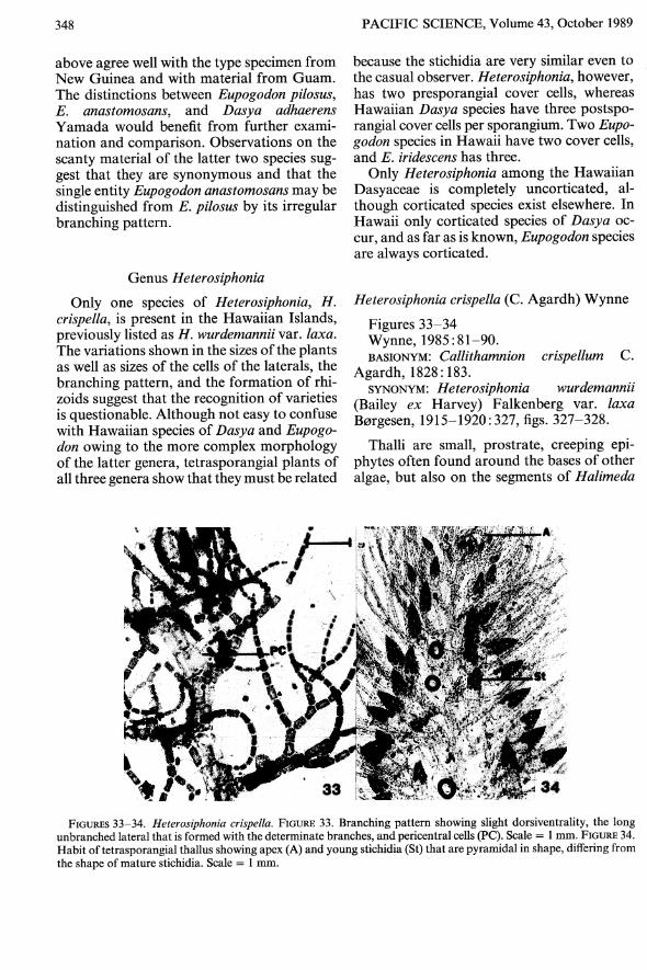
348
above agree well with the type specimen fromNew Guinea and with material from Guam.The distinctions between Eupogodon pilosus,E. anastomosans, and Dasya adhaerensYamada would benefit from further examination and comparison. Observations on thescanty material of the latter two species suggest that they are synonymous and that thesingle entity Eupogodon anastomosans may bedistinguished from E. pilosus by its irregularbranching pattern.
Genus Heterosiphonia
Only one species of Heterosiphonia, H.crispella, is present in the Hawaiian Islands,previously listed as H. wurdemannii var. laxa.The variations shown in the sizes of the plantsas well as sizes of the cells of the laterals, thebranching pattern, and the formation of rhizoids suggest that the recognition of varietiesis questionable. Although not easy to confusewith Hawaiian species of Dasya and Eupogodon owing to the more complex morphologyof the latter genera, tetrasporangial plants ofall three genera show that they must be related
PACIFIC SCIENCE, Volume 43, October 1989
because the stichidia are very similar even tothe casual observer. Heterosiphonia, however,has two presporangial cover cells, whereasHawaiian Dasya species have three postsporangial cover cells per sporangium. Two Eupogodon species in Hawaii have two cover cells,and E. iridescens has three.
Only Heterosiphonia among the HawaiianDasyaceae is completely uncorticated, although corticated species exist elsewhere. InHawaii only corticated species of Dasya occur, and as far as is known, Eupogodon speciesare always corticated.
Heterosiphonia crispella (C. Agardh) Wynne
Figures 33-34Wynne, 1985: 81-90.BASIONYM: Callithamnion crispellum C.
Agardh, 1828: 183.SYNONYM: Heterosiphonia wurdemannii
(Bailey ex Harvey) Falkenberg var. laxaB0rgesen, 1915-1920: 327, figs. 327-328.
Thalli are small, prostrate, creeping epiphytes often found around the bases of otheralgae, but also on the segments of Halimeda
FIGURES 33-34. Heterosiphonia crispella. FIGURE 33. Branching pattern showing slight dorsiventrality, the longunbranched lateral that is formed with the determinate branches, and pericentral cells (PC). Scale = I mm. FIGURE 34.Habit oftetrasporangial thallus showing apex (A) and young stichidia (St) that are pyramidal in shape, differing fromthe shape of mature stichidia. Scale = I mm.
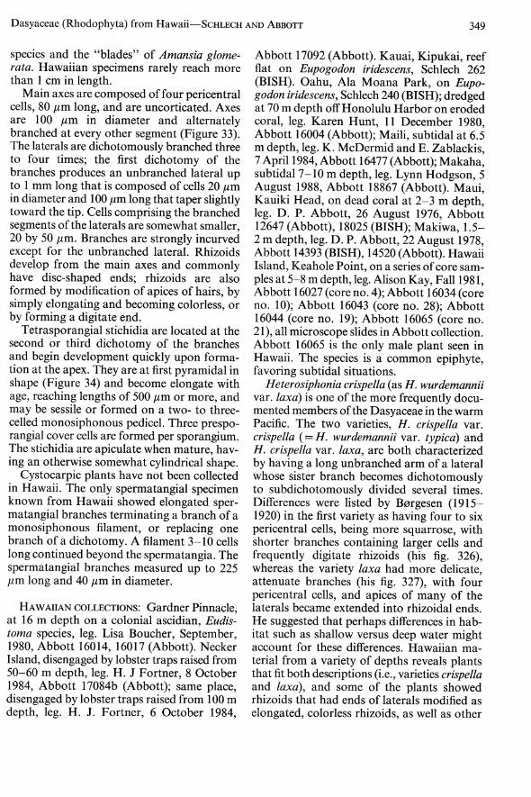
Dasyaceae (Rhodophyta) from Hawaii-SCHLECH AND ABBorr 349
species and the "blades" of Amansia glomerata. Hawaiian specimens rarely reach morethan 1 em in length.
Main axes are composed of four pericentralcells, 80 /lm long, and are uncorticated. Axesare 100 /lm in diameter and alternatelybranched at every other segment (Figure 33).The laterals are dichotomously branched threeto four times; the first dichotomy of thebranches produces an unbranched lateral upto I mm long that is composed of cells 20 /lmin diameter and 100 /lm long that taper slightlytoward the tip. Cells comprising the branchedsegments of the laterals are somewhat smaller,20 by 50 /lm. Branches are strongly incurvedexcept for the unbranched lateral. Rhizoidsdevelop from the main axes and commonlyhave disc-shaped ends; rhizoids are alsoformed by modification of apices of hairs, bysimply elongating and becoming colorless, orby forming a digitate end.
Tetrasporangial stichidia are located at thesecond or third dichotomy of the branchesand begin development quickly upon formation at the apex. They are at first pyramidal inshape (Figure 34) and become elongate withage, reaching lengths of 500 /lm or more, andmay be sessile or formed on a two- to threecelled monosiphonous pedicel. Three presporangial cover cells are formed per sporangium.The stichidia are apiculate when mature, having an otherwise somewhat cylindrical shape.
Cystocarpic plants have not been collectedin Hawaii. The only spermatangial specimenknown from Hawaii showed elongated spermatangial branches terminating a branch of amonosiphonous filament, or replacing onebranch of a dichotomy. A filament 3-10 cellslong continued beyond the spermatangia. Thespermatangial branches measured up to 225/lm long and 40 /lm in diameter.
HAWAIIAN COLLECTIONS: Gardner Pinnacle,at 16 m depth on a colonial ascidian, Eudistoma species, leg. Lisa Boucher, September,1980, Abbott 16014,16017 (Abbott). NeckerIsland, disengaged by lobster traps raised from50-60 m depth, leg. H. J Fortner, 8 October1984, Abbott 17084b (Abbott); same place,disengaged by lobster traps raised from 100 mdepth, leg. H. J. Fortner, 6 October 1984,
Abbott 17092 (Abbott). Kauai, Kipukai, reefflat on Eupogodon iridescens, Schlech 262(BISH). Oahu, Ala Moana Park, on Eupogodon iridescens, Schlech 240 (BISH); dredgedat 70 m depth off Honolulu Harbor on erodedcoral, leg. Karen Hunt, 11 December 1980,Abbott 16004 (Abbott); Maili, subtidal at 6.5m depth, leg. K. McDermid and E. Zablackis,7 April 1984, Abbott 16477 (Abbott); Makaha,subtidal 7-10 m depth, leg. Lynn Hodgson, 5August 1988, Abbott 18867 (Abbott). Maui,Kauiki Head, on dead coral at 2-3 m depth,leg. D. P. Abbott, 26 August 1976, Abbott12647 (Abbott), 18025 (BISH); Makiwa, 1.52 m depth, leg. D. P. Abbott, 22 August 1978,Abbott 14393 (BISH), 14520 (Abbott). HawaiiIsland, Keahole Point, on a series ofcore samples at 5-8 m depth, leg. Alison Kay, Fall 1981,Abbott 16027 (core no. 4); Abbott 16034 (coreno. 10); Abbott 16043 (core no. 28); Abbott16044 (core no. 19); Abbott 16065 (core no.21), all microscope slides in Abbott collection.Abbott 16065 is the only male plant seen inHawaii. The species is a common epiphyte,favoring subtidal situations.
Heterosiphonia crispella (as H. wurdemanniivar. laxa) is one of the more frequently documented members of the Dasyaceae in the warmPacific. The two varieties, H. crispella var.crispella (=H. wurdemannii var. typica) andH. crispella var. laxa, are both characterizedby having a long unbranched arm of a lateralwhose sister branch becomes dichotomouslyto subdichotomously divided several times.Differences were listed by B0rgesen (19151920) in the first variety as having four to sixpericentral cells, being more squarrose, withshorter branches containing larger cells andfrequently digitate rhizoids (his fig. 326),whereas the variety laxa had more delicate,attenuate branches (his fig. 327), with fourpericentral cells, and apices of many of thelaterals became extended into rhizoidal ends.He suggested that perhaps differences in habitat such as shallow versus deep water mightaccount for these differences. Hawaiian material from a variety of depths reveals plantsthat fit both descriptions (i.e., varieties crispellaand laxa), and some of the plants showedrhizoids that had ends of laterals modified aselongated, colorless rhizoids, as well as other

350
branches on the same specimen ending indigitate or disc-shaped ends. A few of theplants had five pericentral cells, but most hadfour. We suggest that only the species shouldbe recognized, with a range of features thatwould include those of the two current varieties. However, more gametangial materialmight aid in separating out the overlap offeatures that at present depend on vegetativedistinctions.
ACKNOWLEDGMENTS
The first author acknowledges with thanksthe help of members of her thesis committee,M. S. Doty, 1. A. Abbott, and C. W. Smith ofthe Department of Botany, University ofHawaii. She is also grateful for loans of important material from the Botanical Museum,Lund University; Rijksherbarium, Leiden;Botanical Museum, Copenhagen; New YorkBotanical Garden; the U. S. National Herbarium, Smithsonian Institution; the B. P.Bishop Museum, Honolulu; and from the personal herbaria of1. A. Abbott, M. S. Doty, R.T. Tsuda, and J. A. West. Both authors appreciate the careful reviews of Murray Parsons and James Norris.
LITERATURE CITED
ABBOTT, 1. A. 1980. Early collections of Hawaiian marine algae. Pac. Sci. 34: 101-107.
AGARDH, C. A. 1822. Species algarum. 1(2):169-531. Lund.
--. 1828. Species algarum. 2(1): 1-189.Griefswald.
AGARDH, J. G. 1841. In historiam algarumsymbolae. Linnea 15: 1-50, 443-457.
B0RGESEN, F. 1915-1920. The marine algae ofthe Danish West Indies. Dan. Bot. Ark.Vols. II-IV. Rhodophyta: Pages 300-331.
---.1937. Contributions to a south Indianmarine algal flora II. J. Indian Bot. Soc.16(6): 311-357.
COLLINS, F. S., and A. B. HERVEY. 1917. Thealgae of Bermuda. Proc. Am. Acad. ArtsSci. 53: 1-195, 6 pIs.
PACIFIC SCIENCE, Volume 43, October 1989
DAWSON, E. Y. 1954. Marine plants in thevicinity of Nha Trang, Viet Nam. Pac. Sci.8 :373-471.
---. 1957. An annotated checklist of marine algae from Eniwetok Atoll, MarshallIslands. Pac. Sci. 2: 92-132.
DIXON, P. S., and L. M. IRVINE. 1970. Miscellaneous notes on algal taxonomy and nomenclature III. Bot. Not. 123: 474-487.
DOTY, M. S., W. J. GILBERT, and 1. A. ABBOTT.1974. Hawaiian marine algae from seawardof the algal ridge. Phycologia 13: 345-357.
GMELIN, S. G. 1768. Historia Fucorum. St.Petersburg. 239 pp., 33 pis.
HOWE, M. A. 1918. Algae. Pages 489-540 inN. L. Britton, Flora of Bermuda.
KYLIN, H. 1956. Die Gattungen Rhodophyceen. Gleerup, Lund. 673 pp.
MONTAGNE, C. J. F. 1841. Plantae cellulares.Pages 161-208, pis. 5, 7, 8 in P. BarkerWebb and S. Berthelot, Histoire naturelledes Iles Canaries. Vol. 3. Paris.
PARSONS, M. J. 1975. Morphology and taxonomy of the Dasyaceae and the Lophothaliae (Rhodomelaceae) of the Rhodophyta. Aust. J. Bot. 23: 549-713.
SCHLECH, KRISTEN E. 1983. A synopsis of theHawaiian Dasyaceae (Rhodophyta, Ceramiales). M.S. thesis, University of Hawaii, Honolulu.
---. 1989. Eupogodon iridescens and Lophocladia kipukai, two new species of Rhodophyta from Hawaii. Br. Phycol. J. (inpress).
SILVA, P. C. 1987. Nomenclatural notes. Pages127-131 in P. C. Silva, E. G. Menez, andR.L. Moe, Catalog of the benthic marine algaeof the Philippines. Smithson. Contrib. Mar.Sci. 27.
TAYLOR, W. R. 1950. Plants of Bikini andother northern Marshall Islands. University ofMichigan Press, Ann Arbor. 227 pp.,79 pIs.
---. 1960. Marine algae of the easterntropical and subtropical coasts of theAmericas. University of Michigan Press,Ann Arbor. 870 pp.
WEBER VAN BOSSE, A. 1921. Liste des algues duSiboga. Premo part. Rhodophyceae. SibogaExpeditie Mongr. 59b: 187-310, pis. 6-8.E. J. Brill, Leiden.

Dasyaceae (Rhodophyta) from Hawaii-SCHLECH AND ABBOTI 351
---. 1923. Liste des algues du Siboga, II.Rhodophyceae. Seconde partie. Ceramiales.Siboga Expeditie Mongr. 59c: 311-392, pIs.9,10.
WYNNE, M. J. 1985. Concerning the namesScagelia coral/ina and Heterosiphonia
wurdemannii (Ceramiales, Rhodophyta).Cryptogamie: Algologie 6: 81-90.
YAMADA, Y. 1944. A list of marine algae fromthe Atoll of Ant. Hokkaido Imp. Univ. Inst.Algol. Res., Fac. Sci., Sci. Papers 3: 31-45,pis. 6-7.