Species diversity and geographical distribution of ...
25
Species diversity and geographical distribution of Fusarium species on winter wheat in two regions in Sweden Fatima El Khosht, 2010 Swedish University of Agricultural Science Department of Forest Mycology and Pathology Examensarbete- Agronomprogrammet, 30 hp, C-level Uppsala 2010
Transcript of Species diversity and geographical distribution of ...
Species diversity and geographical distribution of Fusarium species
on winter wheat in two regions in Sweden
Fatima El Khosht, 2010
Swedish University of Agricultural Science Department of Forest Mycology and Pathology Examensarbete- Agronomprogrammet, 30 hp, C-level Uppsala 2010
SLU, Sveriges lantbruksuniversitet Department of Forest Mycology and Pathology / Institutionen för skoglig mykologi och patologi
Art diversitet och geografisk utbredning av Fusarium arter på höstvete i två regioner i Sverige.
Fatima El Khosht Key words: Winter wheat, Fusarium, Tri5 gene, trichothecenes Supervisors: Annika Djurle & Magdalena Grudzinska-Sterno Examiner: Dan Funck Jensen Självständigt arbete i biologi C 30 hp Uppsala, 2010 Course code: EX0591
2
Table of contents Sammanfattning ...................................................................................................................................... 5
Abstract ................................................................................................................................................... 6
Introduction............................................................................................................................................. 6
Background.............................................................................................................................................. 7
Fusarium species ................................................................................................................................. 7
Trichothecene toxins ........................................................................................................................... 7
Disease and dispersal .......................................................................................................................... 8
Control measures ................................................................................................................................ 8
Materials and Methods ........................................................................................................................... 9
Isolation of fungi from grain................................................................................................................ 9
Single‐spore isolation ........................................................................................................................ 10
Molecular identification of fungi....................................................................................................... 11
DNA extraction .................................................................................................................................. 11
Amplification of TEF region ........................................................................................................... 11
Electrophoresis.............................................................................................................................. 11
Sequencing and species identification .......................................................................................... 12
Screening of Fusarium species for presence of the Tri5 gene .......................................................... 12
Results ................................................................................................................................................... 13
Region 1, Mälardalen – Fusarium spp. and Tri5 gene frequency...................................................... 13
Region 2, Kalmar – Fusarium spp. and Tri5 gene frequency............................................................. 14
Weather data .................................................................................................................................... 15
Data on Fusarium spp. identified by using species‐specific primers and toxin analyses.................. 15
Discussion.............................................................................................................................................. 17
Fusarium spp. and their frequencies................................................................................................. 17
Presence of the Tri5 gene.................................................................................................................. 19
Toxin analyses ................................................................................................................................... 19
Conclusion ............................................................................................................................................. 20
3
Acknowledgements ............................................................................................................................... 20
References.
Appendix 1. .......................................................................................................................................... 23
............................................................................................................................................ 21
Solution recipes and preparation of agar:............................................................................................. 23
CZID agar (Czapek‐Dox agar containing Iprodione and Dicloran) ......................................................... 23
Preparation of CZID agar: ...................................................................................................................... 23
SNA (Spezieller Nährstoffarmer Agar)................................................................................................... 23
Appendix 1, cont. .................................................................................................................................. 24
PDA (Potato Dextrose Agar) .................................................................................................................. 24
WA (Water Agar) ................................................................................................................................... 24
CTAB (3%) buffer ................................................................................................................................... 24
Appendix 2............................................................................................................................................. 25
4
Sammanfattning Arter inom Fusarium-släktet orsakar några av de vanligaste stråsädessjukdomarna världen över. Förutom eventuella skördeförluster orsakade av dessa sjukdomar, kan individer från flera Fusarium-arter producera mykotoxiner varav vissa är mycket giftiga. Syftet med denna studie var att undersöka vilka Fusarium-arter och hur många som förekom på höstvete insamlat från Mälardalen och Kalmar län, om skillnader i utbredning av Fusarium-svampar mellan regionerna fanns samt att undersöka potential inom arter att producera mykotoxiner tillhörande toxingruppen trichothecener. Höstvetekärnor insamlade år 2009 från tio fält (icke fungicidbehandlade) i de två regionerna analyserades med avseende på Fusarium-svampar. PCR användes för att amplifiera TEF- regioner varefter proverna sekvenserades. De prov som identifierats som Fusarium testades för förekomst av Tri5-genen som är nödvändig för trichothecenproduktion. De erhållna resultaten jämfördes med tidigare insamlade data om Fusarium-arter som påträffats i samma tio fält efter att ha använt artspecifika primers, samt toxinanalyser. I Mälardalen isolerades Fusarium-arter från 234 av totalt 500 kärnor. De identifierade arterna var F. acuminatum, F. arthrosporioides, F. avenaceum, F. graminearum, F. poae och F. tricinctum. I Kalmar län var 43 av de 500 kärnorna infekterade av Fusarium-svampar. Sju arter (F. acuminatum, F. avenaceum, F. flocciferum, F. graminearum, F. langsethiae, F. poae och F. tricinctum) identifierades. Skälet till skillnaden av antalet infekterade kärnor mellan regionerna tros ha varit de regniga väderförhållandena i Mälardalen under tiden för blomning vilket gynnar Fusarium-infektioner. I Mälardalen bar 132 individer av Fusarium-isolaten på Tri5-genen och i Kalmar län bar tio individer på genen. Majoriteten av dessa individer var av arten F. poae. Oklarheter uppstod angående isolaten som identifierats som någon av arterna F. avenaceum, F. tricinctum och F. flocciferum då några av dessa visade sig ha Tri5-genen. Dessa tre arter har inte tidigare påträffats med Tri5-genen vilket ger skäl att tro att de individerna som hade genen egentligen var någon annan Fusarium-art. Det är även möjligt att individer från dessa arter kan ha Tri5-genen men att den inte har upptäckts tidigare, eller så har genen inte uttryckts. Ytterligare analyser måste dock göras för att kunna fastställa om Tri5-genen fanns hos individer av arterna F. avenaceum, F. tricinctum and F. flocciferum. Detta kan göras genom att repetera PCR, sekvensering och artidentifiering.
5
Abstract Various Fusarium species causes some of the most common cereal diseases worldwide. Besides the yield losses that can be a result of these diseases, strains from several Fusarium species can produce mycotoxins, some very toxic. The aim of this study was to investigate which Fusarium species and how many that occurred in winter wheat grains from Mälardalen and Kalmar län, if there was any difference in the distribution of Fusarium fungi between the regions and the potential within species to produce mycotoxins belonging to the group trichothecenes. Winter wheat grains collected in 2009 from ten fields (unsprayed plots) in the two regions were analysed for Fusarium species. PCR was used to amplify the TEF region where after the samples were sequenced. The samples identified as Fusarium species were screened for the presence of the Tri5 gene necessary for trichothecene production. The results were compared with previously collected data on Fusarium spp. detected in the same ten fields using species specific primers as well as toxin analyses. In Mälardalen, Fusarium species were isolated from 234 out of 500 grains. The identified species were F. acuminatum, F. arthrosporioides, F. avenaceum, F. graminearum, F. poae and F. tricinctum. In the Kalmar area 43 of the 500 grains were infected with Fusarium species. Seven species (F. acuminatum, F. avenaceum, F. flocciferum, F. graminearum, F. langsethiae, F. poae and F. tricinctum) were identified. The differences between the amounts of infected grains in the two regions were thought to be due to humid weather conditions in Mälardalen during the time of flowering which favours Fusarium infection. In Mälardalen 132 strains of the Fusarium isolates carried the Tri5 gene and ten strains from the Kalmar area carried the gene. The majority of these strains belonged to the species F. poae. Questions arose concerning the isolates identified as F. avenaceum, F. tricinctum and F. flocciferum since some of them showed to have the Tri5 gene. These three species are not known to carry the Tri5 gene which gives reason to believe that the strains in fact belonged to other Fusarium species. It is also possible that strains from these species can carry the Tri5 gene but they have not been detected before or the gene has not been expressed. Further analyses are needed to confirm whether the Tri5 gene was present in strains from F. avenaceum, F. tricinctum and F. flocciferum. This could be made by rerunning PCR, sequencing and species identification.
6
Introduction Food spoilage and yield losses due to fungal growth are common problems all over the world. Besides the economical losses that can arise as a consequence of the fungal infection, strains from some species may have the ability to produce mycotoxins, some very toxic (Pitt & Hocking, 2009). One of the most important cereal diseases worldwide is head blight caused by species from the genus Fusarium (Hörberg, 2001) which also causes other diseases such as root- and stem blight (Bottalico & Perrone, 2002). Various Fusarium species can produce different mycotoxins. Some of the common toxins produced by Fusarium spp. are zearalenone, zearalenols and toxins belonging to the group trichothecenes (Bottalico & Perrone, 2002). This project is part of a bigger project at the Department of Forest Mycology and Pathology which focuses on the identification of factors influencing the contamination of wheat grain with Fusarium mycotoxins. The aim of this thesis was to determine which Fusarium species and how many that occurred in winter wheat grains from two regions (Mälardalen and Kalmar) in Sweden, if there was any difference in distribution of Fusarium fungi between the regions and the potential, within a species, to produce trichothecenes. Data on identified Fusarium spp. as well as toxin analyses in winter wheat samples collected in 2009 in the same fields as those used in this study were provided from a different project. They are presented here and used for comparison with the results achieved in this study.
Background
Fusarium species The most frequent pathogenic Fusarium species globally are F. graminearum (teleomorph: Gibberella zeae), F. culmorum, F. avenaceum, F. poae and F. tricinctum (Champeil et al., 2004). Together they have a variety of host plants e.g. legumes, woody seedlings and cereals and many are also found as saprophytes. F. graminearum, F. culmorum, F. avenaceum and F. poae often occur on cereals (Leslie & Summerell, 2006). In Sweden common Fusarium species on cereals are F. avenaceum, F. culmorum and F. poae (Hörberg, 2001). In recent years the prevalence of F. graminearum in Sweden has increased (Börjesson, 2004). Strains from all of the above mentioned species can carry the ability to produce mycotoxins (Leslie & Summerell, 2006).
Trichothecene toxins Mycotoxins are secondary metabolites produced by fungi. Trichothecenes is a group of toxins produced by many members of the class Ascomycetes. Fusarium, a member of this class contains trichothecene-producing species which can cause intoxication in humans and animals. F. graminearum, F. culmorum, F. poae and F. acuminatum are some of the species able to produce toxins from the group trichothecenes (Leslie & Summerell, 2006). A certain enzyme, trichodiene synthase, is needed to catalyze the first step in the production of trichothecenes. The gene Tri5 encodes for the production of the enzyme trichodiene synthase consequently making the presence of the Tri5 gene necessary for the ability to produce trichothecenes (Doohan et al., 1999). Mycotoxins are stable, chemically and thermally and they may be found both in raw material and processed food and feed products (Champeil et al., 2004).
7
Disease and dispersal Fusarium species are, as mentioned, connected to diseases such as head-, stem- and root blights usually on small-grain crops. Infections are more likely to occur if the plants are somehow weakened by for example drought during a hot and dry spring. Such conditions benefit a possible attack by Fusarium spp. and can lead to seedling- and stem blight. Seedlings surviving the first attack may later develop stem blight. Stem blight may however also be developed from Fusarium fungi sporulating on crop residues from the previous season. If a dry and hot spring is followed by a humid and cooler summer head blight might arise in the field. During the time of flowering the crops are most susceptible to infection. Precipitation during flowering will increase the risk for head blight (Hörberg, 2001). Dispersal of the fungi takes place through seeds and crop residues contaminated with inoculum in forms of conidia, chlamydospores and hyphal fragments (Hörberg, 2001). Conidia and chlamydospores can also be spread through wind and splashing (Champeil et al., 2004). The wind transports dry and light spores/conidia and the heavier conidia are transported through splashing (Hörberg, 2001). Studies have also shown that inoculum, from F. poae, can be spread by several insects and mites (Siteroptes graminum) (Hörberg, 2001, Parry et al., 1995).
Control measures To avoid Fusarium infections in cultivated crops certain measures are recommended. Obtaining a good soil structure, through e.g. drainage, is desirable to keep the crops healthy and resistant to disease development. A varied crop rotation with non-susceptible crops included and healthy seed reduce the risk for infections. Since many Fusarium species can survive as saprophytes, ploughing so that crop residues are turned down into the soil is a measure that can be used to decrease potentially contaminated plant residues on the soil surface. This would in turn decrease the infection risk for the following crops. Regarding the use of fungicides, permitted substances in Sweden are not very effective against Fusarium species (Hörberg, 2001). Fungicides are only recommended if the risk for infection is large. This is when circumstances as high humidity and high temperatures during time of flowering, crop residues on the soil surface, previous crops of wheat or maize and the cultivated crop is susceptible towards Fusarium infections, occurs simultaneously (Jordbruksverket). The use of some fungicides has even showed to increase the attack (Hörberg, 2001).
8
Materials and Methods Winter wheat grains collected randomly before harvesting (growth stage DC 85-90) in the growing season of 2009 from 10 fields (unsprayed plots) located in the regions Mälardalen and Kalmar län were used in the study. Five fields from each region, Mälardalen called region 1 and Kalmar län region 2, were randomly chosen from a larger collection.
Isolation of fungi from grain Sterilization of needed material (distilled water, forceps, scalpels and filter papers) was made by autoclaving them in 121 °C for 30 minutes. Culture media, CZID, PDA, WA and SNA were prepared according to recipes (see Appendix 1). A general description of preparation of the media PDA, WA and SNA is also to be found in Appendix 1 along with a detailed description of CZID preparation. The CZID medium is somewhat selective towards Fusarium fungi allowing them to grow faster than other genera (Abildgren et al., 1987). It also contains antibiotics to avoid bacterial contamination. PDA is a rich medium enhancing growth, pigmentation and colony morphology. The SNA medium is usually used for obtaining conidia which are more abundant on this medium (Summerell et al., 2003). In this study a small piece of sterile filter paper was place in the centre of the SNA dishes prior to usage to further induce sporulation. At least 100 seeds from each field sample were distributed in four plastic cassettes and labeled with the respective field number. The four cassettes from one field were placed into a beaker with sterile water. The beaker was placed in a shaker and left for five minutes. The cassettes were then transferred to a second beaker with sterile water and once again placed in the shaker for five minutes. This procedure was repeated for all the ten field samples with four cassettes each, one field at a time. Consequently; the seeds from each field were washed separately in sterile water for ten minutes in total. The washing was to decrease the presence of possible contaminating fungi on the grains. The cassettes were placed on sterile filter paper in a laminar flow cabinet and thereafter the seeds were placed on new sterile paper and left to dry. For grains from region 1, ten CZID dishes for each field were marked with field number, dish number (1-50) and date. Ten seeds were placed in one CZID dish using sterile forceps. The seeds were placed in columns of three, four and three as shown below and numbered from 1-10 (Figure 1). Altogether, 100 seeds from each of the five fields were distributed on ten dishes resulting in 500 seeds on 50 dishes from region 1. The dishes were then closed with parafilm and incubated for seven days in 25 °C. Regarding region 2 the same procedure was performed except that only five grains were placed in each CZID dish. This resulted in 100 CZID dishes containing five grains each from region 2. Figure 1. Seeds placement on a CZID-dish, region 1.
9
When the incubation time had passed the seeds infected with colonies resembling Fusarium were marked with letters (A-J). Each Fusarium resembling colony was transferred to SNA dishes marked with dish number, number and letter of the infected seed from which the colony was taken, for example 137 (plate number) / 4C (seed number and letter), and date. This way all isolates could be tracked back to their source if needed. The transfer of colonies on CZID to SNA was made by cutting out a piece of CZID agar with mycelium then placing the agar piece upside down on the SNA dish close to the filter paper. The scalpel used was dipped in ethanol which was burned off between each colony transfer. The SNA dishes were incubated in a growth cabinet at 25 °C for about seven days in combined black light (NUV) and cold daylight alternated with darkness, using a 12 hour photoperiod. Subsequently the dishes were studied (upside down) under microscope to spot micro- or macroconidia which then were marked on the bottom of the dish and grouped as either I, III or V. Group I represented found microconidia, group III macroconidia and group V both micro- and macroconidia. If no conidia were spotted and the fungus was believed to be a Fusarium species it was left in the 12 hour photoperiod cabinet for a few days more until it had produced conidia. One single colony was then transferred to the WA medium via spore suspension solution as presented in the protocol for single-spore isolation (below). The remaining fungi that were thought not to be Fusarium species were grown on PDA for grouping based on morphological features. After grouping, 1-40 for region 1 and 1-50 for region 2 were the numbers represent the name of the group, two isolates from each group was randomly picked for later DNA extraction.
Single-spore isolation Micro tubes of 1.5 ml were placed in a rack and prepared with 500 µl of sterile distilled water. A small piece of SNA with conidia was placed into the tube. With a pipette, 10 μl water containing conidia was transferred to a glass slide and covered with a cover glass whereafter the spore concentration was studied under a microscope. If the concentration was 1-10 spores under the low power (x10) microscope, a 10 μl drop was placed on a WA-dish and dispersed using a glass spatula. If the spores were more than 10 the samples were diluted until the desired spore concentration was reached. The WA-dishes were left to incubate over night at room temperature with the lid upwards. The dishes were after incubation studied upside down under microscope (x10) to find germinating spores. A single germinating spore was marked on the bottom of the dish. The piece of agar containing a germinating spore was subsequently cut out with a sterile scalpel and transferred to a SNA-dish. The agar piece was placed upside down close to the filter paper in the centre of the SNA-dish. The second round of SNA-dishes was incubated in the 12 hour photoperiod cabinet. When mycelium was observed on the SNA-dishes it was transferred to PDA-dishes by cutting out a piece of agar containing mycelium and placing it upside down on the new dish. The PDA-dishes were incubated in darkness at 25 °C. The same procedure was performed with randomly picked wheat grains from region 2.
10
Molecular identification of fungi
DNA extraction
Preparation: A piece of the mycelium obtained after single-spore isolation was removed from each PDA-dish through carefully scraping on the surface of the agar. It was then placed in 2 ml tubes (with screw-caps) marked with the dish number from which the mycelium was taken. The tubes were kept in a freezer until the mycelia were frozen and then the samples were freeze dried. Extraction: DNA was extracted from dried mycelium using the CTAB method according to the protocol of Gardens & Burns (1993) but with some modifications. Three autoclaved glass beads were placed into each tube containing dried mycelia. The dried mycelia were then homogenized in a FastPrep (FP120, SAVANT) 2 x 30s with the speed 4000 rpm. Next 1000µl of 3% CTAB buffer was added to the tubes which were left to incubate at 65 ºC for 1-2 hours. Before and after the incubation at 65 ºC the tubes content was properly mixed through vortexing. After incubation the tubes were centrifuged for 10 minutes at 13 000 rpm. Subsequently the supernatants were transferred to a new marked 1.5 ml Eppendorf tube. Chloroform was added to each tube. The maximum volume of the supernatant was used as a measurement for the volume chloroform to be added. The tubes were shaken quickly, thereafter centrifuged for 8 minutes at 13 000 rpm. Known amounts of the aqueous phase were transferred to the new tubes, and the volume of the aqueous phase multiplied with 1.5 gave the volume of cold 2-Propanol to be added to each tube. After the addition of 2-Propanol the tubes were stored at -20 ºC over night. The samples were centrifuged for 20 minutes at 13 000 rpm. The supernatant was removed and 200µl cold 70% ethanol was added. The samples were once again centrifuged, 5 minutes at 13 000 rpm. The supernatant was discarded and the pellet dried. The dry pellet was re-suspended in 50 µl ultrapure water. The DNA-concentration was thereafter measured for each sample in NanoDrop spectrophotometer ND-1000 (Saveen Werner). Samples were diluted to the concentration 1 ng/µl of DNA.
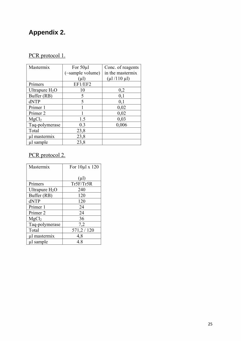
Amplification of TEF region The Transcription Elongation Factor region in fungal DNA was amplified using the primers EF1 (forward primer; 5´- ATGGGTAAGGA (A/G) GACAAGAC-3) and EF2 (reverse primer; 5´-GGA(G/A)GTACCAGT(G/C)ATCATGTT-3´) (Geiser et al., 2004). The polymerase chain reaction (PCR) with primer set EF1/ EF2 was based on an initial denaturation step at 94 °C for 5 min, followed by 35 cycles of denaturation at 94 °C for 30s, primer annealing at 53 °C for 30s, and primer extension at 72 °C for 30s, respectively, and a final elongation step at 72 °C for 7 min using a 2720 Applied Biosystems thermal cycler (Applied Biosystems, California, USA). The used program was according to Macia´-Vicente et al. (2008) with some modifications. The recipe of the PCR mix is presented in appendix 2, protocol 1. In the PCR reactions final concentrations of 2.5 mM MgCl2, 0.2 mM dNTP, 0,2 �M of each primer, 0.03 units/�L DreamTaq TM Green DNA Polymerase (Fermentas International) and corresponding reaction buffer were used. The final DNA-concentration was 0,5 ng/µl.
Electrophoresis The amplified products were separated using 1% agarose gel electrophoresis (Agarose D-1, Conda). The gels, size 25 x 15 cm, were stained by Gel green (Biotium), loaded with 3µl of each sample and 4 µl of a marker (Gene Ruler 100bp) and ran in 220 V for 30 minutes.
11
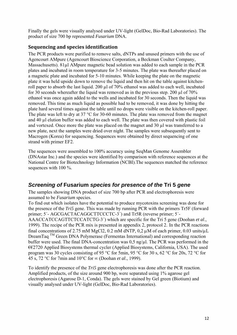
Finally the gels were visually analysed under UV-light (GelDoc, Bio-Rad Laboratories). The product of size 700 bp represented Fusarium DNA.
Sequencing and species identification The PCR products were purified to remove salts, dNTPs and unused primers with the use of Agencourt AMpure (Agencourt Bioscience Corporation, a Beckman Coulter Company, Massachusetts). 81µl AMpure magnetic bead solution was added to each sample in the PCR plates and incubated in room temperature for 3-5 minutes. The plate was thereafter placed on a magnetic plate and incubated for 5-10 minutes. While keeping the plate on the magnetic plate it was held upside down to remove the liquid and then hit on the table against kitchen-roll paper to absorb the last liquid. 200 µl of 70% ethanol was added to each well, incubated for 30 seconds whereafter the liquid was removed as in the previous step. 200 µl of 70% ethanol was once again added to the wells and incubated for 30 seconds. Then the liquid was removed. This time as much liquid as possible had to be removed, it was done by hitting the plate hard several times against the table until no drops were visible on the kitchen-roll paper. The plate was left to dry at 37 °C for 30-60 minutes. The plate was removed from the magnet and 40 µl elution buffer was added to each well. The plate was then covered with plastic foil and vortexed. Once more the plate was placed on the magnet and 30 µl was transferred to a new plate, next the samples were dried over night. The samples were subsequently sent to Macrogen (Korea) for sequencing. Sequences were obtained by direct sequencing of one strand with primer EF2.
The sequences were assembled to 100% accuracy using SeqMan Genome Assembler (DNAstar Inc.) and the species were identified by comparison with reference sequences at the National Centre for Biotechnology Information (NCBI).The sequences matched the reference sequences with 100 %.
Screening of Fusarium species for presence of the Tri 5 gene The samples showing DNA product of size 700 bp after PCR and electrophoresis were assumed to be Fusarium species. To find out which isolates have the potential to produce mycotoxins screening was done for the presence of the Tri5 gene. This was made by running PCR with the primers Tr5F (forward primer; 5´- AGCGACTACAGGCTTCCCTC-3´) and Tr5R (reverse primer; 5´-AAACCATCCAGTTCTCCATCTG-3´) which are specific for the Tri 5 gene (Doohan et al., 1999). The recipe of the PCR mix is presented in appendix 2, protocol 2. In the PCR reactions final concentrations of 2.75 mM MgCl2, 0.2 mM dNTP, 0,2 μM of each primer, 0.03 units/μL DreamTaq TM Green DNA Polymerase (Fermentas International) and corresponding reaction buffer were used. The final DNA-concentration was 0,5 ng/µl. The PCR was performed in the €€2720 Applied Biosystems thermal cycler (Applied Biosystems, California, USA). The used program was 30 cycles consisting of 95 °C for 5min, 95 °C for 30 s, 62 °C for 20s, 72 °C for 45 s, 72 °C for 7min and 10°C for ∞ (Doohan et al., 1999).
To identify the presence of the Tri5 gene electrophoresis was done after the PCR reaction. Amplified products, of the size around 900 bp, were separated using 1% agarose gel electrophoresis (Agarose D-1, Conda). The gels were stained by Gel green (Biotium) and visually analysed under UV-light (GelDoc, Bio-Rad Laboratories).
12
Results
Region 1, Mälardalen – Fusarium spp. and Tri5 gene frequency In region 1, 234 grains of the 500 (46.8%), were infected with fungi. Fusarium DNA products from some of the samples are shown as stripes on one of the gels (Figure 2), on which electrophoresis was performed. Six species were identified. The species were F. acuminatum, F. arthrosporides, F. avenaceum, F. graminearum, F. poae and F. tricinctum. The most common species in this region was F. poae which accounted for 56% of the infected grains. The second most common species was F. tricinctum which infected 35% of the grains, followed by F. avenaceum found on 7% of the grains. The remaining three species (F. acuminatum, F. arthrosporides and F. graminearum) were distributed on only five grains. 700 bp
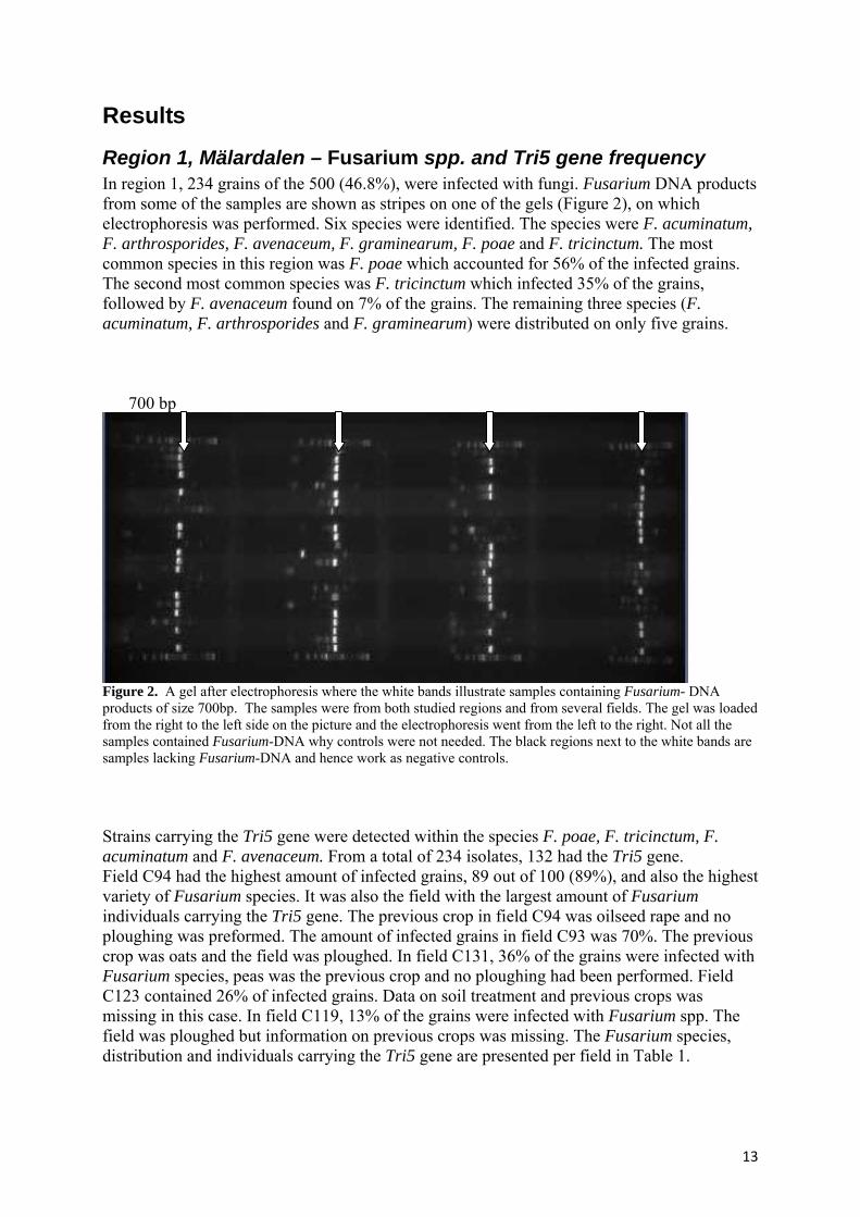
Figure 2. A gel after electrophoresis where the white bands illustrate samples containing Fusarium- DNA products of size 700bp. The samples were from both studied regions and from several fields. The gel was loaded from the right to the left side on the picture and the electrophoresis went from the left to the right. Not all the samples contained Fusarium-DNA why controls were not needed. The black regions next to the white bands are samples lacking Fusarium-DNA and hence work as negative controls. Strains carrying the Tri5 gene were detected within the species F. poae, F. tricinctum, F. acuminatum and F. avenaceum. From a total of 234 isolates, 132 had the Tri5 gene. Field C94 had the highest amount of infected grains, 89 out of 100 (89%), and also the highest variety of Fusarium species. It was also the field with the largest amount of Fusarium individuals carrying the Tri5 gene. The previous crop in field C94 was oilseed rape and no ploughing was preformed. The amount of infected grains in field C93 was 70%. The previous crop was oats and the field was ploughed. In field C131, 36% of the grains were infected with Fusarium species, peas was the previous crop and no ploughing had been performed. Field C123 contained 26% of infected grains. Data on soil treatment and previous crops was missing in this case. In field C119, 13% of the grains were infected with Fusarium spp. The field was ploughed but information on previous crops was missing. The Fusarium species, distribution and individuals carrying the Tri5 gene are presented per field in Table 1.
13
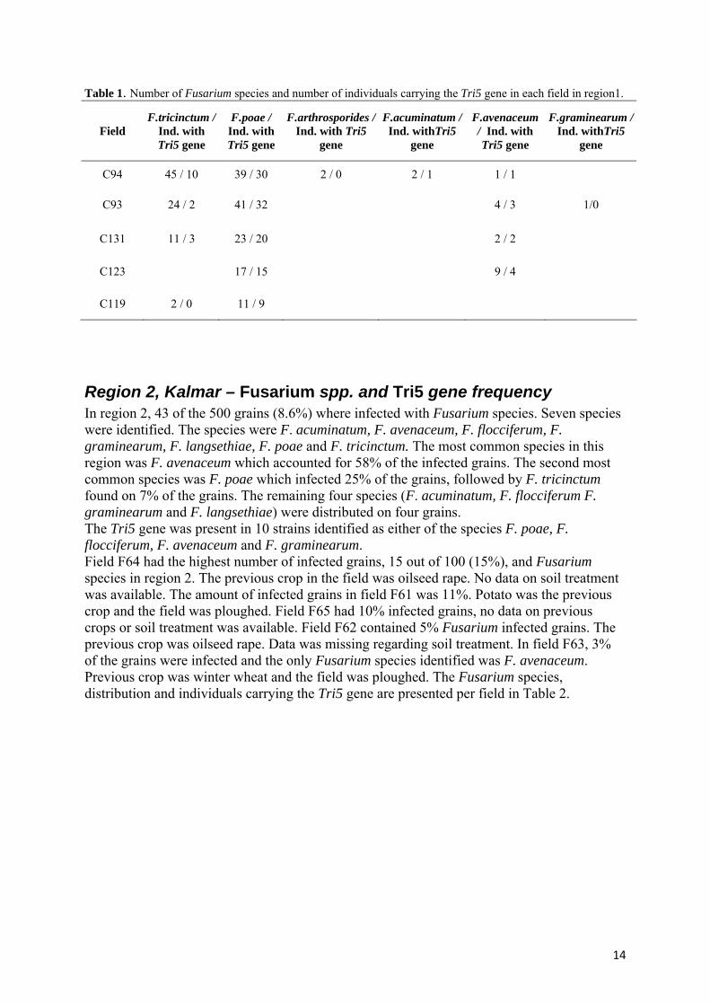
Table 1. Number of Fusarium species and number of individuals carrying the Tri5 gene in each field in region1.
Field F.tricinctum /
Ind. with Tri5 gene
F.poae / Ind. with Tri5 gene
F.arthrosporides / Ind. with Tri5
gene
F.acuminatum / Ind. withTri5
gene
F.avenaceum / Ind. with Tri5 gene
F.graminearum / Ind. withTri5
gene
C94 45 / 10 39 / 30 2 / 0 2 / 1 1 / 1
C93 24 / 2 41 / 32 4 / 3 1/0
C131 11 / 3 23 / 20 2 / 2
C123 17 / 15 9 / 4
C119 2 / 0 11 / 9
Region 2, Kalmar – Fusarium spp. and Tri5 gene frequency In region 2, 43 of the 500 grains (8.6%) where infected with Fusarium species. Seven species were identified. The species were F. acuminatum, F. avenaceum, F. flocciferum, F. graminearum, F. langsethiae, F. poae and F. tricinctum. The most common species in this region was F. avenaceum which accounted for 58% of the infected grains. The second most common species was F. poae which infected 25% of the grains, followed by F. tricinctum found on 7% of the grains. The remaining four species (F. acuminatum, F. flocciferum F. graminearum and F. langsethiae) were distributed on four grains. The Tri5 gene was present in 10 strains identified as either of the species F. poae, F. flocciferum, F. avenaceum and F. graminearum. Field F64 had the highest number of infected grains, 15 out of 100 (15%), and Fusarium species in region 2. The previous crop in the field was oilseed rape. No data on soil treatment was available. The amount of infected grains in field F61 was 11%. Potato was the previous crop and the field was ploughed. Field F65 had 10% infected grains, no data on previous crops or soil treatment was available. Field F62 contained 5% Fusarium infected grains. The previous crop was oilseed rape. Data was missing regarding soil treatment. In field F63, 3% of the grains were infected and the only Fusarium species identified was F. avenaceum. Previous crop was winter wheat and the field was ploughed. The Fusarium species, distribution and individuals carrying the Tri5 gene are presented per field in Table 2.
14
Table 2. Number of Fusarium species and number of individuals carrying the Tri5 gene in each field in region2.
Field F.tricinctum / Ind. with Tri5 gene
F.poae / Ind. with Tri5
F.avenaceum / Ind. with Tri5 gene
F.acuminatum / Ind. with Tri5 gene
F.langsethiae / Ind. with Tri5 gene
F.graminearum / Ind. with Tri5
gene
F.flocciferum / Ind. with Tri5 gene
F64 2 / 0 6 / 4 4 / 0 1 / 0 1 / 0 1 / 1
F61 1 / 1 10 / 0
F65 4 / 2 5 / 1 1 / 1
F62 1 / 0 4 / 0
F63 3 / 0
Weather data The weather in region 1, Mälardalen represented by Uppsala which lies within this region, during the period 1st of May to the 28th of August is presented in Figure 3. The temperature and precipitation were normal in May. June was slightly colder than normal followed by July with rather normal temperatures. Heavy precipitation during these two months took place, especially around the last weeks in June (SMHI). The time of flowering occurred around the last week of June which coincides with the period of heavy precipitation. This weather favours many of the Fusarium species and increases the risk of grain infection. The weather in region 2, Kalmar, during the period 1st of May to the 28th of August is shown in Figure 4. The temperature was normal and the precipitation almost evenly distributed and somewhat low over the period (SMHI). Dry conditions during the time of flowering decreases the risk of grain infection by most of the Fusarium species.
Data on Fusarium spp. identified by using species-specific primers and toxin analyses Identification of Fusarium species in the same fields as those studied in this project was made in a different project using species-specific primers. These samples were also analyzed for mycotoxin content. Data of identified Fusarium spp. is presented below in Table 3 and 4, showing the species found in region 1 and 2 respectively.
15
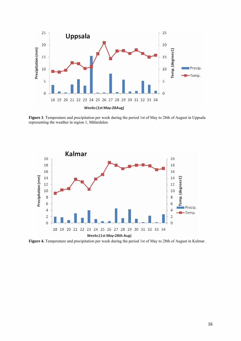
Figure 3. Temperature and precipitation per week during the period 1st of May to 28th of August in Uppsala representing the weather in region 1, Mälardalen.
Figure 4. Temperature and precipitation per week during the period 1st of May to 28th of August in Kalmar.
16
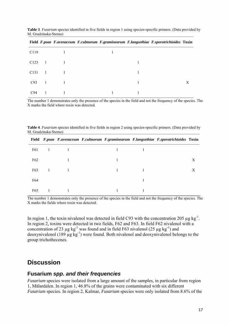
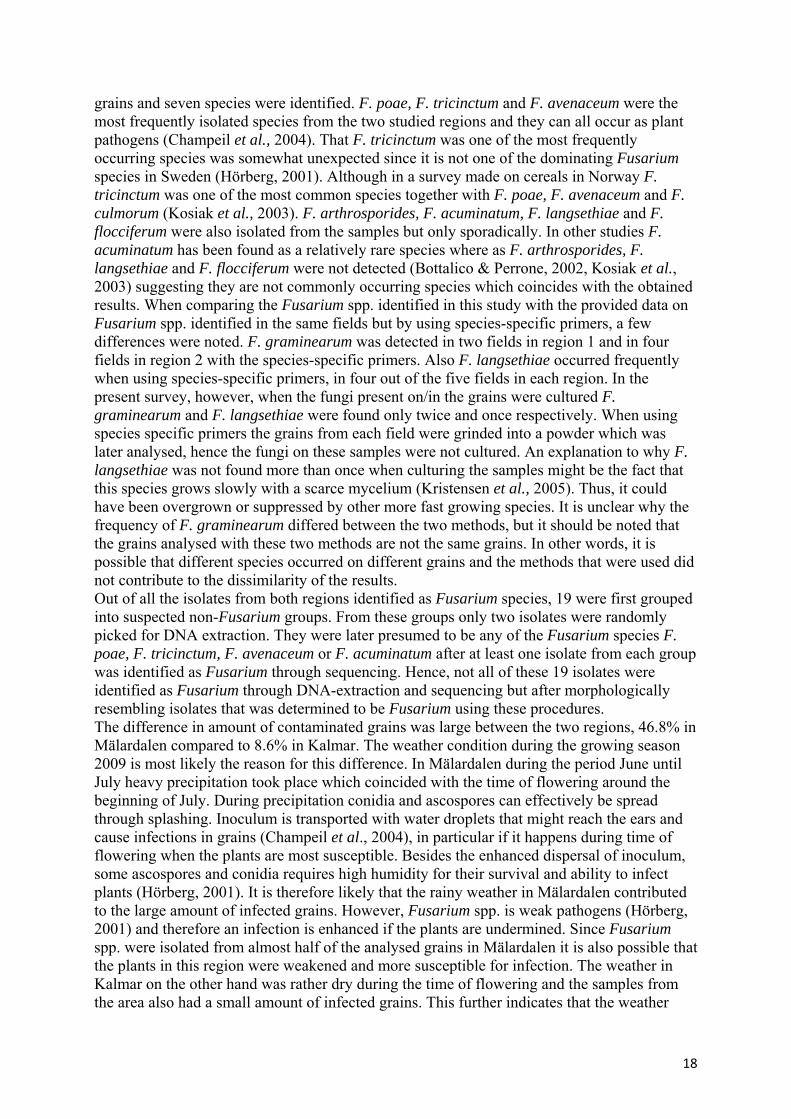
Table 3. Fusarium species identified in five fields in region 1 using species-specific primers. (Data provided by M. Grudzinska-Sterno)
Field F.poae F.avenaceum F.culmorum F.graminearum F.langsethiae F.sporotrichioides Toxin
C119 1 1
C123 1 1 1
C131 1 1 1
C93 1 1 1 X
C94 1 1 1 1
The number 1 demonstrates only the presence of the species in the field and not the frequency of the species. The X marks the field where toxin was detected. Table 4. Fusarium species identified in five fields in region 2 using species-specific primers. (Data provided by M. Grudzinska-Sterno)
Field F.poae F.avenaceum F.culmorum F.graminearum F.langsethiae F.sporotrichioides Toxin
F61 1 1 1 1
F62 1 1 X
F63 1 1 1 1 X
F64 1
F65 1 1 1 1
The number 1 demonstrates only the presence of the species in the field and not the frequency of the species. The X marks the fields where toxin was detected. In region 1, the toxin nivalenol was detected in field C93 with the concentration 205 µg kg-1. In region 2, toxins were detected in two fields, F62 and F63. In field F62 nivalenol with a concentration of 23 µg kg-1 was found and in field F63 nivalenol (25 µg kg-1) and deoxynivalenol (189 µg kg-1) were found. Both nivalenol and deoxynivalenol belongs to the group trichothecenes.
Discussion
Fusarium spp. and their frequencies Fusarium species were isolated from a large amount of the samples, in particular from region 1, Mälardalen. In region 1, 46.8% of the grains were contaminated with six different Fusarium species. In region 2, Kalmar, Fusarium species were only isolated from 8.6% of the
17
grains and seven species were identified. F. poae, F. tricinctum and F. avenaceum were the most frequently isolated species from the two studied regions and they can all occur as plant pathogens (Champeil et al., 2004). That F. tricinctum was one of the most frequently occurring species was somewhat unexpected since it is not one of the dominating Fusarium species in Sweden (Hörberg, 2001). Although in a survey made on cereals in Norway F. tricinctum was one of the most common species together with F. poae, F. avenaceum and F. culmorum (Kosiak et al., 2003). F. arthrosporides, F. acuminatum, F. langsethiae and F. flocciferum were also isolated from the samples but only sporadically. In other studies F. acuminatum has been found as a relatively rare species where as F. arthrosporides, F. langsethiae and F. flocciferum were not detected (Bottalico & Perrone, 2002, Kosiak et al., 2003) suggesting they are not commonly occurring species which coincides with the obtained results. When comparing the Fusarium spp. identified in this study with the provided data on Fusarium spp. identified in the same fields but by using species-specific primers, a few differences were noted. F. graminearum was detected in two fields in region 1 and in four fields in region 2 with the species-specific primers. Also F. langsethiae occurred frequently when using species-specific primers, in four out of the five fields in each region. In the present survey, however, when the fungi present on/in the grains were cultured F. graminearum and F. langsethiae were found only twice and once respectively. When using species specific primers the grains from each field were grinded into a powder which was later analysed, hence the fungi on these samples were not cultured. An explanation to why F. langsethiae was not found more than once when culturing the samples might be the fact that this species grows slowly with a scarce mycelium (Kristensen et al., 2005). Thus, it could have been overgrown or suppressed by other more fast growing species. It is unclear why the frequency of F. graminearum differed between the two methods, but it should be noted that the grains analysed with these two methods are not the same grains. In other words, it is possible that different species occurred on different grains and the methods that were used did not contribute to the dissimilarity of the results. Out of all the isolates from both regions identified as Fusarium species, 19 were first grouped into suspected non-Fusarium groups. From these groups only two isolates were randomly picked for DNA extraction. They were later presumed to be any of the Fusarium species F. poae, F. tricinctum, F. avenaceum or F. acuminatum after at least one isolate from each group was identified as Fusarium through sequencing. Hence, not all of these 19 isolates were identified as Fusarium through DNA-extraction and sequencing but after morphologically resembling isolates that was determined to be Fusarium using these procedures. The difference in amount of contaminated grains was large between the two regions, 46.8% in Mälardalen compared to 8.6% in Kalmar. The weather condition during the growing season 2009 is most likely the reason for this difference. In Mälardalen during the period June until July heavy precipitation took place which coincided with the time of flowering around the beginning of July. During precipitation conidia and ascospores can effectively be spread through splashing. Inoculum is transported with water droplets that might reach the ears and cause infections in grains (Champeil et al., 2004), in particular if it happens during time of flowering when the plants are most susceptible. Besides the enhanced dispersal of inoculum, some ascospores and conidia requires high humidity for their survival and ability to infect plants (Hörberg, 2001). It is therefore likely that the rainy weather in Mälardalen contributed to the large amount of infected grains. However, Fusarium spp. is weak pathogens (Hörberg, 2001) and therefore an infection is enhanced if the plants are undermined. Since Fusarium spp. were isolated from almost half of the analysed grains in Mälardalen it is also possible that the plants in this region were weakened and more susceptible for infection. The weather in Kalmar on the other hand was rather dry during the time of flowering and the samples from the area also had a small amount of infected grains. This further indicates that the weather
18

conditions contributed to the difference of amount of Fusarium contaminated grains between the two studied regions. No clear impact of previous crops on the number of Fusarium isolates found seemed obvious. Neither did soil treatment such as ploughing or no ploughing appear to have made a difference in the amount of contaminated grains in the different fields. However, not every field had data on soil treatment which makes it harder to draw conclusions, and the total number of fields was small.
Presence of the Tri5 gene Several strains from the obtained isolates of the species F. acuminatum, F. avenaceum, F. flocciferum, F. graminearum, F. poae and F. tricinctum showed to carry the Tri5 gene and hence having the potential to produce trichothecene toxins. In total 142 strains from the two studied regions were carrying the Tri5 gene, 132 of them were strains from Mälardalen (region 1) and 10 from Kalmar län (region 2). The absolute majority of these strains belonged to the species F. poae which had 106 strains with the gene in Mälardalen and seven in Kalmar län. After screening the Fusarium isolates for the Tri5 gene some confusion concerning the isolates identified as the species F. avenaceum, F. tricinctum and F. flocciferum arose. These three species should in fact lack the Tri5 gene and thus potential of producing trichothecenes (Leslie & Summerell, 2006). This gives reason to believe that the strains belonging to any of the species F. avenaceum, F. flocciferum and F. tricinctum that were carrying the Tri5 gene were in fact some other Fusarium species with the capability of having the gene. Since the GenBank database does have errors and misidentification can occur (Geiser et al. 2004) it is possible that isolates were wrongly identified. In Mälardalen, the Tri5 gene was present in 15 of the 82 F. tricinctum isolates and regarding F. avenaceum 10 of the total 16 isolates showed to have the Tri5 gene. In Kalmar län one of the total 26 F. avenaceum isolates had the Tri5 gene as well as the only F. flocciferum isolate making it questionable if the species F. flocciferum actually was present in this study. However, it is also possible that these species (F. avenaceum, F. flocciferum and F. tricinctum) actually were carriers of the Tri5 gene but have not been detected with the gene in previous studies. Further analyses must be performed to confirm this which could be made by re-running PCR, sequencing and species identification on these samples.
Toxin analyses Low concentrations of the toxins deoxynivalenol (DON) and nivalenol (NIV) were detected. Deoxynivalenol is produced by the Fusarium species F. graminearum and F. culmorum (Bottalico & Perrone, 2002). F. graminearum and F. poae are species able to produce nivalenol. In field C93, Mälardalen, nivalenol was detected which is explainable since several of the F. poae strains in this field had the Tri5 gene. The field F62 (Kalmar) contained nivalenol. In this field the only two Fusarium species identified were F. tricinctum and F. avenaceum and none had the Tri5 gene. Other species with the ability to produce the toxins were probably present in the field but not detected. In field F63 (Kalmar) where the only species found was F. avenaceum deoxynivalenol and nivalenol were detected. Therefore other species might have been present as in field F62. Nonetheless the toxin concentrations found were low and the toxins were present in just three of the ten fields. In Mälardalen, all of the fields contained strains with the ability to produce trichothecenes but not more than one field had detectable toxin concentrations. In Kalmar, isolates from three fields carried the Tri5 gene and in two of these fields toxin was found. In the third field, F65, the fungi had the gene but no toxin was detected. Different factors affect the formation or the absence of formation of
19
toxins. Some factors are time of colonisation, competition between microorganisms and climate. Trichothecene production is favoured by cold and humid conditions (Champeli et al., 2004). Since the temperatures in neither Mälardalen nor Kalmar during the growing season 2009 were lower than normal, which is desirable for trichothecene production, the high temperatures could be one reason for the low toxin concentrations. Other reasons for the low toxin concentration in spite of the large amount of individuals with the Tri5 gene may be late infection so that the disease did not have time to reach the stage of toxin production or the host plant may be resistant (Champeli et al., 2004). F. poae, which is one of the species that produces trichothecenes, is a common saprophyte on grains (Summerell, et al., 2003). Since many of the F. poae found on the samples had the Tri5 gene but did not produce the toxin they might have occurred on the grains in a late stage as saprophytes and hence did not reach the stage of toxin production. Moreover the Tri5 gene codes for the production of an enzyme required in only the first step of the production of trichothecenes (Doohan et al., 1999). This could mean that the strains carrying the Tri5 not necessarily are capable of producing the trichothecene toxins but only the first step in the production chain. Further studies are needed to establish which factors that contributed to the low trichothecenes concentrations found although many of the isolates carried the Tri5 gene.
Conclusion The amount of Fusarium infected grains in Mälardalen was 47% compared to Kalmar län were 9% of the grains were infected. The weather conditions in the two regions are thought to be the main reason behind the large difference of infected grains. In Mälardalen precipitation took place around the time of flowering which is when the wheat is most susceptible towards infection. During the time of the wheat flowering in Kalmar the weather was dry and hence the risk of grain infection was low. The Fusarium species found in the two regions were in general the same but with some differences in the distribution. F. poae (56%) and F. tricinctum (35%) were the most common species in Mälardalen and in Kalmar län the most common species were F. avenaceum (58%) and F. poae (25%). Altogether the most frequently occurring species in the survey were F. poae, F. tricinctum and F. avenaceum. Several strains from the species F. acuminatum, F. avenaceum, F. flocciferum F. graminearum F. poae and F. tricinctum were found to be carriers of the Tri5 gene. F. avenaceum, F. poae, and F. tricinctum are not known to have this gene why repeated analyses of these samples are recommended. In total 142 strains of the 277 Fusarium isolates had the Tri5 gene. Low toxin concentrations were detected and in only three of the ten studied fields in spite of the fact that eight fields contained strains with the potential of trichothecene production. To re-run the samples that were identified as the species F. avenaceum, F. tricinctum and F. flocciferum and once again screen them for the presence of the Tri5 gene and compare the results to the results obtained in this survey would be interesting.
Acknowledgements I would like to thank my supervisors Annika Djurle and Magdalena Grudzinska-Sterno for their help during my laboratory work and the time of writing this report.
20
Also I want to especially thank Rena Gadjieva for on short notice taking her time to instruct me throughout the second part of the laboratory work and assist me with comments while writing. I appreciate your help and enjoyed working with you. At last I want to thank everyone that helped me in any way during this time, from personnel or students on the Department of Forest Mycology and Pathology, to my family and friends.
References Abildgren, M. P., Lund, F., Thrane, U., & Elmholt, S. 1987. Czapek-Dox agar containing Iprodione and Dicloran as a selective medium for the isolation of Fusarium species. Letters in Applied Microbiology 5(4): 83-86 Biotium, www.biotium.com/product/price_and_info.asp?item=41004&Sub_section=09A Bottalico, A. & Perrone, G. 2002. Toxigenic Fusarium species and mycotoxins associated with head blight in small-grain cereals in Europe. European Journal of Plant Pathology 108:611-624. Börjesson, T. 2004. Mykotoxiner i stråsädeskärna – Förekomst i Sverige. Meddelande från Södra jordbruksförsöksdistriktet nr 57. Instutionen för växtvetenskap, Södra jordbruksförsöksdidtriktet, SLU
Champeil, A., Dorè, T. & Fourbet, J.F. 2004. Fusarium head blight: epidemiological origin of the effects of cultural practices on head blight attacks and the production of mycotoxins by Fusarium in wheat grains. Plant Science 1389-1415.
Doohan, F. M., Weston, G., Rezanoor, H. N., Parry, D. W. & Nicholson, P. 1999. Development and use of a Reverse Transcription-PCR assay to study expression of Tri5 by Fusarium species in vitro and in planta. Applied and Environmental Microbiology 65: 3850-3854
Gardens, M. & Burns, T.D. 1993. ITS primers with enhance specificity for basidiomycetes – application to the identification of mycorrhizae and rusts, Mol. Ecol. 2:113-118 Geiser, D. M., Jimenez-Gasco del Mar, M., Kang, S., Makalowska, I., Veeraraghavam, N., Ward, T.J., Zhang, N., Kuldau, D. A. & O’Donnell, K. 2004. FUSARIUM-ID v. 1.0: A DNA sequence database for identifying Fusarium. European Journal of Plant Pathology 110: 473-479
Hörberg, H. 2001a Fusarium-svampar i stråsäd. Faktablad om växtskydd, Jordbruk, 103J. SLU Publikationstjänst, Uppsala. Jordbruksverket, http://www.jordbruksverket.se/etjanster/etjanster/vaxtskyddsinfo.4.35974d0d12179bec28580002425.html
21
Kosiak, B., Torp, M., Skejerve, E. & Thrane, U. 2003. The Prevalence and Distribution of Fusarium species in Norwegian Cereals: s Survey. Acta Agriculturae Scandinavica, Section B – Plant Soil Science 53:4, 168-176. Kristensen, R., Torp, M., Kosiak, B. & Holst-Jensen, A. 2005. Phylogeny and toxigenic potential is correlated in Fusarium species as revealed by partial translation elongation factor 1 alpha gene sequences. Mycological Research 109 (2): 173-186
Leslie, F.J. & Summerell, B.A.. 2006. The Fusarium Laboratory Manual. First edition. Oxford: Blackwell Publishing. Macia´-Vicente, J. G., Jansson, H., Abdullah, S. K., Descals, E., Salinas, J. & Lopez-Llorca, L. V. (2008). Fungal root endophytes from natural vegetation in Mediterranean environments with special reference to Fusarium spp. FEMS Microbiol Ecol 64:90–105
NCBI, www.ncbi.nlm.nih-gov Parry, D.W., Jenkinson, P. & McLeod, L. 1995. Fusarium ear blight (scab) in small grain cereals – a review. Plant Pathology 44, 207-238. Pitt, J. & Hocking, A. (2009). Fungi and Food Spoilage. 3rd ed. US: Springer SMHI, www.smhi.se/klimatdata Summerell, B.A., Salleh, B. & Leslie, J. F. (2003). A Utilitarian Approach to Fusarium Identification. Plant Disease 87:117-128.
22
Appendix 1. Solution recipes and preparation of agar: CZID agar (Czapek-Dox agar containing Iprodione and Dicloran) For 1 L: Czapek-Dox Agar 48g CuSO4 x 5H2O 0.005g ZnSO4 x 7H2O 0.01g Chloramphenicol 0.05g Dicloran solution: 1.0ml (2, 6-dicloro-4-nitroaniline 20mg to 10ml ethanol) Iprodione suspension (100 mg to 50 ml ethanol) 1.5ml Chlortetracycline solution: 1.0ml (Chlortetracycline hydrochloride 0.5g to 10ml Ethanol) Preparation of CZID agar: The substances were weighed and poured into a glass bottle and distilled water was added to the desired volume. The bottle was autoclaved with the lid only slightly closed in 121�C for 30 minutes. After autoclaving, the medium was left to cool to 50-55�C before adding Chlortetracycline- (broad-spectrum antibiotic) and Iprodione solutions (fungicide) that had been prepared and kept in a freezer until usage. The Iprodione solution was shaken before it was added to the substrate. Thereafter the medium was poured into the sterile Petri-dishes under sterile conditions (in a laminar flow cabinet), about 20ml in each plate. After the medium had solidified the Petri-dishes were put into clean plastic bags and kept in a refrigerator. Due to the lost effect of Iprodione with time the dishes were used within two days after preparation. SNA (Spezieller Nährstoffarmer Agar) For 1 L: KH2PO4 1g KNO3 1g MgSO4 x 7H2O 0.5g KCl 0.5g Glucose 0.2g Sucrose 0.2g Bacto Agar 20g
23
Appendix 1, cont. PDA (Potato Dextrose Agar) For 1 L: PDA 19.5g Bacto Agar 6g WA (Water Agar) For 1 L: Bacto agar 20g Preparation of SNA, PDA and WA: Substances were weighed and poured in a glass bottle and distilled water was added to the desired volume. The bottle with slightly closed lid containing medium was autoclaved in 121�C for 30 minutes. After cooling to a workable temperature the medium was poured in sterile Petri-dishes in a laminar flow cabinet, left to solidify and then packed in clean plastic bags. Prior to usage of the SNA dishes a piece of sterile filter paper, about 1cm2, was placed in the center of each dish. The purpose of the filter paper was to aid sporulation of the Fusarium fungi. CTAB (3%) buffer For 150 ml: CTAB 4.5g H2O 48. 9 ml Tris-HCL (pH 8.0) 22. 5 ml NaCl (1.4 M) 0. 60 ml
24
25
Appendix 2. PCR protocol 1. Mastermix For 50µl
(~sample volume) (µl)
Conc. of reagentsin the mastermix (µl /110 µl)
Primers EF1/EF2 Ultrapure H2O 10 0,2 Buffer (RB) 5 0,1 dNTP 5 0,1 Primer 1 1 0,02 Primer 2 1 0,02 MgCl2 1.5 0,03 Taq-polymerase 0.3 0,006 Total 23,8 µl mastermix 23,8 µl sample 23,8 PCR protocol 2. Mastermix For 10µl x 120
(µl)
Primers Tr5F/Tr5R Ultrapure H2O 240 Buffer (RB) 120 dNTP 120 Primer 1 24 Primer 2 24 MgCl2 36 Taq-polymerase 7,2 Total 571,2 / 120 µl mastermix 4,8 µl sample 4.8


![Biodiversity and conservation Genetic diversity: within species variation (e.g corn [Zea mays] in North vs. Central America) Species diversity: species.](https://static.fdocuments.in/doc/165x107/56649db45503460f94aa4ef5/biodiversity-and-conservation-genetic-diversity-within-species-variation.jpg)