Specialised cells
33
Core and Extended *
-
Upload
circle4biology -
Category
Education
-
view
127 -
download
0
Transcript of Specialised cells

Core and Extended
*

*Cells in organisms
become
specialised to
perform certain
functions
*If similar cells
work together
they form a tissue* These cardiac(heart) muscle cells are
all the same
* They work together, forming cardiac muscle tissue
*
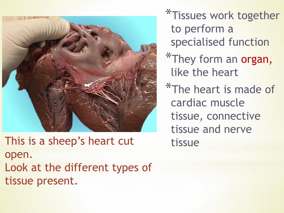
*Tissues work together
to perform a
specialised function
*They form an organ,
like the heart
*The heart is made of
cardiac muscle
tissue, connective
tissue and nerve
tissueThis is a sheep’s heart cut
open.
Look at the different types of
tissue present.
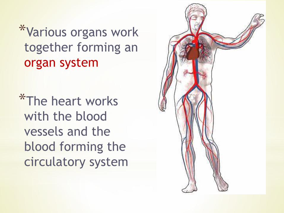
*Various organs work
together forming an
organ system
*The heart works
with the blood
vessels and the
blood forming the
circulatory system
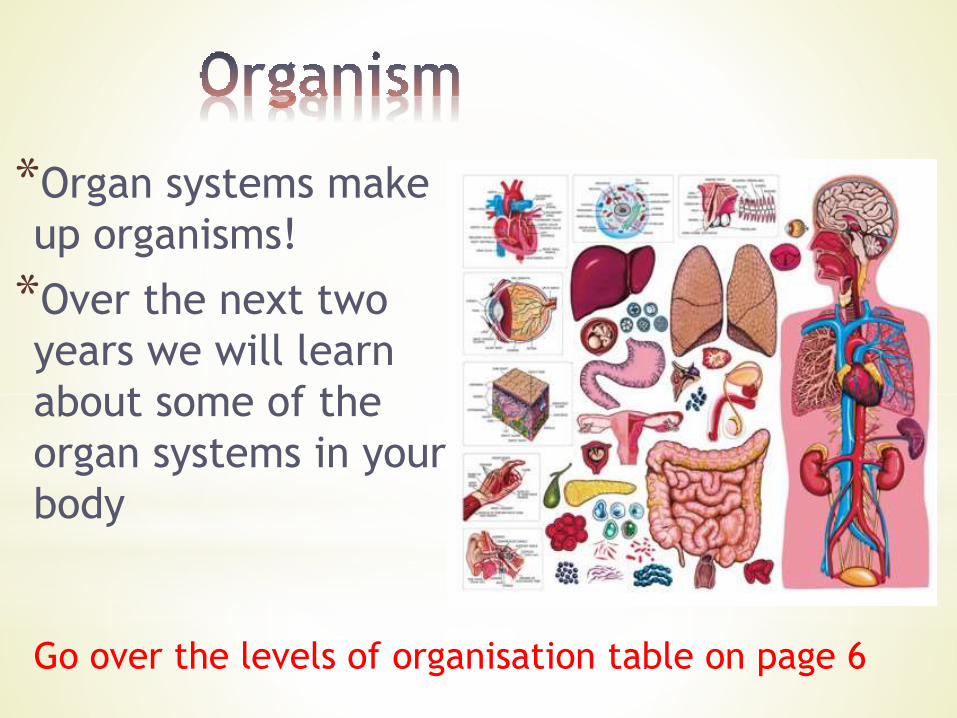
*Organ systems make
up organisms!
*Over the next two
years we will learn
about some of the
organ systems in your
body
Go over the levels of organisation table on page 6

**You need to know some examples of specialised cells in
plants and in animals
*Specifically we will look at Ciliated epithelial cells, muscle
cells and red blood cells in animals and Root Hair cells
and Xylem vessels in pants
*We come back to these cells now and then through the
syllabus, so look out for them
*We will also see other specialised cells as we do other
topics; again make a note of them
*Lets look at the examples…

**The passages into the lungs have a clever system to
keep dust, bacteria and other small particles from
reaching the lungs
*These passages are lined with cells that make mucus,
called Goblet cells, and with ciliated cells
*Small particles are trapped in the mucus

*Cilia are small cytoplasmic
extensions at one end of
the cell
*Protein fibres move these
cilia like a wave
*This movement carries the
mucus out of the airways
to the back of the throat
*it is swallowed here – and
the acid in the stomach
should kill any pathogens
(disease causing
organisms)
Cytoplasm
Cilia move
in waves
forming a
current
Mucus is
moved along
nucleuscilia
Cell surface membrane
Use this information to label the drawing in your notes
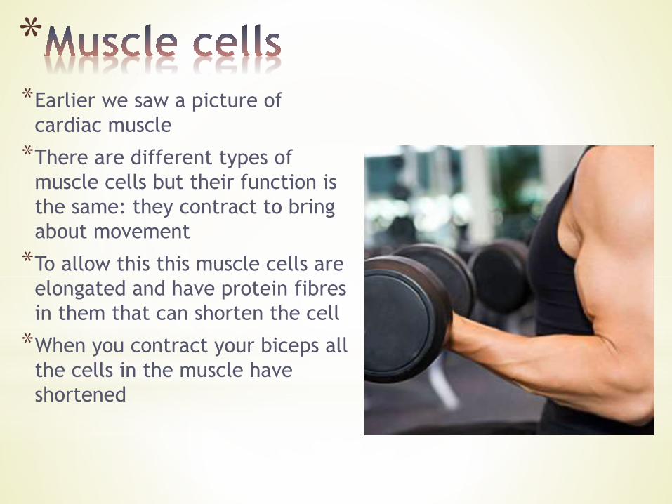
**Earlier we saw a picture of
cardiac muscle
*There are different types of
muscle cells but their function is
the same: they contract to bring
about movement
*To allow this this muscle cells are
elongated and have protein fibres
in them that can shorten the cell
*When you contract your biceps all
the cells in the muscle have
shortened
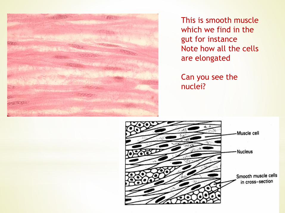
This is smooth muscle
which we find in the
gut for instance
Note how all the cells
are elongated
Can you see the
nuclei?

This is Skeletal muscle. It attaches to bone, so your biceps
muscle looks like this.
The stripes are because of overlapping regions of proteins.
Note how the nuclei are squashed to the edge of the cell
because of the protein fibres in the cytoplasm
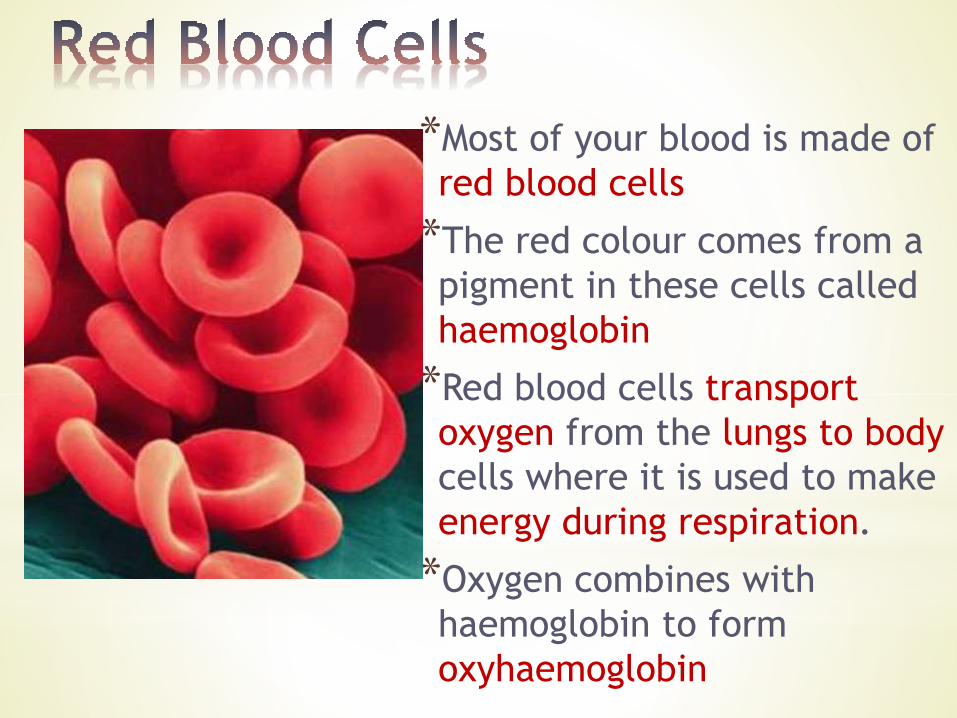
*Most of your blood is made of
red blood cells
*The red colour comes from a
pigment in these cells called
haemoglobin
*Red blood cells transport
oxygen from the lungs to body
cells where it is used to make
energy during respiration.
*Oxygen combines with
haemoglobin to form
oxyhaemoglobin

*Note the shape of the red blood cells: they are
dented on both sides
*This is described as biconcave – “bi” means twice and
“concave” means dented
*This shape gives the cells a larger surface area so
they can absorb more oxygen
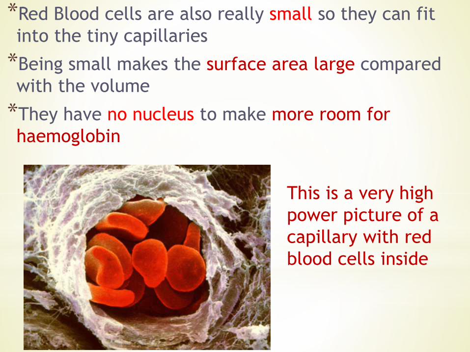
*Red Blood cells are also really small so they can fit
into the tiny capillaries
*Being small makes the surface area large compared
with the volume
*They have no nucleus to make more room for
haemoglobin
This is a very high
power picture of a
capillary with red
blood cells inside

When red blood
cells are looked at
under a light
microscope they
look as if the centre
is empty because it
is so thin the light
comes straight
through
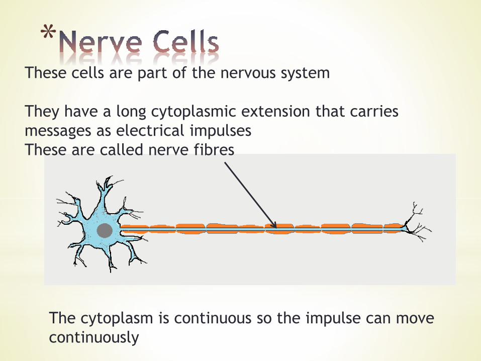
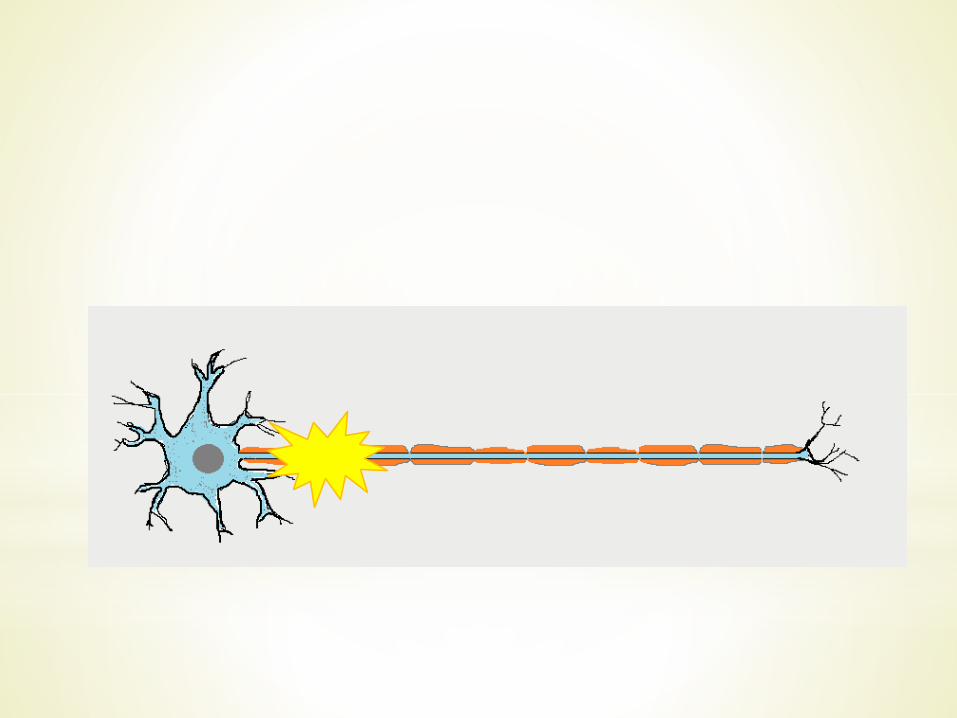
*These cells are part of the nervous system
They have a long cytoplasmic extension that carries
messages as electrical impulses
These are called nerve fibres
The cytoplasm is continuous so the impulse can move
continuously
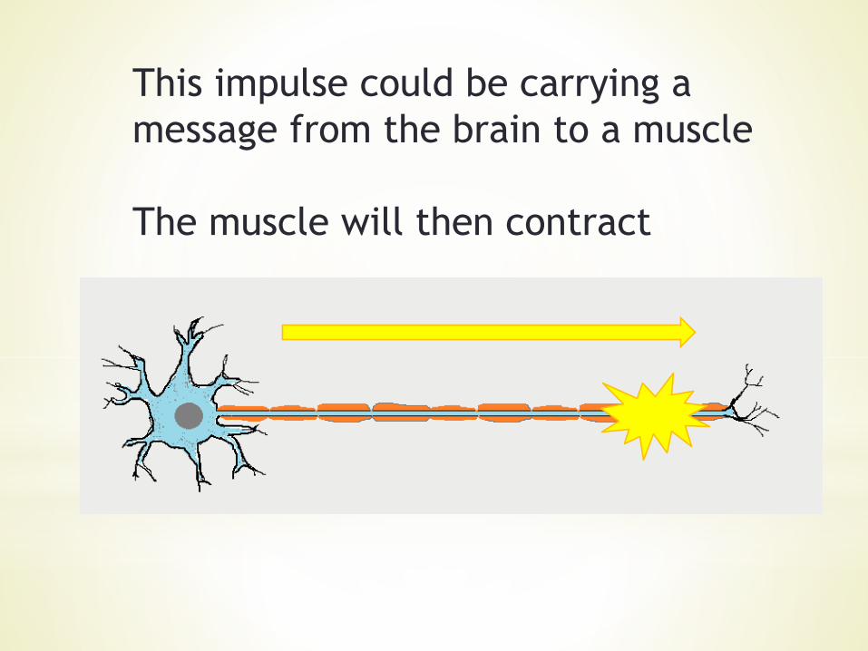
This impulse could be carrying a
message from the brain to a muscle
The muscle will then contract
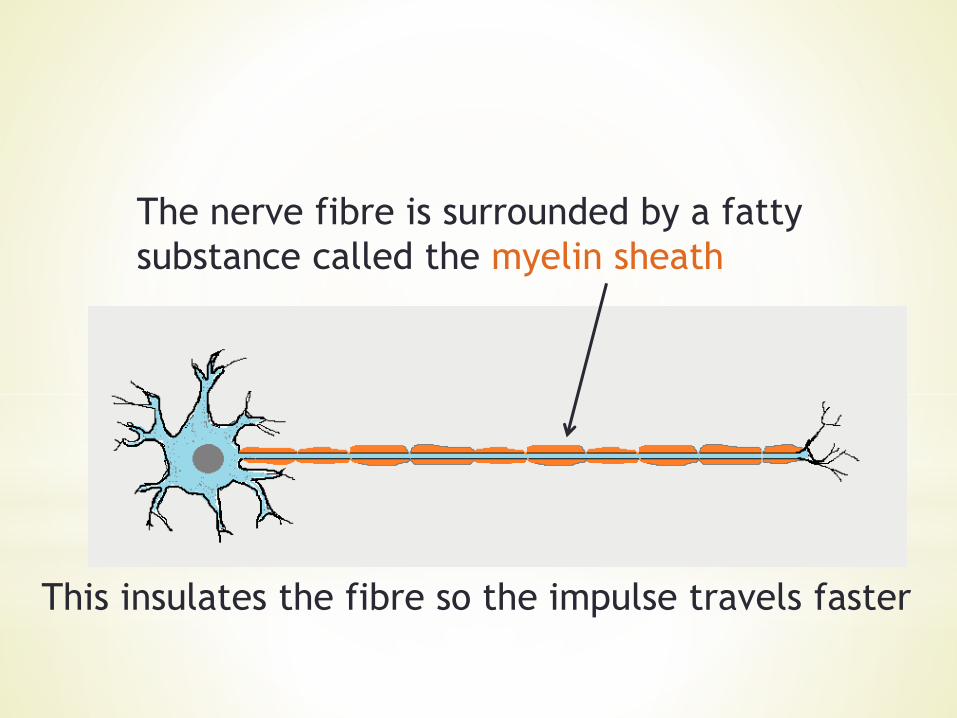
The nerve fibre is surrounded by a fatty
substance called the myelin sheath
This insulates the fibre so the impulse travels faster
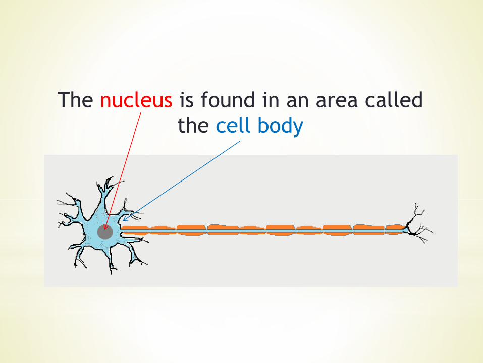
The nucleus is found in an area called
the cell body
*
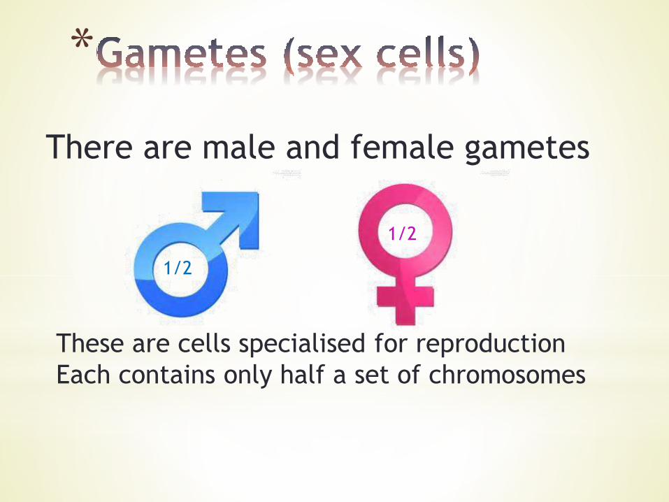
There are male and female gametes
These are cells specialised for reproduction
Each contains only half a set of chromosomes
1/2
1/2
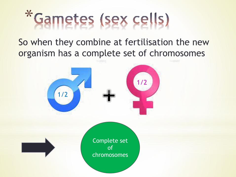
*So when they combine at fertilisation the new
organism has a complete set of chromosomes
1/2
1/2
Complete set
of
chromosomes

Sperm cells These are the male sex cells
The nucleus has half a set of
chromosomes
It is in the head

Sperm cells
The male sex cell swims to the
female sex cell
So it has a tail
And in this region
there are many
mitochondria to
make energy

Egg cells These are the female sex cells
The nucleus contains half
a set of chromosomes

Egg cells
There is a lot of
cytoplasm to store
nutrients and
provide organelles
to the new cell
after fertilisation
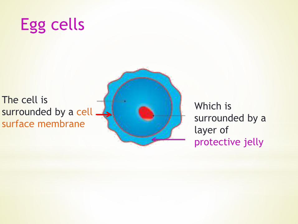
Egg cells
The cell is
surrounded by a cell
surface membrane
Which is
surrounded by a
layer of
protective jelly
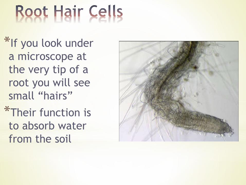
*If you look under
a microscope at
the very tip of a
root you will see
small “hairs”
*Their function is
to absorb water
from the soil

*Root hairs are
cytoplasmic extensions
from cells lining the
outside of the root; they
are called epidermal cells
*The extensions increase
the surface area for water
absorption
Label the diagram in your notes and use arrows to show where
water enters the root hair cell
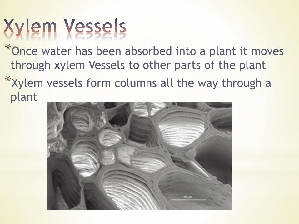
*Once water has been absorbed into a plant it moves
through xylem Vessels to other parts of the plant
*Xylem vessels form columns all the way through a
plant
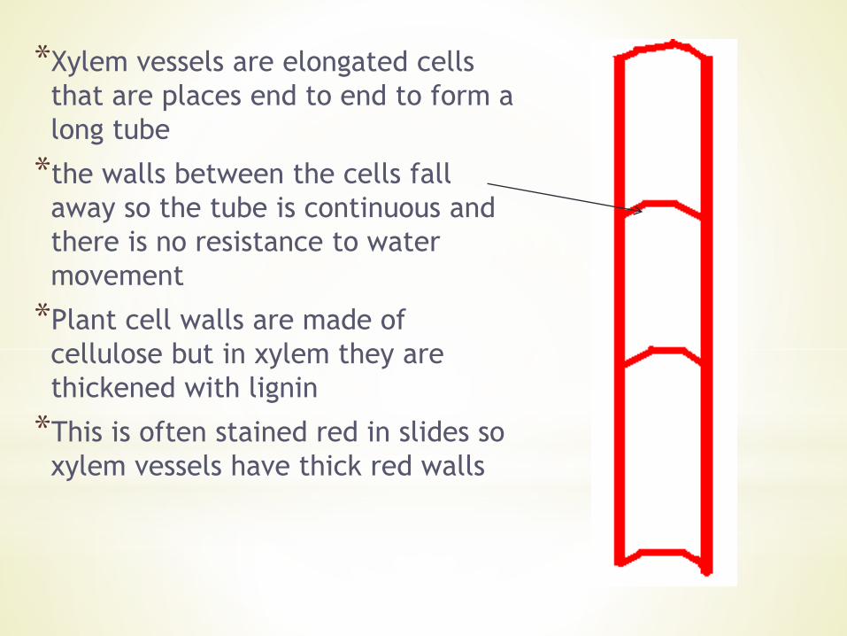
*Xylem vessels are elongated cells
that are places end to end to form a
long tube
*the walls between the cells fall
away so the tube is continuous and
there is no resistance to water
movement
*Plant cell walls are made of
cellulose but in xylem they are
thickened with lignin
*This is often stained red in slides so
xylem vessels have thick red walls
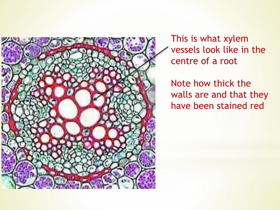
This is what xylem
vessels look like in the
centre of a root
Note how thick the
walls are and that they
have been stained red
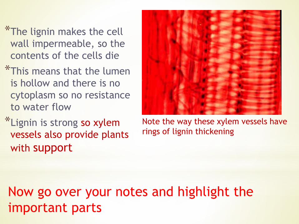
*The lignin makes the cell
wall impermeable, so the
contents of the cells die
*This means that the lumen
is hollow and there is no
cytoplasm so no resistance
to water flow
*Lignin is strong so xylem
vessels also provide plants
with support
Note the way these xylem vessels have
rings of lignin thickening
Now go over your notes and highlight the
important parts