Special thanks to Shire Genzyme The full article and its … · 2017-07-18 · Drugs Antineoplastic...
24
1 Special thanks to Shire Genzyme The full article and its related educational material were produced by and under the sole responsibility of the Working Group on Myocardial and Pericardial Diseases.
Transcript of Special thanks to Shire Genzyme The full article and its … · 2017-07-18 · Drugs Antineoplastic...

1
Special thanks to
ShireGenzyme
The full article and its related educational materialwere produced by and under the sole responsibility
of the Working Group on Myocardial and Pericardial Diseases.

2
ESC WORKING GROUP ON MYOCARDIAL AND PERICARDIAL DISEASES: KEY MESSAGES on Dilated Cardiomyopathy(original title: Proposal for a Revised Definition of Dilated Cardiomyopathy, Hypokinetic Non-Dilated Cardiomyopathy, and its Implications for Clinical Practice: a position statement of the European Society of Cardiology Working Group on Myocardial and Pericardial Diseases. Eur Heart J. 2016 Jan 19. pii: ehv727. [Epub ahead of print]
Table of contentsI. Introduction and scope of the document ...........................................................................................................................................................................................................................................................................................................................................Page 32. Basic definition and Causes ...................................................................................................................................................................................................................................................................................................................................................................................................................Page 33. Refined definitions and diagnostic criteria ........................................................................................................................................................................................................................................................................................................................................Page 6 3.1 Dilated Cardiomyopath..............................................................................................................................................................................................................................................................................................................................................................................................................Page 8 3.2 Non dilated hypokinetic cardiomyopathy ..................................................................................................................................................................................................................................................................................................................Page 8 3.3 Diagnosis in relatives .............................................................................................................................................................................................................................................................................................................................................................................................................................Page 84. Diagnostic work-up ...........................................................................................................................................................................................................................................................................................................................................................................................................................................................Page 11 4.1 General process ....................................................................................................................................................................................................................................................................................................................................................................................................................................................Page 11 4.2 Diagnostic work-up according to age ........................................................................................................................................................................................................................................................................................................................................Page 14 4.3 Diagnostic work-up according to red flags .............................................................................................................................................................................................................................................................................................................Page 165. Etiology directed management and treatment ................................................................................................................................................................................................................................................................................................................Page 18 5.1 Familial Cardiomyopathy ........................................................................................................................................................................................................................................................................................................................................................................................................Page 19 5.2 Inflammatory Cardiomyopathy........................................................................................................................................................................................................................................................................................................................................................................Page 206. Pregnancy counselling................................................................................................................................................................................................................................................................................................................................................................................................................................................Page 207. Summary.................................................................................................................................................................................................................................................................................................................................................................................................................................................................................................................Page 22

3
I. Introduction and scope of the document Research over recent decades has shed new light on the aetiology and natural history of dilated cardiomyopathy (DCM). In particular, it is recognized that many patients have a long preclinical phase characterized by few if any symptoms and minor cardiac abnormalities that fall outside current disease definitions.
It is also clear that distinct subtypes in fact share a common DCM phenotype. The aim of this position paper is to update the definition of DCM to take into account its diverse aetiology and clinical manifestations in patients and relatives.
We do not describe the general management of left ventricular systolic dysfunction as this is covered in existing European Society of Cardiology (ESC) heart failure guidelines but do consider the implications of an aetiology oriented approach to therapy.
2. Basic definition and CausesDCM is currently defined by the presence of left ventricular (LV) or biventricular dilatation and systolic dysfunction in the absence of abnormal loading conditions (hypertension, valve disease) or coronary artery disease sufficient to cause global systolic impairment.
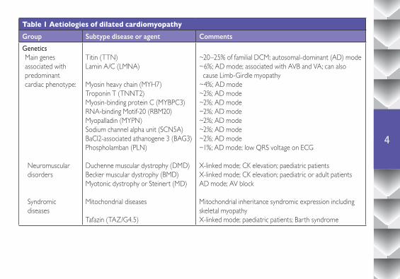
The causes of DCM can be classified as genetic or non-genetic (Table 1), but there are circumstances in which genetic predisposition interacts with extrinsic or environmental factors.
4
Table 1 Aetiologies of dilated cardiomyopathyGroup Subtype disease or agent Comments
GeneticsMain genes associated with predominant cardiac phenotype:
Neuromuscular disorders
Syndromic diseases
Titin (TTN)Lamin A/C (LMNA)
Myosin heavy chain (MYH7) Troponin T (TNNT2)Myosin-binding protein C (MYBPC3) RNA-binding Motif-20 (RBM20) Myopalladin (MYPN)Sodium channel alpha unit (SCN5A) BaCl2-associated athanogene 3 (BAG3) Phospholamban (PLN)
Duchenne muscular dystrophy (DMD) Becker muscular dystrophy (BMD) Myotonic dystrophy or Steinert (MD)
Mitochondrial diseases
Tafazin (TAZ/G4.5)
~20–25% of familial DCM; autosomal-dominant (AD) mode~6%; AD mode; associated with AVB and VA; can also cause Limb-Girdle myopathy~4%; AD mode~2%; AD mode~2%; AD mode~2%; AD mode~2%; AD mode~2%; AD mode~2%; AD mode~1%; AD mode; low QRS voltage on ECG
X-linked mode; CK elevation; paediatric patientsX-linked mode; CK elevation; paediatric or adult patients AD mode; AV block
Mitochondrial inheritance syndromic expression includingskeletal myopathyX-linked mode; paediatric patients; Barth syndrome

5
Table 1 Aetiologies of dilated cardiomyopathy (continued)Group Subtype disease or agent Comments
Drugs Antineoplastic drugs
Psychiatric drugs
Other drugs
Anthracyclines; antimetabolites; alkylating agents; Taxol; hypomethylating agent; monoclonal antibodies; tyrosine kinase inhibitors; immunomodulating agentsClozapine, olanzapine; chlorpromazine, risperidone, lithium;methylphenidate; tricyclic antidepressants;Chloroquine; all-trans retinoic acid; antiretroviral agents; phenothiazines
Toxic and overload Ethanol
Cocaine, amphetamines, ecstasy Other toxicIron overload
Risk proportional to entity and duration of alcohol intake.Frequent good response after withdrawalChronic usersArsenic; cobalt; anabolic/androgenic steroidsTransfusions; haemochromatosis
Nutritional deficiency
Selenium deficiencyThiamine deficiency (Beri-Beri)
Zinc and copper deficiency Carnitine deficiency
Rare, high frequency in some regions in China (Keshan disease) Favoured by malnutrition, alcohol abuse. High-output dilated cardiac failurePossible contributors to DCMPaediatric patients
Electrolyte disturbance
Hypocalcemia, hypophosphatemia
Endocrinology Hypo- and hyper-thyroidism Cushing/addison disease Phaeocromocytoma, AcromegalyDiabetes mellitus

6
3. Refined definitions and diagnostic criteriaExisting definition of DCM has a number of important limitations. Most notable is the fact that the term encompasses a broad range of genetic and acquired disorders that manifest as a spectrum of electrical and functional abnormalities that change with time. This applies particularly to genetic diseases that have delayed or incomplete cardiac expression, with the result that many mutation carriers have intermediate phenotypes that do not meet standard disease definitions. Similarly, systolic LV dysfunction or dilatation in acquired diseases such as myocarditis can be very mild
Table 1 Aetiologies of dilated cardiomyopathy (continued)Group Subtype disease or agent Comments
Infection
Auto-immune diseases
Organ specific
Not organ specific
Viral (including HIV), bacterial (including Lyme disease), mycobacterial, fungal, parasitic (Chagas disease)
Giant-cell myocarditis (GCM)
Inflammatory DCM
Polymyositis/dermatomyositis; Churg–Strauss syndrome; Wegener’s granulomatosis; systemic lupus erythematosus, sarcoidosis
DCM caused by infectious myocarditis. Atrio-ventricular block (AVB) in Lyme disease. Chagas’ disease: DCM develops after a long latent infection.
Multinucleated giant cell; frequent AV block and ventriculararrhythmiaDCM caused by biopsy-proven, non-infectious myocarditis
In cardiac sarcoidosis there is granulomatous myocarditis; AV block is frequent DCM is possible but uncommon in these diseases
Peripartum Risk factors: multiparity, African descent, familial DCM, autoimmunity
7
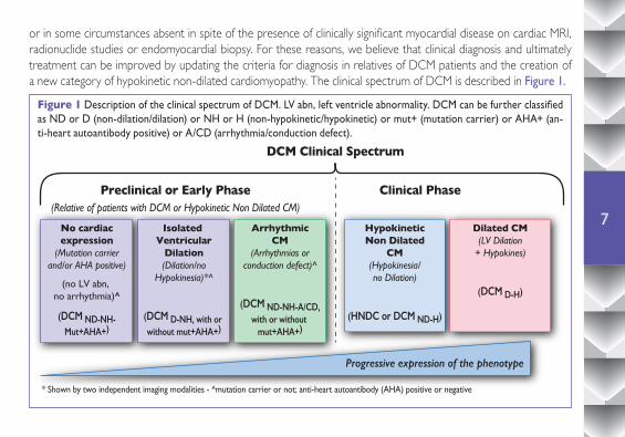
or in some circumstances absent in spite of the presence of clinically significant myocardial disease on cardiac MRI, radionuclide studies or endomyocardial biopsy. For these reasons, we believe that clinical diagnosis and ultimately treatment can be improved by updating the criteria for diagnosis in relatives of DCM patients and the creation of a new category of hypokinetic non-dilated cardiomyopathy. The clinical spectrum of DCM is described in Figure 1.
Figure 1 Description of the clinical spectrum of DCM. LV abn, left ventricle abnormality. DCM can be further classified as ND or D (non-dilation/dilation) or NH or H (non-hypokinetic/hypokinetic) or mut+ (mutation carrier) or AHA+ (an-ti-heart autoantibody positive) or A/CD (arrhythmia/conduction defect).
DCM Clinical Spectrum
Preclinical or Early Phase(Relative of patients with DCM or Hypokinetic Non Dilated CM)
* Shown by two independent imaging modalities - ^mutation carrier or not; anti-heart autoantibody (AHA) positive or negative
Clinical Phase
No cardiacexpression
(Mutation carrierand/or AHA positive)
(no LV abn, no arrhythmia)^
(DCM ND-NH-Mut+AHA+)
Progressive expression of the phenotype
Isolated Ventricular
Dilation(Dilation/no
Hypokinesia)*^
(DCM D-NH, with or without mut+AHA+)
ArrhythmicCM
(Arrhythmias orconduction defect)^
(DCM ND-NH-A/CD,with or without
mut+AHA+)
HypokineticNon Dilated
CM(Hypokinesia/no Dilation)
(HNDC or DCM ND-H)
Dilated CM(LV Dilation
+ Hypokines)
(DCM D-H)

8
3.1 Dilated Cardiomyopathy (DCM)Definition: Left ventricular or biventricular systolic dysfunction and dilatation that are not explained by abnormal loading conditions or coronary artery disease.Notes:Systolic dysfunction is defined by abnormal LV Ejection fraction , measured using any modality and shown either by two independent imaging modalities or on two distinct occasions by the same technique, preferably echocardiograohy or CMR. LV dilatation is defined by LV end diastolic (ED) volumes or diameters >2SD from normal according to normograms (Z scores >2 standard deviations) corrected by body surface area (BSA) and age, or BSA and gender. Normograms for echocardiographic volumes and diameters are available for adults and children and can be calculated using web-based calculators (www.parameterz.com) and by an App (ParameterZan for iPhone/ipad platform).
3.2 Hypokinetic non-dilated cardiomyopathy (HNDC)Definition: Left ventricular or biventricular global systolic dysfunction without dilatation (defined as LVEF <45%), not explained by abnormal loading conditions or coronary artery disease.Note: Strictly decreased LVEF is mandatory in index patient with HNDC since no combination with dilatation is mandatory for the diagnosis.
3.3 Diagnostic criteria in relativesAs the relatives of patients with DCM or with hypokinetic non-dilated cardiomyopathy (HNDC) can develop overt disease, they should be considered for clinical and genetic screening. However, clinical testing in relatives often reveals mild non-diagnostic abnormalities that overlap with normal variation or mimic changes seen in other more common diseases such as hypertension and obesity. In this statement, we propose three new diagnostic categories for relatives of cases with either DCM or HNDC who undergo screening, which takes into account whether a definite causative mutation has been identified as well as the presence of clinical features that are associated with the development of

9
overt DCM (major criteria) or are suggestive of incomplete disease expression (minor criteria). We acknowledge that evidence to support the use of minor criteria in this context is based on small studies or DCM caused by specific mutations.
Box 1 Diagnostic criteria for relatives
MAJOR1. Unexplained decrease of LVEF ≤50% but >45% OR2. Unexplained LVED dilatation (diameter or volume) according to nomograms (LVED diameter/volume >2SD + 5% since this more specific echocardiographic criterion was used in studies that demonstrated the predictive impact of isolated dilatation in relatives)a
MINOR1. Complete LBBB, or AV block (PR >200 ms or higher degree AV block)2. Unexplained ventricular arrhythmia (W100 ventricular premature beats per hour in 24 h or non-sustained ventricular tachycardia, ≥3 beats at a rate of ≥120 beats per minute).3. Segmental wall motion abnormalities in the left ventricle in the absence of intraventricular conduction defect4. Late enhancement (LGE) of non-ischaemic origin on cardiac magnetic resonance imaging.5. Evidence of non-ischaemic myocardial abnormalities (inflammation, necrosis and/or fibrosis) on EMB.6. Presence of serum organ-specific and disease-specific AHA by one or more autoantibody tests.
aFeature shown either by two independent imaging modalities or on two distinct occasions by the same technique.

10
Recommendation 1: Definition of disease in a relative
• DefiniteDiseasewhen: Meets criteria for DCM or hypokinetic non-dilated cardiomyopathy (HNDC)
• Probable Disease when: (i) One major criterion (from Box 1) plus at least one minor criterion (from Box 1) OR (ii) One major criterion (from Box 1) plus carrying the causative mutation identified in the proband
• Possible disease when: (i) Two minor criteria (from Box 1) OR (ii) One minor criterion (from Box 1) plus carrying the causative mutation identified in the proband OR (iii) One major criterion (from Box 1) but without any minor criterion and without genetic data within the family
Recommendation 2: Definition of familial disease (in the absence of conclusive molecular genetic information in a family):1. When two or more individuals (1st or 2nd degree relatives) have DCM or HNDC fulfilling diagnostic criteria for “definite” disease OR 2. In the presence of an index patient fulfilling diagnostic criteria for DCM/HNDC and a first-degree relative with autopsy-proven DCM and sudden death [1] at <50 years of age.
11
4. Diagnostic work-up
4.1 General processConsidering the broad spectrum of disorders that cause DCM, a systematic approach can be helpful (Figure 2) in identifying and managing uncommon but clinically important forms of DCM. In brief, the systematic search for diagnostic clues or “red flags” can suggest particular disorders and guide rational selection of additional diagnostic tests. Clinical workup starts with personal and family history, physical examination, and a focused analysis of ECG and echocardiography (figure2).
Some of the most important diagnostic clues are described in table 2. Identification of clinical features suggestive of specific diseases should then lead to a second level diagnostic work-up that may include biochemical analyses, MRI, endomyocardial biopsy and genetic testing. The role of genetic testing in cardiomyopathies has been the subject of a previous Position Statement of this Working Group.
Once a mutation is identified, and its pathogenic role is established, then this may have multiple impacts since the information is able to confirm the genetic origin and mode of inheritance, may be used for guidance of therapy and can be used for family cascade screening and early diagnosis.

12
Basic evaluation
Personal historyFamily hsitory
Physcalexamination
ECGImaging
AHA
Featuressuggestinga specificaetiology?
No familial DCMNo mutationidentified
Causalmutationidentified
** Restricted = known DCM genes
Yes
Yes
no
DefiniteFamilial/Genetic
DCMDCM diagnosis/Clinical spectrum
Search forPrecipitating factors
Systematiccardiac family
screening
Usualmanagement
of DCM
Etiologydirected
management
Genetic testing(restricted panel**)
Control largepanel of genes
(if appropriate family strcuture)
ImagingBiopsy
Genetic testing(restricted panel**)
Other
Definitespecific
aetiology
Diagnostic cluesSecond-level
evaluation
Figure 2 Diagnostic work-up for aetiology assessment.

13
Recommendation 3: diagnostic work-up1. Coronary artery disease should be excluded in patients more than 35 years of age, or before 35 years if there are significant personal coronary artery disease (CAD) risk factors or a family history of early CAD. 2. First line laboratory testing should include creatine kinase (CK), renal function, urine analysis for proteinuria, liver function tests, haemoglobin and white blood cell count, serum iron, ferritin, calcium, phosphate, natriuretic peptides and thyroid stimulating hormone.3. Second line diagnostics should be targeted to the suspected aetiology4. Cardiac magnetic resonance (CMR) may be useful for assessment of ventricular size and function and for tissue characterization 5. In patients with clinically suspected myocarditis, endomyocardial biopsy (EMB) (including histology, immunohistology and polymerase chain reaction (PCR) for infectious agents is recommended. EMB should also be considered when there is clinical suspicion of storage or metabolic diseases that cannot be confirmed by other means.6. Cardiac screening with echocardiography and ECG is recommended in all first degree-relatives of an index patient with DCM, irrespective of family history. 7. Genetic testing is recommended in the presence of a familial form of DCM OR in sporadic DCM with clinical clues suggestive of a particular/rare genetic disease (such as atrio-ventricular block or CK elevation). 8. Genetic testing should be oriented by clinical diagnostic clues when present, and should be restricted to genes known to cause DCM. The use of next generation sequencing (NGS) for the analysis of very large panels of genes, including titin, may be considered when the family structure permits segregation analysis (i.e. several patients with DCM and DNA available).

14
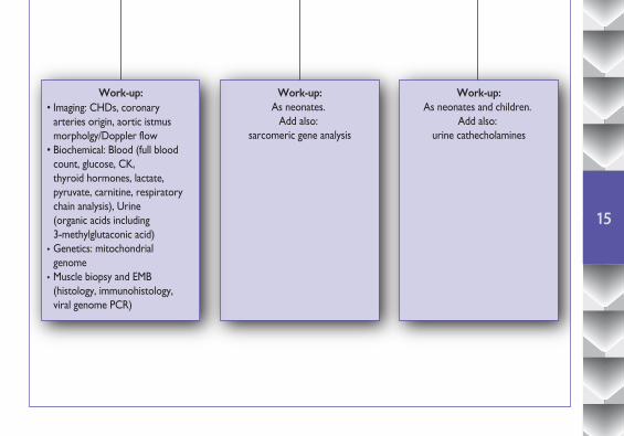
4.2 Diagnostic work-up according to age
Diagnostic work-up according to age when Dilated Cardiomyopathy (DCM) is suspected
ChildrenNeonates Adolescents/Adults
Aetiology:• Familial DCM• Myocarditis• Barth Syndrome• Mitochondrial CMP• Nemaline Myopathy• Carnitine Deficiency• Toxic (Adriamycin)
Aetiology:• Myocarditis• Barth Syndrome• Mitochondrial CMP• Nemaline Myopathy• Carnitine Deficiency
Aetiology:• Familial DCM• Myocarditis• Muscular Dystrophies• Mitochondrial CMP• Toxic (Adriamycin/Alchool/Drugs)• Pheochromocytoma• Eosinophilic CMP
Exclude:• Kawasaki• Congenital coronary anomaly (ALCAPA)• Supraventricular tachycardia (SVT)
Exclude:• Kawasaki• Congenital coronary anomaly (ALCAPA)• CHDs (i.e. VSD, PDA, A-V fistula)• Supraventricular tachycardia (SVT)
Exclude:• Other cardiomyopathies (LVNC, ARVC, end stage HCM)• Supraventricular tachycardia (SVT)
Work-up:As neonates.
Add also: sarcomeric gene analysis
Work-up:• Imaging: CHDs, coronary arteries origin, aortic istmus morpholgy/Doppler flow• Biochemical: Blood (full blood count, glucose, CK, thyroid hormones, lactate, pyruvate, carnitine, respiratory chain analysis), Urine (organic acids including 3-methylglutaconic acid)• Genetics: mitochondrial genome• Muscle biopsy and EMB (histology, immunohistology, viral genome PCR)
Work-up:As neonates and children.
Add also: urine cathecholamines
15
Diagnostic work-up according to age when Dilated Cardiomyopathy (DCM) is suspected
ChildrenNeonates Adolescents/Adults
Aetiology:• Familial DCM• Myocarditis• Barth Syndrome• Mitochondrial CMP• Nemaline Myopathy• Carnitine Deficiency• Toxic (Adriamycin)
Aetiology:• Myocarditis• Barth Syndrome• Mitochondrial CMP• Nemaline Myopathy• Carnitine Deficiency
Aetiology:• Familial DCM• Myocarditis• Muscular Dystrophies• Mitochondrial CMP• Toxic (Adriamycin/Alchool/Drugs)• Pheochromocytoma• Eosinophilic CMP
Exclude:• Kawasaki• Congenital coronary anomaly (ALCAPA)• Supraventricular tachycardia (SVT)
Exclude:• Kawasaki• Congenital coronary anomaly (ALCAPA)• CHDs (i.e. VSD, PDA, A-V fistula)• Supraventricular tachycardia (SVT)
Exclude:• Other cardiomyopathies (LVNC, ARVC, end stage HCM)• Supraventricular tachycardia (SVT)
Work-up:As neonates.
Add also: sarcomeric gene analysis
Work-up:• Imaging: CHDs, coronary arteries origin, aortic istmus morpholgy/Doppler flow• Biochemical: Blood (full blood count, glucose, CK, thyroid hormones, lactate, pyruvate, carnitine, respiratory chain analysis), Urine (organic acids including 3-methylglutaconic acid)• Genetics: mitochondrial genome• Muscle biopsy and EMB (histology, immunohistology, viral genome PCR)
Work-up:As neonates and children.
Add also: urine cathecholamines

16
4.3Diagnosticwork-upaccordingtoredflagsThe most important clues for an appropriate diagnosis of the underlying etiology are described below.
Table 2A: Examples of signs and symptoms that raise the suspicion of specific aetiologies
Finding DCM
Intellectual disability Dystrophinopathies • Mitochondrial diseases • MyotonicdystrophyFKTN mutations
Sensorineuraldeafness Epicardin mutation • Mitochondrial diseases
Visual impairment CRYAB (polar cataract) • Type 2 myotonic dystrophy (subcapsular cataract)
Gaitdisturbance Dystrophinopathies • Sarcoglycanopathies • Myofibrillar myopathies
Myotonia (involuntary muscle contraction with delayed relaxation)
Myotonic dystrophy (type 1 and Type 2)
Muscle weakness Dystrophinopathies • Sarcoglycanopathies • Laminopathies • MyotonicDystrophyDesminopathy
Palpebral ptosis Mitochondrial disease
Pigmentation of skin and scars Haemochromatosis
Palmoplantarkeratoderma and woolly hair Carvajal syndrome
17
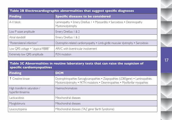
Table 2B Electrocardiographic abnormalities that suggest specific diagnoses
Finding Specific diseases to be considered
A-V block Laminopathy • Emery Dreifuss 1 • Myocarditis • Sarcoidosis • DesminopathyMyotonicdystrophy
Low P wave amplitude Emery Dreifuss 1 & 2
Atrial standstill Emery Dreifuss 1 & 2
“Posterolateral infarction” Dystrophin-related cardiomyopathy • Limb-girdle muscular dystrophy • Sarcoidosis
Low QRS voltage + “atypical RBBB” ARVC with biventricular involvement
Extremely low QRS amplitude PLN mutation
Table 2C Abnormalities in routine laboratory tests that can raise the suspicion of specific cardiomyopathies
Finding DCM↑ Creatine kinase Dystrophinopathies Sarcoglycanopathies • Zaspopathies (LDB3gene) • Laminopathies
Myotonicdystrophy • FKTN mutations • Desminopathies • Myofibrillar myopathies
High transferrin saturation / hyperferritinaemia
Haemochromatosis
Lacticacidosis Mitochondrial diseases
Myoglobinuria Mitochondrial diseases
Leucocytopenia Mitochondrial diseases (TAZ gene/ Barth Syndrome)
18
5. Etiology directed management and therapyThe identification of a specific underlying cause for DCM can have profound consequences for clinical management. For example, identification of a definite genetic cause should lead to genetic counselling and screening of relatives and in some specific circumstances prompt regular monitoring for complications such as conduction disease. It also
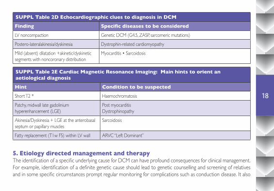
SUPPL Table 2D Echocardiographic clues to diagnosis in DCM
Finding Specific diseases to be considered
LV noncompaction Genetic DCM (G4.5, ZASP, sarcomeric mutations)
Postero-lateralakinesia/dyskinesia Dystrophin-related cardiomyopathy
Mild (absent) dilatation +akinetic/dyskinetic segments with noncoronary distribution
Myocarditis • Sarcoidosis
SUPPL Table 2E Cardiac Magnetic Resonance Imaging: Main hints to orient an aetiological diagnosis
Hint Condition to be suspected
Short T2 * Haemochromatosis
Patchy, midwall late gadolinium hyperenhancement (LGE)
Post myocarditisDystrophinopathy
Akinesia/Dyskinesia + LGE at the anterobasal septum or papillary muscles
Sarcoidosis
Fatty replacement (T1w FS) within LV wall ARVC “Left Dominant”
19
has significant consequences for advice on contraception and reproduction and in a number of examples, early intervention with ICDs, lifestyle modification and specific drug therapy may also be necessary. General advice on the management of heart failure can be found in current ESC guidelines for chronic heart failure. In DCM caused by LMNA mutations, risk assessment also involves gender-specific risk as described elsewhere. Relatives with minor cardiac abnormalities such as LV enlargement are at increased risk of DCM development and may benefit from early medical treatment (although this has not yet demonstrated by placebo controlled trials). 5.1 Familial DCM and follow up of relatives
Recommendation 4:• In the context of familial DCM, cardiac screening with Echo and ECG (± Holter* monitoring depending on main phenotype in proband)should be performed in all first degree-relatives (from childhood) and should be repeated every 2-3 years if cardiovascular tests are normal, every year if minor abnormalities are detected, whenever symptoms develop.
* Search in a relative for conduction defects ar arrhythmia which may be an early representation of DCM, especially in the context of a LMNA gene mutation.
Recommendation 5:• When a causative mutation has been identified in a DCM patient, then predictive genetic testing should be offered to first degree relatives in order to guide cardiac follow-up.
Recommendation 6: • When a definite causative LMNA mutation is identified, early indication for primary prevention by ICD implantation should be considered (guided by the risk factors as detailed elsewhere).

20
5.2InflammatoryDCM
Recommendation 7:• In familial and non-familial pedigrees with biopsy proven inflammatory DCM in the index case, cardiac-specific autoantibody (AHA) test at baseline and at follow-up should be considered in symptom-free relatives with or without cardiac abnormalities(e.g. ECG, echocardiography, CMR).• Non-invasive cardiac screening with echocardiography and ECG may be more frequent in relatives with cardiac autoantibodies.• Immunomodulatory and/or immunosuppressive therapy in biopsy-proven non-infectious inflammatory DCM should be considered. • Physical activity should be restricted in DCM with underlying biopsy-proven active phase of myocarditis.
6. Pregnancy counselingWe provide a summary of advice for the management of pregnancy in women with DCM in Tables 3 & 4.
Table 3 Pregnancy counseling, risk assessment and managementPregnancy risk and management*
Counselling Girls and women of fertile age should receive counseling concerning contraceptives, pregnancy risk, and the risk of genetic transmission. (Regitz 2011).
Risk assessment • Increased risk of heart failure, arrhythmias, thrombo-embolic events (39%) (Grewal 2010)• Predictors of increased risk: NYHA class III/IV, LVEF < 45%• Pregnancy contra-indicated: NYHA III/IV, LVEF < 30%• Risk of offspring events (low birth weight, premature delivery)

21
Table 3 Pregnancy counseling, risk assessment and management (continued)Pregnancy risk and management*
Delivery • Usually vaginal delivery with epidural anesthesia• Caesarean section: for obstetric indication and unstable heart failure• Observation period postpartum: 48-72 hours because of increased risk of heart failure peripartum
Table 4 Contraceptives and medication during pregnancy in women with DCMMedication during pregnancyACE inhibitors and angiotensin receptor blockers
Contraindicated during whole pregnancy because of fetotoxicity. Skull and lung hypoplasia, renal failure, anuria, fetal death, limb and craniofacial deformations. Breastfeeding: captopril and enalapril: low dose in breast milk but breast feeding not advised. Others do not use.
Anticoagulants Vitamin K antagonists
Low molecular weight heparin
Dabigatran, rivaroxaban
Embryopathy when used in first trimester, dose dependant. Vaginal delivery contra-indicated because of risk of intracranial bleeding.
Preferred in first trimester and last month of pregnancy. Dose adjustment according to weight and anti factor X-a levels
Do not use during pregnancy and breast-feedingBeta-blockers • Low birthweight, neonatal bradycardia and hypoglycaemia.
• Preferred beta-blockers during pregnancy and breast feeding are metoprolol and labetalol. Do not use atenolol.
Diuretics Risk of oligohydramnion and electrolyte disbalance in fetus. No fetotoxicity. Breast feeding: hydrochlorothizide preferred over furosemide, do not use bumetanide.

22
7. SummaryIn this paper the Working Group on Myocardial and Pericardial Diseases proposes a revised definition of DCM in an attempt to bridge the gap between our recent understanding of the disease spectrum and its clinical presentation in relatives, which is key for early diagnosis and the institution of potential preventative measures. We also provide practical hints to identify subsets of the DCM syndrome where aetiology-directed management has great clinical relevance.
Table 4 Contraceptives and medication during pregnancy in women with DCM (continued)
Medication during pregnancy
Aldosterone antagonists Antiandrogenic effects and endocrine dysfunction. Only use when no other alternative. Do not use when breastfeeding.
Anti-arrhythmic drugs • Amiodarone: Fetotoxic. Do only use with severe arrhythmias when no other alternative is available. High dose in breast milk, do not use• Flecainide: Limited data, may be used with caution.• Procainamide: Limited data, use only when necessary.• Sotalol: Possibly bradycardia and hypoglycaemia in neonate. No harm in animal studies. May be used with caution.
Other drugs
Digoxin Ivabradin
• Probably not harmful during pregnancy and breastfeeding• Contra-indicated during pregnancy

23
We have below summarized in figure3 the diagnostic tree that emerges from the novel classification of DCM.
Summary of diagnostic criteria for DCM
INDEX CASE
- DCM (Dilated LV & reduced EF)- HNDC (EF <45% & no dilation)
- Familial- Non familial
Yes
Yes
No
Yes
No
No
No
Yes
}RELATIVE
≥2 minor without mutation carrier?OR 1 minor criterion + mutation carrier?
+ ≥1 minor criterion?OR mutation carrier?
Criteria as for index cases?Definitive disease(DCM/HNDC)
Probable disease
Possible disease
Possible disease
No disease
Major criteria for relatives?(dilated LV OR LV EF 45–50%)
Figure 3 Overview of dilated cardiomyopathy criteria in probands and relatives.

24
1. Depts of Cardiology and Experimental Cardiology, Academic Medical Hospital (AMC) at the University of Amsterdam, Amsterdam The Netherlands2. Inherited Cardiac Diseases Unit, The Heart Hospital, University College London, London, UK.3. Center for Inherited Cardiovascular Diseases, IRCCS Foundation Policlinico San Matteo, Pavia, Italy4. Department of Cardiology, Sheba Medical Center, Tel Hashomer, Israel5. First Cardiology Department, University of Athens, Medical School, Athens, Greece6. Klinik fürInnereMedizin III, Universitätsklinikum des Saarlandes, Homburg, Germany7. Assistance Publique Hôpitaux de Paris (AP HP) ,Hôpital Cochin, Université Paris Descartes, Paris, France .8. Department of Cardiology, University Hospital Virgen de Arrixaca, Murcia, Spain9. Service de cardiologie, Pôlecardio-vasculaire et Pulmonaire, CHRU de Lille, Lille, France - Inserm U1167, InstitutPasteur de Lille, Université de Lille 2, Lille, France10. Cardiology Department, Maria Vittoria Hospital and University of Torino, Torino, Italia11. Cardiovascular Research Institute Maastricht, Department of Cardiology, Maastricht University Medical Center, Netherlands AND: ICIN, Netherlands Heart Institute, Utrecht, Netherlands.
12. Department of Molecular Pathology, Institute for Pathology, University Hospital Tübingen, Germany13. INSERM UMRS-956, UPMC Univ Paris 6, AP-HP, Hôpital Pitié-Salpêtrière, Paris, France.14. Division of Cardiology, Monaldi Hospital, Second University of Naples, Naples, Italy15. Second Department of Medicine, Department of Cardiovascular Medicine, General University Hospital and the First Faculty of Medicine, Charles University, Prague, Czech Republic16. Department of Cardiology, Odense University Hospital, Odense, Denmark17. Division of Cardiovascular Imaging and Biostatistics, The Heart Hospital, London, England18. Department of Cardiology, University Medical Center Groningen, University of Groningen, Groningen, the Netherlands19. Department of Cardiology, University Medical Center, Belgrade, Serbia20. Department of Cardiology, Freeman Hospital, Newcastle upon Tyne, UK21. Cardiac Imaging Unit, Ramón y Cajal University Hospital, Madrid, Spain.22. Division of Cardiology, Department of Cardiological Thoracic and Vascular Sciences, University of Padova, Padova, Italy.23. Université de Versailles-Saint Quentin, Hôpital Ambroise Paré, AP-HP, Boulogne-Billancourt, France24. AP-HP, Hôpital Pitié-Salpêtrière, Paris, France
Authors and Task Force membersYigal M Pinto1, Perry M Elliott2, Eloisa Arbustini3^, Yehuda Adler 4, Aris Anastasakis5, Michael Bohm6^, Denis Duboc7, JuanGimeno8, Pascal de Groote9, Massimo Imazio10, Stephane Heymans11§, Karin Klingel12, Michel Komajda13^, Giuseppe Limongelli14, Ales Linhart15, Jens Mogensen16, James Moon17, Petronella G Pieper18, Petar M Seferovic19^§, Stephan Schueler20, JoseL Zamorano21̂ , Alida LP Caforio22, Philippe Charron23, 24.
Authoraffiliations:


![NIOSH List of Antineoplastic and Other Hazardous Drugs in Healthcare Settings… · 2018-12-19 · NIOSH [2016]. NIOSH list of antineoplastic and other hazardous drugs in healthcare](https://static.fdocuments.in/doc/165x107/5ec6554ac9bd9e3e7a09db62/niosh-list-of-antineoplastic-and-other-hazardous-drugs-in-healthcare-settings-2018-12-19.jpg)