special report: sleep blood gas ventilation sars asthma
60
Volume 4 Number 3 June-July 2009 The Journal of Pulmonary Technique SPECIAL REPORT: SLEEP BLOOD GAS VENTILATION SARS ASTHMA
Transcript of special report: sleep blood gas ventilation sars asthma

Volume 4 Number 3 June-July 2009
The Journal of Pulmonary Technique
SPECIAL REPORT: SLEEPBLOOD GASVENTILATIONSARSASTHMA

© 2
007
Card
inal
Hea
lth, I
nc. o
r one
of i
ts s
ubsi
diar
ies.
All
right
s re
serv
ed.
Portable Wireless Sleep Diagnostics
A New Generation of Sleep Diagnostics
• Comfortable and easy to use
• Wireless Technology
• Gold Standard Signals
• Designed for Pediatrics and Adults
• Developed by the pioneers in Portable Sleep Diagnostics
cardinalhealth.com
Cardinal Health GmbHLeibnizstraße 797204 HöchbergTel. +49 (0)931 4972-0Fax. +49 (0)931 4972-423
© 2009 Cardinal Health, Inc. All rights reserved.
T3 NOXternal
T3 NOXternal - Compact and wireless

Nellcor™
Puritan Bennett™
Airox™
Mallinckrodt™
DAR™
Shiley™
Sandman™
COVIDIEN, COVIDIEN with logo, “positive results for life” and ™ marked brands are trademarks of Covidien. © 2009 Covidien. All rights reserved.
Know more than an SpO2 reading—at a glance.
We give you warning about potentially harmful patterns of desaturation in your adult patients, so you can act sooner. With our alerts, you know that your patients may be experiencing repetitive reductions in airflow—even if they haven’t crossed the SpO2 alarm threshold.
That’s just one feature of the new Alarm Management System for the Nellcor™ OxiMax™ N-600x™ pulse oximeter, offering more meaningful alarms and more pulse oximetry information, at a glance.
You live and breathe patient safety. And so do we.
Visit www.nellcor.com or call your Covidien representative at 800-635-5267 for details.
BALANCING PAIN MANAGEMENT AND PATIENT SAFETY? YOU WANT MORE THAN A NUMBER.
ad name: sent date: publication: run date:
size: email
contact: ftp
Covidien Patient Safety 04/14/09 Respiratory Therapy June / July
8.125 x 10.875 [email protected]
Andre Naval xxxxxxxxx

4 Respiratory Therapy Vol. 4 No. 3 n June-July 2009
Editorial
Table of Contents
DEPARTMENTS
4 Editorial
12 News
14 News Feature: Lung Injury
14 Sleep News
16 Products
49 Blood Gas Roundtable
53 Sleep Roundtable
FEATURES
19 Blood Gas
22 Post-Op Ventilation
26 SARS Protection
30 PEEP and Lung Size
32 Assisted Ventilation
37 Sleep Feature: NPPV
41 Sleep Report: Insomnia
44 Cough and Asthma
Vol. 4 No. 3June-July 2009
The Journal of Pulmonary Technique
SPECIAL REPORT: SLEEPBLOOD GASVENTILATIONSARSASTHMA
Let’s Think This ThroughIf the new administration has its way, information technology will soon put all medical records into an electronic database. Sounds like a swell idea, right? According to our news item on page 12, the government plans to spend $19 billion to accelerate the use of computerized medical records in doctors’ offices. The New York Times recently noted, “Electronic patient records, when used wisely, can help curb costs and improve care… The administration has called for more than $40,000 spread over a few years for a physician who buys and uses electronic health records and puts them to “meaningful use.” And that’s the catch, that “meaningful use.” Let’s take a look at an alternate view. A letter writer to the Times noted: “I’ve been a physician for 11 years and worked in three healthcare systems. I’ve never worked in an office with paper charts, only electronic records. I’ve also been an administrator who has wrestled with how to share those records between offices. While the records may sound simple, they are preposterously complicated, with thousands of data points per chart… Before we embark on vastly expanding electronic records, let’s decide how to use them well.”
Anne Armstron-Coben, writing in the NYT, says there’s a whole other side to electronic records to be considered: She writes, “For 20 years, I practiced pediatric medicine with a ‘paper chart.’ I would sit with my young patients and their families, chart in my lap, making eye contact and listening to their stories. I could take patients’ histories in the order they wanted to tell them or as I wanted to ask. I could draw pictures of birthmarks, rashes or injuries… We have all heard about the wonderful ways in which electronic medical records are supposed to transform our broken health care system. The benefits may be real, but we should not sacrifice too much for them. Doctors in every specialty struggle daily to figure out a way to keep the computer from interfering with what should be going on in the exam room — making that crucial connection between doctor and patient. I find myself apologizing often, as I stare at a series of questions and boxes to be clicked on the screen and try to adapt them to the patient sitting before me. I am forced to bring up questions in the order they appear, to ask the parents of a laughing 2-year-old if she is “in pain.” The computer depersonalizes medicine. It ignores nuances that we do not measure but clearly influence care. In the past, I could pick up a chart and flip through it easily. Looking at a note, I could picture the visit and recall the story. Now a chart is a generic outline, screens filled with clicked boxes. Important points often get lost. I have half-joked with residents that they could type ‘child has no head’ in the middle of a computer record—and it might be missed. I have seen how choosing the wrong box can lead to the wrong drug being prescribed. So before we embrace the inevitable, there should be more discussion and study of electronic records, or at a minimum acknowledgment of the downside.” It’s been my own experience with electronic technology, that the law of unintended consequences is quickly made manifest. Let’s be sure we know what we’re getting into.
Les Plesko, Editor
For the full article from the New York Times, see the March 5 issue, “The Computer Will See You Now,” by Anne Armstrong-Coben.
FOR COVERAGE OF PROTECTION FOR HEALTHCARE WORKERS FROM CONTAGIOUS RESPIRATORY INFECTIONS, SEE THE ARTICLE ON SARS ON PAGE 26. WE WILL BE COVERING THE SWINE INFLUENZA, A/H1N1 IN OUR UPCOMING ISSUE.

Cyan
525
50
75
95100
525
50
75
95100
525
50
75
95100
525
50
75
95100
Magenta
Yellow
Black
RG
B
INL-006 NIC-RT.eps 8/9/07 9:10:19 AM
INL-006_NIC-RT_1-Page 1.pgs 08.09.2007 09:17 BLACK YELLOW MAGENTA CYAN

Published six times each year byGoldstein and Associates, Inc.10940 Wilshire Blvd., Suite 600Los Angeles, CA 90024 USATel: 310-443-4109Fax: 310-443-4110E-mail: [email protected]: www.respiratorytherapy.ca Video website: www.rttv.ca
PublisherSteve Goldstein
EditorLes Plesko
Senior EditorCarol Brass
Assistant EditorLaszlo Sandor
Design and Production Managementhttp://accugraphics.net
Circulation, Coverage, Advertising Rates: Complete details regarding circulation, coverage, advertising rates, space sizes, and similar information are available to prospective advertisers. Closing date is 45 days preceding date of issue.
Change of Address notices should be sent promptly to Circulation Department. Provide old mailing label as well as new address. Allow two months for change.
Editorial Contributions will be handled with reasonable care. However, publishers assume no responsibility for the safety of artwork, photographs or manuscripts. All submissions may be emailed to [email protected]. Every precaution is taken to ensure accuracy, but the publish ers cannot accept responsi bility for the correctness or accuracy of information supplied herein or for any opinion expressed. Editorial closing date is the first day of the month preceding month of issue.
©2009 by Goldstein & Associates, Inc. All rights reserved. Reproduction in whole or in part without written permission is strictly prohibited.
Mohammed Al Ahmari, BSRT, MSc., RRT Prince Sultan Military College
of Health Sciences Al-Khobar, Saudi Arabia
Muhammad Aslam, MD Clinical Fellow in Newborn Medicine
Harvard Neonatal-Perinatal Fellowship Program, Children’s Hospital Boston
Instructor in Pediatrics, Harvard Medical School, Boston, MA
Larry Conway, RRT North Mississippi Medical Center
Tupelo, MS
Ed Coombs, MA, RRT Sr. Marketing Manager–Ventilation
Draeger Medical, Telford, PA
Antonio Esquinas, MD, PhD, FCCP Intensive Care Unit, Hospital Morales
Meseguer, Murcia, Spain
Dr. Javier Fernandez Director of Clinical Affairs & Education
Respiratory Division Latin America Miami, FL
Gerardo N. Ferrero, PT Clinical Specialist, Latin America
Buenos Aires, Argentina
Charles J. Gutierrez, PhD, RRT, FAARC Assistant Chief, Neurorespiratory Care
Program–Spinal Cord Injury Center James A. Haley Veterans Hospital
Tampa, FL
Surinder K. Jindal, MD Postgraduate Institute of Medical
Education & Research, Chandigarh, India
Rebecca A. Mabry General Sleep Manager
Viasys Healthcare, Yorba Linda, CA
Paul Mathews, PhD, RRT, FCCM, FCCP, FAARC, Associate Professor,
Respiratory Care, University of Kansas Medical Center, Kansas City, KS
Nawal M. Mofarreh MBBS, Arab Board-Internal Medicine I, Cardiac center- Al-Thawra General Modern Hospital, CPR instructor &
co-ordinator, Saudi Heart Association in affiliation with American Heart
Association, CPR center, Al-Thawra Hospital, Sana’a-Yemen
Hossein Razavi, MD, FCCP Pulmonary, Critical Care &
Sleep Medicine, St. Helena, CA
Daniel D. Rowley, BS, RRT-NPS, RPFT Surgical/Trauma/Burn ICU
University of Virginia Medical Center Charlottesville, VA
J. Kyle Schwab, MD Medical Director
Louisiana Sleep Foundation Baton Rouge, LA
Editorial Advisory Board
VISIT WWW.RESPIRATORYTHERAPY.CA
Respiratory Therapy, The Journal of Pulmonary Technique, can now beaccessed on line. The site features everything you’ll find in our journal,and more.
Visitors to Respiratory Therapy’s official website can see informativevideos of new products, read the current issue of the journal on line,select and review all our previous issues in the Respiratory Therapyarchives, and catch up on the latest in respiratory therapy by viewing theday’s updated news. The site also features information about articlesubmission guidelines, subscriptions, advertising, and opportunities foreditorial participation.
The website, like the journal, offers clinical studies, product reviews,news, facility reports, commentaries, and special sections about thecurrent trends in respiratory care.
Respiratory Therapy’s website, www.respiratorytherapy.ca, is publishedon line by Respiratory Therapy, The Journal of Pulmonary Technique,Goldstein & Associates, Inc., 10940 Wilshire Boulevard, Suite 600, LosAngeles, California 90024. For inquiries please contact us [email protected] or see the website.

Clear the Way for Better Outcomes from Hospital to HomeThe Vest® Airway Clearance System provides safe, easy-to-use and e�ective therapy that can eliminate the need for patient repositioning and reduce the time spent delivering therapy. The Vest® Airway Clearance System o�ers:
Learn more at www.thevest.com or call 800-426-4424 to arrange a demonstration.
Available for Home Care, The Vest® Airway Clearance System, Model 105
Available for Acute Care, The Vest® Airway Clearance System, Model 205
The Vest® Airway Clearance System, Model 105 (Home Care) is distributed by Advanced Respiratory, Inc. a Hill-Rom CompanyThe Vest® Airway Clearance System, Model 205 (Acute Care/Long-term Care) is distributed by Hill-Rom
®
(Community Health Accreditation Program)
Advanced Respiratory, Inc. a Hill-Rom Company is
accredited by CHAP in the home care setting

FIRESAFE™ Cannula ValveBetter patient safety is a choice.
FIRESAFE™ is a trademark of and is manufactured by BPR Medical Limited. The FIRESAFE™ cannula valve is distributed exclusively by LifeGas, a division of Linde Gas North America LLC.
Despite your best efforts to educate and inform, some patients still make the wrong choices. And while you cannot control their behavior, you can help to limit the potentially devastating effects of an oxygen fire with the FIRESAFE™ Cannula Valve.
Installed in the oxygen tubing, the FIRESAFE™ Cannula Valve from LifeGas acts like a fuse to automatically isolate the oxygen supply and potentially:
Prevents the progression to upstream equipment• Lessens the probability that the fire will spread further, minimizing • patient harm and property damageGives the RT a clinical intervention tool to assist in the care of non-• compliant patients
Make the right choice for your patients. Contact LifeGas today for more information about the FIRESAFE™ Cannula Valve.
LifeGas. Living healthcare.
Call us: 1-866-LIFEGAS (543-3427)Email us: [email protected]
Please reference this ad for a special offer.
RTMAG_FIRESAFE_option1.indd 1 5/12/09 9:25:55 AM

Adpage 9
Ohio Medical’s Push-to-Set™ TechnologyHelps Resolve Your Over Suctioning Issues
If the system was not occluded to establish the maximum safe pressure at set-up, pressure will spike to clear the occlusion, and once the occlusion passes, the patient will be subjected to potentially dangerous, unregulated vacuum pressures*
*Patricia Carroll, RN,BC, CEN, RRT, MS: Enhancing the Safety of Medical Suction
Over Suction Artwork Place Holder
1.866.549.6446 - www.ohiomedical.com
Call and ask about our video presentation titled:Hazards of Inadvertent Over-Suctioning
(As seen at the AACN/NTI show in New Orleans, LA [May19-21])
As presented by: Douglas Pursley, M.Ed, RRT

The simple solution for fast, accurate, automated blood gas analysis and data management.
“ I need to manage my blood gas data, without my data managing me.”
To learn more, contact us today at 800 490-6784 or visit us at www.optimedical.com.
OPTI and the OPTI Medical logo are trademarks or registered trademarks of OPTI Medical Systems, Inc. in the United States and/or other countries. Prism POC and Aegis POC are trademarks of Laboratory Data Systems. ©2009 OPTI Medical. NI 2009-0XX Rev A 02/09
www.optimedical.com
STAT Blood Gas AnalyzerOPTI™ R
Patented optical fluorescence technology Offers accurate results and reliability.
Automated quality control Single fluid pack with three levels of QC helps ensure accuracy.
Virtually maintenance-free Innovative design leads to less downtime.
Comprehensive data management Manage every aspect of your OPTI Medical testing—patients, blood gas and QC test results, operator IDs and certifications, and more—with a single, easy-to-use tool.
The OPTI R with PrismPOC combines innovative blood gas sensor technology and automation features with integrated data management to provide a complete solution for obtaining and managing blood gas results and patient and QC data—in the lab or at the point of care.
Data Management System
OPTI_R_PRISM_Focus_ad_030309.indd 1 3/4/09 11:22:26 AM
Everybody has a great story to tell with Optiflow™
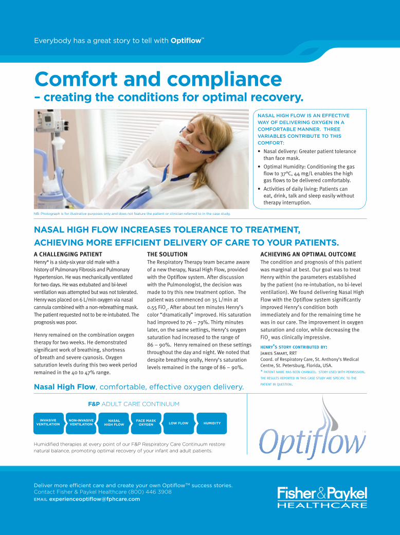
Comfort and compliance – creating the conditions for optimal recovery.
NASAL HIGH FLOW INCREASES TOLERANCE TO TREATMENT, ACHIEVING MORE EFFICIENT DELIVERY OF CARE TO YOUR PATIENTS.
F&P ADULT CARE CONTINUUM
INVASIVE VENTILATION
NON-INVASIVE VENTILATION
NASAL HIGH FLOW
FACE MASK OXYGEN LOW FLOW HUMIDITY
Humidified therapies at every point of our F&P Respiratory Care Continuum restore natural balance, promoting optimal recovery of your infant and adult patients.
NASAL HIGH FLOW IS AN EFFECTIVE WAY OF DELIVERING OXYGEN IN A COMFORTABLE MANNER. THREE VARIABLES CONTRIBUTE TO THIS COMFORT:
• Nasal delivery: Greater patient tolerance than face mask.
• Optimal Humidity: Conditioning the gas flow to 37°C, 44 mg/L enables the high gas flows to be delivered comfortably.
• Activities of daily living: Patients can eat, drink, talk and sleep easily without therapy interruption.
Deliver more efficient care and create your own OptiflowTM success stories.Contact Fisher & Paykel Healthcare (800) 446 3908EMAIL [email protected]
Nasal High Flow, comfortable, effective oxygen delivery.
NB: Photograph is for illustrative purposes only and does not feature the patient or clinician referred to in the case study.
A CHALLENGING PATIENTHenry* is a sixty-six year old male with a history of Pulmonary Fibrosis and Pulmonary Hypertension. He was mechanically ventilated for two days. He was extubated and bi-level ventilation was attempted but was not tolerated. Henry was placed on 6 L/min oxygen via nasal cannula combined with a non-rebreathing mask. The patient requested not to be re-intubated. The prognosis was poor.
Henry remained on the combination oxygen therapy for two weeks. He demonstrated significant work of breathing, shortness of breath and severe cyanosis. Oxygen saturation levels during this two week period remained in the 40 to 47% range.
THE SOLUTIONThe Respiratory Therapy team became aware of a new therapy, Nasal High Flow, provided with the Optiflow system. After discussion with the Pulmonologist, the decision was made to try this new treatment option. The patient was commenced on 35 L/min at 0.55 FiO
2. After about ten minutes Henry’s
color “dramatically” improved. His saturation had improved to 76 – 79%. Thirty minutes later, on the same settings, Henry’s oxygen saturation had increased to the range of 86 – 90%. Henry remained on these settings throughout the day and night. We noted that despite breathing orally, Henry’s saturation levels remained in the range of 86 – 90%.
ACHIEVING AN OPTIMAL OUTCOMEThe condition and prognosis of this patient was marginal at best. Our goal was to treat Henry within the parameters established by the patient (no re-intubation, no bi-level ventilation). We found delivering Nasal High Flow with the Optiflow system significantly improved Henry’s condition both immediately and for the remaining time he was in our care. The improvement in oxygen saturation and color, while decreasing the FiO
2 was clinically impressive.
HENRY’S STORY CONTRIBUTED BY:James Smart, RRTCoord. of Respiratory Care, St. Anthony’s Medical Centre, St. Petersburg, Florida, USA. * PATIENT NAME HAS BEEN CHANGED. STORY USED WITH PERMISSION. THE RESULTS REPORTED IN THIS CASE STUDY ARE SPECIFIC TO THE PATIENT IN QUESTION.

12 Respiratory Therapy Vol. 4 No. 3 n June-July 2009
IT NOWThe New York Times’ Steve Lohr recently reported on the inroads electronic record keeping is making in the medical community. According the article, the government is about to get into electronic technology for health records in a big way. In its economic recovery package, the Obama administration plans to spend $19 billion to accelerate the use of computerized medical records in doctors’ offices. Medical experts agree that electronic patient records, when used wisely, can help curb costs and improve care. According to the Times, such data-processing is already the norm among large medical groups which have invested in IT and say they have benefited from the cost savings. Yet, three-fourths of the nation’s doctors practice in small offices, with 10 doctors or fewer, and only about 17% of the nation’s physicians are using computerized patient records, according to a government-sponsored survey. How come? Getting up to IT-speed hasn’t been reimbursable, for one thing. Now the Obama administration has called for more than $40,000 spread over a few years for a physician who buys and uses electronic health records and puts them to “meaningful use.” Well, there’s the rub: the government has yet to define its terms. Consequently, says the Times, “many health experts predict that the meaningful use will be a requirement to collect and report measurements that can be closely correlated with improved health.” It is predicted that achieving success in implementation will not be easy. The crucial element, it is said, will be how local organizations help doctors in small offices adopt and use electronic records. The new legislation calls for creation of “regional health IT extension centers.” The Primary Care Information Project in New York City is a model. The project began two years ago, with $27 million in financing. The New York team brought in experts to see how doctors operate, and designed its own software for simple, Web-based electronic health records, but “abandoned that idea once they understood that patient records would have to be tightly linked to billing,” per the Times. The staff worked closely with its software supplier, eClinicalWorks, to tailor the system. The Times says, “They began rolling out the records a little more than a year ago. They are now used by more than 1,000 physicians, mainly in poorer neighborhoods, whose workplaces include two hospital outpatient clinics, 10 community health centers, 150 small group physician practices and one women’s jail, serving a total of one million patients. The rollout is progressing, and the government plan promises to accelerate adoption.” According to a physician who uses the system, “Our experience here is that it’s just hard. It’s not impossible.”
CLINICAL REVIEWWAO, worldallergy.org, reported on the recent studies: From Eur Respir J, 2008;32:1548-1554: The effects of rhinovirus infection on asthmatics vary, but upper respiratory infections commonly worsen asthma and lead to exacerbations. In a multicenter study, 413 adult asthmatics were followed for over a year to determine if the severity of a cold could predict loss of asthma control. To quantify the loss of asthma control, subjects completed the mini-Asthma Control Questionnaire and to measure cold severity, the Wisconsin Upper Respiratory Symptom Survey-21. Significant loss of asthma control occurred in 134 subjects and the WURSS-21 scores on the second day were predictive of subsequent worsening of asthma… From Chest 2008;134:1141-1148: The viral etiology of CAP has not been studied extensively and treatment guidelines are not as complete as those for bacterial pneumonia. Nasal swab secretions were acquired from 193 individuals hospitalized with CAP and analyzed by the nucleic acid amplification test for influenza, rhinovirus, hMPV, RSV and PIV. Serum samples were tested for bacterial infection by immunofluorescence methods. Pathogens were identified in 39% and among those, 29 had a viral infection only, 38 had a bacterial infection only, and 8 had both viral and bacterial infections. The most common were influenza, hMPV and RSV. In patients with bacterial infection, Streptococcus pneumoniae was most common (37%). Viral infection occurred more frequently in older patients (median 76 yrs) than younger (median 64 yrs) and was seasonal (October to May), whereas bacterial infections occurred year round. Morbidity and mortality rates were similar for viral and bacterial infections… From Eur Respir J 2008; 32:989-996: The hypothesis of a pilot study was that a pre-asthmatic condition characterized by lung inflammation exists in a significant number of individuals who are not diagnosed with asthma by traditional measures of lung function. The randomized, multicenter, DBPC trial enrolled 144 patients, 12 to 65 years of age, with symptoms suggestive of asthma to undergo treatment with mometasone furoate, 400 µg per day. The subjects scored morning and evening symptoms including cough, sputum production, wheeze, and shortness of breath, among others. Spirometry, eosinophil numbers in induced sputum and airway hyperresponsiveness were also measured. MF or placebo was administered as one puff in the evening from a metered-dose inhaler and salbutamol was used as needed by both groups. The treatment was continued for 4-8 weeks. Despite heterogeneity in the groups, overall symptom scores were lower, eosinophilia decreased and spirometry improved with MF compared to placebo. The researchers concluded that a short course of inhaled corticosteroids may be beneficial for some patients with symptoms suggestive of asthma, but careful follow-up was needed to determine the necessity of continued treatment.
FLUMONIAResearchers have believed that the flu facilitates an infection with pneumonia bacteria because it leads to a decrease of immune cells in the blood and impairs the body’s defenses. But research by two universities has shown that influenza facilitates and intensifies an infection from pneumonia bacteria, while disproving the common idea that this is caused by a lack of immune cells. Helmholz-Centre for Infection researchers infected mice with flu viruses and measured the amount of immune cells in the animals’ blood every day. Some days later, flu-infected mice received a dosage of pneumonia bacteria usually harmless for healthy mice. While the flu-infected mice did develop a superinfection & subsequently died, surprisingly,
NewsM June-July 2009

Respiratory Therapy Vol. 4 No. 3 n June-July 2009 13
they were not suffering from lymphopenia. The healthy, non-flu-infected mice defeated the bacteria successfully and recovered. To discover whether a lack of immune cells encourages an infection with pneumonia bacteria in general, an artificial drug-induced lymphopenia was established in the mice. Without infecting these lymphopenic mice with flu viruses, they received pneumonia bacteria. Despite a severe lack of immune cells, the mice recovered completely. Thus, researchers found that influenza facilitates and intensifies an infection from pneumonia bacteria, while disproving the common idea that this is caused by a lack of immune cells. Now scientists have to find out why the two conditions are related. The original article outlining the above findings is: Stegemann S, et al, Increased Susceptibility for Superinfection with Streptococcus pneumonia during Influenza Virus Infection Is Not Caused by TLR7-Mediated Lymphonia. 2009 PLoS ONE 4(3): e4840.
NOT WITH THE PROGRAMPatients in line for allergy immunotherapy are terrible with follow-up, according to a study by Allergy Partners in North and South Carolina. Investigators found that 71% percent of the nearly 30,000 patients who visited two facilities during the six-year study period received allergy testing but 11% of patients for whom an immunotherapy prescription was prepared never showed up for their first allergy immunotherapy appointment, and 13% discontinued treatment within the first three sessions. The majority of patients who were initially compliant with treatment eventually discontinued immunotherapy. Women were significantly more likely to stop immunotherapy within the first two years than men. Sixty percent of patients in the study did not complete the recommended three year course of treatment and less than a quarter completed two years of immunotherapy.
SHINE AND SPIN Using ultraviolet lights near a ceiling together with fans may reduce the spread of tuberculosis in hospitals, and air treatment with negative ionizers may also be effective, according to research published in PLoS Medicine. Researchers at the Imperial College London used 900 guinea pigs housed on the roof of a hospital in Lima, Peru to test whether simple approaches to disinfecting air could reduce transmission of TB. They (the researchers) found that 35% of those exposed to untreated air from patient rooms developed TB infection, compared to 14% in the negative air-ionizer group, and only 9.5% of those breathing air vented from rooms during treatment with upper-room UV lights and mixing fans. Why the pigs? Guinea pigs are susceptible to airborne infection with M. tuberculosis, which makes them sensitive to detection for infectious particles. By venting air from the rooms of patients with active TB through the guinea pig enclosures, the researchers were able to compare guinea pigs exposed on days when UV lights and air mixing fans were turned on in the patient rooms, to days when UV lights were off. The enclosure of a third group of guinea pigs contained the negative air ionizers. See Escombe, A.R., Upper-room ultraviolet light and negative air ionization to prevent tuberculosis transmission, PLoS Med.
BETTER MEDSKids taking recent asthma medications were found to have better asthma control compared to asthmatic children studied a decade ago, according to researchers at National Jewish Health in Denver, who compared asthmatic children between 2004 and 2007 with those tested between 1993 and 1997. Seventy-six percent of the newer cohort were on leukotriene
receptor antagonists and 66% were using combination inhaled glucocorticoids and long-acting bronchodilators, while none of the older group received these medications. While the current group was younger and had a higher proportion of males, the percentage requiring chronic oral GC therapy, along with the average dose and duration of oral GC, use were less in the present group. The current kids also had fewer GC-induced adverse effects compared to the historic group. The new cohort also had higher FEV1, required less albuterol and had fewer intubations in the past.
NO INSURANCEA study presented at the 2009 Annual Meeting of the AAAAI showed that access to insurance and health care doesn’t lead to better asthma control. Fifty eight percent of the enrollees in the study who had a regular physician used emergency care, as opposed to 27% who didn’t, and 92% of the respondents said they had a physician caring for their asthma, while 89% said they had medical insurance. The researchers found uniformly high rates of prednisone use, hospitalization, emergency care and uncontrolled daytime and nighttime symptoms among the sample of students. Low use of inhaled corticosteroids was also noted for those with and without insurance.
OBVIOUSLYLong-term exposure to smog significantly raises the risk of dying from lung disease, according to a new nationwide study conducted over an 18-year period. Researchers at the NYU School of Medicine found the risk of dying from respiratory disease 30% greater in metropolitan areas with the highest ozone concentration vs the lowest. The new study is the first nationwide population study on the long-term impact of ozone on human health, and the first to separate ozone’s effects from those of fine particulate matter, the tiny particles of pollutants emitted by factories, cars, and power plants. Ozone tends to be higher in concentration in suburbs and rural areas downwind of cities. Fine particulate matter, a primary pollutant, is more prevalent at its source, in the inner city, along roadways and in industrial areas. Background levels of ozone have at least doubled since pre-industrial-revolution times. The study analyzed 450,000 people who were followed from 1982 to 2000. Over that period 118,777 people in the study died. The cause of death data was linked to air pollution levels in 96 cities. California had both the city with the highest and the city with the lowest concentration of ozone pollution in the country. The researchers estimate that the risk of dying from respiratory causes rises 4% for every 10 parts-per-billion increase in exposure to ozone. Based on that result, the city with the highest mean daily maximum ozone concentration over the 18-year period of the study, was Riverside, CA, with 104 ppb. Long-term cumulative exposure corresponded to a 50% increased risk of dying from lung disease compared to no exposure to the pollutant. Los Angeles ran a close second, with an estimated 43% increased risk. In Washington, DC, and New York City, the study results indicate a 27 and 25% increased risk of respiratory death, with ozone concentrations of 75 ppb, which the EPA says is okay. The lowest ozone concentration was in San Francisco (33 ppb which carried a associated 14% increase in risk. The present EPA air quality standards do not protect against the long-term cumulative effects of ozone exposures, but only address the health effects of short-term daily peaks in ozone exposure.
14 Respiratory Therapy Vol. 4 No. 3 n June-July 2009
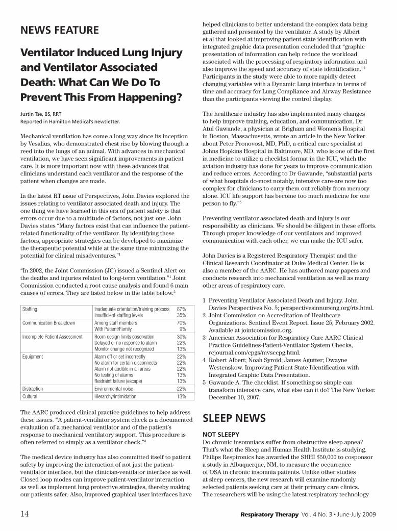
NEWS FEATURE
Ventilator Induced Lung Injury and Ventilator Associated Death: What Can We Do To Prevent This From Happening?Justin Tse, BS, RRT
Reported in Hamilton Medical’s newsletter.
Mechanical ventilation has come a long way since its inception by Vesalius, who demonstrated chest rise by blowing through a reed into the lungs of an animal. With advances in mechanical ventilation, we have seen significant improvements in patient care. It is more important now with these advances that clinicians understand each ventilator and the response of the patient when changes are made.
In the latest RT issue of Perspectives, John Davies explored the issues relating to ventilator associated death and injury. The one thing we have learned in this era of patient safety is that errors occur due to a multitude of factors, not just one. John Davies states “Many factors exist that can influence the patient-related functionality of the ventilator. By identifying these factors, appropriate strategies can be developed to maximize the therapeutic potential while at the same time minimizing the potential for clinical misadventures.”1
“In 2002, the Joint Commission (JC) issued a Sentinel Alert on the deaths and injuries related to long-term ventilation.”1 Joint Commission conducted a root cause analysis and found 6 main causes of errors. They are listed below in the table below.2
Staffing Inadequate orientation/training process 87%Insufficient staffing levels 35%
Communication Breakdown Among staff members 70%With Patient/Family 9%
Incomplete Patient Assessment Room design limits observation 30%Delayed or no response to alarm 22%Monitor change not recognized 13%
Equipment Alarm off or set incorrectly 22%No alarm for certain disconnects 22%Alarm not audible in all areas 22%No testing of alarms 13%Restraint failure (escape) 13%
Distraction Environmental noise 22%
Cultural Hierarchy/intimidation 13%
The AARC produced clinical practice guidelines to help address these issues. “A patient-ventilator system check is a documented evaluation of a mechanical ventilator and of the patient’s response to mechanical ventilatory support. This procedure is often referred to simply as a ventilator check.”3
The medical device industry has also committed itself to patient safety by improving the interaction of not just the patient-ventilator interface, but the clinician-ventilator interface as well. Closed loop modes can improve patient-ventilator interaction as well as implement lung protective strategies, thereby making our patients safer. Also, improved graphical user interfaces have
helped clinicians to better understand the complex data being gathered and presented by the ventilator. A study by Albert et al that looked at improving patient state identification with integrated graphic data presentation concluded that “graphic presentation of information can help reduce the workload associated with the processing of respiratory information and also improve the speed and accuracy of state identification.”4 Participants in the study were able to more rapidly detect changing variables with a Dynamic Lung interface in terms of time and accuracy for Lung Compliance and Airway Resistance than the participants viewing the control display.
The healthcare industry has also implemented many changes to help improve training, education, and communication. Dr Atul Gawande, a physician at Brigham and Women’s Hospital in Boston, Massachusetts, wrote an article in the New Yorker about Peter Pronovost, MD, PhD, a critical care specialist at Johns Hopkins Hospital in Baltimore, MD, who is one of the first in medicine to utilize a checklist format in the ICU, which the aviation industry has done for years to improve communication and reduce errors. According to Dr Gawande, “substantial parts of what hospitals do-most notably, intensive care-are now too complex for clinicians to carry them out reliably from memory alone. ICU life support has become too much medicine for one person to fly.”5
Preventing ventilator associated death and injury is our responsibility as clinicians. We should be diligent in these efforts. Through proper knowledge of our ventilators and improved communication with each other, we can make the ICU safer.
John Davies is a Registered Respiratory Therapist and the Clinical Research Coordinator at Duke Medical Center. He is also a member of the AARC. He has authored many papers and conducts research into mechanical ventilation as well as many other areas of respiratory care.
1 Preventing Ventilator Associated Death and Injury. John Davies Perspectives No. 5; perspectivesinnursing.org/rts.html.
2 Joint Commission on Accreditation of Healthcare Organizations. Sentinel Event Report. Issue 25, February 2002. Available at jointcomission.org.
3 American Association for Respiratory Care AARC Clinical Practice Guidelines-Patient-Ventilator System Checks, rcjournal.com/cpgs/mvsccpg.html.
4 Robert Albert; Noah Syroid; James Agutter; Dwayne Westenskow. Improving Patient State Identification with Integrated Graphic Data Presentation.
5 Gawande A. The checklist. If something so simple can transform intensive care, what else can it do? The New Yorker. December 10, 2007.
SLEEP NEWSNOT SLEEPYDo chronic insomniacs suffer from obstructive sleep apnea? That’s what the Sleep and Human Health Institute is studying. Philips Respironics has awarded the SHHI $50,000 to cosponsor a study in Albuquerque, NM, to measure the occurrence of OSA in chronic insomnia patients. Unlike other studies at sleep centers, the new research will examine randomly selected patients seeking care at their primary care clinics. The researchers will be using the latest respiratory technology

Respiratory Therapy Vol. 4 No. 3 n June-July 2009 15
to measure breathing, and hypothesize that more than half of chronic insomnia patients will turn out to suffer from previously undiagnosed sleep apnea. For more, contact sleeptreatment.com.
ABUSED?Modafinil, which is being used to enhance cognitive abilities, affects the activity of dopamine in the brain in a way that may create the potential for abuse and dependence. It’s currently used off-label for the treatment of cognitive dysfunction in some psychiatric disorders. It can produce psychoactive and euphoric effects typical of central nervous system stimulant drugs, and there is debate surrounding its potential for abuse. Researchers at the National Institute on Alcohol Abuse and Alcoholism, Bethesda and Brookhaven National Laboratory conducted a study to test whether modafinil, at therapeutic doses, would elevate extracellular dopamine in the brain by blocking the dopamine transporter. The study included 10 healthy men, between the ages of 23-46 years, who received either placebo or 200 and 400 mg of modafinil. The researchers found that modafinil acutely increased dopamine levels and blocked dopamine transporters in the human brain. Because drugs that increase dopamine have the potential for abuse, and considering the increasing use of modafinil for multiple purposes, these results suggested that risk for addiction in vulnerable persons merited heightened awareness.
CANCER SHIFTWomen in Denmark who got breast cancer after working night shifts will receive government compensation, according to an article in Medical News Today. A report by BBC Scotland said that working night shifts probably increases people’s risk of developing cancer, and the Danish government has started to pay compensation to women whose breast cancer was probably caused this way. Research by the UN’s International Agency for Research on Cancer said that the risks presented by working night shifts at the same level as those presented by industrial chemicals. Epidemiological studies have shown that long term night workers have a higher risk of breast cancer than women who do not work such patterns. The studies, which have been done mostly on nurses and flight attendants, are consistent with animal studies that show constant light, dim light at night, or simulated chronic jet lag substantially raises the risk of tumors. Other studies have shown that depressing melatonin levels at night also raises the risk of developing tumors. Nearly one fifth of workers in Europe and North America work night shifts. The above was written by Catharine Paddock, PhD, Copyright: Medical News Today.
SWEET SLEEPPeople who sleep less than six hours a night during the work-week are 4.5 times more likely to have elevated levels of blood sugar than those who sleep 6-8 hours, according to a study by the University of Buffalo. The finding was based on data from a six-year follow-up of a study conducted from 1996-2001. The 91 persons with normal fasting glucose levels at baseline who developed pre-diabetes by their follow-up exam were matched to persons from the study who had maintained normal glucose levels who served as controls. Participants were placed into three groups based on the average daily amount of sleep they reported receiving from Sunday through Thursday: short-sleepers, those who reported less than 6 hours of sleep nightly; long-sleepers, who reported sleeping more than eight hours nightly; and a reference group who slept 6-8 hours a night. Short-sleepers had a significantly increased risk of progressing from
normal glucose levels to pre-diabetes, compared to those who slept 6-8 hours nightly. Sleeping an average of more than 8 hours a night had no significant effect on glucose levels.
BUT DON’T TAKE A NAP!Taking regular lunchtime naps could increase the risk of developing Type 2 diabetes, according researchers at the University of Birmingham, who looked at the napping habits of 16,480 people and found that diabetes prevalence increased with napping frequency. Those who napped had a 26% greater risk of developing Type 2 diabetes compared to those who never did. The researchers said that an association between napping and reduced physical activity may be behind the link. Napping during the day may also disrupt night-time sleep. In addition, waking up from napping activates hormones and mechanisms in the body that stop insulin from working effectively.
WAKE UP AND DRIVETruck crashes with OSA are a potential road hazard, and the nature of their jobs contributes to their chances of suffering from the condition. Researchers at Cambridge Health Alliance said that excessive daytime sleepiness is common among truck drivers, and that 2.4 to 3.9 million licensed commercial drivers in the US probably have OSA. Truck drivers with sleep apnea have up to a 7-fold increased risk of being involved in a motor vehicle crash. Over a15-month period, 456 commercial drivers were examined from over 50 different employers. Seventy-eight (17%) met the screening criteria for suspect OSA. These drivers were older and more obese, and had a higher average blood pressure. Of the 53 drivers who were referred for sleep studies, 33 did not comply with the referral and were lost to follow-up. The remaining 20 were all confirmed to have OSA, but after diagnosis, only one of these 20 drivers with confirmed OSA complied with treatment recommendations. Researchers noted that drivers with sleep apnea frequently minimized or underreported symptoms such as snoring and daytime sleepiness. In the study, the majority of truck drivers did not follow through on physician recommendations for sleep studies and sleep apnea treatment. Researchers said it was very likely that most of the drivers who didn’t comply with sleep studies or sleep apnea treatment sought medical certification from examiners who don’t screen for sleep apnea. The Federal Motor Carrier Safety Administration is currently deliberating recommendations to require sleep apnea screening for all obese drivers based on body mass index.
MANY SLEEPLESS NIGHTSThree-fourths of individuals with insomnia report experiencing the condition for at least one year and almost half experience it for three years, according to study by the Université Laval and Centre de recherche Université Laval-Robert Giffard, Québec, Canada. Researchers evaluated insomnia persistence, remission and relapse in 388 middle-aged adults over a course of three years. Seventy-four percent reported insomnia for at least one year and 46% reported insomnia persisting over the entire three-year study. The group with initial insomnia syndrome had a higher persistence rate than the group with symptoms of insomnia (66% vs 37.2%). About 54% of participants went into insomnia remission; however, 26.7% percent experienced relapse. Individuals with subsyndromal insomnia were three times more likely to remit than worsen to syndrome status, although persistence was the most frequent course in that group as well. Of the 269 individuals with baseline symptoms of insomnia, after one year 38.4% were classified as good sleepers,
16 Respiratory Therapy Vol. 4 No. 3 n June-July 2009
48.7% still had insomnia symptoms and 12.9% had insomnia syndrome. Results were similar after the second and third year of follow-up. Of the 119 participants with insomnia syndrome at the beginning of the study, 17% good sleepers after one year, while 37% had symptoms of insomnia and 46% remained in the insomnia syndrome group.
CHEAPER TO SLEEPERDiagnosing and treating obstructive sleep apnea may soon become much less expensive and arduous, thanks to new research showing that a simplified program using experienced nurses, home ambulatory diagnosis and using auto-titrating CPAP machines is not inferior to the traditional model which relies on specialist physicians and sleep studies. A randomized, multicenter study at the Adelaide Institute for Sleep Health in South Australia compared the results of two OSA diagnosis and treatment protocols, simplified and traditional, as well as their respective costs. The simplified model of care was found to be not inferior to the usual physician-led, hospital-based model. Researchers developed a nurse-led diagnosis and treatment model that featured ambulatory overnight oximetry and auto-titrating CPAP machines to set fixed CPAP under nurse supervision. They compared the results of patients thus diagnosed and treated to those who underwent standard sleep medicine pathways, including laboratory-based polysomnography, CPAP titration and physician management of the patient. They assessed the patients’ sleepiness on the validated Epworth Sleepiness Scale (ESS) and set the minimal clinically significant change at +/- 2 points. They also assessed other outcomes of sleep, including quality of life measures, executive neurocognitive function on maze tasks and maintenance of wakefulness tests and CPAP adherence. In all, the study assessed almost 200 patients with moderate to severe OSA who were randomly assigned to the simplified or traditional model. The patients in the nurse-led group spent about 50 minutes longer with the nurse than the patients in the physician-led groups, but were seen by physicians 12% of the time. Patients in the physician-led group, meanwhile, had an average of 2.36 consultations with physicians, as opposed to 0.18 for patients in the nurse-led group. None of the secondary outcomes measured showed significant differences between the groups, and differences in ESS scores between groups were lower than the predetermined minimum for clinical significance. The patients in the nurse-led group were diagnosed and treated for $722 less per patient than those in the physician-led group.
MEDICARE FOR OSAThe Centers for Medicare & Medicaid Services (CMS) announced a new policy for Medicare coverage of sleep testing for the diagnosis of OSA. The decision provides coverage for specified sleep tests that are used to confirm the diagnosis in patients who have clinical signs and symptoms of OSA. The coverage decision establishes nationally consistent coverage. The decision does not apply to the use of these tests for other purposes beyond the diagnosis of OSA. Local Medicare contractors may continue to determine coverage on other uses within their own jurisdictions. See cms.hhs.gov/home/medicare.asp.
OLD, SLEEPY, DEADOlder women who take daily naps have a significantly greater risk of dying, according to researchers at University of Gent, Belgium. Actually, everyone is at risk of dying; however, the researchers likely meant that older women may die sooner. Four communities consisting of 8,101 women aged 69 and
older were studied over a 7-year period. Women who reported napping daily were 44% percent more likely to die from any cause, 58% more likely to die from cardiovascular causes and 59% more likely to die from non-cardiovascular, non-cancer causes. Older women who reported sleeping between 9-10 hours per 24-hour period also had a greater risk of mortality compared to those who slept between 8-9 hours. The association was strongest for cardiovascular-related mortality. Researchers noted, however, that napping and long sleep duration may be caused by sleepiness due to underlying sleep disorders or other medical conditions that actually lead to death. Elderly women who napped less than 3 hours per week were not at increased risk of mortality compared to women who did not nap at all. Criticisms of the study included a low responder rate (37%) that could have introduced an element of bias, and the fact that it lacked objective measures of day time sleepiness (such as polysomnography readings), instead using self reported patient responses. The data left unclear whether sleep complaints were a symptom of underlying cardiovascular disease or whether sleepiness triggered or worsened the disease.
PANTS ON FIREWSJ Blogs, by the Wall Street Journal, reports that a Harvard researcher fabricated and falsified data in a study of sleep apnea. Sarah Rubenstein reports that Robert Fogel, a former assistant prof at Harvard Med School, has retracted a 2003 study in the journal Sleep, titled, “Anatomic and physiologic predictors of apnea severity in morbidly obese subjects.” Fogel was said to change or falsify nearly half of the sleep data so that the data would better conform to his hypothesis, and he was also said to fabricate about 20% of anatomic data that supposedly came from CT scans, according to info Fogel himself volunteered. He told the paper The Scientist, “I moved numbers around to make the data look like there was something there… I never really thought through the consequences, and once I did this I got myself into a loop that I found I couldn’t get out of.”
RESPIRATORY THERAPY PRODUCTSNICU SUCCESSVapotherm’s hospital newsletter recently spotlighted the UMASS Memorial Medical Center’s Newborn Intensive Care Unit. Located in Worcester, MA, it is the region’s only Level III NICU for high-risk neonatal care. The department has earned benchmark status for its encouragement of family participation in newborn care, making it a model for other hospital NICUs throughout the world. The NICU has 43 beds, including 27 intensive care beds in three pods designed for maximum privacy and 16 beds in a Continuing Care Nursery which eases the transition to home as the baby’s health improves. The NICU initially purchased four Vapotherm units, which quickly became so popular, according to hospital staff, that nurses were constantly searching for available units for their patients. The NICU Respiratory Therapists and nurses quickly experienced excellent clinical outcomes as well as strong support from staff members and families. Recently, the NICU migrated to Vapotherm’s new device, Precision Flow. It currently has 10 units and has treated 270 babies, totaling 2,300 days of care with Vapotherm technology. When the first Vapotherm device was nearing the end of its evaluation period, it was still supporting a patient, who was recovering and doing well. The company’s

Respiratory Therapy Vol. 4 No. 3 n June-July 2009 17
clinical products division had expected to retrieve the device, but the patient was recovering and the hospital simply didn’t want to put him back on mechanical ventilation. When the mother of the patient heard the conversation about the status of the device, she said, “I overheard your conversation and I am here to write a check to purchase the unit and donate it to the NICU.” Contact vtherm.com.
TESTING…B&B Medical Technologies’ The Test Lung-Pediatric offers a solution for performing routine OVP testing and demonstrating operation of mechanical ventilators. With certified resistance and compliance, the 0.5 liter Test Lung-Pediatric is made of Latex-free silicone and space age resins to withstand the rigors of daily hospital and classroom use. The ventilation bag is durable, easily removable and can be cleaned or sterilized as needed. Included with each Test Lung is a Test Lung Connector Kit that adapts to all patient circuits and proximal airway flow sensors. The Connector Kit has three adapters, two with Luer Ports and Caps, allowing practitioners the ability to demonstrate leak performance and patient-trigger function. The Test Lung-Pediatric is compact in design and lightweight. Each 0.5L Test Lung is tested and validated for resistance and compliance in the application range, and has a unique serial number to insure its compliance with specification. It is the ideal tool for teaching and demonstration in addition to performing pediatric ventilator verification testing. A separate kit is available to demonstrate changes in airway resistance. The Precision Resistor Kit is adaptable to both Test Lungs and includes three resistors: Rp5, Rp20 and Rp50. The Precision Resistor Kit is factory calibrated, and can be cleaned and sterilized. Visit bandb-medical.com.
VAPOTHERM IN THE HOMECan the Vapotherm 2000i be used at home? The short answer is “yes.” For all patients receiving HFT at home, care providers will need a Vapotherm 2000h and the associated disposable supplies. In addition they will need the HCK-200 (LF or HF) kit. Lastly they’ll will need an oxygen source if the FIO2 delivered is to be greater than 21% (room air). A concentrator, liquid or cylinder system can be used for FIO2 above 21%. Contact vtherm.com.
DUAL-INGNewport Medical Instruments announced the release of the Dual Pac Internal Battery System for the HT50 Ventilator. The ease of use, compact size and durability of the lightweight HT50 have made it the ideal choice for users on the go. With the new Dual Pac Internal Battery System, the primary battery allows up to 10 hours of operation and when the “battery low” alarm sounds, a secondary back-up battery provides a minimum of 30 minutes of operation prior to shutdown. The new battery technology utilizes two batteries that work independently and charge simultaneously so that both the primary and back up batteries are charged by any external AC or DC power. The HT50’s miniature internal gas generator eliminates the need for an external compressed gas source. The HT50 is applicable for > 10 kg infant, pediatric and adult patients, for invasive or non-invasive respiratory support for emergency, hospital, transport and homecare applications. Contact ventilators.com.
PLAY A PARTAs part of its ongoing commitment to promoting open access in the developing world, BioMed Central teamed up with Computer Aid International to support research in Africa. BioMed has chosen to support Kenyatta University in Nairobi
to help local scientists conduct vital research directly relevant to local problems in one of the poorest parts of Africa. Many of the university’s academics have been published in open access journals, including those from BioMed Central. In common with most African universities, however, Kenyatta cannot afford new computers, meaning that academics cannot get the access time that they need for researching and preparing papers. We’re partnering with Computer Aid International, who provide affordable professionally refurbished PCs to the developing world, to resolve this problem. BioMed Central aims to raise £10,760 in order to provide a container of 225 PCs to the university – enough to give all research departments their own dedicated suite of computers and guarantee that the university’s 720 research staff all get the IT access that they need. You can make a contribution to this project today by visiting the site above. In return for your support BioMed said it would let contributors know how the money is spent and provide updates on the progress of the project.
RELAUNCHEDRadiometer has relaunched its site bloodgas.org under a new name: acutecaretesting.org. The relaunch reflects three critical changes in the website: scope, usability and graphics, with the biggest change being the scope of articles to also include other acute care testing topics adjacent to blood gas, such as tight glycemic control and cardiac markers. In addition, the site has improved its usability, with information reorganized in a way that is more intuitive to users and optimizes the search function. The site has also had a graphic makeover. Acutecaretesting.org has more than 17,000 registered users worldwide; registration is free, and content is provided by healthcare professionals around the world. Despite its corporate sponsorship, the site remains true to its policy of non-commercial content.
BREAKTHROUGHMasimo the inventor of Pulse CO-Oximetry and Measure-Through Motion and Low-Perfusion pulse oximetry, announced that it has initiated the full market release of its breakthrough noninvasive and continuous hemoglobin (SpHb) monitoring technology. As the first noninvasive and continuous hemoglobin monitoring technology to receive FDA 510(k) clearance and be available for widespread commercial adoption, Masimo SpHb is already transforming the way hemoglobin testing is performed at over 40 hospitals in the U.S., Europe, Asia, and Africa, that have participated in the technology’s limited market release initiated in September of 2008. The availability of noninvasive, continuous, and immediate hemoglobin measurements is expected to have wide ranging clinical impact, from surgery and intensive care to less acute care settings, including the emergency department, physician office, ambulatory surgery center, and long-term care facility by facilitating prompt detection of internal bleeding and more appropriate administration of blood transfusions. Early benefits and impact of Masimo SpHb were evident in feedback received from clinicians at hospitals around the world who participated in the limited market release. SpHb is part of the Masimo Rainbow SET Pulse CO-Oximetry patient monitoring platform—the first-and-only upgradable technology platform capable of continuously and noninvasively measuring multiple blood constituents and helping to predict fluid responsiveness in patients previously requiring invasive procedures. Masimo Rainbow SET noninvasive measurements—including: total hemoglobin (SpHb), oxygen content (SpOC), carboxyhemoglobin (SpCO), methemoglobin (SpMet), PVI, oxyhemoglobin (SpO2), pulse rate

18 Respiratory Therapy Vol. 4 No. 3 n June-July 2009
(PR), and perfusion index (PI)—have the potential to facilitate faster, easier and safer health decisions. Currently available in bedside Masimo Radical-7 and Rad-87 Pulse CO-Oximeter patient monitors, SpHb will also be offered in handheld monitors and select multiparameter patient monitoring brands through Masimo Rainbow SET Pulse CO-Oximetry technology license agreements. SpHb has received regulatory clearance in the US, Canada, Europe, Korea and Australia, and is now available for sale in most of the countries in the world. Here are some user comments: Ronald Miller, MD, Chief of Anesthesia, Professor and Chairman of the Dept of Anesthesia and Perioperative Care at the University of California, San Francisco, stated, “Masimo SpHb is an impressive new tool that helps us to more safely guide patients in surgery through to recovery.” Randy Marcel, MD, Medical Director and Chief of Anesthesiology at The Heart Hospital Baylor Plano in Plano, TX: “In the past, we’ve only received glimpses of our patients’ hemoglobin levels from lab measurements, but now we have complete and real-time hemoglobin visibility.” Javed Akhtar, MD, FAAP, Medical Director, Pediatric Intensive Care Unit at Creighton University Medical Center in Omaha: “We purchased the SpHb monitor after seeing it in a hands-on demonstration… SpHb has reduced the traumatic experience for pediatric patients, increased the satisfaction of parents, and reduced the workload on our nursing staff, phlebotomist and laboratory personnel.” Madhava Karunarathna, MD, OB/GYN at Balangoda Hospital in Sri Lanka: “With SpHb, we now have accurate hemoglobin measurements available at our fingertips, around the clock. In cases of severe hemorrhaging during and after childbirth, SpHb has enabled us to immediately identify and continuously assess blood loss severity to better manage internal bleeding, prevent overloading of fluid, and decrease maternal death.” Adi Abdussalam Adham, MD, Chief Manager at Accidents Hospital Abu Saleem in Tripoli, Libya: “In the operating room, Masimo SpHb has enabled us to more effectively monitor blood loss and better manage transfusions in surgery.” Bertrand Debaene, MD, Anesthesiologist at the University Hospital Center of Poitiers in Poitiers, France: “SpHb, along with PVI, have been important improvements for both the department and our patients.”
WAO-WOWThe WAO worldallergy.org website has begun to provide a growing list of links to websites in various languages that contain patient information about allergic diseases. The sources of these links include patient organizations, hospitals, clinics and allergy societies that offer these resources on their Web sites. Clinicians will be able to refer patients to this page, and the list also will be a resource to individuals as they search the Internet for patient information about allergic disease. The list of links, which is searchable by country, region, and or language, will be continually expanded and updated in the goal of making this a global patient education resource. Presentations given by WAO members as webinars via the internet are recorded and archived for viewing on the WAO web site in the “Education in Allergy” section. Now available are RSV: Gatewaway to Asthma? and Asthma and GERD… The Global Resources in Allergy (GLORIA) curricula educates medical professionals worldwide, through local, state, regional and national presentations. Modules are created from established guidelines and recommendations to address different aspects of allergy-related patient care… Expert interviews available from WAO are: Annual WAO Update, with Prof G. Walter Canonica; Sublingual Immunotherapy, with Prof Hans de Groot; Drug Hypersensitivity, with Dr David Khan, and Immunologic Advance, with Dr Lanny Rosenwasser.
TEAMING UPTeleflex Medical announced today the signing of a distribution agreement with ResMed Corp which makes Teleflex an exclusive distributor of the ResMed Non-Invasive Ventilation (NIV) mask portfolio for US acute care hospitals not affiliated with the Veterans Administration. Teleflex Medical is focused on providing hospitals with products that address the needs of the non-invasive ventilation patient. The ResMed NIV mask portfolio expands Teleflex Medical’s offering which includes the ConchaTherm Neptune, a heated humidifier designed for use in NIV. The use of NIV has grown rapidly in recent years and this trend is expected to continue as facilities adopt protocols aimed at reducing VAP. Studies have shown that NIV improves oxygenation and is well tolerated by patients with acute respiratory failure. By eliminating the need for endotracheal intubation in certain conditions, the result is fewer complications, shorter hospitals stays and as a result, reduced mortality rates and costs of care. ResMed is known for innovation and setting new standards in the design and manufacture of masks for non-invasive pressure therapy. Their offering in the NIV space displays this commitment to excellence. The Resmed NIV masks are quick-fitting, high-performance products that have earned a reputation for being easy-to-use, secure-sealing and comfortable. These are all critical factors in ensuring patient/ventilator synchrony and effective ventilation. Contact resmed.com or teleflex.com.
PROBINGRespiratory Technology Corporation, Restech, announced that the Division of Gastroenterology at Seattle Children’s Hospital, Seattle, has adopted the Restech Dx-pH Measurement System to detect acid reflux in the airway. The Dx-System provides valuable information about patients’ pharyngeal acid exposure and its role in various comorbidities, helping physicians diagnose the cause of each patient’s symptoms more accurately, and treat the patient more effectively. At Seattle Children’s, the system is being used to evaluate the extra-esophageal manifestations of GERD. The Dx-pH System is said to be noninvasive and well tolerated and enables doctors to more aggressively treat for reflux or to wean off previously started anti-reflux treatments and search for other causative factors. The miniaturized pH sensor at the tip of the Dx-pH Probe is unique in its ability to measure pH in a non-liquid environment, such as the pharynx. The Probe’s miniaturized, patented sensor is housed in the tear-drop shaped tip at the distal end of a thin trans-nasal catheter. An LED blinks during placement, allowing the medical personnel to confirm the proper placement in the oropharynx. The measurements taken by the pH sensor are sent wirelessly to a recording device. Contact restech-corp.com.
U B BREATHINGA new, recently licensed medical device developed by University at Buffalo researchers, the new UB ventilator, has the potential to shorten the length of patient stays in the intensive care unit because it will greatly reduce complications and habituation to sedatives. It also is expected to be more cost-effective than current methods of ventilating ICU patients. The device also may have promising applications in treating large numbers of patients during pandemics or other events with mass casualties because it can safely enable multiple patients to share a single ventilator without the risk of cross-contamination. The device is designed to cost effectively deliver to patients small amounts of powerful inhalation anesthetic agents as they breathe or Continued on page 48…

Respiratory Therapy Vol. 4 No. 3 n June-July 2009 19
Special Report: Blood Gas
IntroductionUpon purchasing a new blood gas analyzer, immediate testing for patient management is not permitted. Regardless of manufacturer’s claims or warranties, a series of quality assurance steps must be conducted to validate instrument performance and ensure analytes are reported with accuracy and precision.1 These implementation procedures are both essential from a Regulatory perspective and necessary to guarantee diagnoses and therapeutic maneuvers remain consistent based on results analyzed from either the existing blood gas instrument or values produced from the new device.
The first three-function (pH, PCO2, PO2) blood gas apparatus was built by Severinghaus and Bradley in 1959.2 Numerous technical advances have occurred since that period which has improved reliability, increased sample throughput and minimized the specimen volume required for analysis. However, the most significant clinical advances have been to increase the analyte panel to include other assays which evaluate metabolic and renal function. Twenty-first century blood gas instruments combine pH and blood gas measurements with electrochemical and enzymatic sensor technology to analyze electrolytes, lactate, glucose, creatinine, BUN/urea, and hematocrit concentrations.3 The methods validation procedures employed for blood gases are also essential for these analytes as well. Whether replacing an existing traditional bench-top instrument or acquiring additional near patient care or point-of-care (POC) analyzers, the same device implementation requirements are recommended.
Regulatory ComplianceThe Centers for Medicare and Medicaid Services (CMS) administers the Clinical Laboratory Improvement Amendments (CLIA) which regulate all facilities that perform testing on materials derived from the human body for the purpose of providing information for the diagnosis, prevention, or treatment of any disease or impairment of, or the assessment of the health of human beings, to meet certain Federal requirements. If a facility performs tests for these purposes, it is considered a laboratory under CLIA and must apply and obtain a certificate from the CLIA program that corresponds to the complexity of tests performed.4
Before a new analyzer is used for reporting patient results, the methods validation and verification of instrument performance procedures must be completed and approved by the medical director named on the facilities CLIA license, issued by CMS. If the blood gas instruments are operating under the clinical
laboratory’s CLIA license, the laboratory POC coordinator and the laboratory medical director are both required to oversee the validation process. If the respiratory care department holds their own independent license to perform testing, they must comply with the standards set by the regulatory inspecting agency indentified on the CLIA application.
Many private agencies have emerged to promulgate standards for laboratory best practices. In addition, these agencies perform site-visits or inspections to the facility to observe and document compliance of laboratory standards. Currently, these agencies include: The College of American Pathologists (CAP), The Joint Commission (formerly known as The Joint Commission on the Accreditation of Healthcare Organizations or JCAHO), and the Commission on Laboratory Accreditation (formally known as COLA). These inspecting agencies also offer services to US military hospital laboratories. State governments have a strong role in laboratory standards as well. Each state’s Department of Health manages the CLIA branch for that region. The state CLIA office can set the standards for laboratories in their jurisdiction and also inspect the testing facility. While the national office of CLIA has published their standards,5 each individual state CLIA branch can modify those regulations to more stringent standards and hold the testing facilities in their state to local guidelines.1
Methods ValidationThe regulations of instrument validation specify all blood gas analyzers, including POC devices, be evaluated in the same environment where actual patient testing will be conducted. When POC devices are being examined, it is important to assess instruments operation in all patient areas where testing will be provided. For example, if the analyzer is a POC device and the anticipated use is a site external to the main facility, or used in patient transport, validating each environment is essential. Consulting with the manufacturer on issues that could affect instrument performance in extreme environmental conditions or nontraditional applications is highly recommended. If the test panel includes analytes that are not intended to be reported, reference the Operator’s Manual or consult with the manufacturer on how to suppress those values from being measured. If the device is not capable of analyte suppression, all analytes must be validated.
A familiarization period is often overlooked as an important step in the implementation of a new system. The Clinical and Laboratory Standards Institute (CLSI) formally the National Committee on Clinical Laboratory Standards (NCCLS) recommends a five day period in which the instrument should be handled by the new operators and data not collected for the statistical analysis of the system.6 Device training is a part of
Guide to Validation of a Blood Gas SystemDiana Blanco, MT-SC (ASCP); Bruce Toben, RRT-NPS, CPFT
The authors are with the Department of Clinical Affairs, International
Technidyne Corporation, Piscataway, NJ

20 Respiratory Therapy Vol. 4 No. 3 n June-July 2009
this process as selected personnel learn the analyzer’s operation, maintenance requirements, and advanced functions of the instrument.
Commonly, the manufacturer will initially educate operators on-site. Maintaining a detailed roster of training is necessary and needs to be filed within the department. The roster should include: date, name/signature of attendee, key components of training, who conducted the class, and results of a written test. In addition, proficiency testing is also required to evaluate operator’s performance. New operators should be tested to ensure they can produce analyte results with liquid quality control (LQC) material and produce values within the pre-established ranges published by the LQC manufacturer. Documents attesting to satisfactorily completing this practicum must also be filed to demonstrate the operator’s skill.
Among the specific laboratory tests that are required for methods validation are: precision/trueness, establishing the analytical measurement range, method correlation, and development of normal ranges.
Trueness and Precision Trueness is replacing the previous term accuracy but the test objective remains the same, how close does the average measured result approach the “true” value. Precision is a measure of reproducibility and is assessed in combination with trueness. Commonly, LQC is used for these purposes since the “known” range for recovery has been predetermined or assayed. This procedure is performed by analyzing multiple levels of analyte concentrations over a period of five days. CLSI recommends one series of tests from multiple analyte concentration levels in triplicate for five days,7 while CAP recommends a minimum of two results from each level for five days. Testing over a span of many days may alter system performance due to changes in ambient temperature, humidity, barometric pressure, but most of all, more than one operator.
The objective of the CLIA program is to ensure quality laboratory testing. CLIA has set guidelines defining the total acceptable variations for analytes reported by blood gas and chemistry analyzers.8 Based on these guidelines, the acceptable range of results for a single instrument can vary markedly. For example, a laboratory may report an absolute difference for PCO2 of 10 mmHg on the same specimen, from repeated tests, using the same analyzer and would be within acceptable limits, eg, if the true PCO2 is 40 mmHg, the blood gas analyzer may report 35 mmHg with the first measurement and 45 mmHg on the repeated test. The CLIA acceptable limit for PCO2 is ± 5 mmHg or 8%, which ever is greater. The degree of measured variance can be designated by the medical director, but cannot exceed specifications set by CLIA.
Analytical Measurement Range vs. Clinical Reportable Range and Linearity ValidationThe Analytical Measurement Range (AMR) is the manufacturer’s range of detection for the device. This range can exceed limits of what can be considered compatible with life, eg, pH 6.00 – 8.00. The Clinical Reportable Range (CRR) is the actual range that can be verified by the facility with either commercially available assayed materials or measuring a blood specimen corroborated by split-sample technique with a calibrated reference instrument. The CRR usually approximates the limits of what is physiologically acceptable in humans, e.g., pH 6.80 – 7.80. Patient
results that are lower or higher than the level verified can only be reported as outside the reportable range, exceeding the limits.9
Establishing the CRR employs testing both the extreme ends of the assay scale but mid-range points as well. This method permits estimation of the linearity, or the degree of sensitivity to incremental changes in analyte concentration and predicts instrument performance at any point along the measurable range. As described in the CRR development, linearity is commonly performed with assayed calibration verification controls. If no commercially available material is compatible for the device being evaluated, protocols need to be developed using spiked whole blood specimens and verified with a laboratory reference instrument. A minimum of two samples each, of 4-5 incremental concentrations are needed for statistical analysis.
The CRR and linearity testing are required for all new devices and each analyte reported, whether replacing an existing instrument or adding multiple devices intended to supplement a current blood gas systems.
Method CorrelationPrior to reporting patient results, all new laboratory analyzers need to be evaluated side-by-side in order to determine if both instruments produce similar values. This comparative procedure is called Method Correlation testing. The objective of the procedure is to ensure that whether blood gas results are reported from the current analyzer or from the new system, diagnostic interpretation of results and therapeutic maneuvers can be conducted in the same fashion, transparent to the end-user of which analyzer made the measurement. This procedure will also validate the current ranges and assess if they need to be adjusted.
The protocol should include patient samples from all applications and analyte concentrations within the CRR. Environments such as the operating room present unique issues. Low ambient temperature and hemodilution from pump priming solution during cardiac surgery10 may affect assay performance. If the new blood gas system is intended for this clinical area, it is vital to include specimens from this patient population.
If more than one new blood gas system of the same manufacturer and model has been purchased, then one analyzer can be selected for evaluation as a representative of the new system. Prior to testing, the reference analyzer (current system) should be qualified for optimal performance by running quality control procedures, including LQC. Similar to the quality checks performed on the reference instrument, the new device needs to successfully pass quality control before correlation testing begins. CLSI recommends forty patient samples to be evaluated on both the current and new systems6 and CLIA suggests a minimum of twenty pair samples for method correlation.9
Arterial sampling is the preferred specimen for evaluating all parameters of a blood gas. Venous sampling is only suitable for pH, PCO2, electrolytes, and some of the dyshemoglobins such as %COHb and %MetHb.11 Collecting a venous sample in a heparinized vacuum tube is not acceptable.
It is paramount to avoid preanalytical errors during correlation testing. One common source of error that could produce erroneous results is the introduction of air in the specimen syringe during sample aspiration into the analyzer. If either instrument employs a closed system sample introduction method, this device should be tested first. If both current and
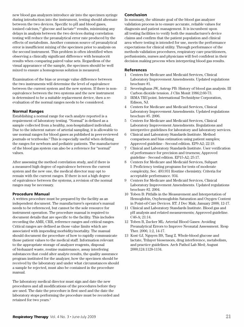
Respiratory Therapy Vol. 4 No. 3 n June-July 2009 21
new blood gas analyzers introduce air into the specimen syringe during introduction into the instrument, testing should alternate between the two devices. Specific to pH and blood gases, ionized calcium,12 glucose and lactate13 results, minimizing any delays in analysis between the two devices during correlation testing will reduce the preanalytical error rate produced by the effects of metabolism. Another common source of preanalytical error is insufficient mixing of the specimen prior to analysis on the second instrument. This problem is often identified when observing a clinically significant difference with hematocrit results when comparing paired value sets. Regardless of the visual appearance of the sample, the specimen should be well mixed to ensure a homogenous solution is measured.
Examination of the bias or average value difference between the two instruments will determine the level of equivalence between the current system and the new system. If there is non-equivalence between the two systems and the new instrument is determined to be a suitable replacement device, then a re-evaluation of the normal ranges needs to be considered.
Normal RangesEstablishing a normal range for each analyte reported is a requirement of laboratory testing. “Normal” is defined as a sample collected from a healthy, non-hospitalized individual. Due to the inherent nature of arterial sampling, it is allowable to use normal ranges for blood gases as published in peer-reviewed journals or textbooks.9 This is especially useful when defining the ranges for newborn and pediatric patients. The manufacturer of the blood gas system can also be a reference for “normal” values.
After assessing the method correlation study, and if there is a measured high degree of equivalence between the current system and the new one, the medical director may opt to remain with the current ranges. If there is not a high degree of equivalence between the systems, a revision of the normal ranges may be necessary.
Procedure ManualA written procedure must be prepared by the facility as an independent document. The manufacturer’s operator’s manual needs to be referenced, but cannot be the sole resource for instrument operation. The procedure manual is required to document details that are specific to the facility. This includes recording the AMR, CRR, reference ranges and critical ranges. Critical ranges are defined as those value limits which are associated with impending morbidity/mortality. The manual should document the procedure of how to rapidly communicate those patient values to the medical staff. Information relevant to the appropriate storage of analyzer reagents, disposal of biohazard waste, routine maintenance, assay interfering substances that could alter analyte results, the quality assurance program instituted for the analyzer, how the specimen should be received by the laboratory and under what circumstances should a sample be rejected, must also be contained in the procedure manual.
The laboratory medical director must sign and date the new procedures and all modifications of the procedures before they are used. The date the procedure is first used and the date the laboratory stops performing the procedure must be recorded and retained for two years.1
ConclusionIn summary, the ultimate goal of the blood gas analyzer validation process is to ensure accurate, reliable values for diagnosis and patient management. It is incumbent upon all testing facilities to verify both the manufacturer’s device claims and confirm that the patient population and clinical area where testing is intended for use, meets the performance expectations for clinical utility. Through performance of the methods validation procedures, respiratory care practitioners, perfusionists, nurses and physicians will feel confident in their decision making process when interpreting blood gas results.
ReferencesCenters for Medicare and Medicaid Services, Clinical 1 Laboratory Improvement Amendments. Updated regulations brochure #1.Severinghaus JW, Astrup PB: History of blood gas analysis. III 2 Carbon dioxide tension. J Clin Monit 1986;2:60-73.IRMA TRUpoint. International Technidyne Corporation, 3 Edison, NJ.Centers for Medicare and Medicaid Services, Clinical 4 Laboratory Improvement Amendments. Updated regulations brochure #5. 2006.Centers for Medicare and Medicaid Services, Clinical 5 Laboratory Improvement Amendments. Regulations and interpretive guidelines for laboratory and laboratory services.Clinical and Laboratory Standards Institute. Method 6 comparison and bias estimation using patient samples; Approved guideline - Second edition. EP9-A2; 22:19.Clinical and Laboratory Standards Institute. User verification 7 of performance for precision and trueness; Approved guideline - Second edition. EP15-A2; 25:17.Centers for Medicare and Medicaid Services, Subpart 8 I: Proficiency testing programs for tests of moderate complexity, Sec. 493.931 Routine chemistry. Criteria for acceptable performance. 934.Centers for Medicare and Medicaid Services, Clinical 9 Laboratory Improvement Amendments. Updated regulations brochure #2. 2004.Toben B: Pitfalls in the Measurement and Interpretation of 10 Hemoglobin, Oxyhemoglobin Saturation and Oxygen Content in Point-of-Care Devices. RT. J Dec Mak; January 2008; 12-17.Clinical and Laboratory Standards Institute. Blood gas and 11 pH analysis and related measurements; Approved guideline. C46-A; 21:14.Toben B, Zucker ML: Arterial Blood Gases: Avoiding 12 Preanalytical Errors to Improve Neonatal Assessment. Resp Ther. 2006; 1:2, 14-17.Kost GJ, Nguyen BS, Tang Z. Whole-blood glucose and 13 lactate, Trilayer biosensors, drug interference, metabolism, and practice guidelines. Arch Pathol Lab Med; August 2000;124:1128-1134.
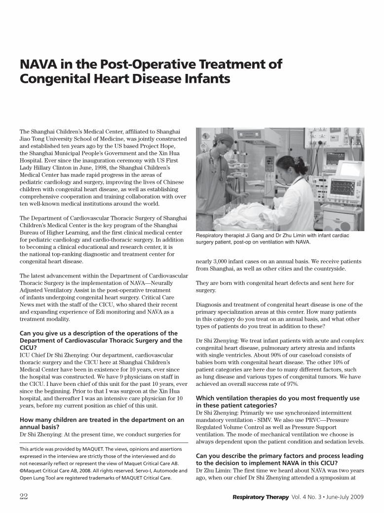
22 Respiratory Therapy Vol. 4 No. 3 n June-July 2009
The Shanghai Children’s Medical Center, affiliated to Shanghai Jiao Tong University School of Medicine, was jointly constructed and established ten years ago by the US based Project Hope, the Shanghai Municipal People’s Government and the Xin Hua Hospital. Ever since the inauguration ceremony with US First Lady Hillary Clinton in June, 1998, the Shanghai Children’s Medical Center has made rapid progress in the areas of pediatric cardiology and surgery, improving the lives of Chinese children with congenital heart disease, as well as establishing comprehensive cooperation and training collaboration with over ten well-known medical institutions around the world.
The Department of Cardiovascular Thoracic Surgery of Shanghai Children’s Medical Center is the key program of the Shanghai Bureau of Higher Learning, and the first clinical medical center for pediatric cardiology and cardio-thoracic surgery. In addition to becoming a clinical educational and research center, it is the national top-ranking diagnostic and treatment center for congenital heart disease.
The latest advancement within the Department of Cardiovascular Thoracic Surgery is the implementation of NAVA—Neurally Adjusted Ventilatory Assist in the post-operative treatment of infants undergoing congenital heart surgery. Critical Care News met with the staff of the CICU, who shared their recent and expanding experience of Edi monitoring and NAVA as a treatment modality.
Can you give us a description of the operations of the Department of Cardiovascular Thoracic Surgery and the CICU?ICU Chief Dr Shi Zhenying: Our department, cardiovascular thoracic surgery and the CICU here at Shanghai Children’s Medical Center have been in existence for 10 years, ever since the hospital was constructed. We have 9 physicians on staff in the CICU. I have been chief of this unit for the past 10 years, ever since the beginning. Prior to that I was surgeon at the Xin Hua hospital, and thereafter I was an intensive care physician for 10 years, before my current position as chief of this unit.
How many children are treated in the department on an annual basis?Dr Shi Zhenying: At the present time, we conduct surgeries for
nearly 3,000 infant cases on an annual basis. We receive patients from Shanghai, as well as other cities and the countryside.
They are born with congenital heart defects and sent here for surgery.
Diagnosis and treatment of congenital heart disease is one of the primary specialization areas at this center. How many patients in this category do you treat on an annual basis, and what other types of patients do you treat in addition to these?
Dr Shi Zhenying: We treat infant patients with acute and complex congenital heart disease, pulmonary artery atresia and infants with single ventricles. About 90% of our caseload consists of babies born with congenital heart disease. The other 10% of patient categories are here due to many different factors, such as lung disease and various types of congenital tumors. We have achieved an overall success rate of 97%.
Which ventilation therapies do you most frequently use in these patient categories?Dr Shi Zhenying: Primarily we use synchronized intermittent mandatory ventilation - SIMV. We also use PRVC—Pressure Regulated Volume Control as well as Pressure Support ventilation. The mode of mechanical ventilation we choose is always dependent upon the patient condition and sedation levels.
Can you describe the primary factors and process leading to the decision to implement NAVA in this CICU?Dr Zhu Limin: The first time we heard about NAVA was two years ago, when our chief Dr Shi Zhenying attended a symposium at
NAVA in the Post-Operative Treatment of Congenital Heart Disease Infants
Respiratory therapist Ji Gang and Dr Zhu Limin with infant cardiac surgery patient, post-op on ventilation with NAVA.
This article was provided by MAQUET. The views, opinions and assertions
expressed in the interview are strictly those of the interviewed and do
not necessarily reflect or represent the view of Maquet Critical Care AB.
©Maquet Critical Care AB, 2008. All rights reserved. Servo-I, Automode and
Open Lung Tool are registered trademarks of MAQUET Critical Care.
Respiratory Therapy Vol. 4 No. 3 n June-July 2009 23
the ESICM meeting in Barcelona, and she heard the lecture by Dr Christer Sinderby. She informed us of this new technology, and I received much information about NAVA, and I became very interested.
When did you have your first patient experience with NAVA, and how many infants have been treated with NAVA so far?Dr Zhu Limin: We had our first patient experience with NAVA only two months ago. We have placed the Edi catheter in about 16 patients, and treated about twelve patients with NAVA. The other four cases were babies with diaphragmatic paralysis, and since the babies were not spontaneously breathing, we could not use NAVA in those patients. However, in using the Edi catheter, we were able to monitor the Edi and detect the paralysis in these four cases. Some patients develop a bilateral diaphragmatic paralysis after surgery, so the Edi monitoring will confirm this by indicating no Edi signal.
How routinely is NAVA used in the ICU?Dr Zhu Limin: We have been gaining a lot of experience since we just started using NAVA only two months ago, so now we are selecting more difficult cases to gain even more experience with Edi monitoring and with NAVA. For the patients we have treated with NAVA, they have been on NAVA for a range of times, between a few hours up to three days, depending on their condition.
Is monitoring of the Edi signal used in conventional ventilatory modes, or in stand-by post-op after extubation?Dr Zhu Limin: For patients that have had diaphragmatic paralysis, we want to leave the Edi catheter in for 2-3 days to monitor the status of the diaphragm. Our surgeons need verification; so now we can give all this information to them, ultrasounds, Edi signals and X-rays, so that they see the actual condition of the diaphragm. It is very interesting and very useful for the surgeons. For our NAVA patients at post extubation, we leave the Edi catheter in for another 24 hours just to monitor the Edi, if we suspect the patient may re-develop something, or just to confirm that their spontaneous breathing is doing well on their own. We have been doing this as a routine for our NAVA patients. We have also monitored the Edi signals in other modes, such as Pressure Support, to monitor diaphragmatic status.
What in your opinion is the advantage or benefit of Edi monitoring as a bedside parameter?Dr Zhu Limin: I think it is very useful. For example, just in the
past two days, right after surgery we had a patient with a low Edi signal and very labored breathing, but after 12 hours the Edi signal became much stronger. We switched the patient to NAVA and yesterday we successfully extubated him, and now we are just monitoring the Edi signal. I think the Edi signal is very helpful and useful; you can get answers to all kinds of questions during the treatment process. It is a new bedside parameter for us. I think that maybe the Edi signal can tell us about sedation levels and the wash-out process and behavior patterns of the patient coming out of sedation that we were not able to see before.
How do you perceive NAVA from a therapeutic perspective?Dr Zhu Limin: From our CICU department, I think we have two patient perspectives. On the one hand, for the simple cases after cardiac surgery, you want to extubate them as safely and as quickly as possible. If you place the Edi catheter and use NAVA, and find the Edi signal returning after surgery, the patient can be extubated as early as possible. On the other hand, in complex cases, the patient needs mechanical ventilation for some longer lengths of time. If we use NAVA, the patient and ventilator are in synchrony, which means that the baby is more comfortable, and the dosage of sedation can be reduced. The Edi monitoring gives the opportunity to extubate earlier as well as monitor the sedation process.
Do you think esophageal ECG is valuable as a diagnostic tool in this patient category?Dr Zhu Limin: Esophageal ECG is very important for us, as our open heart surgery patients sometimes have post-operative arrhythmia. From the normal ECG, we cannot always see a clear
Intensive care physician Dr Zhu Limin, ICU Chief Dr Shi Zhenying and respiratory therapist Ji Gang.
Dr Zhu Limin and colleagues with patient on Edi monitoring, post-extubation after NAVA ventilation.
ICU Chief Dr Shi Zhenying surrounded by her staff members Bian Jun, Ji Gang, Zhu Limin and Liu Liping.

24 Respiratory Therapy Vol. 4 No. 3 n June-July 2009
diagnosis, such as SVT, or sometimes atrial tachycardia. If we have an esophageal ECG, we can see the correct diagnosis of the arrhythmia, which is very useful for our treatment.
Have you had any infant patient cases with NAVA of particular interest you would like to share?Dr Zhu Limin: We had a baby which came to the hospital at two months of age, who was suffering from transposition of the great arteries. An emergency operation was necessary, and it was very difficult to extubate him. We had three failed extubation attempts, with breathing difficulties and bronchospasm. We performed a bronchoscopy contrast CT, which revealed another problem, a vascular ring that compromised the trachea. Another surgery was performed after he had been in the CICU for about one month. After the second surgery, we placed the Edi catheter and treated the child with NAVA. He was spontaneously breathing with NAVA for about three days, followed by a successful extubation. One week later, we were able to finally discharge him. (Editors note: details of this patient case report may be found at criticalcarenews.com).
Are there specific staff members using NAVA, or has the general ICU staff received training?Dr Zhu Limin: All of the CICU staff has received training, doctors and nurses. We have also trained our RT group for special cases, so everyone has been educated. Our respiratory therapists place the Edi catheter and verify the positioning.
How do the respiratory therapists experience the Edi catheter placement and positioning process?Ji Gang, RT: It is not very difficult to place and position the Edi catheter, and we just monitor placement by means of the ECG signals. We use the Edi catheter as a normal feeding tube as well. As a team, we have a follow-up after each NAVA treatment for every case, so that we can all continue to learn about NAVA together.
What role do you think NAVA will have in the future in this patient population of congenital heart defects and disease?Dr Zhu Limin: I think that NAVA will be used increasingly in cardiac surgery, especially for pediatric patients, because of the opportunity of earlier extubation for simple cases post-op, and for complex cases, the opportunity to monitor Edi and
diaphragmatic status, in order to monitor and decrease dosage of sedation. Also, I think that the NAVA technique is easy to learn for any ICU staff member.
Do you think that your institution will be researching and expanding the use of NAVA in future?Dr Zhu Limin: I think that the research is very important, and we are planning to do some research in three areas. First, we would like to compare NAVA with traditional Pressure Support ventilation in terms of patient-ventilator synchrony as well as if we determine reduction of sedation dosages. Secondly, we are interested in research with NAVA to confirm the safety of hemodynamics in cardiac surgery patients. One other area of research we are interested in is to measure the Edi signal after extubation and chart and track to establish the normal range for children.
BiographyDr Shi Zhenying, MD, received her medical degree in 1975. She worked in Xinhua Hospital from 1975 to 1999, and was employed as physician of the cardiac intensive care unit there from 1989.
Dr Shi Zhenying has been the chief of the cardiac intensive care unit of Shanghai Children’s Medical Center, China since 2000. She was versed in the clinical and research work in perioperative treatment for congenital heart disease in children, especially in the prevention of low cardiac output syndrome and the treatment of multiple organ dysfunction syndrome. Dr Zhu Limin, MD, obtained her medical degree in 1999. Thereafter she was employed as a physician of the cardiac intensive care unit at Shanghai Children’s Medical Center, China. She received the fellowship of respiratory therapy and pediatric intensive care in Schneider Children’s Medical Center of Israel in 2006. She has been the manager of the team for respiratory management in the Cardiac Intensive Care Unit since 2006. She specializes in treatment of pulmonary hypertension and post-operative lung protective mechanical ventilation. From 2008, she has conducted clinical research of NAVA in neonates and pediatrics following cardiac surgery.
References1 Sinderby C, Beck J. Proportional Assist Ventilation and
Neurally Adjusted Ventilatory Assist—Better Approaches to Patient Ventilator Synchrony? Clin Chest Med 2008; 29(2):
The Shanghai Children’s Medical Center was established 10 years ago, and has become an internationally known center for treatment and research.
Respiratory therapist Liu Liping and Dr Zhu Limin.

Respiratory Therapy Vol. 4 No. 3 n June-July 2009 25
329-342.2 Sinderby C, Beck J. Neurally Adjusted Ventilatory Assist
(NAVA): An Update and Summary of Experiences. Neth J Crit Care 2007; 11(5): 243-252.
3 Emeriaud G, Beck J, Tucci M, Lacroix J, Sinderby C. Diaphragm electrical activity during expiration in mechanically ventilated infants. Pediatr Res 2006; 59(5): 705-710.
4 Beck J, Tuci M, Emeriaud G, Lacroix J, Sinderby C. Prolonged neural expiratory time induced by mechanical ventilation in infants. Pediatr Res 2004; 55(5): 747-754.
5 Sinderby C. Ventilatory assist driven by patient demand. Am J respire Crit Care Med 2003; 168(7): 729-730.
6 Sinderby C. Neurally adjusted ventilatory assist (NAVA). Minerva Anesthesiol 2002; 68(5): 378-380.
7 Sinderby C, Spahija J, Beck J, Kaminski D, Yan S, Comtois N, Sliwinski P. Diaphragm activation during exercise in chronic obstructive pulmonary disease. Am J Respir Crit Care Med 2001; 163(7): 1637-1642.
8 Sinderby C, Navalesi P, Beck J, Skrobik Y, Comtois N, Friberg S, Gottfried SB, Lindstrom L. Neural control of mechanical ventilation in respiratory failure. Nat Med 1999; 5(12): 1433-1436.
not eliminate the inefficiencies of other adopted measures, due to the fact that we utilized a retrospective rather than an interventional study design.
ConclusionIn summary, good air ventilation in wards and a series of simple control and preventive measures might decrease or prevent SARS transmission among HCWs in hospitals.
References1 Koh D, Lim MK, Chia SE: SARS: health care work can be
hazardous to health. Occup Med (Lond) 2003, 53: 241-3. 2 CDC: Severe acute respiratory syndrome – Singapore, 2003.
MMWR 2003, 52: 405-411. 3 Seto WH, Tsang D, Yung RWH, Ching TY, Ng TK, Ho M,
Ho, LM, Peiris JSM and Advisors of Expert SARS group of Hospital Authority: Effectiveness of precautions against droplets and contact in prevention of nosocomial transmission of severe acute respiratory syndrome (SARS). Lancet 2003, 361: 1519-1520.
4 CDC: Cluster of Severe acute respiratory syndrome cases among protected health-care workers– Toronto, Canada, April 2003. MMWR 2003, 52: 433-436.
5 CDC: Severe acute respiratory syndrome – Taiwan, 2003. MMWR 2003, 52: 461-466.
6 Jiang S, Huang L, Chen X, Wang J, Wu W, Yin S, Chen W, Zhan J, Yan L, Ma L, Li J, Huang Z: Ventilation of wards and nosocomial outbreak of severe acute respiratory syndrome among healthcare workers. Chin Med J 2003, 116: 1293-1297.
7 Lau JT, Fung KS, Wong TW, Kim JH, Wong E, Chung S, Ho, D, Chan LY, Liu SF, Cheng A: SARS transmission among hospital workers in Hong Kong. Emerg Infect Dis 2004, 10: 280-286.
8 Varia M, Wilson S, Sarwal S, McGeer A, Gournis E, Henry B, hospital Outbreak Investigation Team: Investigation of a nosocomial outbreak of severe acute respiratory syndrome (SARS) in Toronto, Canada. CMAJ 2003,169:285-292.
9 CDC: Update: outbreak of severe acute respiratory syndrome – worldwide, 2003. MMWR 2003, 52: 241-248.
10 Ho, AS, Sung JJY, Chan-Yeung M: An outbreak of severe acute respiratory syndrome among hospital workers in a community hospital in Hong Kong. Ann Intern Med 2003, 139: 564-7.
11 Lau JTF, Yang X, Leung PC, Chan L, Wong E, Fong C, Tsui HY: SARS in three categories of hospital workers, Hong Kong. Emerg Infect Dis 2004, 10: 1399-404.
12 Liang W, Zhu Z, Guo J, Liu Z, Zhou W, Chin DP, Schuchat A, Beijing Joint SARS Expert Group: Severe acute respiratory syndrome, Beijing, 2003. Emerg Infect Dis 2004, 10: 25-31.
13 Chen WQ, Lu CY, Wong TW, Ling WH, Lin ZN, Hao YT, Liu Q, Fang JQ, He Y, Luo FT, Jing J, Ling L, Ma X, Liu YM, Chen GH, Huang J, Jiang YS< Jiang WQ, Zou HQ, Yan GM: Anti-SARS-CoV immunoglobulin G in healthcare workers, Guangzhou, China. Emerg Infect Dis 2005, 11: 89-94.
14 SPSS: SPSS Base 11.5 User’s Guide. SPSS Inc.: Chicago, 2002. 15 Wong TW, Chen WQ: The Epidemiology of SARS. In.
Severe Acute Respiratory Syndrome. Edited by: Sung JJY. Singapore:World Scientific Printers, 2004: 9-21.
16 Wong RSM, Hui DS. Index patient and SARS outbreak in Hong Kong. Emerg Infect Dis 2004; 10: 339-341.
17 Wong TW, Lee CK, Tam W, Lau JT, Yu TS, Liu SF, Chan PK, Li Y, Bresee JS, Sung JJ, Parashar UD, Outbreak Study Group: Cluster of SARS among medical students exposed to single patient, Hong Kong. Emerg Infect Dis 2004, 10: 269-276.
18 Olsen SJ, Chang HL, Cheung TYY, Tang AF, Fisk TL, Ooi SP, Kuo HW, Jiang DD, Chen KT, Lando J, Hsu KH, Chen TJ, Dowell SF: Transmission of the Sever Acute Respiratory Syndrome on Aircraft. N Engl J Med 2003, 349: 2416-2422.
19 Yu IT, Li Y, Wong TW, Tam W, Chan AT, Lee JH, Leung DY, Ho T: Evidence of airborne transmission of the Sever Acute Respiratory Syndrome Virus. N Engl J Med 2004, 350: 1731-1739.
20 Twu SJ, Chen TZ, Chen CJ, Olsen SJ, Lee LT, Fisk T, Hsu KH, Chang SC, Chen KT, Chiang IH, Wu YC, Wu JS, Dowell SF: Control measures for severe acute respiratory syndrome (SARS) in Taiwan. Emerg Infect Dis 2003, 9: 718-720.
21 Yu IT, Xie ZH, Tsio KK, Chiu YL, Tang XP, Hui DS, Lee N, Li YM, Huang ZT, Liu T, Wong ZW, Zhong NS, Sung JJ: Why did outbreak of severe acute respiratory syndrome occur in some hospitals but not in others? CID 2007, 44: 1017-1025.
SARS…continued from page 29
26 Respiratory Therapy Vol. 4 No. 3 n June-July 2009
Abstract Background: Despite the use of a series of preventive measures, a high incidence of severe acute respiratory syndrome (SARS) was observed among health care workers (HCWs) during the SARS epidemic. This study aimed to determine which preventive measures may have been effective in protecting HCWs from infection, and which were not effective.
Methods: A retrospective study was performed among 758 ‘frontline’ health care workers who cared for SARS patients at the Second Affiliated Hospital and the Third Affiliated Hospital of Sun Yat-sen University. The HCWs with IgG against SARS and those without IgG against SARS were respectively defined as the ‘case group’ and the ‘control group,’ and logistic regression was conducted to explore the risk factors for SARS infection in HCWs.
Results: After adjusting for age, gender, marital status, educational level, professional title, and the department in which an individual worked, the results of a multivariate logistic regression analysis indicated that incidence of SARS among HCWs was significantly and positively associated with: performing tracheal intubations for SARS patients, methods used for air ventilation in wards, avoiding face-to-face interaction with SARS patients, the number of pairs of gloves worn by HCWs, and caring for serious SARS cases.
Conclusions: Some measures, particularly good air ventilation in SARS wards, may be effective in minimizing or preventing SARS transmission among HCWs in hospitals.
BackgroundSevere acute respiratory syndrome (SARS), a viral respiratory illness caused by the coronavirus SARS-CoV was possibly the
first globally significant occupational disease to emerge in the 21st century, making healthcare work potentially hazardous.1 This was indicated by the high incidence of SARS observed among health care workers (HCWs) in the epidemic of SARS, especially during its earlier stages.2-5 In China, from a total of 5,323 SARS cases, 966 (over 18%) were HCWs, and in the early period of the SARS epidemic, near 90% of the SARS patients were frontline HCWs.6 In Hong Kong, a total of 384 (22.1%) of 1739 suspected or confirmed SARS patients were hospital workers.7 Generally, SARS outbreaks first originated in hospitals where SARS patients were treated and subsequently spread to communities from there.8,9
Several studies indicated that HCWs coming into direct or indirect contact with SARS patients in wards had a greatly increased risk of contracting SARS-Cov, despite some strict infection control measures being taken.7,10,11 A similar situation also arose in the Second Affiliated Hospital and the Third Affiliated Hospital of Sun Yat-sen University during the epidemic of SARS in 2003. A total of 846 HCWs worked on the frontline of caring for SARS patients in the two affiliated hospitals and 112 of them contracted SARS during this time. Throughout the whole period of the SARS epidemic, a series of infection control and protective measures were employed in the two affiliated hospitals. But, why were some of HCWs infected by SARS, and some of them were not? The objective of this study was to determine which preventive measures used were effective in protecting HCWs from SARS, and which were not effective. To answer this question, we conducted a retrospective study of HCWs who worked at the frontline in the two affiliated hospitals during the SARS epidemic.
MethodsStudy population: In mid-May 2003, about 4 months after the initial SARS outbreak in Guangzhou, a retrospective study was conducted in HCWs working at the frontline of the SARS epidemic, a providing primary care in the Second Affiliated Hospital and the Third Affiliated Hospital of Sun Yat-sen Unviersity, where the first and second outbreak of SARS among HCWs occurred in the early stage of SARS epidemic in Guangzhou. Among a total of 846 frontline HCWs who tended to SARS patients from the two hospitals, 758 (89.2%) who were on duty during the investigation were surveyed, and the included HCWs from all departments involved in the care of SARS patients in the two hospitals. But, those who were off-duty during the survey were excluded. During the SARS epidemic, a total of 112 HCWs working on the frontline were diagnosed suffering from “SARS” according to a case definition of SARS by the Ministry of Health, China12 and 90 of them were successfully
Which Preventive Measures Might Protect Healthcare Workers From SARS?Wei-Qing Chen, Wen-Hua Ling, Ci-Yong Lu, Yuan-Tao Hao, Zhong-Ning Lin, Li Ling, Jian Huang, Gang Li, Guang-Mei Yan
Authors Chen, Lu and Ling are with the Department of Biostatistics and
Epidemology; Li Ling is with the Department of Nutrition; Lin is with the
Department of Medicine, School of Public Health; Hao and Huang are
with the Department of Surgery, Second Affiliated Hospital; Li is with the
Department of Infeection, Third Affiliated Hospital; and Yan is with the
Department of Pharmacology, Zhongshan School of Medicine, Sun Yat-
Sen University, Guangzhou, China. The authors would like to thank the
management and healthcare workers in the Second and Third Affiliated
Hospitals of Sun Yat-sen University. This project was supported by a
research grant from both the Ministry of Science and Technology and
the Ministry of Education of China. Reprinted from BioMed Central, BMC
Public Health, © 2009 Chen et al , licensee BioMed Central Ltd. This is an
open access article distributed under the terms of the Creative Commons
Attribution License.

Respiratory Therapy Vol. 4 No. 3 n June-July 2009 27
interviewed, giving a response rate of 80.4% (90/112). Written informed consent was obtained from all the participants prior to enrollment after a detailed explanation of the study objectives and requirements of the survey. The Ethical Committee of the Sun Yat-sen University approved the study.
Definition of a SARS case: A SARS case was defined using the criteria for probable SARS cases provided by the Health Ministry of China.12 Criteria for probable and suspected SARS cases included travel to a SARS epidemic area in the 2 weeks before the onset of symptoms or close contact with a probable SARS patient, fever of ≥38˚C; chest x-ray abnormalities; normal or decreased leukocycte count; and no response to treatment by antimicrobial drugs.
In the present study, 10 mL of peripheral venous blood was collected from all the subjects, and the serum was separated and stored at -70˚C. Immunoglobulin (Ig) G against SARS-CoV was detected using an enzyme-linked immunosorbent assay (ELISA).13 Among the 758 surveyed HCWs, 91 ones (80 cases were diagnosed suffering from “SARS” and 11 were not diagnosed suffering from “SARS”) had IgG antibodies against SARS, and the prevalence rate of IgG antibodies against SARS was 12.01% for the total samples.13 Furthermore, the prevalence of IgG antibodies against SARS-CoV was 88.9% (80/90) for HCW with SARS [86.3% (63/73) in the Second Affiliated Hospital and 100.0% (17/17) in the Third Affiliated Hospital], and 1.6% (11/668) for HCW without SARS who worked on frontline of SARS [2.8% (8/288) in the Second Affiliated Hospital and 0.8% (3/380) in the Third Affiliated Hospital.]13
Data collection: A standardized interview with a structured questionnaire was used to obtain the following information in mid-May 2003, about 4 months after the initial SARS outbreak in Guangzhou. (1) Socio-demographic characteristics, including: age, gender, marital status, educational level, and professional title, and in which department did you work? (2) History of SARS patient care, including: (i) Did you receive any special training for how to handle SARS? (ii) Did you ever perform a tracheotomy? (iii) Did you ever perform tracheal intubations? (iv) Did you ever care for ‘Super Spreading SARS cases’? (3) Use of personal protective and control measures, including: (i) How many gowns did you wear while you cared for SARS patients? (ii) How many multilayered cotton masks did you wear while you cared for SARS patients? (iii) How many pairs of gloves did you wear while you cared for SARS patients? (iv) With what frequency did you wear shoe covers while you cared for SARS patients? (v) With what frequency did you wear a cap while you cared for SARS patients? (vi) With what frequency did you wear a face shield while you worked in SARS wards? (vii) with what frequency did you wear goggles while you performed operations on SARS patients? (4) Health-related behaviors included: (i) With what frequency did you wash uncovered skin after you cared for SARS patients? (ii) With what frequency did you wash hands after you cared for SARS patients? (iii) With what frequency did you wash your nasal cavity after you cared for SARS patients? (iv) With what frequency did you wash your mouth after you cared for SARS patients? (5) Other relevant control measures were: (i) What type of air ventilation system was used in your office and in SARS wards? (ii) What type of hand-washing equipment was used in your office? More details about the name, definition and value of these variables are listed in Table 1.
Data analysis: HCWs who had IgG against SARS (91 cases = 80 cases with SARS and 11 cases without SARS) and those without
� 19
Table�1:�Variables�and�definition�
Variable�name� Definition�
Socio-demographics� �
Age� Year�
Gender� 1=Male,�2=Female�
Marital�status� 1=Unmarried,�2=Married,�3=Others�
Educational�level� 1=Senior� school,� 2=Technical� secondary� school,� 3=Junior�
college,�4=university,�5=Master�degree,�6=PhD�
Professional�title� 1=Doctor,� 2=Nurse,� 3=Health� attendant,� 4=Technician� in�
laboratory,�5=Others�
Department� 1=SARS� ward,� 2=Emergency� department/Fever� clinic,�
3=Infectious�disease,�4=Respiratory�disease,�5=Others�
Use�of�personal�protective�and�control�measures� �
Number�of�gowns�worn� 0=Single,�1=Double�
Number�of�multilayered�cotton�mask�worn� 0=Single,�1=Double�
Number�of�pairs�of�gloves�worn� 0=Single,�1=double�
Frequency�of�wearing�shoe�cover� 0=Never,�1=Sometimes,�2=Often,�3=Every�time�
Frequency�of�wearing�cap� 0=Never,�1=Sometimes,�2=Often,�3=Every�time�
Frequency�of�face�shield�in�SARS�ward� 0=Never,�1=Sometimes,�2=Often,�3=Every�time�
Frequency�of�wearing�goggles�while�performing�operation�for�SARS�
patients�
0=Never,�1=Sometimes,�2=Often,�3=Every�time�
Health-related�behaviors� �
Frequency�of�washing�uncovered�skin�after�caring�for�SARS�patients� 0=Never,�1=Sometimes,�2=Often,�3=Every�time�
Frequency�of�washing�hands�after�caring�for�SARS�patients� 0=Never,�1=Sometimes,�2=Often,�3=Every�time�
Frequency�of�washing�nasal�cavity�after�caring�for�SARS�patients� 0=Never,�1=Sometimes,�2=Often,�3=Every�time�
Frequency�of�washing�oral�cavity�after�caring�for�SARS�patients� 0=Never,�1=Sometimes,�2=Often,�3=Every�time�
SARS�patient�care� �
Special�training�for�SARS� � 0=No,�1=Yes� �
Performing�tracheotomy� 0=No,�1=Yes�
Performing�tracheal�intubations� � 0=No,�1=Yes�
Caring�for�“Super�Spreading�Patient”� 0=No,�1=Yes�
Avoiding�face�to�face�while�caring�for�patient� 0=Never,�1=Sometimes,�2=Often,�3=Every�time�
Other�relevant�control�measures� �
Method�of�air�ventilation�in�offices�and�SARS�wards� 1=Artificial�central�ventilation�(windows�were�closed�in�wards),�
2=Natural� ventilation� (windows� were� opened� in� wards),�
3=Natural� ventilation� and� additional� electronic� exhaust� fan�
(windows� were� opened� in� wards,� at� the� same� time,� electronic�
exhaust�fans�were�used�for�improving�air�circulation�in�wards)�
Type�of�equipment�for�washing�hands� 1=Automatic�tap,�2=Non-automatic�tap,�3=Others�
� 20
Table�2:�Socio-demographic�characteristics�of�HCWs�in�the�two�affiliated�hospitals�
Second�Affiliated�Hospital� � Third�Affiliated�Hospital��
Socio-demographic�characteristics� No.� %� � No.� %�
Age�(years)� � � � � �
<26� 152� 42.1� � 113� 28.5�
26~30� 83� 23.0� � 126� 31.7�
31~35� 59� 16.3� � 67� 16.9�
36~40� 37� 10.3� � 39� 9.8�
>40� 30� 8.3� � 52� 13.1�
Gender� � � � � �
Male� 80� 22.2� � 101� 25.5�
Female� 281� 77.8� � 295� 74.5�
Marital�status� � � � � �
Unmarried� 209� 57.9� � 172� 43.3�
Married� 147� 40.7� � 213� 53.7�
Others� 5� 1.4� � 12� 3.0�
Educational�level� � � � � �
Senior�school� 17� 4.7� � 46� 11.6�
Technical�secondary�school� 157� 43.5� � 138� 34.8�
Junior�college� 58� 16.1� � 72� 18.1�
University� 74� 20.5� � 43� 10.8�
MD/PhD� 55� 15.2� � 98� 24.7�
Department� � � � � �
SARS�ward� � 227� 68.2� � 111� 33.8�
Emergency�department/Fever�clinic� 25� 7.5� � 52� 15.9�
Infectious�disease�department� 5� 1.5� � 120� 36.6�
Respiratory�diseases�department� 42� 12.6� � 15� 4.6�
Others� 34� 10.2� � 30� 9.1�
Professional�title� � � � � �
Doctor� 105� 29.1� � 134� 33.8�
Nurse� 199� 55.1� � 174� 43.8�
Health�attendant� 15� 4.2� � 40� 10.1�
Technician�in�laboratory� 25� 6.9� � 13� 3.3�
Others� 17� 4.7� � 36� 9.1�
�
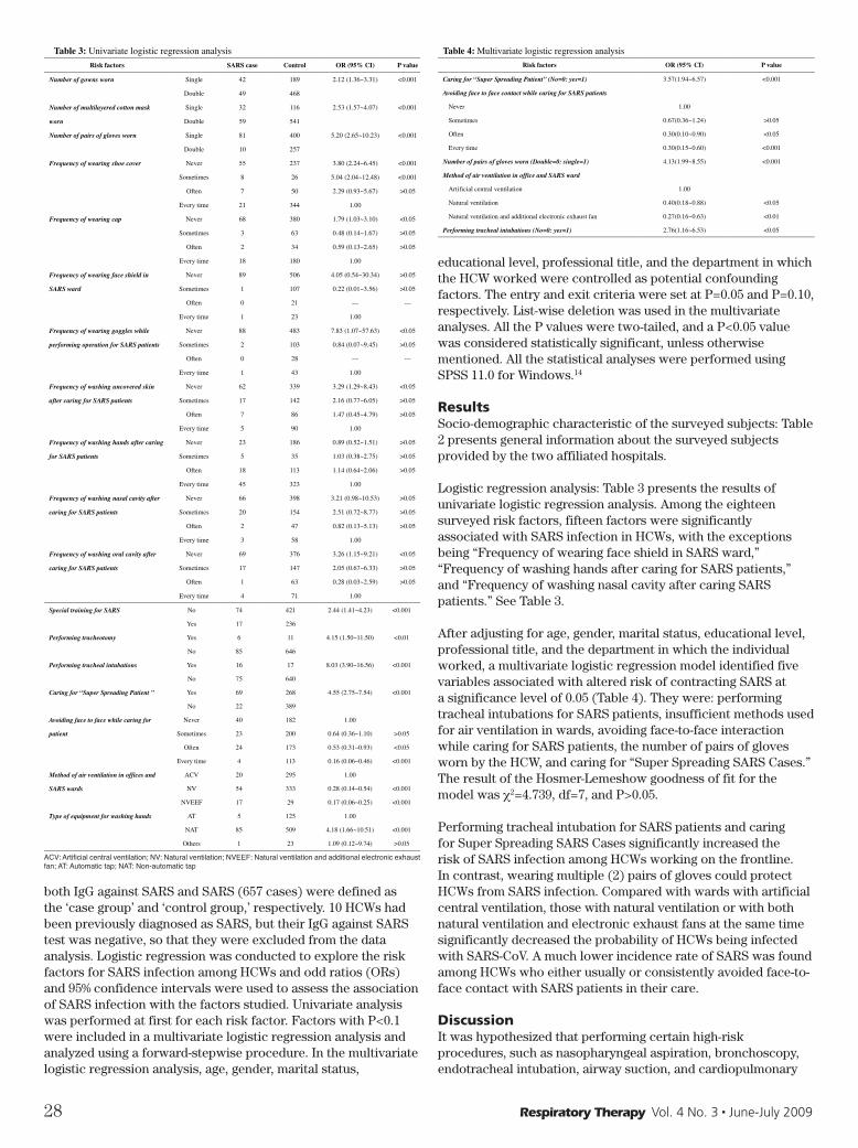
28 Respiratory Therapy Vol. 4 No. 3 n June-July 2009
both IgG against SARS and SARS (657 cases) were defined as the ‘case group’ and ‘control group,’ respectively. 10 HCWs had been previously diagnosed as SARS, but their IgG against SARS test was negative, so that they were excluded from the data analysis. Logistic regression was conducted to explore the risk factors for SARS infection among HCWs and odd ratios (ORs) and 95% confidence intervals were used to assess the association of SARS infection with the factors studied. Univariate analysis was performed at first for each risk factor. Factors with P<0.1 were included in a multivariate logistic regression analysis and analyzed using a forward-stepwise procedure. In the multivariate logistic regression analysis, age, gender, marital status,
educational level, professional title, and the department in which the HCW worked were controlled as potential confounding factors. The entry and exit criteria were set at P=0.05 and P=0.10, respectively. List-wise deletion was used in the multivariate analyses. All the P values were two-tailed, and a P<0.05 value was considered statistically significant, unless otherwise mentioned. All the statistical analyses were performed using SPSS 11.0 for Windows.14
ResultsSocio-demographic characteristic of the surveyed subjects: Table 2 presents general information about the surveyed subjects provided by the two affiliated hospitals.
Logistic regression analysis: Table 3 presents the results of univariate logistic regression analysis. Among the eighteen surveyed risk factors, fifteen factors were significantly associated with SARS infection in HCWs, with the exceptions being “Frequency of wearing face shield in SARS ward,” “Frequency of washing hands after caring for SARS patients,” and “Frequency of washing nasal cavity after caring SARS patients.” See Table 3.
After adjusting for age, gender, marital status, educational level, professional title, and the department in which the individual worked, a multivariate logistic regression model identified five variables associated with altered risk of contracting SARS at a significance level of 0.05 (Table 4). They were: performing tracheal intubations for SARS patients, insufficient methods used for air ventilation in wards, avoiding face-to-face interaction while caring for SARS patients, the number of pairs of gloves worn by the HCW, and caring for “Super Spreading SARS Cases.” The result of the Hosmer-Lemeshow goodness of fit for the model was c2=4.739, df=7, and P>0.05.
Performing tracheal intubation for SARS patients and caring for Super Spreading SARS Cases significantly increased the risk of SARS infection among HCWs working on the frontline. In contrast, wearing multiple (2) pairs of gloves could protect HCWs from SARS infection. Compared with wards with artificial central ventilation, those with natural ventilation or with both natural ventilation and electronic exhaust fans at the same time significantly decreased the probability of HCWs being infected with SARS-CoV. A much lower incidence rate of SARS was found among HCWs who either usually or consistently avoided face-to-face contact with SARS patients in their care.
DiscussionIt was hypothesized that performing certain high-risk procedures, such as nasopharyngeal aspiration, bronchoscopy, endotracheal intubation, airway suction, and cardiopulmonary
� 21
� Table�3:�Univariate�logistic�regression�analysis� �
Risk�factors� � SARS�case� Control� OR�(95%�CI)� P�value�
Number�of�gowns�worn� Single� 42� 189� 2.12�(1.36~3.31)� <0.001�
� Double� 49� 468� � �
Number�of�multilayered�cotton�mask� � Single� 32� 116� 2.53�(1.57~4.07)� <0.001�
worn� Double� 59� 541� � �
Number�of�pairs�of�gloves�worn� Single� 81� 400� 5.20�(2.65~10.23)� <0.001�
� Double� 10� 257� � �
Frequency�of�wearing�shoe�cover� Never� 55� 237� 3.80�(2.24~6.45)� <0.001�
� Sometimes� 8� 26� 5.04�(2.04~12.48)� <0.001�
� Often� 7� 50� 2.29�(0.93~5.67)� >0.05�
� Every�time� 21� 344� 1.00� �
Frequency�of�wearing�cap� Never� 68� 380� 1.79�(1.03~3.10)� <0.05�
� Sometimes� 3� 63� 0.48�(0.14~1.67)� >0.05�
� Often� 2� 34� 0.59�(0.13~2.65)� >0.05�
� Every�time� 18� 180� 1.00� �
Frequency�of�wearing�face�shield�in� � Never� 89� 506� 4.05�(0.54~30.34)� >0.05�
SARS�ward� Sometimes� 1� 107� 0.22�(0.01~3.56)� >0.05�
� Often� 0� 21� ---� ---�
� Every�time� 1� 23� 1.00� �
Frequency�of�wearing�goggles�while� � Never� 88� 483� 7.83�(1.07~57.63)� <0.05�
performing�operation�for�SARS�patients� Sometimes� 2� 103� 0.84�(0.07~9.45)� >0.05�
� Often� 0� 28� ---� ---�
� Every�time� 1� 43� 1.00� �
Frequency�of�washing�uncovered�skin� Never� 62� 339� 3.29�(1.29~8.43)� <0.05�
after�caring�for�SARS�patients� Sometimes� 17� 142� 2.16�(0.77~6.05)� >0.05�
� Often� 7� 86� 1.47�(0.45~4.79)� >0.05�
� Every�time� 5� 90� 1.00� �
Frequency�of�washing�hands�after�caring� Never� 23� 186� 0.89�(0.52~1.51)� >0.05�
for�SARS�patients� Sometimes� 5� 35� 1.03�(0.38~2.75)� >0.05�
� Often� 18� 113� 1.14�(0.64~2.06)� >0.05�
� Every�time� 45� 323� 1.00� �
Frequency�of�washing�nasal�cavity�after� Never� 66� 398� 3.21�(0.98~10.53)� >0.05�
caring�for�SARS�patients� Sometimes� 20� 154� 2.51�(0.72~8.77)� >0.05�
� Often� 2� 47� 0.82�(0.13~5.13)� >0.05�
� Every�time� 3� 58� 1.00� �
Frequency�of�washing�oral�cavity�after� Never� 69� 376� 3.26�(1.15~9.21)� <0.05�
caring�for�SARS�patients� Sometimes� 17� 147� 2.05�(0.67~6.33)� >0.05�
� Often� 1� 63� 0.28�(0.03~2.59)� >0.05�
� Every�time� 4� 71� 1.00� �
�
� 22
Table�3:�Univariate�logistic�regression�analysis�(continued)�
Risk�factors� � SARS�case� Control� OR�(95%�CI)� P�value�
Special�training�for�SARS� No� 74� 421� 2.44�(1.41~4.23)� <0.001�
� Yes� 17� 236� � �
Performing�tracheotomy� Yes� 6� 11� 4.15�(1.50~11.50)� <0.01�
� No� 85� 646� � �
Performing�tracheal�intubations� Yes� 16� 17� 8.03�(3.90~16.56)� <0.001�
� No� 75� 640� � �
Caring�for�“Super�Spreading�Patient�”� Yes� 69� 268� 4.55�(2.75~7.54)� <0.001�
� No� 22� 389� � �
Avoiding�face�to�face�while�caring�for� � Never� 40� 182� 1.00� �
patient� Sometimes� 23� 200� 0.64�(0.36~1.10)� >0.05�
� Often� 24� 173� 0.53�(0.31~0.93)� <0.05�
� Every�time� 4� 113� 0.16�(0.06~0.46)� <0.001�
Method�of�air�ventilation�in�offices�and� � ACV� 20� 295� 1.00� �
SARS�wards� NV� 54� 333� 0.28�(0.14~0.54)� <0.001�
� NVEEF� 17� 29� 0.17�(0.06~0.25)� <0.001�
Type�of�equipment�for�washing�hands� AT� 5� 125� 1.00� �
� NAT� 85� 509� 4.18�(1.66~10.51)� <0.001�
� Others� 1� 23� 1.09�(0.12~9.74)� >0.05�
ACV:�Artificial�central�ventilation;�NV:�Natural�ventilation;�NVEEF:�Natural�ventilation�and�additional�electronic�exhaust�fan;�AT:�Automatic�tap;�NAT:�Non-automatic�tap�
�
�
�
Table�4:�Multivariate�logistic�regression�analysis� �
Risk�factors� OR�(95%�CI)� P�value�
Caring�for�“Super�Spreading�Patient”�(No=0:�yes=1)� 3.57(1.94~6.57)� <0.001�
Avoiding�face�to�face�contact�while�caring�for�SARS�patients� � �
Never� 1.00� �
Sometimes� 0.67(0.36~1.24)� >0.05�
Often� 0.30(0.10~0.90)� <0.05�
Every�time� 0.30(0.15~0.60)� <0.001�
Number�of�pairs�of�gloves�worn�(Double=0:�single=1)� 4.13(1.99~8.55)� <0.001�
Method�of�air�ventilation�in�office�and�SARS�ward� � �
Artificial�central�ventilation� 1.00� �
Natural�ventilation� 0.40(0.18~0.88)� <0.05�
Natural�ventilation�and�additional�electronic�exhaust�fan� � 0.27(0.16~0.63)� <0.01�
Performing�tracheal�intubations�(No=0:�yes=1)� 2.76(1.16~6.53)� <0.05�
�
�
�
ACV: Artificial central ventilation; NV: Natural ventilation; NVEEF: Natural ventilation and additional electronic exhaust fan; AT: Automatic tap; NAT: Non-automatic tap
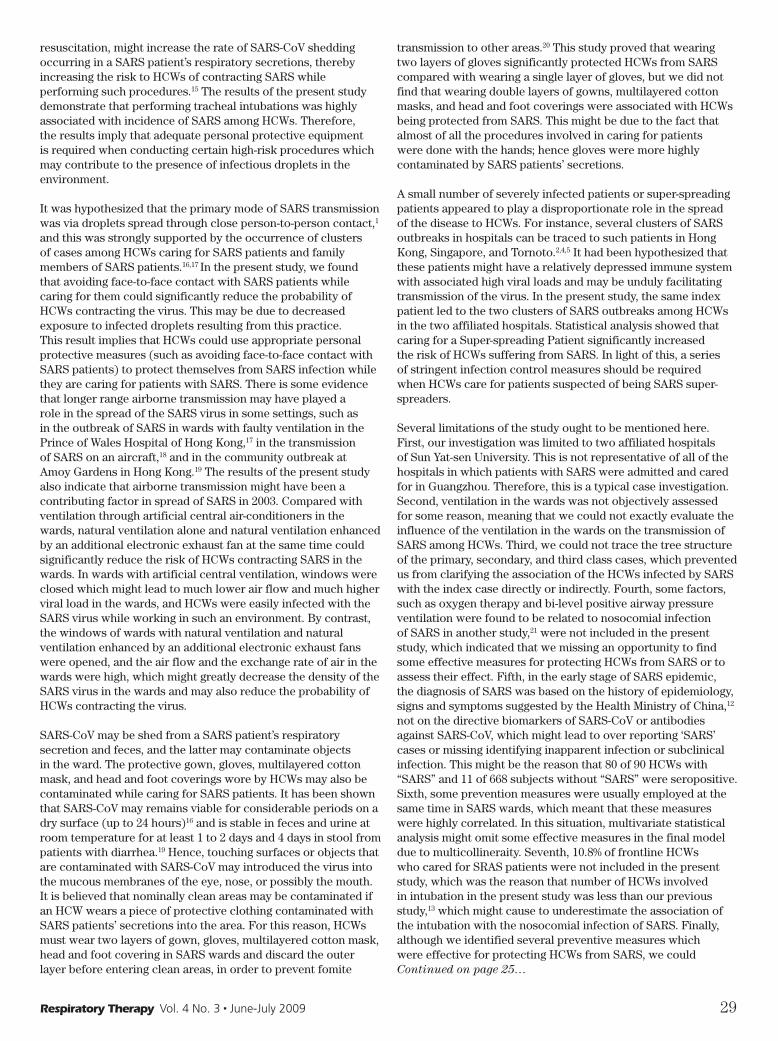
Respiratory Therapy Vol. 4 No. 3 n June-July 2009 29
resuscitation, might increase the rate of SARS-CoV shedding occurring in a SARS patient’s respiratory secretions, thereby increasing the risk to HCWs of contracting SARS while performing such procedures.15 The results of the present study demonstrate that performing tracheal intubations was highly associated with incidence of SARS among HCWs. Therefore, the results imply that adequate personal protective equipment is required when conducting certain high-risk procedures which may contribute to the presence of infectious droplets in the environment.
It was hypothesized that the primary mode of SARS transmission was via droplets spread through close person-to-person contact,1 and this was strongly supported by the occurrence of clusters of cases among HCWs caring for SARS patients and family members of SARS patients.16,17 In the present study, we found that avoiding face-to-face contact with SARS patients while caring for them could significantly reduce the probability of HCWs contracting the virus. This may be due to decreased exposure to infected droplets resulting from this practice. This result implies that HCWs could use appropriate personal protective measures (such as avoiding face-to-face contact with SARS patients) to protect themselves from SARS infection while they are caring for patients with SARS. There is some evidence that longer range airborne transmission may have played a role in the spread of the SARS virus in some settings, such as in the outbreak of SARS in wards with faulty ventilation in the Prince of Wales Hospital of Hong Kong,17 in the transmission of SARS on an aircraft,18 and in the community outbreak at Amoy Gardens in Hong Kong.19 The results of the present study also indicate that airborne transmission might have been a contributing factor in spread of SARS in 2003. Compared with ventilation through artificial central air-conditioners in the wards, natural ventilation alone and natural ventilation enhanced by an additional electronic exhaust fan at the same time could significantly reduce the risk of HCWs contracting SARS in the wards. In wards with artificial central ventilation, windows were closed which might lead to much lower air flow and much higher viral load in the wards, and HCWs were easily infected with the SARS virus while working in such an environment. By contrast, the windows of wards with natural ventilation and natural ventilation enhanced by an additional electronic exhaust fans were opened, and the air flow and the exchange rate of air in the wards were high, which might greatly decrease the density of the SARS virus in the wards and may also reduce the probability of HCWs contracting the virus.
SARS-CoV may be shed from a SARS patient’s respiratory secretion and feces, and the latter may contaminate objects in the ward. The protective gown, gloves, multilayered cotton mask, and head and foot coverings wore by HCWs may also be contaminated while caring for SARS patients. It has been shown that SARS-CoV may remains viable for considerable periods on a dry surface (up to 24 hours)16 and is stable in feces and urine at room temperature for at least 1 to 2 days and 4 days in stool from patients with diarrhea.19 Hence, touching surfaces or objects that are contaminated with SARS-CoV may introduced the virus into the mucous membranes of the eye, nose, or possibly the mouth. It is believed that nominally clean areas may be contaminated if an HCW wears a piece of protective clothing contaminated with SARS patients’ secretions into the area. For this reason, HCWs must wear two layers of gown, gloves, multilayered cotton mask, head and foot covering in SARS wards and discard the outer layer before entering clean areas, in order to prevent fomite
transmission to other areas.20 This study proved that wearing two layers of gloves significantly protected HCWs from SARS compared with wearing a single layer of gloves, but we did not find that wearing double layers of gowns, multilayered cotton masks, and head and foot coverings were associated with HCWs being protected from SARS. This might be due to the fact that almost of all the procedures involved in caring for patients were done with the hands; hence gloves were more highly contaminated by SARS patients’ secretions.
A small number of severely infected patients or super-spreading patients appeared to play a disproportionate role in the spread of the disease to HCWs. For instance, several clusters of SARS outbreaks in hospitals can be traced to such patients in Hong Kong, Singapore, and Tornoto.2,4,5 It had been hypothesized that these patients might have a relatively depressed immune system with associated high viral loads and may be unduly facilitating transmission of the virus. In the present study, the same index patient led to the two clusters of SARS outbreaks among HCWs in the two affiliated hospitals. Statistical analysis showed that caring for a Super-spreading Patient significantly increased the risk of HCWs suffering from SARS. In light of this, a series of stringent infection control measures should be required when HCWs care for patients suspected of being SARS super-spreaders.
Several limitations of the study ought to be mentioned here. First, our investigation was limited to two affiliated hospitals of Sun Yat-sen University. This is not representative of all of the hospitals in which patients with SARS were admitted and cared for in Guangzhou. Therefore, this is a typical case investigation. Second, ventilation in the wards was not objectively assessed for some reason, meaning that we could not exactly evaluate the influence of the ventilation in the wards on the transmission of SARS among HCWs. Third, we could not trace the tree structure of the primary, secondary, and third class cases, which prevented us from clarifying the association of the HCWs infected by SARS with the index case directly or indirectly. Fourth, some factors, such as oxygen therapy and bi-level positive airway pressure ventilation were found to be related to nosocomial infection of SARS in another study,21 were not included in the present study, which indicated that we missing an opportunity to find some effective measures for protecting HCWs from SARS or to assess their effect. Fifth, in the early stage of SARS epidemic, the diagnosis of SARS was based on the history of epidemiology, signs and symptoms suggested by the Health Ministry of China,12
not on the directive biomarkers of SARS-CoV or antibodies against SARS-CoV, which might lead to over reporting ‘SARS’ cases or missing identifying inapparent infection or subclinical infection. This might be the reason that 80 of 90 HCWs with “SARS” and 11 of 668 subjects without “SARS” were seropositive. Sixth, some prevention measures were usually employed at the same time in SARS wards, which meant that these measures were highly correlated. In this situation, multivariate statistical analysis might omit some effective measures in the final model due to multicollineraity. Seventh, 10.8% of frontline HCWs who cared for SRAS patients were not included in the present study, which was the reason that number of HCWs involved in intubation in the present study was less than our previous study,13 which might cause to underestimate the association of the intubation with the nosocomial infection of SARS. Finally, although we identified several preventive measures which were effective for protecting HCWs from SARS, we could Continued on page 25…

30 Respiratory Therapy Vol. 4 No. 3 n June-July 2009
Mechanical ventilation has traditionally been viewed as supportive therapy in treating acute respiratory distress syndrome (ARDS) and acute lung injury (ALI). We now know that our mechanical ventilation strategy impacts the development and course of acute lung injury. “It (mechanical ventilation) may, however, overstretch lung tissue and cyclically open and close lung units, inducing eg mechanical injury and immune response.”1 There is now consensus that reductions in tidal volume impact outcomes in ARDS patients. Clinicians debate the relative role of tidal volume vs. plateau pressure in the management of ARDS. However, these parameters are surrogates for assessing the stress and strain injury associated with mechanical ventilation. The stress applied to the lung is best assessed by measuring transpulmonary pressure, and the strain to the lung is best assessed by quantifying the volume applied above the functional residual capacity. A recent study utilizing pressure volume curves and transpulmonary measurements revealed that plateau pressure and Vt (cc/kg) levels do not correlate with stress and strain to the lung.2 The authors did note in the discussion section that limiting plateau pressure to a threshold of ~30cm was consistent with their findings. Their results showed that changes in lung volume relative to functional residual capacity correlate with stress and strain injury. Large variations found in stress and strain occurred in patients titrated to the same Vt and PEEP settings and these variations occurred in both normal and ARDS lungs. The data showed that differences in factors such as chest wall compliance, specific compliance (change in volume/functional residual capacity), lung recruitability, intra vs. extra-pulmonary source of ARDS, etc. determines the potential adverse effects of tidal volume and PEEP on individual patients. Indeed the actual size of the lung as affected by lung pathology (functional residual capacity) relative to the size of the volume change applied above FRC (due to PEEP and Vt settings) determines the stress/strain injury to the lungs.
Not accounting for these factors may explain why PEEP studies in ARDS patients that simply applied high vs. low PEEP levels have yielded inconsistent results.
Other studies that titrated PEEP to the patient’s individual pres-
sure volume curves have shown differences in outcome. How these findings relate to the afore mentioned study remains to be elicited, but the properties of the pressure volume curve have been shown to be related to many of the same factors such as source of ARDS, start and end volumes from which the curves are obtained and the amount of recruitable vs consolidated vs ‘normal’ lung present.
Let us review the pressure-volume curve (Figure 1) and discuss some recent PV curve studies.
The Lower Inflection Point (LIP) is the point of maximal curvature below the linear portion of the inflation P/V curve, in other words, the point where the rate of increase in respiratory system compliance is maximal. This point is where alveoli start to open as airway pressure increases. The recommendation is to set minimum PEEP at 1-2 cm H2O above the LIP to mitigate atelectrauma. (Note: we refer to minimum PEEP setting as more recent data shows little correlation between ‘inflection’ points and the degree of lung under/overdistension.)
The linear compliance of the inflation pressure/volume curve is the part of the curve where the maximal compliance is obtained. In that zone, changes in volume will require a minimal increase in pressure. The recommendation has been to set PEEP and tidal volume to obtain tidal ventilation in that zone.
The Upper Inflection Point (UIP) is the point of maximal curvature at the end of the linear portion of the inflation PV curve, in other words the point where the rate of decrease in respiratory system compliance is maximal. This point is where potentially all recruitable alveoli are open and risk of over distension increases. This may also represent a decreased rate
Individualizing PEEP Therapy: Lung Size MattersPaul Garbarini, MS, RRT and Justin Tse, BS, RRT
Figure 1
This article was provided by Hamilton Medical, from its newsletter.

Respiratory Therapy Vol. 4 No. 3 n June-July 2009 31
of recruitment. The recommendation is to limit Vt and plateau pressure per local practice, but take into account that the UIP may reflect Vt/plateau pressure limit in some patients. Again, more recent studies cast some doubt as to how to interpret the UIP inflection point due to such factors as lung vs chest wall compliance.
The most common current method is to look at the pressure-volume curve and set PEEP 1 to 2 cm H2O above the Lower Inflection Point. See Figure 2.
Another method is to look at the expiratory limb of the pressure-volume loop. The Point of Maximal Curvature (PMC) is the point of de-recruitment and proponents of this method suggest setting PEEP to the pressure at PMC. “LIP may only reflect the pressure at which closed lung units start to open and not at which they collapse and PMC indicates the pressure at which lung volume starts to decrease and may not indicate whether the lungs start to collapse.”1
Another method which has recently been in the spotlight looks at the maximal hysteresis as a way to set PEEP. The vertical difference in volume between the inflation and the deflation limb at a given pressure or hysteresis may have several meanings. Recent studies suggest that in patients with acute lung injury or acute respiratory distress syndrome this hysteresis could indicate the volume that might be gained by increasing PEEP or by using a special maneuver to open alveoli (recruitment maneuver). The response to a recruitment maneuver correlates with the degree of hysteresis on the PV curve (studies pending publication).
Nielsen et al “hypothesized that PEEP set at the pressure where the hysteresis is 90% of maximum (90%MH) would give similar oxygenation, but less cardiovascular depression than PEEP set at the LIP on the inspiratory limb or at the PMC on the expiratory
limb in ALI.”
The study involved 12 mechanically ventilated pigs where ALI was induced randomly with various methods. PEEPaverage for the 90%MH was 19 cm H2O where the LIPaverage and PMCaverage was 25 and 24 cm H2O respectively. There was no significant statistical difference between PF ratio and end expiratory lung volume in the 3 groups. However, the 90%MH group showed better compliance, lower plateau pressures, and less hemodynamic effects than the LIP and PMC groups.
Ongoing studies will further clarify the role of PV curves in applying protective ventilation strategies and their relation to lung volume and pressure measurements.
Current evidence does support the need to titrate tidal volume and PEEP in relation to the individual patient’s lung ‘size’. Indeed, the concept of treating ARDS/ALI patients as if they have ‘baby lungs’ may be the working principle for clinicians to understand.
ReferencesJ. Koefoed-Nielsen,G. Andersen, A. Barklin, A. Bach, S. 1 Lunde, E. Tonnesen and A. Larsson. Maximal hysteresis: A new method to set positive end-expiratory pressure in acute lung injury? Acta Anaesthesiol Scand 2008; 52: 641-649 L. Gattinoni et al. Lung stress and strain during mechanical 2 ventilation of the Acute Respiratory Distress Syndrome. Am J Respir Crit Care Med. 2008 May 1. [Epub ahead of print] Amato MB, Barbas CS, Medeiros DM et al. Effect of a 3 protective-ventilation strategy on mortality in the acute respiratory distress syndrome. N Engl J Med 1998; 338: 347-54. V. Neve, F. Leclerc, E. Dumas de la Roque, S. Leteurtre, and 4 Y Riou. Overdistension in ventilated children. Critical Care, August 2001; Vol 5 No 4. A. Rotta. Lung -Protective Ventilation in Pediatrics. Business 5 Briefing: US Respiratory Care 2006; 101 - 103. Hickling KG. Best compliance during a decremental, but 6 not incremental, positive end-expiratory pressure trial is related to open-lung positive end-expiratory pressure: a mathematical model of acute respiratory distress syndrome lungs. Am J Respir Crit Care Med 2001; 163: 69-78.
Figure 2
Figure 3

32 Respiratory Therapy Vol. 4 No. 3 n June-July 2009
Editor’s Note: The FlexCycle feature is not currently available in the USA. Contact Newport Medical for more information.
Introduction For many years Pressure Support has been a popular form of assisted ventilation. This partial ventilatory support breath type uses physiologic inputs to determine breath delivery timing so that the ventilator’s activity can be synchronized with that of the patient.1
Improved patient ventilator synchrony can reduce
sedation needs which can result in earlier liberation from the ventilator.
When it functions as intended, Pressure Support can be useful for helping patients overcome the excessive load associated with acute disease states, working as a pump that takes care of most of the load, but still allowing the patient to breathe spontaneously and in a physiological pattern that seems to preserve the integrity of the diaphragm.2,3
In cases of diaphragm
atrophy, disuse or fatigue, the unloading of respiratory muscles provided by pressure support can also be of help, by providing work support while the patient builds the strength needed to breathe without the ventilator.4,5
When Pressure Support does not function as intended and breath delivery is not synchronous with patient efforts, under-assistance, dynamic hyperinflation, double triggering, and missed triggering may occur—all of which can interfere with the process of liberating someone from the ventilator. Unfortunately, Pressure Support dys-synchrony problems are fairly common.6-9
Following is a discussion about how FlexCycle (microprocessor-managed automation) can prevent dys-synchrony and the clinical problems it causes so that Pressure Support can be used more successfully for its intended purpose.
Breath Cycling-off Mechanisms Pressure Support breaths cycle from inspiration to exhalation when the measured flow, airway pressure or elapsed inspiratory time reaches a ventilator-selected or user-selected threshold. The flow threshold is intended to provide the primary breath cycling off mechanism with the pressure and time thresholds acting as backups.
Newport FlexCycle: Automating the Expiratory Threshold Setting for Pressure Support and Volume Target Pressure Support Breaths Cyndy Miller, RRT, Clinical Education, Newport Medical Instruments, Inc.
PRODUCT REVIEW
Figure 1
“For the most effective unloading of the inspiratory muscles, the ventilator should cycle in synchrony with the activity of a patient’s own respiratory rhythm. The interplay between these two pumps is complex, and problems can arise at several points in the respiratory cycle: the onset of ventilator triggering, the rest of inspiration after triggering, the switch from inspiration to expiration, and the end of expiration.”12
How the Expiratory (Flow) Threshold WorksVentilator control systems monitor the peak-flow achieved during the initial phase of each Pressure Support breath and then cycle from inspiration to exhalation after detecting that the rate of flow being delivered to the patient has decayed to less than a certain (X) percentage of the peak-flow for that breath. See Figure 1.
The setting which determines the percentage of peak flow at which the breath will cycle to exhalation is labeled differently on different ventilators. On Newport Medical ventilators, it is called the Expiratory Threshold. An ideal Expiratory Threshold setting allows Pressure Support breaths to deliver a suitable level of tidal volume and cycle to exhalation simultaneously with the patient’s transition from inspiration to exhalation.9
“The Expiratory Threshold optimization process should obey a simple principle and that is to maximize ventilator assistance within the widow of time determined by the patient’s respiratory muscle contraction.” Dr. Marcelo Amato
Initially, major ventilator manufacturers selected either an absolute level of flow (between 2 and 6 L/min) or a fixed

Respiratory Therapy Vol. 4 No. 3 n June-July 2009 33
Figure 2
Figure 3-A
Figure 3-B
Figure 5
Figure 6
Figure 7-A
Figure 7-BFigure 4

34 Respiratory Therapy Vol. 4 No. 3 n June-July 2009
percentage of the peak inspiratory flow (12 or 25 percent) as the standard, built-in Expiratory Threshold setting. For years, not much consideration was given to whether or not these Expiratory Threshold values would provide a good match between the patient’s natural inspiratory time and the machine delivered inspiratory time during Pressure Support breaths. Recent investigations have demonstrated the limitations of these settings.7
Clinical evidence shows that a 25% flow criterion provides good breath timing synchrony for patients with specific clinical conditions but it fails to do so for patients with other clinical conditions. For those patients, Pressure Support breath cycling off may occur too late or too early, either of which can result in dys-synchrony and delayed weaning.6
Late Cycling Off Late cycling off of Pressure Support breaths may be seen in patients with Chronic Obstructive Pulmonary Disease (COPD) when the Expiratory Threshold is set at a traditional setting like 25%.These patients often have long time constants which means that it takes longer for the inspiratory flowrate to decrease from the peak flow value to 25% of the peak flow during a Pressure Support breath.7
See Figure 2.
The COPD patient may finish inhaling and feel ready to exhale when inspiratory flow has decayed to approximately 50% of
the peak flow but the ventilator is programmed to delay breath cycling off until flow drops to 25% of the peak flow. See Figure 3-A and 3-B.
This prolongation of breath delivery can cause the patient to experience dys-synchrony in the form of a recognizable breathing pattern which consists of hyperinflation for one breath-cycle and then a missed trigger (due to auto-PEEP)and under-inflation for the next breath.10,11See Figure 4.
This problem may be identified easily by observing monitored values. The monitored tidal volume will oscillate up and down while the total breath count is unexpectedly low, since the ventilator is not responding to and therefore not counting every patient effort.
High levels of Pressure Support and slow rise-time adjustments can also cause late cycling off when the Expiratory Threshold is set at 25%.12,13,14
Even though a 25% Expiratory Threshold results in late cycling off for many patients, we can’t use a higher setting all the time because some patients experience early cycling off unless the Expiratory Threshold is set below 25%.7
Early Cycling OffEarly cycling off may be seen in patients with restrictive disease when the traditional Expiratory Threshold value of 25% is used. Flow decays rapidly on these patients due to their short time constants. The traditional 25% threshold may be met very early in their breathing effort and support may end before the patient stops inhaling. See Figure 5. This truncated support of patient
Figure 8
Figure 9
Figure 10
Respiratory Therapy Vol. 4 No. 3 n June-July 2009 35
effort can cause the patient to experience dys-synchrony with another recognizable breathing pattern which consists of double triggering, hyperinflation and missed triggers.15
See Figure 6.
Early cycling off can also happen when a clinician adjusts the Slope/Rise setting to a faster value without readjusting the Expiratory Threshold to a lower value. See Figure 7-A and 7-B.
The faster Slope/Rise causes a higher or even spiked peak flow, the Expiratory Threshold is met sooner and therefore the breath ends sooner than it did at the slower Slope/Rise setting.
Some clinicians mistakenly attribute the apparent tachypnea and dys-synchrony that results from this situation with the fast Slope/Rise setting rather than with the inappropriate Expiratory Threshold setting and truncated inspiratory time,16,17 therefore they shy from using faster Slope/Rise settings even when the slower Slope/Rise setting causes their patients to be under-assisted.12 Having a better understanding about the way Slope/Rise and Expiratory Threshold settings interact may give clinicians the confidence to assist their patients more appropriately.7
User-Selectable versus Automated Selection of Expiratory Threshold With the understanding that a single level of flow threshold is unlikely to bring good patient ventilator breath cycling off synchrony to all patient categories, most ventilator manufacturers have introduced controls on their ventilators that allow the user to select the Expiratory Threshold that is most appropriate for their patient. The control is named differently on different ventilators. Examples include Expiratory Threshold, Expiratory Trigger Sensitivity, Inspiratory Cycle Off, and Expiratory Sensitivity (Esens).
This pathway adds flexibility to clinical management in comparison with a fixed Expiratory Threshold, but when you take a closer look you can see some fundamental difficulties. To achieve good breath cycling off timing synchrony for their patients, the bedside clinician may need to adjust the Expiratory Threshold setting with each change in Pressure Support level, change in Slope/Rise, change in amount of patient effort, change in patient respiratory time constant (i.e., respiratory compliance and airway resistance) and change in patient neural inspiratory time. This can be very burdensome if it is done well.
Evidence has shown that Pressure Support can provide good support and good synchrony if the Expiratory Threshold setting is selected properly but clinicians are not often able to devote the time it takes to manage the setting appropriately.
Instead, it is more common to find that the Expiratory Threshold is not adjusted as needed and therefore many patients experience dys-synchrony and discomfort when ventilated with Pressure Support.
“I believe that Pressure Support can be used more broadly, without fears about dys-synchrony, if a more physiological approach to the problem of termination criteria is applied.” Dr. Marcelo Amato
Dr. Hong Lin Du, President of Newport Medical, led a research team that used mathematical models to analyze the mechanisms of expiratory dys-synchrony during Pressure Support ventilation.
Solving the models allowed the team to discover several interesting relationships that occur during Pressure Support breathing.
• Flowdecelerationislargelyafunctionoftheresistanceand compliance of the respiratory system. The product of resistance and compliance is the respiratory time constant. Measuring time constant is a way of determining how fast or slow the flow will decelerate during a pressure support breath.
•Identifyinghowfastorslowflowwilldeceleratedetermineshow a given flow cycling off threshold will affect (1) the duration of inspiration for the breath and (2) the degree of breath cycling off synchrony.
•Patienteffortwillalsoimpacthowagivenflowcyclingoffthreshold will affect (1) the duration of inspiration for the breath and (2) the degree of breath cycling off synchrony.
•Theendinspiratoryslopeofthepressurewaveformisimpacted by cycling off synchrony.
Introduction to FlexCycle: The Intelligent Control Solution This information was used to develop FlexCycle, Newport Medical’s automated algorithm which makes Pressure Support more physiologically adaptable by adjusting the Expiratory Threshold setting in response to a stream of ongoing physiological data input.
FlexCycle is the only system to use a patented feedback control algorithm to actively manage cycling off timing, breath-by-breath. This allows Newport Medical ventilators to maintain good patient-ventilator synchrony even as the patient’s lung dynamics change.
There are two stages to the automated selection of the Expiratory Threshold for each Pressure Support breath. First, the microprocessor selects the Expiratory Threshold range on the basis of the measured respiratory time constant for previous breaths. Then the threshold is adjusted up and down by 5% within the range based on the slope of the pressure waveform at end inspiration of the previous breath.
Using the change in end inspiratory slope as a second control factor makes the system more robust and considers patient effort and patient response in the selection.
•Thehigherthemeasuredrespiratorytimeconstant,thehigherrange of flow cycle threshold the automated selector chooses for the upcoming breath, and vise verse;
•Thehighertheslopeoftheendinspiratorypressure,thehigher the threshold the automated selector chooses for the upcoming breath, and vise verse. The automated selector always chooses a threshold between 5 and 55% of the peak inspiratory flow. Figure 8 shows a schematic diagram for Newport Medical’s closed loop control system of expiratory threshold during pressure support ventilation.
Evaluation of Newport Medical’s Closed Loop System for Automating the Expiratory Threshold for Pressure Support and Volume Target Pressure Support Breaths Newport Medical’s automated Expiratory Threshold selector was evaluated in a bench set up against a fixed Expiratory Threshold setting of 5% (Siemens SV300A Ventilator).
36 Respiratory Therapy Vol. 4 No. 3 n June-July 2009
Setup The laboratory test set up was designed to simulate real patients with a fixed 200 mL/cmH2O chest wall compliance, but varying lung compliance, airway resistance and breathing effort levels.
Results Under the conditions of high inspiratory effort (60 L/min peak flow), the Siemens SV300A ventilator with a fixed Expiratory Threshold terminated inspiratory flow delivery within 0.2 seconds after the simulated “patient” stopped inspiratory effort. When the patient inspiratory effort was low (30L/min peak flow) the Siemens SV300A delayed cycling off inspiratory flow by up to 1.0 second. A 1.0 second delay would essentially double the patient’s natural inspiratory time. The cycling off delay was higher as airway resistance or lung compliance increased. The delay was longest at R5 and C80, representing the longest respiratory time constant in the tested conditions. See Figure 10.
In contrast, the Newport Medical ventilator with automated selection of the Expiratory Threshold terminated inspiratory flow delivery within 0.1 second of the completion of “patient” inspiratory effort in all tested conditions. In other words, the Newport Medical ventilator provided good patient ventilator inspiratory to expiratory synchrony with Pressure Support under all combinations of respiratory mechanics and patient effort. See Figure 10.
Summary Pressure Support ventilation can provide optimal muscle unloading while the patient recovers the ability to breathe without the ventilator. It has lost some popularity because of misunderstandings about the reasons why some patients fall into dys-synchrony and are uncomfortable during use of Pressure Support. Research has helped us gain a better understanding of the reasons for this dys-synchrony.
When a traditional Expiratory Threshold setting is applied, late cycling off can be a problem for patients with long time constants (such as those with obstructive lung disease). Late cycling off can also occur with other patients when Slope/Rise is made slower without readjusting the Expiratory Threshold. Late Cycling off and the symptoms that accompany it are often blamed on using too high a level of Pressure Support but can usually be solved at all suitable levels of Pressure Support by adjusting the Expiratory Threshold properly. At a traditional Expiratory Threshold setting, early cycling off can be a problem for patients with short time constants such as those with restrictive lung disorders. Early cycling off can also occur on other patients when Slope/Rise is made faster without readjusting the Expiratory Threshold. Early cycling off and the symptoms that accompany it are often blamed on using too fast of a Slope/Rise but can usually be solved at nearly any Slope/Rise setting by adjusting Expiratory Threshold properly. When Slope/Rise is adjusted so that it provides the lowest work of breathing, lowest pressure time product, and highest volume delivery the Expiratory Threshold must be re-adjusted in order to restore end-inspiratory synchrony at the new Slope/Rise setting. Laboratory results suggest that during Pressure Support breathing, good patient ventilator inspiratory to expiratory synchrony can be achieved through automatic control of the Expiratory Threshold using feedback from the patient’s respiratory time constant and end inspiratory pressure slope. Newport Medical’s Intelligent Control System with FlexCycle does this automatically. The patients’ expiratory time constant
and the slope of the end-inspiratory pressure waveform are used in order to determine a good Expiratory Threshold setting from breath to breath. FlexCycle improves Pressure Support breath cycling off management so that patient ventilator synchrony can be maintained along the full range of patient conditions. This results in allowing Pressure Support to be comfortable for patients for which it was not previously useful. More comfortable ventilation can mean less sedation and faster liberation from mechanical ventilation. On Newport Medical critical care ventilators, Expiratory Threshold may be adjusted by the user or managed by the ventilator, breath by breath, using the FlexCycle (AUTO) feature.
“FlexCycle allows us to return the simplicity to Pressure Support.” Dr. Hong Lin Du
References1 Brochard L. Pressure Support Ventilation. In Tobin MJ, editor.
Principles and practice of mechanical ventilation. New York: McGraw-Hill; 1994. p. 239-257.
2 Brochard L, Harf A, Lorino H, Lemaire F, Inspiratory Pressure Support prevents diaphragmatic fatigue during weaning from mechanical ventilation. Am Rev Respir Dis, 1989 Feb;139(2):513-21.
3 EDITORIALS Weinberger, Steven E., Weiss, J. Woodrow Weaning from Ventilatory Support. N Engl J Med 1995 332: 388-389
4 Brochard L, Rauss A, Benito S, et al. Comparison of three methods of gradual withdrawal from ventilatory support during weaning from mechanical ventilation. Am J Respir Crit Care Med 1994;150:896-903. [Abstract]
5 Knebel AR, Janson-Bjerklie SL, Malley JD, Wilson AG, Marini JJ: Comparison of breathing comfort during weaning with two ventilatory modes. Am J Respir Crit Care Med 1994;149:14–18.
6 Du HL, Yamada Y, Expiratory Asynchrony. Respir Care Clin 11 (2005) 265–280
7 Yamada, Y, Du, HL Analysis of the mechanisms of expiratory asynchrony in pressure support ventilation: a mathematical approach. J Appl Physiol 2000;88,2143-2150
8 Chao, DC, Scheinhorn, DJ, Stearn-Hassenpflug, M Patient-ventilator trigger asynchrony in prolonged mechanical ventilation. Chest 1997;112,1592-1599
9 Thille AW, Cabello B, Galia F, Lyazidi A, Brochard L. Reduction of patient-ventilator asynchrony by reducing tidal volume during pressure-support ventilation. Intensive Care Med. 2008 Aug;34(8):1477-86. Epub 2008 Apr 24
10 Tassaux D, Gainnier M, Battisti A, Jolliet P. Impact of expiratory trigger setting on delayed cycling and inspiratory muscle workload. Am J Respir Crit Care Med 2005;172:1283–1289.
11 Parthasarathy S, Jubran A, Tobin MJ, Cycling of Inspiratory and Expiratory Muscle Groups with the Ventilator in Airflow Limitation. Am. J. Respir. Crit. Care Med., Volume 158, Number 5, November 1998, 1471-1478
12 Tobin MJ, Jubran A, Laghi F, Patient-Ventilator Interaction. Am. J. Respir. Crit. Care Med., Volume 163, Number 5, April 2001, 1059-1063
13 Jubran A; Van de Graaff WB; Tobin MJ Variability of patient-ventilator interaction with pressure support ventilation in patients with chronic obstructive pulmonary disease. Am J Respir Crit Care Med 1995 Jul;152(1):129-36.
14 Parthasarathy, S, Jubran, A, Tobin, MJ. Cycling of inspiratory Continued on page 43…

Respiratory Therapy Vol. 4 No. 3 n June-July 2009 37
SLEEP FEATURE
AbstractBackground: Amyotrophic lateral sclerosis (ALS) is a neurodegenerative disease, which rapidly leads to chronic respiratory failure requiring mechanical ventilation. Currently, forced vital capacity (FVC) < 50% is considered as physiologic marker for admitting patients to Noninvasive Positive Pressure Ventilation (NPPV) intervention, although it has been recently shown the median survival of patients with baseline FVC < 75% much shorter than median survival of patients with baseline FVC > 75%, independently by any treatment.
Aim: To assess the role of NPPV in improving outcome of ALS, a retrospective analysis was performed to investigate 1 year survival of ALS patients with FVC < 75% and nocturnal respiratory insufficiency, treated with NPPV, compared to a well-matched population of ALS patients, who refused or was intolerant to NPPV.
Methods: We investigated seventy-two consecutive ALS patients who underwent pulmonary function test. Forty-four presented a FVC > 75% and served as control group. Twenty-eight patients presented a FVC < 75% and showed, at polysomnography analysis, nocturnal respiratory insufficiency, requiring NPPV; sixteen were treated with NPPV, while twelve refused or were intolerant.
Results: Increased survival rate at 1 year in patients with FVC < 75% treated with NPPV, as compared to those who refused or could not tolerate NPPV (p = 0.02), was observed . The median rate of decline in FVC% was slower in NPPV patients than in patients who did not use NPPV (95% CI: 0.72 to 1.85; p < 0.0001).
Conclusion: This report demonstrates that early treatment with NPPV prolongs survival and reduces decline of FVC% in ALS.
IntroductionAmyotrophic lateral sclerosis (ALS) is a neurodegenerative disease characterized by progressive neuromuscular atrophy with early involvement of respiratory system, rapidly leading to pulmonary collapse, which requires mechanical ventilation and represents the major cause of mortality.1 Over the last decade, noninvasive positive pressure ventilation (NPPV) has been indicated and widely recommended in ALS patients with chronic respiratory failure,2 since not only reduces dyspnea and improves persistent hypoventilation,3 but may also extend life of individuals affected by this fatal disease.2,4 Few patients receive NPPV, although early prediction of respiratory muscle involvement might be useful to plan mechanical ventilation interventions before chronic respiratory failure occurs.5 Currently, forced vital capacity (FVC) is considered as physiologic marker to admit ALS patients to NPPV treatment.6 According to previous studies, NPPV should be offered to all subjects with a FVC of less than 50%.7,8 Recently reports have shown the ultimate role of FVC in ALS patients: a study disclosed that chronic respiratory failure might initiate within 1 year from the first presentation of ALS, in a great proportion of patients, independently of their first respiratory functional status (median FVC% 87),9 suggesting that the progressive decline of respiratory function might be due to a sudden weakness of respiratory muscles. By the contrast, in a large cohort observation of 1034 patients with ALS, the median survival of patients with baseline FVC < 75% was much shorter than the median survival of patients with baseline FVC > 75%,10 independently by the medical treatment, indicating that a single FVC value is a predictor of survival and disease progression.
Interestingly, neuromuscular deterioration and respiratory decline are also involved in severe sleep disordered breathing11 which occurs at an early stage of ALS.12 Polysomnography analysis reveals impaired nightly sleep hypoventilation, increasing nocturnal oxygen desaturation index, high frequency of apnea/hypopnea events,13 resulting in nocturnal respiratory insufficiency and decline of cognitive function.14 Recent studies demonstrated that noninvasive positive pressure ventilation, decreasing sleep disordered breathing, improves quality of life of patients with ALS.15,16 These substantiate data address the hypothesis that sleep disturbance together with the impairment of the respiratory functional status are the earliest indication of
Early Treatment With Noninvasive Positive Pressure Ventilation Prolongs Survival in Amyotrophic Lateral Sclerosis Patients with Nocturnal Respiratory InsufficiencyPierluigi Carratù, Lucia Spicuzza, Anna Cassano, Mauro Maniscalco, Felice Gadaleta, Donato Lacedonia, Cristina Scoditti, Ester Boniello, Giuseppe Di Maria, Onofrio Resta
Authors Carratu, Cassano, Gadaleta, Lacedonia, Scoditti, Boniello and Resta
are with the Institute of Pulmonary Disease, University of Bari; Spicuzza
and Di Maria are with the Institute of Pulmonary Disease, University of
Catania, and Maniscalco is with the Institute of Pulmonary Disease, Naples,
Italy. Reprinted from BioMed Central, Orphanet Journal of Rare Diseases©
2009 Carratu et al. licensee BioMed Central Ltd. This is an open access
article distributed under the terms of the Creative Commons Attribution
License.

38 Respiratory Therapy Vol. 4 No. 3 n June-July 2009
respiratory insufficiency in ALS patients.17 In our experience, all patients with FVC < 75% showed nocturnal respiratory disorders.
Aim of the present study was to assess the role of early NPPV treatment in improving outcome of ALS patients. A retrospective analysis was performed to investigate 1 year survival of ALS patients with FVC < 75% and nocturnal respiratory insufficiency, who were treated with NPPV, compared to a well-matched group of ALS patients who did refuse or was intolerant to NPPV.
MethodsStudy design and population: We investigated seventy-two (43 males) consecutive patients (mean symptom duration at diagnosis was 16.2 ± 5.3 months) who were referred to the pulmonary function and polysomnography laboratories at University of Bari and Catania, from July 2003 to January 2008. Patients were eligible for the analysis if their ages were between 18 and 80 years, had definite or probable diagnosis of ALS. Patients who had other neurological disease, or lung disease unrelated to ALS, were excluded. The protocol was accepted by the local Institutional Review Board. Patients were divided into three groups: forty-four patients (27 males) showed a FVC > 75%, did not undergo polysomnography test, and served as control group (group 1). Twenty-eight patients (16 males) presented a FVC < 75% and nocturnal respiratory insufficiency at polysomnography, requiring NPPV. Sixteen were treated with NPPV (group 2), while twelve refused or were intolerant to NPPV (group 3). Table 1 shows the demographic features and measures of disease severity of the three groups of patients.
Amyotrophic Lateral Sclerosis Functional Rating Scale (ALSFRS) and revised ALSFRS (ALSFRS-R): Disease severity of the
patients with FVC < 75% (groups 2 and 3) was measured using the Revised- Amyotrophic Lateral Sclerosis Functional Rating Scale (ALSFRS-R) (Table 1).18 ALSFRS is a 10-item functional inventory which was devised for use in therapeutic trials in ALS.19 Each item is rated on a 0-4 scale, inversely related to disease severity, by the patient and/or caregiver. The ALSFRS assesses patients’ levels of self-sufficiency in areas of feeding, grooming, ambulation and communication. The revised ALSFRS-R, ultimately, incorporates three additional assessments including dyspnea, orthopnea, and the need for ventilatory support.18
Pulmonary function testing and arterial blood gas analysis: Pulmonary function tests were performed in the pulmonary function laboratory of our Institutes using a spirometer (PK Morgan Ltd, Gillingham, UK). The equipment was calibrated daily using a 3-L syringe and the analysis was performed in accordance to the guidelines of the ATS.20 The best of three reproducible values was expressed as a percentage of the predicted normal value. To overcome mouth leaks, a full-face mask was adapted for bulbar ALS patients. Patients unable to perform the test were excluded from the study.
Arterial blood for the analysis of gases during room air breathing was drawn with the patient in the supine position, and PaO2, PaCO2 and pH were measured in a blood gas analyzer (Model 1312; Instrumentation Laboratory; Milan, Italy).
Sleep study: All subjects of groups 2 and 3 were evaluated in the sleep laboratory of the Institutes of Respiratory Diseases of the Universities of Bari and Catania for one night. They were monitored continuously for 8 hours using a 19-channel polysomnograph (Compumedic; Sydney, Australia). Polysomnography was performed after one night of adaptation in the hospital, according to standard methods.21 Polysomnography consisted of continuous polygraphic recording from surface leads for electroencephalogram, electro-oculography, electromyography, electrocardiogram, thermistors for nasal and oral airflow, thoracic and abdominal impedance belts for respiratory effort, pulse oximetry for oxyhemoglobin level, and sensor for the position during sleep. Apnea was defined as complete cessation of airflow lasting 10 seconds; hypopnea was defined as either a 50% reduction in airflow for 10 seconds or a < 50% but discernible reduction in airflow accompanied either by a decrease in oxyhemoglobin saturation of > 4% or an arousal. Severity of nocturnal respiratory insufficiency was assessed by the apnea-hypopnea index (AHI), mean total sleeping time, and mean arterial oxygen saturation (SpO2). Table 2 shows the polysomnographic analysis of the groups 2 and 3.
Noninvasive ventilation: Patients were prescribed NPPV per standard guidelines, when FVC was less than 75% and nocturnal respiratory symptoms were present. Pressures were routinely begun at 8 cmH2O IPAP and 3 cmH2O EPAP.22 The
Table 1. Baseline Characteristics of Patients Entering the Study
FVC > 75% FVC < 75 %
p-value between groups
2-3
Group 1 Group 2 (NPPV) Group 3 (not vent)
N 44 16 12
male/female 27/17 9/7 7/5
Age 51.16 (7.39) 55.93 (5.09) 57.54 (6.54) ns
BMI 22.72 (3.90) 21.98 (4.48) 22.85 (3.16) ns
Bulbar onset 13/44 5/16 4/12 ns
Spinal onset 23/44 8/16 6/12 ns
FVC% 83.3 (11.84) 65.13 (13.37) 62.35 (12.75) ns
FEV1% 88.12 (13.98) 64.53 (14.17) 61.6 (13.8) ns
PaO2 mmHg 90.35 (9.58) 79.91 (12.48) 77.35 (8.54) ns
PaCO2 mmHg 36.43 (6.72) 41.83 (9.27) 39.48 (8.92) ns
ALSFRS-R 28.7 (6.1) 26.7 (7.1) ns
Data are presented as mean (±SD); ns = not significant
Table 2. Polysomnographic features of the groups 2 (FVC < 75% with NPPV) and 3 (FVC < 75% without NPPV)
FVC< 75%Group 2 (16)
(NPPV)Group 3 (12) (no NPPV) p-value
SpO2 per min, (%±SD) 89±5.2 88±7.4 ns
Total sleep time, min, (mean±SD) 312±54 306±62 ns
AHI median (range±SD) 12±7 11±8 ns
AI/h 7± 1,38 3.8± 6,5 ns
HI/h 17± 5,61 19± 1.5 ns
Data are presented as mean (±SD); ns = not significant
Table 3. Causes of death of Patients of groups 2 and 3
Causes of death Group 2 (NPPV) (16)
Group3 (not vent) (12)
Heart failure 1 3
Broncho-Pneumonia and diaphragmatic respiratory insufficiency
2 2
Pulmonary embolism 0 2
Uncertain 1 1
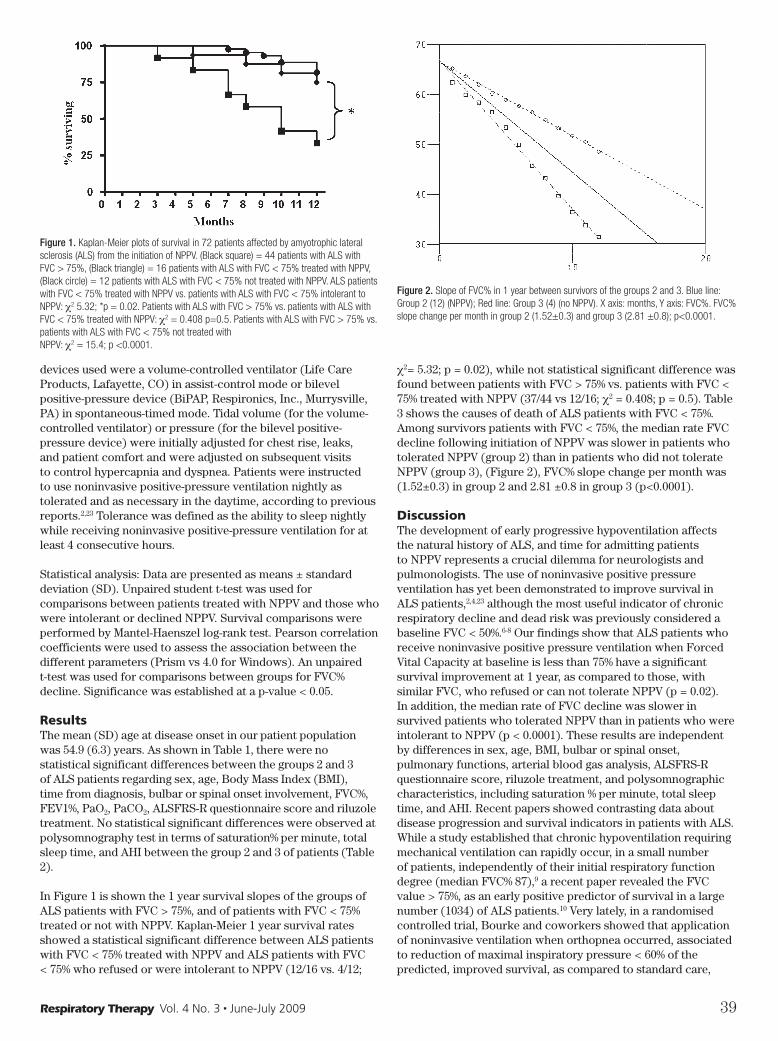
Respiratory Therapy Vol. 4 No. 3 n June-July 2009 39
devices used were a volume-controlled ventilator (Life Care Products, Lafayette, CO) in assist-control mode or bilevel positive-pressure device (BiPAP, Respironics, Inc., Murrysville, PA) in spontaneous-timed mode. Tidal volume (for the volume-controlled ventilator) or pressure (for the bilevel positive-pressure device) were initially adjusted for chest rise, leaks, and patient comfort and were adjusted on subsequent visits to control hypercapnia and dyspnea. Patients were instructed to use noninvasive positive-pressure ventilation nightly as tolerated and as necessary in the daytime, according to previous reports.2,23 Tolerance was defined as the ability to sleep nightly while receiving noninvasive positive-pressure ventilation for at least 4 consecutive hours.
Statistical analysis: Data are presented as means ± standard deviation (SD). Unpaired student t-test was used for comparisons between patients treated with NPPV and those who were intolerant or declined NPPV. Survival comparisons were performed by Mantel-Haenszel log-rank test. Pearson correlation coefficients were used to assess the association between the different parameters (Prism vs 4.0 for Windows). An unpaired t-test was used for comparisons between groups for FVC% decline. Significance was established at a p-value < 0.05.
ResultsThe mean (SD) age at disease onset in our patient population was 54.9 (6.3) years. As shown in Table 1, there were no statistical significant differences between the groups 2 and 3 of ALS patients regarding sex, age, Body Mass Index (BMI), time from diagnosis, bulbar or spinal onset involvement, FVC%, FEV1%, PaO2, PaCO2, ALSFRS-R questionnaire score and riluzole treatment. No statistical significant differences were observed at polysomnography test in terms of saturation% per minute, total sleep time, and AHI between the group 2 and 3 of patients (Table 2).
In Figure 1 is shown the 1 year survival slopes of the groups of ALS patients with FVC > 75%, and of patients with FVC < 75% treated or not with NPPV. Kaplan-Meier 1 year survival rates showed a statistical significant difference between ALS patients with FVC < 75% treated with NPPV and ALS patients with FVC < 75% who refused or were intolerant to NPPV (12/16 vs. 4/12;
c2= 5.32; p = 0.02), while not statistical significant difference was found between patients with FVC > 75% vs. patients with FVC < 75% treated with NPPV (37/44 vs 12/16; c2 = 0.408; p = 0.5). Table 3 shows the causes of death of ALS patients with FVC < 75%. Among survivors patients with FVC < 75%, the median rate FVC decline following initiation of NPPV was slower in patients who tolerated NPPV (group 2) than in patients who did not tolerate NPPV (group 3), (Figure 2), FVC% slope change per month was (1.52±0.3) in group 2 and 2.81 ±0.8 in group 3 (p<0.0001).
DiscussionThe development of early progressive hypoventilation affects the natural history of ALS, and time for admitting patients to NPPV represents a crucial dilemma for neurologists and pulmonologists. The use of noninvasive positive pressure ventilation has yet been demonstrated to improve survival in ALS patients,2,4,23 although the most useful indicator of chronic respiratory decline and dead risk was previously considered a baseline FVC < 50%.6-8 Our findings show that ALS patients who receive noninvasive positive pressure ventilation when Forced Vital Capacity at baseline is less than 75% have a significant survival improvement at 1 year, as compared to those, with similar FVC, who refused or can not tolerate NPPV (p = 0.02). In addition, the median rate of FVC decline was slower in survived patients who tolerated NPPV than in patients who were intolerant to NPPV (p < 0.0001). These results are independent by differences in sex, age, BMI, bulbar or spinal onset, pulmonary functions, arterial blood gas analysis, ALSFRS-R questionnaire score, riluzole treatment, and polysomnographic characteristics, including saturation % per minute, total sleep time, and AHI. Recent papers showed contrasting data about disease progression and survival indicators in patients with ALS. While a study established that chronic hypoventilation requiring mechanical ventilation can rapidly occur, in a small number of patients, independently of their initial respiratory function degree (median FVC% 87),9 a recent paper revealed the FVC value > 75%, as an early positive predictor of survival in a large number (1034) of ALS patients.10 Very lately, in a randomised controlled trial, Bourke and coworkers showed that application of noninvasive ventilation when orthopnea occurred, associated to reduction of maximal inspiratory pressure < 60% of the predicted, improved survival, as compared to standard care,
Figure 1. Kaplan-Meier plots of survival in 72 patients affected by amyotrophic lateral sclerosis (ALS) from the initiation of NPPV. (Black square) = 44 patients with ALS with FVC > 75%, (Black triangle) = 16 patients with ALS with FVC < 75% treated with NPPV, (Black circle) = 12 patients with ALS with FVC < 75% not treated with NPPV. ALS patients with FVC < 75% treated with NPPV vs. patients with ALS with FVC < 75% intolerant to NPPV: c2 5.32; *p = 0.02. Patients with ALS with FVC > 75% vs. patients with ALS with FVC < 75% treated with NPPV: c2 = 0.408 p=0.5. Patients with ALS with FVC > 75% vs. patients with ALS with FVC < 75% not treated withNPPV: c2 = 15.4; p <0.0001.
Figure 2. Slope of FVC% in 1 year between survivors of the groups 2 and 3. Blue line: Group 2 (12) (NPPV); Red line: Group 3 (4) (no NPPV). X axis: months, Y axis: FVC%. FVC% slope change per month in group 2 (1.52±0.3) and group 3 (2.81 ±0.8); p<0.0001.
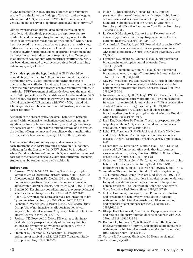
40 Respiratory Therapy Vol. 4 No. 3 n June-July 2009
in ALS patients.24 Our data, already published as preliminary results,25 are similar to the findings of Lechtzin and colleagues, who admitted ALS patients with FVC < 65% to mechanical ventilation and observed a significant prolongation of survival.26
Our study provides additional data about the role of sleep disorders, which actively participate to respiratory failure in ALS. Indeed, the respiratory failure may be present in the absence of breathlessness at rest, or orthopnoea,27 while, it has been observed that sleep disturbance appears at an early stage of disease,12 when respiratory muscle weakness is not sufficient to cause daytime orthopnea. Sleep-disordered breathing might be likely the earliest indication of respiratory insufficiency.17 In addition, in ALS patients with nocturnal insufficiency, NPPV has been demonstrated to correct sleep-disordered breathing, enhancing quality of life.16
This study supports the hypothesis that NPPV should be immediately prescribed to ALS patients with mild respiratory dysfunction (FVC < 75%) and polysomnographic signs of nocturnal hypoventilation, for at least 4 hours per day, in order to delay the rapid progression toward chronic respiratory failure. In particular, NPPV treatment significantly decreased the mortality rate of ALS patients with FVC < 75%, as well as the median FVC% rate decline, resulting much slower when compared to the slope of vital capacity of ALS patients with FVC < 50%, treated with 4 hours per day with bi-level intermittent positive pressure, as earlier reported.23
Although in the present study, the small number of patients treated with noninvasive mechanical ventilation can not give significance for a definitive conclusion, our findings encourage the early use of NPPV, in order to extend survival and to reduce the decline of lung volumes and compliance, thus ameliorating the respiratory function and quality of life of these patients.
In conclusion, this preliminary report demonstrates that early treatment with NPPV prolongs survival in ALS patients, indicating for the first time that NPPV should be introduced when FVC drops below 75% and not 50%, as considered standard care for these patients previously, although further multicentric studies must be conducted to well establish it.
References1 Caroscio JT, Mulvihill MN, Sterling R et al. Amyotrophic
lateral sclerosis. Its natural history. Neurol Clin. 1987;5,1-8. 2 Aboussouan LS, Khan SU, Meeker DP et al. Effect of
noninvasive positive-pressure ventilation on survival in amyotrophic lateral sclerosis. Ann Intern Med. 1997;127,450-3
3 Benditt JO. Respiratory complications of amyotrophic lateral sclerosis. Semin Respir Crit Care Med. 2002;23,239-47.
4 Bach JR. Amyotrophic lateral sclerosis: prolongation of life by noninvasive respiratory AIDS. Chest. 2002;122,92-8.
5 Lechtzin N, Wiener CM, Clawson L, et al. ALS CARE Study Group. Use of noninvasive ventilation in patients with amyotrophic lateral sclerosis. Amyotroph Lateral Scler Other Motor Neuron Disord. 2004;5,9-15.
6 Jackson CE, Rosenfeld J, Moore DH et al. A preliminary evaluation of a prospective study of pulmonary function studies and symptoms of hypoventilation in ALS/MND patients. J Neurol Sci. 2001;191,75-8.
7 Stambler N, Charatan M, Cedarbaum JM. Prognostic indicators of survival in ALS. ALS CNTF Treatment Study Group. Neurology. 1998;50,66-72.
8 Miller RG, Rosenberg JA, Gelinas DF et al. Practice parameter: the care of the patient with amyotrophic lateral sclerosis (an evidence-based review): report of the Quality Standards Subcommittee of the American Academy of Neurology: ALS Practice Parameters Task Force. Neurology. 1999;52,1311-23.
9 Lo Coco D, Marchese S, Corrao S et al. Development of chronic hypoventilation in amyotrophic lateral sclerosis patients. Respir Med 2006;100,1028-1036
10 Czaplinski A, Yen AA, Appel SH. Forced vital capacity (FVC) as an indicator of survival and disease progression in an ALS clinic population. J Neurol Neurosurg Psychiatry 2006; 77,390-2.
11 Ferguson KA, Strong MJ, Ahmad D et al. Sleep-disordered breathing in amyotrophic lateral sclerosis. Chest. 1996;110,664-9.
12 Kimura K, Tachibana N, Kimura J et al. Sleep-disordered breathing at an early stage of \ amyotrophic lateral sclerosis. J Neurol Sci. 1999;164,37-43.
13 Gay PC, Westbrook PR, Daube JR et al. Effects of alterations in pulmonary function and sleep variables on survival in patients with amyotrophic lateral sclerosis. Mayo Clin Proc. 1991;66,686-94.
14 Newsom-Davis IC, Lyall RA, Leigh PN et al. The effect of non-invasive positive pressure ventilation (NIPPV) on cognitive function in amyotrophic lateral sclerosis (ALS): a prospective study. J Neurol Neurosurg Psychiatry. 2001;71,482-7.
15 Santos C, Braghiroli A, Mazzini L et al. Sleep-related breathing disorders in amyotrophic lateral sclerosis.Monaldi Arch Chest Dis. 2003;59,160-5.
16 Lyall RA, Donaldson N, Fleming T et al. A prospective study of quality of life in ALS patients treated with noninvasive ventilation. Neurology. 2001;57,153-6.
17 Leigh PN, Abrahams S, Al-Chalabi A et al. King’s MND Care and Research Team. The management of motor neurone disease. J Neurol Neurosurg Psychiatry. 2003;74 Suppl 4:iv32-iv47
18 Cedarbaum JM, Stambler N, Malta E et al. The ALSFRS-R: a revised ALS functional rating scale that incorporates assessments of respiratory function. BDNF ALS Study Group (Phase III). J Neurol Sci. 1999;169(1-2)
19 Cedarbaum JM, Stambler N. Performance of the Amyotrophic Lateral Sclerosis Functional Rating Scale (ALSFRS) in multicenter clinical trials. J Neurol Sci. 1997;152 Suppl 1:S1-9.
20 American Thoracic Society. Standardization of spirometry, 1994 update. Am J Respir Crit Care Med 1994;152,1107-1136
21 Sleep-related breathing disorders in adults: recommendations for syndrome definition and measurement techniques in clinical research. The Report of an American Academy of Sleep Medicine Task Force. Sleep. 1999;22,667-89.
22 Melo J, Homma A, Iturriaga E, et al. Pulmonary evaluation and prevalence of non-invasive ventilation in patients with amyotrophic lateral sclerosis: a multicenter survey and proposal of a pulmonary protocol. J Neurol Sci. 1999;169,114-7.
23 Kleopa KA, Sherman M, Neal B et al. Bipap improves survival and rate of pulmonary function decline in patients with ALS. J Neurol Sci. 1999;164,82-8.
24 Bourke SC, Tomlinson M, Williams TL et al.Effects of non-invasive ventilation on survival and quality of life in patients with amyotrophic lateral sclerosis: a randomised controlled trial. Lancet Neurol. 2006;5,140-7
25 Carratu P, Cassano A, Maniscalco M, Home mechanical Continued on page 43…

Respiratory Therapy Vol. 4 No. 3 n June-July 2009 41
Not much is known about insomnia, but an ongoing study will soon shed more light, so to speak, on why people stay awake. The study, Sleep Disturbances in An Arctic Population: The Tromsø Study, is by Arne Fetveit, Jørund Straand, Bjørn Bjorvatn. Authors Fetveit and Straand are with the Department of General Practice and Community Medicine, University of Oslo; Bjorvatn is with the Department of Public Health and Primary Health Care, University of Bergen and with the Norwegian Competence Centre for Sleep Disorders, Haukeland University Hospital, Bergen, Norway. The background information below is reprinted from BMC Health Services Research, BioMed Central, © 2008 Fetveit et al; licensee BioMed Central Ltd. This is an Open Access article distributed under the terms of the Creative Commons Attribution License. This article has been edited for readers of Respiratory Therapy. To see the complete article, please go to BioMed Central and type in the full title.
Prevalence estimates for insomnia range from 10 to 50% in the adult general population. Sleep disturbances cause great impairment in quality of life, which might even rival or exceed the impairment in other chronic medical disorders. The economic implications and use of health-care services related to chronic insomnia represent a clinical concern as well as a pronounced public health problem. Hypnotics are frequently prescribed for insomnia, but alcohol and over-the-counter sleep aids seem to be more widely used by insomniacs than prescription medications. Despite the complex relationship between insomnia and physical and mental health factors, the condition appears to be underrecognized and undertreated by healthcare providers, probably due to the generally limited knowledge of the causes and natural development of insomnia.
Insomnia is the subjective complaint of insufficient sleep quantity or quality leading to impaired daytime functioning, usually linked to difficulties in falling asleep, trouble staying asleep, waking up too early in the morning or non-restorative sleep. The real prevalence of insomnia is not known, but prevalence estimates, which in part are influenced by diagnostic definitions, range from 10 to 50% in the adult population.
Insomnia is classified as transient (no more than a few nights), acute (less than 3 to 4 weeks), and chronic (more than 3 to 4
weeks). Transient or acute insomnia usually occurs in people with no history of sleep disturbances and is often related to an identifiable cause. Chronic insomnia is further divided into primary and secondary conditions, depending on the cause. When the complaint of insomnia is not caused by other medical, psychiatric or medication-related disorders, it is considered to be primary in nature. It is estimated that primary insomnia accounts for approximately 25% of all chronic insomnia. Recently, The American National Institutes of Health State-of-the-Science Conference suggested that insomnia should be conceptualized as a disorder in and of itself, which is or is not comorbid with other disorders—rather than drawing conclusions about insomnia’s primary or secondary status.
Excessive daytime sleepiness is a key symptom of any type of disturbed sleep. Sleepiness is defined by recurrent episodes of drowsiness or involuntary dozing that arise mainly in sedentary situations. Sleepiness should not be confused with fatigue or tiredness. Although subjective reports of sleepiness are common in people with insomnia, many studies report normal or even heightened levels of daytime alertness among insomniacs. On the other hand, insomnia patients usually experience more fatigue than patients with other sleep disturbances, such as obstructive sleep apnea, narcolepsy or periodic limb movements during sleep.
A wide range of factors are associated with chronic insomnia. Precipitants of acute insomnia may include acute medical illness, hospitalization, changes in the sleeping environment, medications, jet lag, and acute or recurring psychosocial stressors. Chronic insomnia is often linked to or associated with various underlying medical, behavioral, and environmental conditions, and various medications. Especially mental disorders and discomfort are frequently reported by insomniacs. In a survey of 811 respondents, 40% of those with insomnia (compared with 16% of those without), met the criteria for one or more psychiatric diagnoses.17 Although it is difficult to discern whether a psychiatric disorder precipitates insomnia or whether insomnia makes an individual vulnerable to the emergence of a psychiatric disorder, investigations indicate an increased risk for new onset of depression, anxiety disorders, and substance abuse in people with persistent insomnia. Additionally, insomnia may be precipitated by factors like stress and perpetuated by
SLEEP RESEARCH
Insomnia
42 Respiratory Therapy Vol. 4 No. 3 n June-July 2009
behavioral factors, or unstable sleep schedules. Shift work or other lifestyle factors that disrupt circadian rhythm increase the risk of sleep disturbances.
An association between elevated body mass index (BMI) and short sleep duration has been demonstrated in recent years. A possible causality between short sleep duration and the development of diabetes mellitus (DM) has also been explored. Gottlieb et al. reported that short sleep duration is associated with DM and impaired glucose tolerance (IGT) in community-dwelling middle-aged and older adults. This association persisted after adjustment for known DM risk factors. Sleep restriction to four hours of sleep per night increased blood pressure, decreased parasympathetic tone, increased evening cortisol and insulin levels, and promoted increased appetite, possibly through the elevation of ghrelin, a pro-appetitive hormone, and decrease in levels of leptin. The correlation between short sleep duration with DM and IGT may partly explain the association between short sleep duration and coronary heart disease and all over mortality.
Sleep patterns change throughout life. It seems as if ageing makes it more difficult to stay asleep at night and to stay awake during the day. Total sleep time during the 24-hour period appears to decline with advancing age, accompanied by a redistribution of sleep—with decreased nocturnal sleep and increased daytime napping. Progressive physical inactivity, dissatisfaction with social life, and presence of physical
and mental problems may be the most predictive factors of insomnia in the elderly. Opposed to healthy elderly, sleep duration during the 24-h day in demented nursing home patients seems to increase according to the degree of dementia.31 Some researchers claim that ageing probably is the single most important factor that determines human sleep—more so than gender and even most physical and mental illnesses. However, challenging findings of no reduction of nocturnal sleep with age in healthy elderly have also been presented, suggesting that insomnia in the elderly is not related to ageing itself.
It is known that hypnotics are frequently prescribed for insomnia. However, alcohol and over-the-counter (OTC) medications seem to be more widely used by insomniacs than prescription medications, perhaps due to the fact that most insomniacs do not discuss their sleep problems with their doctor. In a general population sample of 2,181 adults aged 18 to 45 years, Johnson et al found that 25.9% had used any substance as a sleep aid in the past 12 months. Approximately 18% reported using medications (57% of these being OTC-drugs), and 13% reported using alcohol to fall asleep. The choice of substance used as a sleep aid seems to be affected by sociodemographic characteristics: Most chronic benzodiazepine users tend to be elderly, and more men than women use alcohol to induce sleep. Rokstad et al examined the prescribing patterns among general practitioners (GPs) in a Norwegian county in relation to the patients’ age, gender and the diagnosis for prescribing, and found that insomnia was among the most commonly recorded diagnosis for prescribing. Most drugs have not been sufficiently studied to determine their primary effects on sleep and waking behavior. Even when the effects of a drug are known, the medication may act differently in normal individuals and individuals who are ill.
The economic implications and use of healthcare services related to chronic insomnia represent a major problem. Costs related to chronic insomnia do not only include the direct costs of health-care, but also the indirect costs related to absence from work, diminished productivity, accidents, and other health problems that are, at least in part, secondary to insomnia.
Compared to other sleep disturbances, such as narcolepsy and sleep apnea, the understanding of the basic pathophysiology of insomnia has lagged behind. This discrepancy probably stems from the heterogeneous nature of insomnia. On one hand, insomnia may be a primary condition with a pathophysiology, like a general state of hyperarousal—which includes changes like increased levels of catecholamines,46 increased basal metabolic rate, increased body temperature, altered heart rate, increased level of central nervous system (CNS) metabolic rate, and elevated electroencephalograph activity. On the other hand, insomnia may be a co-existing condition with numerous physical and mental disorders.
Further knowledge of the impact of latitude, daytime illumination and season, on the prevalence of insomnia is needed. The term “midwinter insomnia” has been applied for the seasonal type of insomnia observed in arctic areas during the dark periods of the year, but the possible consequences of annual variations in environmental light on sleep are still not settled, and make arctic sleep studies especially relevant.
Semantic as well as definition confusions are present in many epidemiological sleep studies. This situation probably rises from
Table 1: Factors related to chronic insomnia
Specific sleep disturbances:• Circadian rhythm disorders:
n Advanced sleep-phase syndromen Delayed sleep-phase syndrome
• Sleep apnea (obstructive, central, or mixed)• Restless leg syndrome• Periodic limb movement disorders (nocturnal myoclonus)• Parasomnias, i.e. REM-sleep-behavior-disorderPhysical illness:• Pain: arthritis, musculoskeletal pain, other painful conditions• Cardiovascular: heart failure, nocturnal breathlessness, nocturnal angina• Pulmonary: chronic obstructive pulmonary disease, allergic rhinitis (nasal
obstruction)• Gastrointestinal: gastroesophageal reflux disease, peptic ulcer disease,
constipation, diarrhea, pruritus ani• Urinary: nocturia, incomplete bladder emptying, incontinence• Central nervous system: stroke, Parkinson disease, Alzheimer disease, seizure
disorder• Psychiatric illness: anxiety, depression, psychosis, dementia, delirium• Pruritus• Menopause (hot flushes)Behavioral: daytime nap, early retirement to bed, use of bed for other activities (eg, reading and watching television), heavy meals, lack of exercise,and sedentary lifestyleEnvironmental: noise, light and other disturbances, extreme temperatures, uncomfortable bedding, and lack of exposure to sunlightMedications:• Central nervous system stimulants: sympathomimetics, caffeine, nicotine,
amphetamines, ephedrine, phenytoin• Antidepressants: bupropion, selective serotonin reuptake inhibitors, venlafaxine• Anti-Parkinsonian agents: levodopa• Bronchodilators: theophylline• Cardiovascular: B-blockers, diuretics• Histamines, H2 blockers: cimetidine• Anticholinergics• Corticosteroids• Alcohol• Herbal remedies• Stimulant laxative
REM = rapid eye movement.

Respiratory Therapy Vol. 4 No. 3 n June-July 2009 43
the fact that no single and clear definition exists of insomnia and how to assess it. As a result, very different and non-comparable sleep assessment tools exist—making it is almost impossible to compare and summarize studies.
Our knowledge of insomnia is still limited, especially regarding its evolution and consequences in the general population. Future sleep research is in need of validated measurements of sleep complaints, in order to provide common metrics for describing insomnia and insomniacs in epidemiological studies.
Below, we describe the protocol for a longitudinally, retrospective cohort survey and a cross-sectional study in a large representative adult community sample in northern Norway (The Tromsø Study). Using validated sleep assessment tools, our aim is to describe and analyze sleep disturbances, with their correlating and predisposing factors.
The Tromsø Study is an ongoing population-based cohort study with five previous health studies undertaken between 1974 and 2001. This protocol outlines a planned study within the sixth Tromsø Study (Tromsø VI), aiming at; 1) describing sleep patterns in a community-based sample representative of the general population of northern Norway, and 2) examining outcome variables of sleep disturbances against possible explanatory and confounding variables, both within a cross-sectional approach, as well as retrospectively in a longitudinal study—exploring sleep patterns in subjects who have attended two or more of the previous Tromsø studies between 1974 and 2009. First, we plan to perform a simple screening in order to identify those participants with probable sleep disturbances, and secondly to investigate these sleep disturbances further, using an extensive sleep-questionnaire. We will also collect biological explanatory variables, i.e. blood samples, weight, height and blood pressure. We plan to merge data on an individual level from the Tromsø VI Study with data from the Norwegian Prescription Database (NorPD), which is a national registry including data for all prescription drugs issued at Norwegian pharmacies. Participants with sleep disturbances will be compared with pair-matched controls without sleep disturbances.
Despite ongoing research, many challenges remain in the characterization of sleep disturbances and its correlates. Future mapping of the biological dimensions, natural history, as well as the behavioral and drug-related aspects of sleep disturbances in a representative population samples is clearly needed.
ventilation decreases mortality in patients affected by ALS. European Respiratory Journal Abstract Suppl. September 2006, Volume 28 Number 3
26 Lechtzin N, Scott Y, Busse AM, Clawson LL, Kimball R, Wiener CM. Early use of noninvasive ventilation prolongs survival in subjects with ALS. Amyotroph Lateral Scler. 2007 Jun;8(3):185-8.
27 Sivak ED, Shefner JM, Mitsumoto H, et al. The use of non-invasive positive pressure ventilation (NIPPV) in ALS patients. A need for improved determination of intervention timing. Amyotroph Lateral Scler Other Motor Neuron Disord. 2001;2,139-45.
Early Treatment…continued from page 40
and expiratory muscle groups with the ventilator in airflow limitation. Am J Respir Crit Care Med 1998; 158:1471.
15 Jubran, A. Inspiratory flow rate: More may not be better. Crit Care Med 1999 Vol. 27, No 4
16 Sydow M, Thies K, Engel J, Golish W, Buscher H, Zinserling J, Burchardi H, Variation in inspiratory gas flow in pressure support ventilation. The effect on respiratory mechanics and respiratory work. Anaesthesist.1996 Nov;45(11):1051-8.
17 Laghi F, Karamchandani K, Tobin MJ, Influence of ventilator settings in determining respiratory frequency during mechanical ventilation. Am J Respir Crit Care Med 1999;Vol 160: 1766-1770.
Newport FlexCycle…continued from page 36

44 Respiratory Therapy Vol. 4 No. 3 n June-July 2009
Features of Cough Variant Asthma and Classic Asthma During Methacholine-Induced Brochoconstriction: a cross-sectional studyHisako Matsumoto, Akio Niimi, Masaya Takemura, Tetsuya Ueda, Masafumi Yamaguchi, Hirofumi Matsuoka, Makiko Jinnai, Kazuo Chin and Michiaki Mishima
AbstractBackground: Little is known regarding mechanistic and phenotypic differences between cough variant asthma (CVA), presenting with a chronic cough as the sole symptom that responds to bronchodilators, and classic asthma with wheezing during methacholine inhalation. Here we reported airway sensitivity, airway reactivity, and as the main concern, the appearance of cough and wheezes during methacholine inhalation in patients with CVA or classic asthma.
Methods: We cross-sectionally examined the degrees of airway sensitivity, the point where resistance started to increase, and reactivity, the slope of the methacholine-resistance curve, and the appearance of cough and wheezes in steroid-naïve adult patients with classic asthma (n = 58) or CVA (n = 55) while they were continuously inhaling methacholine during simultaneous measurement of respiratory resistance.
Results: Patients with CVA were less sensitive and less reactive to inhaled methacholine and wheezed less frequently but coughed more frequently during methacholine-induced bronchoconstriction than did patients with classic asthma. Multivariate analysis revealed that airway hypersensitivity and lower baseline FEV1/FVC were associated with the appearance of wheezes, whereas a diagnosis of CVA was associated with coughing.
Conclusion: There are mechanistic and phenotypic differences between CVA and classic asthma during methacholine inhalation. Frequent coughing during bronchoconstriction may be a distinctive feature of CVA.
BackgroundPatients with cough variant asthma (CVA) present with a chronic
cough as the sole symptom that responds to bronchodilator treatment and show airway hyperresponsiveness (AHR). CVA, one of the most common causes of chronic cough,1-4 is considered a precursor5-9 and a variant form of classic asthma with typical symptoms of wheezing and dyspnea.5 Several studies have examined mechanistic differences between CVA and classic asthma. Airway sensitivity, a component of airway responsiveness that is defined as the inflection point where respiratory resistance (Rrs) starts to increase, did not differ between patients with CVA and those with classic asthma in a few small studies.10,11 In contrast, airway reactivity, another component of airway responsiveness expressed as the slope of the dose-response curve, is attenuated in children with CVA as compared with those with classic asthma.12 In adults with CVA, however, no study has separately examined airway sensitivity and reactivity in a large number of patients.
Methacholine, a non-specific cholinergic stimulant, induces bronchoconstriction without exacerbating airway inflammation. Apart from an analysis of mechanistic aspects, analyses of phenotypes, such as the appearance of cough and wheezes during methacholine-induced bronchoconstriction, may provide clues to understanding the unique features of CVA. To our knowledge, however, such an approach has not been attempted thus far. In one study in asthmatic children, detection of wheezes during methacholine inhalation depended on the degree of airway narrowing, while factors related to coughing during methacholine inhalation were not specified.13
In this study, we initially examined airway sensitivity and reactivity to methacholine in adults with CVA and in those with classic asthma, using a continuous inhalation method that can separately evaluate these two components.12-14 Our major concern was the presence or absence of cough and wheezes during methacholine-induced bronchoconstriction. Factors associated with the appearance of cough and wheezes were then analyzed.
MethodsStudy subjects and design: We cross-sectionally studied adults with classic asthma (n = 58) or with CVA (55) who presented at the outpatient clinic of Kyoto University Hospital from April 1993 to September 2001. Classic asthma was diagnosed according to the American Thoracic Society criteria:15 the symptoms
Authors Matsumoto, Niimi, Takemura, Ueda, Yamaguchi, Matsuoka, Jinnai
and Mishima are with the Department of Respiratory Medicine, Kyoto
University, Kyoto; Takemura and Ueda are also with the Department of
Respiratory Medicine, Kitano Hospital, Osaka; Chin is with the Department
of Respiratory Care and Sleep Control Medicine, Kyoto University, Japan.
Reprinted from BioMed Central, Cough, © 2009 Matsumoto et al; licensee
BioMed Central Ltd. This is an Open Access article distributed under the
terms of the Creative Commons Attribution License.

Respiratory Therapy Vol. 4 No. 3 n June-July 2009 45
of episodic wheezing and dyspnea within the previous year that responds to bronchodilators, and AHR to methacholine inhalation.
CVA was diagnosed according to the following criteria:5-10 an isolated chronic cough without wheezing or dyspnea that had persisted for more than 8 weeks, AHR to methacholine, and symptomatic improvement of coughing in response to inhaled beta-2 agonists, sustained-release theophylline, or both. Wheezing or rhonchi were not audible on chest auscultation, even with forced expiration. No patient had a past history of asthma or had an upper respiratory tract infection within the past 8 weeks. No other apparent causes of chronic cough, such as gastroesophageal reflux, chronic sinusitis, or medication with angiotensin-converting enzyme inhibitors, were present. Patients with CVA had normal chest radiographs and were steroid-naïve, similar to those with classic asthma. The ethics committee of our institution approved the study protocol, and written informed consent was obtained from each participant.
Prebronchodilator FEV1 was tested using a spirometer (Chestac-65V, Chest, Tokyo, Japan) according to the standards of the American Thoracic Society.16
Airway responsiveness was tested by directly recording a dose-response curve of Rrs (cmH2O/L/sec) during continuous inhalation of methacholine in two-fold incremental concentrations (49 to 25,000 µg/ml) under tidal breathing from nebulizers with an output of 0.15 ml/minute (Astograph; Chest, Tokyo), as described previously in detail.14,17 If bronchodilators were being used, their use was suspended 24 hours before the methacholine inhalation. In short, after we recorded the baseline Rrs during inhalation of physiologic saline for 1 minute, patients inhaled methacholine, starting with the lowest concentration, at 1-minute intervals. The index of airway sensitivity that we adopted was Dmin: the cumulative dose of inhaled methacholine at the inflection point where which Rrs began to increase continuously. One unit of Dmin is equivalent to dose of 1 mg/ml of methacholine inhalation for one minute. Inhalation of methacholine was discontinued, and switched to bronchodilator inhalation when Rrs reached twice the baseline
value. The plateau of the dose-response curve was not, therefore, examined. The slope of the methacholine-Rrs dose-response curve (SRrs) was used as an index of airway reactivity. FEV1 was not measured after the methacholine challenge test since holding the administration of a bronchodilator and addition of forced expiratory maneuver might induce severe bronchoconstriction.
Cough was considered to have appeared during the methacholine inhalation when patients coughed one or more times after the inflection point of Dmin. Cough before the point of Dmin, if any, was also documented. Coughing caused a transient spike-shape increase in Rrs, but it did not interfere with the determination of the inflection point or the slope of the dose-response curve. When the methacholine inhalation was discontinued and switched to bronchodilator inhalation, whether wheezing was audible on auscultation was assessed by either of the attending physicians (AN, HM). The assessment of cough or wheezes was performed in a blinded manner.
In 18 patients with classic asthma and in 22 with CVA, cough sensitivity test in addition to methacholine inhalation test was done one to two weeks apart. Cough sensitivity was tested by a continuous inhalation method of capsaicin solution using the Astograph as described previously.18 Ten doubling concentrations of capsaicin solution (0.61–312 µM) were inhaled until 5 or more coughs were induced (cough threshold, C5). Each concentration of capsaicin was inhaled for 15 seconds during tidal breathing every 60 seconds. Remaining patients were not examined for capsaicin cough sensitivity because informed consents for the test were not obtained mostly due to time constraint.
Statistical analysis: Data were analyzed using GraphPad Prism 4.00 (GraphPad Software, Inc, La Jolla, CA) and StatView software 5.0 (SAS Institute Inc, Cary, NC). To compare the two patient groups, the t-test was used when data were normally distributed, and the Mann-Whitney test was used for nonparametric data. The c2 test was used for the comparisons of nominal data between groups. Univariate and stepwise multivariate regression analyses were performed to test for independent effects of disease diagnosis, blood eosinophil counts, atopic status, FEV1/FVC, current smoking, log Dmin,
Cough 2009, 5:3 http://www.coughjournal.com/content/5/1/3
Page 3 of 6(page number not for citation purposes)
attending physicians (AN, HM). The assessment of coughor wheezes was performed in a blinded manner.
Capsaicin cough sensitivity testIn 18 patients with classic asthma and in 22 with CVA,cough sensitivity test in addition to methacholine inhala-tion test was done one to two weeks apart. Cough sensitiv-ity was tested by a continuous inhalation method ofcapsaicin solution using the Astograph™ as described pre-viously [18]. Ten doubling concentrations of capsaicinsolution (0.61–312 M) were inhaled until 5 or morecoughs were induced (cough threshold, C5). Each concen-tration of capsaicin was inhaled for 15 seconds duringtidal breathing every 60 seconds. Remaining patients werenot examined for capsaicin cough sensitivity becauseinformed consents for the test were not obtained mostlydue to time constraint.
Statistical analysisData were analyzed using GraphPad Prism 4.00 (Graph-Pad Software, Inc., La Jolla, CA, USA) and StatView soft-ware 5.0 (SAS Institute Inc, Cary, NC, USA). To comparethe two patient groups, the t-test was used when data werenormally distributed, and the Mann-Whitney test wasused for nonparametric data. The 2 test was used for thecomparisons of nominal data between groups. Univariateand stepwise multivariate regression analyses were per-formed to test for independent effects of disease diagno-sis, blood eosinophil counts, atopic status, FEV1/FVC,current smoking, log Dmin, and SRrs levels on the appear-ance of cough or wheezes during methacholine inhala-tion, setting the absence of cough or wheezes as 0 and thepresence as 1. We did not include C5 levels as an inde-pendent variable since less than half of the patients under-
went the capsaicin cough sensitivity test. For inclusion ofvariables into multivariate analyses, the F value, a measureof the extent to which a variable makes a unique contribu-tion to the prediction of the dependent variable, was set at4.0. Data are expressed as means ± SD. We considered pvalues of < 0.05 to indicate statistical significance.
ResultsPatients' characteristics are shown in Table 1. As com-pared with classic asthma group, CVA group includedfewer smokers, had a lower blood eosinophil count, ahigher baseline FEV1/FVC value, a lower baseline Rrsvalue, and less sensitivity and less reactivity to inhaledmethacholine.
As for phenotypic characteristics during methacholine-induced bronchoconstriction, cough appeared in 19patients (35%) in the CVA group and 10 (17%) in theclassic asthma group (p = 0.035), whereas wheezes weredetected at the end of inhalation in 9 patients (16%) inthe CVA group and 28 (48%) in the classic asthma group(p = 0.0003). Four patients with CVA started to coughbefore the inflection point of Dmin. In three of thesepatients, cough was relieved when methacholine wasswitched to a bronchodilator, associated with a two-foldincrease in Rrs from baseline. Multivariate analyses of theappearance of wheezes and cough showed that lowerbaseline FEV1/FVC and airway hypersensitivity were inde-pendently associated with the detection of wheezes (Table2), whereas the appearance of cough was solely associatedwith a diagnosis of CVA (Table 3). These results wereunchanged even when four patients with CVA who startedto cough before the inflection point of Dmin wereexcluded from the analyses.
Table 1: Patients' characteristics
Classic asthma Cough variant asthma p-valuen = 58 n = 55
Age (yr) 44.4 ± 15.9 43.2 ± 16.5 0.71Male/Female (No) 29/29 33/22 0.29Disease duration at diagnosis (yr) 6.7 ± 10.0 2.8 ± 4.4 0.10Current smoking (Yes, %) 19.0 5.2 0.029Atopic status* (Yes, %) 71.4 67.9 0.69Blood eosinophils (number/ l) 389 ± 247 310 ± 404 0.011FEV1 (% predicted) 86.8 ± 19.2 92.1 ± 17.6 0.14FEV1/FVC (%) 72.6 ± 11.3 81.8 ± 8.9 < 0.0001Baseline Rrs (cm H2O/L/sec) 4.3 ± 2.0 4.0 ± 3.2 0.040Log Dmin (units) -0.20 ± 0.82 0.36 ± 0.60 < 0.0001SRrs (cm H2O/L/sec/min) 2.9 ± 3.2 2.1 ± 2.1 0.042Log C5 ( M) 1.51 ± 0.79 (n = 18) 1.17 ± 0.71 (n = 22) 0.15
Values are given as the means ± SD.*: measured in 56 patients with classic asthma and 53 patients with cough variant asthma. Patients were considered atopic when 1 or more specific IgE antibodies were positive for cat dander, dog dander, weed, grass pollen, mold, or house dust mite.Abbreviation: Rrs, respiratory resistance. Dmin, cumulative dose of inhaled methacholine at the inflection point, where which respiratory resistance begins to increase. SRrs, slope of the methacholine dose-response curve for Rrs. C5, cough threshold, the lowest concentration of capsaicin that induces 5 or more cough.
46 Respiratory Therapy Vol. 4 No. 3 n June-July 2009
and SRrs levels on the appearance of cough or wheezes during methacholine inhalation, setting the absence of cough or wheezes as 0 and the presence as 1. We did not include C5 levels as an independent variable since less than half of the patients underwent the capsaicin cough sensitivity test. For inclusion of variables into multivariate analyses, the F value, a measure of the extent to which a variable makes a unique contribution to the prediction of the dependent variable, was set at 4.0. Data are expressed as means ± SD. We considered p values of < 0.05 to indicate statistical significance.
ResultsPatients’ characteristics are shown in Table 1. As compared with classic asthma group, CVA group included fewer smokers, had a lower blood eosinophil count, a higher baseline FEV1/FVC value, a lower baseline Rrs value, and less sensitivity and less reactivity to inhaled methacholine.
As for phenotypic characteristics during methacholine-induced bronchoconstriction, cough appeared in 19 patients (35%) in the CVA group and 10 (17%) in the classic asthma group (p = 0.035), whereas wheezes were detected at the end of inhalation in 9 patients (16%) in the CVA group and 28 (48%) in the classic asthma group (p = 0.0003). Four patients with CVA started to cough before the inflection point of Dmin. In three of these patients, cough was relieved when methacholine was switched to a bronchodilator, associated with a two-fold increase in Rrs from baseline. Multivariate analyses of the appearance of wheezes and cough showed that lower baseline FEV1/FVC and airway hypersensitivity were independently associated with
the detection of wheezes (Table 2), whereas the appearance of cough was solely associated with a diagnosis of CVA (Table 3). These results were unchanged even when four patients with CVA who started to cough before the inflection point of Dmin were excluded from the analyses.
Cough sensitivity did not differ between patients with classic asthma (n = 18) and those with CVA (n = 22) (Table 1). However, in CVA group 9 patients who coughed during the methacholine-induced bronchoconstriction showed more enhanced cough sensitivity to inhaled capsaicin (log C5, 0.72 ± 0.65 µM) than 13 non-coughers (1.48 ± 0.58 µM)(p = 0.015). Meanwhile, 2 coughers and 16 non-coughers in classic asthma group did not differ in their cough sensitivity (0.69 ± 0.85 µM; 1.62 ± 0.75 µM, respectively)(p = 0.12).
DiscussionTo our knowledge, this is the first study to comprehensively examine mechanistic and phenotypic differences during methacholine inhalation between adults with CVA and those with classic asthma. Patients with CVA were less sensitive and less reactive to inhaled methacholine than were those with classic asthma. Coughing was more frequent during methacholine-induced bronchoconstriction in the CVA group, whereas wheezes were more frequent in the classic asthma group at the end of methacholine inhalation. Multivariate analysis of factors related to cough and wheezes revealed that wheezes were associated with airway hypersensitivity and baseline airflow obstruction, whereas cough triggered by bronchoconstriction was related to CVA.
Cough 2009, 5:3 http://www.coughjournal.com/content/5/1/3
Page 4 of 6(page number not for citation purposes)
Cough sensitivity did not differ between patients withclassic asthma (n = 18) and those with CVA (n = 22)(Table 1). However, in CVA group 9 patients whocoughed during the methacholine-induced bronchocon-striction showed more enhanced cough sensitivity toinhaled capsaicin (log C5, 0.72 ± 0.65 M) than 13 non-coughers (1.48 ± 0.58 M)(p = 0.015). Meanwhile, 2coughers and 16 non-coughers in classic asthma groupdid not differ in their cough sensitivity (0.69 ± 0.85 M;1.62 ± 0.75 M, respectively)(p = 0.12).
DiscussionTo our knowledge, this is the first study to comprehen-sively examine mechanistic and phenotypic differencesduring methacholine inhalation between adults with CVAand those with classic asthma. Patients with CVA were lesssensitive and less reactive to inhaled methacholine thanwere those with classic asthma. Coughing was more fre-quent during methacholine-induced bronchoconstrictionin the CVA group, whereas wheezes were more frequent inthe classic asthma group at the end of methacholine inha-lation. Multivariate analysis of factors related to coughand wheezes revealed that wheezes were associated withairway hypersensitivity and baseline airflow obstruction,
whereas cough triggered by bronchoconstriction wasrelated to CVA.
Airway sensitivity and reactivity are thought to be differ-ently regulated [19,20]. Airway sensitivity is most likelyassociated with airway inflammation, epithelial damageor malfunction, abnormal neural control, and increasedinflammatory cell number and activity. In contrast, airwayreactivity is considered most strongly related to smoothmuscle contractility. Airway sensitivity was substantiallylower in patients with CVA than in those with classicasthma. Previous studies showed no significant differencein airway sensitivity between these two asthmatic condi-tions [10-12,21]. The discrepancy may be attributed todifferences in patient selection [10-12,21] and methodol-ogy [21]. Children with CVA were studied by Koh et al[21] and Mochizuki et al [12]. The airway physiology ofchildren may differ from that of adults, as suggested bydifferences between mature and immature animals in theresponses of airway smooth muscle to cholinergic stimu-lation [22]. In previous studies of adults with CVA (n =14) [10], (n = 10) [11], sample sizes were relatively small.In addition, our previous study was conducted in patientswith CVA who agreed to be hospitalized and to undergobronchoscopic examination [10], conditions that might
Table 2: Univariate and multivariate regression analysis of appearance of wheezes
Univariate analysisCorrelation coefficient
P WheezesStandardized partial regression coefficient
F value
Disease 0.34 0.0002 not entered < 4.0Blood eosinophils (number/ l) 0.11 0.35 not entered < 4.0Atopy -0.05 0.60 not entered < 4.0Baseline FEV1/FVC (%) -0.31 0.001 -0.31 9.2Smoking 0.08 0.39 not entered < 4.0Log Dmin (units) -0.30 0.001 -0.33 10.7SRrs (cmH2O/L/sec/min) 0.28 0.003 not entered < 4.0
Adjusted R2 = 0.22, p < 0.0001 for the multivariate analysis of appearance of wheezes. Wheezes, atopy, and smoking status are rated as 0 for absent and 1 for present. Disease is labeled as 0 for cough variant asthma and 1 for classic asthma. F value is a measure of the extent to which a variable makes a unique contribution to the prediction of the dependent variable. Dmin, cumulative dose of inhaled methacholine at the inflection point, where which respiratory resistance begins to increase. SRrs, slope of the methacholine dose-response curve for respiratory resistance.
Table 3: Univariate and multivariate regression analyses of appearance of cough
Univariate analysisCorrelation coefficient
P CoughStandardized partial regression coefficient
F value
Disease -0.20 0.036 -0.27 6.2Blood eosinophils (number/ l) -0.12 0.27 Not entered < 4.0Atopy 0.02 0.83 not entered < 4.0Baseline FEV1/FVC (%) 0.11 0.25 not entered < 4.0Smoking -0.10 0.30 not entered < 4.0Log Dmin (units) 0.09 0.33 not entered < 4.0SRrs (cmH2O/L/sec/min) -0.02 0.87 not entered < 4.0
Adjusted R2 = 0.06, p = 0.015 for the multivariate analysis of appearance of cough. Cough, atopy, and smoking status are rated as 0 for absent and 1 for present. Disease is labeled as 0 for cough variant asthma and 1 for classic asthma. F value is a measure of the extent to which a variable makes a unique contribution to the prediction of the dependent variable. Dmin, cumulative dose of inhaled methacholine at the inflection point, where which respiratory resistance begins to increase. SRrs, slope of the methacholine dose-response curve for respiratory resistance.

Respiratory Therapy Vol. 4 No. 3 n June-July 2009 47
Airway sensitivity and reactivity are thought to be differently regulated.19,20 Airway sensitivity is most likely associated with airway inflammation, epithelial damage or malfunction, abnormal neural control, and increased inflammatory cell number and activity. In contrast, airway reactivity is considered most strongly related to smooth muscle contractility. Airway sensitivity was substantially lower in patients with CVA than in those with classic asthma. Previous studies showed no significant difference in airway sensitivity between these two asthmatic conditions.10-12,21 The discrepancy may be attributed to differences in patient selection10-12,21 and methodology.21 Children with CVA were studied by Koh et al21 and Mochizuki et al.12 The airway physiology of children may differ from that of adults, as suggested by differences between mature and immature animals in the responses of airway smooth muscle to cholinergic stimulation.22 In previous studies of adults with CVA (n = 14),10 (n = 10),11 sample sizes were relatively small. In addition, our previous study was conducted in patients with CVA who agreed to be hospitalized and to undergo bronchoscopic examination,10 conditions that might have lead to a selection bias toward patients with more severe CVA. In contrast, all subjects with CVA in the present study were outpatients. Our subjects may therefore be more representative of patients encountered in daily practice.
Several studies in children indicate that the degree of excessive airway narrowing is modest in patients with CVA. Yoo et al have shown that children with CVA more frequently reach a maximal response plateau on the dose-response curve to methacholine than those with classic asthma.23 Moreover, plateau levels are lower in children with CVA.24 In agreement with these results in children with CVA,12,23,24 we demonstrated for the first time that adults with CVA were significantly less reactive to methacholine than were those with classic asthma, although the difference in airway reactivity between the two groups was small in our study of adults.
The presence or absence of cough and wheezes during methacholine-induced bronchoconstriction was our main interest. Bronchoconstriction is a well-known stimulant of cough that is thought to be mediated by mechanosensitive, rapidly adapting receptors.25 In a guinea pig model of CVA, degree of antigen-induced bronchoconstriction is strongly correlated with cough counts that are inhibited by procaterol administration.26 Clinical studies examining the appearance of cough during bronchoconstriction are scant, however. Springer et al. performed methacholine provocation tests by the forced expiration method in asthmatic children with wheezing.13 Cough appeared in most (81%) of the asthmatic children, but the background characteristics of the coughers were not described. We showed that the appearance of cough was solely associated with a diagnosis of CVA and not with mechanistic variables. It was also surprising that only 17% of the adults with classic asthma coughed during bronchoconstriction in our study. Mechanisms underlying the discrepancy in bronchoconstriction-induced cough between classic asthma and CVA were not clarified since cough sensitivity did not differ between the two asthmatic conditions. However given that in CVA group coughers had more heightened cough sensitivity to inhaled capsaicin than non-coughers, cough during methacholine-induced bronchoconstriction might be on a background of enhanced capsaicin cough reflex in CVA. However the guinea pig model of CVA described above is not sensitive to inhaled capsaicin and the authors negate the involvement of tachykinins
in bronchoconstriction-induced cough.26 Further studies are necessary to elucidate a linkage between bronchoconstriction-triggered cough and capsaicin-induced cough in CVA patients.
As expected, wheezes were more frequent in the classic asthma group than in the CVA group at the end of methacholine inhalation. In contrast to cough, wheezes were not classic asthma-specific on multivariate analysis. Baseline airflow obstruction and airway hypersensitivity contributed to the presence of wheezes, consistent with the theory that wheezes are generated by airflow turbulence.27 Mochizuki et al. proposed that lower airway reactivity or slower airway constriction may explain the absence of wheezing or dyspnea in children with CVA.12 We found no independent contribution of airway hyperreactivity to the appearance of wheezes. The lower frequency of wheezes in patients with CVA may be inherently related to their better pulmonary function and modest airway sensitivity.
Needless to say, airway inflammation has an important role in the pathogenesis of both CVA and classic asthma. Lack of the information on airway inflammation in the present study may not weaken our results, however, since methacholine contracts airway smooth muscle without modulating airway inflammation. Methacholine provocation test may not reproduce clinical conditions, but we believe that our findings regarding the frequency of cough and wheezes triggered by airway smooth muscle contraction in classic asthma and CVA are novel and relevant. One may argue that methacholine worked as a non-specific nociceptor stimulant for cough. However, given that cough subsided after methacholine was switched to a bronchodilator, we are convinced that cough was triggered directly by bronchoconstriction. Another possible limitation of this study was that wheezes were not automatically detected. Albeit auscultation is less sensitive than automatic analysis, their agreement is fairly good,27 and auscultation was done in a constant and blinded manner by either of the two examiners (HM, AN) to minimize bias.
ConclusionIn conclusion, there are mechanistic and phenotypic differences between CVA and classic asthma during methacholine-induced bronchoconstriction. The milder mechanistic impairment in patients with CVA may explain their lower frequency of wheezing. Frequent coughing triggered by bronchoconstriction was predominantly associated with CVA and was unrelated to mechanistic variables. Our findings may provide important clues to better understanding the unique features of CVA.
References1 Irwin RS, Curley FJ, French CL: Chronic cough. The
spectrum and frequency of causes, key components of the diagnostic evaluation, and outcome of specific therapy. Am Rev Respir Dis 1990, 141:640-647.
2 Dicpinigaitis PV: Chronic cough due to asthma: ACCP evidence- based clinical practice guidelines. Chest 2006, 129:75S-79S.
3 Niimi A: Geography and cough aetiology. Pulm Pharmacol Ther 2007, 20:383-387.
4 Matsumoto H, Niimi A, Takemura M, Ueda T, Yamaguchi M, Matsuoka H, Jinnai M, Chin K, Mishima M: Prevalence and clinical manifestations of gastro-oesophageal reflux-associated chronic cough in the Japanese population. Cough 2007, 3:1.
48 Respiratory Therapy Vol. 4 No. 3 n June-July 2009
5 Corrao WM, Braman SS, Irwin RS: Chronic cough as the sole presenting manifestation of bronchial asthma. N Engl J Med 1979, 300:633-637.
6 Johnson D, Osborn LM: Cough variant asthma: a review of the clinical literature. J Asthma 1991, 28:85-90.
7 Koh YY, Jeong JH, Park Y, Kim CK: Development of wheezing in patients with cough variant asthma during an increase in airway responsiveness. Eur Respir J 1999, 14:302-308.
8 Fujimura M, Ogawa H, Nishizawa Y, Nishi K: Comparison of atopic cough with cough variant asthma: is atopic cough a precursor of asthma? Thorax 2003, 58:14-18.
9 Matsumoto H, Niimi A, Takemura M, Ueda T, Tabuena R, Yamaguchi M, Matsuoka H, Hirai T, Muro S, Ito Y, Mio T, Chin K, Nishiyama H, Mishima M: Prognosis of cough variant asthma: a retrospective analysis. J Asthma 2006, 43:131-135.
10 Niimi A, Amitani R, Suzuki K, Tanaka E, Murayama T, Kuze F: Eosinophilic inflammation in cough variant asthma. Eur Respir J 1998, 11:1064-1069.
11 Okada C, Horiba M, Matsumoto H, Torigoe R, Mizuuchi H, Murao M, Soda R, Takahashi K, Kimura G, Tanimoto Y: A study of clinical features of cough variant asthma. Int Arch Allergy Immunol 2001, 125(Suppl 1):51-54.
12 Mochizuki H, Arakawa H, Tokuyama K, Morikawa A: Bronchial sensitivity and bronchial reactivity in children with cough variant asthma. Chest 2005, 128:2427-2434.
13 Springer C, Godfrey S, Picard E, Uwyyed K, Rotschild M, Hananya S, Noviski N, Avital A: Efficacy and safety of methacholine bronchial challenge performed by auscultation in young asthmatic children. Am J Respir Crit Care Med 2000, 162:857-860.
14 Niimi A, Matsumoto H, Takemura M, Ueda T, Chin K, Mishima M: Relationship of airway wall thickness to airway sensitivity and airway reactivity in asthma. Am J Respir Crit Care Med 2003, 168:983-988.
15 Standards for the diagnosis and care of patients with chronic obstructive pulmonary disease (COPD) and asthma. This official statement of the American Thoracic Society was adopted by the ATS Board of Directors, November 1986. Am Rev Respir Dis 1987, 136:225-244.
16 Standardization of Spirometry, 1994 Update. American Thoracic Society. Am J Respir Crit Care Med 1995, 152:1107-1136.
17 Takishima T, Hida W, Sasaki H, Suzuki S, Sasaki T: Direct-writing recorder of the dose-response curves of the airway to methacholine. Clinical application. Chest 1981, 80:600-606.
18 Matsumoto H, Niimi A, Tabuena RP, Takemura M, Ueda T, Yamaguchi M, Matsuoka H, Jinnai M, Chin K, Mishima M: Airway wall thickening in patients with cough variant asthma and nonasthmatic chronic cough. Chest 2007, 131:1042-1049.
19 Orehek J, Gayrard P, Smith AP, Grimaud C, Charpin J: Airway response to carbachol in normal and asthmatic subjects: distinction between bronchial sensitivity and reactivity. Am Rev Respir Dis 1977, 115:937-943.
20 Sterk PJ, Bel EH: Bronchial hyperresponsiveness: the need for a distinction between hypersensitivity and excessive airway narrowing. Eur Respir J 1989, 2:267-274.
21 Koh YY, Chae SA, Min KU: Cough variant asthma is associated with a higher wheezing threshold than classic asthma. Clin Exp Allergy 1993, 23:696-701.
22 Wills M, Douglas JS: Aging and cholinergic responses in bovine trachealis muscle. Br J Pharmacol 1988, 93:918-924.
23 Yoo Y, Koh YY, Kang H, Yu J, Nah KM, Kim CK: Sputum eosinophil counts and eosinophil cationic protein levels in coughvariant asthma and in classic asthma, and their
relationships to airway hypersensitivity or maximal airway response to methacholine. Allergy 2004, 59:1055-1062.
24 Kang H, Koh YY, Yoo Y, Yu J, Kim DK, Kim CK: Maximal airway response to methacholine in cough-variant asthma: comparison with classic asthma and its relationship to peak expiratory flow variability. Chest 2005, 128:3881-3887.
25 Widdicombe JG: Sensory neurophysiology of the cough reflex. J Allergy Clin Immunol 1996, 98:S84-89. discussion S89–90
26 Nishitsuji M, Fujimura M, Oribe Y, Nakao S: A guinea pig model for cough variant asthma and role of tachykinins. Exp Lung Res 2004, 30:723-737.
27 Meslier N, Charbonneau G, Racineux JL: Wheezes. Eur Respir J 1995, 8:1942-1948.
are mechanically ventilated. The portable patient ventilator was invented by Bradley Fuhrman, MD, professor of pediatrics and anesthesiology and chief of critical care at Women & Children’s Hospital of Buffalo, and Mark Dowhy, director of the Pediatric Critical Care Laboratory in the UB Department of Pediatrics; both are on staff in the UB School of Medicine and Biomedical Sciences. A key advantage of inhaled anesthetics over intravenous sedation, which is the current approach in the ICU, is that inhaled anesthesia delivers and clears sedatives by way of the lungs, bypassing the metabolic and excretory systems. When anesthesia is delivered through the lung, there is a much more rapid onset of effect and much quicker reversal once it is removed, an important consideration especially in patients who need to be frequently or abruptly awakened, such as children who have suffered trauma to the skull. Patients in operating rooms are sedated using intravenous sedatives combined with precisely controlled concentrations of inhalation agents delivered by an expensive, specially designed anesthesia ventilator. An anesthesiologist or nurse anesthetist then monitors and controls a patient’s vital signs and depth of anesthesia on a moment-by-moment basis.
IT’S A PEPPERPepper Medical offers its Vent-Tie Tracheostomy Tube Neckband. It features a combination trach tie and ventilator anti-disconnect device which is easy and quick to use because you don’t have to use two or more products. It’s an all-in-one system that eliminates use of rubber bands and/or shoestrings. The Anti-Disconnect Strap aids in securing the ventilator circuitry to trach Tube. It has a “no roping” effect, with 100% cotton fabric. It is made from 100% cotton flannel and open loop, with “moisture wicking” properties. The Neckband is Latex-free and has a thicker foam padding. Contact peppermedical.com.
News…continued from page 18

Respiratory Therapy Vol. 4 No. 3 n June-July 2009 49
Product Roundtables
BLOOD GAS ROUNDTABLEWeaskedourrespondentstoanswerthefollowingquestions:•What new product developments enhance the effectiveness of yourequipment?•Wheredoyouseeyourproductusedmost?•Whattechnicalsupportprogramsdoyouhaveinplacetomaximizeequipmentuptime?•Discusstheimplementationofpointofcaresystems.•Describeyouruserassistanceprogramfortechnicalissuesorcomplianceissues.•Whatdoesyourproduct offer in terms of cost savings/benefits?
Roche DiagnosticsLarry Healy, Marketing Manager, Blood Gas Systems, Roche Diagnostics
Corporation.
New Products: The new firmware (v7.02) for the cobas b 221 blood gas system gives healthcare providers more actionable information, with the addition of patient trending of a single analyte. The system will now trend any one of eighteen parameters, including glucose, lactic acid, MetHb and COHb. This provides a diagnostic platform for a number of clinical applications. For example, lactic acid trending gives the clinician the ability to monitor the level of lactic acid and track the patient’s response to treatment in cases of tissue hypoxia, sepsis and the onset of myocardial infarction. The new firmware helps make testing more efficient by enabling user-defined test panels and measurement reports. The QC set-up wizard also helps the user configure new QC lot ranges and run times quickly and easily. The new system login function helps healthcare providers comply with security policies for regulatory agencies and their own facility with an Operator ID and password requirement that is administered by the Lab Manager, RT Manager or POC Coordinator.
Product Applications: The cobas b 221 blood gas system is used in all locations of the hospital. To make operation easier for a diverse group of users, the system offers a POC, or Point of Care, run mode that reduces the number of screens the operator uses. The mode can be configured by the RT / Lab Manager or POC Coordinator for up to 3,000 qualified users in the decentralized setting. Other features also help the facility ensure patient safety and centralized control when several instruments are being used or when multiple operators use the same instrument. For example, the system’s barcode reader allows patient IDs and consumables information to be entered automatically, helping eliminate transcription errors. And cobas bge link Instrument Manager software provides the RT / Lab Director or POC Coordinator with real-time screen sharing and control of all linked cobas b 221 systems from a central location.
Technical Support: First, there are several features inherent in the cobas b 221 system and software that help RT Directors, Lab Managers and POC Coordinators maximize uptime for the analyzer. The cobas bge link Instrument Manager software and Axeda remote access provide centralized command and control of analyzer operation, documentation and reporting. The system provides remote real-time monitoring of calibration data, QC, maintenance and operator activity of all connected analyzers
from one central location—without interrupting analyzer workflow. With this software and available site permissions, the Roche Diagnostics call center can also access and share the analyzer’s screen. This helps facilitate faster troubleshooting and resolution, maximizing analyzer uptime for patient result reporting.
POC Systems: The Roche Instrument Placement Team manages the delivery of the cobas b 221 system and all the materials needed to get started and coordinates the installation and training with the customer. A qualified Roche Field Service Representative (FSR) installs the cobas b 221 blood gas system at the point-of-care site and makes sure the system is running properly, a process that only takes a few hours. After the installation is completed, a Roche Technical Service Representative (TSR) performs the linearity and correlation studies, followed by a one-day training session for all qualified operators and supervisors.
User Assistance: Roche provides a number of support materials to help the operator and supervisor with technical and compliance issues that may occur with the system. The cobas b 221 blood gas system comes with onboard video tutorials to instruct the user in the proper operation of the system. A Short Instruction for Use guide supports the tutorials, and a customer-based-training CD-ROM and reference manuals provide detailed operating and troubleshooting instructions. Roche also has a fully staffed Technical Support Center available 24/7 to provide technical support. The Technical Support staff, who are medical technologists with an average of 15 years of experience in the clinical diagnostic testing field, offer total system support. In addition, customers with cobas bge link with Axeda are supported with remote real-time monitoring to help resolve issues faster.
Cost-Savings/Benefits: The Roche cobas b 221 blood gas system offers a number of cost-saving features. Having load-and-go reagents with 42 days of onboard stability extends reagent use and can reduce waste. Zero-maintenance electrodes eliminate the need to refill, soak, polish or replace caps, saving time and lowering costs. Onboard Auto QC holds up to 40 days of QC results and enables automatic lot-to-lot comparisons for improved convenience, capacity and compliance. And with liquid calibration, the cobas b 221 does not need gas tanks, reducing cost and eliminating the related safety concern. Barcode scanning and continuous self-monitoring of consumables helps ensure accurate documentation of lot numbers and ranges and helps prevent the use of expired controls, reagents and electrodes. QC and calibration default settings for all parameters prevent patient samples from being run until QC and calibration are in range. And continuous electronic monitoring provides operational status checks between calibration intervals, alerting the operator to a problem or re-running a calibration to maximize uptime.
Nova BiomedicalRichard Rollins, Marketing Generalist.
New Products: Nova has recently introduced total bilirubin (tBil) to the comprehensive test menu on the Stat Profile CCX. Bilirubin is an important indicator of liver function particularly in neonatal applications. With the addition of total bilirubin, the CCX analyzer now offers 20 measured tests, including pH,

50 Respiratory Therapy Vol. 4 No. 3 n June-July 2009
PCO2, PO2, (measured) SO2%, hematocrit and hemoglobin, sodium, potassium, chloride, ionized calcium, ionized magnesium, glucose, BUN, creatinine, lactate, deoxyhemoglobin, oxyhemoglobin, methemoglobin, and carboxyhemoglobin, in a single, compact instrument. We have also introduced, last year, a new feature on the CCX analyzer that allows reporting of estimated Glomerular Filtration Rate (eGFR) with the CCX serum creatinine measurement. eGFR is a calculation that uses the creatinine reading along with the patient’s age, gender, and body weight to give an estimate of renal function. The National Institutes of Health has developed the National Kidney Disease Education Program (NKDEP) that includes recommendations for reporting eGFR with every serum creatinine measurement. Many U.S. states have mandated by law that eGFR be reported with every creatinine measurement.
Applications: Nova blood gas/critical care analyzers are used routinely in centralized testing locations and at the point of care (OR, ICU, ED).
Technical Support: Nova is committed to providing proactive responsible customer service. Toward that end, we offer a 24/7/365 Technical Support Hotline and on-site technical service within 8 working hours to perform corrective maintenance. In addition, Nova Applications Support Specialists can assist with operator training, linearity and correlation studies, regulatory assistance, and onsite education and training. Nova analyzers also combine on-board, liquid QC materials and specialized software to provide an automated, continuous QC system. These automated systems check the entire analyzing system, utilize independent control materials that are different from the calibrating materials, set control values at or near clinical decision levels, provide peer-group comparisons, and offer electronic self-checks of correct system performance on each sample and calibration. Further, on our Stat Profile Critical Care Xpress analyzer, our SmartCheck Automated Maintenance system allows the operator to initiate a maintenance cycle and then walk away from the analyzer.
Point Of Care Implementation: For implementation of Nova analyzers, Nova offers its Peak Performance Program where during the first 12 weeks of “go-live” operation, a Nova Biomedical support professional will make weekly visits to the facility. The Peak Performance Program virtually guarantees a successful implementation during the initial startup period by providing assistance with operational, applications and regulatory implementation of a new Nova CCX analyzer. Nova personnel also provide ongoing training throughout the period for all operator skill levels. In addition, Nova will help establish inventory management and assist in establishing QC programs and enrollment in Nova’s Quality Assurance Program.
User Assistance: The purchase of a Nova analyzer is the beginning of a long series of commitments and responsibilities from Nova to our customers. Immediately following analyzer installation, training of operators on all shifts is provided by Nova training and applications staff. Correlation and transition studies are included as part of the training process. We maintain a highly skilled and experienced technical support “hotline” staff to answer calls 24/7/365, as well as one-day on-site service by a trained factory representative. Nova views the relationship with our customers as a partnership to provide timely, reliable blood gas/critical care testing within their hospital. Due to the urgency of blood gas/critical care testing, such a partnership requires
Nova to provide dependable equipment and comprehensive, responsive support. Nova support includes immediate telephone technical assistance 24 hours per day, 365 days per year; on-site service within 8 working hours; same day shipment of supplies; flexible service agreements such as our Point of Care Transition and Vendor Transition programs; and seminars and grand rounds lectures regarding various aspects of blood gas/critical care testing. Nova administers a periodic Customer Satisfaction Survey to allow customers to grade our performance and offer suggestions thereby helping us improve in ways that are meaningful to them. Nova develops CLIA-formatted procedure manuals including topics such as pre-analytical sample handling to help the user follow the appropriate procedures and avoid pre-analytical or equipment usage errors.
Cost & Benefits: Nova offers the best value in blood gas/critical care testing with the broadest test menu of any blood gas/critical care analyzer available, at the lowest cost. With up to 20 tests on board, fast, economical critical care results, and the industry’s best overall user satisfaction, Nova’s products are the best value in critical care testing. CCX is the only blood gas/critical care analyzer to provide a comprehensive stat menu including blood gases and essential chemistry and hematology tests. No other blood gas/critical care analyzer can match the clinical value of CCX to effectively manage high acuity, critically ill patients. CCX analyzers combine the lowest capital cost, lowest operating cost, and labor saving automation to provide the best economic value of any comparable blood gas critical care analyzer. Nova’s comprehensive test menus combine with fast test results to provide clinicians with more information faster, allowing patients to be moved through the hospital more quickly. This can result in shorter length of stay that can lead to significant cost savings for both the hospital and the patient. Each year, we survey customers and ask “What is your overall level of satisfaction with blood gas products and services you use?” For the third consecutive year, Nova received the number 1 ranking over all other blood gas competitors.
Instrumentation LaboratoryNew Products: Instrumentation Laboratory has been at the forefront of breakthroughs in quality and information management throughout our fifty-year history which have provided significant enhancements to our critical care analyzers. Intelligent Quality Management (iQM), IL’s patented quality control assurance system represents the “new standard for the future of QC.” [James Westgard, PhD, developer of “Westgard Rules”.] Developed specifically for the GEM Premier systems, iQM automatically and continuously detects, corrects and documents errors in real-time, to assure quality results and regulatory compliance 24/7, regardless of operator or testing location. Further, IL has developed GEMweb Plus software that allows managers to oversee all analyzers in the network for complete control. Users have unprecedented remote access to and control over any networked analyzer, from a networked analyzer or a PC, anywhere in or out of the hospital. Additionally, in January 2009, IL launched the GEM Premier 3500 critical care analyzer. Building on the unprecedented testing simplicity, flexibility and reliability of the GEM Premier 3000, the GEM Premier 3500 offers new capabilities, such as wireless communication to the LIS/HIS, in an enhanced system adaptable to the needs—and volume—of any hospital and lab. Coming soon on IL’s flagship critical care analyzer, the GEM Premier 4000,

Respiratory Therapy Vol. 4 No. 3 n June-July 2009 51
an expanded test menu, which will include BUN*, Creatinine*, Total Bilirubin*, and HCO3*, enhancing its applications and effectiveness in critical care testing [*in development]. Improvements will also be added to further enhance GEMweb
Plus, such as: onboard user training, onboard user certification and new interfacing capabilities.
Product Applications: Ideal for both the laboratory and the point-of-care, including RT, ICU, NICU, CVOR, and ED are the GEM Premier 4000 and GEM Premier 3500 critical care analyzers. These analyzers measure pH, blood gases, electrolytes, metabolites and CO-Oximetry from a single sample of whole blood [integrated on the GEM Premier 4000 and with portable GEM OPL module on the GEM Premier 3500]. They are exceptionally easy-to-use, allowing users to perform time-sensitive diagnostic tests efficiently and accurately.
Technical Support: An interactive Training Guide and Training Video accompany each installation of the GEM Premier 4000. IL’s dedicated technical field representatives perform a comprehensive training program to ensure that end-users are not only comfortable running the system, but are fully competent in running different types of samples (from capillary tubes to syringes) by addressing both analytical testing and pre-analytical sample handling. These field-based technical representatives provide on-going and on-site training and support to maximize operator efficiency for the lifetime of the product. Additionally, IL’s technical support group provides telephone assistance 24 hours a day, 7 days a week.
POC Implementation: Simplicity, flexibility, standardization, quality control and remote management are the key components
to implementing a successful point-of-care testing program. The GEM Premier family of critical care analyzers represents a breakthrough in each of these arenas. The GEM Premier system is a single instrument platform that standardizes testing across the hospital—from the lab to the Intensive Care Unit, streamlining training, QC and ensuring comparable test results from varying locations. GEM Premier 4000 and GEM Premier 3500 cartridges offer flexibility through a comprehensive offering of analyte menu and test size options to meet the needs of each testing location. iQM standardizes quality so that you can assure quality results and regulatory compliance 24/7, regardless of operator or testing location. Additionally, GEMweb Plus software allows managers to oversee all analyzers in the network for complete control. Users have remote access to any networked analyzer, from a networked analyzer or a PC, anywhere in or out of the hospital.
User Assistance and Compliance: IL’s GEM Premier 4000 and GEM Premier 3500 feature the only single-component, multi-use cartridge on the market today. Since all components for critical care testing are contained in the cartridge itself, there is virtually no need for maintenance or technical support. A single cartridge, which can be stored at room temperature at any testing site, is simply installed when needed. However, IL does have technical support staff in the field for customers to ensure optimal product performance and customer satisfaction. And, our technical support group is just a toll-free phone call away, 24 hours a day, 7 days a week. To assist customers with regulatory compliance, IL also offers customers a comprehensive document outlining how the GEM Premier 4000 and GEM Premier 3500, with iQM, meet every regulatory requirement of each regulatory agency. Additionally, IL conducts educational seminars throughout
Whole Blood CO-Oximeter
Avoximeter® 4000
The Point of Care
®
?
?FUNCTIONAL O2 SATURATION (SO2) FRACTIONAL O2 SATURATION (FHbO2)
HbO2
HHb
COHb
MetHb
HbO2
HHb
COHb
MetHb
Avoximeter puts all the pieces together.Only by measuring all of the hemoglobinderivatives can a true reflection of patient’s oxygenation be assessed. The Gold Standard Avoximeter 4000 directly measures the concentrations of oxyhemoglobin, carboxyhemoglobin, methemoglobin, total hemoglobin and a true oxygen content in less than 10 seconds, allowing the right treatment decisions to be made at the patient’s side.
To find out more about how the Avoximeter delivers the full picture of patient oxygenation, contact us at 1-800-631-5945 or visit www.itcmed.com
© 2009 ITC. All rights reserved. ITC is a wholly-owned subsidiary of Thoratec Corporation. Avoximeter and ITC are registered trademark of International Technidyne Corporation in the United States and other jurisdictions.
Are you seeing the full picture of patient oxygenation?

52 Respiratory Therapy Vol. 4 No. 3 n June-July 2009
the year at customer hospitals and at national conferences. These seminars include experts in the field of diagnostics and quality control who discuss best practices in quality, regulatory compliance and other key components of a point-of-care testing program. These seminars provide Continuing Education Units (CEU) for attendees.
Cost Savings/Benefits: iQM, featured on the GEM Premier 4000 and GEM Premier 3500, automates the most manual and skill-intensive tasks in critical care testing. Traditional QC, both manual and auto, requires significant staff time (up to 16 hours/month/instrument) to meet regulatory and routine testing requirements. iQM automatically and continuously monitors all testing processes and components and provides continuous error detection, correction, and documentation in real-time, requiring no operator intervention, for maximum efficiency and better patient care. Moreover, the single-component, multi-use, non-refrigerated cartridge, is replaced every 30 days, significantly reducing inventory management-related costs. [GEM Premier 3500 PAK is replaced every 21 days.] GEMweb Plus, the unique information management software for the GEM Premier 4000, now available with the GEM Premier 3500 via GEMlink, allows remote management and control of all networked analyzers regardless of location, from any networked GEM Premier 4000 analyzer or PC, anywhere in or out of the hospital, optimizing analyzer up-time and saving staff time.
ITCBeth O’Connell, Hospital Marketing Director; Bruce Toben , RRT-NPS,CPFT,
Clinical Affairs Director.
New Products: ITC has implemented several new compliance, connectivity and menu features that enhance the effectiveness of our products. Recently lactate and creatinine tests were added to the IRMA TRUpoint Blood Analysis System due to the high demand and value these diagnostic tests deliver. Barcode scanning and new compliance features including automatic electronic quality control and an Ethernet port for direct communication have been added to the HEMOCHRON Signature Elite Whole Blood Coagulation System. ITC also plans to release ITC Ensemble, a single-source web-based data management and configuration platform for all ITC products. The Ensemble connects ITC devices and can assist end-users with improving efficiency and enhancing compliance of their point-of-care testing program.
Applications: ITC’s product portfolio is focused on delivering immediate diagnostic test results and monitoring at the patient’s side, the true point of care. Our products are used throughout the hospital in the emergency room (ER), cardiac cath lab, operating room, critical care units, or wherever your patient may be. We are the leading hospital point-of-care supplier for coagulation point-of-care systems, including the HEMOCHRON
and PROTIME brand. The HEMOCHRON is used throughout the hospital to monitor anticoagulation therapy and is found in operating rooms, cardiac cath labs, coronary care units, the emergency room for adherence to stroke protocols, and in oral anti-coagulation clinics for PT testing. PROTIME is a CLIA waived system used to monitor warfarin therapy at home, in physicians’ offices and oral anti-coagulation clinics. The AVOXimeter 1000E is a point-of-care whole blood oximeter that is optimized for the cardiac cath lab and can assist with
diagnosing and detecting intracardiac and great vessel shunts. The AVOXimeter 4000 is a portable whole blood CO-oximeter used in the ER to determine carbon monoxide toxicity and can be an effective tool in managing nitric oxide therapy in the NICU. The portable IRMA TRUpoint analyzer measures blood gases, electrolytes and other chemistries. Clinicians using the IRMA TRUpoint System in the operating room and critical care units benefit from receiving immediate results allowing them to make treatment decisions without delay.
Technical Support: All of our instruments utilize single-use, self-contained disposable cartridges. Our devices use whole blood, and do not require sample preparation, waste chambers or fluidic systems. As a result, ITC instruments are low maintenance and customers experience maximum device uptime. ITC’s HEMOCHRON Signature Elite and IRMA TRUpoint also feature onboard automatic electronic quality control which provides a comprehensive diagnostic check and verification of internal circuitry prior to running a test. This helps customers maintain compliance and realize maximum uptime for their testing program. ITC’s customer technical support/customer care center is available 24 hours-a-day, 7 days-a-week, to help customers troubleshoot and maintain their devices. In the event a repair is required, ITC provides warranty and extended warranty service. Loaners are also available to help support our customers.
POC Implementation: Prior to implementing a new point-of-care system, the facility must conduct a series of quality assurance steps to validate the instrument and test system. Regulatory agencies require this validation and verification prior to implementing any new test system. This validation consists of precision, trueness or accuracy, linearity testing, determining the analytical measurement range, correlation testing comparing the previous system with the new test system, and verifying normal ranges. Written procedures must be developed and signed by the Medical Director. Staff training that includes instrument operation, troubleshooting, maintenance and a quality assurance program needs to be documented. ITC provides a variety of tools to assist customers with this process. Our professional field based clinical specialist team provides CEU based implementation and training courses. In addition, a specially developed Implementation Manual, training CD, and statistical templates assist customers efficiently through the conversion process. These resources enable customers to complete the requisite regulatory compliance documents and procedures to get new instruments safely operational in an expedited timeframe.
User Assistance & Compliance: Our technical support/customer care center is staffed 24 hours-a-day, 7 days-a-week to assist and support our customers. The support center is staffed with trained technical professionals with expertise in the area of point–of-care testing, hemostasis, and blood gas and electrolyte management. ITC’s Clinical Affairs Department is actively involved in the scientific community and provides assistance for our customers through a number of initiatives including customer education and participation on scientific advisory committees, industry standards and compliance organizations. The Clinical Affairs Department is comprised of personnel who have clinical experience in many disciplines including hemostasis, cardiovascular, respiratory care management, blood gas monitoring, and point-of-care compliance. The Department hosts worldwide educational seminars for our customers to

Respiratory Therapy Vol. 4 No. 3 n June-July 2009 53
educate in science and technology associated with improving patient management through point-of-care testing.
Cost Savings/Benefits: Our devices are cost effective and low maintenance. They use single-use test disposables and do not require additional equipment such as gas tanks, electrodes, tubing or waste chambers. These enhancements further reduce the overall cost of testing and reduce the time of maintaining and supporting point-of-care testing systems. It has been shown that earlier inventions can improve patient outcomes while reducing overall health care costs. ITC’s entire portfolio of products are point-of-care oriented, measuring blood tests within seconds, providing accurate laboratory quality results. This essential benefit of ITC’s products enables respiratory care practitioners, nurses, perfusionists and physicians immediate feedback of critical care blood tests to diagnose and intervene appropriately, with confidence, without delay.
FACILITY REVIEW
OPTI MedicalChris Southerland, VP of Sales and Marketing
Describe the product(s) you offer for use in a hospital setting.OPTI Medical is focused on manufacturing and marketing portable analyzers for the measurement of time sensitive diagnostics tests such as blood gas, electrolyte and metabolite analysis. These products include: OPTI R Automated Blood Gas Analyzer; OPTI CCA-TS Blood Gas Analyzer; OPTI LION Stat Electrolyte Analyzer, ComfortSampler Arterial Blood Gas Collection Kit.
What education and training do you offer hospital staff and administrators?OPTI Medical provides on-site training for all operators of OPTI instruments. OPTI Medical also provides remote, web based training with competency exams.
Discuss end-user input by hospital staff and administrators.All OPTI instruments allow the entry of full patient demographics as well as information such as ventilator settings and operator comments.
Discuss technical support you offer for hospitals using your products.OPTI Medical provides telephone technical support 24/7/365 including holidays.
What new technologies do you anticipate for hospital use of your product?Data Management utilizing customized patient and QC reports. For more about OPTI Medical’s distribution of its products to hospitals, references are available upon request.
Sleep RoundtableWe asked companies providing sleep products and applications toreplytothefollowingquestions:•Whatsleepdiagnosticandtherapyproductsdoyouoffer?•HowareyouapplyingthelatestadvancesintechnologytoR&D?•Whattrainingand education do you provide for your staff and/or for users ofyourproducts?•Howdoyourproductsenhancepatientcomplianceandoutcomes?•Discusscosts,reimbursementandrecords management as they impact on the use of your product. Respondents were given a choice about which questions to address. Their responses are below.
CleveMed Sarah Weimer, Clinical Coordinator.
Products, Compliance and Outcomes: CleveMed is developing and pioneering the use of compact, easy-to-use, wireless monitor-ing systems for patients with sleep disorders. The Type I, 22-chan-nel Sapphire PSG and Type II, 14-channel Crystal Monitor PSG Series are suitable for attended, remotely attended and unattend-ed full PSG recordings. The headbox, amplifiers, transducers, and data telemetry are integrated into one handheld unit. The Type III, 9-channel SleepScout is a portable sleep monitoring system exceeding AASM and CMS guidelines. The SleepScout can be eas-ily linked to CPAP machines, aiding in easy diagnosis and patient follow-up, allowing for supplemented recording of CPAP compli-ance. CleveMed’s PSG and portable sleep monitoring systems are wireless, and ideal for the traditional sleep-lab setting, as well as non-traditional settings, like hospital rooms, pre-surgical suites, patients’ homes, hotels and nursing homes. Mobile studies mean that a comprehensive sleep diagnostic service can come to the patient instead of the patient having to go to the lab. CleveMed’s iPSG, a PSG system upgrade, provides remotely attended sleep studies in a hospital inpatient setting. Technicians can monitor and respond to problems, while the patient is under the imme-diate supervision of skilled nurses. While expanding baseline evaluations to multiple settings is critical to improving access to diagnosis, the future success of sleep medicine lies in continuum of care. Like all chronic diseases, documenting compliance and more importantly ensuring long-term therapy efficacy in a cost effective manner will become even more important for all stake-holders in the years to come.
HST vs In-Lab: In-lab sleep testing requires extensive resources: onsite staff, medical equipment, and a full bedroom set. Not only is this an additional expense, but the unfamiliar environment can affect the patient’s normal sleep patterns and skew test results. Expanding sleep labs to HST solves many of these problems; it can be beneficial where the patient is unwilling or unable to come to the sleep lab, like the home bound or those suffering from chronic pain. Also, moving a patient who requires a paid medical assistant can be expensive. However, HST cannot replace in-lab testing; patients with complicated disease states will continue to require the full attention of a watchful technologist and the full montage of in-lab hookup. Sleep labs volume will not diminish but their patient mix will differ. In addition to handling more demanding cases, sleep labs are expected to conduct more CPAP titration and patient follow-up. CleveMed offers the technologies that can expand the sleep lab services with its wireless systems that are suitable for both HST, follow-up and in-lab sleep studies.

54 Respiratory Therapy Vol. 4 No. 3 n June-July 2009
R&D: CleveMed is consistently involved in various clinical trials, including the pre-surgical PSG assessment for cardiac surgery inpatients with Cleveland Clinic and John Hopkins Hospital. The efficacy of a novel, wireless PSG system built specifically for the hospital setting is being tested in a large prospective evaluation on 400 cardiovascular surgery patients preoperatively. The goal of the study is to provide a fast-deployable PSG device for real-time inpatient sleep assessment. Another goal is to demonstrate the high prevalence of sleep disordered breathing in that patient population, which will encourage hospitals to adjust their pre-surgical protocols to include more sophisticated inpatient PSG assessment for at-risk patients. CleveMed has also entered into an exclusive agreement with Robert Thomas, MD of Beth Israel Deaconess Medical Center (BIDMC, a Harvard Medical School affiliate) to develop and commercialize a new therapeutic technology for certain CPAP resistant sleep apnea patients. Technology completion and clinical validation on more than 100 patients will be supported by a recent NIH SBIR Fast Track grant awarded to CleveMed. CleveMed has also continued to grow its Crystal PSG software to score and report RERA, Cardiac and EtCO2 levels (beneficial in the monitoring of pediatric patients, using capnography from Nonin LifeSense). The new Crystal PSG Endeavor software, designed to work in unison with Crystal PSG, allows for seamless integration of multi-user management and improves accessibility of past studies on additional work stations.
Philips RespironicsGretchen Jezerc, Director, US Marketing, Sleep Disordered Breathing.
Training and Education: Philips Respironics offers a variety of education and training resources for clinicians to help patients achieve positive treatment outcomes. Resources include self-directed written and Web-based tutorials and face-to-face instruction. Clinicians learn key concepts and practical application in the area of sleep medicine that they can apply to the care of their patients. Clinicians can earn continuing education credits as required by their states to maintain credentials. We also offer patient education materials for distribution by caregivers.
Compliance and Outcomes: The combination of Respironics products, technology and programs helps to ensure that patients are compliant with sleep therapy and that providers are equipped with the tools they need to address the challenges of new reimbursement criteria. Respironics M Series sleep therapy devices feature Flex pressure relief technology to help make therapy more comfortable. This clinically-proven technology can help your patients achieve compliance within the first 90 days and beyond. One study found that during the first 90 days of therapy, patients using CPAP with C-Flex achieved an average of 4.8 hours of nightly use compared to 3.1 hours of nightly use by patients on CPAP [Aloia, M.S., et al. Treatment, Adherence and Outcomes in Flexible versus Standard Continuous Positive Airway Pressure Therapy. Chest 2005;127(6):2085-93.] We also have masks to fit every type and shape of face, to help you fit 100 percent of your patients, 100 percent of the time. Both devices and masks are backed by “promise” programs that help homecare providers find the right therapy for their patients should they need to be converted from CPAP to bi-level, or if they stop using a certain mask because of fit or preference-related issues. The EncoreAnywhere patient management system is a
web-based system that connects various caregivers with access to a common view of patient compliance data and information related to their patients’ sleep therapy. It enables caregivers to proactively gather, report and share information through a secure web portal. The system is intended to provide a lower cost, more timely and accurate method for monitoring patient acceptance to sleep therapy over manually-intensive legacy alternatives and to facilitate improved collaboration and intervention by caregivers to increase positive health outcomes. EncoreAnywhere makes it easier and faster for the entire care team to access and act on accurate compliance data and recorded patient care information. With a wired modem, prescriptions can be changed over the internet. Faster data access and easier collaboration will enable more efficient and effective efforts to improve compliance outcomes. The care team can identify struggling patients sooner and, if necessary, recommend a change in therapy, such as a different mask or a bi-level system. More efficient data management means that clinicians will have more time for patient education and follow up that is a key part of patient compliance.
Nihon Kohden USALarry Orbeta, MBA, RPSGT, Associate Product Manager, Sleep.
It has been stated that approximately 20-30 million Americans may have diagnosable obstructive sleep apnea (OSA). The majority of those prospective patients go undiagnosed. With the recent CMS decision, we see the need in providing a reliable, less obtrusive and cost effective alternate to in-lab sleep studies in capturing this undiagnosed population.
Products: The Nomad (pending 510K approval) home sleep testing (HST) device is Nihon Kohden’s most recent addition to our line of Type II (Trackit Sleepwalker and 18+8) and III portable monitoring recorders. The Nomad’s design is in direct response to the AASM’s recommendations for Portable Monitoring (PM) and consumer feedback. The Nomad is a Type III recorder that includes channels for pressure transducer, thermocouple, chest and abdominal effort, pulse oximetry, snore, body position, EMG leg channels and a DC input for CPAP connectivity. The use of airflow and respiratory measurements provide the added benefit of differential information regarding whether the respiratory events recorded are obstructive, central or mixed apneas.
Features: The Polysmith program is the main interface for the Nomad portable recorder. For existing Polysmith customers, this allows for a common platform for both in-lab studies and HST recordings. The Polysmith program will initialize the Nomad recording startup (the technician can chose between starting at a specified time or when the patient applies the pulse oximeter), download data for manual or automated scoring, present raw data for interpretation and generate customizable reports.
Compliance and Outcomes: The site has the option to wirelessly initialize the Nomad and verify that the recording signals are good before sending the patient home through Bluetooth. In addition, the built-in Bluetooth also allows the Nomad to be paired with cellular technology to notify the technician via a text message when a lead has fallen off. Through the cellular interface, the technician can notify the patient (similar to an intercom) to fix the channel, thus reducing the risk of a “failed” recording and the need for repeat study.

Respiratory Therapy Vol. 4 No. 3 n June-July 2009 55
Reimbursement: Upon FDA 510K approval, the Nomad will qualify for the CMS’s G0399TC reimbursement. As such, the Nomad provides the sleep lab with the flexibility to test for OSA from the comforts of the patient’s home or in a hospital environment when a patient cannot be transported to a sleep facility.
EmblaGretchen Main, Marketing Coordinator.
Training and Education: Embla has a variety of training programs that are tailored to complement each of our systems. We offer extensive onsite training for new customers upon installation, and for existing customers who may have experienced staff turnover or wish to learn about the more advanced features available in our products. Our training courses are conducted by Embla’s Clinical Application Specialists, many of whom are registered technologists. Comprehensive web-based training is also offered, which many find ideal for question and answer sessions with our Technical or Clinical Application team. Web-based training sessions offer significant value for your training dollar with minimal disruption to your staff schedule or work day.
Compliance and Outcomes: Our Enterprise Business Management software allows users to manage patient data and information for large, networked groups and small sleep centers alike. Enterprise software is compatible with Hospital or Practice Management applications, and allows the effortless generation of reports, questionnaires, and marketing trends using custom or standard fields. One of the many benefits of Enterprise is the ability to track and document patient follow up activities by attaching reports to the patient’s file. These therapy reports can also be flagged, allowing easy access to patients with indications of poor treatment efficacy or lack of overall improvement. This increased visibility to therapy information can indicate which patients need further attention, the percentage of patients that are compliant within the lab’s patient profile, and a comparison of individual patient compliance levels over time.
Reimbursement: Reimbursement for PSG studies is the same no matter what system you use, therefore, many are quick to assume that the lowest priced system is the right business decision. This is a shortsighted view. Efficiency tools, such as Enterprise Business Management software, RemLogic CPC module, standard 2-year hardware warranty, and no cost software upgrades are just a few of the value added benefits offered by Embla. Also of note is Embla’s commitment to ensure our customers are the best served in the industry through our free, full service 24/7 Technical Support, Embla User Group meetings
featuring CEU credits, and our outstanding customer service.
HST vs In-Lab: Numerous studies have shown the effectiveness of diagnosing specific OSA patients through HST (Home Sleep Testing). With the final version of the CMS National Coverage Determination release in early March, the market is set for advancement in HST. While CMS’s position on HST has evolved slowly, private payers are taking a more aggressive approach. Many third party payers are specifically calling out the use of Level III devices such as the Embletta. Most HST scenarios will require partnerships with sleep labs to ensure the patient is provided with a total continuum of care. Embla has encouraged and facilitated these partnerships. Many labs with a patient backlog have turned to HST for those patients who meet the criteria for an in-home study. Several of our PSG customers have found that a diagnostic HST followed by an in-lab titration study is a good model to use, which opens up beds for the technical aspect of titration. It is also much easier to convince a patient who can see their own results from a HST that they need to schedule an in-lab titration study. Another growing area of interest in HST is the Dental Sleep Medicine market. Through Embla’s partnership with Sleep Group Solutions, we are educating more dentists about OSA and HST. Dentists can send a patient home with an Embletta to pre-qualify for oral appliance therapy, and refer patients to a partnering sleep lab if there are indications of OSA. Dentists who are trained in Dental Sleep Medicine are making great strides with OSA patients through the HST market combined with sleep lab partnerships.
R&D: Over the last 20 years the sleep industry has seen a transition from paper recording and analogue filters to real time
Fisher Paykel

56 Respiratory Therapy Vol. 4 No. 3 n June-July 2009
data collection systems, but there has been very little in the way of real innovation. Embla has recently released a tool called Cardio Pulmonary Coupling (CPC), which presents a picture of sleep quality or “Pictograph.” With only one channel of ECG data, it can predict treatment outcomes and identify the type of SDB a patient may have, including Complex Sleep Apnea (CSA). In some cases, patients with CSA can be identified prior to therapy titration, allowing the appropriate therapy to be initiated immediately—saving time, money and unnecessary therapy attempts. Additionally, CPC analysis can predict patient success or failure of Positive Airway Pressure (PAP) treatment with a 90.9% accuracy rate. Sleep labs can become more efficient by minimizing repeat PSG studies and by initiating the appropriate therapy so patients will be more compliant to treatment. The CPC module is available exclusively in both RemLogic and RemLogic-E so that it can also be used in combination with an Embletta portable monitoring device. Uses include pre-treatment screening or to track CPAP treatment efficacy. The CPC module works both in “real-time” for online studies or will analyze a previous PSG in less than one minute.
Fisher & Paykel HealthcareProducts: Fisher & Paykel Healthcare provides solutions for the treatment of OSA that include a comprehensive range of CPAP units and humidification technologies in addition to a full range of interfaces.
R&D: Fisher & Paykel Healthcare is a world leader in the design, manufacture and marketing of heated humidification devices used in respiratory care and in the treatment of Obstructive Sleep Apnea. We combine leading-edge technologies with rigorous R&D to provide a line-up of high-performance products for sleeping well and living well. The development team derives its success by a process that generates unique technology solutions from listening to patients, providers and clinicians. This process enables Fisher & Paykel Healthcare to achieve a very valuable objective: research and development with targeted markets and customers and designing treatment solutions that are easy to use and have valuable therapeutic benefits such as ThermoSmart, Ambient Tracking Plus, and SensAwake, to name a few.
Training And Education: In addition to an intensive new hire training program, all Fisher & Paykel Healthcare sales and clinical staff are trained periodically throughout the year. Today our customers have access to multiple CEU courses provided at no cost, as well as new and existing product in-services on demand. We also offer our customers and patients a training, education, and support website (vigor8.com) which includes instructional streaming videos on our product line, literature reviews, and clinical pathways. We are also looking to even further improve our employee and customer training utilizing the latest online technologies available.
Compliance And Outcomes: Patient adherence to therapy is an area of strength for Fisher & Paykel Healthcare. Patient compliance requires a total solution. This solution must be focused on the three primary areas that create challenges for the patient such as interface, humidification, and pressure relief. Our Interfaces offer 3 primary market differentiators: 1. Patient ease of use: no dials and no need for complicated adjustments to adjust the T-piece to relieve bridge of the nose pressure and prevent leaks. Instead, by utilizing the FlexiFit auto-contouring
technology available in all of our nasal and full face masks, we provide one-step ease of fitting and optimized seal for the patient. 2. Patient freedom of movement: By offering the unique Glider Strap, patients can rotate their heads side to side while maintaining the mask seal and minimizing the occurrence of leaks. 3. Simple: Less parts to deal with which simplifies the cleaning and maintenance for patients.
Humidification: Patient comfort is improved when adverse effects of therapy are reduced. Evidence suggests that improving patient comfort by providing heated humidification increases patients’ CPAP acceptance and compliance. Fisher & Paykel Healthcare’s innovative ThermoSmart technology which is available on our SleepStyle 600 CPAP Series offers a unique heated breathing tube that allows for the delivery of higher levels of humidity throughout the night, while preventing condensation in the tubing. ThermoSmart technology clears the way for optimal therapy success and unsurpassed levels of patient comfort. We have also developed and manufactured a patented, self-adjusting humidification technology available in our SleepStyle 200 CPAP Series that minimizes condensation and maximizes humidity called Ambient Tracking Plus.
Pressure Relief: Clinical evidence has shown that patients commonly arouse from sleep (~10/hr) which can sometimes lead to full awakenings. During these awake states, patients can be intolerant of the pressure and patient comfort is critical for the patient to return to sleep. Unique SensAwake technology, available in the SleepStyle 200 Auto Series, detects when a patient is transitioning to a wakeful state and promptly lowers the pressure to aid the transition back to sleep. The result is a more personalized therapy during sleep and awake states.
Outcomes: In today’s market measuring compliance has more importance than ever before. Keeping it simple is the hard part. The SmartStick is our compliance measurement solution that is very simple. The SmartStick uses a USB port to download patient data. No need for modems, readers or more expensive solutions that have geographical coverage limitations.
Costs, Reimbursement, Record Management: Given this is a very difficult economic environment, solutions to reduce costs associated with inventory management and augmenting existing revenues while improving staff efficiencies remain critical. We have taken several steps to help provide solutions for our customers such as offering masks with greater normal population fitting range (Zest Nasal Mask) or masks with multiple sizes in one package (Forma Full Face Mask). This type of solution will allow our customers to manage their inventory more efficiently while ensuring a great fit for their patients. We continue to work with our customers to provide them with effective and efficient solutions to allow them to be competitive in this environment.
Ambu IncTraining and Education: Sleepmate’s Professional Training Series (PTS) is a modular web-based interactive approach to training that both the new and experienced sleep technician and clinician will find extremely valuable for professional development. It’s the perfect complement to traditional sleep school and proctored education. The entire Professional Training Series provides extensive video demonstrations, audio clips with helpful tips, and addendums that guide you through the training

Respiratory Therapy Vol. 4 No. 3 n June-July 2009 57
process. Free upgrades via the web are available for one year from the date of purchase for any module in the series. Each module in the Professional Training Series allows users to test themselves, or assess staff and potential employees, using the competency tests. Many modules also qualify the user to earn AARC or APT Continuing Education Credits/Units through our education partners. Interactive Sleep Scoring Teaches basic wave form recognition for sleep staging, respiratory events, limb movements and arousals with voice narration and easy-to-use interactive tutorials designed to provide immediate feedback. Includes hundreds of epochs of actual PSG data tracings. Preparing the Adult Patient for Polysomnography: A step-by-step program teaching 10-20 electrode application and full complement of sleep sensor placement on a variety of patients. Learn from experienced sleep technicians how to correctly prepare the patient for their Polysomnogram when faced with such challenges as hair styles, wheelchairs, and the bed ridden. Performing a Diagnostic Polysomnogram: Learn all aspects of performing a polysomnogram including calibrations, signal amplification and filtering, artifact recognition and correction, re-referencing, monitoring and documentation, patient safety, and arrhythmias. Performing a Titration Polysomnogram: Positive Airway Pressure is widely accepted for the treatment of OSA. In this module you will learn the differences in PAP modalities, how to perform a full or split-night titration, and appreciate any increased risks to the patient. Polysomnogram Core Program includes Interactive Sleep Scoring; Preparing the Adult Patient for Polysomnography; Performing a Diagnostic Polysomnogram; Performing a Titration Polysomnogram. Sleep Center Management Tools: A complete customized Sleep Management Tool Kit includes courses on Administrative Standards, Policies and Procedures as Complete, Basic or Supplemental Package, Patient File Forms and Marketing. Business Practices for Sleep Centers includes chapters on selecting the type of site; equipment; supplies, technicians and other essential personnel; job descriptions; policies; quality assurance; accreditation; reimbursement; cost structures; DME; marketing; legal issues; and buying and selling a sleep practice. A Guide to Sleep Center/Lab Reimbursement and Ancillary Revenue Streams includes chapters on contracting; cost structure; billing and coding; claim submission; monitoring your bottom line; marketing; legal issues; buying and selling a sleep medicine center; DME; managed care worksheets; selected glossary; and sample letters and contracts.
Compliance and Outcomes: Products that are manufactured by Ambu Sleepmate meet all requirements and expectations of the AASM. Our recent release of the Ambu CannuTherm completes our list of required Diagnostic Sensors for PSG testing by the AASM. The Ambu CannuTherm is the smallest and most patient friendly dual recorder for Nasal/Oral Temperature and Nasal Pressure. The Ambu RIPMate has been on the market for just over a year and our latest modification make it the easiest Inductance Belt to place on the patient and comes with the most reliable warranty on the market.
HST vs In-Lab: Though home sleep testing has not been utilized as anticipated, the impact home testing could have on the Sleep industry is immeasurable. The disposable Ambu sleep products are ideal for the future of home testing. Ambu offers a full range of highly conductive and easy to apply electrodes for EOG, EMG and ECG. Our unique off-center snap electrodes are designed to move freely with the patient with no interruption of the signal or risk of the electrode being removed. These are all necessary features no matter if the patient travel from the sleep lab or is
applying the electrodes at home for their sleep study. Cross contamination is a big risk factor when any product leaves the controlled sleep center environment. To help eliminate these risks, and in addition to our disposable surface electrodes, Ambu manufactures the first fully disposable cup electrode for EEG recording. The Ambu Neuroline Disposable Cup needs no cleaning, no additional prep and will provide the most reliable and clean signal compared to any reusable cup on the market.
Cardinal HealthProducts: For 21 years, Cardinal Health has been the trusted manufacturer and supplier of leading sleep laboratory and therapy products. Over time, our core values of quality, integrity and innovation, have always guided our commitment to manufacturing reliable, high quality products at a fair and reasonable market price.
Training and Education: Our commitment to our valued customers is demonstrated in making our customers success a number one priority. Towards this goal we offer CEU programs which help our customers maintain the highest educational standards. By making our customers success a priority we improve the healthcare environment for providers and patients alike.
R&D: Innovation has been a pillar by which we have built our loyal customer base which was recently demonstrated with the introductions of new platforms such as the Nox \ T3 portable Sleep Monitor and the PureSom CPAP. These introduction are the result of combining years of technical expertise and clinical experience, that when combined result in the best in class products. Cardinal Health is focused on bringing customers what they need to serve their patients effectively and efficiently.
Reimbursement: In addition to our innovative new products, we are continuously improving our in markets products. We understand what complications arise from improvements to our industry’s guidelines and continue to keep pace with enhancements to our product lines. Our latest revision of SomnoStar, version 9.1, incorporates the latest recommendations from the AASM.
In-Home vs Lab Testing: Portable sleep testing is a growing trend in our marketplace that is voicing a lot of mixed emotions. Our approach to this topic is to view it as complementary, and not of one or the other. We are promoting our new Portable System Nox / T3 as a significant component in the area of diagnostic care, and not one that challenges the existing paradigm. This is why we are integrating our systems together helping to make our new device a component of your lab portfolio. We see a very large pool of uncomplicated OSA patients out there that would benefit from a home diagnostic study and want to make it a valuable option for all of our customers.
New Products: As a company focused on all aspects of respiratory care, Cardinal Health also offers a full line of therapeutic products. From the new line of PureSom CPAP devices to the distribution of the innovative SleepNet Gel Masks, we are dedicated to fulfilling all of your respiratory needs. Looking to the future, we are actively investing in therapeutic products that will create more profitability to our DME providers. Keep your eye out for the continuation of the PureSom line into

58 Respiratory Therapy Vol. 4 No. 3 n June-July 2009
new spaces. The IQ nasal mask, by SleepNet, is one of the best masks for a lab to carry. It is a “one size fits most” nasal mask using softer than skin gel to keep your inventory low, eliminating up to 3 SKUs, while keeping the patient happy and comfortable. With a formable shell that maintains its formed shape, this mask can be adjusted to fit any facial structure, male or female, making it the most diverse option to try on any patient.
Health Costs: An integrated portfolio of diagnostic and therapeutic products should be expected as it is the best way to reduce the costs of healthcare. Having that said we will continue offering the most detailed, highly precise diagnostic tools we can manufacture to properly diagnose complicated patients using the latest breakthroughs in medical understanding. We will not prioritize the therapy over the diagnosis, which so often reduces the integrity of our industry.
ResMedDrew Terry, Sr. Director, Sleep Product Management.
Products: ResMed offers a full range of sleep therapy solutions including class-leading masks and flow generators. Over the last several years ResMed has introduced a number of new technologies to ensure the best therapy is provided to the patient no matter what form of sleep-disordered breathing they suffer from. For example, three years ago ResMed introduced the first device specifically designed to treat all forms of central and complex sleep apnea, bringing relief to many patients who simply could not be adequately treated. ResMed has also introduced technologies that improve the treatment and comfort of patients suffering from obstructive sleep apnea. Easy-Breathe motor technology enables the quietest therapy on the market and extremely comfortable pressure delivery. ResMed has also provided many innovations in the masks worn by the patient. This is the most critical and most personal part of therapy, and probably have the most important influence in successful therapy. ResMed has introduced new masks that are smaller, quieter and more comfortable. With the Swift LT for Her, ResMed introduced the first mask designed for women. The Mirage Activa LT enables a great seal in a very flexible nasal mask. The Mirage Quattro is the world’s best selling full face mask with its innovative sealing technology that accommodates jaw drop. Each of these masks is quieter than its predecessors and less intrusive for the patient and bed partner. For diagnostic solutions, ResMed offers products for titrating patients in the traditional sleep lab setting and for home testing. In the sleep lab, ResMed provides on-screen titration controls with TxControl software and bedside titration equipment that offers the same technologies as the home therapies. For home or portable testing, the ApneaLink is a simple and low-cost device that provides nasal flow, SpO2 and heart rate signals.
R&D: Each year, we invest heavily in research and product development for the improvement of our devices and to provide the clinical evidence to objectively demonstrate the benefits. As outlined above, we have placed much of our focus on acoustic engineering and ensuring that the therapy is as quiet and unobtrusive as possible. Those who have been in the field for some time will recognize the tremendous progress that we have made in this area. In addition, complex sleep apnea and hypoventilation require much more sophisticated algorithms to respond promptly and appropriately to changing breathing
conditions of the patient. ResMed has developed some of the most advanced models of the physiologic systems involved in patient ventilation in the industry. These models help us to fine-tune the algorithms and have the added benefit of enabling educational tools. Recently, ResMed embarked on what may be the largest privately sponsored clinical study in sleep-disordered breathing with the Serve-HF study. The goal is to show objectively that when you treat a patient’s sleep-disordered breathing associated with heart failure, you improve not only the patient’s sleep, but mortality outcomes as well.
Training and Education: Everyone who joins the ResMed team goes through an intensive internal training process to ensure good knowledge of our products and the conditions that we treat. We provide a variety of collateral, online tools, CDs and other materials to help our customers. ResMed also provides live training through our clinical education department and web-based training programs through regular webinars. Patients are invited to visit MyResMed.com for patient-focused information on sleep-disordered breathing.
Compliance and Outcomes: ResMed designs our products with the primary goal of treating the patient well and meeting the needs of the clinicians and service providers who care for them. We design our devices to be small, quiet and smooth to make them easy to use every day and easy to travel with. We design our masks to be extremely lightweight and flexible to accommodate motion and to minimize contact in the sensitive areas of the face. Many of our devices now include compliance and efficacy data and we have the industry’s only wireless compliance monitoring system. Used together, this system allows clinicians to know during the critical first few days which of their patients require additional follow-up and coaching to maximize patient success.
Costs, Reimbursement, Records Management: Recent reductions in Medicare reimbursement rates squeeze our customers and we work hard to ensure that we are providing the best products and best technology at the best value. Solutions like our ResTraxx™ wireless compliance monitoring system ensure that our customers can both maximize compliance through early intervention, but also automate the process of collecting the objective data required to demonstrate compliance. Because these data are automatically collected and stored in a web-accessible database, it makes meeting the record requirements straightforward whatever the technical capabilities of the patient are. ResMed expects that the requirements of payers, patients and customers will continue to evolve and we want to earn the honor of being the first choice for sleep therapy products. We will continue to improve the features that improve compliance and the ways that we provide the right information back to the dedicated clinicians who care for them.

CRITICAL CARE
SERVO-i wNAVA Ad_RespTher_print.indd 1 7/2/08 3:19:00 PM

Now you won’t have to do backflipsto meet regulatory compliance.Why? Because the cobas b 221 blood gas analyzer:� Is the only analyzer with FDA 510(k) clearance for pleural fluid pH testing� Provides innovative and reliable IT solutions for remote control, patient data management
and QC reporting� Features an extensive, labor-saving AutoQC� module with automatic lot-to-lot comparisons� Offers eQAP, online CEU programs and remote troubleshooting capability
To find out more, contact your Roche Diagnostics representative or call 800-428-5076.
COBAS, LIFE NEEDS ANSWERS, AUTOQC and EQAP are trademarks of Roche.©2006 Roche Diagnostics. All rights reserved. 574-31643-0606
http://us.labsystems.roche.com
��
��
574-31643 RespTherapy_v11.qxd 6/14/06 1:54 PM Page 1