Sounding the Alarm: Studying the Role of Signal Cascades ... · POGIL roles 26 ... accuracy of data...
46
Sounding the Alarm: Studying the Role of Signal Cascades in the Immune Response Mr. Mark Trice Frederick High School 650 Carroll Parkway Frederick, MD 21701 [email protected] Funded by: The American Association of Immunologists Mentored by David Rozak, Ph. D United States Army Medical Research Institute of Infectious Diseases (USAMRIID) Frederick, MD 21701
Transcript of Sounding the Alarm: Studying the Role of Signal Cascades ... · POGIL roles 26 ... accuracy of data...

Sounding the Alarm: Studying the Role of Signal Cascades in the Immune Response
Mr. Mark Trice Frederick High School 650 Carroll Parkway Frederick, MD 21701
Funded by: The American Association of Immunologists Mentored by David Rozak, Ph. D
United States Army Medical Research Institute of Infectious Diseases (USAMRIID) Frederick, MD 21701

2
Table of Contents ABSTRACT 3 INSTRUCTOR SECTION 4 Science Background 4 What will students do? 5 Placement 5 Student Outcomes 5 Learning Objectives 6
AP Biology Standards Addressed: 6 Next Generation Science Standards 7
Time Requirements 8 Materials and Equipment 9 Teacher Materials 10
Lesson Plans 11-16 Teacher Version: Cell Signaling 17-18 Teacher Version: Tag- You’re Lit! 19-21 Class Notes 22
Giving the Green Light! 23-25 POGIL roles 26 Group Quiz 27-29 STUDENT SECTION 30 Cell Signaling Journal Article Reading 31 Cell Signaling Reading Guide 32 Cornell Notes Template 33 Tag- You’re Lit! 34-38 Fire! 39 Erectile Dysfunction and the Cell Signaling Pathway 40 Giving the Green Light! 41-42 CELL SIGNAL PROJECT 43 Group Quiz 44-46

3
Abstract Sounding the Alarm: Studying the Role of Signal Cascades in the Immune Response. Mark Trice1 and David Rozak, Ph. D.2 1 Frederick High School, Frederick, MD 2 United States Army Medical Research Institute of Infectious Diseases (USAMRIID), Frederick, MD The purpose of this research opportunity was to investigate the methods and roles of cell signaling via protein cascades and their connections to the functionality of the immune system. During my summer internship, I worked with investigators at the United States Army Medical Research Institute for Infectious Diseases (USAMRIID) to develop and test the activity of man-made protease cascades that could someday mirror the complement system in its aggressive response to specific signal molecules. My research experience aimed at understanding the role and function of protein cascades in immune responses inspired me to develop a unit for a second year Biology/AP Biology course. Students examine the effects of several factors, including protease concentration, substrate concentration, and a non-cascade reaction, on reaction rates through construction of both physical models and simulated protease cascades. In this unit, students first work with a hands-on physical, manipulative model to familiarize them with how a protein cascade works. Then, all of the students will work in tandem on a kinesthetic activity modeling exactly how protein cascades behave. This activity allows for a cost-effective method of demonstrating protease cascades without expensive lab equipment and reagents. Students translate their knowledge into the application stage when they examine how protease cascades work in the human body and discuss various uses and effects of cascades, such as protein kinases, on the immune system.

4
Instructor Section
Science Background Proteases are enzymes that catalytically break down proteins. Protein cascades can occur simply, as in the digestive system, or be found to be more complicated, such as those that take place in the immune system in the form of the complement system, or the apoptotic pathway. When proteases are bound to specific inhibitors, protein cascades will self-activate, self-amplify, and propagate signals.1
Two specific proteases investigated in the labs and activities in this unit are caspases and subtilisins. Subtilisins are derived from soil bacteria and play a role in attacking serine residues on the active site.2 Caspases are cysteine proteases and play an important role in the maturation of lymphocytes in the immune system.3
Their absence can lead to tumor development and autoimmune diseases due to failure to undergo apoptosis. This phenomenon, coupled with unwanted apoptosis seen in Alzheimer’s, is the reason why caspases are now being looked at as a therapeutic agent.
Phage display is a scientific technique to study the interaction between one protein and either another protein, a peptide, or a DNA molecule. This technique uses bacteriophages in order to connect the protein with the corresponding genetic information encoding them.4 Our experiments utilized the filamentous phage that would adhere to subtilisin at the peptide of interest. This process can be used to generate large libraries of proteins to be screened and subsequently amplified through in vitro selection, which can be somewhat paralleled to the process of natural selection.5,6 Ultimately, our purpose for using this phage was to determine the interaction between the protein and its “partner” to determine the sequence of function of the protein of interest.7 These techniques can be utilized in drug discovery to find new ligands to target specific proteins, as well as to determine tumor antigens to study the protein-DNA interactions that take place. 8,9,10
1 Ruan, Biao, Kathryn E. Fisher, Molly A. Bryan, Eun J. Choi, Sofia Hu, Sanjay R. Krishnaswamy, David A. Rozak, and Philip N. Bryan. "Engineering Programmable Protease Cascades and Self-amplifying Chain Reactions Useful for Ultra-sensitive Detection." Potomac Affinity Proteins/University of Maryland IBBR/Department of Bioengineering/Diagnostic Systems and Bacteriology Divisions, US Army Medical Institute of Infectious Disease, n.d.. 07 July 2013.
2 Ottesen Mm, Svendsen I (1970). "The subtilisins". Methods Enzymol. 19: 199–215. 3 Alnemri ES, Emad S; et al. (1996). "Human ICE/CED-3 Protease Nomenclature". Cell 87 (2): 171. Retrieved 6 March 2011 4 Smith GP (June 1985). "Filamentous fusion phage: novel expression vectors that display cloned antigens on the virion surface". Science 228 (4705): 1315–7. 5 Smith GP, Petrenko VA (April 1997). "Phage Display". Chem. Rev. 97 (2): 391–410. 6 Kehoe JW, Kay BK (November 2005). "Filamentous phage display in the new millennium". Chem. Rev. 105 (11): 4056–72. 7 "Protein Interaction Mapping." In the Genome. N.p., n.d. Web. 17 July 2013. 8 Lunder M, Bratkovic T, Doljak B, Kreft S, Urleb U, Strukelj B, Plazar N (November 2005). "Comparison of bacterial and phage display peptide libraries in search of target-binding motif". Appl. Biochem. Biotechnol. 127 (2): 125–31. 9 Bratkovic T, Lunder M, Popovic T, Kreft S, Turk B, Strukelj B, Urleb U (July 2005). "Affinity selection to papain yields potent peptide inhibitors of cathepsins L, B, H, and K". Biochem. Biophys. Res. Commun. 332 (3): 897–903.

5
These mechanistic cascades can be used to create a programmable molecular sensor which would respond to an analyte11
and subsequently amplify and measure the signal of the reaction. The onset of the signal can then be correlated with the concentration of the initiating analyte.
What will students do? Students will utilize a physical, manipulative model to demonstrate the pathway of a protease cascade. With background knowledge and this model, students will take part in creating a kinesthetic representation of a protease cascade, namely the subtilisin cascade. During the simulation, students will collect data and graph their results to conclude the effects of several variables on reaction rate. Students will then apply their knowledge to additional comprehension and application assessments that allow them to explain the model in their own words. Placement This unit is placed in the cellular signaling and pathways chapter. Students are already familiar with biochemistry applications, such as enzyme kinetics, and are also familiar with the types of cells and cell communication. The concepts are also reiterated in the chapter about immunology within the human body unit in the AP Biology class. Student Outcomes
• Students will be able to model and explain what a protein cascade is and how it works. • Students will be able to explain the interplay between enzymes and active sites, and how
these interactions affect protein cascades. • Students will model the release of the prodomain from the subtilisin complex and
physically model how the cascade works. • Students will explain the importance of fluorescence in the protein cascade and how these
molecules allow for measurement of reaction rate. • Students will construct, analysis, and extrapolate information from graphs to determine
conclusions when various parameters are manipulated.
10 Lunder M, Bratkovic T, Kreft S, Strukelj B (July 2005). "Peptide inhibitor of pancreatic lipase selected by phage display using different elution strategies". J. Lipid Res. 46 (7): 1512–6 11 Analyte- substance undergoing analysis. "Analyte." Merriam-Webster. Merriam-Webster, n.d. Web. 10 July 2013. <http://www.merriam-webster.com/medical/analyte>.

6
Learning Objectives AP Biology Standards Addressed:
• 2.D.3: Biological Systems are affected by disruptions to their dynamic homeostasis o a. disruption at the molecular and cellular level affect the health of the organism
• 2.D.4: Plants and animals have a variety of chemical defenses against infections that affect dynamic homeostasis
o A. Plants, invertebrates, and vertebrates have multiple, nonspecific immune responses
o B. mammals use specific immune responses triggered by natural or artificial agents that disrupt dynamic homeostasis
• 2.E.1.: Timing and coordination of specific events are necessary for the normal development of an organism, and these events are regulated by a variety of mechanisms
o Programmed cell death (apoptosis) plays a role in the normal development and differentiation
• 3.A.1. DNA, and in some cases RNA, is the primary source of hereditable information o E. Genetic engineering techniques can manipulate the heritable information of
DNA and, in special cases, RNA • 3.B.1. Gene regulation results in differential gene expression, leading to cell
specialization o B. both positive and negative control mechanisms regulate gene expression in
bacteria and viruses • 3.C.3. Viral replication results in genetic variation and viral infection can introduce
genetic variation into the hosts. o A. Viral replication differs from other reproductive strategies and generates
genetic variation via various mechanisms o B. The reproductive cycles of viruses facilitate transfer of genetic information
• 3.D.1. Cell communication processes share common features that reflect a shared evolutionary history
o A. communication involves transduction of stimulatory or inhibitory signals from other cells, organisms, or environment
o B. Correct and appropriate signal transduction processes are generally under strong selective pressure
o C. In single-celled organisms, signal transduction pathways influence how the cell responds to its environment
• 3. D.2. Cells communicate with each other through direct contact with other cells or from a distance via chemical signaling
o A. cells communicate by cell-to-cell contact o B. cells communicate over short distances by using local regulators that target
cells in the vicinity of the emitting cells • 3. D.3. Signal transduction pathways link signal reception with cellular response
o A. Signaling begins with the recognition of a chemical messenger, a ligand, by a receptor protein
o B. Signal transduction is the process by which a signal is converted to a cellular response

7
• 4. B.1. Interactions between molecules affect their structure and function o The shape of enzymes, active sites, and interaction with specific molecules are
essential for basic functioning of the enzyme. • 4. C.1. Variation in molecular units provides cells a wider range of functions
o Variations within molecular classes provide cells and organisms with a wider range of functions
Next Generation Science Standards • Develop and use a model based on evidence to illustrate the relationships between
systems or between components of a system (HS-LS-1-2) • Use a model based on evidence to illustrate the relationships between systems or between
components of a system (HS-LS1-4), (HS-LS1-5), (HS-LS1-7) • Plan and conduct an investigation individually and collaboratively to produce data to
serve as the basis for evidence, and in the design: decide on types, how much, and accuracy of data needed to produce reliable measurements and consider limitations on the precision of the data (e.g., number of trials, cost, risk, time), and refine the design accordingly. (HS-LS1-3)
• Systems of specialized cells within organisms help them perform the essential functions of life. (HS-LS1-1)
• Multicellular organisms have a hierarchical structural organization, in which any one system is made up of numerous parts and is itself a component of the next level. (HS-LS1-2)
• Feedback mechanisms maintain a living system’s internal conditions within certain limits and mediate behaviors, allowing it to remain alive and functional even as external conditions change within some range. Feedback mechanisms can encourage (through positive feedback) or discourage (negative feedback) what is going on inside the living system. (HS-LS1-3)
• Empirical evidence is required to differentiate between cause and correlation and make claims about specific causes and effects. (HS-LS4-2),(HS-LS4-4),(HS-LS4-5)

8
Time Requirements This unit is designed for a 90 minute block period for a 6 day period with an additional day for project presentation. Below is the unit schedule: Day Number Activities 1 • Introduction to cell signaling- Cell signaling reading
• Domino/Rube Goldberg analogy https://www.youtube.com/watch?v=dLRjiiAawGg https://www.youtube.com/watch?v=uF3nV0r87v8
• YouTube Video: Bozeman Biology: Cell Signaling: https://www.youtube.com/watch?v=qOVkedxDqQo
2 • Protease cascade manipulative • Protease cascade kinesthetic simulations and data graphing
3 • Cell Signaling notes 4 • Group Quiz
• Fire! Application essay 5 • Erectile Dysfunction and the Cell Signaling Pathway 6 • Giving the Green Light- Cell Signaling Pathways group activity and
review • Immune System brochure assignment • Computer research time
**At some point in the future, students will complete a gallery walk handing out brochures to their classmates and explaining the importance and relevance of their disease and connections to protease cascades and cell signaling***

9
Materials and Equipment For Each Student:
• 1 copy of the cell signaling article • 1 copy of the cell signaling article reading guide • 1 copy of laminated subtilisin and prodomain protease cascade manipulative • 1 copy of lab handout- Protease cascade manipulative • 1 copy of lab handout- Tag- You’re Lit! • Stopwatch (or a phone with a stopwatch) • Glow sticks • Cameras or phones to take pictures • Cell Signaling Notes • 1 copy of “Fire!” application essay • 1 group quiz for each group/partner • 1 copy of the case study Erectile Dysfunction Group Activity • 1 copy of the group activity- “Giving the Green Light” • 1 copy of the immune system/cell signaling presentation

10
Teacher Materials

11
Lesson Plans
Day 1: Introduction to Cell Signaling and Communication
Engagement: Students will view the following two videos, one of dominoes falling, and the other on Rube Goldberg machines. After watching the video, brainstorm on the board what the similarities are between the two videos. Write all answers on the board, and then direct the conversation to the fact that both have a “cascading” effect, where one object sets off a chain reaction.
o Domino/Rube Goldberg analogy https://www.youtube.com/watch?v=dLRjiiAawGg https://www.youtube.com/watch?v=uF3nV0r87v8
Elaboration: Students will view the following video on general cell signaling. Have them take notes and/or write down any important information from the video.
YouTube Video: Bozeman Biology: Cell Signaling: https://www.youtube.com/watch?v=qOVkedxDqQo
Explanation: Students will read the cell signaling article on cellular signaling. They will underline all important information as they read.
Exploration: Students will then complete the reading comprehension guide about the cell signaling article.
Evaluation: As a class, review the main ideas of the article and how to use the reading guide to facilitate comprehension.

12
Day 2: Protease Cascade manipulative and “Tag- You’re Lit”
Explanation: Students will each receive a bag with the manipulative pieces already cut out and ready. Follow directions on the paper and as a class, simulate a protease cascade.
Engagement: Students will receive a paper copy of the “Tag-You’re Lit” lab paper. Students will be given roles and act out each of the three trials of the activity.
Evaluation: Students will collect their data and graph the data to show trends.
Exploration: Students must discuss in their group what trends exist, what the trends in the data suggest, and how that relates to real-life protease cascades

13
Day 3: Cell Signaling Notes
Engagement: Students will write how a protease cascade works in their own words. Their definitions will be placed on a post-it note.
Exploration: Then as a class, all notes are reviewed and one master definition will be made to help students make a comprehensive example.
Explanation: Students will take Cornell notes on cell signaling pathways Teachers: Please be aware that these notes outline the basics of immunology, however further detail will need to be supplemented through audio visual resources, kinesthetic activities, manipulatives, etc. Details, such as Helper T cells, macrophages, interferons, etc. are asked on the quiz; however there are not DETAILED notes on all of the immunological processes. This allows freedom for the teacher to explore these concepts however he/she chooses.
Evaluation: Students will receive a cell-signaling critical thinking question. They may answer as partners and turn in one slip per group as an exit ticket. These will be used to spur any conversations or fix any misconceptions at the beginning of the next class.
14
Day 4: Group Quiz and “Fire!” Analogy
Engagement: Students will review the exit tickets from the prior day and review any concepts that were unclear.
Evaluation: Students will complete a group quiz of 10 questions. They will first try to answer the questions independently, and then as a group, they will discuss their answers to arrive at their final answers. (This promotes “Team Based Learning” that allows for collaboration among students.)
Elaboration: Students will receive a copy of the “Fire!” Analogy paper. As a group, they must derive their constructs for how a fire evacuation mimics a protease cascade. All Final drafts of their proposals are due at the end of the class.

15
Day 5: Erectile Dysfunction Group Activity
Engagement: As a class, all “Fire!” analogies are presented, and students are asked to vote for their favorite construct that helps to parallel the protease cascade.
Exploration: Students will receive the application activity demonstrating the link between erectile dysfunction and cell signaling pathways. Students will work in groups to complete the activity as a team.
Evaluation: As a class, the case study is reviewed and major concepts are made clear.
Elaboration: Students will begin to work in a team on the group activity on cell signaling entitled “Cell Signaling Pathways-Giving the Green Light!” Each student will receive their role and begin to work on the questions.
16
Day 6: Group Activity and Immunology presentation introduction**
Explanation: Students will continue to work on the group activity and share their answers with other groups.
Engagement: Students will receive a copy of the immunology/cell signaling activity and begin to brainstorm ideas. By the end of class, students must present to the teacher what disease they would like to work with and a rough outline of their important points/main ideas they want to cover. They are allowed to use computers for the entire class.
**this activity can be continued for however long you’d like to give for research
17
Teacher Version: Cell Signaling
What Kind of Signals Do Cells Receive? Cells must be able to receive signals, as well as propagate signals to elicit a response. Chemical in nature, cell signals can be used as sensor mechanisms, growth factors, hormones, and neurotransmitters. Neurotransmitters, a class of short-range signaling molecules that travel across the neuromuscular junction, aid in intracellular signaling among neurons. Other molecules, such as FSH (Follicle Stimulating Hormone), move further from the targeted cell traveling from the brain to the ovary, resulting in egg release form the ovary. Other signaling molecules aid in mechanical stimulation of cells, such as cells in the ear reacting to movement of sound waves, baroreceptors that detect changes in blood pressure in the vascular system, or pressure sensors in the skin. How Do Cells Recognize Signals? Extracellular receptor proteins bind to signaling molecules in order to initiate certain types of responses, whether they are chemical or physiological. There exist different types of receptors, depending upon their target and/or function; dopamine receptors bind to dopamine, insulin receptors bind to insulin, etc. The signal pathway begins when transmembrane protein receptors bind to extracellular signaling molecules and subsequently transmit the signal through a series of “molecular switches” which in turn initiates signaling pathways within the cell resulting in a cell signal being propagated within the cell. The membrane receptors that initiate the intracellular signals include three different classes: G-protein coupled receptors, ion channel receptors, and enzyme-linked receptors. For example, gated ion channels, such as acetylcholine receptors allow for soluble materials to travel across the membrane when the pore is open, and, when the pore is closed, no external signal is present. Therefore, when an acetylcholine molecules bind to a receptor, a conformational change within the membrane allows for transport of ion is through the membrane. How Do Cells Respond to Signals? After the signal is received from the receptor protein, a conformational change occurs, thus launching series of biochemical reactions known as a signal transduction cascade. This amplifies the message and propagates the signal throughout the cell. After the receptor activates the cascade, secondary messengers are triggered which initiate and coordinate the intracellular signaling pathways. The quintessential example is cAMP, a common secondary messenger which contributes the increasing propagation of the signal. cAMP, synthesized from ATP by adenyl cyclase, activates the enzyme protein kinase which subsequently phosphorylates multiple protein substrates. Each step further amplifies the initial signal and continues to further the propagation of the signal within the cell.

18
How Do Signals Affect Cell Function? Through the process of phosphorylation, protein kinase enzymes catalyze the transfer of phosphate groups, specifically with proteins containing the amino acid serine, threonine, and tyrosine, as these are common phosphorylation sites. This process of phosphorylation causes a conformational change, resulting in either activation or inhibition of enzyme activity. Phosphorylation also allows for control of protein function, receiving of signals, and response to stimuli. Adapted from: http://www.nature.com/scitable/topicpage/cell-signaling-14047077

19
Teacher Version: Tag- You’re Lit! The goal of this activity is to show how reaction rate and fluorescence varies when different parameters of the protein cascade reaction are manipulated. ______________________________________________________________________ ATTENTION! For all simulations, you will need to:
1. Choose one person as a “student timer” that will sit at the “fluorescent end zone” and record the time from when the simulation started until each person crosses the line into the designated area.
a. Alternatively, the teacher could set up a video camera that would run for the entirety of the simulation. This provides the opportunity for all students to participate as well as provides a digital record of data.
2. Choose one student as the “free subtilisin” (FS) molecule that will begin the simulation by tagging one of his/her fellow students, thus beginning the reaction.
Simulation 1: Reaction Rate Here the speed with which a person gets up from a sitting position will show how reaction rates can vary. Trial 1:
1. Spread out all students on a large field, i.e. football or soccer field, or large gymnasium. These students should be sitting on the ground.
2. Hand out a “secret card” to all persons playing, EXCEPT the FS student. These cards will have various times written on them, such as:
a. 5 seconds b. 10 seconds c. 15 seconds d. 20 seconds e. 30 seconds
**The time on these cards represent how long that person has to wait after being tagged by FS student before they can get up and proceed to tag another student***
3. When the teacher says “GO”, the FS student will proceed to go and tag one person. When that person is tagged, they look at their card and wait for the designated amount of time. The FS student can still go tag other students but he/she must be making his/her way to the “Fluorescent Endzone” which would be across the field. Each person who has a card, after waiting their designated time, can get up and proceed towards the fluorescent endzone, along the way tagging other students who are still sitting down.
4. Once the person reaches the “fluorescent endzone”, they will bend a glowstick and make it glow. This represents that the subtilisin molecules have triggered a fluorescent molecule to light up. This rate of fluorescence is indicative of reaction rate.

20
5. Once all students have entered the endzone, stop data collection, either by your timekeeper or stopping the tape if you chose the videotape method. (This can also be adapted to iPhone usage if you have a tall enough tripod.)
Simulation 2: Concentration of Reactants The demonstration shows variation of how the size of the test tube, or concentration of reactants, changes the reaction rate. This will be measured by changing distance between students, i.e. increasing and decreasing concentration. Trial 1:
1. Students will spread out along the playing area of your choice (football field, etc.). The more spread out for this simulation, the better!
2. When teacher says “GO”, the student timer will begin his/her timer for data collection and count how many students enter the endzone and the corresponding time. See the ATTENTION note at the top of the page 19 for alternative procedures for student timer.
3. After the teacher says “GO”, the FS student will proceed towards the end zone, tagging anyone he/she comes across.
a. This “tagging” represents the prodomain of the complex coming off. 4. As each person is tagged, he/she will proceed towards the endzone, tagging others along
the way. Those students can get up and tag other students. a. This provides the cascade effect for students to see.
5. As each student enters the end zone, he/she breaks a light stick, showing fluorescence. 6. Stop and Discuss Data. Did it take long for one person to begin the cascade and get
everyone in the endzone? Trial 2:
1. Assign your student timer as noted before. 2. This time, three students will become FS students. Each of them will proceed to the
endzone as in Trial 1, tagging others along the way. 3. Record times that each person entered the endzone and broke their lightstick to show
fluorescence. Trial 3:
1. Repeat the above procedure, however this time, start with five FS students. 2. Gather data and record times that each person entered the endzone and broke their
lightstick to show fluorescence.
21
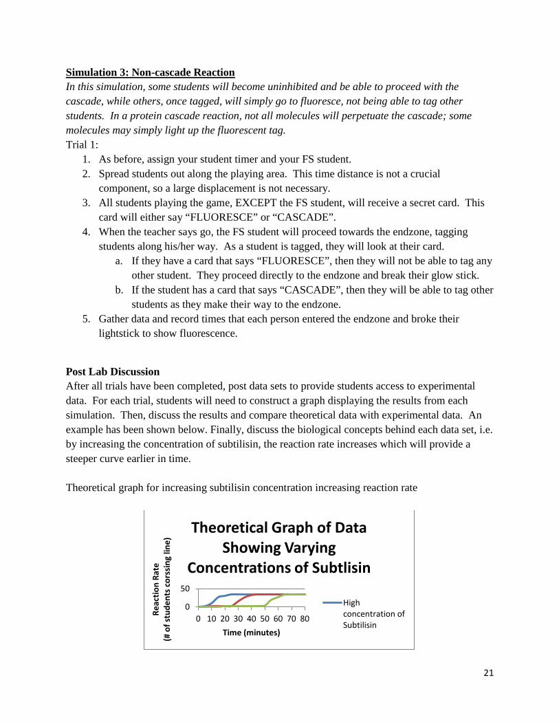
Simulation 3: Non-cascade Reaction In this simulation, some students will become uninhibited and be able to proceed with the cascade, while others, once tagged, will simply go to fluoresce, not being able to tag other students. In a protein cascade reaction, not all molecules will perpetuate the cascade; some molecules may simply light up the fluorescent tag. Trial 1:
1. As before, assign your student timer and your FS student. 2. Spread students out along the playing area. This time distance is not a crucial
component, so a large displacement is not necessary. 3. All students playing the game, EXCEPT the FS student, will receive a secret card. This
card will either say “FLUORESCE” or “CASCADE”. 4. When the teacher says go, the FS student will proceed towards the endzone, tagging
students along his/her way. As a student is tagged, they will look at their card. a. If they have a card that says “FLUORESCE”, then they will not be able to tag any
other student. They proceed directly to the endzone and break their glow stick. b. If the student has a card that says “CASCADE”, then they will be able to tag other
students as they make their way to the endzone. 5. Gather data and record times that each person entered the endzone and broke their
lightstick to show fluorescence.
Post Lab Discussion After all trials have been completed, post data sets to provide students access to experimental data. For each trial, students will need to construct a graph displaying the results from each simulation. Then, discuss the results and compare theoretical data with experimental data. An example has been shown below. Finally, discuss the biological concepts behind each data set, i.e. by increasing the concentration of subtilisin, the reaction rate increases which will provide a steeper curve earlier in time. Theoretical graph for increasing subtilisin concentration increasing reaction rate
0
50
0 10 20 30 40 50 60 70 80 Reac
tion
Rate
(# o
f stu
dent
s cor
ssin
g lin
e)
Time (minutes)
Theoretical Graph of Data Showing Varying
Concentrations of Subtlisin
High concentration of Subtilisin
22
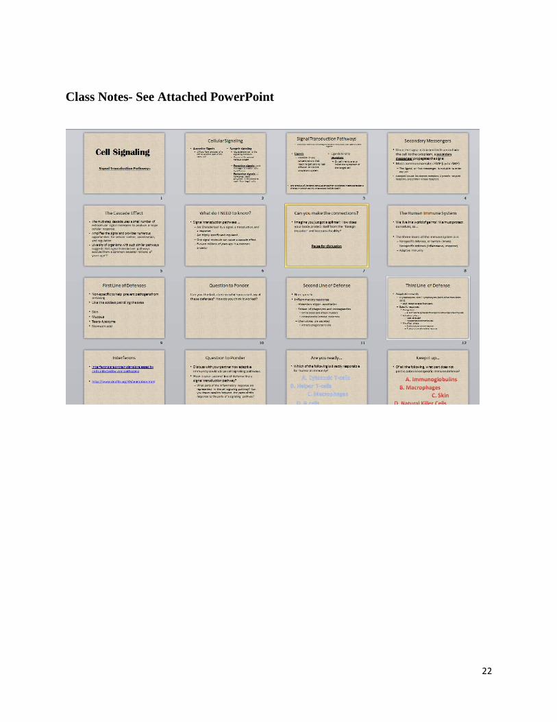
Class Notes- See Attached PowerPoint
23
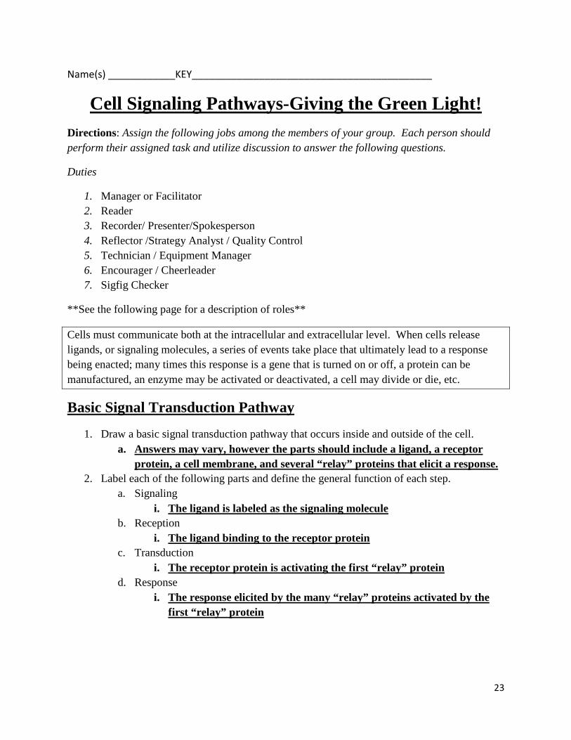
Name(s) ____________KEY___________________________________________
Cell Signaling Pathways-Giving the Green Light! Directions: Assign the following jobs among the members of your group. Each person should perform their assigned task and utilize discussion to answer the following questions.
Duties
1. Manager or Facilitator 2. Reader 3. Recorder/ Presenter/Spokesperson 4. Reflector /Strategy Analyst / Quality Control 5. Technician / Equipment Manager 6. Encourager / Cheerleader 7. Sigfig Checker
**See the following page for a description of roles**
Cells must communicate both at the intracellular and extracellular level. When cells release ligands, or signaling molecules, a series of events take place that ultimately lead to a response being enacted; many times this response is a gene that is turned on or off, a protein can be manufactured, an enzyme may be activated or deactivated, a cell may divide or die, etc.
Basic Signal Transduction Pathway
1. Draw a basic signal transduction pathway that occurs inside and outside of the cell. a. Answers may vary, however the parts should include a ligand, a receptor
protein, a cell membrane, and several “relay” proteins that elicit a response. 2. Label each of the following parts and define the general function of each step.
a. Signaling i. The ligand is labeled as the signaling molecule
b. Reception i. The ligand binding to the receptor protein
c. Transduction i. The receptor protein is activating the first “relay” protein
d. Response i. The response elicited by the many “relay” proteins activated by the
first “relay” protein

24
3. Cell signaling pathways rely on signal amplification to produce a signaling cascade. a. Define amplification in your own words
i. Answers may vary, however amplification generally means “to make larger”
b. How can a signal be amplified? BE SPECIFIC! i. One relay protein activates several other relay proteins, which
activate other relay proteins, which ultimately elicit an end response. Immune responses with B and T cells, as well as the general histamine response, are all examples of this type of pathway.
c. Provide several examples of possible responses that occur in a cell due to a signal being received
i. Producing a protein, opening a protein channel, cell division and/or cell death, a gene being turned on, etc.
d. Once the cell fulfills and completes a response, what must happen in order to stop the response?
i. The activated relay protein must become deactivated/inhibited.
Phosphorylation Cascade
1. Draw a cell signaling cascade. Now show specifically when phosphorylation occurs and what the major function of phosphorylation is.
a. Answers will vary, however the drawing should include the ligand binding the receptor protein in the cell membrane, activating a relay protein, which in turns activates other inactive protein complexes through adding a phosphate group and hydrolyzing ATP to produce energy.
2. Where are the phosphate groups that are added to the proteins during phosphorylation derived from? Show this in your diagram.
a. Answers may vary, however the drawing should show that ATP ADP and through removing the phosphate group, an inactive protein kinase becomes active and propagates the signal down the cell eliciting a response.
3. What kind of enzymes performs phosphorylation? a. Kinases
4. Describe what effects amplification has on the signal transduction pathway. a. The signal can be propagated quicker because each kinase can activate more
than one subsequent kinase enzyme in the sequence. 5. How is it advantageous for an organism to possess a signal transduction pathway that has
several amplification steps? a. Amplification of the signal can allow the receptor of one single ligand to
cause response in several cellular locations, which allows for quicker response to a stimulus.
25
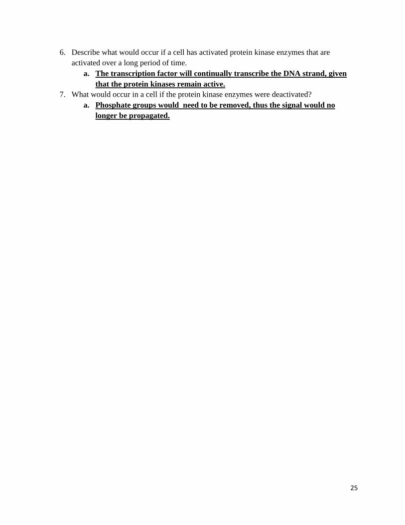
6. Describe what would occur if a cell has activated protein kinase enzymes that are activated over a long period of time.
a. The transcription factor will continually transcribe the DNA strand, given that the protein kinases remain active.
7. What would occur in a cell if the protein kinase enzymes were deactivated? a. Phosphate groups would need to be removed, thus the signal would no
longer be propagated.

26
Description of Roles
https://pogil.org/uploads/media_items/descriptions-of-roles-expanded.original.pdf
In POGIL courses, most of the class work is done in groups of about four. The membership of the groups may change. The roles within a group will change to allow everyone an opportunity to try out a role. Here are some roles that are commonly used:
Manager or Facilitator: Manages the group. Ensures that members are fulfilling their roles, that the assigned tasks are being accomplished on time, and that all members of the group participate in activities and understand the concepts. Your instructor will respond to questions from the manager only (who must raise his or her hand to be recognized).
Reader: Reads the activity out loud to the group. The reader must monitor their volume so that their group can hear them, but other groups are not disturbed. This helps to keep everyone in the group together. The Manager will tell the Reader when it is time to read the next part of the activity.
Recorder/ Presenter/Spokesperson: Records the names and roles of the group members at the beginning of each activity. Records the important aspects of group discussions, observations, insights, etc. The recorder’s report is a log of the important concepts that the group has learned. This person is also responsible for reporting orally to the class when called for in class discussions.
Reflector /Strategy Analyst / Quality Control: Observes and comments on group dynamics and behavior with respect to the learning process. These observations should be made to the manager on a regular basis (no more than 20 minutes between reports) in an effort to constantly improve group performance. The reflector/analyst may be called upon to report to the group (or the entire class) about how well the group is operating (or what needs improvement) and why.
Technician / Equipment Manager: Gathers materials and uses any equipment other than paper and pencil. Performs all technical operations for the group, including the use of a calculator or computer. Unless otherwise instructed, only the technician in each group may operate equipment such as this.
Encourager / Cheerleader: Acknowledges the good ideas and insights of group members (or the group as a whole) through expressions such as “That was a really good point!” at appropriate times.
Sigfig Checker: This role should be self-evident
27
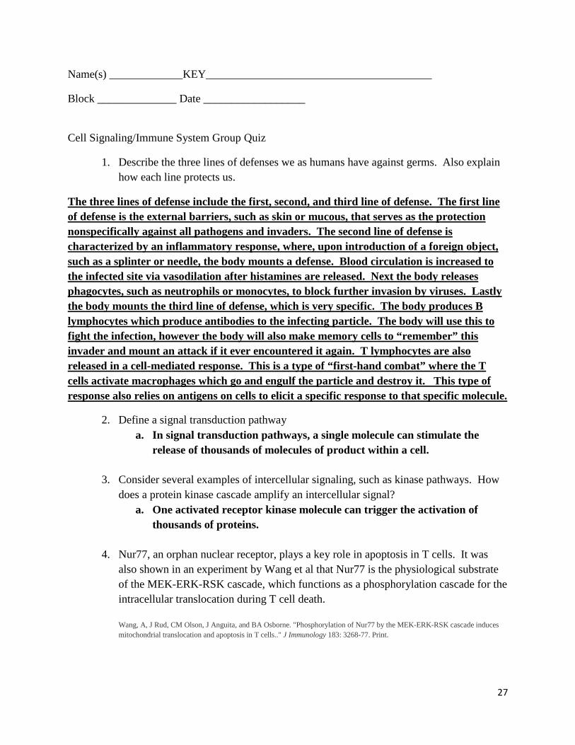
Name(s) _____________KEY________________________________________
Block ______________ Date __________________
Cell Signaling/Immune System Group Quiz
1. Describe the three lines of defenses we as humans have against germs. Also explain how each line protects us.
The three lines of defense include the first, second, and third line of defense. The first line of defense is the external barriers, such as skin or mucous, that serves as the protection nonspecifically against all pathogens and invaders. The second line of defense is characterized by an inflammatory response, where, upon introduction of a foreign object, such as a splinter or needle, the body mounts a defense. Blood circulation is increased to the infected site via vasodilation after histamines are released. Next the body releases phagocytes, such as neutrophils or monocytes, to block further invasion by viruses. Lastly the body mounts the third line of defense, which is very specific. The body produces B lymphocytes which produce antibodies to the infecting particle. The body will use this to fight the infection, however the body will also make memory cells to “remember” this invader and mount an attack if it ever encountered it again. T lymphocytes are also released in a cell-mediated response. This is a type of “first-hand combat” where the T cells activate macrophages which go and engulf the particle and destroy it. This type of response also relies on antigens on cells to elicit a specific response to that specific molecule.
2. Define a signal transduction pathway a. In signal transduction pathways, a single molecule can stimulate the
release of thousands of molecules of product within a cell.
3. Consider several examples of intercellular signaling, such as kinase pathways. How does a protein kinase cascade amplify an intercellular signal?
a. One activated receptor kinase molecule can trigger the activation of thousands of proteins.
4. Nur77, an orphan nuclear receptor, plays a key role in apoptosis in T cells. It was also shown in an experiment by Wang et al that Nur77 is the physiological substrate of the MEK-ERK-RSK cascade, which functions as a phosphorylation cascade for the intracellular translocation during T cell death. Wang, A, J Rud, CM Olson, J Anguita, and BA Osborne. "Phosphorylation of Nur77 by the MEK-ERK-RSK cascade induces mitochondrial translocation and apoptosis in T cells.." J Immunology 183: 3268-77. Print.

28
Phosphorylation cascades involving a series of protein kinases are useful for cellular signal transduction because
a. They are species specific b. They always lead to the same cellular response c. They amplify the original signal manyfold d. They counter the harmful effects of phosphatases
5. Explain how interferons are an example of cell communication. Include source and
targets in your answer. Interferons are protein signals released by cells infected by viral pathogens. Interferons increase the resistance of neighboring cells to infection by binding to receptors on the plasma membranes of uninfected cells, stimulating a signaling pathway that inhibits viral reproduction. Interferons also bind to receptors of other cells in the immune system, activating them to fight the infection.
6. What is the relationship between signal transduction pathways and second messengers? How does this relate to immunological responses?
a. Secondary messengers relay the message from the inside of the membrane throughout the cytoplasm. This can be related to T cells being activated from macrophages and perpetuating a further immunological response to a foreign invader.
Matching: Match the word with the description below.
B cells Macrophages Helper T cells Cytotoxic T cell
7. Bonds to class II MHC molecules ___Helper T cells_________________ 8. An APC that is also a part of innate immunity___Macrophages_____________ 9. Responsible for humoral immunity ___B cells__________________ 10. A lymphocyte that secretes cytokines to stimulate other lymphocytes ____Helper T
cells____________
11. Describe three responses by the innate immune system in response to a pathogen.
Increase in tear, or lysozyme, production; inflammatory response at the exposure site; phagocytosis of the foreign invader, or pathogen; and increased flow of bodily fluids, such as mucus.

29
12. Explain the positive feedback loop in regards to how Helper T cells use cell signaling to initiate an active immune response.
a. Helper T cells are activated by direct cell-to-cell signaling and use cytokines released from macrophages. Macrophages then ingest the antigens, process and present them on the cell surface. This is where the helper T cells recognizes the antigens and subsequently allow for activation of helper T cells, which in turn release more cytokines causing proliferation of helper T cells. This positive feedback helps to activate cytotoxic T cells and antibody-producing B cells.

30
Student Section

31
Name _____________________________________________________
Cell Signaling Article What Kind of Signals Do Cells Receive? Cells must be able to receive signals, as well as propagate signals to elicit a response. Cell signals can be used as sensor mechanisms, growth factors, hormones, and neurotransmitters. Neurotransmitters, a class of signaling molecules that travel across the neuromuscular junction, aid in intracellular signaling among neurons. Other molecules, such as FSH (Follicle Stimulating Hormone), move further from the targeted cell traveling from the brain to the ovary, resulting in egg release from the ovary. Other signaling molecules aid in mechanical stimulation of cells, such as cells in the ear reacting to movement of sound waves, sensors (baroreceptors) that detect changes in blood pressure in the vascular system, or pressure sensors in the skin. How Do Cells Recognize Signals? Receptor proteins bind to signaling molecules and ultimately initiate a physiological response. Depending on functionality, there exist different receptors for different molecules; dopamine receptors bind to dopamine, insulin receptors bind to insulin, etc. Receptors can be transmembrane proteins (span across the membrane) which consequently bind to signaling molecules outside the cell and transmit the signal through a series of molecular switches, internal signaling pathways, and propagation of a signal through the cell. Membrane receptors fall into three main classes: G-protein-coupled receptors, ion channel receptors, and enzyme-linked receptors. All three types transmit extracellular signals within the cell. They allow these signaling molecules to affect the cell functions without entering the cell. Within the membrane exist gated ion channels, such as acetylcholine receptors that allow for soluble materials to travel across the membrane when the pore is open, and when the pore is closed, no external signal is present. Therefore, when an acetylcholine molecule binds to a receptor, a conformational change within the membrane allows for transport of ion through the membrane. How Do Cells Respond to Signals? After the signal is received from the receptor protein, a conformational change occurs and launches a series of biochemical reactions known as a signal transduction cascade. This amplifies the message and propagates the signal throughout the cell. After the receptor activates the cascade, secondary messengers are triggered which initiate and coordinate the intracellular signaling pathways. The quintessential example is cAMP, a common secondary messenger which contributes the increasing propagation of the signal. cAMP, synthesized from ATP by adenyl cyclase, activates the enzyme protein kinase which subsequently phosphorylates multiple protein substrates. Each step further amplifies the initial signal and continues to further the propagation of the signal within the cell. How Do Signals Affect Cell Function? Through the process of phosphorylation, protein kinase enzymes catalyze the transfer of phosphate groups, specifically with proteins containing the amino acids serine, threonine, and tyrosine, as these are common phosphorylation sites. This process of phosphorylation causes a conformational change in the enzymes resulting in either activation or inhibition on enzyme activity. Phosphorylation also allows for control of protein function, receiving of signals, and response to stimuli.
32
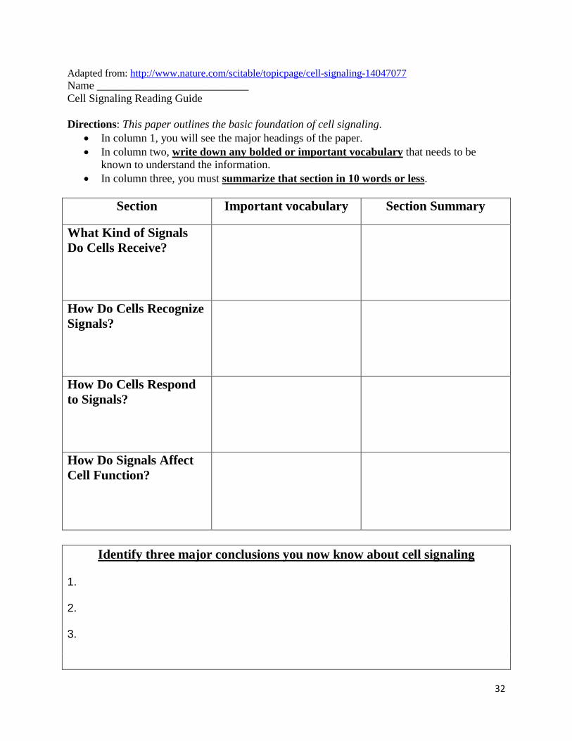
Adapted from: http://www.nature.com/scitable/topicpage/cell-signaling-14047077 Name ___________________________ Cell Signaling Reading Guide Directions: This paper outlines the basic foundation of cell signaling.
• In column 1, you will see the major headings of the paper. • In column two, write down any bolded or important vocabulary that needs to be
known to understand the information. • In column three, you must summarize that section in 10 words or less.
Section Important vocabulary Section Summary
What Kind of Signals Do Cells Receive?
How Do Cells Recognize Signals?
How Do Cells Respond to Signals?
How Do Signals Affect Cell Function?
Identify three major conclusions you now know about cell signaling
1.
2.
3.

33
Cornell Notes Template
34
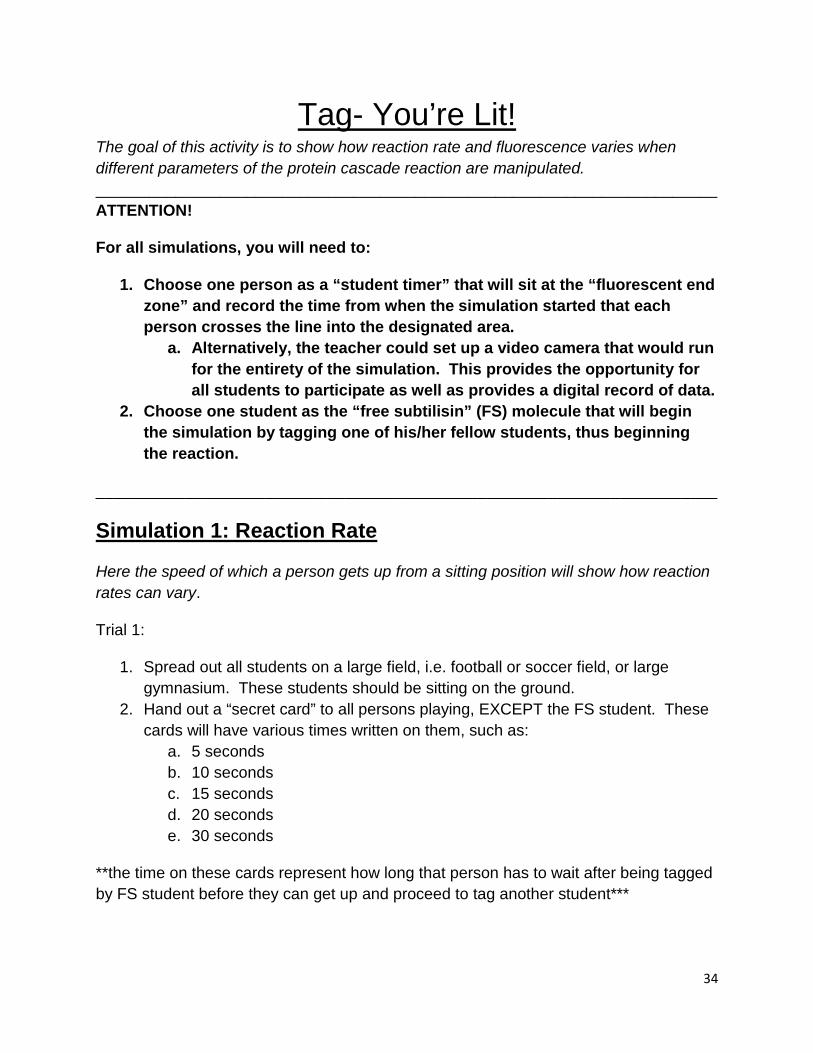
Tag- You’re Lit! The goal of this activity is to show how reaction rate and fluorescence varies when different parameters of the protein cascade reaction are manipulated. ______________________________________________________________________ ATTENTION!
For all simulations, you will need to:
1. Choose one person as a “student timer” that will sit at the “fluorescent end zone” and record the time from when the simulation started that each person crosses the line into the designated area.
a. Alternatively, the teacher could set up a video camera that would run for the entirety of the simulation. This provides the opportunity for all students to participate as well as provides a digital record of data.
2. Choose one student as the “free subtilisin” (FS) molecule that will begin the simulation by tagging one of his/her fellow students, thus beginning the reaction.
______________________________________________________________________
Simulation 1: Reaction Rate
Here the speed of which a person gets up from a sitting position will show how reaction rates can vary.
Trial 1:
1. Spread out all students on a large field, i.e. football or soccer field, or large gymnasium. These students should be sitting on the ground.
2. Hand out a “secret card” to all persons playing, EXCEPT the FS student. These cards will have various times written on them, such as:
a. 5 seconds b. 10 seconds c. 15 seconds d. 20 seconds e. 30 seconds
**the time on these cards represent how long that person has to wait after being tagged by FS student before they can get up and proceed to tag another student***

35
3. When the teacher says “GO”, the FS student will proceed to go and tag one person. When that person is tagged, they look at their card and wait for the designated amount of time. The FS student can still go tag other students but he/she must be making his/her way to the “Fluorescent Endzone” which would be across the field. Each person who has a card, after waiting their designated time, can get up and proceed towards the fluorescent endzone, along the way tagging other students who are still sitting down.
4. Once the person reaches the “fluorescent endzone”, they will bend a glowstick and make it glow. This represents that the molecules has triggered a fluorescent molecule to light up, which is indicative of reaction rate.
5. Once all students have entered the endzone, stop data collection, either by your timekeeper or stopping the tape if you chose the videotape method. (This can also be adapted to iPhone usage if you have a tall enough tripod.)
Simulation 2: Concentration of Reactants
This demonstration shows how the size of the test tube, or concentration of reactants, can vary in a reaction, changing reaction rate. This will be measured by changing distance between students, i.e. increasing and decreasing concentration.
Trial 1:
1. Students will spread out along the playing area of your choice (football field, etc.). The more spread out for this simulation, the better!
2. When teacher says “GO”, the student timer will begin his/her timer for data collection and count how many students enter the endzone and the corresponding time. See the ATTENTION note at the top of the preceding page for alternative procedures for student timer.
3. After the teacher says “GO”, the FS student will proceed towards the end zone, tagging anyone he comes across.
a. This “tagging” represents the prodomain of the complex coming off 4. As each person is tagged, they will proceed towards the endzone, tagging others
along the way. Those students can get up and tag other students. a. This provides the cascade effect for students to see.
5. As each student enters the endzone, they break a lightstick, showing fluorescence.
6. Stop and Discuss Data. Did it take long for one person to begin the cascade and get everyone in the endzone?

36
Trial 2:
1. Gather your student timer as noted before. 2. This time, three students will become FS students. Each of them will proceed to
the endzone as in Trial 1, tagging others along the way. 3. Record times that each person entered the endzone and broke their lightstick to
show fluorescence.
Trial 3:
1. Repeat the above procedure, however this time, start with five FS students. 2. Gather data and record times that each person entered the endzone and broke
their lightstick to show fluorescence.
Simulation 3: Non-cascade Reaction
In this simulation, some students will become uninhibited and be able to proceed with the cascade, while others, once tagged, will simply go to fluoresce, not being able to tag other students. In a protein cascade reaction, not all molecules will perpetuate the cascade; some molecules may simply light up the fluorescent tag.
Trial 1:
1. As before, gather your student timer and your FS student. 2. Spread students out along the playing area. This time distance is not a crucial
component, so a large displacement is not necessary. 3. All students playing the game, EXCEPT the FS student, will receive a secret
card. This card will either say “FLUORESCE” or “CASCADE”. 4. When the teacher says go, the FS student will proceed towards the endzone,
tagging students along his/her way. As a student is tagged, they will look at their card.
a. If they have a card that says “FLUORESCE”, then they will not be able to tag any other student. They proceed directly to the endzone and break their glow stick.
b. If the student has a card that says “CASCADE”, then they will be able to tag other students as they make their way to the endzone.
5. Gather data and record times that each person entered the endzone and broke their lightstick to show fluorescence.

37
Data Table Analysis

38

39
Fire! Near closing time on Saturday afternoon, March 25, 1911, a fire broke out on the top floors of the Asch Building in the Triangle Waist Company. Within minutes, the quiet spring afternoon erupted into madness, a terrifying moment in time, disrupting forever the lives of young workers. By the time the fire was over, 146 of the 500 employees had died. The survivors were left to live and relive those agonizing moments. The victims and their families, the people passing by who witnessed the desperate leaps from ninth floor windows and the City of New York would never be the same. Survivors recounted the horrors they had to endure, and passers-by and reporters also told stories of pain and terror they had witnessed. The images of death were seared deeply in their mind's eye.12
The above scenario actually occurred in New York in 1911. To escape the fire, an evacuation plan must be in place that allows all inhabitants to know of the danger, whether by signals or by word of mouth.
• You will be using the above scenario as a springboard for modeling your own plan for a protein cascade model. Just like some type of signal must be propagated to all the workers, you must devise a plan to create the most efficient way to amplify a signal in a protein cascade. You have learned about the parts of the protein cascade, i.e. the prodomain, the subtilisin molecule, the fluorescent tags, and the free molecules. Explain how much of each component you believe you will need and create a plan for maximum amplification.
• Once you have completed this, use that cascade as a springboard to explain the most efficient way that the human body can mount an immune response. You may choose either the inflammatory response or the cell-mediated response. Utilize the principles of cellular signaling to explain how the response would commence and explain any/all products of the cascade/reaction.
• Every student will submit their plans, and as a class, will decide upon the best plans. At the teacher’s discretion, these plans could be acted out as the simulations were to justify predictions.
12 "FIRE!" Remembering the 1911 Triangle Factory Fire. Cornell University, n.d. Web. 10 July 2013. <http://www.ilr.cornell.edu/trianglefire/story/fire.html>.
40
Name (s) ______________________________________________________
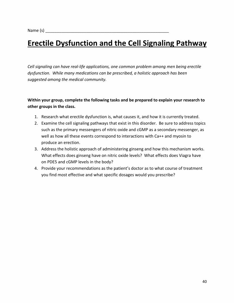
Erectile Dysfunction and the Cell Signaling Pathway
Cell signaling can have real-life applications, one common problem among men being erectile dysfunction. While many medications can be prescribed, a holistic approach has been suggested among the medical community.
Within your group, complete the following tasks and be prepared to explain your research to other groups in the class.
1. Research what erectile dysfunction is, what causes it, and how it is currently treated. 2. Examine the cell signaling pathways that exist in this disorder. Be sure to address topics
such as the primary messengers of nitric oxide and cGMP as a secondary messenger, as well as how all these events correspond to interactions with Ca++ and myosin to produce an erection.
3. Address the holistic approach of administering ginseng and how this mechanism works. What effects does ginseng have on nitric oxide levels? What effects does Viagra have on PDE5 and cGMP levels in the body?
4. Provide your recommendations as the patient’s doctor as to what course of treatment you find most effective and what specific dosages would you prescribe?

41
Name(s) _______________________________________________________
Cell Signaling Pathways-Giving the Green Light! Directions: Assign the following jobs among the members of your group. Each person should perform their assigned task and utilize discussion to answer the following questions.
Duties
1. Manager or Facilitator 2. Reader 3. Recorder/ Presenter/Spokesperson 4. Reflector /Strategy Analyst / Quality Control 5. Technician / Equipment Manager 6. Encourager / Cheerleader 7. Sigfig Checker
Cells must communicate both at the intracellular and extracellular level. When cells release ligands, or signaling molecules, a series of events take place that ultimately lead to a response being enacted; many times this response is a gene that is turned on or off, a protein can be manufactured, an enzyme may be activated or deactivated, a cell may divide or die, etc.
Basic Signal Transduction Pathway
1. Draw a basic signal transduction pathway that occurs inside and outside of the cell. 2. Label each of the following parts and define the general function of each step.
a. Signaling b. Reception c. Transduction d. Response
3. Cell signaling pathways rely on signal amplification to produce a signaling cascade. a. Define amplification in your own words b. How can a signal be amplified? BE SPECIFIC! c. Provide several examples of possible responses that occur in a cell due to a signal
being received d. Once the cell fulfills and completes a response, what must happen in order to stop
the response?

42
Phosphorylation Cascade
1. Draw a cell signaling cascade. Now show specifically when phosphorylation occurs and what the major function of phosphorylation is.
2. Where are the phosphate groups that are added to the proteins during phosphorylation derived from?
3. What kind of enzymes performs phosphorylation? 4. Describe what amplification means and what effects does that have on the signal
transduction pathway. 5. How is it advantageous for an organism to possess a signal transduction pathway that has
several amplification steps? 6. Describe what would occur if a cell has activated protein kinase enzymes that are
activated over a long period of time. 7. What would occur in a cell if the protein kinase enzymes were deactivated?

43
IMMUNOLOGY AND CELL SIGNAL PROJECT
Each pair of individuals will choose from the list below. No topics may be chosen twice. Students who wish to work alone may also do so. I suggest that when you begin your research you Google the disease and cell signaling or signal transduction. I have tried this for each disease and it will get you off to a good start. Your responsibility is to develop a poster that explains the basic immunology of the disease and its relationship to a faulty signal transduction pathway. Much of this research is "cutting edge." Your poster must explain both the "normal" pathway and the "faulty" pathway. Pictures and graphics are also necessary. I do not expect you to work at the graduate level on this project but I do expect you to be able to explain, in your own words, the basics of the mechanism and the way in which the failure of the mechanism leads to disease.
Grading: Total Points - 80
Format - poster - 15 points Traditional Poster
• 18" X 24" minimum • Typed headings • Pictures and graphics as needed • Neat presentation - no spelling or
grammatical errors Glogster
• At least 6 subtopics, excluding the main idea
• Must include pictures, videos, etc. • Neat Presentation- no spelling or
grammatical errors
Prezi • Must flow coherently • Must address all topics • Must include pictures, videos, etc. • Neat Presentation- no spelling or
grammatical errors
Content - 45 points • Disease – immunology, including the cause
and core symptoms • Cell signaling pathway involved • Correct mechanism • Incorrect mechanism • Graphics of pathway • Current direction of research
Presentation - 20 points
• Five minute presentation of poster/presentation to class
• Be able to put content into "own words" and explain poster
• No direct reading off poster Timeliness
• 11% per day penalty for lateness for poster
• 11% per day penalty for not being prepared for oral presentation
Disease Choices
• Crohn’s disease • Asthma • AIDS • Allergic Rhinitis • Tuberculosis • Type 1 Diabetes Mellitus • Celiac disease • Rheumatoid Arthritis • Leprosy • Multiple sclerosis • Chronic myelogenous leukemia - Chronic
myeloid leukemia – CML • Systemic Lupus

44
Name(s) _____________________________________________________ Block ______________ Date __________________
Cell Signaling/Immune System Group Quiz
1. Describe the three lines of defenses we as humans have against germs. Also explain how each line protects us.
2. Define a signal transduction pathway
3. Consider several examples of intercellular signaling, such as kinase
pathways. How does a protein kinase cascade amplify an intercellular signal?

45
4. Nur77, an orphan nuclear receptor, plays a key role in apoptosis in T cells. It was also shown in an experiment by Wang et al that Nur77 is the physiological substrate of the MEK-ERK-RSK cascade, which functions as a phosphorylation cascade for the intracellular translocation during T cell death. Wang, A, J Rud, CM Olson, J Anguita, and BA Osborne. "Phosphorylation of Nur77 by the MEK-ERK-RSK cascade induces mitochondrial translocation and apoptosis in T cells.." J Immunology 183: 3268-77. Print. Phosphorylation cascades involving a series of protein kinases are useful for cellular signal transduction because
a. They are species specific b. They always lead to the same cellular response c. They amplify the original signal manyfold d. They counter the harmful effects of phosphatases
5. Explain how interferons are an example of cell communication. Include source and targets in your answer.
6. What is the relationship between signal transduction pathways and second messengers? How does this relate to immunological responses?
.

46
Matching: Match the word with the description below. You may use words more than once, once, or not at all.
B cells Macrophages
Helper T cells Cytotoxic T cell
7. Bonds to class II MHC molecules ____________________ 8. An APC that is also a part of innate immunity________________ 9. Responsible for humoral immunity _____________________ 10. A lymphocyte that secretes cytokines to stimulate other lymphocytes
_______________
11. Describe three responses by the innate immune system in response to a pathogen.
12. Explain the positive feedback loop in regards to how Helper T cells use cell
signaling to initiate an active immune response.