Sonographic Imaging of Scrotal Emergencies …...Sonographic Imaging of Scrotal Emergencies...
23
Sonographic Imaging of Scrotal Emergencies Including Potential Pitfalls and Mimickers Jennifer Trinh, MD Shawn Van Bockel, MD Santa Clara Valley Medical Center, San Jose, CA
Transcript of Sonographic Imaging of Scrotal Emergencies …...Sonographic Imaging of Scrotal Emergencies...

Sonographic Imaging of Scrotal Emergencies Including Potential
Pitfalls and Mimickers
Jennifer Trinh, MDShawn Van Bockel, MD
Santa Clara Valley Medical Center, San Jose, CA
Disclosures
• There are no financial disclosures.
OverviewObjectives• Describe normal scrotal anatomy.• Understand the role of ultrasound in scrotal emergencies.• Identify characteristic sonographic features of scrotal
emergencies including ischemia, infection, and trauma.• Be aware of potential mimickers of scrotal emergencies.
Target audience• Radiology residents and practicing radiologists who would
like a refresher on scrotal emergencies.

Background• Ultrasound is the initial modality of choice in evaluating scrotal
emergencies.
• Common diseases seen in the emergency department include epididymitis, orchitis, abscess, testicular torsion, and trauma.
• Familiarity with the characteristic sonographic features and common pitfalls and mimickers is essential to differentiate these conditions and initiate treatment.
• This is particularly important for Fournier’s gangrene, testicular torsion, and testicular rupture because timely treatment is crucial to preserving fertility and hormonal activity.

Anatomy• Sperm is produced in the seminiferous
tubules (S).• The seminiferous tubules converge in the
mediastinum testes as a network of tubules called the rete testes (RT).
• The efferent ductules (curved black arrow) bridge the testicle and epididymal head (EH), which leads to the epididymal body (EB) and tail (ET).
• Sperm exit the scrotum through the vas deferens (straight black arrow).
• The tunica albuginea (A) surrounds the testicle.• The tunica vaginalis (arrowhead) has two layers
and also partially surrounds the testicle. • The testicular (internal spermatic) artery (white
arrow) supplies the testicle.
Diagram courtesy of Avery et al. Radiographics 2013

Normal Anatomy
Tunica albuginea –thin echogenic line
Mediastinum testes –horizontal echogenic band
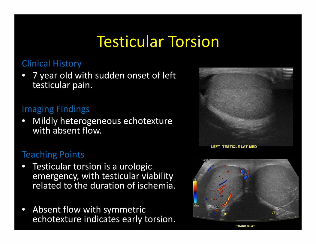
Testicular TorsionClinical History• 7 year old with sudden onset of left
testicular pain.
Imaging Findings• Mildly heterogeneous echotexture
with absent flow.
Teaching Points• Testicular torsion is a urologic
emergency, with testicular viability related to the duration of ischemia.
• Absent flow with symmetric echotexture indicates early torsion.
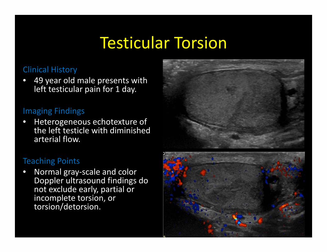
Testicular TorsionClinical History• 49 year old male presents with
left testicular pain for 1 day.
Imaging Findings• Heterogeneous echotexture of
the left testicle with diminished arterial flow.
Teaching Points• Normal gray‐scale and color
Doppler ultrasound findings do not exclude early, partial or incomplete torsion, or torsion/detorsion.

Torsed AppendixClinical History• 64 year‐old male with right testicular pain
for 1 year.
Imaging Findings• Small echogenic extratesticular lesion.
Teaching Points• This usually occurs in pediatric population
with a mean age of 9 years.• Pedunculated nature of the appendix
testis makes it prone to torsion.• An enlarged appendix testis (>5.6 mm)
with absence flow is characteristic.• A reactive hydrocele is common.

Epididymitis & Epididymal Abscess
Clinical History• Two weeks of right testicular
swelling.
Imaging Findings• Enlarged, hyperemic left
epididymis containing a large cystic structure with layering echogenic fluid (abscess).
• Complex left hydrocele.
Teaching Points• Infection spreads in a
retrograde fashion. The epididymal tail is involved before the body and head and should be carefully evaluated
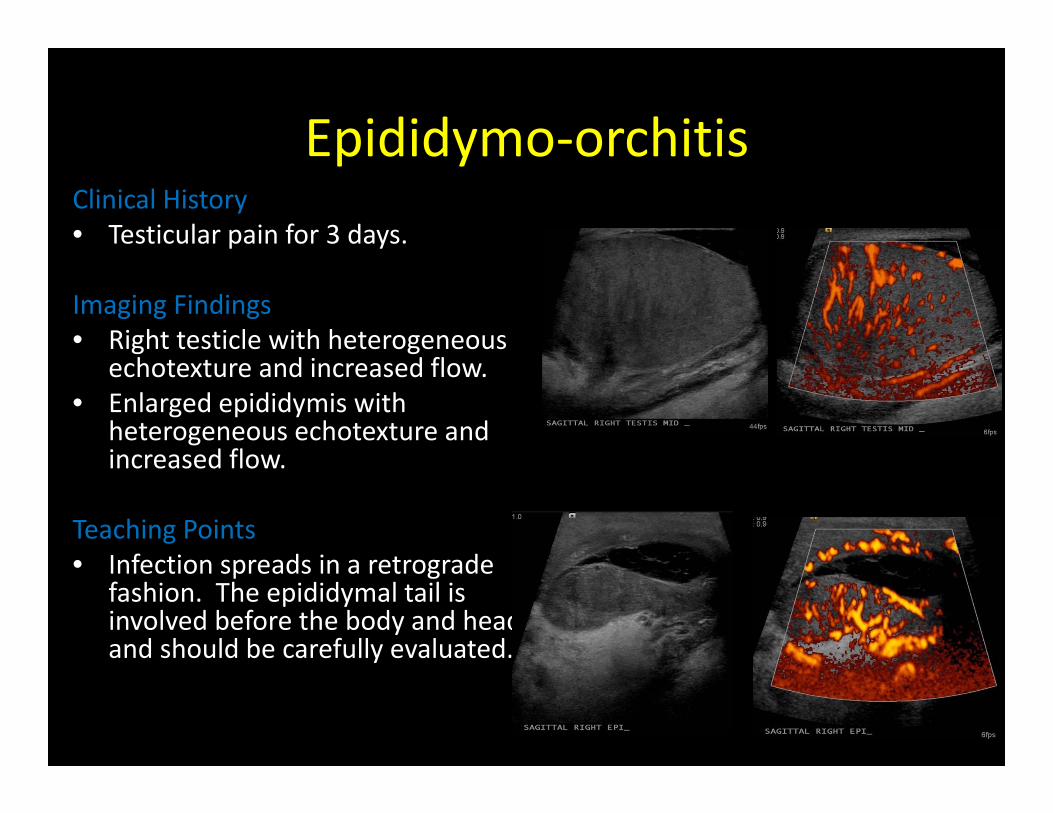
Epididymo‐orchitisClinical History• Testicular pain for 3 days.
Imaging Findings• Right testicle with heterogeneous
echotexture and increased flow.• Enlarged epididymis with
heterogeneous echotexture and increased flow.
Teaching Points• Infection spreads in a retrograde
fashion. The epididymal tail is involved before the body and head and should be carefully evaluated.
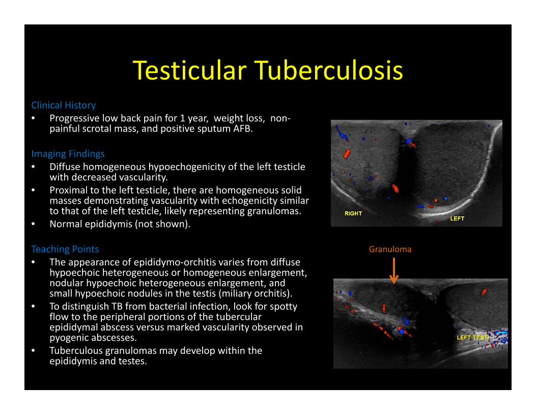
Testicular TuberculosisClinical History• Progressive low back pain for 1 year, weight loss, non‐
painful scrotal mass, and positive sputum AFB.
Imaging Findings• Diffuse homogeneous hypoechogenicity of the left testicle
with decreased vascularity.• Proximal to the left testicle, there are homogeneous solid
masses demonstrating vascularity with echogenicity similar to that of the left testicle, likely representing granulomas.
• Normal epididymis (not shown).
Teaching Points• The appearance of epididymo‐orchitis varies from diffuse
hypoechoic heterogeneous or homogeneous enlargement, nodular hypoechoic heterogeneous enlargement, and small hypoechoic nodules in the testis (miliary orchitis).
• To distinguish TB from bacterial infection, look for spotty flow to the peripheral portions of the tubercular epididymal abscess versus marked vascularity observed in pyogenic abscesses.
• Tuberculous granulomas may develop within the epididymis and testes.
Granuloma

Scrotal Abscess & Nonviable TestisClinical History• Homeless male with history of grade
1 scrotal trauma with hematocele who presents a month later with acute onset of worsening right testicular pain and swelling.
Imaging Findings• Complex fluid collection in the right
scrotum.• Thickening of the right scrotal soft
tissues.• Asymmetrically smaller right testicle
without flow.
Teaching Points• Orchitis unresponsive to antibiotics
requires surgical drainage.• Untreated orchitis can lead to
vascular compromise resulting in testicular infarction and atrophy.
Scrotum

Scrotal Abscess & Necrotic, Torsed Testicle
Clinical History• Leukocytosis and worsening right testicular pain
and swelling despite 4 days of antibiotics
Imaging Findings• Enlarged, heterogeneous epididymis with
increased vascularity.• Enlarged, edematous right testicle with minimal
flow, representing severe orchitis and torsion.• Large complex septated right hydrocele,
compatible with an abscess.
Teaching Points• Avascular hypoechoic area can represent
orchitis, but absent or minimal flow throughout the testis is suspicious for torsion.
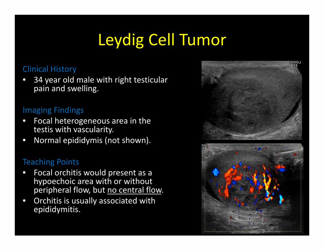
Leydig Cell TumorClinical History• 34 year old male with right testicular
pain and swelling.
Imaging Findings• Focal heterogeneous area in the
testis with vascularity.• Normal epididymis (not shown).
Teaching Points• Focal orchitis would present as a
hypoechoic area with or without peripheral flow, but no central flow.
• Orchitis is usually associated with epididymitis.
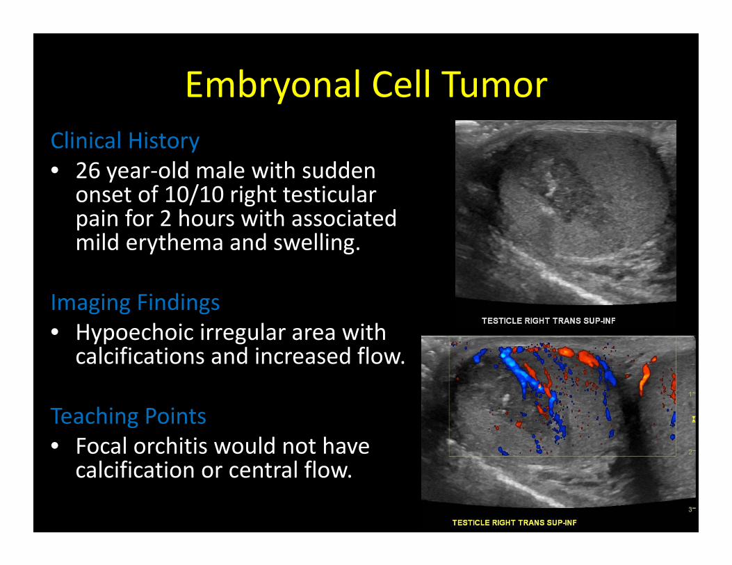
Embryonal Cell TumorClinical History• 26 year‐old male with sudden onset of 10/10 right testicular pain for 2 hours with associated mild erythema and swelling.
Imaging Findings• Hypoechoic irregular area with calcifications and increased flow.
Teaching Points• Focal orchitis would not have calcification or central flow.

Fournier’s GangreneClinical History• 34 year old male with status post right orchiectomy
3 weeks ago due to trauma, who presents with left scrotal swelling, pain and fever for 3 days.
Imaging Findings• US: Punctate echogenic foci within the left testicle,
concerning for gas.
• CT: Gas in the left scrotum, which tracks into the inguinal canal, retroperitoneum, and along the left gonadal vein.
Teaching Points• Fournier’s gangrene is a rapid progressive
necrotizing infection involving both the superficial and deep fascial planes.
• It is a urologic emergency due to the high mortality.
• Gas within the soft tissues is characteristic, but it’s absence does not exclude the diagnosis.
• CT is the modality of choice because it may depict the source of infection and its pathways of spread.
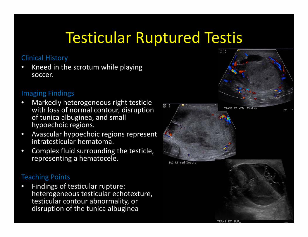
Testicular Ruptured TestisClinical History• Kneed in the scrotum while playing
soccer.
Imaging Findings• Markedly heterogeneous right testicle
with loss of normal contour, disruption of tunica albuginea, and small hypoechoic regions.
• Avascular hypoechoic regions represent intratesticular hematoma.
• Complex fluid surrounding the testicle, representing a hematocele.
Teaching Points• Findings of testicular rupture:
heterogeneous testicular echotexture, testicular contour abnormality, or disruption of the tunica albuginea
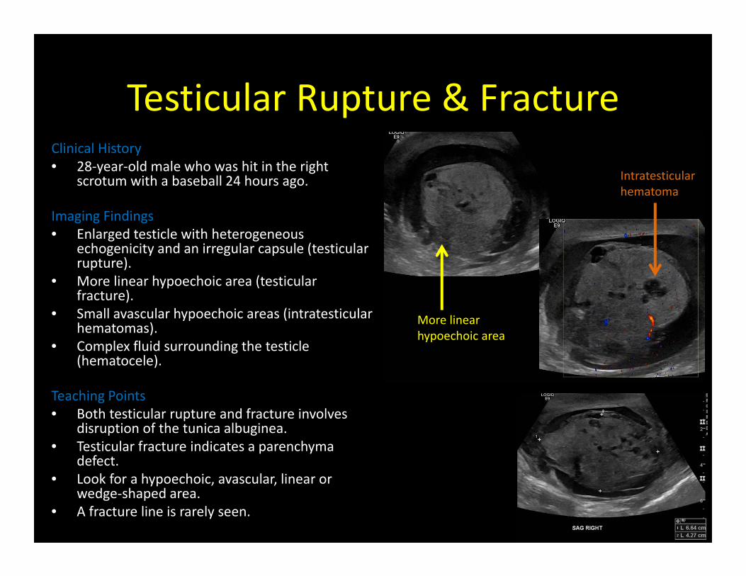
Testicular Rupture & FractureClinical History• 28‐year‐old male who was hit in the right
scrotum with a baseball 24 hours ago.
Imaging Findings• Enlarged testicle with heterogeneous
echogenicity and an irregular capsule (testicular rupture).
• More linear hypoechoic area (testicular fracture).
• Small avascular hypoechoic areas (intratesticular hematomas).
• Complex fluid surrounding the testicle (hematocele).
Teaching Points• Both testicular rupture and fracture involves
disruption of the tunica albuginea.• Testicular fracture indicates a parenchyma
defect. • Look for a hypoechoic, avascular, linear or
wedge‐shaped area.• A fracture line is rarely seen.
More linear hypoechoic area
Intratesticular hematoma
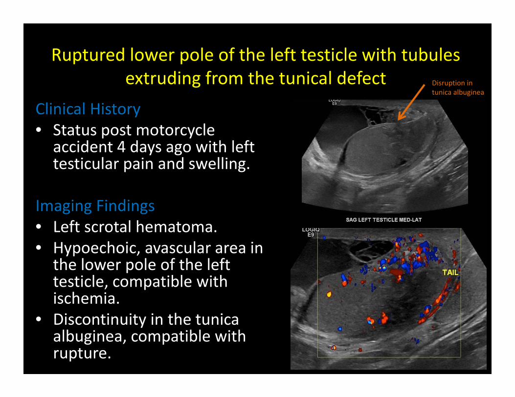
Ruptured lower pole of the left testicle with tubules extruding from the tunical defect
Clinical History• Status post motorcycle accident 4 days ago with left testicular pain and swelling.
Imaging Findings• Left scrotal hematoma.• Hypoechoic, avascular area in the lower pole of the left testicle, compatible with ischemia.
• Discontinuity in the tunica albuginea, compatible with rupture.
Disruption in tunica albuginea
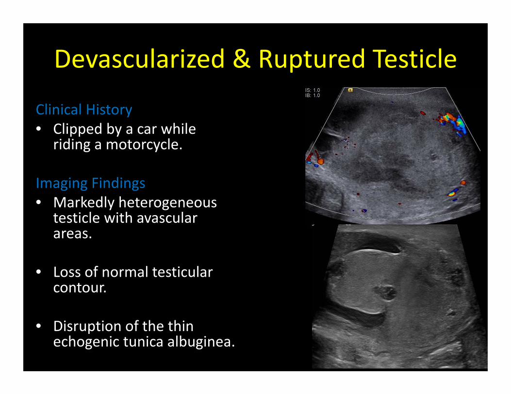
Devascularized & Ruptured Testicle
Clinical History• Clipped by a car while
riding a motorcycle.
Imaging Findings• Markedly heterogeneous
testicle with avascular areas.
• Loss of normal testicular contour.
• Disruption of the thin echogenic tunica albuginea.
References1. Avery LL, Scheinfeld MH. Imaging of penile and scrotal
emergencies. Radiographics 2013; 33:721‐402. Bhatt S, Dogra VS. Role of US in testicular and scrotal
trauma. Radiographics 2008;28:1617‐293. Davis JE, Silverman M. Scrotal emergencies. Emerg Med
Clin North Am 2011;29:469‐844. Yusuf GT, Sidhu PS. A review of ultrasound imaging in
scrotal emergencies. J Ultrasound 2013;16:171‐85. Chirindel A, Martinez F, Gagliardi JA, ArmmMF. Testicular
Tuberculosis Without Epididymitis Simulating Neoplasm. Radiology Case Reports 2008;3:133 http://radiology.casereports.net/index.php/rcr/article/view/133/434

Contact Information
Jennifer Trinh, MDDepartment of Radiology
Santa Clara Valley Medical Center751 S. Bascom Ave.San Jose, CA 95125