Somerset, Wiltshire, Avon and Gloucestershire (SWAG ...
17
Somerset, Wiltshire, Avon and Gloucestershire (SWAG) Cancer Alliance SWAG Head & Neck Clinical Advisory Group Clinical Guidelines Version 1.4 Page 1 of 17 Somerset, Wiltshire, Avon and Gloucestershire (SWAG) Cancer Services Head and Neck Cancer Clinical Advisory Group Clinical Guidelines June 2019 Revision due: April 2021
Transcript of Somerset, Wiltshire, Avon and Gloucestershire (SWAG ...
Somerset, Wiltshire, Avon and Gloucestershire (SWAG) Cancer Alliance
SWAG Head & Neck Clinical
Advisory Group Clinical Guidelines
Version 1.4
Page 1 of 17
Somerset, Wiltshire, Avon and Gloucestershire (SWAG) Cancer Services
Head and Neck Cancer Clinical Advisory Group
Clinical Guidelines
June 2019
Revision due: April 2021
Somerset, Wiltshire, Avon and Gloucestershire (SWAG) Cancer Alliance
SWAG Head & Neck Clinical
Advisory Group Clinical Guidelines
Version 1.4
Page 2 of 17
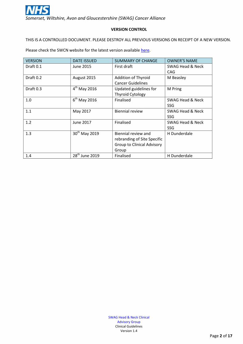
VERSION CONTROL
THIS IS A CONTROLLED DOCUMENT. PLEASE DESTROY ALL PREVIOUS VERSIONS ON RECEIPT OF A NEW VERSION.
Please check the SWCN website for the latest version available here.
VERSION DATE ISSUED SUMMARY OF CHANGE OWNER’S NAME
Draft 0.1 June 2015 First draft SWAG Head & Neck CAG
Draft 0.2 August 2015 Addition of Thyroid Cancer Guidelines
M Beasley
Draft 0.3 4th May 2016 Updated guidelines for Thyroid Cytology
M Pring
1.0 6th May 2016 Finalised SWAG Head & Neck SSG
1.1 May 2017 Biennial review SWAG Head & Neck SSG
1.2 June 2017 Finalised SWAG Head & Neck SSG
1.3 30th May 2019 Biennial review and rebranding of Site Specific Group to Clinical Advisory Group
H Dunderdale
1.4 28th June 2019 Finalised H Dunderdale
Somerset, Wiltshire, Avon and Gloucestershire (SWAG) Cancer Alliance
SWAG Head & Neck Clinical
Advisory Group Clinical Guidelines
Version 1.4
Page 3 of 17
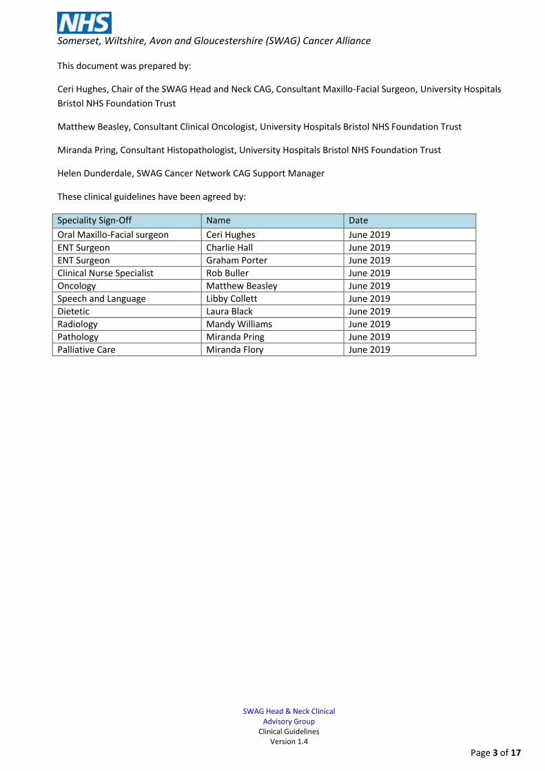
This document was prepared by:
Ceri Hughes, Chair of the SWAG Head and Neck CAG, Consultant Maxillo-Facial Surgeon, University Hospitals
Bristol NHS Foundation Trust
Matthew Beasley, Consultant Clinical Oncologist, University Hospitals Bristol NHS Foundation Trust
Miranda Pring, Consultant Histopathologist, University Hospitals Bristol NHS Foundation Trust
Helen Dunderdale, SWAG Cancer Network CAG Support Manager
These clinical guidelines have been agreed by:
Speciality Sign-Off Name Date
Oral Maxillo-Facial surgeon Ceri Hughes June 2019
ENT Surgeon Charlie Hall June 2019
ENT Surgeon Graham Porter June 2019
Clinical Nurse Specialist Rob Buller June 2019
Oncology Matthew Beasley June 2019
Speech and Language Libby Collett June 2019
Dietetic Laura Black June 2019
Radiology Mandy Williams June 2019
Pathology Miranda Pring June 2019
Palliative Care Miranda Flory June 2019
Somerset, Wiltshire, Avon and Gloucestershire (SWAG) Cancer Alliance
SWAG Head & Neck Clinical
Advisory Group Clinical Guidelines
Version 1.4
Page 4 of 17
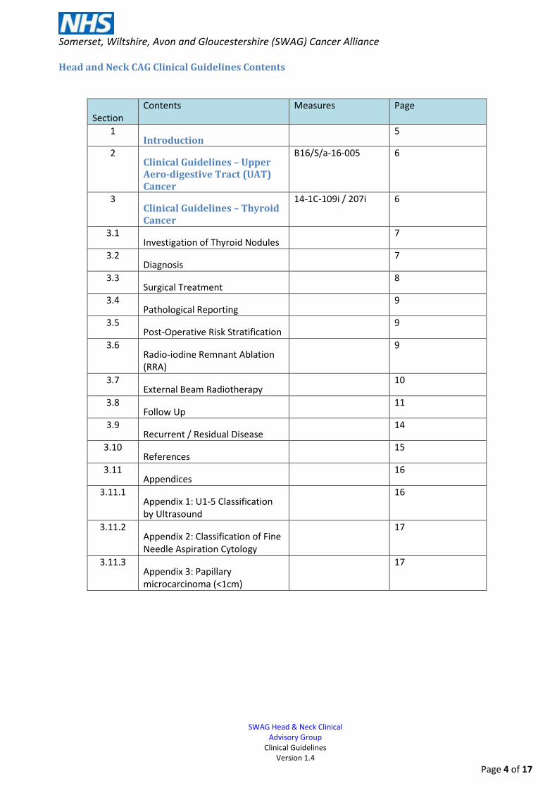
Head and Neck CAG Clinical Guidelines Contents
Section
Contents Measures Page
1 Introduction
5
2 Clinical Guidelines – Upper Aero-digestive Tract (UAT) Cancer
B16/S/a-16-005 6
3 Clinical Guidelines – Thyroid Cancer
14-1C-109i / 207i 6
3.1 Investigation of Thyroid Nodules
7
3.2 Diagnosis
7
3.3 Surgical Treatment
8
3.4 Pathological Reporting
9
3.5 Post-Operative Risk Stratification
9
3.6 Radio-iodine Remnant Ablation (RRA)
9
3.7 External Beam Radiotherapy
10
3.8 Follow Up
11
3.9 Recurrent / Residual Disease
14
3.10 References
15
3.11 Appendices
16
3.11.1 Appendix 1: U1-5 Classification by Ultrasound
16
3.11.2 Appendix 2: Classification of Fine Needle Aspiration Cytology
17
3.11.3 Appendix 3: Papillary microcarcinoma (<1cm)
17
Somerset, Wiltshire, Avon and Gloucestershire (SWAG) Cancer Alliance
SWAG Head & Neck Clinical
Advisory Group Clinical Guidelines
Version 1.4
Page 5 of 17
1. Introduction
The following guidelines pertain to the local management of Head and Neck (H&N) malignancies for the
Somerset, Wiltshire, Avon and Gloucestershire (SWAG) Network H&N Clinical Advisory Group (CAG).
The CAG refers to the Scottish Intercollegiate Guidelines for the Diagnosis and Management of Head and Neck
Cancers (SIGN), the British Association of Head and Neck Oncologists (BAHNO) guidelines and the NICE Guidance
for cancer of the upper aero-digestive tract: assessment and management in people aged 16 or over (2016).
These local guidelines should be reviewed alongside three other key documents for the CAG: the Constitution,
Annual Report and the Work Programme. The Constitution provides an overview of how the CAG operates,
outlining the general working processes of the CAG, the patient referral pathways and the guidelines to which
the CAG adheres. The Annual Report reflects the period of activity for the CAG from the previous year and it
contains a summary of this activity measured against several key performance indicators that have been
outlined in the National Cancer Peer Review Programme. The Work Programme summarises the key areas for
growth, development and improvement of the CAG over the next financial year (and beyond where
appropriate). All four documents should be reviewed together to give a full overview of the CAG, its
performance and future plans.
Primary care clinicians should refer to the NICE guidelines Suspected Cancer: recognition and management of
suspected cancer in children, young people and adults (2015) for the signs and symptoms relevant when referring
to Head and Neck services. Further details on the local process for referral can be found in the CAG constitution.
The CAG is committed to offering all eligible patients entry into clinical trials where available. Consent to provide
tissue for research purposes will also be sought wherever appropriate.
Somerset, Wiltshire, Avon and Gloucestershire (SWAG) Cancer Alliance
SWAG Head & Neck Clinical
Advisory Group Clinical Guidelines
Version 1.4
Page 6 of 17
2. The CAG Agreed Clinical Guidelines for Upper Aero-digestive Tract Cancer (B16/S/a-16-005)
The diagnosis, staging, treatment with surgery, radiotherapy and chemotherapy and the follow-up schedule for
UAT cancers is performed as recommended by the SIGN, BAHNO and NICE guidelines. A sentinel lymph node
biopsy service for early oral cancer as an alternative to an elective neck dissection is currently being developed;
training will commence on Friday 19th May 2017.
3. The CAG Agreed Clinical Guidelines for Thyroid Cancer
Bristol and Bath Differentiated Thyroid Cancer Guidelines
3.1 Investigation of Thyroid Nodules 1. Check thyroid function and refer to endocrinology if hyperthyroid. 2. Ultrasound evaluation of the neck should be performed in all patients with clinically evident thyroid nodules. 3. Ultrasound evaluation of the neck should be performed in all patients with solitary nodules found incidentally on PET-CT or found to be malignant in pathology specimens taken for other reasons. Nodules noted on CT without suspicious features such as extra-capsular extension, tracheal invasion or lymphadenopathy can be assessed clinically without the need for ultrasound. 4. The use of the U1-U5 scoring system is recommended for assessing risk of malignancy and guiding FNA. 5. For nodules in patients without lymphadenopathy, suspicious ultrasound findings, previous neck irradiation, hoarse voice or first degree relative with thyroid cancer (or associated syndrome) no further investigation is mandatory. In certain circumstances follow up may be appropriate but is not mandatory. 6. Nodules with potentially suspicious sonographic features (U3-5) should be investigated with FNA. Suspicious nodules <5mm may be observed with repeat ultrasound 3-6 months later. 7. FNA should be performed under ultrasound guidance, particularly if it is a repeat FNA after a non-diagnostic specimen. 8. All cases with Thy4 and Thy5 cytology should be discussed at the thyroid MDT prior to surgery. 9. Thy2 cytology should be repeated where there is clinical or ultrasound suspicion of malignancy. 10. Patient with nodules with benign ultrasound features may be discharged
3.2 Diagnosis (a) All patients with thyroid cancer should be discussed at the regional thyroid MDT and the plan from this discussion should be recorded in the patient’s notes. (b) At diagnosis all patients should meet or be given the contact details of the appropriate clinical nurse specialist and be given information about thyroid cancer and its treatment. (c) The GP should be informed within 24 hours of the diagnosis of cancer being communicated to the patient. (d) Delaying surgery until after delivery in patients diagnosed in pregnancy with differentiated thyroid cancer does not result in a poorer outcome but some patients may prefer to elect for a mid-trimester surgery.
Somerset, Wiltshire, Avon and Gloucestershire (SWAG) Cancer Alliance
SWAG Head & Neck Clinical
Advisory Group Clinical Guidelines
Version 1.4
Page 7 of 17
3.3 Surgical Treatment
(i) Pre-surgical investigations (a) Ultrasound of thyroid and bilateral neck should be performed by a specialist radiologist in all patients prior to diagnostic hemithyroidectomy or surgery for thyroid cancer (if not already performed) with FNA of suspicious nodes if this would change management. (b) Assessment of vocal cord function should be performed in all patients. (c) In patients with thyroid cancer cross sectional imaging (preferably MRI) may be undertaken pre-operatively to assess extra-thyroidal extension and lymph node disease. If CT is used there should be a 2 month delay between the use of iodinated contrast media and subsequent radio-iodine therapy unless there is a clinical urgency. (d) Measurement of thyroglobulin before thyroidectomy has no value and should not be undertaken.
(ii) Diagnostic thyroid surgery
(a) For patients with Thy3 and Thy4 FNA a diagnostic hemi-thyroidectomy is recommended. (b) Where there are additional indications for thyroidectomy (including patient preference), a total
thyroidectomy may be justified if supported by the MDT.
(iii) Therapeutic surgery for thyroid cancer (a) Surgeons who operate on patients with thyroid cancer should perform a minimum of 20 thyroidectomies
per year and publish audit outcome data. (b) Total thyroidectomy is recommended for patients with tumours greater than 4cm or tumours of any size
in association with multifocal disease, bilateral disease, extrathyroidal spread (T3 and T4a), familial disease and those with clinically or radiologically involved nodes and/or distant metastases.
(c) Patient with unifocal papillary microcarcinomas and minimally invasive follicular carcinoma without evidence of nodal spread or disease in the contralateral lobe should be offered hemithyroidectomy.
(d) Hemithyroidectomy without radio-iodine is an option that can be discussed with patients aged < 45 years who have tumours < 4cm without additional risk factors (evidence of nodal spread or suspicious features in the contralateral lobe on ultrasound, high risk histology including Hurtle cell) as part of Personalised Decision Making.
(e) In patients with radiation induced tumours 1-4cm then Personalized Decision Making should be employed to determine the extent of surgery (either lobectomy or total thyroidectomy is acceptable).
(f) In locally advanced disease a small residue of tumour may be left to preserve at least one recurrent laryngeal nerve but if tracheal or oesophageal resections are considered the patient should be referred to ENT.
(g) Completion thyroidectomy should occur within 8 weeks of initial lobectomy.
(iv) Nodal surgery
(a) Central compartment neck dissection is not recommended for patients without clinical or radiological evidence of lymph node involvement or overt extension outside the thyroid gland.
(b) Perform a Level IIa-Vb selective lateral neck dissection if clinically or radiologically involved nodes or suspicious nodes confirmed on FNA (or intra-operatively confirmed nodes on frozen section). I, IIb and Va dissection not recommended unless clinically involved. Isolated lymph node excision should not be performed as a planned therapeutic procedure.
(c) Central neck dissection is recommended if lateral neck dissection is performed.
Somerset, Wiltshire, Avon and Gloucestershire (SWAG) Cancer Alliance
SWAG Head & Neck Clinical
Advisory Group Clinical Guidelines
Version 1.4
Page 8 of 17
(v) Discharge Following total thyroidectomy patients should be discharged on levothyroxine 2mcg/kg (or lower initial dose if
elderly or obese).
3.4 Pathological Reporting (a) FNA specimens should be reported by a Head and Neck Pathologist or cytologist with significant experience in reporting of thyroid cytology. (b) Aspirates should be classified as Thy1-5 as per Royal College of Pathologists guidelines (January 2016) (c) Thyroid histology should be reported by a Head and neck or endocrine pathologist and should be pathologically staged using the AJCC TNM 7th Edition. (d) The dataset for thyroid cancer histopathology should be completed as per Royal College of Pathologists Standards and Datasets for reporting cancers (Dataset for thyroid cancer histopathology reports February 2014).
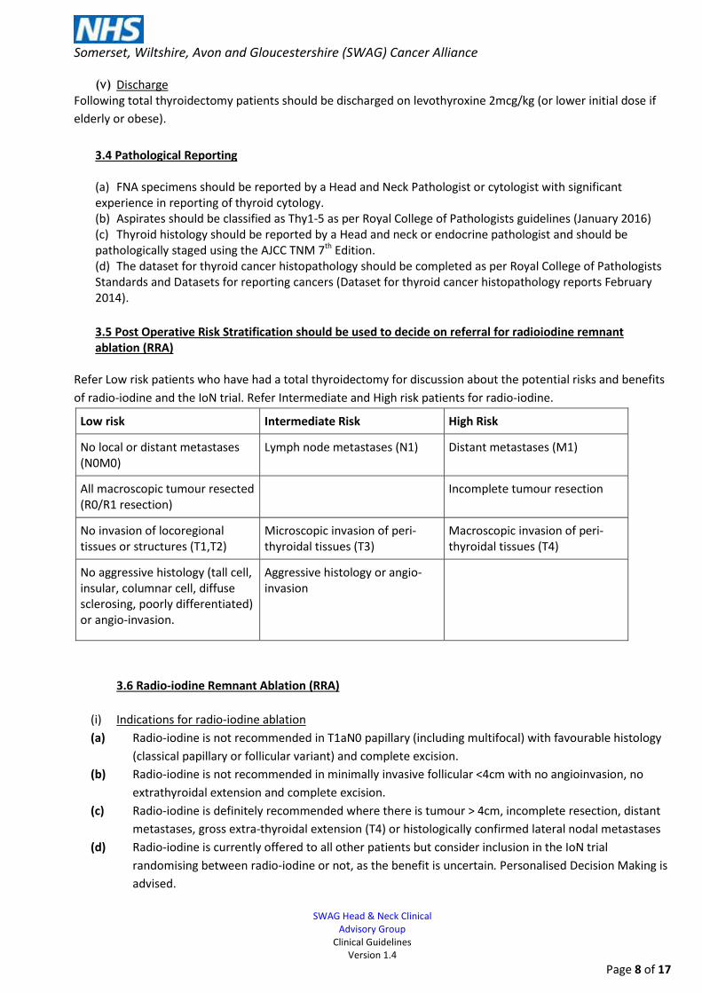
3.5 Post Operative Risk Stratification should be used to decide on referral for radioiodine remnant ablation (RRA)
Refer Low risk patients who have had a total thyroidectomy for discussion about the potential risks and benefits
of radio-iodine and the IoN trial. Refer Intermediate and High risk patients for radio-iodine.
Low risk Intermediate Risk High Risk
No local or distant metastases (N0M0)
Lymph node metastases (N1) Distant metastases (M1)
All macroscopic tumour resected (R0/R1 resection)
Incomplete tumour resection
No invasion of locoregional tissues or structures (T1,T2)
Microscopic invasion of peri-thyroidal tissues (T3)
Macroscopic invasion of peri-thyroidal tissues (T4)
No aggressive histology (tall cell, insular, columnar cell, diffuse sclerosing, poorly differentiated) or angio-invasion.
Aggressive histology or angio-invasion
3.6 Radio-iodine Remnant Ablation (RRA)
(i) Indications for radio-iodine ablation
(a) Radio-iodine is not recommended in T1aN0 papillary (including multifocal) with favourable histology
(classical papillary or follicular variant) and complete excision.
(b) Radio-iodine is not recommended in minimally invasive follicular <4cm with no angioinvasion, no
extrathyroidal extension and complete excision.
(c) Radio-iodine is definitely recommended where there is tumour > 4cm, incomplete resection, distant
metastases, gross extra-thyroidal extension (T4) or histologically confirmed lateral nodal metastases
(d) Radio-iodine is currently offered to all other patients but consider inclusion in the IoN trial
randomising between radio-iodine or not, as the benefit is uncertain. Personalised Decision Making is
advised.
Somerset, Wiltshire, Avon and Gloucestershire (SWAG) Cancer Alliance
SWAG Head & Neck Clinical
Advisory Group Clinical Guidelines
Version 1.4
Page 9 of 17
(ii) Radio-iodine ablation preparation
(a) Radio-iodine ablation should be offered 3-8 weeks post surgery. (b) Check renal function has been assessed by a serum creatinine within the last 3 months (c) Recombinant humanised TSH administration is preferred to thyroid hormone withdrawal except in
patients where the thyroid gland has not been completely removed. (d) If the patient is being prepared with recombinant humanised TSH injections these should be given
for 2 days prior to radio-iodine. (e) If withdrawing from thyroid hormones, stop T4 4 weeks beforehand and change to T3. Stop T3 and
commence low iodine diet 2 weeks pre-ablation (or therapy) dose. Ideally iodine intake should be <50mcg/day for 2 weeks prior.
(f) Avoid Amiodarone for 12 months and iodinated multi-vitamins for 4-6 weeks prior to radio-iodine. (g) If possible, delay radio-iodine for 2 months post iodine containing contrast media. (h) Consider pre-treatment sperm banking for male patients likely to need > 2 doses of radio-iodine. (i) Exclude pregnancy before giving radio-iodine and stop breast feeding 8 weeks beforehand. If there is
any doubt about pregnancy status a human beta-hCG urine test should be undertaken in women of child bearing age.
(j) Fast 2 hours before and 1 hour post radio-iodine. (k) There is not enough evidence to recommend for or against measures such as sucking sweets to
protect salivary function (beyond liberal oral hydration). (l) Where thyroid hormone withdrawal is used, a TSH level of >30 should be confirmed on the day of
admission. Use one injection of recombinant TSH and treat 24 hours later if TSH<30 (unless a thyroidectomy has not been performed, in which case recombinant TSH is not recommended and a longer period of thyroid hormone withdrawal will be required).
(iii) Activity of radio-iodine for ablation
(a) 1.1GBq is recommended for T1N0 and T2N0 disease with R0 resection and is an option for patients with
T3 and N1 disease.
(b) 3.7GBq remains the recommended dose for patients with T4, or M1 disease or with other risk factors
such as high risk histology (Hurthle cell carcinoma, tall cell, insular, poorly differentiated and diffuse
sclerosing).
(iv) Post radio-iodine ablation
(a) Commence 2mcg/kg levothyroxine at discharge if not already taking this (unless elderly or cardiac issues
where the initial dose should be 1mcg/kg titrated up slowly).
(b) Levothyroxine should be taken on an empty stomach 20-30 minutes prior to breakfast.
(c) Post-ablation whole body gamma scan and SPECT-CT 2-10 days post treatment.
3.7 External Beam Radiotherapy
(i) Only recommended adjuvantly for macroscopic residual disease or for irresectable residual or recurrent
disease that fails to concentrate radio-iodine.
(ii) 50-60Gy with boost of up to 10Gy for gross disease, using Intensity Modulated Radiotherapy.
(iii) Palliative use for metastases.
Somerset, Wiltshire, Avon and Gloucestershire (SWAG) Cancer Alliance
SWAG Head & Neck Clinical
Advisory Group Clinical Guidelines
Version 1.4
Page 10 of 17
3.8 Follow-up
(i) Early post treatment (0-12 months)
a) Vocal cords should be checked in all patients post-operatively. b) Patients with postoperative hypocalcaemia discharged on alfacalcidol /calcium should be weaned off
their supplements with close monitoring of calcium levels. If this is not possible or if there is ongoing hypoparathyroidism, consider referral to an endocrinologist.
c) 2 months post RRA the patient should be seen by their oncologist or by their surgeon/endocrinologist if no radio-iodine given, to check TSH and T4 and titrate levothyroxine dose as required: a. to achieve a TSH of <0.1 if RRA given b. to achieve TSH 0.3-2.0 if no RRA given (section 6 i (a) and (b)).
d) Patients with pT1a disease and no other risk factors who were treated with hemithyroidectomy can be discharged back to primary care with advice including to check thyroid function once per year as they are at risk of developing hypothyroidism.
e) All patients (male and female) should use effective contraception for six months post radio-iodine. f) 9-12 months post radio-iodine patients should have a recombinant TSH stimulated thyroglobulin
measurement (measured on day 5 after the first rhTSH injection) (see below (8(v)) and neck ultrasound performed by a specialist (with FNAC if recurrent / metastatic disease suspected), to confirm success of ablation.
g) A diagnostic whole body scan and SPECT-CT should be performed only where measurement of serum Tg is unreliable or there was uptake beyond the neck on the post ablation scan.
h) Cross sectional imaging or PET scanning should be considered for patients with an incomplete response based on Tg but normal neck US.
i) Patients can then be assigned to a dynamic risk stratified group to determine ongoing requirement for TSH suppression and frequency of follow up.
Somerset, Wiltshire, Avon and Gloucestershire (SWAG) Cancer Alliance
SWAG Head & Neck Clinical
Advisory Group Clinical Guidelines
Version 1.4
Page 11 of 17
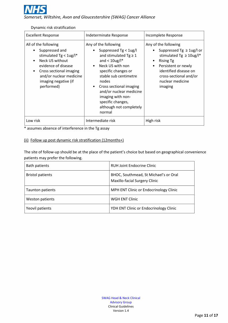
Dynamic risk stratification
Excellent Response Indeterminate Response Incomplete Response
All of the following
• Suppressed and stimulated Tg < 1ug/l*
• Neck US without evidence of disease
• Cross sectional imaging and/or nuclear medicine imaging negative (if performed)
Any of the following
• Suppressed Tg < 1ug/l and stimulated Tg ≥ 1 and < 10ug/l*
• Neck US with non specific changes or stable sub centimetre nodes
• Cross sectional imaging and/or nuclear medicine imaging with non-specific changes, although not completely normal
Any of the following
• Suppressed Tg ≥ 1ug/l or stimulated Tg ≥ 10ug/l*
• Rising Tg • Persistent or newly
identified disease on cross-sectional and/or nuclear medicine imaging
Low risk Intermediate risk High risk
* assumes absence of interference in the Tg assay
(ii) Follow up post dynamic risk stratification (12months+)
The site of follow-up should be at the place of the patient’s choice but based on geographical convenience
patients may prefer the following.
Bath patients RUH Joint Endocrine Clinic
Bristol patients BHOC, Southmead, St Michael’s or Oral
Maxillo-facial Surgery Clinic
Taunton patients MPH ENT Clinic or Endocrinology Clinic
Weston patients WGH ENT Clinic
Yeovil patients YDH ENT Clinic or Endocrinology Clinic
Somerset, Wiltshire, Avon and Gloucestershire (SWAG) Cancer Alliance
SWAG Head & Neck Clinical
Advisory Group Clinical Guidelines
Version 1.4
Page 12 of 17
1. Excellent response
a) TSH should be maintained in the low normal range (between 0.3 and 2) b) Frequency of follow up: 6 monthly for 1 year then annually c) Tg measurement: pre clinic annually unless antibodies present (see below 8(v))
2. Indeterminate response
a) TSH should be maintained between 0.1 and 0.5 for 5-10 years unless the patient develops relapsed disease in which case they should revert to TSH suppression <0.1
b) Frequency of follow up: minimum 6 monthly for 1 year then annually depending on individual need c) Tg measurement: pre clinic.
a. Patients with positive antibodies should be monitored 6 monthly until it is evident that these are antibodies are stable or falling (see below 8(v))
b. Patients with positive stimulated Tg (sTg) at 9-12 months, but negative US and otherwise low risk on ATA criteria should be managed expectantly with annual stimulated Tg (with the expectation that this will gradually decline)
3. Incomplete response (persistent or recurrent disease)
a) TSH should be suppressed to <0.1 indefinitely in the absence of specific contraindications b) Frequency of follow up: minimum 6 monthly for 5 years then annually depending on individual need c) Tg measurement: 6 monthly d) Patients with an incomplete response should be considered for further radio-iodine therapy once any
surgically resectable disease has been excluded. e) Patients with metastatic disease should be seen at least 6 monthly, either solely in oncology or
alternating with the surgical/endocrinology team and oncology. Stable patients who have to travel some distance to get to oncology could be seen locally with referral back to oncology as appropriate.
(iii) Follow up of historical patients who have not undergone Dynamic Risk Stratification (use ATA risk –
section 5)
(a) TSH should be suppressed to <0.1 for 5-10 years then relax to the low normal range (0.3-2.0) in the absence of any relapse.
(b) ATA Low risk patients in remission should be followed up yearly for 5 years (c) ATA Intermediate risk patients in remission with an uncomplicated course post treatment should then be
seen 6 monthly for the first year, then yearly for 10 years. (d) ATA high risk patients should be seen 6 monthly for the first 5 years then yearly although this could still
be at the site of their choice or alternating between BHOC and local team
(iv) Principles of TSH suppression
(a) The patient should be maintained on the lowest dose of levothyroxine that will keep the TSH suppressed to the required level, adjusting by 25mcg every 6-8 weeks. (b) During follow up, the degree of TSH suppression should be re-evaluated every few years to ensure that the target range correlates with the reassessed risk of recurrence and death, balancing potential benefits against risk to heart and skeleton. (c) In high risk groups (e.g. post-menopausal women) osteoporotic fracture risk should be assessed using the WHO FRAX tool (www.shef.ac.uk/FRAX). (d) The GP should be advised of the reason for TSH suppression and the target TSH.
(v) Principles of thyroglobulin (Tg) measurement
Somerset, Wiltshire, Avon and Gloucestershire (SWAG) Cancer Alliance
SWAG Head & Neck Clinical
Advisory Group Clinical Guidelines
Version 1.4
Page 13 of 17
a) All laboratories within the region should use the same assays (or send samples to the same central
laboratory) to give consistent results. b) Tg should not be routinely measured in patients who have had a hemi-thyroidectomy only with no
adjuvant radio-iodine ablation. c) Samples should not be collected sooner than 6 weeks post thyroidectomy or RRA. d) As Tg levels are TSH dependent, TSH levels should be measured concurrently. e) A rising Tg level over time indicates disease that is likely to become clinically apparent. f) Anti-thyroglobulin antibodies (Tg Ab) may falsely increase or decrease Tg levels. g) Where present at the outset, TgAb should decrease and disappear over a median of 3 years. De novo
and rising antibody levels or persistent levels beyond 3 years should be investigated. h) Tg should be measured six monthly if TgAb are detected or Tg remains elevated. In other patients
thyroglobulin should be measured annually. i) Stimulated Tg is measured on day 5 post recombinant human TSH (rh TSH) – given 0.9mg IM on days 1
and 2.
(vi) Use of routine imaging (a) Neck ultrasound should be requested in patients suspected of harbouring residual/relapsed disease such as persistent or rising Tg or TgAb or significant signs or symptoms in the neck with FNAC of any suspicious areas. (b) Suspicious lymph nodes on ultrasound <5mm only need explicit description and may be monitored by repeat ultrasound.
(vii) Long term follow-up (a) Patients treated with hemi-thyroidectomy alone because of a low risk of recurrence do not require long term follow in secondary care. (b) Patients with persisting disease will usually remain under hospital follow up. (c) Patients with excellent response / low risk on ATA criteria who are disease free at 5 years and no longer judged to require TSH suppression may be discharged to a to primary care or a nurse-led clinic with explicit instructions. Currently the advice is for this to continue life-long. If discharged to primary care the following is recommended.
Annual history and clinical examination
Annual Tg and TgAb measurement
Adjust Levothyroxine dose to maintain TSH 0.3 – 2.0
Refer back if new neck lump or rise in Tg level.
3.9 Recurrent/residual disease Surgical resection of recurrent or residual neck disease is the management of choice often followed by radio-
iodine therapy or external beam radiotherapy if the disease is non-iodine avid. Low volume recurrent or
persistent disease in the neck that is not progressing may be managed with active surveillance.
(i) Investigation of a rising thyroglobulin (a) A single elevated Tg should be confirmed by repeating the test before proceeding to additional investigations (b) For patients with low levels of Tg that are not rising, the decision to proceed to further investigation should be balanced against the low probability of detecting disease for which treatment is likely to be beneficial (c) Patients with newly detectable (>1.0) or rising Tg or TgAb should be investigated for relapse
Somerset, Wiltshire, Avon and Gloucestershire (SWAG) Cancer Alliance
SWAG Head & Neck Clinical
Advisory Group Clinical Guidelines
Version 1.4
Page 14 of 17
(d) The initial investigation should be a detailed neck USS performed by a specialist head and neck radiologist (e) For Tg +ve, US –ve, subsequent investigations should be guided by symptoms, but if asymptomatic a recombinant TSH stimulated radio-iodine tracer scan should be performed. If these investigations do not reveal a site of recurrence then a CT scan of neck and chest and/ or recombinant TSH stimulated PET-CT scan should be considered. If this does not reveal any site of disease then either the patient can be kept under observation or a therapeutic dose of radio-iodine could be considered depending on the risk group of the patient (f) High risk patients could be treated with a therapeutic dose of radio-iodine rather than a tracer scan as this may lead to stunning and affect subsequent treatment. In this situation, if the post treatment uptake scan is negative further iodine therapy should not be given unless there is a significant decrease in Tg.
(ii) Distant metastatic disease This is most frequently in the lungs or bones and can be treated by radio-iodine (3.7-7.4 GBq 4-12 monthly.
(a) Total thyroidectomy should be performed to improve the efficacy of radio-iodine if possible (b) For isolated metastases or non-iodine avid disease, surgical resection, radio-frequency ablation or palliative radiotherapy can be helpful (c) Even in radio-iodine avid disease, resection of predominant, large metastases should be considered to improve the effectiveness of radio-iodine (d) Palliative radiotherapy should be considered as an initial measure prior to radio-iodine in lesions where oedema would result in potential morbidity, such as vertebral metastases with potential to cause spinal cord compression. Steroids should be considered in such cases to try to avoid any swelling.
(iii) Systemic treatment There is no role for the routine use of chemotherapy. Single agent Doxorubicin can be discussed as palliation of
metastatic disease that does not respond to radio-iodine or radiotherapy. Consider applying for exceptional
funding for tyrosine kinase inhibitors or appropriate clinical trials.
3.10 References
Cooper at al. Revised American Thyroid Association Management Guidelines for Patients with Thyroid Nodules
and Differentiated Thyroid Cancer. Thyroid (2009) 19;11: 1167-1214
Pacini et al. European consensus for the management of patients with differentiated thyroid carcinoma of the
follicular epithelium. European Journal of Endocrinology (2006) 154 787-803
Luster et al. Guidelines for radioiodine therapy of differentiated thyroid cancer. Eur J Nucl Med Mol Imaging
(2008)
Royal College of Pathologists Standards and Datasets for reporting cancers. Dataset for thyroid cancer
histopathology reports February 2014
3.11 Appendices
3.11.1 Appendix 1 U1-5 Classification by Ultrasound
Somerset, Wiltshire, Avon and Gloucestershire (SWAG) Cancer Alliance
SWAG Head & Neck Clinical
Advisory Group Clinical Guidelines
Version 1.4
Page 15 of 17
U1. Normal.
U2. Benign:
(a) halo, iso-echoic / mildly hyper-echoic
(b) cystic change +/- ring down sign (colloid) (c) micro- cystic / spongiform
(d & e) peripheral egg shell calcification
(f) peripheral vascularity.
U3. Indeterminate/Equivocal:
(a) homogenous, hyper-echoic (markedly), solid, halo (follicular lesion).
(b) ? hypo-echoic, equivocal echogenic foci, cystic change
(c) mixed/central vascularity.
U4. Suspicious:
(a) solid, hypo-echoic (cf thyroid)
(b) solid, very hypo-echoic (cf strap muscle)
(c) disrupted peripheral calcification, hypo-echoic (d) lobulated outline
U5. Malignant
(a) solid, hypo-echoic, lobulated / irregular outline, micro-calcification. (? Papillary carcinoma)
(b) solid, hypo-echoic, lobulated/irregular outline, globular calcification (? Medullary carcinoma)
(c) intra-nodular vascularity
(d) shape (taller >wide) (AP>TR)
(e) characteristic associated lymphadenopathy
3.11.2 Appendix 2 Classification of Fine Needle Aspiration Cytology
Thy1/THY1c – non-diagnostic. This will include samples reflecting poor operator or preparation technique such as insufficient epithelial cells (the target is 6 groups each of at least 10 well- visualised follicular epithelial cells) or only poorly preserved cells, as well as those reflecting the lesion such as cysts. The suffix Thy1c is used for cyst fluid samples with insufficient colloid and epithelial cells.
Thy2/THY2c – non-neoplastic. These samples are adequately cellular and suggest a non-neoplastic lesion such as a hyperplastic or colloid nodule or thyroiditis. The cytological appearances should be consistent with the
Somerset, Wiltshire, Avon and Gloucestershire (SWAG) Cancer Alliance
SWAG Head & Neck Clinical
Advisory Group Clinical Guidelines
Version 1.4
Page 16 of 17
clinical information. Cyst samples containing abundant colloid, even if less that the target number of epithelial cells, can be categorised as Thy2c.
Thy3 – neoplasm possible. This is subdivided into:
Thy3a – Samples that exhibit cytological/nuclear or architectural atypia or other features that raise the possibility of neoplasia, but which are insufficient to enable confident placement into any other category. Situations would include sparsely cellular samples containing predominantly microfollicles and a compromised specimen (eg poorly spread) where some cells appear mildly abnormal. In many cases, a repeat aspirate is able to be placed into a more definitive category.
Thy3f – Samples that raise the possibility of a Follicular neoplasm and require histological assessment for a definitive diagnosis. The histological possibilities include a hyperplastic nodule, follicular adenoma or follicular carcinoma and also a follicular variant of papillary carcinoma (without clear characterising nuclear features of papillary carcinoma).
Thy4 – suspicious of malignancy but a confident diagnosis is not possible. The type of malignancy suspected should be stated in the report if possible.
Thy5 – A confident diagnosis of malignancy can be made. The type of malignancy should be stated, for example papillary, medullary or anaplastic thyroid carcinoma, lymphoma, or metastatic disease.
3.11.3 Appendix 3 Papillary microcarcinoma (<1cm) Found in up to 49.9% specimens (usually 5-12%) LN involvement in 12.3-50% In FU series: deaths 0.34%, local recurrence 2.5%, new distant mets 0.4% (median FU 3.7-11.2yrs) Recurrences virtually all in LNs, up to 20yrs post diagnosis, good prognosis with treatment Risk factors for recurrence or mets: o Clinical presentation o PET +ve o Larger size(6-10mm) o Multifocality/bilateral o Extrathyroidal extension o Poorly differentiated component o Desmoplastic fibrosis +/- infiltrative growth pattern. Management: 1. Surgery: Lobectomy for unifocal microPTC and no other risk factors Total thyroidectomy for multifocal involving both lobes, familial non-medullary thyroid cancer PDM (table 2.4): MicroPTC with Hx of neck irradiation – total vs hemi – consider additional RFs LN dissection if present with involved nodes PDM: Multifocal (both lobes)/minimal extrathyroidal extension through capsule (pT3) – prophylactic CCND vs none – consider poorly differentiated component/desmoplastic fibrosis
Somerset, Wiltshire, Avon and Gloucestershire (SWAG) Cancer Alliance
SWAG Head & Neck Clinical
Advisory Group Clinical Guidelines
Version 1.4
Page 17 of 17
2. RAA No indication for RRA – unifocal or multifocal, classical papillary or follicular variant, no extrathyroiddal extension PDM: MicroPTC with minimal extrathyroidal (pT3) or LN mets resected – RRA vs no RRA – consider unfavourable histological type, >5 LNs, LNs > 6mm, high ratio of positive LNs, 3. Follow up Unifocal microPTC with no other RFs (lobectomy only) – same risk of dying as general population and much lower risk of PTC than any other cancer over the lifetime. • No indication for TSH suppression • Discharge to GP - annual TFTs to prevent overt hypothyroidism.
All other groups - risk stratify after treatment and follow up accordingly.
-END-