Some Observations on Plant Karyology and Investigation...
40
Chapter 10 Some Observations on Plant Karyology and Investigation Methods Feyza Candan Additional information is available at the end of the chapter http://dx.doi.org/10.5772/56081 1. Introduction Karyology deals with the structure of cell nuclei, especially chromosomes. Cytology dealing with the study of cells in terms of structure also, function is known nowadays to mean only the study of chromosomes or nucleus and made synonymous with karyology wrongly. Cytotaxonomy means the application of cytological data to taxonomy. Cytotaxonomy studies the morphological and cytological characteristics of the organism along with their chromo‐ some numbers and structures (karyotype)[1, 2]. It is a secondary discipline that reinforces the principles of plant and animal taxonomy by abiding to the phylogenetic kinships. In classical taxonomy, the plants are categorized through determining their natural kinships in accordance with their morphological characteristics. Especially, the taxonomists are advisable in connec‐ tion with chromosomes. So, often chromosome number is assumed to be the all important, if not the only, chromosome character of interest to taxonomists, but size, shape, and behavior of chromosomes may throw more light on a taxonomic problem than their number alone. F. Ehrendorfer, in an erudite essay on Cytologie, Taxonomie und Evolution bei Samenpflan‐ zen [Cytology, taxonomy and evolution in seed plants], gives a detailed outline of these developments in cytogenetics since 1900 that have had a bearing on problems of taxono‐ my. Examples of taxonomic corrections based on chromosome studies include the remov‐ al of Yucca and Agave from the Amaryllidaceae to the Agavaceae and a number of rearrangements of species and genera in the Gramineae, Liliaceae, Compositae and other families; warnings are sounded against an uncritical use of chromosome pairing as a criterion of affinity, secondary pairing being dismissed altogether. A concrete example of the use of morphological and cytological considerations in deciding questions of relation‐ ships and evolution is given for the Dipsacaceae, where two distinct lines of phylogenetic development are traced: one of bushy species with clear polyploid series and the other of © 2013 Candan; licensee InTech. This is an open access article distributed under the terms of the Creative Commons Attribution License (http://creativecommons.org/licenses/by/3.0), which permits unrestricted use, distribution, and reproduction in any medium, provided the original work is properly cited.
Transcript of Some Observations on Plant Karyology and Investigation...

Chapter 10
Some Observations on Plant Karyology andInvestigation Methods
Feyza Candan
Additional information is available at the end of the chapter
http://dx.doi.org/10.5772/56081
1. Introduction
Karyology deals with the structure of cell nuclei, especially chromosomes. Cytology dealingwith the study of cells in terms of structure also, function is known nowadays to mean onlythe study of chromosomes or nucleus and made synonymous with karyology wrongly.Cytotaxonomy means the application of cytological data to taxonomy. Cytotaxonomy studiesthe morphological and cytological characteristics of the organism along with their chromo‐some numbers and structures (karyotype)[1, 2]. It is a secondary discipline that reinforces theprinciples of plant and animal taxonomy by abiding to the phylogenetic kinships. In classicaltaxonomy, the plants are categorized through determining their natural kinships in accordancewith their morphological characteristics. Especially, the taxonomists are advisable in connec‐tion with chromosomes. So, often chromosome number is assumed to be the all important, ifnot the only, chromosome character of interest to taxonomists, but size, shape, and behaviorof chromosomes may throw more light on a taxonomic problem than their number alone.
F. Ehrendorfer, in an erudite essay on Cytologie, Taxonomie und Evolution bei Samenpflan‐zen [Cytology, taxonomy and evolution in seed plants], gives a detailed outline of thesedevelopments in cytogenetics since 1900 that have had a bearing on problems of taxono‐my. Examples of taxonomic corrections based on chromosome studies include the remov‐al of Yucca and Agave from the Amaryllidaceae to the Agavaceae and a number ofrearrangements of species and genera in the Gramineae, Liliaceae, Compositae and otherfamilies; warnings are sounded against an uncritical use of chromosome pairing as acriterion of affinity, secondary pairing being dismissed altogether. A concrete example ofthe use of morphological and cytological considerations in deciding questions of relation‐ships and evolution is given for the Dipsacaceae, where two distinct lines of phylogeneticdevelopment are traced: one of bushy species with clear polyploid series and the other of
© 2013 Candan; licensee InTech. This is an open access article distributed under the terms of the CreativeCommons Attribution License (http://creativecommons.org/licenses/by/3.0), which permits unrestricted use,distribution, and reproduction in any medium, provided the original work is properly cited.

annuals with diploid series and a strong tendency towards structural chromosomedifferentiation [1].
In addition, findings about the status of the karyotypes, the chromosome numbers, thechromosome structures, sizes and enlightening data about the controversial situations of themembers of the genus such as Pandanus, Typha, Sparganium, Funcia, Polyantes [2]. As it can beseen, karyological studies were helpful in classification considering the cytological character‐istics. Today, palinogical and micromorphological characteristics detected with a scanningelectron microscope (SEM) and the DNA sequence analysis are helpful in classification alongwith the karyological studies. However, in plant taxonomy, the emergence and geographicaldiffusion of cytotaxonomic new karyotypes continues to be an important problem. The studyof karyotypes is important for cell biology and genetics, and the results may be used inevolutionary biology and medicine [2, 3]. Karyotypes can be used for many purposes; such asto study chromosomal aberrations, cellular function, taxonomic relationships, and to gatherinformation about past evolutionary events.
In recent years numerous plants have been published in the field of cytotaxonomy which havebeen concerned with the cytological aspects of many species. For example, the morphological,anatomical and molecular biological studies and the cytological examinations carried out havecaused modifications to be made on the classification of algaes [4].
There are two types of cell division for high plants; mitosis and meiosis. Mitosis is the type ofdivision required for the plants to grow, develop and for the plant parts to be ingenerated.Mitosis is observed at the cambium, root tips and the somatic cells of other growth points.Mitosis allows for the genetic content of a cell to be transferred to the new generations withoutbeing distorted. Because, mitosis process of the haploid and diploid cells takes place after thechromosome duplication process.
Plant cells contain 3 genomes; in the nucleus, in the mitochondria and in plastids. Duringmitosis, the main cell doubles the chromosomes and its organelles such as mitochondria andchloroplast which proceed from the prokaryotic cells and membranes. Prokaryotes aremultiplied with the method of binary fission. Mitochondria and chloroplast are organelleswhich are multiplied by division, as their ancestors. First the DNAs of these organelles aredoubled. The small chromosomes produced, hold on to the inner membrane of the organelles.As the organelles grow in length, these two chromosomes move away from each other. At thisstage, the membrane collapses inwards reciprocally at one point and it becomes narrower. Asa result, two small organelles are produced.
In Eukaryotic cells, the cell cycle is consisted of the constant repetition of consecutive processes.The cell cycle includes the time period between the beginning of one cell division and thebeginning of the following cell division. The cell division is divided into two stages; one longinterphase and a short division stage. The division stage, or the M stage, includes the nucleusdivision (mitosis) and the cytokinesis (the division of the cytoplasm). The interphase takesplace before the mitosis and cytokinesis. Interphase is the stage where the cell members aresynthesized and the active growth occurs. Replication (DNA replication) and the duplicationof chromosomes (doubling) occur in the interphase stage. The interphase is consisted of G1, S
Current Progress in Biological Research218

and G2 stages. G is the abbreviation for the word 'gap' and S is the abbreviation for the word'synthesis'. G1 is the stage where the molecules and the intracellular elements are intensivelysynthesized. In the S stage, replication of DNA takes place. G2 stage is where the requiredpreparations for cell division are completed [3].
As the prophase progresses, the length of the chromosomes are contracted. As the contractionincreases, it can be observed that the chromosomes are consisted of two chromotids and thatthey are connected to each other at the centromere. Although in some sources, centromere andkinetochore are used as synonyms, they have differences. Centromere has a special DNAsequence which is present in every chromosome and which connects the chromosome tomitotic fibers. Kinetochore, on the other hand, is a special protein complex formed by eachchromotid in the centromere. The mitotic fibers consisted of microtubes emerge at the end ofthe prophase stage. The nucleus and the nuclear membrane are dissolved at the end of theprophase [3, 5].
During metaphase, the chromosomes are lined through the equator of the mitotic fibers. Thekinetochores are connected at the equator level to the ends of the cell which is thought to bethe + end of the microtubes. The - ends of the microtubes are on the polar side. When akinetochore is connected to the microtubes, the chromosome begins to move towards the polarside to which the microtube is extended. As all the chromosomes are aligned reciprocally atthe equator level, the anaphase ends. The sister chromotids are separated from each other atthe anaphase stage [3].
The nucleuses of the plant cells which do not divide are generally close to the cell walls. Thenucleus takes place at the center of the cell before the division. Cytokinesis begins in thetelophase stage. Phragmosome is produced firstly in the section where the cell is to be dividedinto two. Later, after the division of nucleus, the cell wall and fragmoplast allows for thecytoplasm to be portioned in two small cells. During the telophase, fragmoplast and the cellwall are visible.
As it is seen, if the division of the full-length of the chromosomes in mitosis did not occurequally, the new cells coming into existence as a result of the cell division would be verydifferent from each other. If the number of chromosomes was not divided equally to both cells,large, immoderate and excessive cells would originate. In this case, the cells originated wouldnot be able to perform fully.
Sometimes the chromosome number can be doubled without cell or nucleus divisions. Theincrease in the chromosome numbers of antipode nucleuses provides a good example for thissituation [6, 7]. If the chromotids produced with endomitosis are separated from each other toform independent chromosomes, this is called endopolyploidy.
As it is in other organisms, the number and the morphology of chromosomes vary in plants.The number of chromosomes in a plant does not provide information with regard to itsdevelopment level. For example, Ophioglossum petiolatum, which belongs to Pteridophyta, has2n=2000 chromosomes, whereas Allium cepa (onion), which is a monocotyle has 2n=16 chro‐mosomes. Also, one of the two plants which have equal chromosomes may be more developedthan the other. For example, the number of chromosomes of Acetabularia mediterranea, which
Some Observations on Plant Karyology and Investigation Methodshttp://dx.doi.org/10.5772/56081
219

is a green algae is equal to the number of chromosomes Zea mays (corn) has 2n=20. As it canbe seen, the important point is the data included in the chromosomes.
Some studies have shown that the changes in the number of chromosomes of the same speciesaffect the flower sizes [8]. Also the idea that the chromosome number variety observed in thesame species is related to the changes in the morphological characteristics of the plant wasproposed [9, 10]. In reference [10], the differences seen in the number of chromosomes amongthe problematic genus such as the Crocus which is a monocotyle or among the problematicspecies such as the Crocus chrysanthus can be related to the differences in the morphologicalcharacteristics of the plant, such as the color of the anther, tepal or throat of the flower. Thisalso constitutes a good example regarding the reflection of the karyotype differences on thephenotype (Figure 1). In references [10-15], the researcher even claims that these differencesare indications of new taxa.
Figure 1. Three different anther types (wholly yellow, blackish lobed, blackish lined) of Crocus chrysanthus with differ‐ent chromosome numbers [10]
Each chromosome includes a single DNA molecule in the form of a chain and in the length ofthousands of nucleotides. DNA includes one nucleotide chain. Adenine (a), Guanine (g),Thymine (t) and Cytosine (c), which are the bases in the nucleotides which contain nitrogen,only connect to deoxyribose. The spine of the nucleotide chain is held together with thechemical bonds of sugar deoxyribose and phosphate groups. Chromosomes carry the genescoded for the synthesis of genetic proteins. Genes are units of genetic data.
Prokaryotes contain the smallest amount of DNA. Mycoplasma, which is a bacterium, is oneof the organisms known that consist the minimal amount of DNA. Eukaryotes, as opposed toprokaryotes, contain greater amounts of DNA. However, approximately 90% of the DNA ofeukaryotes do not code any proteins. 20-40% of the DNA in eukaryotes are consisted ofunnecessary repetition series and their functions are unknown. Introns, which interrupt theprotein coding series are an another example for DNAs which do not carry any coded data.Prokaryotes do not have any introns. Exons are regions which code a certain protein. The data
Current Progress in Biological Research220

coded in mRNA are transformed into proteins in the ribosome. A protein includes 300-400amino acids in average. In order to code this amount of amino acids, 1,000 base pairs arerequired unknown [3]. According to this view, it can be said that the Zea mays with 4,500,000base pairs contain 4,500-5,000 coded proteins.
Arabidopsisthaliana species have a small amount of DNA. Its small size, its short reproductionperiod, the scarcity of the number of the chromosomes it contains, its availability for crossbreeding experiments and the high production of seeds are required for an experimentalorganism and because of these characteristics, this plant is preferred by plant geneticists. Thestudies conducted have revealed that the DNA of the plant Arabidopsis thaliana is similar tomany plants used by humans [3].
Trillium sp., which is a flower that blossoms in spring, is the plant with the greatest genomeknown. The DNA of this plant contains 100 billion base pairs. The reason this plant needs thatmany DNA is unknown [3].
Meiosis is the cell division that allows for the male and female gametes to originate in orderto ensure the continuity of developed plants. During meiosis, one main cell produces 4daughter cells with halved chromosome numbers after two consecutive divisions. In meiosis,there are two stages; reduction, which reduces the chromosome number in half and mitosis,which preserved the halved number. If mitosis takes place first and is followed with reductiondivision, it is called postreduction. If reduction takes place first and is followed with mitosis,it is called prereduction [6, 7].
Cytokinesis is the division of cytoplasm into two young cells after the division of nucleus. Inplants, following the production of the young nucleuses, with cytokinesis, 4 cells (gons) areproduced [6-8]. The microspore main cell produces 4 microspores. The group formed by these4 microspores is called a tetrad. Later, these microspores are separated from each other andeach form a pollen as they develop. The pollens are produce in the anthers. Anemone coronar‐ia var. coccinea anthers show this structure clearly (Figure2, 3) [16].
Figure 2. Anther cross section of Anemone coronaria var. coccinea (P:pollen, E:endotesium) [16]
Some Observations on Plant Karyology and Investigation Methodshttp://dx.doi.org/10.5772/56081
221

Figure 3. Anther transversal section of Anemone coronaria var. coccinea (P:pollen, E:endotesium) [16].
Proteins exist on the intine and exine layers of the pollen wall (Figure 4). These proteins areespecially concentrated around the germination pores and the exine dents. The proteins in theintine have a gametophytic origin, whereas the proteins in the exine have a saprophytic origin.Following the contact of the pollens to the humid surface of the stigma, the proteins in theintine and exine are rapidly released and are diffused on the contact surface [17, 18]. The firstsignificant stage of this interaction between the stigma and pollen is the pollen tube producedas a result of the intine stemming out of the germination pore (Figure 5).
Figure 4. Crocussieheanus pollen exine layer (SEM photograph) [10]
Figure 5. Crocus ancyrensis pollen with pollen tube (SEM photograph) [10]
Current Progress in Biological Research222

Different fertility values may be observed as a result of the studies carried out on the pollengrains obtained from the stamens of male and female flowers of the same plant. The fertilityvalues obtained from the male flowers are generally higher. If the fertility value of thefemale flower is also high, the plant will be able to self-fertilize, because the stamens arerich in pollens and that they almost cover the stigma. Thus, the main typical characteris‐tics are often preserved. For example, Turkey constitutes a great variation center for thespecies, Cucumis melo. It is known that especially in the Eastern Anatolia region, geneexchanges are made and as a result of natural cross-breeding of the cultural and wide formsof this species [19].
Tissue culture studies generally use mitosis and meiosis divisions basically. The plant tissueculture, is the production of new tissue, plant or herbal products (such as metabolites) from acomplete plant or plant parts such as cells (meristematic cells, suspension or callus cells),tissues (various plant parts=explants) or organs (apical meristem, root, etc.). Explant is theplant parts which can be collected from various sections of the plant and which can be usedfor culture. Creating new species and causing variability in the existing species can be regardedas the main purposes of tissue culture. For this reason, plant tissue cultures are important withregard to genetic optimization studies. Also various tissue culture methods are used for thepreservation of endangered species and the reproduction of the species which are not easilyreproduced [20].
The main method used in plant tissue culture processes and genetic optimizations is theregeneration capability of plants. Plant regeneration can be assessed in three parts with regardto the characteristics of the cultured cells: 1) regeneration from the somatic tissue consistingof organized meristematic cells, 2) regeneration from the somatic tissue consisting of non-meristematic cells, 3) regeneration from the gametic cells divided with meiosis. The first kindof regeneration consists of the reproduction of plants from the apical and lateral meristems.This is called clone reproduction with meristem culture method. The cells obtained look exactlylike the donor plant. The second kind of regeneration is the formation of an embryo or acomplete plant by the constant division of a somatic cell (direct somatic embryogenesis) or theformation of organs and then a complete plant by the division of some of the certain somaticcells on the cut surfaces of a plant explants, generally caused by plant growth regulators(especially auxins and cytokinins) and the organization of these divided cells (direct organo‐genesis). Also, both situations can occur following a certain callus, proto-callus or cell suspen‐sion generation phase (indirect regeneration). Some genetic or temporary variations can occurin the plants produced. Lastly, plants can regenerate directly or indirectly from the cells whichinclude half the number of chromosomes it should normally have. With this method it ispossible to reproduce haploid plants which are generally sterile and have half of the chromo‐somes the donor plant has [20, 21].
Explant should be chosen carefully with regard to tissue culture studies. Younger tissue ismore easily dividable and has a higher capacity to form a callus. Cells should be activelydividable and they should not have the tendency to get in a dormancy period [22].
Comparative genomics, the study of the similarities and differences in structure andfunction of hereditary information across taxa, uses molecular tools to investigate many
Some Observations on Plant Karyology and Investigation Methodshttp://dx.doi.org/10.5772/56081
223
notions that long preceded identification of DNA as the hereditary molecule. Over the pasttwo decades, multiple investigations of many additional taxa have delivered two broadmessages:(1) In most plants, the evolution of the small but essential portion of the ge‐nome that actually encodes the organism’s genes has proceeded relatively slowly; as a result,taxa that have been reproductively isolated for millions of years haveretained recogniza‐ble intragenic DNA sequences as well as similar arrangements of genes along the chromo‐somes. (2) A wide range of factors, such as ancient chromosomal or segmental duplications,mobility of DNA sequences, gene deletion, and localized rearrangements, has beensuperimposed on the relatively slow tempo of chromosomal evolution and causes manydeviations from colinearity [23].
2. Chromosome morphology
Prokaryotes have one chromosome and haploid genomes. The chromosomes of prokaryoteshave an annular structure. The DNA of eukaryotes is consisted of long and linear shapedmolecules to form distinctive and different chromosomes, as opposed to the annular chromo‐somes mentioned.
Every chromosome of eukaryotic cells is found in pairs and they have a diploid (2n) structure.Thus, diploid organisms carry two copies of each gene. Every member of the chromosomepairs are the homologue chromosomes of each other (Figure 6). The chromosomes are foundmetacentrically, submetacentrically, acrocentrically and telocentrically in the cell. Haploidcells (n) have one complete set of chromosomes. Chromatin is a mass of uncoiled DNA andassociated proteins called histones. A small segment of DNA that contains the informationnecessary to construct a protein or part of a protein (polypeptide) is called a gene. Genes arethe unit of inheritance.
Figure 6. Two chromosome pairs of submetacentric homologue chromosomes [10]
Karyogram: Chromosomes are cut or taken from the photos as regards metaphase stagesof an individual, in which chromosomes can be observed clearly. Chromosomes which aremorphologically, which similar have the similar or the same length are placed in juxtapo‐sition in the horizontal axis. The karyograms allow for the chromosome characteristics ofthe individual to be in comparison in themselves, and it helps to present the relationbetween different individuals with regard to different chromosome properties. Accordingto references [9, 10, 24, 25], performed karyograms for various Crocus taxa and comparedthem to each other. In reference [10], samples of the problematic species Crocus chrysan‐thus have been grouped up, which have different morphological structures and examined
Current Progress in Biological Research224
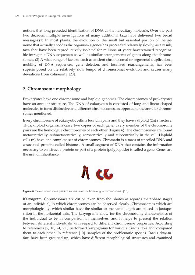
them with respect to their cytological, palinogical and micromorphological propertiesduring and after her doctorate thesis. Consequently, she compared the karyograms of thesamples (Figure 7, 8) she has examined and stated that their cytological properties differas well as their other characteristics [10]. Then, she suggested a new identification key for7 new taxa of Crocus chrysanthus [11-15].
Ideogram: Some cells can be observed in the right metaphase stage; however, since thechromosomes are small, these are not in the size to be made a karyogram with. In these cases,ideograms are prepared with chromosomes. The chromosomes are examined with a micro‐scope and the drawings of chromosomes are made with camera lucida or another photographiccomputer program. Later, drawings of the chromosomes are made by beginning from thelongest chromosome as straight lines which determine the average branch lengths. First thelower branch is drawn, than 1 mm space is left for the centromere and the short branch isdrawn on it. Later, other chromosomes are drawn on the same axis by leaving 4-5 mm spacein between. Thus the ideogram is prepared.
Figure 7. Karyogram of the Crocus chrysanthus sample which is suggested as a new taxon, 2n=8 [10, 15]
Figure 8. Karyogram of the Crocus chrysanthus sample which is suggested as a new taxon, 2n=12 [10, 15]
Sometimes the karyotype analysis are made by measuring the lengths of the chromo‐somes of the species similar to each other and the letters J and V are used in order tofacilitate the comparison. The capital and lower case letters are used to determine the sizesof the chromosomes and each chromosome is classified in two types depending on theposition of the centromere. If the centromere of the chromosome is close by 1/3 of the totallength, this chromosome is represented with the letter J or j in the karyotype formula.According to this representation, the centromere is called subterminal or terminal. If thecentromere of the chromosomes is near to the center or at the center, the chromosome isrepresented with the letter V or v. The centromere is called metacentric or submetacen‐tric. In V type chromosomes the ratio between the length of the short branch and the total
Some Observations on Plant Karyology and Investigation Methodshttp://dx.doi.org/10.5772/56081
225

chromosome length is more than 33.3%, and in J type chromosomes this ratio is less than33.3%. On the other hand, if the length of the chromosome is more than half the length ofthe longest chromosome, it is represented with V or J and if it is less than half the lengthof the longest chromosome, it is shown with v or j [26, 27].
2.1. B Chromosomes
B chromosomes can be observed in plants and animals. The chromosomes which are part ofthe genome and carry the essential genetic data are known as the ‘A’ chromosomes. Theorganism does not need the ‘B’ chromosomes for survival. For this reason, they are called as‘B’ chromosomes. If an organism has many B chromosomes, its development (phenotypicdefects), fertilization and its capacity to produce effective seeds (differences in pollen sizes,sterile seeds or seeds which include different genetic data) are negatively affected. B chromo‐somes are not observed in polypoid plants. It can be observed in monocotyles, particularly inherbaceous dicotyles and in primitive plants such as bryophytes. B chromosomes may existon the individuals of a species. It may also be observed in the pollen mother cells of anindividual, even if its root cells do not consist of B chromosomes.
B chromosomes may be observed in monocotyles and dicotyles in the populations of the samespecies, which have been subject to differentiation, and to ensure the adaptation to theenvironmental conditions. For example, some samples of Crocus ancyrensis, which is anendemic species with 2n=12 chromosomes [28], were observed to have 2n=12+1B chromosomes(Figure 9). The fact that there were some individuals of Crocus ancryensis species with Bchromosomes and that no individuals of the subspecies Crocus flavus subsp. flavus and Crocusflavus subsp. dissectus with B chromosomes were observed, has led us to believe that theenvironmental adaptation capability of Crocus ancryensis is stronger, when compared to thespecies Crocus flavus [10].
B chromosomes were observed in the samples collected from different populations, as seen inthe samples of the species Crocus chrysanthus with 2n=20+2B chromosomes (Figure 10). Inreference [15], the researchers have observed during their field studies and herbariumexaminations that these samples, which also have a different morphology and blackish linedanthers (Figure 1), are differentiated within the species and they proposed for them to beaccepted as a subspecies.
B chromosomes are not observed in all of the cells of a certain plant. It only can be observedin some of its cells. Smaller numbers of B chromosomes in a plant (1-3, sometimes 4) may be
Figure 9. Karyogram of the Crocus ancyrensis, 2n=12+1B [10]
Current Progress in Biological Research226

accepted as an indicator of a good adaptive capability. For a study conducted, which wasrelated to B chromosomes, the samples of the Secale cereale with 2n=14, 2n=14+1B, 2n=14+2B,2n=14+3B and 2n=14+4B chromosomes were compared with regard to the growth rate of thepollen tube. The results revealed that the pollen percentage and the pollen tube length of thesample with 2n=14+2B chromosomes was higher than the other samples. The second in rankingwas the sample with 2n=14 chromosomes [29, 30].
The studies performed revealed that when there are more than two B chromosomes, multiva‐lents produces. The B chromosomes in the mother pollen cells sometimes replicate themselvesand they are observed in all the cells produced with the division. Sometimes, in meiosis, it isobserved that when there is one B chromosome in the cell, this chromosome remains out ofthe equatorial level. In this case, the B chromosome is an underdeveloped chromosome. It isnot observed in the other nucleuses. However, this chromosome may exist as a micronuclei in80-85% of the mother pollen cells at the end of the meiosis stage [30, 31].
2.2. Chromosome enormity
The chromosome enormity is regarded as an indicator of primitiveness. Small chromo‐somes have been differentiated throughout evolution as a result of chromosome disassoci‐ations. If all the chromosomes are metacentric, the karyotype will be symmetrical. Thedifferences in sizes of the chromosomes and the existence of acrocentric chromosomes showthat the karyotype is asymmetrical. Excess asymmetry of the karyotype may point out thatthe species is at a more advanced level with regard to evolution. In other words, be‐tween the similar species, the one with metacentric chromosomes is regarded more primitivethan the species which include submetacentric chromosomes in majority. Thus, the specieswith submetacentric chromosomes in majority is a more recent flora element than the other.The chromosome enormity may be regarded as a reliable characteristic in the phylogenet‐ic aspect, if similar species are being compared. During evolution, the karyotype maytransform from being asymmetrical when it is symmetrical and chromosomes may havedisassociate. With an advanced specialization, even polyploidy may emerge. In similar taxait can be said that the ones with smaller chromosomes are more specialized than the oneswith larger chromosomes [32, 33].
Different environment factors cause ecotypes to emerge. Ecotypes can be examined under 5categories; climatic ecotypes, edaphic ecotypes, culture ecotypes, physiologic ecotypes andchemotypes [33]. It would be imperfect to state that the chromosome number, size and formof a species will be constant. Because of ecological properties and edaphic factors, the total size
Figure 10. Karyogram of the Crocus chrysanthus samples with balckish lined anthers suggested as a new subspecies,2n=20+2B [10]
Some Observations on Plant Karyology and Investigation Methodshttp://dx.doi.org/10.5772/56081
227
of the chromosomes of the members of the same species grown in different places. For thisreason, it is natural for a plant with edaphic ecotypes such as Lythrum salicaria, which is grownin clayed, sandy and salty soil, to have different chromosome lengths in different soils.
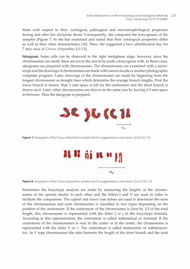
For example, it was determined that the chromosomes of Allium cepa grown in an environmentrich in phosphate, are twice the size of the ones grown in environments with less phosphate[30]. In reference [10], the researcher has determined the chromosome sizes on the karyogramsas regards the samples collected from two different populations of Crocus flavus subsp. flavusand has resulted that the chromosome lengths of these two populations are different from eachother (Figure 11). It is stated that this difference is caused by the differences in the chemicalcontent of the soil [10, 34].
Enormous chromosomes are most generally found in animal cells. Also, if the chromotidesemerging with endomitosis stay stuck to each other, they may form enormous chromosomeswhich consist of chromotide packs. This is called polyteny.
Figure 11. Karyograms of Crocus flavus subsp. flavus collected from different localities [10]
3. Procurement of the material, the first treatment and fixation
Root tips obtained from the plant grown in land or in a pot, root tips from the seed germinatedin petri dish, tips of very young leaves, flower primordiums, growth points of the buds oflateral branches, very young petals, glumes of some monocotyle plants can be used for theexamination of somatic chromosomes. Root tips are the most commonly used part among allthese, when conducting a mitosis examination.
In order for the chromosomes to be examined quantitatively and structurally in the somaticsense in detail, first, the material needs to be pretreated. The pretreatment allows for thechromosomes to remain at the metaphase level, to decrease in length by increasing the number
Current Progress in Biological Research228

of spirals and to be observed clearly. The pretreatment solutions alter the viscosity of theplasma in the cell, the chromosomes diverge from each other and this allows for them to beobserved separately. In addition to this, all pretreatment solutions insure the coagulation ofproteins in organelles of the cell and perform the fixation process at a certain level. Thecleanness and the pH and oxygen levels of the pretreatment solution are extremely important.If the root tips are collected from the land or a field, they should be decontaminated of soilparticles and dirt before being rested in the pretreatment solution. The most practical methodwhen cleaning root tips is to use water. However, they should be wiped with a blotting paperbefore being put in the pretreatment solution. If the root tips are placed in the pretreatmentsolution without being cleaned or with water drops on them, the concentration of the solutionmay change and it may not perform well. Also, the bottle which holds the pretreatmentsolution should be left in an airy environment with its lid open at least one day before thetreatment, so that it would aerate. There are several pretreatment solutions. It may be necessaryto use different kinds of pretreatment solutions for different kinds of plants. Also, the periodsof the samples are to be rested in the solutions differ according to each plant. Thus, it wouldbe beneficial to collect many samples and perform many trials.
Generally, the most commonly used pretreatment solutions are iced water, monobromonaph‐thalene (α-bromonaphthalene), colchicine, 8-hydroxyquinoline, coumarin, paradicloroben‐zene, acenaphtene. Samples should not be rested for a long time in pretreatment solutions suchas 8-hydroxyquinoline or α-bromonaphthalene. Each sample should be tested for the restingduration and the duration which gives good results should be determined. For example, it wasobserved that if Fabaceae and Poaceae family members are rested in α-bromonaphthalene, at+4 ⁰C for 16 hours or at room temperature for 1 hour, the results would be satisfactory [30]. Itwould be sensible to use this solution with plants which have large chromosomes. Also, itworks when 0.01% colchicine is applied on the root tips of the plants for 3 hours [33, 35].Applying 2% colchicine on the young leaf tips is also a good solution. The root tips are restedin 8-hidroxyquinoline at 10-18 ⁰C for 3-6 hours. In references [10, 15, 24, 25], the root tips arerested in 8-hidroxyquinoline at room temperature for 4 hours during the cytological studiesconducted on the Crocus species and the results were satisfactory. 8-hidroxyquinoline pre‐treatment can also be applied to the root tips on the plant. It can be prepared with the slidetaken from the root tip in 8-hidroxyquinoline. However, it would be more convenient if theslide is prepared after being dyed with aseto-orcein and after maceration is employed. Whenthe root tips are being rested in this liquid, the excess temperature of the room may cause theroot tips to get stuck to each other. The coumarin application suggests for the root tips to berested in solution at 16 ⁰C for 2-3 hours and this application facilitates the examination. Thewater saturated solution of coumarin (2%) should be used. If the coumarin is used with chloralhydrate, paradiclorobenzene sulphanilamide and bromonaphthalene combinations, betterresults will be obtained. If paradiclorobenzene will be used for the examination of somaticchromosomes, the root tips are rested in the saturated solution of paradiclorobenzene in purewater (1-2%) for 1-4 hours. For example, if the root tips as regards members of Poaceae andCyperaceae are heated in the acid solution and dyed with aceto-orcein, after being rested inthis solution at 12-16 ⁰C for 3 hours, satisfactory results will be obtained [33]. With the
Some Observations on Plant Karyology and Investigation Methodshttp://dx.doi.org/10.5772/56081
229

application of paradiclorobenzene the images of many plants with 120 or more chromosomeswere obtained in the metaphase where the chromosome length is reduced. Acenaphtene wasused in the karyotype studies of pollen tube chromosomes. This liquid was not regarded asthe appropriate solution for the karyotype examinations of root tips [36].
After the pretreatment, the sample should be put to fixation. There are many kinds of fixatives.The most important ones among all are; alcohol, acetic alcohol (1 measure of glacial acetic acid:3 measures of absolute alcohol), Carnoy’s fixative (1 measeure of glacial acetic acid:3 measuresof chloroform:6 measures of absolute alcohol or 1 measure of chloroform:3 measures of aceticacid:6 measures of absolute alcohol), Helly’s fixative and Navaschin’s fixative. In reference[10], the root tips have been collected from the plants in pots in different hours of the day basedon the weather being sunny or rainy and rested them in 8-hidroxyquinoline pretreatmentsolution for 4 hours and fixated them in acetic alcohol during the studies on the genus Crocus.
In reference [19], α-bromonaphthalene is used as the pretreatment solution on the root tips theresearcher has collected for the examination of mitosis during the studies on the speciesCucumis melo. In references [19, 37], researchers have rested the flower sprouts directly in theCarnoy’s fixative without using a pretreatment solution during their meiosis examinationsthey have conducted using pollen mother cells.
In reference [27], cytotaxonomical studies were made on several Musci species deployedamong the Aegean region of Turkey. The researcher cultured the samples she has brought inthe laboratory by ensuring the humidity of the material at 10-15 ⁰C in petri dishes. As the newsuckers develop, they were cut from the plant at an hour close to midnight and were fixatedin the Carnoy's fixative at 18-20 ⁰C for 3 hours.
If acetic alcohol is used as a fixative and the samples are put in this solution after beingtaken from the pretreatment solution, there is no need to place the samples in anothersolution or in ethyl alcohol to be able to preserve them for a long period, because, aceticalcohol esterifies and the acid loses its effect in time. Also, since the glacial acetic acid andalcohol mix esterifies in time and since they lose their effect as a fixative in time, this mixtureshould be prepared right after the root tips are removed from the pretreatment solution.Also, the small bottles in which the samples are to be preserved should be chosen carefully.Bottles with a capacity of 5 cc would be adequate to hold the samples. These small bottlesmay be used during the period when the root tips collected from the plant for thepretreatment. The time and whether conditions when the root tips are collected for thepretreatment should definitely be noted on the bottle, because this data will provideinformation about when mitosis divisions occur in general. This information will be helpfulin determining on what time and in which weather conditions root tips should be collect‐ed in order to observe the chromosomes in the metaphase stage. It would be practical totake the pretreatment solution from the bottle with an injector carefully and this way, itwill be ensured that there are not any liquids left in the bottle. Also, before the root tipsare taken from the pretreatment solution and put in the fixative, the pretreatment solu‐tion drops remaining on the samples should be removed with a blotting paper. If thesamples are put in the fixative without paying attention to this step, the purity of the fixative
Current Progress in Biological Research230

may change and the required effects may not be realized on the chromosomes. If the bottlecaps are made of plastic or if the structure of the caps is similar to plastic, it may getfractured in time. Because the bottle includes acid and the corrosive effects of acid are wellknown. If the caps are fractured, the acetic alcohol inside the bottle will evaporate and thesamples will dry. On the other hand, even if the cap is not fractured, the acetic alcohol inthe bottle will vaporize, if the sides of the cap allow air to get in; thus the samples will dryand they will become unusable. For this reason, the cap should be covered with a clothplaster (1 cm wide depending on the size of the bottle) carefully, without leaving any spacefor air between. If the samples will be preserved for a long time, they should be checkedregularly to see if they have dried. If the acetic alcohol level of the bottle has decreased,adding ethyl alcohol on the sample would be sufficient. If the samples are kept in therefrigerator (+4⁰C), their storage time would increase and it would be ensured that thesamples are maintained without decaying. The samples stored in this manner could beexamined for years after they were prepared. In order to examine the chromosomes duringmeiosis, the formation of pollens (microspore) and the ovum (megaspore) of the phanero‐gams should be assessed. Pollen mother cells (microspore mother cells) produce pollens asa result of meiosis. Ovule mother cells (megaspore mother cells) produce ovums as a resultof meiosis. In short, in order to examine the meiosis of the flower, the pollen mother cells,mother cells of the embryo sac and the embryo cells (ovaries) can be used.
In order to examine pollen mother cells, pretreatment should be applied to very young flowerblossoms. After the proper procedure is carried out on the fixated samples, the blossom isopened, the pollen mother cells without a fractured callus wall are chosen by disjoining theanthers and meiosis is observed.
3.1. Dying the chromosomes
For the chromosomes of the collected root tips, at different stages of mitosis to be examinedclearly, the samples should be dyed. Also, in order to examine the meiosis of the fixated flowerprimordium pollens, the samples should be dyed thoroughly. Some researchers have exam‐ined the root tips by dying them with Feulgen dye [30, 38, 39]. Crystallized basic fuchsineshould be used when preparing the Feulgen dye. If basic fuchsine is in powdered form, theFeulgen dye prepared with this will not dye the chromosomes adequately.
Bands are formed when the chromomeres in polythene chromosomes are in juxtaposition. Thepart between two bands is called an interband. The bands can be dyed with Feulgen dye orwith basic dyes, but interbands cannot be dyed.
If mitosis will be examined by using fixated root tip samples, the most convenient material todye the chromosomes with would be aceto-orcein. If the pollens in the anthers of flowerprimordiums will be used as samples to examine meiosis, chromosomes should be dyed withaceto-carmine.
In reference [10, 24, 25], aceto-orcein is used to dye the root tips taken from the fixative whenworking with some Crocus taxa. This way, the photos of the available metaphase stages weretaken and the karyograms were prepared. In reference [40], they used the same method to
Some Observations on Plant Karyology and Investigation Methodshttp://dx.doi.org/10.5772/56081
231

examine the karyotypes in their study related to the seed development and DNA structure ofCynara scolymus and Phaseolus coccineus seeds with Pb and Cu heavy metal stress.
In reference [27], the researcher rested the Musci samples at 15-20 ⁰C for 10 hours in 2% aceto-orcein after fixation. After the dying process, the samples were examined with Feulgenpreparation technique, performed the karyotype analysis and prepared ideograms. Theresearcher has also stated that the fixation period and the dying characteristics of each speciesthey have worked with was different and for this reason, they had to develop differentmethods.
The dye acceptance of pollens and consequently the adequate dying of the chromosomes is indirect proportion with their vitality. For that matter, it would be appropriate to carry out apollen vitality test before the karyological study. The dye acceptability of pollens can be testedon alive samples, samples which are fixated with Carnoy's fixative or 80% alcohol andherbarium samples. In cytological studies, pollen infertility is generally regarded as a measurefor meiosis irregularity.
In reference [19], the meiosis of the pollen mother cells examined were obtained from the flowerbuds along with the root tips when conducting a cytological study on some Cucumis melo taxa.The researcher dyed the root tips with nigrosin and examined the chromosomes. He alsoexamined some of the root tip samples with pectinase enzyme and aceto-orcein dye. Theresearcher examined the meiosis divisions with Fuelgen method. He has dyed the pollenmother cells he had obtained from the anthers of flower buds with 2% aceto-carmine.
There are several ways to prepare aceto-orcein and aceto-carmine and the procedures givenbelow have been tested many times and satisfactory results were obtained [10, 19, 37, 41].
3.2. Preparation of aceto-orcein
5 grams of powdered orcein and 250 cc 45% acetic acid are mixed and shaken. They are boiledfor 30 minutes by using a Soxhlet apparatus. The apparatus should certainly be held by astative. The Soxhlet apparatus looks like a glass tube with a spiral back cooler connected to thevolumetric flask, it has a tap for water intake below and a place for water outlet. The volumetricflask under the Soxhlet apparatus is placed on the amiant wire on the heater. A boiling chipshould be placed inside the volumetric flask. If there are not any boiling chips available, 1-2cm long glass bars can be used for the same function. Acetic acid dye mix is poured in thevolumetric flask and the heating process is started. The aceto-orcein prepared is filtered afterit has been cooled down. Aceto-orcein prepared in this manner shall be ready for use.
3.3. Preparation of aceto-carmine
1 gram of powdered carmine and 200 cc 45% acetic acid are mixed and shaken. They are boiledfor 5 minutes by using a Soxhlet apparatus. This duration is ideal for Turkey. For example, ifthe boiling process for dye preparation would take place at a location in the north of Turkey,a country with colder weather, the boiling duration may be 30 minutes. After the boilingprocess, 1-2 drops of Fe acetate solution saturated with 45% acetic acid is added to the dye.
Current Progress in Biological Research232
Excess Fe acetate settles the carmine. In this case, the dye cannot be used expediently. Theaceto-carmine is filtered after being cooled down. Aceto-carmine prepared in this manner shallbe ready for use.
3-4 cc of aceto-carmine should be spared before every use. If Fe acetate is not used whenpreparing aceto-carmine, a nail is placed in the aceto-carmine spared. The iron nail should notremain in the dye for a long time. Because, aceto-carmine with a heightened Fe proportiondyes the cytoplasm in very dark color as well as the chromosomes and it gets extremely difficultto examine the slide. However, if the cytoplasms of the cells are dyed in a dark color, 45% aceticacid can be dropped on the side of the cover slip with the help of a thin pipette. When the aceticacid reaches below the cover slip, it may lighten the color of the cytoplasm. The reason a lightercytoplasm is required is that, it is preferred when there is a contrast between the cytoplasmand the dyed chromosomes. When there is a contrast between the cytoplasm and the chro‐mosomes, it is easier to examine the chromosomes. Also, the photos of the meiosis phases takenare clearer when there is contrast.
4. The points to take into consideration when taking examination samples
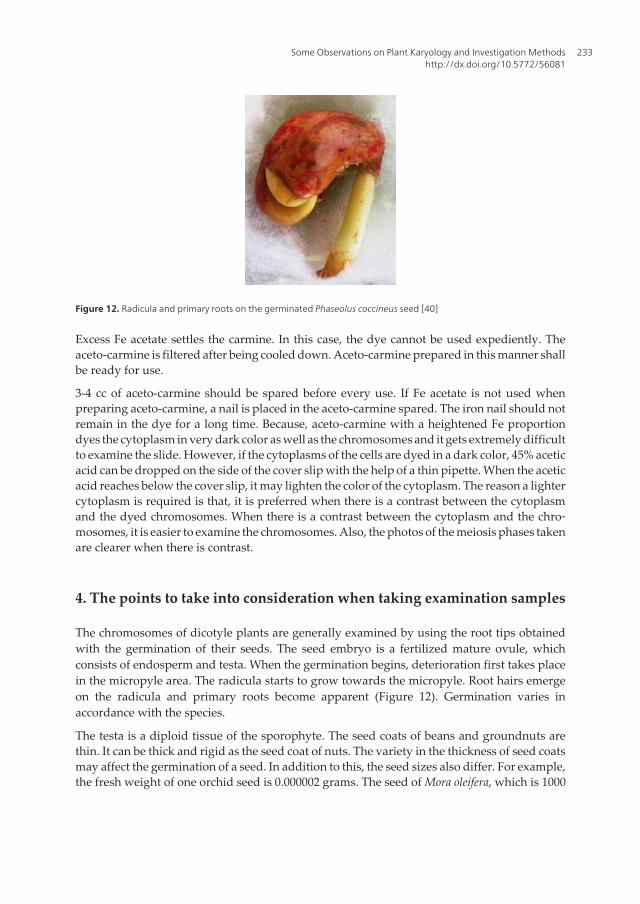
The chromosomes of dicotyle plants are generally examined by using the root tips obtainedwith the germination of their seeds. The seed embryo is a fertilized mature ovule, whichconsists of endosperm and testa. When the germination begins, deterioration first takes placein the micropyle area. The radicula starts to grow towards the micropyle. Root hairs emergeon the radicula and primary roots become apparent (Figure 12). Germination varies inaccordance with the species.
The testa is a diploid tissue of the sporophyte. The seed coats of beans and groundnuts arethin. It can be thick and rigid as the seed coat of nuts. The variety in the thickness of seed coatsmay affect the germination of a seed. In addition to this, the seed sizes also differ. For example,the fresh weight of one orchid seed is 0.000002 grams. The seed of Mora oleifera, which is 1000
Figure 12. Radicula and primary roots on the germinated Phaseolus coccineus seed [40]
Some Observations on Plant Karyology and Investigation Methodshttp://dx.doi.org/10.5772/56081
233

gr, is one of the heaviest seeds known [18]. As it can be seen, if the chromosome examinationwill be conducted by using the root tips obtained with seed germination, the characteristics ofthe seed need to be determined clearly and if necessary some pretreatment procedures shouldbe applied before the germination. For example, if the seed coat is thick, it should be sandedor perforated to facilitate the water intake.
The impact of environmental conditions is also important with regard to seed germination.For a successful germination process; the temperature, water and oxygen levels should beadequate. Water is required for the development of the embryo and the enzymatic reactionsto start by secreting hormones. Temperature is significant with respect to the functioning ofthe enzyme. Oxygen is required for the respiration need of the developing seedlings. Althoughthe seeds of some plants germinate in dark, seeds of several plants can only start the germi‐nation process after being left under light for a certain amount of time. This pretreatmentperformed with light can be significant when the seeds absorb water [18, 42].
Therefore, the ecological factors of the environment plant lives in should be determined anda seed germination environment should be prepared accordingly. For this, generally climatecabinets are used. In some instances, even though the conditions required for the seed to begerminated are present, germination does not take place. For example, for the orchid seeds tobegin germinating, they should form a mycorrhiza with some fungi types. As it can be seenfrom the examples, the germination factors of seeds may vary significantly. Thus, moredetailed preliminary examinations may be due for the germination of some seeds. In additionto this, if the dicotyle plant examined is endemic, its seeds may be small in quantity or thefertile seeds may be scarce. Also, it is possible for the mature seeds obtained from endemicplants to require more care and time for germination. If the seed of the dicotyle plant is spreadaround the land as the fruit dries, such as endemic Linaria corifolia of the Scrophulariaceaefamily (Figure 13), it would be appropriate to take the root tips by collecting and germinatingthese seeds [42]. However, if achene fruits, such as the members of the family Asteraceae; forexample endemic Centaurea zeybekii or endemic Jurinea pontica, are examined (Figure 14, 15),they should directly be subject to germination without attempting to remove the seeds [43-45].Because, pericarp is fused to the thin seed coat in the grain.
Figure 13. Seed of Linaria corifolia (SEM photograph) [43]
Current Progress in Biological Research234

Figure 14. Seed of Jurinea pontica (SEM photograph) [43]
Figure 15. Seed of Centaurea zeybekii (SEM photograph) [44, 45]
The germination durations of seeds may vary significantly among plants. Also, the optimumtemperature required for the germination of the seed may also vary. Thus, many tests andobservations in different places and periods could be needed for the determination of the rightgermination duration and the optimum temperature for the plant.
Also, sometimes, in order to end the dormancy of the genetic material inside the seeds,germination tests can be made after it is rested in the refrigerator (+4⁰C) for hours or days. Thegermination test can be carried out inside a petri dish with a humid blotting paper inside,covered with another petri dish. The point to take into consideration here is this; before placingthe blotting paper inside, first a piece of glass in the shape of a square or a rectangle should beput in the petri dish. Later the blotting paper cut in the shape of a circle is placed on the pieceof glass. The size of the blotting paper should be smaller than the petri dish. Later, water ispoured on the blotting paper, whose center is heightened with the piece of glass. The watershould be pure. The water poured on the blotting paper may be 1-2 mm above the surfacelevel. Later, the seeds are placed on the center of the blotting paper which is heightened withthe piece of glass. A circular and humid blotting paper of an appropriate size may be placed
Some Observations on Plant Karyology and Investigation Methodshttp://dx.doi.org/10.5772/56081
235

on the seed as well. This blotting paper can also be placed in the second petri dish after beinghumidified. This method can work in some instances. For example, if the radicula and theprimary roots grow upwards, this humid blotting paper prevents the root tips from gettingdry. Because, it is impossible to examine mitosis in dry root tips.
The number of seeds to be placed in the petri dish varies depending on the size and the waterabsorption capacity of the seed. For example, the swelling capacity of the seeds which belongto Fabaceae family, is high. If a seed with these characteristics is being used, the seeds to beput in the petri dish should be small in number and the water to be poured needs to be plenty.It is important to adjust the amount of water to be poured in the petri dish. If one poursexcessive amount of water, it may spill from the sides of the petri dish. If the seeds are observedwell during the germination period, water may be added after the level of water in petri dishhas decreased and the beginning stages of dryness are observed.
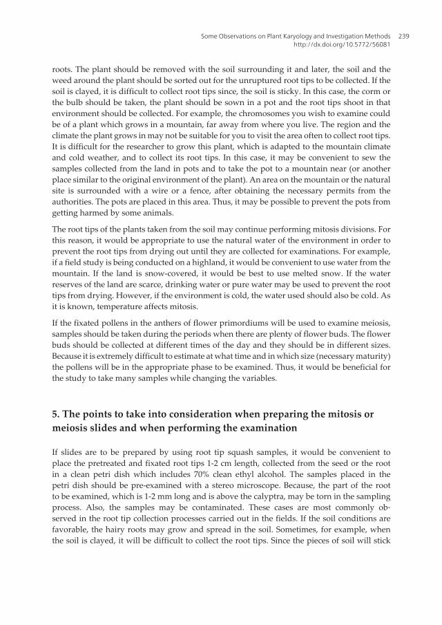
Collecting root tips from a germinated seed in the petri dish relatively easier than collectingthe root tips of plants which are grown in fields or on land. The root tip collected from agerminated seed in petri dish is cleaner and there is no risk of tearing the part above thecalyptra, where mitosis takes place. Also, the most important point to take into considerationwhen applying this method is the length of the primary roots shooting from the seed. If theroot tip is too long, the part where mitosis takes place can dry. In addition to this, if the roottip is too small, it may get damaged during the dying process and it cannot be examined. It issufficient for the primary roots to be 1-2 cm long (Figure 16).
Sometimes the seeds placed in a petri dish for germination can be contaminated, they can bemolded for example. In this case, they do not shoot root tips. Also, in some instances the seedgets infected after germination. There is a root tip; however, it is impossible to carry out theappropriate chromosome examinations on this root tip, because it is infected. Therefore, theseeds placed in the petri dish should be observed frequently (1-2 times a day).
Figure 16. Primary root of the Phaseolus coccineus on the left is suitable to collect [40]
It is difficult to collect root tips from dicotyle plants on the land. Especially, if the soil the plantis located on is clayed or stoned, if the plant is on a cliff or on a sloped environment, it is difficultto collect root tips, even though the roots and the lateral roots of the plant do not grow toodeep. Also, even if the place the plant is on is flat, if the roots grow deep or if the soil type isnot suitable for collecting the root tips, the process of taking root tips will require effort. For
Current Progress in Biological Research236

the chromosome examinations of these plants, germinating their seeds would be an easiermethod which gives useful results.
Perlite can be used for the seeds which are to be germinated. Perlite also is used in greenhousesto grow seedlings from seeds. Torf can also be used for the tests of growing plants in pots. Thepots are turned upside down, the torf surrounding the plant is collected carefully and thelateral roots are reached. One should be very careful in order not to damage the root tips.
If the chromosomes to be examined belong to a monocotyle plant and it reproduces with stemsor corms like the genus Crocus (Figure 17, 18), it should be known that the number of the seedsmay be less for these plants. Even if there are plenty of seeds, there is a great probability thatmany of them are sterile (Figure 19, 20) [10].
Figure 17. Young corms of Crocus chrysanthus [10]
Figure 18. Corms of Crocus chrysanthus [10]
Some Observations on Plant Karyology and Investigation Methodshttp://dx.doi.org/10.5772/56081
237

Figure 19. Riped fruit and scattered seeds of Crocus chrysanthus [10]
Figure 20. Riped seed of Crocus flavus subsp. flavus [10]
If the chromosomes of monocotyles (for example Iridaceae or Liliaceae) are to be examined,root tips may be collected on the land. However, the most appropriate and easy method wouldbe to obtain them on the land, sew them on the pot and to try and collect the root tips. If theplants are on a mountain or on highlands, the environment in your house or your garden maynot be suitable for the plant to grow. In this case, the plants sown will die. However; if thecorms and bulbs are preserved under convenient conditions (in a dry and dark environment),they will give roots the next year. The corms of Crocus (Iridaceae) and bulbs of Colchicum(Liliaceae) species shoot primary or contractile root tips the following year even if they are notplaced under soil or torf.
If root tips are to be collected from monocotyles on land, it should be noted that this processrequires extreme attention and patience. The root depth of the plant is examined and a propergarden tool (such as digger, shovel and hoe) needs to be taken to the land. When removingthe plant from the land, the distance between the tool and the plant should be maintained.Otherwise, there is a risk of tearing the root or the stem of the plant and rupturing the lateral
Current Progress in Biological Research238
roots. The plant should be removed with the soil surrounding it and later, the soil and theweed around the plant should be sorted out for the unruptured root tips to be collected. If thesoil is clayed, it is difficult to collect root tips since, the soil is sticky. In this case, the corm orthe bulb should be taken, the plant should be sown in a pot and the root tips shoot in thatenvironment should be collected. For example, the chromosomes you wish to examine couldbe of a plant which grows in a mountain, far away from where you live. The region and theclimate the plant grows in may not be suitable for you to visit the area often to collect root tips.It is difficult for the researcher to grow this plant, which is adapted to the mountain climateand cold weather, and to collect its root tips. In this case, it may be convenient to sew thesamples collected from the land in pots and to take the pot to a mountain near (or anotherplace similar to the original environment of the plant). An area on the mountain or the naturalsite is surrounded with a wire or a fence, after obtaining the necessary permits from theauthorities. The pots are placed in this area. Thus, it may be possible to prevent the pots fromgetting harmed by some animals.
The root tips of the plants taken from the soil may continue performing mitosis divisions. Forthis reason, it would be appropriate to use the natural water of the environment in order toprevent the root tips from drying out until they are collected for examinations. For example,if a field study is being conducted on a highland, it would be convenient to use water from themountain. If the land is snow-covered, it would be best to use melted snow. If the waterreserves of the land are scarce, drinking water or pure water may be used to prevent the roottips from drying. However, if the environment is cold, the water used should also be cold. Asit is known, temperature affects mitosis.
If the fixated pollens in the anthers of flower primordiums will be used to examine meiosis,samples should be taken during the periods when there are plenty of flower buds. The flowerbuds should be collected at different times of the day and they should be in different sizes.Because it is extremely difficult to estimate at what time and in which size (necessary maturity)the pollens will be in the appropriate phase to be examined. Thus, it would be beneficial forthe study to take many samples while changing the variables.
5. The points to take into consideration when preparing the mitosis ormeiosis slides and when performing the examination
If slides are to be prepared by using root tip squash samples, it would be convenient toplace the pretreated and fixated root tips 1-2 cm length, collected from the seed or the rootin a clean petri dish which includes 70% clean ethyl alcohol. The samples placed in thepetri dish should be pre-examined with a stereo microscope. Because, the part of the rootto be examined, which is 1-2 mm long and is above the calyptra, may be torn in the samplingprocess. Also, the samples may be contaminated. These cases are most commonly ob‐served in the root tip collection processes carried out in the fields. If the soil conditions arefavorable, the hairy roots may grow and spread in the soil. Sometimes, for example, whenthe soil is clayed, it will be difficult to collect the root tips. Since the pieces of soil will stick
Some Observations on Plant Karyology and Investigation Methodshttp://dx.doi.org/10.5772/56081
239

to the roots, the sensitive root tips will be ruptured. Even when the root tip is collectedproperly from such an environment or a different environment, the soil pieces stuck on theroots should be cleaned before the slides are prepared. There is also a possibility for particlesand dirt to remain on the root tips collected from torf or perlite. Thus, before starting theslide preparation process, the samples should be examined in alcohol with a stereomicroscope. If the root tips are very dirty, the alcohol in petri dish should be replaced fora couple of times. The cleaning of root tips can be carried out by shaking the samples inalcohol and removing the dirt by using a forceps or a needle.
It would be sufficient for aceto-orcein or aceto-carmine dyes to be filtered for a short timeduring the preparation process, because, there may be sedimentations in the dyes which arerested for a long period. It may be convenient to transfer small amounts of dye from the totalamount stored in dark glass bottles in a dark place to small bottles which can contain 5 cc ofliquid. Because, the amount of dye required for an examination is 3-5 cc. The required amountof dye is filtered with a diameter of 2-3 cm prepared blotting or filtering paper. The filtereddye is poured in a middle sized watch-glass. The fixated root tips are placed in here. The watch-glass which contains dye and root tips heated on the burner until it boils. Since there is a smallamount of liquid, the boiling process should not take a lot of time. Also, the acetic acid in thedye will evaporate. The samples may dry if the heating process lasts too long, since the acidwill be vaporized. In this case, the root tips in the watch-glass will be unusable. The heatingprocess made in a careful way should be repeated three times. After each heating process, thewatch-glass is taken aside and another watch-glass larger than the one used is covered on topof it. This should be done very quickly. The purpose here is to prevent the acetic acid in thedye to vaporize. Also, the point to take into consideration is the size of the watch-glass whichis used to cover the watch-glass used. If a significantly larger watch-glass is used to cover theheated watch glass, the acetic acid would vaporize and the dye would dry. If the heatingprocess is carried out carefully, it will enable the maceration of the roots as well as allowingfor the dye to perforate in the cells and ensuring the dying of the chromosomes; therefore,there will be no need for hydrolysis.
The slide prepared for mitosis examination does not only reflect one phase of the mitosis.When some cells are in interphase, some may be in anaphase, some in telophase and somemay be in metaphase (Figure 21). If lengthened cells are observed in the slide, this showsthat not only the necessary part of the root tip required to examine mitosis is squashed.The excessive parts squashed are the cells in the stage of elongation. Also, vascular tissuecan be seen in the slide. The reason of this observation is similar to the reason for seeingthe elongated cells. Even if there are proper metaphase cells in the slide, the existence ofelongated cells and vascular tissue could make it difficult for the chromosomes to beexamined. For this reason, one should decide very carefully when determining how muchof the root piece will be left in the slide when examining the length of the calyptra shotfrom the root tip.
The root piece on the slide which is 1-2 mm long has become very soft and is ready to besquashed. The dyed root tip is placed on the slide. After the calyptra is cut with a sharp razorblade (a piece almost in the size of a pinhead), 1-2 mm of the remaining piece is cut and this
Current Progress in Biological Research240

piece is placed on the slide. The remaining root piece is thrown away. 1 drop of the aceto-orceinfiltered for the study shall be poured on the material. The cover slip is closed on the samplewithout allowing any air to get in (with an angle equal to 45⁰C). A piece of blotting paper (alittle larger than the cover slip) is placed on the cover slip carefully. The blotting paper absorbsthe aceto-orcein spilling from the sides of the cover slip. The thumb is placed on the cover slipcarefully, without moving the cover slip on the slide. The thumb is not removed, but is movedfrom side to side for the mitosis cells of the root tip squashed to spread under the cover slip.Thus, the cells which have overlapped and the chromosomes of the cells will be separated fromeach other and are spread on one plane. As a result, the cells and the chromosomes will beeasily examinable.
However, there may be cells in the slide which are not spread on the plane as well as the cellswhich reflect the chromosomes clearly. In this case, various images of the same cell should betaken using the microscrew (Figure 22). Later, the images are put together and examined. Forexample, a chromosome observed in one image may not be present in the other. As a result, akaryogram can be prepared with these images.
Figure 22. Various images of the same cell of Crocuschrysanthus using the microscrew [15]
Figure 21. Different phases in a squashed root tip of Crocus chrysanthus [15]
Some Observations on Plant Karyology and Investigation Methodshttp://dx.doi.org/10.5772/56081
241

Sometimes the photographs taken by replacing the microscrew may not be sufficient inproviding the chromosome data. In this case, the microscope coordinates of the cellexamined are noted in accordance with the objective used. Later, the slide is taken fromthe microscope flange and is put on a hard surface. A blotting paper is placed on the coverslip once more and the cover slip is pressed with the thumb carefully. This pressure mayallow for the chromosomes of the cell, whose coordinates were noted, to be observedindividually. If this operation is carried out carefully, there is a great chance that it will beuseful. However, even the slightest gliding of the slide may cause the cells to overlap or itmay result in the emergence of new cells which have been flattened and cannot be observedclearly. This second squashing process can only work after many practices. As it can beseen, a minor error in the second squashing process may cause the slide to be unusable.Thus, before the second squash and after the coordinates of the examined cell is noted,other photographs of the cells should be taken. So that, when later, the slide is taken fromthe flange to be squashed once more and the slide ends up being unusable, the negativeimpact will partially be eliminated.
In some instances, the researcher may need to take a break when observing the phases ofmitosis or meiosis. When the slide is being examined as described above, it may only bepreserved for 15 minutes, depending on the temperature, because the acetic acid in theaceto-orcein used to dye the chromosomes and the acetic acid in the aceto-carmine used inmeiosis examinations will vaporize. Thus, a dried slide is not functional and it cannot beused for examination. In this case, it would be convenient to cover the all sides of the coverslip with paraffin in order to be able to examine it in 1–2 days. For this appliance, the backof the metal spatula should be heated on the burner and it is contacted with the paraffin.The liquid paraffin poured on the metal is applied thinly around the cover slip. The slideprepared in this manner can be maintained for 1-2 days without being dried and it can beused. If the slide is preserved in the refrigerator (+4⁰C) in a petri dish covered with anotherpetri dish, the life cycle of the slide will increase. Covering of the sides of the cover slipwith rubber solution, has the same effect.
It may take a long time to make a careful examination on all the root tip squash under thecover slip with microscope. If the slide is prepared with aceto-orcein (for the examinationof mitosis) or with aceto-carmine (for the examination of meiosis), under the sides of thecover slip will begin drying circularly. Since the drying of the slide will prevent us fromcompleting the examination, a small amount of dye shall be added on the side of the coverslip with the help of a needle. It will be observed that the dye steals into the dried partsunder the cover slip after a while. The excess dye on the side of the cover slip can becleaned with a blotting paper. This process will delay the drying process and increase thetime allowed for examination.
In some instances Feulgen’s dye is used for the chromosomes dying. In this case, 45% aceticacid is dropped on the sample and the slide is covered with the cover slip. The squashingprocess is carried out carefully. The cover slip may start to dry from the sides when examiningthe slide, as it does with other dying methods. In order to prevent the drying of the slide, theresearcher should drop 45% acetic acid on the side of the cover slip with a needle and should
Current Progress in Biological Research242

wait for it to steal into the dried parts of the squashed root tip under the cover slip. The excessacetic acid on the side of the cover slip is dried with a blotting paper.
6. Chromosomal abnormalities
Chromosomal abnormalities are observed in plants for various reasons. The existence of achromosomal abnormality in the cell can also be understood from the morphology of the rootor another part of the plant. The chromosomal abnormalities which reflect themselves in themorphological properties can occur with relation to the differentiation and adaptationprocesses. For example, in reference [10], when examining the problematic species Crocuschrysanthus, observed lagging chromosomes (Figure 23) and determined 2n=20+2B as thechromosome number for the plant samples with blackish anthers, which are differentiated andmorphologically different suggested as a subspecies in reference 15.
Figure 23. Lagging chromosomes of Crocus chrysanthus in different cells [10]
Rapidly increasing world population in recent years brings about an increasing demand fornutrition, which is one of the most important problems for mankind. Various methods havebeen discussed in order to solve this problem. One of these methods is using chemicals againstharmful organisms in plants. The use of pesticides in agricultural areas increases the plantyield; however, some chemical substances may result in pollution in nature and healthproblems [46].
Modern agriculture and industry depend on a wide variety of synthetically producedchemicals, including insecticides, fungicides, herbicides, and other pesticides. Continualwidespread use and release of such synthetics has become an everyday occurrence, resultingin environmental pollution [47]. It was indicated that many cytogenetic studies have beencarried out to detect the harmful effect of different pesticides on different plants [48]. Thesechemicals used at recommended dosage and double the recommended can give rise toabnormal chromosomes and degeneration in meiosis cycle, such as ring shaped chromosomes,linear chromosomes and binding chromosomes [37]. Benomyl, a systemic fungicide, affectsgermination, mitotic and meiotic activity, and pollen fertility in barley [49]. Besides, the
Some Observations on Plant Karyology and Investigation Methodshttp://dx.doi.org/10.5772/56081
243

fungicide phosphite reduced polen fertility in Petunia hybrida, Tradescantia virginiana and Viciafaba, while phosphite, application increased the number of abnormal meiotic cells at all stagesin Tradescantia virginiana microspores [50]. Furthermore, some common pesticides (Thiodan,Folithion, Lebaycid, and Kitazin) caused a spectrum of cytogenetic abnormalities such aschromosome fragmentation, lagging of chromosomes, anaphase bridge formation as well astripolar and tetrapolar spindle formation in barley [51].
The abnormalities occurring in meiosis are very important because they cause sterility inpollens and genetic damage can be transmitted to the offspring via male gametes, leading tocongenital abnormalities. It was studied on Lycopersicon esculentum that all dosages of thefungicides Agri Fos 400 [80% fosetyl-Al (400 g/l mono- and di-potassium phosphonate)] causedvarious abnormalities in meiosis when compared with the control (Figure 24-27) [37].
Figure 24. Ring-shaped chromosomes in 400 ml/100 l group a (1, 2) [37]
Figure 25. Binding chromosomes in 800 ml/100 l group b [37]
Figure 26. Binding chromosomes and abnormal shape in 800 ml/100 l group c (3, 4) [37]
Current Progress in Biological Research244

Figure 27. Abnormal shape and linear chromosomes in 800 ml/100 l group d (5) [37]
In another studies, the fungicides Fosetyl-Al (80% Aliette WG 800) and Equation Pro (22.5 %Famoxadone + 30 % Cymoxanil) widely applied on tomato plants (Lycopersicon esculentum)grown in greenhouse in Turkey caused various anomalies in polen meiosis such as thread-like, ring shaped, linear and binding chromosomes (Figure 24, 25). This situation could leadto a decreased in the productivity of fruits [52, 53]. In references [54- 56], the effects of fungicideand aplicator application on pollen structure in tomato (Lycopersicon esculentum) were inves‐tigated. SEM photographs of Lycopersicon esculentum as regards control group and non-viablepollen grains in the ACT-2 groups are given below (Figure 28-33).
Figure 28. Pollen grain of control group (SEM photograph) [56]
Some Observations on Plant Karyology and Investigation Methodshttp://dx.doi.org/10.5772/56081
245

Figure 29. Pollen grain of control group, polar view (SEM photograph) [56]
Figure 30. Abnormally shaped pollen grain in 150 cc/100 L. (SEM photograph) [56]
Figure 31. Abnormally shaped pollen grain in 150 cc/100 L. (SEM photograph) [56]
Current Progress in Biological Research246

Figure 32. Abnormally shaped pollen grain in 300 cc/100 L. (SEM photograph) [56]
Figure 33. Wrinkled pollen grain in 300 cc/100 L. (SEM photograph) [56]
Agricultural pesticides or hormones commercialized may cause the flowers and pollens of theflowers to have formal differences and abnormalities and it may reduce productivity. Thespecies, Anemone coronaria is a flower with corm, which can be grown in greenhouses with noheating between October and May and is very popular in domestic and foreign markets, also,its origin is Mediterranean and it is ecologically advantageous; because of these reasons it hasa significant status in export with regard to differentiation. In references [16, 57], it has beenpointed out that while there is homogeneity in the pollen sizes of the natural forms of thevariety within the species, the pollen sizes, pollen deformations, pollen failure to thrive andvitality of the pollens taken from the members to be traded are significantly different (Figure34-40). The agricultural pesticides used to enhance and accelerate the growth and bloomingcan be the reason behind this differentiation. The pollen vitality percentages were determinedas a result of the test made with TTC (triphenyltetrazolium chloride) in order to observe thepollen vitality of natural and commercial forms of Anemone coronaria taxa. In reference [57],
Some Observations on Plant Karyology and Investigation Methodshttp://dx.doi.org/10.5772/56081
247

viable pollen ratio of natural Anemone coronaria var. coccinea is 94%, viable pollen ratio of
commercial forms of that taxon is 69%.
Figure 34. Pollen grains of Anemonecoronaria var. coccinea as regards control group, scale bar:10 μm (WodehouseMethod used) [16]
Figure 35. Pollen grains of Anemone coronaria var. coccinea as regards control group, scale bar:10 μm (ErdtmanMethod used) [16]
Figure 36. Pollen grain of Anemone coronaria subsp. coccinea as regards control group (SEM photograph) [57]
Current Progress in Biological Research248

Figure 37. Abnormal shaped pollen grains of Anemone coronaria var. coccinea as regards commercial group (SEMphotograph) [57]
Figure 38. Abnormal shaped pollen grains of Anemone coronaria var. rosea as regards commercial group (SEM photo‐graph) [57]
Figure 39. Abnormal shaped pollen grains of Anemone coronaria var. cyanea as regards commercial group (SEM pho‐tograph) [57]
Some Observations on Plant Karyology and Investigation Methodshttp://dx.doi.org/10.5772/56081
249

Figure 40. Abnormal shaped pollen grains of Anemone coronaria var. alba as regards commercial group (SEM photo‐graph) [57]
Also, environmental conditions which are stem from pollution, such as heavy metal stress maycause development disorders and chromosomal abnormalities in plants. For example, inreference [40], they applied different values of Pb stress to artichokes for 5 days and haveobserved their development. 40, 80, 160, 320, 1280 ppm Pb acetate solution was used. Theresearchers have also germinated control groups in Hoagland solution. The seeds to whichstress are applied, delays in germination were observed and some did not even germinatedepending on the stress level (Figure 41). Also, the root tips collected from the seeds haverevealed chromosomal abnormalities and deviations.
Figure 41. Photographs of germination related Cynarascolymus seeds in a row of control group and 40, 80, 160, 320ppm Pb application [40]
7. Uncontrolled and abnormal cell reproduction of plants
Galls are abnormal growths that occur on leaves, twigs, roots, or flowers of many plants. Mostgalls are caused by irritation and/or stimulation of plant cells due to feeding or egg-laying byinsects such as aphids, midges, wasps, or mites. Some galls are the result of infections by
Current Progress in Biological Research250

bacteria, fungi, or nematodes and are difficult to tell apart from insect-caused galls. Seeing theinsect or its eggs may help you tell an insect gall from a gall caused by other organisms. Ingeneral, galls provide a home for the insect, where it can feed, lay eggs, and develop. Eachtype of gall-producer is specific to a particular kind of plant [58].
The gall structure observed in oak, apple, cherry, nut, walnut and maple trees and grapefruitcontain a liquid similar to the gall of the mammals with its green tone and bitter taste. Althoughmany galls do not critically harm the plant, each one of them is a parasite. The parasites causingthe gall generation accelerate the uncontrolled cell division of the organism on which they liveand as a result cause the number of the cells to increase.
Acknowledgements
Author wants to thank Prof. Dr. Teoman Kesercioğlu for his considerable helpings regardingsome chromosomal studies and Dr. İlkay Öztürk Çali for giving the photographs used atfigures 24-27. However, author wants to thank her family because of their valuable support inall her life.
Author details
Feyza Candan*
Celal Bayar University, Arts & Science Faculty, Biology Department, MANISA, Turkey
References
[1] Turrill WB. Vistas in Botany. London, New York:Pergamon Press, International ser‐ies of monographs on pure and applied biology. Division, Botany, 1959-1964.
[2] Tokur S, Zeybek N, Kesercioğlu T. Importance of Cytotaxonomy in Determination ofPlants. Anadolu University, Journal of Arts&Science Faculty 1988; 1(1) 17-23.
[3] Graham LE, Graham JM, Wilcox LW. Plant Biology. Prentice Hall, Inc. USA, 1st ed;2002.
[4] Tsarenko PM. Nomenclatural and taxonomic changes in the classification of "green"algae. International Journal on Algae, 2005; 299-309.
[5] Klug WS, Cummings MR. Spencer CA. Concepts of Genetics. Pearson Education, Inc.Publishing as Prentice Hall, USA, 8th edition; 2006.
[6] Fahn A. Plant Anatomy. Pergamon Press, Oxford; 1982.
Some Observations on Plant Karyology and Investigation Methodshttp://dx.doi.org/10.5772/56081
251

[7] Ozyurt S. Plant Anatomy. Erciyes University Publisher, Kayseri, Turkey; 1992.
[8] Sharp LW. Introduction to Cytology. McGraw-Hill Book Company, London; 1934.
[9] Candan F, Kesercioğlu T. Cytaxonomic Investigations on the Species Crocus chrysan‐thus (Herbert) Herbert: proceedings of the International Conference on Environ‐ment:Survival and Sustainability, Vol. 2, 19-24 February 2007a, Near East University,Nicosia-Northern Cyprus.
[10] Candan, F. Morphological, anatomical, cytological and palinological investigationson C. ancyrencis, C. sieheanus, C. chrysanthus and C. flavus taxa of the genus Crocus L.PhD thesis, Science Institute, Celal Bayar University, Manisa, Turkey; 2007b.
[11] Candan F. 7 New Taxa of Crocus chrysanthus (Herbert) Herbert from Turkey:proceed‐ings of the Antalya, Turkey:proceedings of the XIᵗh International Symposium onFlower Bulbs and Herbaceous Perennials, 28 Mart-01 Nisan 2012a, Antalya, Turkey.
[12] Candan F. Seed Macromorphological Investigations On 7 New Taxa of Crocus chrys‐anthus (Herbert) Herbert from Turkey:proceedings of the XIᵗh International Symposi‐um on Flower Bulbs and Herbaceous Perennials, 28 Mart-01 Nisan 2012b, Antalya,Turkey.
[13] Candan F. Leaf Anatomical Investigations On 3 New Taxa of Crocus chrysanthus(Herbert) Herbert from Turkey:proceedings of the XIᵗh International Symposium onFlower Bulbs and Herbaceous Perennials, 28 Mart-01 Nisan 2012c, Antalya, Turkey.
[14] Candan F. Seed Micromorphological Investigations On 7 New Taxa of Crocus chrys‐anthus (Herbert) Herbert from Turkey:proceedings of the 3rd International Symposi‐um on Sustainable Development. International Burch University, 31 May-01 June2012d, Sarajevo, Bosnia and Herzegovina.
[15] Candan F, Özhatay N. Crocus chrysanthus (Herbert) Herbert s. l. in Turkey, 2012e (ar‐ticle is in press).
[16] Candan F. A Biological investigation on Anemone coronaria L. Ms Thesis. Celal BayarUniversity, Arts&Science Faculty, Biology Department, Manisa, Turkey; 2001.
[17] Heslop-Harrison Y, Shivanna KR. The receptive Surface of the angiosperm Stigma.Annals of Botany, 1977; 41:1233-1258.
[18] Ünal M. Plant Angiosperm Embryology. Nobel Academical Publisher, 5th edition,Turkey; 2011.
[19] Kesercioğlu T. Cytotaxonomical and taxonomical investigations on forms of Cucumismelo L. which exist in west Anatolia and which are under culture studies. PhD The‐sis, Ege University, Science Faculty, Biology Department, Izmir, Turkey; 1981.
[20] Baboğlu M, Gürel E, Özcan S. Plant Biotechnology Tissue Culture and Applications.Selçuk University Publisher, Konya, Turkey; 2001.
Current Progress in Biological Research252

[21] Çapan S. An Investigation on Mitosis Activity and Chromosome Number of Cucurbi‐ta pepo L. Embryo Cultures. Ms Thesis. Trakya University, Arts&Science Faculty, Bi‐ology Department, Edirne, Turkey; 2006.
[22] Collin HA, Edwards HS. Plant Cell Culture. BIOS Scientific Publisher; England, 1998.
[23] Paterson A, Bowers JE, Burow MD, Draye X, Elsik CG, Jiang CX, Katsar CS, Lan TH,Lin YR, Ming R, Wright RJ. Comparative Genomics of Plant Chromosomes. ThePlant Cell, 2000; Vol. 12, 1523–1539.
[24] Candan F, Şık L, Kesercioğlu T. Cytotaxonomical studies on some Crocus L. taxa inTurkey, African Journal of Biotechnology, 2009; Vol. 8(18), 4374-4377.
[25] Candan F, Kesercioğlu T, Öztürk Çalı İ. A Cytotaxonomical Investigation on 4 Cro‐cus L. Taxa in Anatolia:proceedings in 20. National Biology Congress, 21-25 June2010, Pamukkale University, Denizli, Turkey.
[26] Inoue S, Iwatsuki Z. A cytotaxonomic study of the genus Rhizogonium Brid. (Musci).J. Hattori Bot. Lab.1976; 41:389-403.
[27] Aydın Ş. Cytotaxonomical investigations on some Musci species which exist in Ae‐gean Region. PhD Thesis, Dokuz Eylül University, Biology Education Department,Izmir, Turkey; 2000.
[28] Mathew B. Crocus: In Davis, P.H. (ed.), The Flora of Turkey and The East Aegean Is‐lands, Edinburgh University, 1984; 8:413-438.
[29] Peurtas MJ, Carmona R. Greater Ability of Pollen Tube Growth in Rye Plants with 2BChromosomes. Theoretical and Applied Genetics, 1976; 47:41-43.
[30] Elçi Ş. Investigation Methods and Observations of Cytogenetics. 100. Year UniversityPublisher, Van, Turkey; 1994.
[31] Jones RN. B Chromosome Systems in Flowering Plants and Animal Species. Interna‐tional Review of Cytology, 1975; 40:1-100.
[32] Stebbins GL. Chromosomal Evolution In Higher Plants. Addison-Wesley, Reading,MA; 1971.
[33] Gönüz A, Kesercioğlu T, Akı C. Basic Principles of Cytotaxonomy. Çanakkale 18Mart University Publisher, Çanakkale, Turkey; 2009.
[34] Şık L, Candan F. Ecological properties of some Crocus taxa in Turkey. African Journalof Biotechnology, 2009; Vol. 8(9), 1895-1899.
[35] O’ Mara JG. Observations on the immediate effects of colchicine. Journal of Heredity,1939; 30:35-37.
[36] İnce HH. Plant Preperation Techniques. Ege University Publisher; 1989.
Some Observations on Plant Karyology and Investigation Methodshttp://dx.doi.org/10.5772/56081
253

[37] Öztürk Çalı İ. Cytogenetic Effects of Fungicide Applications on Meiosis of Tomato(Lycopersicon esculentum Mill.). Turkish Journal of Biology, 2009; 33(3): 205-209.
[38] Feulgen R, Rossenbeck H. Mikroskopich-chemischer nachweis einer nucleinsäurevon typus der thymonucleinsäure und die darauf beruhende elektive färbung vonzellkeren in mikroskopischer präparaten. Hoppe-Seyler’s Zeitschrift für Physiologi‐sche Chemie, 1924; 135:203-248.
[39] Darlington CD, La Cour LF. The Handling of Chromosomes. Sixth edition, London,George Allen and Unwin Ltd.; 1976.
[40] Candan F, Batır MB, Büyük İ. Determination Of DNA Changes in Seedlings Of Arti‐choke (Cynara scolymus L.) And Runner Bean (Phaseolus coccineus L.) Seeds ExposedTo Copper (Cu) And Lead (Pb) Heavy Metal Stress, Celal Bayar University ScientificResearch Project, 2012 (the project is not finished).
[41] Kesercioğlu T, Öztürk Çalı İ. Effects of pyrimethanil on pollen meiosis of tomatoplant (Lycopersıcon lycopersicum Mill.). Bangladesh Journal of Botany, 2008; 36(1):85-88.
[42] Simpkins J, Williams JI. Advenced Biology. Bell&Tlyman LTD. London; 1986.
[43] Öztürk Çalı İ, Candan F. Seed Macromorphological Investigations On Two EndemicSpecies (Linaria corifolia Desf. and Jurinea pontica Hausskn. & Freyn ex Hausskn.)From Turkey:proceedings of the International Conference on Climate, Environmentand Water Related Matters, BALWOIS, 28 May-2 June 2012f, Ohrid, Republic of Mac‐edonia.
[44] Candan F, Uysal T, Tugay O, Ertuğrul K. Achene Macromorphological Investigationson 21 Endemic Taxa of Centaurea Section Acrolophus (Asteraceae) From Turkey:pro‐ceedings of the International Symposium on Biology of Rare and Endemic Plant Spe‐cies, BIORARE, 23-27 April 2012g, Fethiye, Turkey.
[45] Candan F, Tugay O, Uysal T, Ertuğrul K. Achene Micromorphological Investigationson Centaurea Section Acrolophus (Asteraceae) From Turkey:proceedings of the Inter‐national Symposium on Biology of Rare and Endemic Plant Species, BIORARE, 23-27April 2012h, Fethiye, Turkey.
[46] Öztürk Çalı, İ. The Effects of Fosetyl-Al Application on morphology and viability ofLycopersicon esculentum Mill. pollen. Plant, Soil and Environment, 2008a; 54(8):336-340.
[47] Martinez-Toledo MV, Salmeron V, Rodelas B. Effects of the fungicide Captan onsome functional groups of soil microflora. Applied Soil Ecology, 1998; 7: 245-255.
[48] Marcano l, Carruyo I, Del Campo A. Cytotoxicity and made of action of Maleic hy‐drazide in root tips of Allium cepa L. Environmental Research, 2004; 94: 221-226.
Current Progress in Biological Research254

[49] Behera BN, Sahu RK, Sharma CBSR. Cytogenetic hazards from agricultural chemicals4. Sequential screening in the barley progeny test for cytogenetic activity of some sys‐temic fungicides and a metabolite. Toxicology Letters, 1982; 10(2-3), 195-203.
[50] Fairbanks MM, Hardy GESJ, McComb JA. Mitosis and meiosis in plants are affectedby the fungicide phosphite. Australasian Plant Pathology, 2002; 31(3), 281-289.
[51] Grover IS, Tyagi PS. Cytological effects of some common pesticides in barley. Envi‐ronmental and Experimental Botany, 1980; 20(3): 243-245.
[52] Öztürk Çalı İ. Effects of fungicide on meiosis of tomato (Lycopersicon esculentumMill.). Bangladesh Journal of Botany, 2008b; 37(2): 121-125.
[53] Öztürk Çalı İ, Kesercioğlu T. Effects of Fosetyl-Al, a fungicide on meiosis of Lycoper‐sicon esculentum Mill. Bangladesh Journal of Botany, 2010; 39(2): 237-240
[54] Öztürk Çalı İ, Candan F. Effects of a fungucide on the morphology and viability ofpollens of tomato (Lycopersicon esculentum Mill.). Bangladesh Journal of Botany, 2009;38 (2):115-118.
[55] Öztürk Çalı İ, Candan F. The effect of fungicide application on pollen structure in to‐mato (Lycopersicon esculentum Mill.) plant. Journal of Applied Biological Sciences,2009; 3(1): 56-59.
[56] Öztürk Çalı İ, Candan F. The effect of activator application on the anatomy, morphol‐ogy and viability of Lycopersicon esculentum Mill. Pollen. Turkish Journal of Biology,2010; 34, 281-286.
[57] Candan F, Öztürk Çalı İ. A Palinological Investigation on Natural and Commercialforms of Anemone coronaria L. (A. coronaria var. coccinea (Jord.) Burn, A. coronaria var.rosea (Hanry) Batt, A. coronaria var. cyanea, A. coronaria var. alba Goaty&Pens):pro‐ceedings in 10. National Ecology and Environmental Congress, 04-07 October 2011,Çanakkale 18 Mart University, Çanakkale, Turkey.
[58] http://www.mortonarb.org/tree-plant-advice/article/751/plant-galls.html (accesed 10August 2012).
Some Observations on Plant Karyology and Investigation Methodshttp://dx.doi.org/10.5772/56081
255