Solving NMR Structures II: Calculation and evaluation What NMR-based (solution) structures look like...
22
Solving NMR Structures II: Calculation and evaluation What NMR-based (solution) structures look like the NMR ensemble inclusion of hydrogen coordinates Methods for calculating structures distance geometry, restrained molecular dynamics, simulated annealing Evaluating the quality of NMR structures resolution, stereochemical quality, restraint violations, etc
-
Upload
georgiana-carmel-bond -
Category
Documents
-
view
230 -
download
0
Transcript of Solving NMR Structures II: Calculation and evaluation What NMR-based (solution) structures look like...

Solving NMR Structures II:Calculation and evaluation
What NMR-based (solution) structures look like
the NMR ensemble
inclusion of hydrogen coordinates
Methods for calculating structures
distance geometry, restrained molecular dynamics, simulated annealing
Evaluating the quality of NMR structures
resolution, stereochemical quality, restraint violations, etc

NMR data do not uniquely define a 3D protein structure (single set of
coordinates)
• Restraints are ranges of allowed distances, angles etc. rather than single values, reflecting the fact that the experimental data contain uncertainties both in measurement and interpretation.
• Only a limited number of the possible restraints are observable experimentally
due to peak overlap/chemical shift degeneracy, lack of stereospecific assignments, etc.
• View of protein structure as a single set of atomic coordinates may itself be physically unrealistic!
proteins are dynamic molecules

The NMR Ensemble
• NMR methods not calculate a single structure, but rather repeat a structure calculation many times to generate an ensemble of structures
• The structure calculations are designed to thoroughly explore all regions of conformational space that satisfy the experimentally derived restraints
• At the same time, they often impose some physical reasonableness on the system, such as bond angles, distances and proper stereochemistry.
• The ideal result is an ensemble which
A. satisfies all the experimental restraints (minimizes violations)
B. at the same time accurately represents the full permissible conformational space under the restraints (maximizes RMSD between ensemble members)
C. looks like a real protein

The NMR Ensemble
At right, an ensemble of 25 structures for Syrian hamster prion protein(only the backbone is shown)
Liu et al. Biochemistry (1999) 38, 5362.
The fact that NMR structures are reported as ensembles gives them a “fuzzy” appearance which is both informative and sometimes annoying

NMR structures include hydrogen coordinates
• X-ray structures do not generally include hydrogen atoms in atomic coordinate files, because the heavy atoms dominate the diffraction pattern and the hydrogen atoms are not explicitly seen.
• By contrast, NMR restraints such as NOE distance restraints and hydrogen bond restraints often explicitly include the positions of hydrogen atoms. Therefore, these positions are reported in the PDB coordinate files.

Methods for structure calculation
• distance geometry (DG)• restrained molecular dynamics (rMD)• simulated annealing (SA)• hybrid methods

Starting points for calculations
• to get the most unbiased, representative ensemble, it is wise to start the calculations from a set of randomly generated starting structures.
• Alternatively, in some methods the same initial structure is used for each trial structure calculation, but the calculation trajectory is pushed in a different initial direction each time using a random-number generator.

DG--Distance geometry
• In distance geometry, one uses the nOe-derived distance restraints to generate a distance matrix, which one then uses as a guide in calculating a structure
• Structures calculated from distance geometry will produce the correct overall fold but usually have poor local geometry (e.g. improper bond angles, distances)
• hence distance geometry must be combined with some extensive energy minimization method to generate physically reasonable structures

rMD--Restrained molecular dynamics
• Molecular dynamics involves computing the potential energy V with respect to the atomic coordinates. Usually this is defined as the sum of a number of terms:
Vtotal= Vbond+ Vangle+ Vdihedr+ VvdW+ Vcoulomb+ VNMR
• the first five terms here are “real” energy terms corresponding to such forces as van der Waals and electrostatic repulsions and attractions, cost of deforming bond lengths and angles...these come from some standard molecular force field like CHARMM or AMBER
• the NMR restraints are incorporated into the VNMR term, which is a “pseudoenergy” or “pseudopotential” term included to represent the cost of violating the restraints
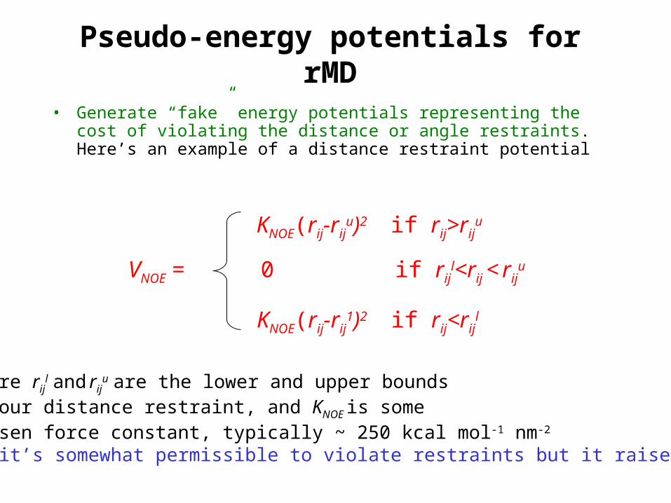
Pseudo-energy potentials for rMD
• Generate “fake” energy potentials representing the cost of violating the distance or angle restraints. Here’s an example of a distance restraint potential
KNOE(rij-rij1)2 if rij<rij
l
KNOE(rij-riju)2 if rij>rij
u
0 if rijl<rij < rij
u VNOE =
where rijl and rij
u are the lower and upper boundsof our distance restraint, and KNOE is somechosen force constant, typically ~ 250 kcal mol-1 nm-2
So it’s somewhat permissible to violate restraints but it raises V

Example of nOe pseudopotential
rijl rij
u
0
VNOE potentialrises steeply with degreeof violation
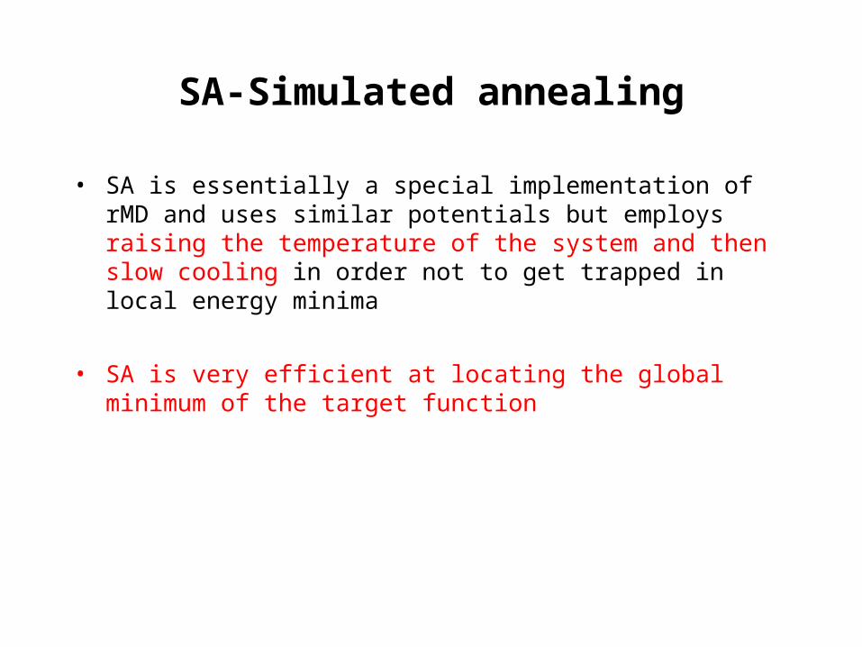
SA-Simulated annealing
• SA is essentially a special implementation of rMD and uses similar potentials but employs raising the temperature of the system and then slow cooling in order not to get trapped in local energy minima
• SA is very efficient at locating the global minimum of the target function

Dealing with ambiguous restraints
• often not possible to tell which atoms are involved in a NOESY crosspeak, either because of a lack of stereospecific assignments or because multiple protons have the same chemical shift
• sometimes an ambiguous restraint is included but is expressed ambiguously in the restraint file, e.g. 3 HA --> 6 HB#, where the # wildcard indicates that the beta protons of residue 6 are not stereospecifically assigned. This is quite commonly done for stereochemical ambiguities.
• it is also possible to leave ambiguous restraints out and then try to resolve them iteratively using multiple cycles of calculation. This is often done for restraints that involve more complicated ambiguities, e.g. 3 HA-->10 HN, 43 HN, or 57 HN, where three amides all have the same shift.
• can also make stereospecific assignments iteratively using what are called floating chirality methods

A B
C
9.52 ppm
4.34 ppm
4.34 ppm
Due to resonance overlapbetween atoms B and C,an NOE crosspeakbetween 9.52 ppmand 4.34 ppm couldbe A to C or A to B--this restraint is ambiguous
But if an ensemble generated with this ambiguous restraintleft out shows that A is neverclose to B, then the restraint mustbe A to C.
Example of resolving an ambiguityduring structure calculation
9-11 Å
3-4 Årange of interatomicdistances observedin trial ensemble

Iterative structure calculation with assignment of ambiguous restraints
source:http://www.pasteur.fr/recherche/unites/Binfs/aria/
•there are programs such as ARIA, with automatic routines for iterative assignment of ambiguous restraints. The key to success is to make absolutely sure the restraints you start with are right!
start with some setof unambiguous NOEsand calculate an ensemble

Acceptance criteria: choosing structures for an ensemble
• typical to generate 50 or more trial structures, but not all will converge to a final structure that is physically reasonable or consistent with the experimentally derived NMR restraints. We want to throw such structures away rather than include them in our reported ensemble.
• these are typical acceptance criteria for including calculated structures in the ensemble:– no more than 1 nOe distance restraint violation greater than 0.4 Å– no dihedral angle restraint violations greater than 5– no gross violations of reasonable molecular geometry
• sometimes structures are rejected on other grounds as well:– too many residues with backbone angles in disfavored regions of
Ramachandran space– too high a final potential energy in the rMD calculation

Precision of NMR Structures (Resolution)
• judged by RMSD of superimposed ensemble of accepted structures
• RMSDs for both backbone (C, N, CC=O) and all heavy atoms (i.e. everything except hydrogen) are typically reported, e.g.
bb 0.6 Å
heavy 1.4 Å
• sometimes only the more ordered regions are included in the reported RMSD, e.g. for a 58 residue protein you will see RMSD (residues 5-58)
if residues 1-4 are completely disordered.
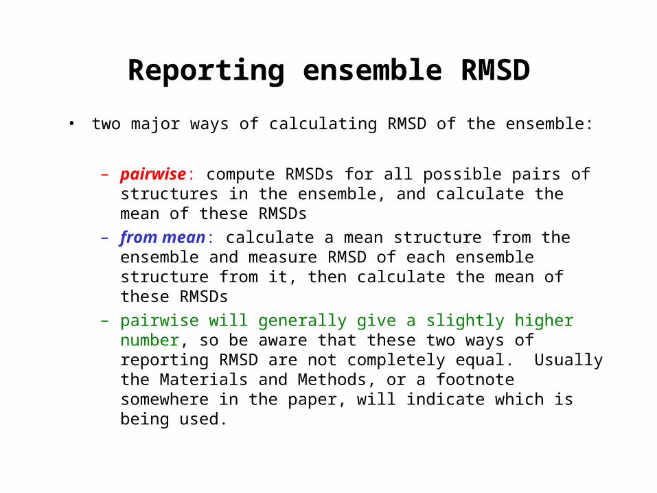
Reporting ensemble RMSD
• two major ways of calculating RMSD of the ensemble:
– pairwise: compute RMSDs for all possible pairs of structures in the ensemble, and calculate the mean of these RMSDs
– from mean: calculate a mean structure from the ensemble and measure RMSD of each ensemble structure from it, then calculate the mean of these RMSDs
– pairwise will generally give a slightly higher number, so be aware that these two ways of reporting RMSD are not completely equal. Usually the Materials and Methods, or a footnote somewhere in the paper, will indicate which is being used.

“Minimized average” structure
• a minimized average is just that: a mean structure is calculated from the ensemble and then subjected to energy minimization to restore reasonable geometry, which is often lost in the calculation of a mean
• this is NMR’s way of generating a single representative structure from the data. It is much easier to visualize structural features from a minimized average than from the ensemble.
• for highly disordered regions a minimized average will not be informative and may even be misleading--such regions are sometimes left out of the minimized average
• sometimes when an NMR structure is deposited in the PDB, there will be separate entries for both the ensemble and the minimized average. It is nice when people do this. Alternatively, a member of the ensemble may be identified which is considered the most representative (often the one closest to the mean).

How many restraints do we need to get a high-resolution NMR structure?
• usually ~15-20 nOe distance restraints per residue, but the total # is not as important as how many long-range restraints you have, meaning long-range in the sequence: |i-j|> 5, where i and j are the two residues involved
• good NMR structures usually have ≥ ~ 3.5 long-range distance restraints per residue in the structured regions
• to get a very good quality structure, it is usually also necessary to have some stereospecific assignments, e.g. hydrogens; Leu, Val methyls

Assessing Structure Quality
• NMR spectroscopists usually run their ensemble through the program PROCHECK-NMR to assess its quality
• high-resolution structure will have backbone RMSD ≤ ~0.8 Å, heavy atom RMSD ≤ ~1.5 Å
• low RMS deviation from restraints (good agreement w/restraints)
• will have good stereochemical quality:
– ideally >90% of residues in core (most favorable) regions of Ramachandran plot
– very few “unusual” side chain angles and rotamers (as judged by those commonly found in crystal structures)
– low deviations from idealized covalent geometry

Structural Statistics Tables
list of restraints,# and type
precision ofstructure (RMSD)
agreement of ensemble structureswith restraints (RMS)
calculated energies
sometimes also see listings of Ramachandran statistics, deviationsfrom ideal covalent geometry, etc.


![TERPENOID NMR STUDIES: NMR PARAMETERS FOR … · 2020. 4. 2. · entitled Terpenoid MR Studies': NMR Parameters for 6107010" [jS. 1. i] heptanes and Revised Structures for Archangelin](https://static.fdocuments.in/doc/165x107/60b24d5ace76f2582736c36b/terpenoid-nmr-studies-nmr-parameters-for-2020-4-2-entitled-terpenoid-mr-studies.jpg)